Article

Genomic, Antimicrobial, and Aphicidal Traits of Bacillus velezensis ATR2, and Its Biocontrol Potential against Ginger Rhizome Rot Disease Caused by Bacillus pumilus

1
Wuhan Institute of Virology, Chinese Academy of Sciences, Wuhan 430071, China
2
University of Chinese Academy of Sciences, Beijing 100039, China
*
Author to whom correspondence should be addressed.
Microorganisms 2022, 10(1), 63; https://doi.org/10.3390/microorganisms10010063
Submission received: 22 November 2021 / Revised: 10 December 2021 / Accepted: 23 December 2021 / Published: 29 December 2021

Abstract

:
Ginger rhizome rot disease, caused by the pathogen Bacillus pumilus GR8, could result in severe rot of ginger rhizomes and heavily threaten ginger production. In this study, we identified and characterized a new Bacillus velezensis strain, designated ATR2. Genome analysis revealed B. velezensis ATR2 harbored a series of genes closely related to promoting plant growth and triggering plant immunity. Meanwhile, ten gene clusters involved in the biosynthesis of various secondary metabolites (surfactin, bacillomycin, fengycin, bacillibactin, bacilysin, difficidin, macrolactin, bacillaene, plantazolicin, and amylocyclicin) and two clusters encoding a putative lipopeptide and a putative phosphonate which might be explored as novel bioactive compounds were also present in the ATR2 genome. Moreover, B. velezensis ATR2 showed excellent antagonistic activities against multiple plant pathogenic bacteria, plant pathogenic fungi, human pathogenic bacteria, and human pathogenic fungus. B. velezensis ATR2 was also efficacious in control of aphids. The antagonistic compound from B. velezensis ATR2 against B. pumilus GR8 was purified and identified as bacillomycin D. In addition, B. velezensis ATR2 exhibited excellent biocontrol efficacy against ginger rhizome rot disease on ginger slices. These findings showed the potential of further applications of B. velezensis ATR2 as a biocontrol agent in agricultural diseases and pests management.

1. Introduction

Ginger (Zingiber officinale Roscoe), a popular spice with aromatic and pungent components, is also utilized in traditional medicine for its healing properties of various gastrointestinal diseases, metabolic diseases, rheumatism, arthritis, pain, and muscle discomfort [1,2,3]. Moreover, numerous studies have reported that ginger exhibits antibacterial, antifungal, and anticancer properties [4,5,6]. Ginger rhizome is widely cultivated as an important economic crop. In ginger cultivation, agricultural diseases and pests are major factors restricting the production and quality of ginger [7,8,9,10,11]. Ginger rhizome rot disease, one of the most destructive diseases of ginger in Shandong Province of China, caused severe rot of ginger rhizomes during later growth stages and heavily threaten ginger production. The pathogen of ginger rhizome rot disease was identified as B. pumilus GR8 [12]. Currently, the control of ginger rhizome rot disease still primarily depends on preventive before-planting measures and agricultural chemicals.
In recent years, biological control agents (BCAs) with safe and eco-friendly merits have been regarded as an alternative strategy to chemical pesticides for controlling plant diseases and insect pests in the modern agricultural field [13]. Some Bacillus species, such as B. subtilis, B. amyloliquefaciens, and B. licheniformis, have been extensively studied and widely used to control various agricultural diseases and pests [14,15,16,17,18]. These Bacillus species harbor many advantages, such as high viability in different environmental conditions, harmlessness to plants and animals, low cost, and environmentally friendly.
B. velezensis, a Bacillus species first reported by Ruiz-García et al. in 2005 [19], was originally characterized as a later heterotypic synonym of B. amyloliquefaciens [20]. Recently, B. methylotrophicus, B. oryzicola, and B. amyloliquefaciens subsp. plantarum were reclassified as the later heterotypic synonyms of B. velezensis [21]. B. velezensis has many excellent merits, such as producing a variety of antimicrobial metabolites to suppress plant pathogens, enhancing plant defenses, and promoting plant growth and efficient colonization on plants [22,23,24,25]. Given these biocontrol properties, many strains of B. velezensis have been widely used in agricultural field [25,26,27]. For example, B. velezensis FZB42, a well-studied biocontrol strain, has been widely used as a disease suppressor and plant growth promoter for multiple economic crops and vegetables, such as lettuce, tobacco, cotton, tomato, and cucumber [23,28,29,30,31]. It was reported that B. velezensis CC09 exhibited strong antimicrobial activities and showed potential applications in control of wheat powdery mildew, take-all, and spot blotch diseases [13,32]. Furthermore, some B. velezensis strains encoded lignocellulolytic enzymes and exhibited the potential for degrading lignocellulosic biomass [33]. In addition, B. velezensis BZR 86 significantly controlled the root-knot disease on plants and noticeably increased growth and biomass of plants [34]. It was reported that bacterial treatment with B. velezensis YC7010 elicited the induced systemic resistance (ISR) and noticeably decreased settling, feeding, and reproduction of green peach aphid on Arabidopsis leaves [35]. However, few strains within B. velezensis have been reported with insecticidal activities against aphids.
In this study, we characterized a new B. velezensis strain, ATR2, which exhibited extensive antibacterial, antifungal, and aphicidal activities, identified the antagonistic compound from B. velezensis ATR2 against the pathogen of ginger rhizome rot disease, and evaluated the biocontrol efficacy of B. velezensis ATR2 against ginger rhizome rot disease. The results revealed the potential of further applications of B. velezensis ATR2 as a biocontrol agent in agricultural diseases and pest management.

2. Materials and Methods

2.1. Bacteria, Fungi, and Aphids Used in Assays

Strain ATR2 was originally isolated from the ginger rhizosphere in Shangdong Province (China) as the antagonistic strain against the pathogen of ginger rhizome rot disease (B. pumilus GR8). The pathogenic bacteria, B. pumilus GR8, Yersinia pseudotuberculosis YpⅢ, B. anthracis, Klebsiella pneumoniae, and Acinetobacter baumannii were stocked in our laboratory, and Ralstonia solanacearum (Smith), Xanthomonas campestris pv. campestris, Enterobacter cloacae, Escherichia coli EHEC O157:H7, Shigella dysenteriae, Staphylococcus aureus subsp. aureus, Salmonella Choleraesuis, and Salmonella Typhimurium were purchased from the BeNa Culture Collection (BNCC, Beijing, China).
The pathogenic fungi, Fusarium graminearum Sehw, Botrytis cinerea, Ceratocystis paradoxa (Dode) Moreau, and Colletotrichum higginsianum Sacc were kindly provided by Dr. Zhang from State Key Laboratory of Agricultural Microbiology, Huazhong Agricultural University, China, and Microsporum gypseum was purchased from the BNCC.
The aphids, Uroleucon formosanum Takahashi, Aphis gossypii Glover, Aphis craccivora Koch, and Myzus percicae, were obtained from local vegetable farms in Wuhan, Hubei, China.

2.2. Taxonomic Identification of Strain ATR2

To determine species identity of strain ATR2, bacterial cell and colony morphology, physiological and biochemical characteristics, 16S rRNA sequence, and complete genome sequence analyses were conducted [24,36]. Briefly, the features of cell morphology including shape, Gram staining, and spore forming were described using light microscopy (Olympus, Tokyo, Japan). The features of colony morphology were directly observed. The API 50 CHB strips were used to assess carbon source utilization and other physiological and biochemical properties of strain ATR2 according to the manufacturer’s instructions (bioMerieux, Marcy l’Etoile, France) [37].
For molecular identification, total bacterial DNA of strain ATR2 was extracted according to the protocol described by Gao et al. [38]. The 16S rRNA sequence was first amplified by polymerase chain reaction (PCR) using the primer pairs 27f/1492r with cycling conditions reported by Guardado-Valdivia et al. [39], detected with an agarose gel electrophoresis, purified with a Gel Extraction kit (Omega Bio-tek, Inc., Norcross, GA, USA), sequenced by TSINGKE Biological Technology Co., Ltd. (Beijing, China), and searched against the nucleotide sequences available in the National Center for Biotechnology Information (NCBI) nucleotide database. Based on 16S rRNA sequences of strain ATR2 and the type strains of other species from the NCBI nucleotide database, a phylogenetic tree was built using the neighbor-joining method with 1000 bootstrap replicates in MEGA software, version 7.0 [40]. Second, pairwise average nucleotide identity (ANI) values for genomes of strain ATR2 and 14 related type strains available in NCBI nucleotide database were calculated using JspeciesWS (http://jspecies.ribohost.com/jspeciesws/#home, accessed on 25 November 2019) [41]. A heatmap visualizing and hierarchal clustering the ANI values (%) was constructed using the online Morpheus software (https://software.broadinstitute.org/morpheus/, accessed on 24 August 2020).

2.3. Genome Sequencing, Assembly, Annotation, and Bioinformatics Analysis

The genomic DNA of strain ATR2 was sequenced on the Illumina HiSeq 2500 platform using a 500-bp paired-end library with read lengths of 150 bp. The raw reads were quality filtered by Quake [42]. The de novo assembly was performed by SPAdes 3.5.0 [43], and the resulting contigs longer than 500 bp were further arranged by comparing them with three reference genome sequences available in NCBI nucleotide database: B. velezensis YAU B9601-Y2 (GenBank accession no. NC_017061), B. amyloliquefaciens Y2 (GenBank accession no. NC_017912), and B. subtilis strain Bs-916 (GenBank accession no. NZ_CP009611) with Mauve (V2.4.0) to generate a draft genome with gaps. Finally, the gaps were bridged by high-fidelity PCR to acquire the complete genome. Genome annotation was conducted by NCBI Prokaryotic Genome Annotation Pipeline (PGAP, 2 March 2021, http://www.ncbi.nlm.nih.gov/genome/annotation_prok/) with best-placed reference protein set and GeneMarkS-2+ methods. Secondary metabolite clusters were identified by comparing ATR2 genome sequence with the antiSMASH database (11 May 2020, https://antismash.secondarymetabolites.org/) with default parameters [44]. The ATR2 genome sequence was also compared with the REBASE database (12 May 2020, http://rebase.neb.com/rebase/rebase.html) to predict the restriction modification (R-M) system [45]. The CRISPR/Cas (clustered regularly interspaced short palindromic repeats) system in the ATR2 genome was evaluated using the CRISPR finder online service (12 May 2020, https://crispr.i2bc.paris-saclay.fr/) [46]. The BLAST Ring Image Generator (BRIG) software was used to visualize the genome comparisons of strain ATR2 with four representative Bacillus strains: B. subtilis subsp. subtilis 168T (GenBank accession no. AL009126), B. subtilis subsp. inaquosorum KCTC 13429T (GenBank accession no. CP029465), B. amyloliquefaciens DSM 7T (GenBank accession no. FN597644), and B. velezensis FZB42 (GenBank accession no. CP000560) [47].

2.4. UHPLC-QE-MS Analysis

For identifying the secondary metabolites synthesized by strain ATR2, an ultra-high performance liquid chromatography coupled with Q Exactive Orbitrap mass spectrometry (UHPLC-QE-MS) analysis was performed. Strain ATR2 was precultured overnight in 10 mL of Luria-Bertani (LB) liquid medium (tryptone 10 g/L, yeast extract 5 g/L, sodium chloride 10 g/L, the pH was adjusted to 7.0) at 28 °C and 180 rpm with a rotatory shaker (Zhicheng Industry Co., Ltd., Shanghai, China). An aliquot (2 mL) of the preculture (108 CFU/mL) was added in a 500 mL shake flask containing 100 mL of YPD liquid medium (yeast extract 10 g/L, peptone 20 g/L, dextrose 20 g/L), and then incubated at 28 °C and 180 rpm with a rotatory shaker for 64 h. The cell free supernatant (CFS) was acquired by centrifugation of bacterial fermentation liquid at 4 °C and 8000 rpm for 10 min and filtrating the supernatant with a 0.22 μm filter (Merk Millipore Ltd., Tullagreen, Carrigtwohill, Co. Cork, Ireland).
The UHPLC separation conditions were as follows: a UPLC BEH Amide column (2.1 × 100 mm, 1.7 μm, Waters Corp., Milford, MA, USA) was attached to a 1290 Infinity series UHPLC System (Agilent Technologies Inc., Santa Clara, CA, USA), eluent A was 25 mM ammonium acetate and 25 mM ammonia hydroxide in water (pH = 9.75), eluent B was acetonitrile, flow rate was 0.5 mL/min, column temperature was 25 °C, injection volume was 3 μL, elution gradient was as follows: 0.0 min, 95% B; 0.5 min, 95% B; 7.0 min, 65% B; 8.0 min, 40% B; 9.0 min, 40% B; 9.1 min, 95% B; 12.0 min, 95% B. The Q Exactive Orbitrap mass spectrometer (Thermo Fisher Scientific Inc., Waltham, MA, USA) was used to obtain MS/MS spectra on information-dependent acquisition (IDA) mode using Xcalibur 4.0.27 software (Thermo Fisher Scientific Inc., Waltham, MA, USA) under the following conditions: sheath gas flow rate of 45 Arb, aux gas flow rate of 15 Arb, capillary temperature of 400 °C, full MS resolution of 70,000, mass range of 100–1500 m/z, MS/MS resolution of 17,500, collision energy of 10/30/60 in NCE mode, spray voltage of 4.0 kV (positive) or −3.6 kV (negative), respectively [48].

2.5. Antimicrobial Assays of Extracellular Metabolites from Strain ATR2

To evaluate the antagonistic effect of the extracellular metabolites on some pathogenic microorganisms, antimicrobial assays were performed with the CFS of strain ATR2. For the antibacterial activity detection, hole-plate diffusion assays were conducted as described by Njimoh et al. with minor modification [49]. Briefly, the pathogenic bacteria were cultured in LB liquid mediums at 37 °C except Ralstonia solanacearum in 950 liquid medium (peptone 5 g/L, dextrose 5 g/L, beef extract 3 g/L, yeast extract 0.25 g/L, sodium chloride 0.5 g/L, magnesium sulfate heptahydrate 0.5 g/L, the pH was adjusted to 7.0) at 28 °C using a rotatory shaker (180 rpm). Thereafter, 200 μL of the pathogenic bacterial culture (0.5 McFarland standard) was added into 20 mL of melted and tempered (50 °C) LB agar mediums except Ralstonia solanacearum was added into 950 agar medium which was mixed rapidly and poured in the sterile petri dishes (90 mm in diameter). After solidification of the medium at room temperature, three holes evenly distributed on the LB agar plate were created with a sterilized puncher (10 mm in diameter) and filled with 100 μL of the CFS. An equal volume of YPD liquid medium was used to serve as the blank control. After being kept at room temperature for 1 h, all the plates were cultivated at 37 °C for 24 h except Ralstonia solanacearum at 28 °C. The antibacterial activities were determined based on the presence of inhibition zones around the holes, and the diameters of inhibition zones were measured. The experiments were carried out three times.
The potential antifungal activities of extracellular metabolites from strain ATR2 were evaluated using the plate confrontation method described by Jiang et al. with minor modification [25]. Briefly, the pathogenic fungi were cultured on potato dextrose agar (PDA) plates (potato 200 g/L, dextrose 20 g/L, agar 15 g/L) at 28 °C for 5 days, then a mycelia disc was made with a sterilized puncher (10 mm in diameter) and inoculated at the center of a petri dish containing 25 mL of PDA medium. Four holes were punched at 20 mm from the center, and two holes on both sides of the plate were filled with 200 μL of the CFS. As a control, the other two holes were filled with the same volume of YPD liquid medium. The plates were cultured at 28 °C until the mycelial growth on the control sides was almost complete. The diameter of mycelial growth on treated sides (T) and the diameter of mycelial growth on control sides (C) were measured and the inhibition ratio of fungal growth (I) was calculated by the formula: I% = ((CT)/(C − 10)) × 100 [24,39]. Tests were repeated three times.

2.6. Aphicidal Assays of Extracellular Metabolites from Strain ATR2

The aphicidal activities of extracellular metabolites from strain ATR2 were evaluated using leaf-disc spraying method according to the guideline for laboratory bioassay of pesticides. Briefly, the leaves of host plants with uniform growth were divided into small pieces (60 × 30 mm) and placed in petri dishes (90 mm in diameter). Thirty aphids with the same physiological state were picked from the local vegetable farms and allowed to locate on a leaf for 3 h prior to application [16]. Then, 2 mL of the fermentation supernatant diluent (10% dilution, v/v) was evenly sprayed on the aphids with a small sprayer. After the fermentation supernatant diluent settled for 1 min, the leaf was transferred to a new petri dish which was layered with a piece of wet filter paper and punched with a needle to make some air vents. The same volume of PBS buffer (KH2PO4 0.24 g/L, Na2HPO4 1.42 g/L, NaCl 8 g/L, KCl 0.2 g/L, the pH was adjusted to 7.4) was used as the blank control. The dishes were kept in a growth chamber at 25 °C, a relative humidity of 40 to 70% and a photoperiod of 16:8 h (L:D) for 48 h after application. When the mortality of the control group was less than 10%, the corrected mortality was calculated by Abbott’s formula [50]: corrected mortality (%) = ((AB)/A) × 100, where A and B represented the number of the living aphids in the control group and the number of the living aphids in the treated group, respectively. The experiments were repeated three times.
To assess the aphicidal activities in the field, the fermentation supernatant of strain ATR2 was sprayed evenly on stems and leaves of the vegetables severely attacked by aphids. An equal volume of PBS buffer was served as the blank control. The control efficacy was assessed at 24 h after application. The experiments were repeated three times when the weather was sunny, the temperature was between 15 °C and 25 °C, and the wind speed was level 2 to level 3.

2.7. Purification and Mass Spectrometry Analysis of the Antagonistic Compound against B. pumilus GR8

For identifying the antagonistic compound from strain ATR2 against B. pumilus GR8, crude antibacterial substances were first obtained following the extraction method described by Gong et al. with minor modification [51]. Briefly, the fermentation supernatant of strain ATR2 was treated with 4 M HCl to adjust pH to 2.0 and then kept at 4 °C overnight. When the supernatant after acid precipitate had no antagonistic activity against B. pumilus GR8, the precipitation was obtained by centrifugation at 8000 rpm, 4 °C for 20 min and solubilized in a minimal volume of PBS buffer with 10 M NaOH to pH 7.5. After recentrifugation at 8000 rpm for 15 min, the crude antibacterial substances were thus acquired in the soluble fraction. Then, the crude solution was purified by preparative thin layer chromatography (TLC) on silica gel GF254 plates (1 mm, Xinchu Industry Co., Ltd., Shanghai, China) developed with a methanol: acetone (9:1, v/v) solvent system. Each fraction of the plate was extracted with methanol, and then the methanol extract was evaporated to dryness under vacuum with a rotary evaporator, solubilized in PBS buffer, and tested for its antagonistic activity against B. pumilus GR8. The active fraction was further purified by a semi-preparative HPLC system (1200 series, Agilent Technologies Inc., Santa Clara, CA, USA) using a Polar-HILIC column (10.0 × 250 mm, 5 μm, Sepax Technologies, Inc., Newark, DE, USA) eluting with a solvent containing 60% A (HPLC grade methanol) and 40% B (5 mM ammonium acetate in HPLC grade water, pH 7.5) at a flow rate of 1 mL/min. Elution was monitored by an ultraviolet (UV) detector (Agilent Technologies Inc., Santa Clara, CA, USA) at 220 nm.
A liquid chromatography coupled with electron spray ionization tandem mass spectrometry (LC–ESI–MS/MS) system (Ultimate 3000 LCQ FLEET, Thermo Fisher Scientific Inc., Waltham, MA, USA) was employed for the identification of accurate molecular weight and molecular formula of the antagonistic compound from strain ATR2 against B. pumilus GR8. The elution solvent was the same as the semi-preparative HPLC above at a flow rate of 0.3 mL/min, and the program was supervised by UV detection at 254 nm. The MS analysis was performed at a spray voltage of 4.0 kV for positive mode, a capillary temperature of 300 °C and a mass range of 100–2000 m/z.

2.8. Biocontrol Assays of Strain ATR2 against Ginger Rhizome Rot Disease

To evaluate the biocontrol efficacy of strain ATR2 against ginger rhizome rot disease, biocontrol assays were performed on ginger rhizome slices as described by Ye et al. with minor modification [52]. Briefly, antagonistic bacterium strain ATR2 and pathogenic bacterium B. pumilus GR8 were cultivated in YPD liquid medium and LB liquid medium at 28 °C and 180 rpm with a rotatory shaker for 16 h, respectively. Ginger rhizomes purchased from a market were washed in tap water and dried at room temperature. Surface disinfection was conducted by immersing ginger rhizomes sequentially in 70% ethanol and sterile distilled water (SDW) for 1 min followed by air-drying at room temperature for 20 min. After the disinfection procedures were repeated three times, ginger rhizome slices 3 mm in thickness were prepared with a flame-sterilized knife. A 100-μL bacterial suspension of GR8 (2 × 10⁸ CFU/mL) and a 100-μL bacterial suspension of ATR2 at approximately 2 × 10⁹, 2 × 10⁸, or 2 × 10⁷ CFU/mL were mixed and inoculated into the center of ginger rhizome slices. After inoculation, the slices were placed in sterile petri dishes and incubated at 30 °C and 40 to 70% relative humidity for about 2 days. Three control groups included 200-μL bacterial suspension of ATR2 (109 CFU/mL), 200-μL bacterial suspension of GR8 (108 CFU/mL), and 200-μL YPD liquid medium. The symptom severity of the ginger slices was assessed as described by Nishijima et al. [53]. Ginger slices were rated using a scale of 1 to 5 according to the percentage of rot region, where 1 = healthy tissue or tissue without symptoms, 2 = slight (1 to 25%) rot (slight discoloration), 3 = moderate (26 to 50%) rot (discoloration and tissue breakdown), 4 = severe rot (51 to 75%), and 5 = complete rot (76 to 100%). The trials were performed in triplicate.

2.9. Accession Numbers

The 16S rRNA sequence and complete genome sequence of strain ATR2 have been deposited in GenBank under the accession number KP685408 and NZ_CP018133, respectively. Strain ATR2 is available in China Center for Type Culture Collection (CCTCC NO. M2016071).

3. Results

3.1. Strain Identification

Morphological observation revealed the cells of strain ATR2 were Gram-positive, with spores and rod-shaped (Figure S1A). Strain ATR2 generally formed creamy white, opaque, rough colonies with irregular edge on LB agar medium. The colony morphology of strain ATR2 was quite different from that of B. velezensis FZB42, B. amyloliquefaciens DSM 7T, and B. subtilis 168T (Figure S1B). Analysis of physiological and biochemical characteristics showed strain ATR2 was positive for starch hydrolysis and oxidase reaction, but negative for anaerobic growth. Meanwhile, strain ATR2 utilized L-arabinose, D-ribose, D-xylose, sorbitol, methyl-α-D-glucoside, D-lactose, D-melibiose, D-trehalose, D-raffinose, glycogen, and D-turanose (Table S1). Based on the 16S rRNA sequences analysis, strain ATR2 (GenBank accession KP685408) exhibited 98.46% sequence identity with B. velezensis strain EH9 (GenBank accession MN750764) and B. velezensis strain H34 (GenBank accession KJ149808). The phylogenetic analysis of 16S rRNA sequences showed strain ATR2 also clustered with B. velezensis (Figure S2). Furthermore, strain ATR2 shared 99.07% identity with the type B. velezensis strain NRRL B-41580 according to ANI values calculation. It could be considered as B. velezensis based on the generally accepted species boundary of 95% cut-off ANI value [41,54] (Figure 1), in accordance with the result of 16S rRNA sequences analysis. Therefore, strain ATR2 was identified as a member of B. velezensis based on cell and colony morphology, physiological and biochemical characteristics, phylogenetic taxonomy, and ANI values.

3.2. Genomic Features of B. velezensis ATR2

Based on the genome sequencing and assembly, a total of 9,779,334 reads were obtained and assembled into 40 contigs with a sequencing depth of 294-fold. The gaps between contigs were bridged by high-fidelity PCR. The complete genome of B. velezensis ATR2 was 4,006,746 bp, containing a circular chromosome with 46.34% of G + C content, and possessed 3745 protein-coding genes, 21 rRNA genes, and 69 tRNA genes. Comparative genomic analysis revealed that the genome of B. velezensis ATR2 showed 90.69% similarity to the genome of B. velezensis FZB42 (Figure S3), which had been widely used as biocontrol agent and biofertilizer. The R-M system and CRISPR/Cas system were two main defense systems that widely existed in the prokaryotic organisms to protect the bacteria from the foreign DNA. Only one type IV R-M system was identified in the ATR2 genome by comparing with the REBASE database. The type IV R-M system was composed of one or two genes encoding restriction enzymes that cleaved only modified DNA, including methylated, hydroxymethylated, and glucosyl-hydroxymethylated bases. Moreover, only a questionable CRISPR/Cas system was detected in the ATR2 genome using the CRISPR finder online service, which had only two direct repeats (DRs) and the repeated motifs (DR in CRISPR) were not 100% identical. The lack of strict defense system in the ATR2 genome laid a promising foundation for genetic manipulation of B. velezensis ATR2.

3.3. Genes Involved in Plant Growth Promotion

Considering that some biocontrol agents not only prevented plant disease but also participated in plant growth promotion, we also analyzed the genes involved in plant growth promotion in the genome of B. velezensis ATR2. As shown in Table 1, B. velezensis ATR2 harbored a series of genes associated with nutrition utilization and production of multiple growth-promoting substances, including 3-hydroxy-2-butanone, 2,3-butanediol, indole acetic acid (IAA), phytase, trehalose, spermidine, and bacillibactin. The dhb gene cluster was responsible for encoding the synthetase of the iron-siderophore bacillibactin which absorbed and accumulated iron ions from the environment under iron limitation. The 3-hydroxy-2-butanone and 2,3-butanediol, two well-known volatile organic compounds (VOC), exhibited large capacity to promote plant growth and elicit the ISR against plant pathogens. Genes involved in the synthesis of the two growth-promoting VOCs including acetolactate synthase small subunit (ilvN), acetolactate synthase large subunit (ilvB), alpha-acetolactate decarboxylase (alsD), acetolactate synthase (alsS), and butanediol dehydrogenase (bdhA) were present in the ATR2 genome. Meanwhile, genes associated with the synthesis of growth-promoting hormones, including indole-3-glycerol phosphate synthase (trpC), 3-phytase (phyC), trehalose permease IIC protein (treP) and trehalose operon repressor (treR), were also found in the ATR2 genome. Indole acetic acid (IAA), a well-characterized phytohormone, effectively promoted plant growth and its biosynthesis significantly affected the plant growth-promoting activity. The phytase, a functional enzyme degrading the extracellular phytate, was important for the plant growth-promoting effect under phosphate limitation. Trehalose, a nonreducing disaccharide, was accumulated by bacteria as a strategy for protecting cells from desiccation and water loss. In addition, genes responsible to produce spermidine, including agmatinase (speB) and spermidine synthase (speE), were discovered in the ATR2 genome. Spermidine, a type of polyamine (PA), played an important role in improving plant growth and drought tolerance. Genes related to the transport and utilization of nitrogen, potassium, and magnesium were summarized in Table 1.

3.4. Genes Involved in Secondary Metabolite Synthesis

As we all know, some biocontrol Bacillus species exhibit powerful antimicrobial capacities as a result of producing kinds of bioactive metabolites. A total of twelve gene clusters associated with secondary metabolite synthesis were found in the ATR2 genome. Among them, three giant gene clusters (mln, bae, and dif), covering altogether over 196 kb, were predicted to synthesize three polyketides (macrolactin, bacillaene, and difficidin). Moreover, five gene clusters (srf, bmy, fen, bac, and dhb) participated in nonribosomal synthesis of three cyclic lipopeptides (surfactin, bacillomycin, and fengycin), one dipeptide (bacilysin), and one siderophore (bacillibactin). In addition, two gene clusters (acn and ptn) involved in ribosomal synthesis of one bacteriocin (amylocyclicin) and one modified peptide (plantazolicin) were also found in the ATR2 genome (Figure S3, Table 2).
In addition to ten gene clusters associated with the known bioactive metabolites, the other two clusters encoding a putative lipopeptide and a putative phosphonate were also detected in the ATR2 genome which were not found in the genomes of B. velezensis FZB42, B. amyloliquefaciens DSM 7T, and B. subtilis 168T (Figure S3, Table 2). Interestingly, genes surrounding the gene cluster encoding the putative lipopeptide on B. velezensis ATR2 chromosome shared high degrees of homology with the same locus of B. velezensis FZB42 and B. amyloquefaciens DSM 7T. Figure 2A suggested that the gene cluster encoding the putative lipopeptide might insert into B. velezensis ATR2 chromosome as a single copy. The gene cluster should encode a hexapeptide when the biosynthesis obeyed the linear mechanism. Meanwhile, the amino acid sequence of the putative lipopeptide was determined by the specificity and order of the hexamodular NRPSs. However, the hexamodular NRPSs might also encode other lipopeptide when the biosynthesis deviated from the linear rule and presented a nonlinear assembly like locillomycins [55]. Moreover, the putative lipopeptide was predicted to contain two cysteine residues (Figure 2B) which were the signatures for some antibacterial and antifungal polypeptides with cysteine-knot structural motifs [56,57].
Furthermore, three cyclic lipopeptides (surfactin, bacillomycin D, and fengycin), three polyketides (macrolactin, difficidin, and bacillaene) and one dipeptide (bacilysin) were detected by UHPLC-QE-MS analysis (Figure 3), confirming their successful production from B. velezensis ATR2. Analysis of the secondary metabolites synthesized by B. velezensis ATR2 offered an effective way to understand the mechanisms of its multiple bioactivities and facilitated the exploitation for novel bioactive metabolites.

3.5. Antimicrobial Activities of Extracellular Metabolites from B. velezensis ATR2

The antimicrobial activities of extracellular metabolites from B. velezensis ATR2 were measured by hole-plate diffusion assays for pathogenic bacteria and plate confrontation assays for pathogenic fungi. The results of antibacterial assays indicated that B. velezensis ATR2 displayed broad antibacterial spectrum and strong bacteriostatic activities against multiple plant pathogenic bacteria, e.g., Bacillus pumilus GR8, Ralstonia solanacearum (Smith), Xanthomonas campestris pv. campestris, and Enterobacter cloacae, and human pathogenic bacteria, e.g., Yersinia pseudotuberculosis YpⅢ, Bacillus anthracis, Escherichia coli EHEC O157:H7, Klebsiella pneumoniae, Shigella dysenteriae, Staphylococcus aureus subsp. aureus, Salmonella Choleraesuis, Salmonella Typhimurium, and Acinetobacter baumannii (Table 3). Furthermore, B. velezensis ATR2 also significantly inhibited the mycelia growth of Fusarium graminearum Sehw, Botrytis cinerea, Ceratocystis paradoxa (Dode) Moreau, Colletotrichum higginsianum Sacc, and Microsporum gypseum, with 75.42%, 80.83%, 91.67%, 81.67%, and 52.5% of inhibition ratio, respectively (Table 3).

3.6. Aphicidal Activities of Extracellular Metabolites from B. velezensis ATR2

The aphicidal activities of extracellular metabolites from B. velezensis ATR2 were assessed according to the guideline for laboratory bioassay of pesticides, and the results revealed that the fermentation supernatant diluent (10% dilution, v/v) exhibited excellent aphicidal activities on Uroleucon formosanum Takahashi, Aphis gossypii Glover, Aphis craccivora Koch, and Myzus percicae, with 90.24%, 89.15%, 94.32%, and 96.63% of corrected mortality, respectively (Figure 4A). The aphicidal activities of B. velezensis ATR2 in the field were observed at 0 h and 24 h after application, respectively. The results indicated that the fermentation supernatant of B. velezensis ATR2 showed a strong toxic effect on bean aphids (Aphis craccivora Koch) and the aphids on the leaves almost died at 24 h after application, and the dead insect body shriveled and blackened. The same toxic effect was also exhibited on lettuce aphids (Uroleucon formosanum Takahashi) at 24 h after application (Figure 4B). These results indicated that B. velezensis ATR2 was able to effectively control aphids as a biological pesticide.

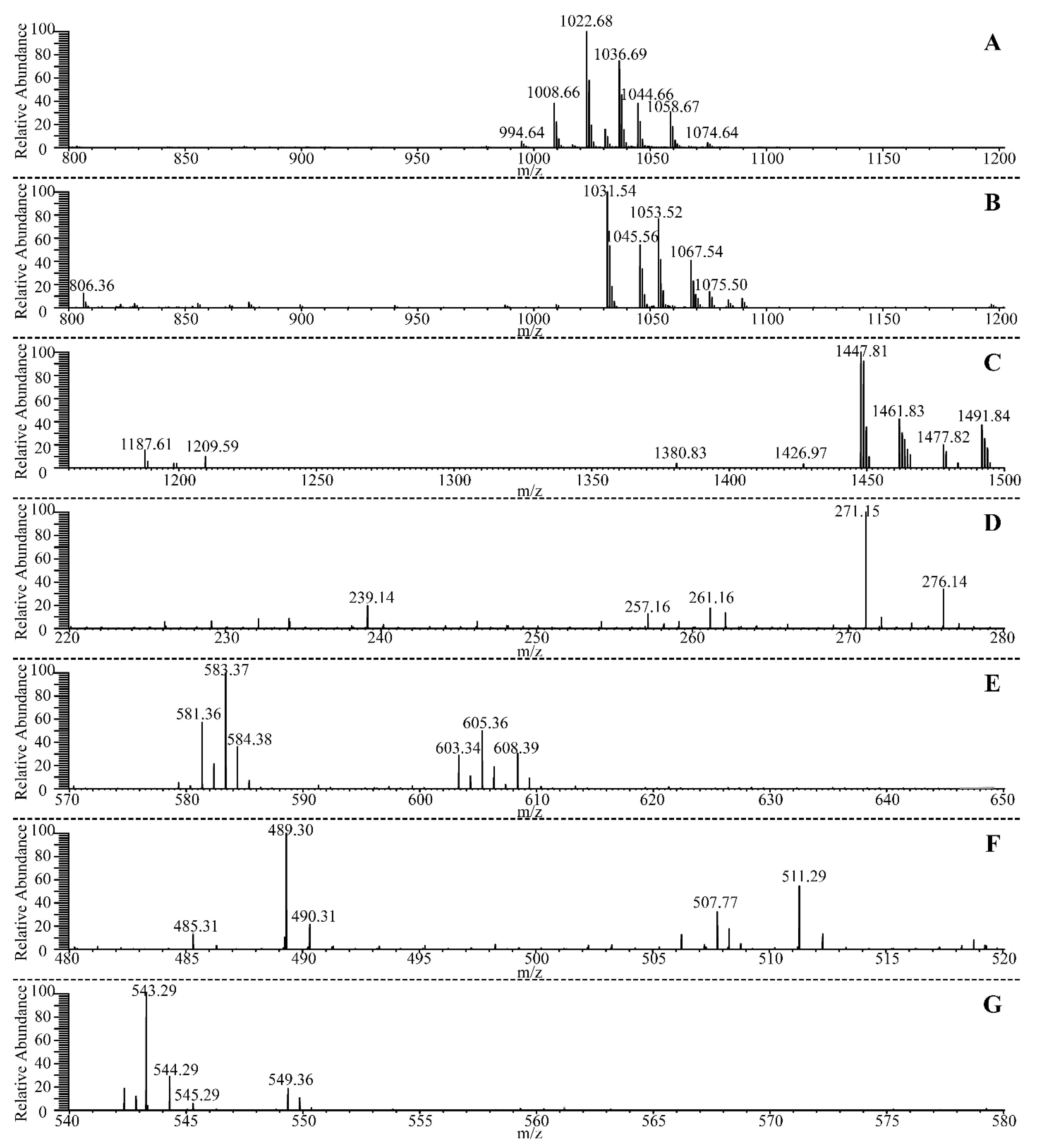
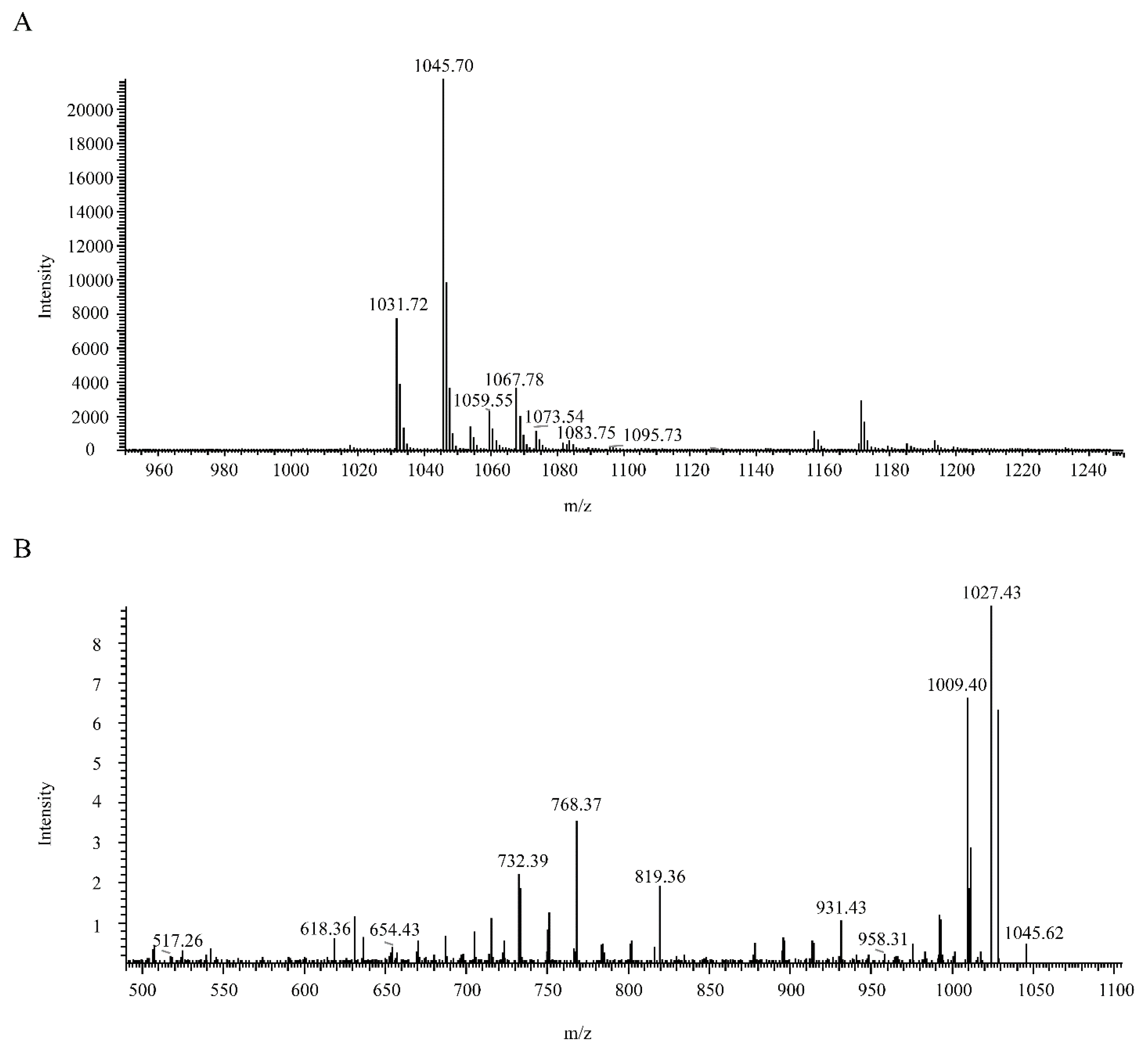
3.7. Identification of the Antagonistic Compound against B. pumilus GR8

By acid precipitate from the fermentation supernatant of B. velezensis ATR2, the crude antibacterial substances were obtained, and most of the impurities were excluded. With preparative TLC and subsequent semi-preparative HPLC, a peak with antagonistic activity was purified (Figure S4). Therefore, the active peak was subjected to LC–ESI–MS/MS for its molecular weight and molecular formula identification. The data from mass spectrum analysis in the positive-ion mode revealed that (M + H) + ions were at 1031.72, 1045.70, 1059.55, and 1073.54 m/z, respectively (Figure 5A). The interval of 14 mass unit was a common phenomenon for molecular weight of many cyclic lipopeptides secreted by Bacillus strains as a result of different numbers of methylene units in lipid chains. Based on the mass data in published studies [75,76], the active peak may be recognized as the homologues of bacillomycin D with molecular weights of 1030 Da (C14), 1044 Da (C15), 1058 Da (C16), and 1072 Da (C17).
ESI/CID-MS spectrometry analysis of precursor ion of m/z 1045.70 was performed to further confirm the molecular formula of the antagonistic compound against B. pumilus GR8. The product ion of m/z 1027.43 in the CID spectrum could be elucidated as (1045.70 − 18) + which was identical to (M + H − H2O) +. Similarly, product ions of m/z 958.31, 931.43, 819.36, 768.37, 732.39, 654.43, 618.36, and 517.26 in the CID spectrum presented (M + H − Ser) +, (M + H − Asn) +, (M + H − Pro − Glu) +, (M + H − Tyr − Asn) +, (M + H − Pro − Glu − Ser) +, (M + H − Asn − Tyr − Asn) +, (M + H − Asn − Pro − Glu − Ser) +, and (M + H − Asn − Pro − Glu − Ser − Thr) +, respectively (Figure 5B). Therefore, the amino acid composition of the antagonistic compound was (Asn-Tyr-Asn-Pro-Glu-Ser-Thr), which was identical to the amino acid residue of bacillomycin D [51,77]. Collectively, the antagonistic compound from B. velezensis ATR2 against B. pumilus GR8 was identified as bacillomycin D.

3.8. Biocontrol Efficacy of B. velezensis ATR2 against Ginger Rhizome Rot Disease

Given that B. velezensis ATR2 exhibited strong antagonistic activity against B. pumilus GR8, biocontrol assays on ginger rhizome slices were performed to further evaluate its efficacy against ginger rhizome rot disease. As shown in Figure 6, the slices inoculated with B. pumilus GR8 were extensively rotten within 2 days. By contrast, the symptoms of discoloration and tissue breakdown were obviously reduced on the slices inoculated with a mixture of GR8 and ATR2 (109 CFU/mL), even when the bacterial concentration of B. velezensis ATR2 was diluted 10-fold (108 CFU/mL) and 100-fold (107 CFU/mL). Additionally, the result of sole inoculation with B. velezensis ATR2 on slices, similar to the treatment of YPD liquid medium, proved that B. velezensis ATR2 was harmless to the host plant and safe for further use as a biocontrol agent.

4. Discussion

Agriculture is the foundation of a country, and the stability and development of agriculture determine the economic level of the country. However, plant diseases and insect pests have always been restricting agricultural development. Currently, control of plant diseases and insect pests mainly depends on agricultural chemicals, whereas long-term and massive use of chemicals has already caused many problems, including drug resistance, environmental contamination, and health hazards [78]. To control plant diseases and insect pests, the demand for safe and eco-friendly alternative products is increasing. In the present study, we characterized the genomic, antimicrobial, and insecticidal properties of a potential biocontrol strain, ATR2. Meanwhile, we identified the antagonistic compound from strain ATR2 against the pathogen of ginger rhizome rot disease and analyzed the biocontrol efficacy of strain ATR2 against ginger rhizome rot disease.
According to the cell and colony morphology, physiological and biochemical characteristics, phylogenetic taxonomy and ANI values analysis, strain ATR2 was finally identified as a member of B. velezensis. Genome analysis showed that B. velezensis ATR2 possessed a great capacity for synthesizing kinds of secondary metabolites, including three polyketides (macrolactin, bacillaene, and difficidin), three cyclic lipopeptides (surfactin, bacillomycin, and fengycin), one dipeptide (bacilysin), one siderophore (bacillibactin), one bacteriocin (amylocyclicin), one modified peptide (plantazolicin), one putative lipopeptide, and one putative phosphonate. In total, more than 9.25% of the ATR2 genome was devoted to synthesizing secondary metabolites, whereas the genomes of some closely related members of the B. subtilis group dedicated only approximately 5% of their capability in production of secondary metabolites [29].
Macrolactin, an efficient peptide deformylase inhibitor, was found in both B. subtilis and B. amyloliquefaciens [79,80]. Notably, macrolactin A, 7-O-malonyl macrolactin A and 7-O-succinyl macrolactin A all exhibited antibacterial activities against methicillin-resistant Staphylococcus aureus and vancomycin-resistant enterococci [81,82]. Another two polyketides, bacillaene and difficidin, inhibited the biosynthesis of prokaryotic protein [83,84]. Surfactin, a cyclic heptapeptide, was named for its powerful surface-activity [85] and exerted antibacterial, antiviral, and hemolytic performances by altering membrane integrity [86,87]. Both bacillomycin and fengycin exhibited a high fungitoxicity for their membrane permeabilization properties [88,89,90]. In addition to direct suppression against the phytopathogens, the cyclic lipopeptides surfactin and fengycin played a role in eliciting the ISR phenomenon in plants [91]. Bacilysin, a dipeptide antibiotic composed of a nonproteinogenic L-anticapsin at the C-terminus and an L-alanine residue at the N-terminus, exhibited antagonistic activities against kinds of bacteria, Candida albicans, and the yeast [92,93,94]. The iron-siderophore bacillibactin participated in specific transport system of taking up and accumulating iron ions from the environment under iron limitation [95]. These nonribosomal peptides and polyketides were synthetized by modularly organized enzymes and all depended on a functional 4′-phosphopantetheine transferase (Sfp), except for the dipeptide bacilysin [96,97]. Amylocyclicin was a circular bacteriocin, which was ribosomally synthesized and showed distinct antibacterial activity against some Gram-positive bacteria [98]. Plantazolicin was a bioactive peptide with thiazole and oxazole heterocycles, which was posttranslationally modified and exhibited antibacterial activity against closely related Gram-positive bacteria [99].
In addition to the ten known bioactive metabolites, one putative lipopeptide and one putative phosphonate predicted in the ATR2 genome were hitherto unknown. According to the previous reports, some organisms produced kinds of phosphonates with antiviral, antibacterial, herbicidal, and antiparasitic properties to provide a competitive advantage [100,101,102]. The lipopeptides, as extensively studied bioactive metabolites, have been related to the biocontrol of multiple plant diseases [51,103,104]. Meanwhile, a series of genes closely related to promoting plant growth and triggering plant immunity were also found in ATR2 genome. These findings suggested that B. velezensis ATR2 should be an excellent biological control agent and an important microorganism resource for further exploitation of novel bioactive metabolites.
According to our results, the extracellular metabolites from B. velezensis ATR2 exhibited distinct antagonistic activities against multiple plant pathogenic bacteria, plant pathogenic fungi, human pathogenic bacteria, and human pathogenic fungus. Aphids, as widely distributed agricultural piercing-sucking mouthpart pests, mainly damaged the leaves, stems and flowers of vegetables, trees, and flowers. The current control measures for aphids mainly rely on chemical pesticides. The extracellular metabolites from B. velezensis ATR2 were also proven to be efficacious in control of aphids in laboratory bioassays and in the field, where corrected mortalities of multiple aphids were almost up to 90%. Collectively, we recommend applying B. velezensis ATR2 in agricultural and medical fields, based on its outstanding potential.
The antagonistic compound from B. velezensis ATR2 against the pathogen of ginger rhizome rot disease was purified by acid precipitate, preparative thin layer chromatography, and semi-preparative HPLC, and identified as bacillomycin D by LC–ESI–MS/MS analysis. Bacillomycin D, a cyclic lipopeptide from the iturins family, was known to have considerable antifungal activities and effectively inhibit various fungi growth, e.g., Aspergillus flavus, Malassezia globosa, and Fusarium graminearum [51,105,106]. However, the research on the antibacterial activities of bacillomycin D was little reported. According to previous reports, bacillomycin D effectively changed cell membrane permeability, disrupted the cell membrane, advanced the release of cellular contents, and thus induced cell death by reducing the content of ergosterol. Ergosterol was the major sterol moiety which inserted into phospholipids for stabilizing the fungal cell membrane structure [105]. Although bacillomycin D could disrupt the fungal cell membrane, Gong et al. reported that the prime action site for bacillomycin D should be cell wall because the cell walls of mycelia and spores were seriously damaged and deformed in the presence of bacillomycin D [51]. Further work to elucidate the antibacterial mechanism of bacillomycin D against B. pumilus GR8 is currently in progress in our laboratory.
Ginger rhizome rot disease, caused by B. pumilus GR8, was a soil-borne bacterial disease which gained access via wounds and caused severe rot of ginger rhizomes. In recent years, ginger rhizome rot disease has seriously broken out and threatened ginger production [12]. According to our results, B. velezensis ATR2 exerted excellent biocontrol efficacy against ginger rhizome rot disease on ginger rhizome slices. Moreover, B. velezensis ATR2 displayed strong bacteriostatic activity against Ralstonia solanacearum (Smith) which was the pathogen of bacterial wilt. Bacterial wilt was one of the significant and destructive diseases of ginger [8,107]. In addition, B. velezensis ATR2 also showed antibacterial activity against Enterobacter cloacae which could cause rhizome rot of edible ginger [53,108]. Based on these results, we propose that B. velezensis ATR2 have an excellent potential to control multiple soil-borne bacterial diseases of ginger and could effectively improve food safety by reducing the use of agricultural chemicals.

Supplementary Materials

The following supporting information can be downloaded at: https://www.mdpi.com/article/10.3390/microorganisms10010063/s1, Table S1: Physiological and biochemical characteristics of strain ATR2. Figure S1: Bacterial cell and colony morphology of strain ATR2. Figure S2: Phylogenetic tree constructed with 16S rRNA sequences using the neighbor-joining method and bootstrap analysis (1000 replicates) in MEGA. Figure S3: Genomic comparison of the B. velezensis ATR2 against closely related Bacillus strains. Figure S4: HPLC analysis of the purified antagonistic compound from B. velezensis ATR2 against B. pumilus GR8.

Author Contributions

Conceptualization, M.G. and L.L.; methodology, M.G. and L.L.; software, Y.F.; validation, M.G. and Y.W.; formal analysis, L.L.; investigation, L.L.; resources, M.G.; data curation, L.L.; writing—original draft preparation, L.L.; writing—review and editing, M.G.; visualization, S.D.; supervision, M.G.; project administration, M.G.; funding acquisition, M.G. All authors have read and agreed to the published version of the manuscript.

Funding

This work was financially supported by “One-Three-Five” Strategic Programs, Wuhan Institute of Virology, Chinese Academy of Sciences (Y602111SA1), the Science and Technology Service Network Initiative, Chinese Academy of Sciences (KFJ-STS-ZDTP-058) and the National Natural Science Foundation of China (grant no. 31170123).

Institutional Review Board Statement

Not applicable.

Informed Consent Statement

Not applicable.

Conflicts of Interest

The authors declare no conflict of interest.

References

  1. Bodagh, M.N.; Maleki, I.; Hekmatdoost, A. Ginger in gastrointestinal disorders: A systematic review of clinical trials. Food Sci. Nutr. 2019, 7, 96–108. [Google Scholar] [CrossRef]
  2. Giacosa, A.; Morazzoni, P.; Bombardelli, E.; Riva, A.; Porro, G.B.; Rondanelli, M. Can nausea and vomiting be treated with gin-ger extract. Eur. Rev. Med. Pharm. 2015, 19, 1291–1296. [Google Scholar]
  3. Haniadka, R.; Saldanha, E.; Sunita, V.; Palatty, P.L.; Fayad, R.; Baliga, M.S. A review of the gastroprotective effects of ginger (Zingiber officinale Roscoe). Food Funct. 2013, 4, 845–855. [Google Scholar] [CrossRef] [PubMed]
  4. Kamada, T.; Kulip, J.; Tani, K.; Phan, C.-S.; Hatai, K.; Vairappan, C.S. Phillipsins A and B from Zingiber phillippsii Mood & Theilade in Borneo. Rec. Nat. Prod. 2018, 12, 317–322. [Google Scholar] [CrossRef]
  5. Kaur, I.P.; Deol, P.; Kondepudi, K.K.; Bishnoi, M. Anticancer Potential of Ginger: Mechanistic and Pharmaceutical Aspects. Curr. Pharm. Des. 2016, 22, 4160–4172. [Google Scholar] [CrossRef]
  6. Ohara, A.; Saito, F.; Matsuhisa, T. Screening of Antibacterial Activities of Edible Plants against Streptococcus mutans. Food Sci. Technol. Res. 2008, 14, 190–193. [Google Scholar] [CrossRef] [Green Version]
  7. Hayward, A.C. Characteristics of Pseudomonas solanacearum. J. Appl. Bacteriol. 1964, 27, 265–277. [Google Scholar] [CrossRef]
  8. Mathew, J.; Abraham, K.; Indrasenan, G.; Samuel, M. A New record of bacterial wilt of ginger incited by Pseudomonas solanacearum Smith, E.F. from India. Curr. Sci. India 1979, 48, 213–214. [Google Scholar]
  9. Koya, K.M.A. Role of rhizome maggot Mimegralla coeruleifrons macquart in rhizome rot of ginger. Entomon 1990, 15, 75–77. [Google Scholar]
  10. Li, Y.; Chi, L.D.; Mao, L.G.; Yan, D.D.; Wu, Z.F.; Ma, T.T.; Guo, M.X.; Wang, Q.X.; Ouyang, C.B.; Cao, A.C. First Report of Ginger Rhizome Rot Caused by Fusarium oxysporum in China. Plant Dis. 2014, 98, 282. [Google Scholar] [CrossRef]
  11. Wang, P.; Chung, C.; Lin, Y.; Yeh, Y. Use of polymerase chain reaction to detect the soft rot pathogen, Pythium myriotylum, in infected ginger rhizomes. Lett. Appl. Microbiol. 2003, 36, 116–120. [Google Scholar] [CrossRef]
  12. Peng, Q.; Yuan, Y.; Gao, M. Bacillus pumilus, a Novel Ginger Rhizome Rot Pathogen in China. Plant Dis. 2013, 97, 1308–1315. [Google Scholar] [CrossRef] [Green Version]
  13. Cai, X.-C.; Liu, C.-H.; Wang, B.-T.; Xue, Y.-R. Genomic and metabolic traits endow Bacillus velezensis CC09 with a potential biocontrol agent in control of wheat powdery mildew disease. Microbiol. Res. 2017, 196, 89–94. [Google Scholar] [CrossRef]
  14. García-Gutiérrez, L.; Zeriouh, H.; Romero, D.; Cubero, J.; de Vicente, A.; Pérez-García, A. The antagonistic strain Bacillus subtilis UMAF6639 also confers protection to melon plants against cucurbit powdery mildew by activation of jasmonate- and salicylic acid-dependent defence responses. Microb. Biotechnol. 2013, 6, 264–274. [Google Scholar] [CrossRef] [Green Version]
  15. Ghribi, D.; Abdelkefi-Mesrati, L.; Boukedi, H.; Elleuch, M.; Ellouze-Chaabouni, S.; Tounsi, S. The impact of the Bacillus subtilis SPB1 biosurfactant on the midgut histology of Spodoptera littoralis (Lepidoptera: Noctuidae) and determination of its putative receptor. J. Invertebr. Pathol. 2012, 109, 183–186. [Google Scholar] [CrossRef]
  16. Yun, D.C.; Yang, S.Y.; Kim, Y.C.; Kim, I.S.; Kim, Y.H. Identification of surfactin as an aphicidal metabolite produced by Bacillus amyloliquefaciens G1. J. Korean Soc. Appl. Biol. Chem. 2013, 56, 751–753. [Google Scholar] [CrossRef]
  17. Romero, D.; García, A.P.; Rivera, M.E.; Cazorla, F.M.; De Vicente, A. Isolation and evaluation of antagonistic bacteria towards the cucurbit powdery mildew fungus Podosphaera fusca. Appl. Microbiol. Biotechnol. 2003, 64, 263–269. [Google Scholar] [CrossRef]
  18. Arguelles-Arias, A.; Ongena, M.; Halimi, B.; Lara, Y.; Brans, A.; Joris, B.; Fickers, P. Bacillus amyloliquefaciens GA1 as a source of potent antibiotics and other secondary metabolites for biocontrol of plant pathogens. Microb. Cell Factories 2009, 8, 63. [Google Scholar] [CrossRef] [PubMed] [Green Version]
  19. Ruiz-García, C.; Béjar, V.; Martínez-Checa, F.; Llamas, I.; Quesada, E. Bacillus velezensis sp. nov., a surfactant-producing bacterium isolated from the river Vélez in Málaga, southern Spain. Int. J. Syst. Evol. Microbiol. 2005, 55, 191–195. [Google Scholar] [CrossRef] [Green Version]
  20. Wang, L.-T.; Lee, F.-L.; Tai, C.-J.; Kuo, H.-P. Bacillus velezensis is a later heterotypic synonym of Bacillus amyloliquefaciens. Int. J. Syst. Evol. Microbiol. 2008, 58, 671–675. [Google Scholar] [CrossRef] [Green Version]
  21. Dunlap, C.A.; Kim, S.-J.; Kwon, S.-W.; Rooney, A.P. Bacillus velezensis is not a later heterotypic synonym of Bacillus amyloliquefaciens; Bacillus methylotrophicus, Bacillus amyloliquefaciens subsp. plantarum and ‘Bacillus oryzicola’ are later heterotypic synonyms of Bacillus velezensis based on phylogenomics. Int. J. Syst. Evol. Microbiol. 2016, 66, 1212–1217. [Google Scholar] [CrossRef] [PubMed]
  22. Rashid, H.-O.-R.; Kim, H.-J.; Yeom, S.-I.; Yu, H.-A.; Manir, M.; Moon, S.-S.; Kang, Y.J.; Chung, Y.R. Bacillus velezensis YC7010 Enhances Plant Defenses Against Brown Planthopper through Transcriptomic and Metabolic Changes in Rice. Front. Plant Sci. 2018, 9, 1904. [Google Scholar] [CrossRef] [Green Version]
  23. Al-Ali, A.; Deravel, J.; Krier, F.; Béchet, M.; Ongena, M.; Jacques, P. Biofilm formation is determinant in tomato rhizosphere colonization by Bacillus velezensis FZB42. Environ. Sci. Pollut. Res. 2018, 25, 29910–29920. [Google Scholar] [CrossRef] [PubMed]
  24. Chen, L.; Shi, H.; Heng, J.; Wang, D.; Bian, K. Antimicrobial, plant growth-promoting and genomic properties of the peanut endophyte Bacillus velezensis LDO2. Microbiol. Res. 2019, 218, 41–48. [Google Scholar] [CrossRef] [PubMed]
  25. Jiang, C.-H.; Liao, M.-J.; Wang, H.; Zheng, M.-Z.; Xu, J.-J.; Guo, J.-H. Bacillus velezensis, a potential and efficient biocontrol agent in control of pepper gray mold caused by Botrytis cinerea. Biol. Control 2018, 126, 147–157. [Google Scholar] [CrossRef]
  26. Gao, Z.; Zhang, B.; Liu, H.; Han, J.; Zhang, Y. Identification of endophytic Bacillus velezensis ZSY-1 strain and antifungal activity of its volatile compounds against Alternaria solani and Botrytis cinerea. Biol. Control 2017, 105, 27–39. [Google Scholar] [CrossRef]
  27. Huang, L.; Li, Q.-C.; Hou, Y.; Li, G.-Q.; Yang, J.-Y.; Li, D.-W.; Ye, J.-R. Bacillus velezensis strain HYEB5-6 as a potential biocontrol agent against anthracnose on Euonymus japonicus. Biocontrol Sci. Technol. 2017, 27, 636–653. [Google Scholar] [CrossRef]
  28. Chowdhury, S.P.; Dietel, K.; Rändler, M.; Schmid, M.; Junge, H.; Borriss, R.; Hartmann, A.; Grosch, R. Effects of Bacillus amyloliquefaciens FZB42 on Lettuce Growth and Health under Pathogen Pressure and Its Impact on the Rhizosphere Bacterial Community. PLoS ONE 2013, 8, e68818. [Google Scholar] [CrossRef] [Green Version]
  29. Chowdhury, S.P.; Hartmann, A.; Gao, X.; Borriss, R. Biocontrol mechanism by root-associated Bacillus amyloliquefaciens FZB42—A review. Front. Microbiol. 2015, 6, 780. [Google Scholar] [CrossRef] [Green Version]
  30. Chowdhury, S.P.; Uhl, J.; Grosch, R.; Alquéres, S.; Pittroff, S.M.; Dietel, K.; Schmitt-Kopplin, P.; Borriss, R.; Hartmann, A. Cyclic Lipopeptides of Bacillus amyloliquefaciens subsp. plantarum Colonizing the Lettuce Rhizosphere Enhance Plant Defense Responses Toward the Bottom Rot Pathogen Rhizoctonia solani. Mol. Plant-Microbe Interact. 2015, 28, 984–995. [Google Scholar] [CrossRef] [Green Version]
  31. Gül, A.; Kidoglu, F.; Tüzel, Y.; Tüzel, I.H. Effects of nutrition and Bacillus amyloliquefaciens on tomato (Solanum lycopersicum, L.) growing in perlite. Span. J. Agric. Res. 2008, 6, 422. [Google Scholar] [CrossRef]
  32. Kang, X.; Zhang, W.; Cai, X.; Zhu, T.; Xue, Y.; Liu, C. Bacillus velezensis CC09: A Potential ‘Vaccine’ for Controlling Wheat Diseases. Mol. Plant-Microbe Interact. 2018, 31, 623–632. [Google Scholar] [CrossRef]
  33. Chen, L.; Gu, W.; Xu, H.-Y.; Yang, G.-L.; Shan, X.-F.; Chen, G.; Kang, Y.-H.; Wang, C.-F.; Qian, A.-D. Comparative genome analysis of Bacillus velezensis reveals a potential for degrading lignocellulosic biomass. 3 Biotech 2018, 8, 253. [Google Scholar] [CrossRef]
  34. Migunova, V.; Tomashevich, N.; Konrat, A.; Lychagina, S.; Dubyaga, V.; D’Addabbo, T.; Sasanelli, N.; Asaturova, A. Selection of Bacterial Strains for Control of Root-Knot Disease Caused by Meloidogyne incognita. Microorganisms 2021, 9, 1698. [Google Scholar] [CrossRef]
  35. Rashid, H.-O.-R.; Khan, A.; Hossain, M.T.; Chung, Y.R. Induction of Systemic Resistance against Aphids by Endophytic Bacillus velezensis YC7010 via Expressing PHYTOALEXIN DEFICIENT4 in Arabidopsis. Front. Plant Sci. 2017, 8, 211. [Google Scholar] [CrossRef] [Green Version]
  36. Susič, N.; Janežič, S.; Rupnik, M.; Stare, B.G. Whole Genome Sequencing and Comparative Genomics of Two Nematicidal Bacillus Strains Reveals a Wide Range of Possible Virulence Factors. G3 Genes|Genomes|Genetics 2020, 10, 881–890. [Google Scholar] [CrossRef] [Green Version]
  37. Logan, N.A.; Berkeley, R.C.W. Identification of Bacillus Strains Using the API System. Microbiology 1984, 130, 1871–1882. [Google Scholar] [CrossRef] [Green Version]
  38. Gao, M.; Li, R.; Dai, S.; Wu, Y.; Yi, D. Diversity of Bacillus thuringiensis strains from soil in China and their pesticidal activities. Biol. Control 2008, 44, 380–388. [Google Scholar] [CrossRef]
  39. Guardado-Valdivia, L.; Tovar-Pérez, E.; Chacón-López, A.; López-García, U.; Gutiérrez-Martínez, P.; Stoll, A.; Aguilera, S. Identification and characterization of a new Bacillus atrophaeus strain B5 as biocontrol agent of postharvest anthracnose disease in soursop (Annona muricata) and avocado (Persea americana). Microbiol. Res. 2018, 210, 26–32. [Google Scholar] [CrossRef]
  40. Kumar, S.; Stecher, G.; Tamura, K. MEGA7: Molecular Evolutionary Genetics Analysis Version 7.0 for Bigger Datasets. Mol. Biol. Evol. 2016, 33, 1870–1874. [Google Scholar] [CrossRef] [Green Version]
  41. Richter, M.; Rosselló-Móra, R.; Glöckner, F.O.; Peplies, J. JSpeciesWS: A web server for prokaryotic species circumscription based on pairwise genome comparison. Bioinformatics 2016, 32, 929–931. [Google Scholar] [CrossRef]
  42. Kelley, D.R.; Schatz, M.C.; Salzberg, S.L. Quake: Quality-aware detection and correction of sequencing errors. Genome Biol. 2010, 11, R116. [Google Scholar] [CrossRef] [Green Version]
  43. Bankevich, A.; Nurk, S.; Antipov, D.; Gurevich, A.A.; Dvorkin, M.; Kulikov, A.S.; Lesin, V.M.; Nikolenko, S.I.; Pham, S.; Prjibelski, A.D.; et al. SPAdes: A New Genome Assembly Algorithm and Its Applications to Single-Cell Sequencing. J. Comput. Biol. 2012, 19, 455–477. [Google Scholar] [CrossRef] [Green Version]
  44. Blin, K.; Shaw, S.; Steinke, K.; Villebro, R.; Ziemert, N.; Lee, S.Y.; Medema, M.H.; Weber, T. antiSMASH 5.0: Updates to the secondary metabolite genome mining pipeline. Nucleic Acids Res. 2019, 47, W81–W87. [Google Scholar] [CrossRef] [Green Version]
  45. Roberts, R.J.; Vincze, T.; Posfai, J.; Macelis, D. REBASE—A database for DNA restriction and modification: Enzymes, genes and genomes. Nucleic Acids Res. 2015, 43, D298–D299. [Google Scholar] [CrossRef]
  46. Grissa, I.; Vergnaud, G.; Pourcel, C. CRISPRFinder: A web tool to identify clustered regularly interspaced short palindromic repeats. Nucleic Acids Res. 2007, 35, W52–W57. [Google Scholar] [CrossRef] [Green Version]
  47. Alikhan, N.-F.; Petty, N.K.; Ben Zakour, N.L.; Beatson, S.A. BLAST Ring Image Generator (BRIG): Simple prokaryote genome comparisons. BMC Genom. 2011, 12, 402. [Google Scholar] [CrossRef] [Green Version]
  48. Wang, J.; Zhang, T.; Shen, X.; Liu, J.; Zhao, D.; Sun, Y.; Wang, L.; Liu, Y.; Gong, X.; Liu, Y.; et al. Serum metabolomics for early diagnosis of esophageal squamous cell carcinoma by UHPLC-QTOF/MS. Metabolomics 2016, 12, 116. [Google Scholar] [CrossRef]
  49. Njimoh, D.L.; Assob, J.C.N.; Mokake, S.E.; Nyhalah, D.J.; Yinda, C.K.; Sandjon, B. Antimicrobial Activities of a Plethora of Medicinal Plant Extracts and Hydrolates against Human Pathogens and Their Potential to Reverse Antibiotic Resistance. Int. J. Microbiol. 2015, 2015, 547156. [Google Scholar] [CrossRef]
  50. Abbott, W.S. A Method of Computing the Effectiveness of an Insecticide. J. Econ. Èntomol. 1925, 18, 265–267. [Google Scholar] [CrossRef]
  51. Gong, Q.; Zhang, C.; Lu, F.; Zhao, H.; Bie, X.; Lu, Z. Identification of bacillomycin D from Bacillus subtilis fmbJ and its inhibition effects against Aspergillus flavus. Food Control 2014, 36, 8–14. [Google Scholar] [CrossRef]
  52. Ye, T.; Zhou, T.; Li, Q.; Xu, X.; Fan, X.; Zhang, L.; Chen, S. Cupriavidus sp. HN-2, a Novel Quorum Quenching Bacterial Isolate, is a Potent Biocontrol Agent against Xanthomonas campestris pv. campestris. Microorganisms 2019, 8, 45. [Google Scholar] [CrossRef] [Green Version]
  53. Nishijima, K.A.; Alvarez, A.M.; Hepperly, P.R.; Shintaku, M.H.; Keith, L.M.; Sato, D.M.; Bushe, B.C.; Armstrong, J.W.; Zee, F.T. Association of Enterobacter cloacae with Rhizome Rot of Edible Ginger in Hawaii. Plant Dis. 2004, 88, 1318–1327. [Google Scholar] [CrossRef] [PubMed]
  54. Chun, J.; Oren, A.; Ventosa, A.; Christensen, H.; Arahal, D.R.; Da Costa, M.S.; Rooney, A.P.; Yi, H.; Xu, X.-W.; De Meyer, S.; et al. Proposed minimal standards for the use of genome data for the taxonomy of prokaryotes. Int. J. Syst. Evol. Microbiol. 2018, 68, 461–466. [Google Scholar] [CrossRef]
  55. Luo, C.; Liu, X.; Zhou, H.; Wang, X.; Chen, Z. Nonribosomal Peptide Synthase Gene Clusters for Lipopeptide Biosynthesis in Bacillus subtilis 916 and Their Phenotypic Functions. Appl. Environ. Microbiol. 2014, 81, 422–431. [Google Scholar] [CrossRef] [Green Version]
  56. Iyer, S.; Acharya, K.R. Tying the knot: The cystine signature and molecular-recognition processes of the vascular endothelial growth factor family of angiogenic cytokines. FEBS J. 2011, 278, 4304–4322. [Google Scholar] [CrossRef] [PubMed] [Green Version]
  57. Reva, O.N.; Larisa, S.A.; Mwakilili, A.D.; Tibuhwa, D.; Lyantagaye, S.; Chan, W.Y.; Lutz, S.; Ahrens, C.H.; Vater, J.; Borriss, R. Complete genome sequence and epigenetic profile of Bacillus velezensis UCMB5140 used for plant and crop protection in comparison with other plant-associated Bacillus strains. Appl. Microbiol. Biotechnol. 2020, 104, 7643–7656. [Google Scholar] [CrossRef]
  58. Sullivan, M.J.; Petty, N.K.; Beatson, S.A. Easyfig: A genome comparison visualizer. Bioinformatics 2011, 27, 1009–1010. [Google Scholar] [CrossRef]
  59. Kurabachew, H.; Wydra, K. Characterization of plant growth promoting rhizobacteria and their potential as bioprotectant against tomato bacterial wilt caused by Ralstonia solanacearum. Biol. Control 2013, 67, 75–83. [Google Scholar] [CrossRef]
  60. Mácha, H.; Marešová, H.; Juříková, T.; Švecová, M.; Benada, O.; Škríba, A.; Baránek, M.; Novotný, Č.; Palyzová, A. Killing Effect of Bacillus velezensis FZB42 on a Xanthomonas campestris pv. Campestris (Xcc) Strain Newly Isolated from Cabbage Brassica oleracea Convar. Capitata (L.): A Metabolomic Study. Microorganisms 2021, 9, 1410. [Google Scholar] [CrossRef]
  61. Hu, Y.; Lu, P.; Wang, Y.; Ding, L.; Atkinson, S.; Chen, S. OmpR positively regulates urease expression to enhance acid survival of Yersinia pseudotuberculosis. Microbiology 2009, 155, 2522–2531. [Google Scholar] [CrossRef] [Green Version]
  62. Yuan, Y.; Zheng, D.; Hu, X.; Cai, Q.; Yuan, Z. Conjugative Transfer of Insecticidal Plasmid pHT73 from Bacillus thuringiensis to B. anthracis and Compatibility of This Plasmid with pXO1 and pXO2. Appl. Environ. Microbiol. 2010, 76, 468–473. [Google Scholar] [CrossRef] [Green Version]
  63. Sagi, S.; Konduru, B.; Parida, M. Heterologous expression of Intimin and IpaB fusion protein in Lactococcus lactis and its mucosal delivery elicit protection against pathogenicity of Escherichia coli O157 and Shigella flexneri in a murine model. Int. Immunopharmacol. 2020, 85, 106617. [Google Scholar] [CrossRef]
  64. Casella, F.; Manenti, M.; Conca, C.; Repetti, V.; Longhi, P.; Lazzaroni, S.; Mercieri, A.; Furlan, R. Liver abscess caused by Klebsiella pneumoniae. Dig. Liver Dis. 2009, 41, 838. [Google Scholar] [CrossRef]
  65. Mazumder, R.N.; Salam, M.A.; Ali, M.; Bhattacharya, M.K. Reactive arthritis associated with Shigella dysenteriae type 1 infection. J. Diarrhoeal Dis. Res. 1997, 15, 21–24. [Google Scholar]
  66. Ellis, M.W.; Johnson, R.; Crawford, K.; Lanier, J.B.; Merrell, D.S. Molecular Characterization of a Catalase-Negative Methicillin-Susceptible Staphylococcus aureus subsp. aureus Strain Collected from a Patient with Cutaneous Abscess. J. Clin. Microbiol. 2014, 52, 344–346. [Google Scholar] [CrossRef] [Green Version]
  67. Liu, Q.; Yi, J.; Liang, K.; Zhang, X. Salmonella Choleraesuis outer membrane vesicles: Proteomics and immunogenicity. J. Basic Microbiol. 2017, 57, 852–861. [Google Scholar] [CrossRef]
  68. Santos, R.; Zhang, S.; Tsolis, R.; Kingsley, R.; Adams, L.G.; Bäumler, A.J. Animal models of Salmonella infections: Enteritis versus typhoid fever. Microbes Infect. 2001, 3, 1335–1344. [Google Scholar] [CrossRef]
  69. Zhou, H.; Yao, Y.; Zhu, B.; Ren, D.; Yang, Q.; Fu, Y.; Yu, Y.; Zhou, J. Risk factors for acquisition and mortality of multidrug-resistant Acinetobacter baumannii bacteremia. Medicine 2019, 98, e14937. [Google Scholar] [CrossRef]
  70. Zhou, X.; Heyer, C.; Choi, Y.-E.; Mehrabi, R.; Xu, J.-R. The CID1 cyclin C-like gene is important for plant infection in Fusarium graminearum. Fungal Genet. Biol. 2010, 47, 143–151. [Google Scholar] [CrossRef]
  71. Tomioka, K.; Sawada, H.; Aoki, T.; Sato, T. Gray mold of pearl lupine caused by Botrytis cinerea. J. Gen. Plant Pathol. 2008, 74, 405–407. [Google Scholar] [CrossRef]
  72. Romão-Dumaresq, A.S.; Araújo, W.L.; Talbot, N.J.; Thornton, C.R. RNA Interference of Endochitinases in the Sugarcane Endophyte Trichoderma virens 223 Reduces Its Fitness as a Biocontrol Agent of Pineapple Disease. PLoS ONE 2012, 7, e47888. [Google Scholar] [CrossRef] [PubMed]
  73. Zhu, W.; Zhou, M.; Xiong, Z.; Peng, F.; Wei, W. The cAMP-PKA Signaling Pathway Regulates Pathogenicity, Hyphal Growth, Appressorial Formation, Conidiation, and Stress Tolerance in Colletotrichum higginsianum. Front. Microbiol. 2017, 8, 1416. [Google Scholar] [CrossRef] [Green Version]
  74. Starace, M.; Carpanese, M.A.; Alessandrini, A.; Piraccini, B.M.; Patrizi, A.; Neri, I. Tinea corporis incognito due to Microsporum Gypseum: Report of eight cases in children. Pediatr. Dermatol. 2021, 38, 652–654. [Google Scholar] [CrossRef]
  75. Peypoux, F.; Besson, F.; Michel, G.; Delcambe, L. Structure of Bacillomycin D, a New Antibiotic of the Iturin Group. JBIC J. Biol. Inorg. Chem. 2005, 118, 323–327. [Google Scholar] [CrossRef] [PubMed]
  76. Moyne, A.-L.; Shelby, R.; Cleveland, T.; Tuzun, S. Bacillomycin D: An iturin with antifungal activity against Aspergillus flavus. J. Appl. Microbiol. 2001, 90, 622–629. [Google Scholar] [CrossRef]
  77. Moyne, A.-L.; Cleveland, T.E.; Tuzun, S. Molecular characterization and analysis of the operon encoding the antifungal lipopeptide bacillomycin D. FEMS Microbiol. Lett. 2004, 234, 43–49. [Google Scholar] [CrossRef] [PubMed]
  78. Russell, P.E. Fungicide resistance: Occurrence and management. J. Agric. Sci. 1995, 124, 317–323. [Google Scholar] [CrossRef]
  79. Yoo, J.-S.; Zheng, C.-J.; Lee, S.; Kwak, J.-H.; Kim, W.-G. Macrolactin N, a new peptide deformylase inhibitor produced by Bacillus subtilis. Bioorgan. Med. Chem. Lett. 2006, 16, 4889–4892. [Google Scholar] [CrossRef]
  80. Lee, S.J.; Cho, J.Y.; Cho, J.I.; Moon, J.H.; Park, K.D.; Lee, Y.J.; Park, K.H. Isolation and characterization of antimicrobial substance macrolactin a produced from Bacillus amyloliquefaciens CHO104 isolated from soil. J. Microbiol. Biotechnol. 2004, 14, 525–531. [Google Scholar]
  81. Kim, D.H.; Kim, H.K.; Kim, K.M.; Kim, C.K.; Jeong, M.H.; Ko, C.Y.; Moon, K.H.; Kang, J.S. Antibacterial activities of macrolactin a and 7-O-succinyl macrolactin a from Bacillus polyfermenticus KJS-2 against vancomycin-resistant enterococci and methicillin-resistant Staphylococcus aureus. Arch. Pharmacal Res. 2011, 34, 147–152. [Google Scholar] [CrossRef]
  82. Romero-Tabarez, M.; Jansen, R.; Sylla, M.; Lünsdorf, H.; Häußler, S.; Santosa, D.A.; Timmis, K.N.; Molinari, G. 7-O-Malonyl Macrolactin A, a New Macrolactin Antibiotic from Bacillus subtilis Active against Methicillin-Resistant Staphylococcus aureus, Vancomycin-Resistant Enterococci, and a Small-Colony Variant of Burkholderia cepacia. Antimicrob. Agents Chemother. 2006, 50, 1701–1709. [Google Scholar] [CrossRef] [Green Version]
  83. Patel, P.S.; Huang, S.; Fisher, S.; Pirnik, D.; Aklonis, C.; Dean, L.; Meyers, E.; Fernandes, P.; Mayerl, F. Bacillaene, a novel inhibitor of prokaryotic protein synthesis produced by Bacillus subtilis: Production, taxonomy, isolation, physico-chemical characteriza-tion and biological activity. J. Antibiot. 1995, 48, 997–1003. [Google Scholar] [CrossRef] [PubMed] [Green Version]
  84. Zweerink, M.M.; Edison, A. Difficidin and oxydifficidin: Novel broad spectrum antibacterial antibiotics produced by Bacillus subtilis. III. Mode of action of difficidin. J. Antibiot. 1987, 40, 1692–1697. [Google Scholar] [CrossRef] [PubMed] [Green Version]
  85. Arima, K.; Kakinuma, A.; Tamura, G. Surfactin, a crystalline peptidelipid surfactant produced by Bacillussubtilis: Isolation, characterization and its inhibition of fibrin clot formation. Biochem. Biophys. Res. Commun. 1968, 31, 488–494. [Google Scholar] [CrossRef]
  86. Peypoux, F.; Bonmatin, J.M.; Wallach, J. Recent trends in the biochemistry of surfactin. Appl. Microbiol. Biotechnol. 1999, 51, 553–563. [Google Scholar] [CrossRef]
  87. Kracht, M.; Rokos, H.; Özel, M.; Kowall, M.; Pauli, G.; Vater, J. Antiviral and Hemolytic Activities of Surfactin Isoforms and Their Methyl Ester Derivatives. J. Antibiot. 1999, 52, 613–619. [Google Scholar] [CrossRef] [PubMed] [Green Version]
  88. Liu, J.; Zhou, T.; He, D.; Li, X.-Z.; Wu, H.; Liu, W.; Gao, X. Functions of Lipopeptides Bacillomycin D and Fengycin in Antagonism of Bacillus amyloliquefaciens C06 towards Monilinia fructicola. J. Mol. Microbiol. Biotechnol. 2011, 20, 43–52. [Google Scholar] [CrossRef]
  89. Meena, K.R.; Kanwar, S.S. Lipopeptides as the Antifungal and Antibacterial Agents: Applications in Food Safety and Therapeutics. BioMed Res. Int. 2015, 2015, 473050. [Google Scholar] [CrossRef] [Green Version]
  90. Hu, L.B.; Shi, Z.Q.; Zhang, T.; Yang, Z.M. Fengycin antibiotics isolated from B-FS01 culture inhibit the growth of Fusarium moniliforme Sheldon ATCC 38932. FEMS Microbiol. Lett. 2007, 272, 91–98. [Google Scholar] [CrossRef] [Green Version]
  91. Ongena, M.; Jourdan, E.; Adam, A.; Paquot, M.; Brans, A.; Joris, B.; Arpigny, J.-L.; Thonart, P. Surfactin and fengycin lipopeptides of Bacillus subtilis as elicitors of induced systemic resistance in plants. Environ. Microbiol. 2007, 9, 1084–1090. [Google Scholar] [CrossRef] [PubMed]
  92. Loeffler, W.; Tschen, J.S.M.; Vanittanakom, N.; Kugler, M.; Knorpp, E.; Hsieh, T.F.; Wu, T.G. Antifungal Effects of Bacilysin and Fengymycin from Bacillus subtilis F-29-3 A Comparison with Activities of Other Bacillus Antibiotics. J. Phytopathol. 1986, 115, 204–213. [Google Scholar] [CrossRef]
  93. Kenig, M.; Abraham, E.P. Antimicrobial Activities and Antagonists of Bacilysin and Anticapsin. J. Gen. Microbiol. 1976, 94, 37–45. [Google Scholar] [CrossRef] [Green Version]
  94. Wu, L.; Wu, H.; Chen, L.; Xie, S.; Zang, H.; Borriss, R.; Gao, X. Bacilysin from Bacillus amyloliquefaciens FZB42 Has Specific Bactericidal Activity against Harmful Algal Bloom Species. Appl. Environ. Microbiol. 2014, 80, 7512–7520. [Google Scholar] [CrossRef] [PubMed] [Green Version]
  95. Chen, X.H.; Koumoutsi, A.; Scholz, R.; Schneider, K.; Vater, J.; Süssmuth, R.; Piel, J.; Borriss, R. Genome analysis of Bacillus amyloliquefaciens FZB42 reveals its potential for biocontrol of plant pathogens. J. Biotechnol. 2009, 140, 27–37. [Google Scholar] [CrossRef]
  96. Chen, X.H.; Koumoutsi, A.; Scholz, R.; Eisenreich, A.; Schneider, K.; Heinemeyer, I.; Morgenstern, B.; Voss, B.; Hess, W.R.; Reva, O.; et al. Comparative analysis of the complete genome sequence of the plant growth–promoting bacterium Bacillus amyloliquefaciens FZB42. Nat. Biotechnol. 2007, 25, 1007–1014. [Google Scholar] [CrossRef] [Green Version]
  97. Chen, X.-H.; Vater, J.; Piel, J.; Franke, P.; Scholz, R.; Schneider, K.; Koumoutsi, A.; Hitzeroth, G.; Grammel, N.; Strittmatter, A.W.; et al. Structural and Functional Characterization of Three Polyketide Synthase Gene Clusters in Bacillus amyloliquefaciens FZB 42. J. Bacteriol. 2006, 188, 4024–4036. [Google Scholar] [CrossRef] [Green Version]
  98. Scholz, R.; Vater, J.; Budiharjo, A.; Wang, Z.; He, Y.; Dietel, K.; Schwecke, T.; Herfort, S.; Lasch, P.; Borriss, R. Amylocyclicin, a Novel Circular Bacteriocin Produced by Bacillus amyloliquefaciens FZB42. J. Bacteriol. 2014, 196, 1842–1852. [Google Scholar] [CrossRef] [PubMed] [Green Version]
  99. Scholz, R.; Molohon, K.J.; Nachtigall, J.; Vater, J.; Markley, A.L.; Süssmuth, R.D.; Mitchell, D.A.; Borriss, R. Plantazolicin, a Novel Microcin B17/Streptolysin S-Like Natural Product from Bacillus amyloliquefaciens FZB42. J. Bacteriol. 2011, 193, 215–224. [Google Scholar] [CrossRef] [PubMed] [Green Version]
  100. Yu, X.; Doroghazi, J.R.; Janga, S.C.; Zhang, J.K.; Circello, B.; Griffin, B.M.; Labeda, D.P.; Metcalf, W.W. Diversity and abundance of phosphonate biosynthetic genes in nature. Proc. Natl. Acad. Sci. USA 2013, 110, 20759–20764. [Google Scholar] [CrossRef] [Green Version]
  101. Metcalf, W.W.; van der Donk, W.A. Biosynthesis of Phosphonic and Phosphinic Acid Natural Products. Annu. Rev. Biochem. 2009, 78, 65–94. [Google Scholar] [CrossRef] [PubMed] [Green Version]
  102. Seto, H.; Kuzuyama, T. Bioactive natural products with carbon–phosphorus bonds and their biosynthesis. Nat. Prod. Rep. 1999, 16, 589–596. [Google Scholar] [CrossRef] [PubMed]
  103. Bais, H.P.; Fall, R.; Vivanco, J.M. Biocontrol of Bacillus subtilis against Infection of Arabidopsis Roots by Pseudomonas syringae Is Facilitated by Biofilm Formation and Surfactin Production. Plant Physiol. 2004, 134, 307–319. [Google Scholar] [CrossRef] [PubMed] [Green Version]
  104. Guo, Q.; Dong, W.; Li, S.; Lu, X.; Wang, P.; Zhang, X.; Wang, Y.; Ma, P. Fengycin produced by Bacillus subtilis NCD-2 plays a major role in biocontrol of cotton seedling damping-off disease. Microbiol. Res. 2014, 169, 533–540. [Google Scholar] [CrossRef]
  105. Wu, T.; Chen, M.; Zhou, L.; Lu, F.; Bie, X.; Lu, Z. Bacillomycin D effectively controls growth of Malassezia globosa by disrupting the cell membrane. Appl. Microbiol. Biotechnol. 2020, 104, 3529–3540. [Google Scholar] [CrossRef] [PubMed]
  106. Tanaka, K.; Ishihara, A.; Nakajima, H. Isolation of anteiso-C17, iso-C17, iso-C16, and iso-C15 Bacillomycin D from Bacillus amyloliquefaciens SD-32 and Their Antifungal Activities against Plant Pathogens. J. Agric. Food Chem. 2014, 62, 1469–1476. [Google Scholar] [CrossRef] [PubMed]
  107. Dutta, R.; Banerjee, A.; Behere, G.T.; Devi, K.J.; Chandra, S.; Ngachan, V. PCR detection of Ralstonia solanacearum: A new ap-proach for rapid detection of bacterium from infected plants. Indian J. Agric. Sci. 2015, 85, 1007–1011. [Google Scholar]
  108. Liu, M.J.; Zhao, Z.; Wang, C. First Report of Rhizome Rot on Ginger (Zingiber officinale) Caused by Enterobacter cloacae in Shandong Province, China. Plant Dis. 2021, 105, 210. [Google Scholar] [CrossRef] [PubMed]
Figure 1. Heat-map analysis based on the ANI values of strain ATR2 and related strains. Strain ATR2 was highlighted in bold with red letters. B. velezensis NRRL B-41580T was highlighted with red letters. Numbers in parentheses represented the GenBank accession numbers.
Microorganisms 10 00063 g001
Figure 2. Gene cluster for the putative lipopeptide. (A), The putative lipopeptide gene cluster and surrounding genes on the B. velezensis ATR2 chromosome. The same locus of B. velezensis FZB42 and B. amyloquefaciens DSM 7T were also presented for comparison. Gray bars indicated the similarity which was calculated via tblastx with the Easyfig tool [58]. (B), Schematic representation of the putative lipopeptide gene cluster. Three NRPS subunits were indicated with arrows. The predicted amino acids were also shown (X for unidentified amino acid). In addition, functional protein domains were indicated as A for adenylation domain; C, condensation domain; CP, carrier protein domain; E, epimerization domain; CAL, CoA ligase domain; and TE, thioesterase domain.
Microorganisms 10 00063 g002
Figure 3. UHPLC-QE-MS analysis of secondary metabolites synthesized by B. velezensis ATR2. (A), Ions of m/z 994.64, 1008.66, 1022.68, and 1036.69 corresponded to the surfactin A (M + H)+, and ions of m/z 1044.66 and 1058.67 corresponded to surfactin A (M + Na)+; (B), ions of m/z 1031.54 and 1045.56 corresponded to bacillomycin D (M + H)+, and ions of m/z 1053.52 and 1067.54 corresponded to bacillomycin D (M + Na)+; (C), ions of m/z 1447.81, 1461.83, 1477.82, and 1491.84 corresponded to fengycin (M + H)+; (D), ion of m/z 271.15 corresponded to bacilysin (M + H)+; (E), ions of m/z 581.36 and 583.37 corresponded to bacillaene A (M + H)+ and bacillaene B (M + H)+, respectively, and ions of m/z 603.34 and 605.36 corresponded to bacillaene A (M + Na)+ and bacillaene B (M + Na)+, respectively; (F), ion of m/z 489.30 corresponded to 7-O-malonyl macrolactin A (M + H)+, and ion of m/z 511.29 corresponded to 7-O-malonyl macrolactin A (M + Na)+; (G), Ion of m/z 543.29 corresponded to difficidin (M − H).
Microorganisms 10 00063 g003
Figure 4. Insecticidal activity of the fermentation supernatant from B. velezensis ATR2. (A), Aphicidal activity of the fermentation supernatant diluent (10% dilution) against the tested aphids in laboratory bioassays. (B), Aphicidal activity of the fermentation supernatant against Aphis craccivora Koch and Uroleucon formosanum Takahashi in the field. Means and standard deviations were statistically calculated using the software package SPSS v18.0.
Microorganisms 10 00063 g004
Figure 5. HPLC-ESI-MS analysis of the purified antagonistic compound from B. velezensis ATR2 against B. pumilus GR8 (A) and CID spectrum resulting from (M + H)+ ion of m/z 1045.70 (B). (A), Ions of m/z 1031.72, 1045.70, 1059.55 and 1073.54 corresponded to bacillomycin D (M + H)+, ions of m/z 1067.78 and 1095.73 corresponded to bacillomycin D (M + Na)+, and ion of m/z 1083.75 corresponded to bacillomycin D (M + K)+; (B), ion of m/z 1027.43 corresponded to (M + H − H2O)+, ion of m/z 958.31 corresponded to (M + H − Ser)+, ion of m/z 931.43 corresponded to (M + H − Asn)+, ion of m/z 819.36 corresponded to (M + H − Pro − Glu)+, ion of m/z 768.37 corresponded to (M + H − Tyr − Asn)+, ion of m/z 732.39 corresponded to (M + H − Pro − Glu − Ser)+, ion of m/z 654.43 corresponded to (M + H − Asn − Tyr − Asn)+, ion of m/z 618.36 corresponded to (M + H − Asn − Pro − Glu − Ser)+, and ion of m/z 517.26 corresponded to (M + H − Asn − Pro − Glu − Ser − Thr)+.
Microorganisms 10 00063 g005
Figure 6. Biocontrol efficacy of B. velezensis ATR2 against ginger rhizome rot disease on ginger rhizome slices. The disease severity rating values were calculated using the unpaired two-tailed t-test with the GraphPad software (GraphPad Prism 8.4.0, San Diego, CA, USA) (*** p < 0.001). The marks represented the disease severity rating values of 9 inoculated ginger rhizome slices. The red lines in the plot area represented the average disease severity rating values.
Microorganisms 10 00063 g006
Table 1. Genes related to plant growth promotion in B. velezensis ATR2 genome.
Gene and Gene ClusterPositionProductDescription
phyC2,149,490–2,150,6413-PhytasePhytase synthesis
trpC2,265,499–2,266,251Indole-3-glycerol phosphate synthaseIAA synthesis
bdhA660,137–661,177Butanediol dehydrogenase2,3-butanediol synthesis
ilvN2,764,326–2,764,844Acetolactate synthase small subunit3-hydroxy-2-butanone synthesis
ilvB2,764,841–2,766,565Acetolactate synthase large subunit
alsD3,559,880–3,560,647alpha-acetolactate decarboxylase
alsS3,560,708–3,562,420Acetolactate synthase
treP820,991–822,406Trehalose permease IIC proteinTrehalose synthesis
treR824,189–824,905Trehalose operon repressor
speE3,685,891–3,686,721Spermidine synthaseSpermidine synthesis
speB3,684,960–3,685,832Agmatinase
dhbABCEF3,110,521–3,129,932BacillibactinIron transport and utilization
nirD351,031–351,351Nitrite reductase small subunitNitrogen transport and utilization
nasD351,372–353,789Nitrite reductase large subunit
nasC353,909–356,041Assimilatory nitrate reductase catalytic subunit
narI3,668,425–3,669,096Nitrate reductase subunit gamma
narJ3,669,093–3,669,650Nitrate reductase molybdenum cofactor assembly chaperone
narH3,669,676–3,671,139Nitrate reductase subunit beta
narG3,671,129–3,674,815Nitrate reductase subunit alpha
khtT1,027,931–1,028,428Potassium: proton antiporter subunit KhtTPotassium transport and utilization
ktrC1,448,732–1,449,397Ktr system potassium transporter KtrC
ktrA3,028,064–3,028,732Potassium uptake protein KtrA
corA847,676–848,635Magnesium and cobalt transport protein CorAMagnesium transport and utilization
mgtE1,338,402–1,339,757Magnesium transporter MgtE
Table 2. Predicted secondary metabolite clusters in genomes of B. velezensis ATR2 and related representative Bacillus strains.
MetaboliteGenes and Gene ClustersGenbank AccessionMain FunctionStrains (Genetic Similarity)
B. velezensis ATR2B. velezensis FZB42B. amyloliquefaciens DSM 7TB. subtilis 168T
Sfp-dependent nonribosomal synthesis
SurfactinsrfABCDAJ575642.1Antibacterial, ISR82%91%82%82%
BacillomycinbmyABCDCP000560.1Antifungal100%100%93%ND
FengycinfenABCDECP000560.1Antifungal, ISR100%100%ND100%
BacillibactindhbABCEFAL009126.3Siderophore100%100%100%100%
BacillaenebaeBCDEGHIJLMNRSAJ634060.2Antibacterial100%100%100%100%
Macrolactinpks2ABCDEFGHIAJ634061.2Antibacterial100%100%NDND
DifficidindifABCDEFGHIJKLMNOAJ634062.2Antibacterial100%100%NDND
Sfp-independent nonribosomal synthesis
BacilysinbacABCDE, ywfAGCP000560.1Antimicrobial100%100%100%100%
Ribosomal synthesis of bacteriocins and modified peptides
PlantazolicinptnABCDEFGHIJKLFN668567.1Antibacterial91%91%NDND
AmylocyclicinacnABCDEFCP000560.1Antibacterial98%100%93%ND
Unknown metabolite
Putative lipopeptide//UnknownD aNDNDND
Putative phosphonate//UnknownDNDNDND
a D and ND indicate “detected” and “not detected,” respectively.
Table 3. Antagonistic activities of extracellular metabolites from B. velezensis ATR2 against some pathogenic microorganisms.
MicroorganismDescriptionSource or ReferenceDiameter of Inhibition Zone (mm) aInhibition Ratio (%)
Bacillus pumilus GR8Pathogen of ginger rhizome rot[12]27 ± 0.67/
Ralstonia solanacearum (Smith)Pathogen of tomato bacterial wilt[59]33.89 ± 0.19/
Xanthomonas campestris pv. campestrisPathogen of crucifers black rot[60]35.11 ± 0.19/
Enterobacter cloacaePathogen of rhizome rot of ginger[53]19 ± 0/
Yersinia pseudotuberculosis YpⅢPathogen of gastroenteritis[61]24.78 ± 0.38/
Bacillus anthracisPathogen of anthrax[62]20 ± 0.67/
Escherichia coli EHEC O157:H7Pathogen of diarrhea[63]20 ± 0/
Klebsiella pneumoniaePathogen of liver abscess[64]16 ± 0/
Shigella dysenteriaePathogen of bacillary dysentery[65]20.56 ± 0.51/
Staphylococcus aureus subsp. aureusPathogen of cutaneous abscess[66]15.11 ± 0.19/
Salmonella CholeraesuisPathogen of swine paratyphoid[67]17.78 ± 0.38/
Salmonella TyphimuriumPathogen of enteritis[68]16.33 ± 0.33/
Acinetobacter baumanniiPathogen of bloodstream infection[69]14.89 ± 0.19/
Fusarium graminearum SehwPathogen of wheat head scab[70]/75.42 ± 0.72
Botrytis cinereaPathogen of gray mold[71]/80.83 ± 0.72
Ceratocystis paradoxa (Dode) MoreauPathogen of pineapple disease of sugarcane[72]/91.67 ± 0.72
Colletotrichum higginsianum SaccPathogen of crucifer anthracnose disease[73]/81.67 ± 0.72
Microsporum gypseumPathogen of tinea corporis[74]/52.5 ± 1.25
a Means and standard deviations were statistically calculated using the software package SPSS v18.0.
Publisher’s Note: MDPI stays neutral with regard to jurisdictional claims in published maps and institutional affiliations.

