Article

Cyclodepsipeptide Biosynthesis in Hypocreales Fungi and Sequence Divergence of The Non-Ribosomal Peptide Synthase Genes

1
Plant-Pathogen Interaction Team, Department of Pathogen Genetics and Plant Resistance, Institute of Plant Genetics of the Polish Academy of Sciences, Strzeszyńska 34, 60-479 Poznań, Poland
2
Department of Chemistry, Poznań University of Life Sciences, Wojska Polskiego 75, 60-625 Poznań, Poland
3
Molecular Biology Techniques Laboratory, Faculty of Biology, Adam Mickiewicz University Poznan, Uniwersytetu Poznańskiego 6, 61–614 Poznan, Poland
4
Functional Evolution of Biological Systems Team, Department of Biometrics and Bioinformatics, Institute of Plant Genetics of the Polish Academy of Sciences, Strzeszyńska 34, 60-479 Poznań, Poland
*
Author to whom correspondence should be addressed.
Pathogens 2020, 9(7), 552; https://doi.org/10.3390/pathogens9070552
Submission received: 19 June 2020 / Revised: 3 July 2020 / Accepted: 7 July 2020 / Published: 9 July 2020

Abstract

:
Fungi from the Hypocreales order synthesize a range of toxic non-ribosomal cyclic peptides with antimicrobial, insecticidal and cytotoxic activities. Entomopathogenic Beauveria, Isaria and Cordyceps as well as phytopathogenic Fusarium spp. are known producers of beauvericins (BEAs), beauvenniatins (BEAEs) or enniatins (ENNs). The compounds are synthesized by beauvericin/enniatin synthase (BEAS/ESYN1), which shows significant sequence divergence among Hypocreales members. We investigated ENN, BEA and BEAE production among entomopathogenic (Beauveria, Cordyceps, Isaria) and phytopathogenic (Fusarium) fungi; BEA and ENNs were quantified using an LC-MS/MS method. Phylogenetic analysis of partial sequences of putative BEAS/ESYN1 amplicons was also made. Nineteen fungal strains were identified based on sequence analysis of amplified ITS and tef-1α regions. BEA was produced by all investigated fungi, with F. proliferatum and F. concentricum being the most efficient producers. ENNs were synthesized mostly by F. acuminatum, F. avenaceum and C. confragosa. The phylogeny reconstruction suggests that ancestral BEA biosynthesis independently diverged into biosynthesis of other compounds. The divergent positioning of three Fusarium isolates raises the possibility of parallel acquisition of cyclic depsipeptide synthases in ancient complexes within Fusarium genus. Different fungi have independently evolved NRPS genes involved in depsipeptide biosynthesis, with functional adaptation towards biosynthesis of overlapping yet diversified metabolite profiles.

1. Introduction

The higher fungi (Dikarya) constitute one of the largest groups of microbial eukaryotes, responsible for producing numerous bioactive secondary metabolites. A diverse set of fungi belonging to the Hypocreales order synthesize non-ribosomal cyclic peptides [1,2]. Some of these have been recognized as emerging toxins and agents of competition between divergent taxa. In particular, the entomopathogenic genera Beauveria, Isaria and Cordyceps as well as various phytopathogenic Fusarium spp. are known to biosynthesize and accumulate different depsipeptides like beauvericins (BEAs), beauvenniatins (BEAEs) or enniatins (ENNs) [3,4,5,6,7,8,9]. ENNs, BEAs and BEAEs are structurally related mycotoxins consisting most often of three alternating D-2-hydroxyisovaleric (D-Hiv) acid and three N-methyl L-amino acid residues (Figure 1) [1,2,10,11]. However, in some BEA and BEAE congeners, one to three D-Hiv units are exchanged for 2-hydroxyisocaproic acid (D-Hmp), e.g., BEA A, B, C, F or BEAE G1, G2, G3. Moreover, yet another hydroxy acid, D-2-hydroxybutyric acid (D-Hbu), may take part in depsipeptides, e.g., in BEA G1 and BEA G2 [10,11,12,13]. The cyclodepsipeptides most frequently investigated and detected in foods and feeds are enniatin A (ENN A), A1 (ENN A1), B (ENN B), B1 (ENN B1) and beauvericin (BEA) (Figure 1) [5,7,14,15,16,17].
BEAs, BEAEs and ENNs exhibit cytotoxic effects to mammalian and human cell lines, inducing apoptosis and DNA fragmentation. They show activity against human cancer cell lines by triggering apoptotic pathways and inhibiting cancer cell motility [10,13,18,19,20,21]. The toxicity of these cyclodepsipeptides is associated with their impact on ion transport through cell membranes [18,22,23]. They exhibit antibacterial and antifungal properties and inhibit the growth of Mycobacterium tuberculosis as well as Plasmodium falciparum [10,12,24,25,26]. Many cyclodepsipeptides are of pharmaceutical interest, due to their biological activities and possible anticancer effects [18,20,27]. On the other hand, these compounds frequently contaminate cereal products. Thus, their full impact on human and animal health remains largely unexplored [17,28,29,30].
As a rule, non-ribosomal peptide synthesis occurs through a mechanism of multistep condensation catalyzed by non-ribosomal peptide synthases (NRPSs) [31,32]. These are modularly organized multienzyme complexes, in which each module is responsible for the elongation of a growing molecule either proteinogenic or non-protein amino acids, as well as carboxyl and hydroxy acids. Each module of the NRPS system is composed of several domains, which themselves carry out distinct catalytic activities and can be characterized by conserved, core motifs of functional importance (e.g., catalytic sites) [32,33,34].
Some depsipeptide synthases have already been characterized in vitro. The enniatin synthase (ESYN1, 347 kDa), which was described by Zocher et al. from Fusarium oxysporum, catalyzed the enniatin synthesis [35]. The production of ENNs is carried out by the condensation of three dipeptidol units through cyclization. ESYN1 synthesizes enniatins from amino acid precursors, mainly, valine, leucine and isoleucine, moreover, from D-2- hydroxyisovaleric acid and S-adenosylmethionine. In the individual functional modules, two adenylation domains are responsible for the specific recruitment of the substrates, respectively, D-2-hydroxyisovaleric acid and L-amino acid, both activated as acyl adenylate intermediates [36,37,38,39,40]. The entire biosynthetic gene cluster responsible for beauvericin production, from entomopathogen—Beauveria bassiana—has been characterized [33,41]. Moreover, a few years later, the beauvericin synthase (FpBEAS) from Fusarium proliferatum was also described [34]. The elucidated beauvericin synthase (bbBEAS) gene encodes a single polypeptide chain with a molecular mass of about 351 kDa [33]. Similarly to ENN biosynthesis, BEA is also assembled by a thiol template mechanism [33,34]. Nevertheless, ESYN1 and BEAS can diverge in selection of substrate needed for chain construction. Beauvericin synthase adenylation domains preferably utilize amino acid residues like N-methyl-L-phenylalanine, N-methyl-L-leucine, N-methyl-L-norleucine or N-methyl-L-isoleucine [41].
Significant sequence similarities between the enniatin and beauvericin synthases were found, where BEAS from B. bassiana showed 60% identity to ESYN from F. equiseti [33]. Indeed, some Fusarium species like F. proliferatum, F. poae, or F. oxysporum, isolated from infected field samples, have been described as beauvericin and enniatin producers simultaneously, which can be explained by both cyclodepsipeptides having a common metabolic pathway [7,16,17,29,42].
In the present work, we investigated the cyclodepsipeptide (ENN, BEA and BEAE) production among selected entomopathogenic (Beauveria, Cordyceps, Isaria), as well as phytopathogenic (Fusarium) fungi genotypes, representing the ecologically diverse Hypocreales order. The results of bioinformatic and phylogenetic analyses allowed us to pinpoint novel strains of cyclodepsipeptide producers and place their chemotypes into the context of extant genetic diversity.

2. Results and Discussion

2.1. Fungal Species Identification

Nineteen fungal strains were identified and used in the study. Some of the analyzed strains represented a group of plant pathogens (nine Fusarium strains belonging to six species), often isolated from such crop species as wheat, rye, pea or asparagus [42]. The second group of identified fungi consisted of selected strains representing the well-known entomopathogenic genera. Among these, representatives of Beauveria, Cordyceps and Isaria genera were present. All the above genera are comprised of species largely characterized as the broad host range facultative entomopathogens, important for control of insect populations in nature and with a possible use in biocontrol [43,44,45]. As such, they are frequently considered as a possible microbial insecticide against insects feeding on crops, including lepidopterans and coleopterans [46,47,48].
The molecular identification of all fungal strains was performed using sequence analysis of the obtained amplicons from polymerase chain reaction (PCR). The DNA regions were amplified with ITS4/ITS5 primers (for entomopathogens) and Ef728M/TefR1 primers (for phytopathogens) and were subsequently sequenced. To confirm species identification, amplified DNA fragments were compared with reference genes from the GenBank Database.
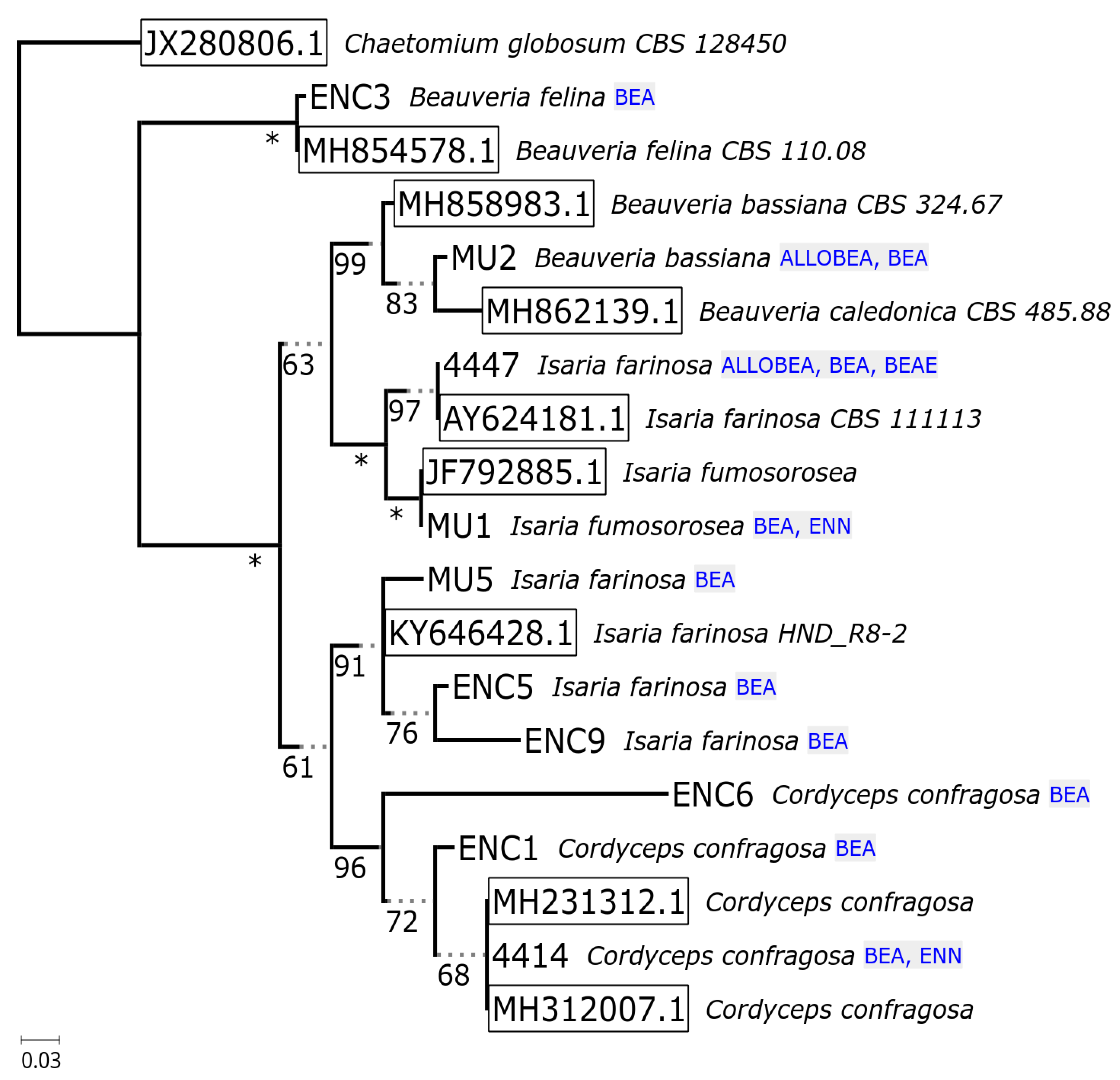
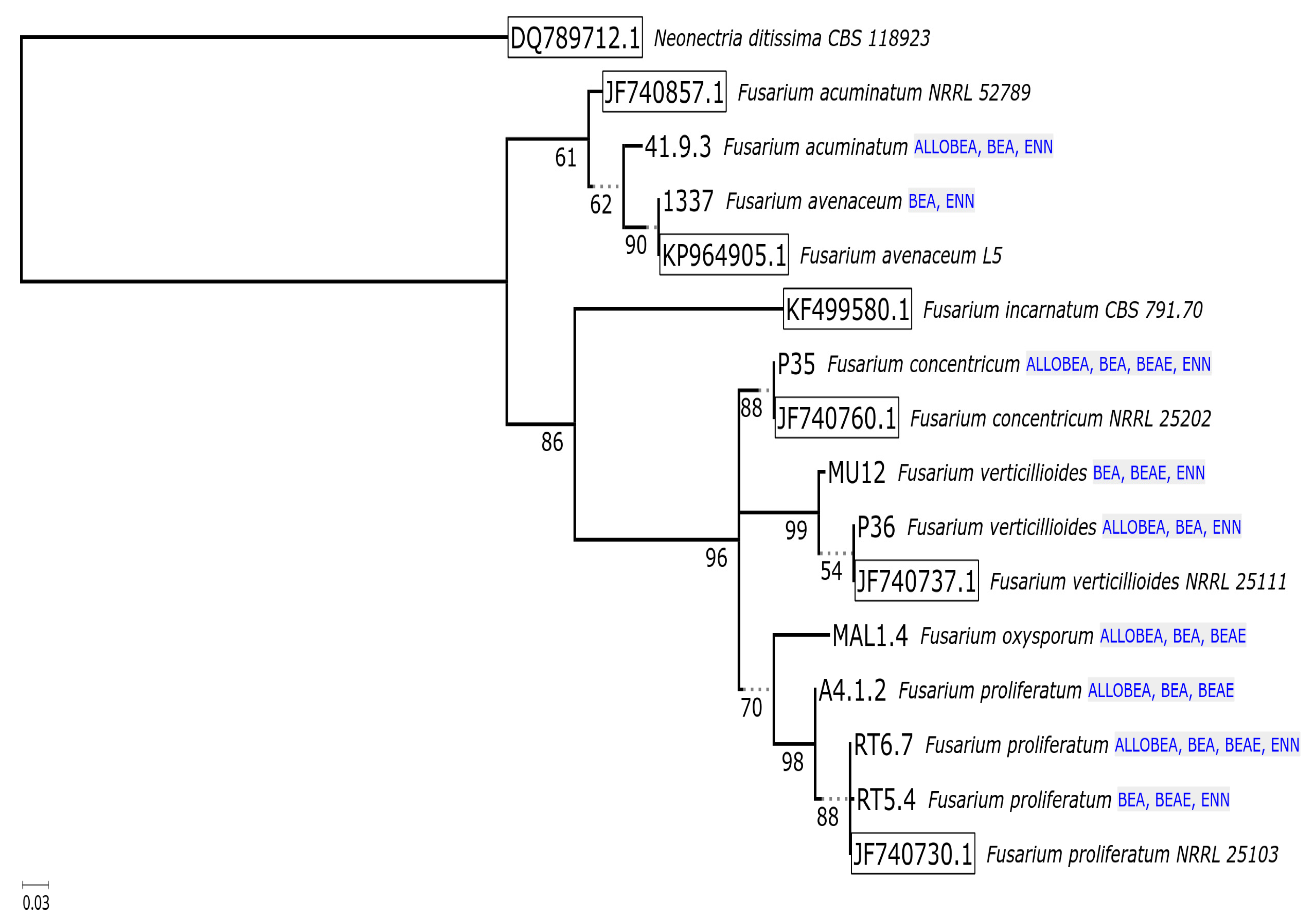
Acquired sequences by the amplification of ITS and tef-1α fragments from the investigated fungal strains showed over 98% similarity to the reference genes. The high identity level to the reference sequences from GenBank showed that all fungal species have been identified correctly. Results of molecular identification and characterization of fungal strains are described in Table 1. Relationships between examined isolates were ascertained as maximum likelihood phylogenetic tree reconstructions based on the ITS1 and tef-1α sequences. For phylogeny of the internal transcribed spacer (Figure 2), the additional reference sequences of Chaetomium globosum (JX280806.1), Beauveria felina (MH854578.1), Beauveria bassiana (MH858983.1), Isaria farinosa (AY624181.1; KY64628.1), Isaria fumosorosea (JF792885.1) and Cordyceps confragosa (MH231312.1; MH312007.1) were included in the analysis. For the reconstruction based on tef-1α marker (Figure 3), the sequences of Neonectria ditissima (DQ789712.1), Fusarium acuminatum (JF740857.1), Fusarium avenaceum (KP964905.1), Fusarium incarnatum (KF499580.1), Fusarium concentricum (JF740760.1), Fusarium verticillioides (JF740737.1) and Fusarium proliferatum (JF740730.1) were included in the analysis.
The analyses of the ITS and tef-1α genes have been frequently employed in the phylogenetic studies of fungi belonging to the Hypocreales order [3,49,50,51,52,53]. However, based only on these conserved taxonomic markers, the determination of differences between closely related genotypes is often limited. The analysis of secondary metabolite biosynthesis-related genes’ sequences, revealing the intraspecific and interspecific polymorphisms in datasets comprised of isolates from multiple species/genera, is often a better solution [5,42,54].

2.2. Non-Ribosomal Peptide Synthetase Genes Divergence

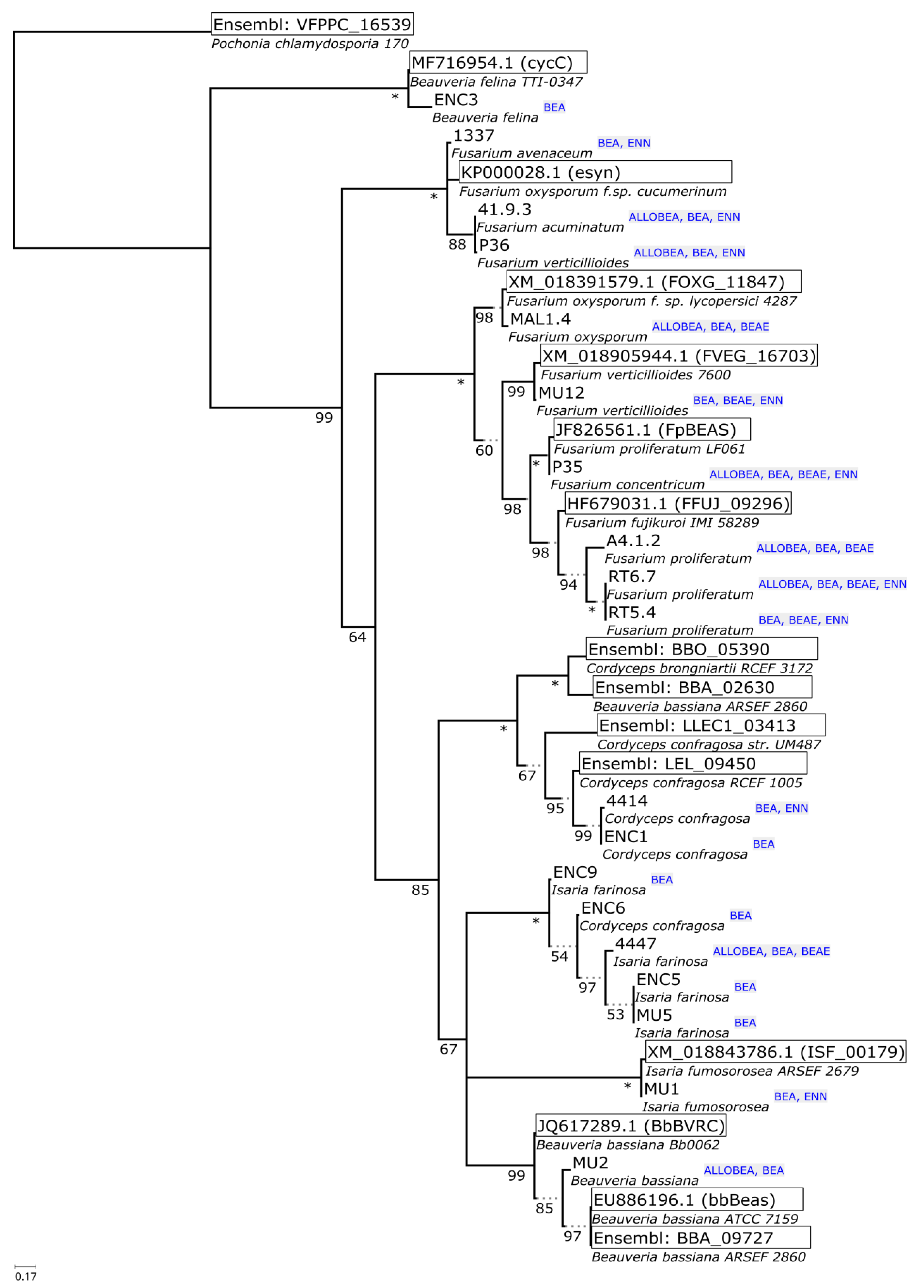
The phylogeny reconstructions of housekeeping sequences (ITS—Figure 2, tef-1α—Figure 3) point to the ancestral biosynthesis of beauvericin independently diverging into biosynthesis of other compounds (enniatins, beauvenniatins, allobeauvericins) across different taxa. The evolutionary relationships suggested by beauvericin gene fragments seem to support the notion of B. felina as an early diverging beauvericin producer, however, the obtained sequence fragment closely matches the cyclosporin synthase recently elucidated by Xu et al. [55], so verification by cloning, functional genomics and/or whole genome sequencing is likely needed to fully verify the synthase sequence. The divergent positioning of three Fusarium isolates (1337, 41.9.3, P36) as well as an unusual GenBank reference sequence for F. oxysporum f.sp. cucumerinum (itself highly similar to the known enniatin synthase from F. scirpi, accession Z18755) raises the possibility of parallel acquisition of cyclic depsipeptide synthases in ancient complexes within Fusarium genus.
Notably, such separation of two diverging variants of the synthase was previously observed in our earlier parsimony-based reconstruction [42], where predominantly enniatin-producing F. avenaceum/acuminatum/tricinctum isolates clustered distinctly separate from beauvericin-producing strains. Regardless of this, save for the B. felina sequence, all Cordycipitaceae isolate sequences form a well-supported clade with distinct separation between bassianolide synthase-like sequences (canonical bassianolide synthase from B. bassiana, model sequences from C. confragosa and C. brongniartii, isolates 4414 and ENC4) and beauvericin synthase-like fragments (canonical beauvericin synthase from B. bassiana, sequences from isolates tentatively identified as I. farinosa, I. fumosorosea, as well as single C. confragosa isolate ENC6). It is worth noting that the fairly recent update of Cordycipitaceae nomenclature and phylogeny shows that past identification of even reference Isaria/Cordyceps strains available from well-curated collections was not well supported by newly available molecular data [56]. In the context of continued research in the field [57], I. farinosa in particular is demonstrated to be a wide label spanning different species within the redefined, monophyletic Cordyceps and Akanthomyces genera (Figure 4).
With regards to the sequencing and phylogeny of putative BEAS homolog fragments, the validation of these sequences on the level of full genomic sequence and functional experiments is a desired follow-up. In particular, the further study of the ENC3 Beauveria felina isolate on genomic and functional levels is likely to provide insight as to the diversification of beauvericin/beauvenniatin/enniatin biosynthesis across this important genus of insect pathogens (Figure 4).

2.3. In Vitro Cyclodepsipeptide Biosynthesis

Quantitative analysis of ENN A (compound 12 in Table 1), ENN A1 (13), ENN B (14), ENN B1 (15) and BEA (1) was conducted using LC-MS/MS (Table 2). Beauvericin was produced by all investigated fungi, with the most efficient producers being Fusarium strains, such as F. proliferatum (RT6.7, RT 5.4) and F. concentricum (P35). To some extent, all of the studied fungi are known producers of beauvericin [42], however, it is important to note that each fungal strain possesses unique abilities to produce different amounts of BEA, which can also be affected by the cultivation conditions [58,59].
In contrast, the most efficient producers of the four enniatin analogues were F. acuminatum (41/9/3) and F. avenaceum (1337). The production of enniatins by phytopathogenic fungi belonging to the genus Fusarium has been described earlier [5,6,7,17,28,29,30,42]. The most efficient enniatin producer among entomopathogenic fungi was the isolate of C. confragosa (4414). Notably, some of the investigated fungal strains were not able to biosynthesize any of the four enniatins, and this group included entomopathogenic fungi B. bassiana (MU3), B. felina (ENC3), I. farinosa (4447, ENC5, ENC9, MU5) and C. confragosa (ENC1, ENC6). The same entomopathogenic fungal species were previously only investigated for beauvericin production, or production of beauvericin analogues [9,11,12,13]. Indeed, we were not able to find any previous reports concerning the production of enniatins by those entomopathogenic fungi. On the contrary, our own analyses showed that for at least two strains—I. fumosorosea (MU1) and C. confragosa (4414)—a limited capacity to produce enniatins exists, as each isolate was able to synthesize two of the four enniatin analogues (Table 2). Moreover, the two phytopathogenic fungal species F. proliferatum and F. oxysporum did not synthesize enniatin analogues, although they have earlier been described as enniatin producers [16,29,42,60]. This suggests a need for comprehensive reevaluation of cyclodepsipeptide profiles across multiple representatives of known producing taxa, in particular as the production of enniatins may depend on cultivation conditions, storage conditions or the host and the environment from which the fungi were isolated [59].
Liquid chromatography–ion-trap mass spectrometry (LC-ITMS) was used for the analysis of BEAs and BEAEs appearance in all investigated fungal crude extracts (Table 3). Two selected strains (P35 and 4447) were also analyzed using liquid chromatography–high-resolution mass spectrometry (LC-HRMS) for accurate mass measurements and HRMS/MS (Table 4). We have previously tentatively shown that these two strains produce BEA A, BEA C, BEA D, BEA F, BEA J, BEA K, BEAE A, and BEAE L [3]. Only five of the studied strains (P35, RT 6.7, 4447, MAL 1.4, and A4.12) were found to produce a wide range of different BEA and BEAE analogues (>15) (Table 3, Figure 1).
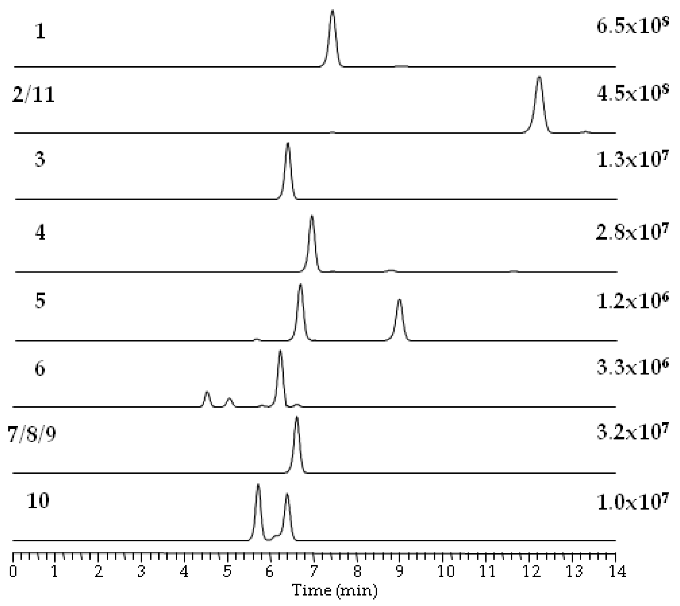
Using reversed-phase liquid chromatography (RPLC), the elution of individual BEAs and BEAEs was highly related to their molecular weights, i.e., beauvericin analogues with a higher molecular weight eluted later (Table 4). The presumed BEAs and BEAEs provided plentiful [M + NH4]+ ions during electrospray ionization, while [M + H]+ and [M + Na]+ ions accounted for only few percent of the total ion yield. In total, five beauvericins (compounds 15), five beauvenniatins (610) and one allobeauvericin (11) were tentatively detected in different fungal extracts (Figure 1 and Figure 5, and Table 4).
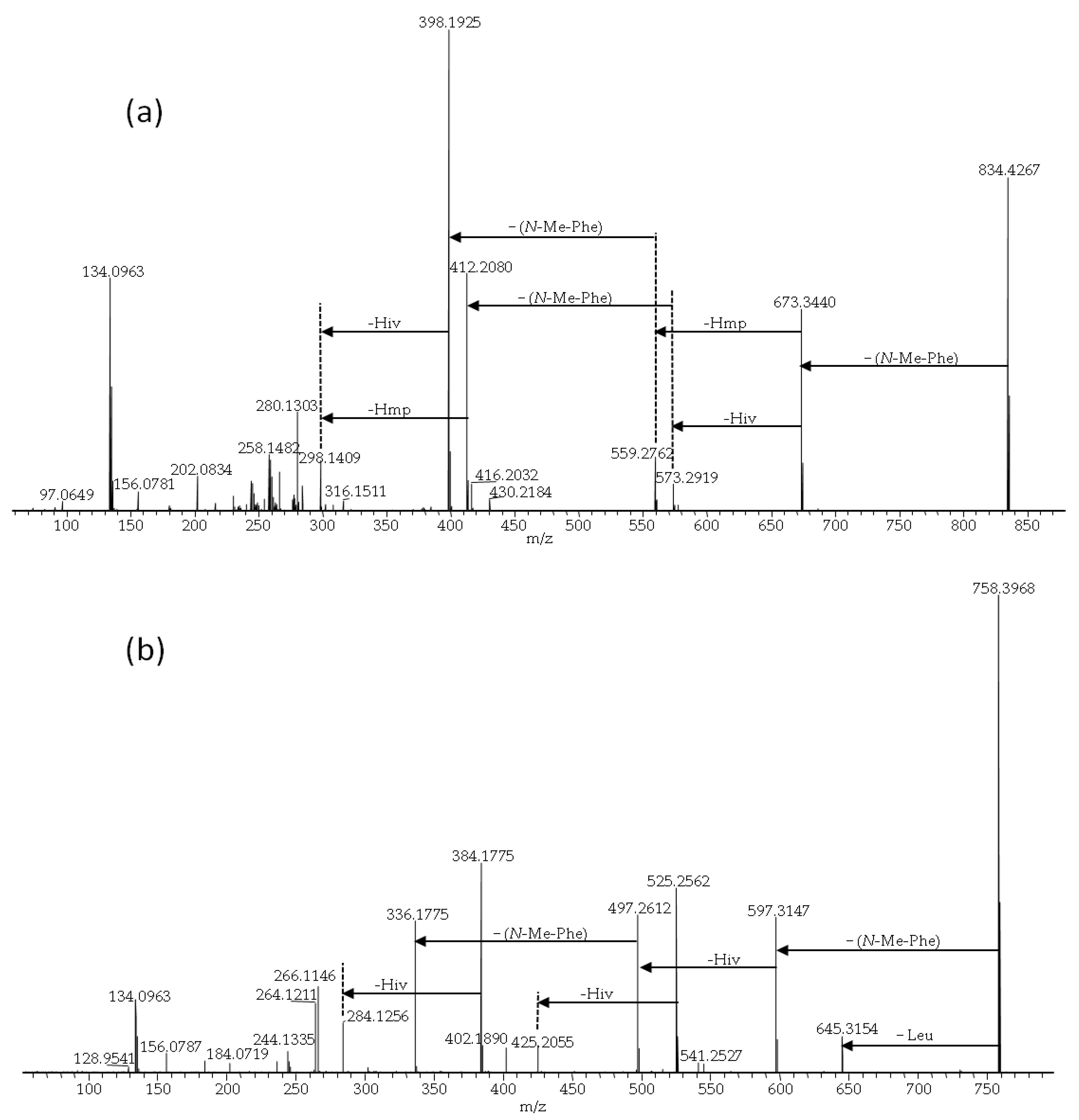
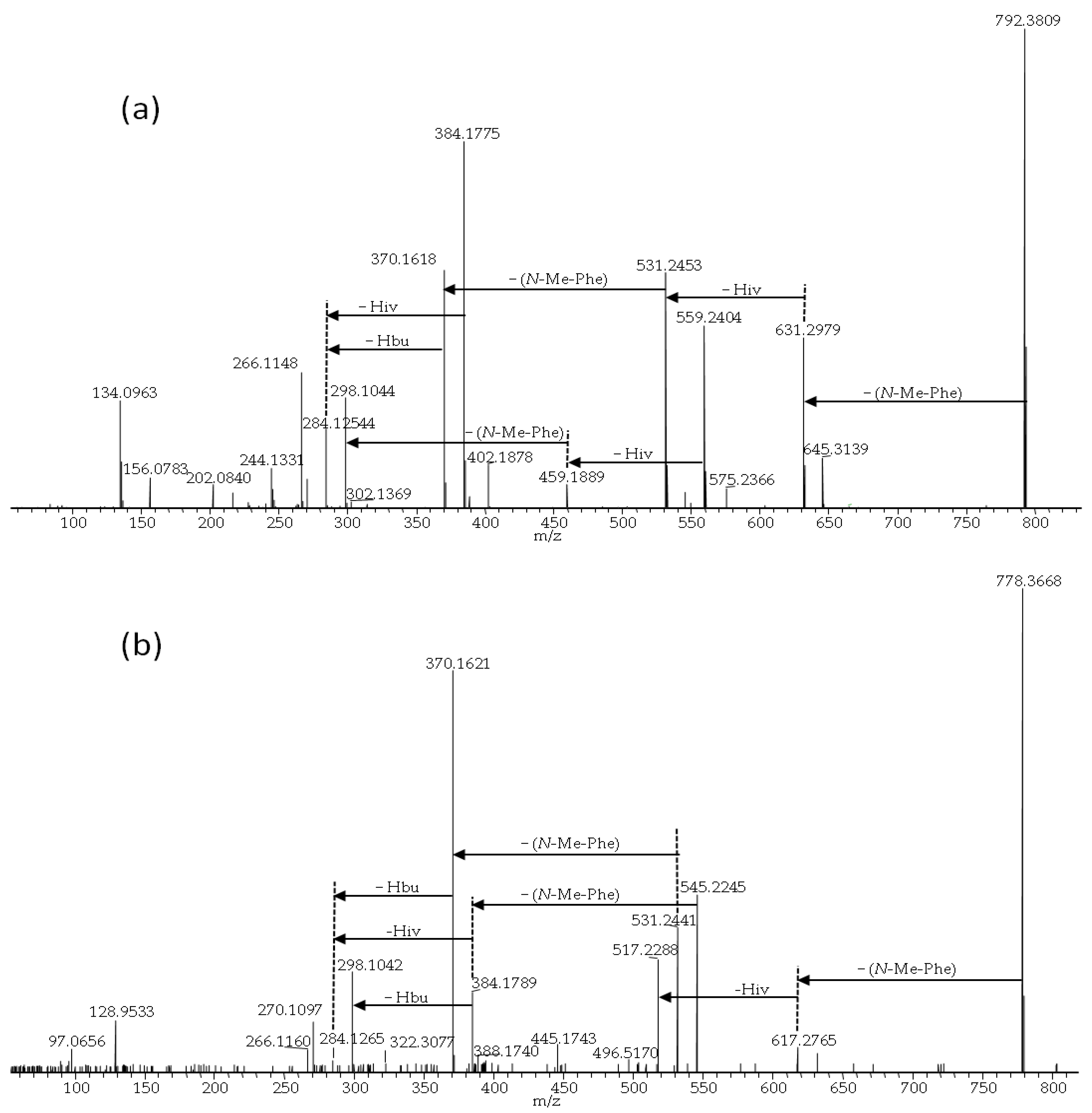
BEA itself was the most common analogue and was produced in the highest relative amounts (in terms of LC-MS peak areas) by all studied fungal strains. We used BEA (1) as a reference for comparing product-ion spectra of the sodiated ions of different analogues [3]. All BEA analogues, characterized by the presence of N-methyl-phenylalanine in their structure, were tentatively identified by the presence of a product-ion due to a loss of −161 Da. The beauvericins that were observed in the studied strains varied in hydroxy acid composition (Figure 6a,b). BEA E (3) contains one leucine moiety in addition to two N-methyl-phenylalanine units, and this analogue was tentatively identified by the presence of a product-ion corresponding to a loss of −127 Da. BEA E (3) possesses three D-Hiv units giving rise to product-ions corresponding to a loss of −100 Da (Figure 6b) [11,24]. In contrast, BEA B (2) possesses two D-Hmp units and one D-Hiv unit, giving rise to product-ions corresponding to −114 and −100 Da, respectively (Figure 6a) [12]. Furthermore, BEA G1, and G2 (4, 5) consist of one or two D-Hbu groups, respectively, which can be observed by product-ions, corresponding to a loss of −86 Da (Figure 7a,b) [13].
ALLOBEAs A, B (11) and C are isomers of BEAs A/F, B (2) and C, which are composed of three N-methyl-phenylalanine moieties and one, two or three D-Hmp groups, respectively [12]. BEAEs 79, which are also isomers [10], were produced in the smallest relative amounts. The HRMS/MS spectra of their sodiated ions were of poor signal/noise ratios; still, they were better than spectra of their ammoniated ions. The detected isomers of depsipeptides consist of the same moieties, therefore, we are not able to tell which isomer/s is/are present in the sample (Figure 5). The differences between studied compounds (2/11 and 7/8/9) were described earlier, on the basis of NMR analyses [10,12]. All investigated BEAEs (610) contain one group of N-methyl-valine and two N-methyl-phenylalanine residues, which was manifested by product-ions corresponding to losses of −161 and −113 Da (Figure 8a–c). BEAE B (6) contains three D-Hiv groups (Figure 8a) [10], while BEAEs 79 (Figure 8b) appeared to contain one D-Hmp group instead of D-Hiv, based on our HRMS/MS data. Moreover, we detected one compound (10) in the crude extract from F. concentricum (P35) pure rice cultures that appeared to be composed of an unreported combination of amino and hydroxy acids. Our HRMS/MS data showed that it could be an unreported BEAE consisting of three D-Hiv groups, one N-methyl-valine, N-methyl-phenylalanine and N-methyl-leucine or -isoleucine group (Figure 8c). The latter two cannot be distinguished by mass spectrometry alone. For future investigations, nuclear magnetic resonance (NMR) will be considered in order to better discern the actual depsipeptide structure.

3. Materials and Methods

3.1. Fungal Strains, Media and Growth Conditions

Fungal strains from the Hypocreales order were isolated from infected plants and insects found in Poland. All isolated strains were placed in the fungi collection of the Institute of Plant Genetics, Polish Academy of Sciences, Poznań, Poland. For characterization of fungal isolates, the species, strain number and host species have been summarized in Table 1. For genomic DNA extraction, the growing mycelia of individual Hypocreales fungi were purified and cultivated on potato dextrose agar medium (PDA, Oxoid, Basingstoke, UK) for seven days at controlled conditions (20/25 °C, 12 h photoperiod). Harvested mycelia were stored at −20 °C. For qualitative and quantitative cyclodepsipeptide analyses, fourteen day old pure rice cultures of each fungal species were used, according to the procedure described previously [53].

3.2. DNA Extraction, Molecular Identification, PCR Primers and DNA Sequencing

Genomic DNA extraction from fungal samples was performed using a modified method with the CTAB (hexadecyltrimethylammonium bromide), according to Gorczyca et al. [61]. The DNA concentration in extracts was quantified using an ND-1000 spectrophotometer (Thermo Scientific, West Palm Beach, FL, USA) and stored in a freezer at −20 °C. For Fusarium species identification, the sequence analysis of a variable fragment of the translation elongation factor 1α gene (tef-1α) was performed, whereas for the other species, the molecular identification was carried out on the basis of the sequence analysis of the internal transcribed spacers of the ribosomal DNA region (ITS1–ITS2).
Polymerase chain reactions (PCRs) were carried out using DreamTaq Green DNA polymerase (Thermo Scientific, Espoo, Finland). The conditions for PCR amplification of the tef-1α gene fragment were described earlier by Gálvez et al. [28], where primers Ef728M (5′-CATCGAGAAGTTCGAGAAGG-3′) and TefR1 (5′-GCCATCCTTGGAGATACCAGC-3′) [62] were used. The conditions for the PCR amplification of the ITS1–ITS2 DNA region were described earlier by Kozłowska et al. [51], where primers ITS4 (5′-TCCTCCGCTTATTGATATGC-3′) and ITS5 (5′-GGAAGTAAAAGTCGTAACAAGG-3′) [63] were used. Obtained sequences from PCR reaction were separated in 1.5% agarose gel (EURx, Gdańsk, Poland) with GelGreen Nucleic Acid Stain (Biotium, Inc., San Francisco, CA, USA).
The beauvericin synthase gene (BEAS) was partially amplified using the beas_1 (5′-TKGARCAGCGBCAYGAGACM-3′) and beas_2 (5′-GGWCGRGGGAARTCRGTDGG-3′) primers designed earlier by Stępień and Waśkiewicz [42].
The PCRs were done in 25 μL volumes containing Color PfuPlus! DNA Polymerase (EURx, Gdańsk, Poland); 2.5 μL of 10 x Pfu Buffer contains 15 mM MgSO4, 0.5 µmol of forward/reverse primers and 0.25 mM of each dNTP and 10–20 ng of fungal DNA. The PCR conditions were as follows: 5 min at 95 °C, 35 cycles of 30 s at 95 °C, 30 s at 61 °C, 1 min at 72 °C and 7 min at 72 °C.
In order to analyze the sequences, amplicons were purified with exonuclease I (Thermo Scientific, Espoo, Finland) and FastAP shrimp alkaline phosphatase (Thermo Scientific, Espoo, Finland), afterwards they were labeled using forward primer and the BigDyeTerminator 3.1 kit (Applied Biosystems, Foster City, CA, USA). Finally, PCR-amplified DNA fragments were precipitated with 96% ethanol according to Tomczyk et al. [49].

3.3. Sequence Analysis and Phylogeny Reconstruction

Individual DNA sequences were aligned using MUSCLE 3.8.31, available via SeaView 4.6.4 environment [64] (Linux 64bit version). In the case of the BEAS coding sequence, the sequences were first translated and aligned as proteins, then backtranslated into codon alignments. In the case of TEF, exon boundaries were corrected manually to match known splicing site consensus (GT-AG type introns). To investigate grouping of obtained sequences, their top BLAST hits (discontiguous MegaBLAST was performed on the non-redundant GenBank database through NCBI/BLAST stand-alone service, accessed on 22 May 2019) were trimmed appropriately and were also included in the final alignments. In the case of BEAS homologs, additional Beauveria/Cordyceps sensu lato representatives were retrieved from Ensembl/Fungi database (accessed on 17 May 2019).
The phylogeny reconstructions were carried out with IQTREE 1.6.9 [65] (Linux 64-bit version) using automated model fitting, partitioning and ultrafast bootstrap [66]. During the reconstruction, ITS rDNA was modelled as non-coding DNA sequence, while BEAS was investigated using codon-based models. In the case of the translation elongation factor (tef-1α), the coding fragment was investigated as a single coding partition, while the DNA model was ascertained for intronic parts present in the amplified fragments.

3.4. Mycotoxin Analyses

3.4.1. Chemicals

Mycotoxin standards (>99%) (enniatins A, A1, B, B1 and beauvericin), ammonium formate (>99.99%) and methanol (HPLC grade > 99.9%) were purchased from Sigma-Aldrich (St. Louis, MO, USA). Standard solutions of ENNs (2.0 ng/μL of each analogue) and BEA (1.0 ng/μL) were prepared in methanol and kept in a freezer at −20 °C. Acetonitrile and water for LC-MS were from Thermo Fisher Scientific (Optima LC-MS grade, Waltham, MA, USA).

3.4.2. Extraction and purification

For screening the fungal rice cultures for the presence of different depsipeptide analogues using LC-ion-trap MS, 0.15 g aliquots weighed (after lyophilized and ground into powder) of each culture were extracted using 1.5 mL of methanol/water (9:1, v/v) by shaking on an orbital shaker (225 min−1, 90 min) and by sonication for 20 min. After centrifugation using a Beckman J2-MC centrifuge (Beckman Coulter Inc., Fullerton, CA) at 15,000× g, for 10 min, extracts were filtered through a 0.22 µm nylon membrane (Costar, Corning Inc., Corning, NY, USA) and transferred to chromatography vials. For quantification of mycotoxins (BEA, ENNs) by LC-MS/MS, individual culture aliquots (1 g) were mixed with 2 mL of acetonitrile-water (9:1, v/v) and after homogenization (homogenizer H500, Pol-Ekoaparatura, Poland), centrifugation (at 4500× g for 5 min), and filtration (0.20 µm syringe filter—Chromafil, Macherey-Nagel, Duren, Germany) were prepared for chromatographic analysis.

3.4.3. Liquid Chromatography Mass Spectrometry Analyses

Culture extracts were screened for the presence of ENNs, BEA, BEAE and ALLOBEA analogues using an LCQ Fleet ion trap mass spectrometer (Thermo Fisher Scientific) coupled to a Waters Acquity UPLC (Milford, MA, USA) with chromatographic column—SunFire C18; instrumental conditions were described earlier by Urbaniak et al. 2019 [3].
Quantitative analysis of mycotoxins (BEA and ENNs) were performed using an LC-MS/MS instrument consisting of an UPLC™ system (Acquity, Waters, Milford, MA, USA) coupled to a triple quadrupole mass spectrometer (TQD, Waters Micromass, Manchester, UK), equipped with an electrospray ionization interface according to Stanciu et al. [67] with our own modifications, described below. The separation of beauvericin and enniatins was achieved using a BEH C18 chromatographic column (100 × 2.1 mm i.d., 1.7 µm particle size; Waters) with injection volume—3 µL and flow rate of mobile phase—0.3 mL/min. The mobile phase consisted of water (A) and methanol (B), both containing 5 mM ammonium formate and 0.1% (v/v) formic acid. The gradient program was as follows: initial conditions at 80% A, 20% B for 2 min; then, from 20% to 90% B in 5 min; next, 90% B for 6 min; from 90% to 100% B in 3 min; return to initial conditions in 2 min. For data processing, EmpowerTM 1 software was used (Waters, Manchester, UK). Chromatographic parameters are shown in Table 5.

3.4.4. Liquid Chromatography High-Resolution Mass Spectrometry (HRMS)

A Q-Exactive Fourier-transform high-resolution mass spectrometer (Thermo Fisher Scientific) coupled to a Vanquish Horizon UHPLC (Thermo Fisher Scientific) was used to acquire accurate mass data and high-resolution product-ion spectra of putative beauvericin analogues. Separation was achieved on a Kinetex C18 column (75 × 2.1 mm; 2.6 µm particles; Phenomenex, Torrance, CA, USA) held at 30 °C with the injection volume—3 µL and flow rate—0.3 mL/min. The mobile phase consisted of line A (5 mM ammonium formate) and line B (5 mM of ammonium formate in MeOH/water, 95:5, v/v) with gradient program as follows: 0–15 min, 75% B; 15–15.1 min, 80% B; 15.1–18 min, 100% B; 18.1–21 min, 75%. The instrument was run in the full-scan mode (m/z 600–900). The fragmentation of the sodiated molecular ions (MS/MS) from eleven metabolites was performed in the full-scan mode (m/z 150–1200), according to the previous work [3]. Elemental compositions were calculated using Xcalibur, version 2.3. (Thermo Fisher Scientific).

4. Conclusions

The present study demonstrates the high variability of naturally produced cyclohexadepsipeptides by Hypocreales fungi. It also emphasizes that different fungi have independently evolved a significant level of divergence inside the non-ribosomal peptide synthase genes involved in depsipeptide biosynthesis, along with functional adaptation towards biosynthesis of overlapping yet diversified metabolite profiles. Additionally, a new compound has been described as a naturally occurring beauvenniatin analogue (10).

Author Contributions

M.U. and Ł.S. conceived and designed the experiments. M.U. and A.T. performed molecular analyses. A.W. and M.U. performed the chemical analyses. A.W., and M.U. discussed the data and results of the chemical analyses. M.U. wrote the manuscript. G.K. and M.U. performed sequence analyses and phylogeny reconstruction. G.K. and Ł.S. reviewed the manuscript. All authors have read and agreed to the published version of the manuscript.

Funding

This research was funded by the National Science Centre OPUS 8 grant: NCN 2014/15/B/NZ9/01544. The National Science Centre PRELUDIUM 13 grant: NCN 2017/25/N/NZ9/02525.

Acknowledgments

The authors wish to thank all, who assisted in conduction of this work, in particular Silvio Uhlig, from the Norwegian Veterinary Institute, who helped with the chemical analyses.

Conflicts of Interest

The authors declare no conflict of interest.

References

  1. Jestoi, M. Emerging fusarium-mycotoxins fusaproliferin, beauvericin, enniatins, and moniliformin: A Review. Crit. Rev. Food Sci. Nutr. 2008, 48, 21–49. [Google Scholar] [CrossRef] [PubMed]
  2. Sivanathan, S.; Scherkenbeck, J. Cyclodepsipeptides: A rich source of biologically active compounds for drug research. Molecules 2014, 19, 12368–12420. [Google Scholar] [CrossRef]
  3. Urbaniak, M.; Stepien, L.; Uhlig, S. Evidence for naturally produced beauvericins containing N-Methyl-Tyrosine in hypocreales fungi. Toxins 2019, 11, 182. [Google Scholar] [CrossRef] [PubMed] [Green Version]
  4. Hornbogen, T.; Glinski, M.; Zocher, R. Biosynthesis of depsipeptide mycotoxins in fusarium. Eur. J. Plant. Pathol. 2002, 108, 713–718. [Google Scholar] [CrossRef]
  5. Stepien, L.; Jestoi, M.; Chelkowski, J. Cyclic hexadepsipeptides in wheat field samples and Esyn1 gene divergence among enniatin producing fusarium avenaceum strains. World Mycotoxin J. 2013, 6, 399–409. [Google Scholar] [CrossRef]
  6. Logrieco, A.; Moretti, A.; Castella, G.; Kostecki, M.; Golinski, P.; Ritieni, A.; Chelkowski, J. Beauvericin production by fusarium species. Appl. Environ. Microbiol. 1998, 64, 3084–3088. [Google Scholar] [CrossRef] [Green Version]
  7. Covarelli, L.; Beccari, G.; Prodi, A.; Generotti, S.; Etruschi, F.; Meca, G.; Juan, C.; Manes, J. Biosynthesis of beauvericin and enniatins in vitro by wheat fusarium species and natural grain contamination in an area of central Italy. Food Microbiol. 2015, 46, 618–626. [Google Scholar] [CrossRef]
  8. Weng, Q.; Zhang, X.; Chen, W.; Hu, Q. Secondary metabolites and the risks of isaria fumosorosea and isaria farinosa. Molecules 2019, 24, 664. [Google Scholar] [CrossRef] [Green Version]
  9. Luangsa-Ard, J.; Berkaew, P.; Ridkaew, R.; Hywel-Jones, N.L.; Isaka, M. A Beauvericin Hot spot in the genus isaria. Mycol. Res. 2009, 113, 1389–1395. [Google Scholar] [CrossRef]
  10. Bunyapaiboonsri, T.; Vongvilai, P.; Auncharoen, P.; Isaka, M. Cyclohexadepsipeptides from the filamentous fungus acremonium sp. Bcc 2629. Helv. Chim. Acta 2012, 95, 963–972. [Google Scholar] [CrossRef]
  11. Fukuda, T.; Arai, M.; Tomoda, H.; Omura, S. New beauvericins, potentiators of antifungal miconazole activity, produced by beauveria sp Fki-1366. II. structure elucidation. J. Antibiot. 2004, 57, 117–124. [Google Scholar] [CrossRef] [Green Version]
  12. Nilanonta, C.; Isaka, M.; Kittakoop, P.; Trakulnaleamsai, S.; Tanticharoen, M.; Thebtaranonth, Y. Precursor-directed biosynthesis of beauvericin analogs by the insect pathogenic fungus paecilomyces tenuipes Bcc 1614. Tetrahedron 2002, 58, 3355–3360. [Google Scholar] [CrossRef]
  13. Xu, Y.; Zhan, J.; Wijeratne, E.M.K.; Burns, A.M.; Gunatilaka, A.A.L.; Molnar, I. Cytotoxic and antihaptotactic beauvericin analogues from precursor-directed biosynthesis with the insect pathogen beauveria bassiana atcc 7159. J. Nat. Prod. 2007, 70, 1467–1471. [Google Scholar] [CrossRef] [PubMed]
  14. Ivanova, L.; Skjerve, E.; Eriksen, G.S.; Uhlig, S. Cytotoxicity of enniatins a, A1, B, B1, B2 and B3 from fusarium avenaceum. Toxicon 2006, 47, 868–876. [Google Scholar] [CrossRef]
  15. Uhlig, S.; Ivanova, L. Determination of beauvericin and four other enniatins in grain by liquid chromatography-mass spectrometry. J. Chromatogr. A 2004, 1050, 173–178. [Google Scholar] [CrossRef]
  16. Song, H.H.; Lee, H.S.; Jeong, J.H.; Park, H.S.; Lee, C. Diversity in beauvericin and enniatins H, I, and Mk1688 by fusarium oxysporum isolated from potato. Int. J. Food Microbiol. 2008, 122, 296–301. [Google Scholar] [CrossRef] [PubMed]
  17. Chełkowski, J.; Ritieni, A.; Wiśniewska, H.; Mulè, G.; Logrieco, A. Occurrence of toxic hexadepsipeptides in preharvest maize ear rot infected by fusarium poae in Poland. J. Phytopathol. 2006, 155, 8–12. [Google Scholar] [CrossRef]
  18. Jow, G.M.; Chou, C.J.; Chen, B.F.; Tsai, J.H. Beauvericin induces cytotoxic effects in human acute lymphoblastic leukemia cells through cytochrome C release, caspase 3 activation: The causative role of calcium. Cancer Lett. 2004, 216, 165–173. [Google Scholar] [CrossRef]
  19. Shin, C.G.; An, D.G.; Song, H.H.; Lee, C. Beauvericin and enniatins H, I and Mk1688 are new potent inhibitors of human immunodeficiency virus type-1 integrase. J. Antibiot. 2009, 62, 687–690. [Google Scholar] [CrossRef]
  20. Dornetshuber, R.; Heffeter, P.; Kamyar, M.R.; Peterbauer, T.; Berger, W.; Lemmens-Gruber, R. Enniatin exerts p53-dependent cytostatic and p53-independent cytotoxic activities against human cancer cells. Chem. Res. Toxicol. 2007, 20, 465–473. [Google Scholar] [CrossRef]
  21. Watjen, W.; Debbab, A.; Hohlfeld, A.; Chovolou, Y.; Kampkotter, A.; Edrada, R.A.; Ebel, R.; Hakiki, A.; Mosaddak, M.; Totzke, F.; et al. Enniatins A1, B and B1 from an endophytic strain of fusarium tricinctum induce apoptotic cell death in H4IIE hepatoma cells accompanied by inhibition of ERK phosphorylation. Mol. Nutr. Food Res. 2009, 53, 431–440. [Google Scholar] [CrossRef] [PubMed]
  22. Chen, B.F.; Tsai, M.C.; Jow, G.M. Induction of calcium influx from extracellular fluid by beauvericin in human leukemia cells. Biochem. Biophys. Res. Commun. 2006, 340, 134–139. [Google Scholar] [CrossRef] [PubMed]
  23. Kamyar, M.; Rawnduzi, P.; Studenik, C.R.; Kouri, K.; Lemmens-Gruber, R. Investigation of the electrophysiological properties of enniatins. Arch. Biochem. Biophys. 2004, 429, 215–223. [Google Scholar] [CrossRef]
  24. Fukuda, T.; Arai, M.; Yamaguchi, Y.; Masuma, R.; Tomoda, H.; Omura, S. New beauvericins, potentiators of antifungal miconazole activity, produced by beauveria sp FKI-1366. I. taxonomy, fermentation, Isolation and biological properties. J. Antibiot. 2004, 57, 110–116. [Google Scholar] [CrossRef] [PubMed] [Green Version]
  25. Zhang, L.X.; Yan, K.Z.; Zhang, Y.; Huang, R.; Bian, J.; Zheng, C.S.; Sun, H.X.; Chen, Z.H.; Sun, N.; An, R.; et al. High-Throughput synergy screening identifies microbial metabolites as combination agents for the treatment of fungal infections. Proc. Natl. Acad. Sci. USA 2007, 104, 4606–4611. [Google Scholar] [CrossRef] [PubMed] [Green Version]
  26. Supothina, S.; Isaka, M.; Kirtikara, K.; Tanticharoen, M.; Thebtaranonth, Y. Enniatin production by the entomopathogenic fungus verticillium hemipterigenum BCC 1449. J. Antibiot. 2004, 57, 732–738. [Google Scholar] [CrossRef] [Green Version]
  27. Lin, H.I.; Lee, Y.J.; Chen, B.F.; Tsai, M.C.; Lu, J.L.; Chou, C.J.; Jow, G.M. Involvement of Bcl-2 family, cytochrome C and caspase 3 in induction of apoptosis by beauvericin in human non-small cell lung Cancer cells. Cancer Lett. 2005, 230, 248–259. [Google Scholar] [CrossRef]
  28. Galvez, L.; Urbaniak, M.; Waskiewicz, A.; Stepien, L.; Palmero, D. fusarium proliferatum–Causal agent of garlic bulb rot in Spain: Genetic variability and mycotoxin production. Food Microbiol. 2017, 67, 41–48. [Google Scholar] [CrossRef]
  29. Jestoi, M.; Rokka, M.; Yli-Mattila, T.; Parikka, P.; Rizzo, A.; Peltonen, K. Presence and concentrations of the fusarium-related mycotoxins beauvericin, enniatins and moniliformin in finnish grain samples. Food Addit. Contam. 2004, 21, 794–802. [Google Scholar] [CrossRef]
  30. Logrieco, A.; Rizzo, A.; Ferracane, R.; Ritieni, A. Occurrence of beauvericin and enniatins in wheat affected by fusarium avenaceum head blight. Appl. Environ. Microbiol. 2002, 68, 82–85. [Google Scholar] [CrossRef] [Green Version]
  31. Bushley, K.E.; Turgeon, B.G. Phylogenomics reveals subfamilies of fungal nonribosomal peptide synthetases and their evolutionary relationships. BMC Evol. Biol. 2010, 10, 26. [Google Scholar] [CrossRef] [Green Version]
  32. Gallo, A.; Ferrara, M.; Perrone, G. Phylogenetic study of polyketide synthases and nonribosomal peptide synthetases involved in the biosynthesis of mycotoxins. Toxins 2013, 5, 717–742. [Google Scholar] [CrossRef] [PubMed] [Green Version]
  33. Xu, Y.; Orozco, R.; Wijeratne, E.M.; Gunatilaka, A.A.; Stock, S.P.; Molnar, I. Biosynthesis of the cyclooligomer depsipeptide beauvericin, a virulence factor of the entomopathogenic fungus beauveria bassiana. Chem. Biol. 2008, 15, 898–907. [Google Scholar] [CrossRef] [Green Version]
  34. Zhang, T.; Zhuo, Y.; Jia, X.; Liu, J.; Gao, H.; Song, F.; Liu, M.; Zhang, L. Cloning and characterization of the gene cluster required for beauvericin biosynthesis in fusarium proliferatum. Sci. China Life Sci. 2013, 56, 628–637. [Google Scholar] [CrossRef] [Green Version]
  35. Zocher, R.; Keller, U.; Kleinkauf, H. Enniatin synthetase, a novel type of multifunctional enzyme catalyzing depsipeptide synthesis in fusarium oxysporum. Biochemistry 1982, 21, 43–48. [Google Scholar] [CrossRef]
  36. Billich, A.; Zocher, R. Constitutive expression of enniatin synthetase during fermentative growth of fusarium scirpi. Appl. Environ. Microbiol. 1988, 54, 2504–2509. [Google Scholar] [CrossRef] [PubMed] [Green Version]
  37. Hornbogen, T.; Riechers, S.P.; Prinz, B.; Schultchen, J.; Lang, C.; Schmidt, S.; Mugge, C.; Turkanovic, S.; Sussmuth, R.D.; Tauberger, E.; et al. Functional characterization of the recombinant N-Methyltransferase domain from the multienzyme enniatin synthetase. ChemBioChem 2007, 8, 1048–1054. [Google Scholar] [CrossRef] [PubMed]
  38. Glinski, M.; Urbanke, C.; Hornbogen, T.; Zocher, R. Enniatin synthetase is a monomer with extended structure: Evidence for an intramolecular reaction mechanism. Arch. Microbiol. 2002, 178, 267–273. [Google Scholar] [CrossRef]
  39. Zocher, R.; Keller, U.; Kleinkauf, H. Mechanism of depsipeptide formation catalyzed by enniatin synthetase. Biochem. Biophys. Res. Commun. 1983, 110, 292–299. [Google Scholar] [CrossRef]
  40. Haese, A.; Schubert, M.; Herrmann, M.; Zocher, R. Molecular characterization of the enniatin synthetase gene encoding a multifunctional enzyme catalysing N-Methyldepsipeptide formation in fusarium scirpi. Mol. Microbiol. 1993, 7, 905–914. [Google Scholar] [CrossRef]
  41. Peeters, H.; Zocher, R.; Kleinkauf, H. Synthesis of beauvericin by a multifunctional enzyme. J. Antibiot. 1988, 41, 352–359. [Google Scholar] [CrossRef] [Green Version]
  42. Stepien, L.; Waskiewicz, A. Sequence divergence of the enniatin synthase gene in relation to production of beauvericin and enniatins in fusarium Species. Toxins 2013, 5, 537–555. [Google Scholar] [CrossRef]
  43. Gallou, A.; Serna-Dominguez, M.G.; Berlanga-Padilla, A.M.; Ayala-Zermeno, M.A.; Mellin-Rosas, M.A.; Montesinos-Matias, R.; Arredondo-Bernal, H.C. Species clarification of isaria isolates used as biocontrol agents against diaphorina citri (hemiptera: Liviidae) in Mexico. Fungal Biol. 2016, 120, 414–423. [Google Scholar] [CrossRef]
  44. Zhang, J.; Fu, B.; Lin, Q.; Riley, I.T.; Ding, S.; Chen, L.; Cui, J.; Yang, L.; Li, H. Colonization of beauveria bassiana 08f04 in root-zone soil and its biocontrol of cereal cyst nematode (heterodera filipjevi). PLoS ONE 2020, 15, e0232770. [Google Scholar] [CrossRef] [PubMed]
  45. Wang, J.J.; Qiu, L.; Chu, Z.J.; Ying, S.H.; Feng, M.G. The connection of protein O-mannosyltransferase family to the biocontrol potential of beauveria bassiana, a fungal entomopathogen. Glycobiology 2014, 24, 638–648. [Google Scholar] [CrossRef] [PubMed] [Green Version]
  46. Yang, H.; Qin, C.S.; Chen, Y.M.; Zhang, G.Y.; Dong, L.H.; Wan, S.Q. Persistence of metarhizium (hypocreales: Clavicipitaceae) and beauveria bassiana (hypocreales: Clavicipitaceae) in Tobacco Soils and Potential as Biocontrol Agents of Spodoptera litura (lepidoptera: Noctuidae). Environ. Entomol. 2019, 48, 147–155. [Google Scholar] [CrossRef] [PubMed] [Green Version]
  47. Bhadauria, B.P.; Singh, P.K.; Shailesh, P.; Zaidi, N.W.; Singh, U.S. Characterization and biocontrol potential of entomopathogenic fungus, beauveria bassiana isolates against spilarctia obliqua. J. Environ. Biol. 2013, 34, 917–921. [Google Scholar] [PubMed]
  48. Batta, Y.A. Biocontrol of almond bark beetle (scolytus amygdali geurin-meneville, coleoptera: Scolytidae) using beauveria bassiana (Bals.) vuill. (deuteromycotina: Hyphomycetes). J. Appl. Microbiol. 2007, 103, 1406–1414. [Google Scholar] [CrossRef] [PubMed]
  49. Tomczyk, L.; Stepien, L.; Urbaniak, M.; Szablewski, T.; Cegielska-Radziejewska, R.; Stuper-Szablewska, K. Characterisation of the mycobiota on the shell surface of table eggs acquired from different egg-laying hen breeding systems. Toxins 2018, 10, 293. [Google Scholar] [CrossRef] [Green Version]
  50. Kozlowska, E.; Hoc, N.; Sycz, J.; Urbaniak, M.; Dymarska, M.; Grzeszczuk, J.; Kostrzewa-Suslow, E.; Stepien, L.; Plaskowska, E.; Janeczko, T. Biotransformation of steroids by entomopathogenic strains of isaria farinosa. Microb. Cell Factories 2018, 17, 71. [Google Scholar] [CrossRef] [Green Version]
  51. Kozlowska, E.; Urbaniak, M.; Hoc, N.; Grzeszczuk, J.; Dymarska, M.; Stepien, L.; Plaskowska, E.; Kostrzewa-Suslow, E.; Janeczko, T. Cascade biotransformation of dehydroepiandrosterone (DHEA) by beauveria Species. Sci. Rep. 2018, 8, 13449. [Google Scholar] [CrossRef] [PubMed]
  52. Stepien, L.; Koczyk, G.; Waskiewicz, A. Diversity of fusarium species and mycotoxins contaminating pineapple. J. Appl. Genet. 2013, 54, 367–380. [Google Scholar] [CrossRef] [Green Version]
  53. Stepien, L.; Waskiewicz, A.; Urbaniak, M. Wildly growing asparagus (Asparagus officinalis L.) hosts pathogenic fusarium species and accumulates their mycotoxins. Microb. Ecol. 2016, 71, 927–937. [Google Scholar] [CrossRef] [Green Version]
  54. Von Bargen, S.; Martinez, O.; Schadock, I.; Eisold, A.M.; Gossmann, M.; Buttner, C. Genetic variability of phytopathogenic fusarium proliferatum associated with crown rot in asparagus officinalis. J. Phytopathol. 2009, 157, 446–456. [Google Scholar] [CrossRef]
  55. Xu, L.; Li, Y.; Biggins, J.B.; Bowman, B.R.; Verdine, G.L.; Gloer, J.B.; Alspaugh, J.A.; Bills, G.F. Identification of cyclosporin C from amphichorda felina using a cryptococcus neoformans differential temperature sensitivity assay. Appl. Microbiol. Biotechnol. 2018, 102, 2337–2350. [Google Scholar] [CrossRef] [PubMed] [Green Version]
  56. Kepler, R.M.; Luangsa-Ard, J.J.; Hywel-Jones, N.L.; Quandt, C.A.; Sung, G.H.; Rehner, S.A.; Aime, M.C.; Henkel, T.W.; Sanjuan, T.; Zare, R.; et al. A phylogenetically-based nomenclature for cordycipitaceae (hypocreales). IMA Fungus 2017, 8, 335–353. [Google Scholar] [CrossRef]
  57. Mongkolsamrit, S.; Noisripoom, W.; Thanakitpipattana, D.; Wutikhun, T.; Spatafora, J.W.; Luangsa-Ard, J. Disentangling cryptic species with isaria-like morphs in cordycipitaceae. Mycologia 2018, 110, 230–257. [Google Scholar] [CrossRef]
  58. Xu, L.J.; Liu, Y.S.; Zhou, L.G.; Wu, J.Y. Enhanced beauvericin production with in situ adsorption in mycelial liquid culture of fusarium redolens Dzf2. Process. Biochem. 2009, 44, 1063–1067. [Google Scholar] [CrossRef]
  59. Decleer, M.; Landschoot, S.; De Saeger, S.; Rajkovic, A.; Audenaert, K. Impact of fungicides and weather on cyclodepsipeptide-producing fusarium spp. and beauvericin and enniatin levels in wheat grains. J. Sci. Food Agric. 2019, 99, 253–262. [Google Scholar] [CrossRef] [Green Version]
  60. Song, H.H.; Ahn, J.H.; Lim, Y.H.; Lee, C. Analysis of beauvericin and unusual enniatins co-produced by fusarium oxysporum Fb1501 (Kfcc 11363p). J. Microbiol. Biotechn. 2006, 16, 1111–1119. [Google Scholar]
  61. Gorczyca, A.; Oleksy, A.; Gala-Czekaj, D.; Urbaniak, M.; Laskowska, M.; Waskiewicz, A.; Stepien, L. fusarium head blight incidence and mycotoxin accumulation in three durum wheat cultivars in relation to sowing date and density. Sci. Nat. 2018, 105. [Google Scholar] [CrossRef] [PubMed] [Green Version]
  62. Carbone, I.; Kohn, L.M. A method for designing primer sets for speciation studies in filamentous ascomycetes. Mycologia 1999, 91, 553–556. [Google Scholar] [CrossRef]
  63. White, T.J.; Bruns, T.; Lee, S.; Taylor, J. Amplification and direct sequencing of fungal ribosomal RNA genes for phylogenetic. PCR Protoc. Guide Methods Appl. 1990, 5, 315–322. [Google Scholar]
  64. Gouy, M.; Guindon, S.; Gascuel, O. Seaview version 4: A multiplatform graphical user interface for sequence alignment and phylogenetic tree building. Mol. Biol. Evol. 2010, 27, 221–224. [Google Scholar] [CrossRef] [Green Version]
  65. Nguyen, L.T.; Schmidt, H.A.; von Haeseler, A.; Minh, B.Q. Iq-Tree: A fast and effective stochastic algorithm for estimating maximum-likelihood phylogenies. Mol. Biol. Evol. 2015, 32, 268–274. [Google Scholar] [CrossRef]
  66. Hoang, D.T.; Chernomor, O.; von Haeseler, A.; Minh, B.Q.; Vinh, L.S. UFBoot2: Improving the ultrafast bootstrap approximation. Mol. Biol. Evol. 2018, 35, 518–522. [Google Scholar] [CrossRef]
  67. Stanciu, O.; Juan, C.; Miere, D.; Loghin, F.; Manes, J. Analysis of enniatins and beauvericin by LC-MS/MS in wheat-based products. CyTA–J. Food 2017, 15, 433–440. [Google Scholar] [CrossRef]
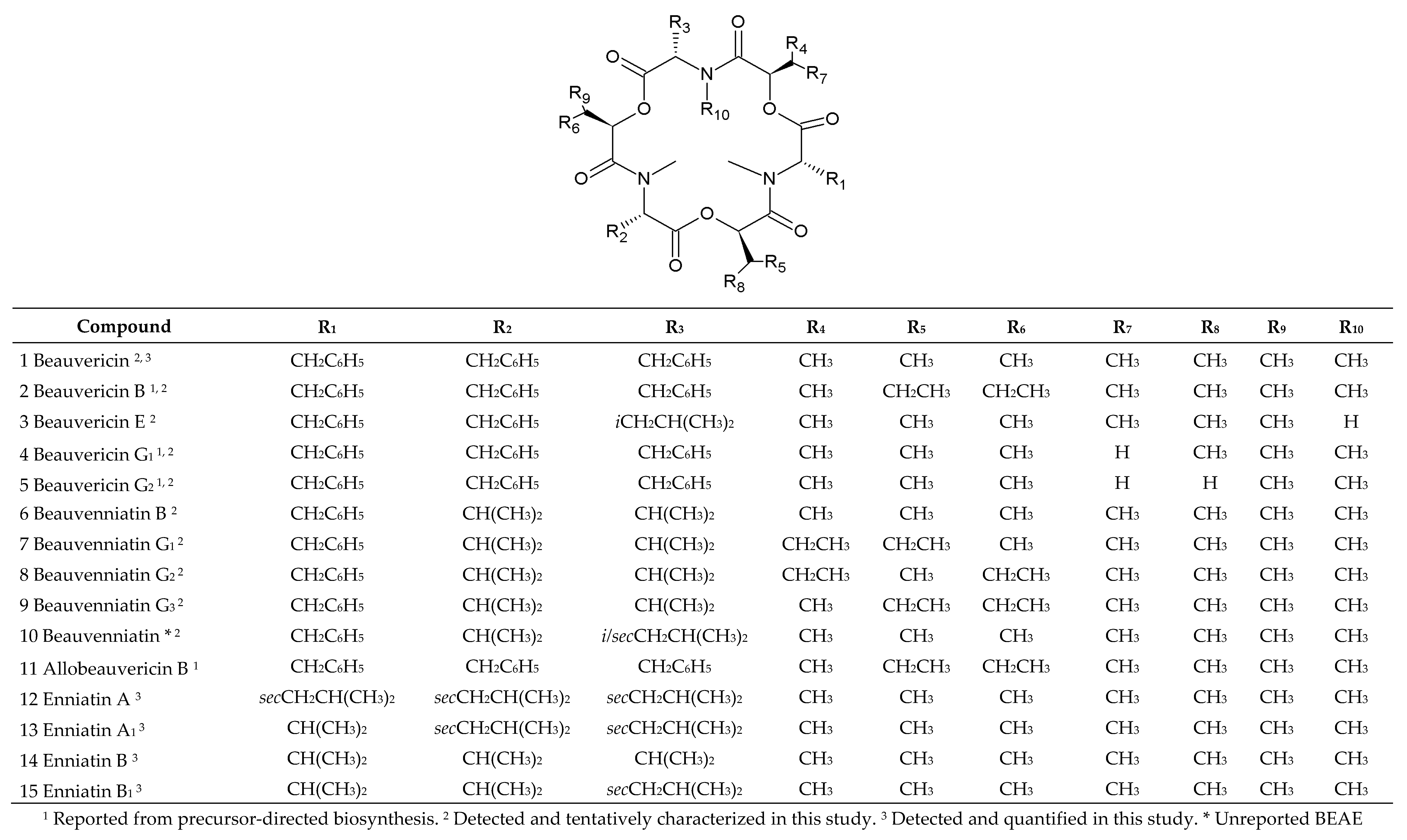
Figure 1. Overview of chemical structures of the beauvericin, allobeauvericin, enniatin and beauvenniatin analogues discussed and identified in this study.
Pathogens 09 00552 g001
Figure 2. Phylogeny of 19 ITS rDNA partial sequences. Numbers below branches indicate support (‘*’ indicates total support for bipartition), box outlines mark reference sequences, and annotated compound classes are listed beside the leaves. The majority-rule consensus tree was based on the alignment of 475 columns with 117 parsimony informative sites. The TIM3e + G4 model was chosen by IQTREE based on BIC criterion. A total of 103 ultrafast bootstrap iterations were carried out (automated stopping criterion for tree search). All bipartitions with below 50% ultrafast bootstrap support were collapsed in the final representation.
Pathogens 09 00552 g002
Figure 3. Phylogeny of 16 tef-1α partial sequences. Numbers below branches indicate support, box outlines mark reference sequences, and annotated compound classes are listed beside the leaves. The majority-rule consensus tree was based on the alignment of 435 columns (including 87 coding triplets) with 101 parsimony informative sites. The following models were chosen by IQTREE based on BIC criterion: TN + F + G4 for intronic fragments and MG + F3X4 + I for coding parts. A total of 102 ultrafast bootstrap iterations were carried out (automated stopping criterion for tree search). All bipartitions with below 50% ultrafast bootstrap support were collapsed in the final representation.
Pathogens 09 00552 g003
Figure 4. Phylogeny of 34 partial sequences of putative BEAS amplicons. Numbers below branches indicate support (‘*’ indicates total support for bipartition), box outlines mark reference sequences from GenBank and Ensembl/Fungi, annotated compound classes are listed beside the leaves. The majority-rule consensus tree was based on the alignment of 157 codons with 147 parsimony informative codons. The MGK + F3X4 + G4 model was automatically fit by IQTREE based on BIC criterion. A total of 115 ultrafast bootstrap iterations were carried out (automated stopping criterion for tree search). All bipartitions with below 50% ultrafast bootstrap support were collapsed in the final representation.
Pathogens 09 00552 g004
Figure 5. Extracted ion LC-HRMS chromatograms (±5 ppm) of the [M + NH4]+ ions for beauvericin, allobeauvericin and beauvenniatin analogues in the crude extract from rice cultures of Fusarium concentricum (P35) and Isaria farinosa (4447). The numbers refer to the compounds in Figure 1.
Pathogens 09 00552 g005
Figure 6. LC-HRMS/MS spectra from higher collision dissociation of the [M + Na]+ ions of putative beauvericin analogues: beauvericin B (a) and beauvericin E (b).
Pathogens 09 00552 g006
Figure 7. LC-HRMS/MS spectra from higher collision dissociation of the [M + Na]+ ions of putative beauvericin analogues: beauvericin G1 (a) and beauvericin G2 (b).
Pathogens 09 00552 g007
Figure 8. LC-HRMS/MS spectra from higher collision dissociation of the [M + Na]+ ions of beauvenniatin analogues: beauvenniatin B (a), beauvenniatin G1/G2/ G3 (b) and beauvenniatin * (c).
Pathogens 09 00552 g008
Table 1. Hypocreales fungi used in this study, host species, fungal strain identification based on ITS1–ITS2 or tef-1α sequence analysis and comparison with reference sequences from the GenBank Database.
No.StrainSpeciesHostSequence Nucleotide Identity
11337Fusarium avenaceumwheat99.76% identity to the Fusarium avenaceum acc. number KP964905.1
(Triticum L.)
2MU2Beauveria bassianabark beetle100% identity to the Beauveria bassiana acc. number KU158425.1
(Trypodendron lineatum)
3ENC3 Beauveria felina caterpillar98.37% identity to the Beauveria felina acc. number MH854578.1
(Lepidoptera sp.)
415222Fusarium acuminatumpea98.19% identity to the Fusarium acuminatum acc. number JF740857.1
(Pisum L.)
5P35Fusarium concentricumpineapple100% identity to the Fusarium concentricum, acc. number JF740760.1
(Ananas comosus)
6RT6.7Fusarium proliferatumrice99.78% identity to the Fusarium proliferatum acc. number JF740730.1
(Oryza sativa)
7RT5.4Fusarium proliferatumrice99.78% identity to the Fusarium proliferatum acc. number JF740730.1
(Oryza sativa)
8MU12Fusarium verticillioidesbanana98.66% identity to the Fusarium verticillioides acc. number JF740717.1
(Musa L.)
9P36Fusarium verticillioidespineapple100% identity to the Fusarium verticillioides acc. number JF740717.1
(Ananas comosus)
104447 Isaria farinosa bark beetle100% identity to the Isaria farinosa, acc. number AY624181.1
(Trypodendron lineatum)
11ENC5 Isaria farinosa bark beetle98.92% identity to the Isaria farinosa, acc. number KY646428.1
(Trypodendron lineatum)
12ENC9 Isaria farinosa beetle 98.89% identity to the Isaria farinosa, acc. number DQ888729.1
(Coleoptera sp.)
13MU5 Isaria farinosa pine beetle99.53% identity to the Isaria farinosa, acc. number KY646428.1
(Dendroctonus ponderosae)
14MU1 Isaria fumosorosea beetle 98.38% identity to the Isaria fumosorosea, acc. number JF792885.1
(Coleoptera sp.)
154414 Cordyceps confragosa parent bug 100% identity to the Cordyceps confragosa, acc. number MH231312.1
(Elasmucha sp.)
16ENC1 Cordyceps confragosa bark beetle98.08% identity to the Cordyceps confragosa, acc. number MH312006.1
(Trypodendron lineatum)
17ENC6 Cordyceps confragosa caterpillar99.82% identity to the Cordyceps confragosa, acc. number MH312006.1
(Lepidoptera sp.)
18MAL 1.4Fusarium oxysporumasparagus100% identity to the Fusarium oxysporum acc. number KP964890.1
(Asparagus officinalis L.)
19A 4.12Fusarium proliferatumwheat100% identity to the Fusarium proliferatum acc. number KU939029.1
(Triticum L.)
Table 2. Mean concentrations (±standard deviations) of beauvericin and enniatins A, A1, B, and B1 [ng/g] produced in vitro by 19 fungal strains belonging to 11 species.
StrainBEA [ng/g]ENN A [ng/g]ENN A1 [ng/g]ENN B [ng/g]ENN B1 [ng/g]
1337 (Fav)49.3 ± 5.64300 ± 16063800 ± 2430NDND
MU2 (Bb)389 ± 22.4NDNDNDND
ENC3 (Bf)211 ± 12NDNDNDND
41/9/3 (Fac)150 ± 1056600 ± 196068970 ± 2535NDND
P35 (Fco)130400 ± 2300127 ± 20ND350 ± 37ND
RT6.7 (Fpr)135540 ± 3225NDND1983 ± 37.4ND
RT5.4 (Fpr)55420 ± 411NDND2130 ± 130ND
MU12 (Fve)125 ± 4.3NDND191 ± 12.751 ± 4.5
P36 (Fve)150 ± 8.3NDND23 ± 2.6ND
4447 (Ifa)10240 ± 246NDNDNDND
ENC5 (Ifa)215 ± 15NDNDNDND
ENC9 (Ifa)352 ± 9.6NDNDNDND
MU5 (Ifa)155 ± 9.3NDNDNDND
MU1 (Ifu)900 ± 26.2NDND720 ± 23.471 ± 5
4414 (Cco)125 ± 15.75730 ± 157NDND9130 ± 128
ENC1 (Cco)128±12NDNDNDND
ENC6 (Cco)115 ± 13NDNDNDND
MAL 1.4 (Fox)433 ± 11.3NDNDNDND
A 4.12 (Fpr)730 ± 60NDNDNDND
ND—not detected. Fav—Fusariumm avenaceum, Bb—Beauveria bassiana, Bf—Beauveria felina, Fac—Fusarium acuminatum, Fco—Fusarium concentricum, Fpr—Fusarium proliferatum, Fve—Fusarium verticillioides, Ifa—Isaria farinosa, Ifu—Isaria fumosorosea, Cco—Cordyceps confragosa, Fox—Fusarium oxysporum.
Table 3. Qualitative analysis of BEAs, BEAEs and ENNs produced by Hypocreales fungi in this study.
No.StrainSpeciesMetabolic Profile
11337Fusarium avenaceumBEA, ENN A, ENN A1
2MU2 Beauveria bassiana BEA, BEA B, BEA C, BEA F/A, ALLOBEA A, ALLOBEA B, ALLOBEA C
3ENC3 Beauveria felina BEA,
415222Fusarium acuminatumBEA, BEA C, BEA D, BEA G1, ALLOBEA C, ENN A, ENN A1
5P35Fusarium concentricumBEA, BEA B, BEA C, BEA D, BEA E, BEA F/A, BEA J, BEA G1, BEA G2, BEAE A, BEAE B, BEAE G1/G2/G3, BEAE *, ALLOBEA A, ALLOBEA B, ALLOBEA C, BEAE L, BEA K, BEA L, ENN A, ENN B
6RT6.7Fusarium proliferatumBEA, BEA B, BEA C, BEA D, BEA E, BEA F/A, BEA J, BEA G1, BEA G2, BEAE A, BEAE B, ALLOBEA A, ALLOBEA B, ALLOBEA C, BEAE L, BEA K, ENN B
7RT5.4Fusarium proliferatumBEA, BEA D, BEA J, BEA G1, BEAE L, ENN B
8MU12Fusarium verticillioidesBEA, BEA D, BEA G1, BEAE A, BEA K, ENN B, ENN B1
9P36Fusarium verticillioidesBEA, BEA C, ALLOBEA C, ENN B
104447 Isaria farinosa BEA, BEA B, BEA C, BEA D, BEA E, BEA F/A, BEA J, BEA G1, BEA G2, BEAE A, ALLOBEA A, ALLOBEA B, ALLOBEA C, BEAE L, BEA K, BEA L
11ENC5 Isaria farinosa BEA
12ENC9 Isaria farinosa BEA
13MU5 Isaria farinosa BEA
14MU1 Isaria fumosorosea BEA, ENN B, ENN B1
154414 Cordyceps confragosa BEA, ENN A, ENN B1
16ENC1 Cordyceps confragosa BEA
17ENC6 Cordyceps confragosa BEA
18MAL 1.4Fusarium oxysporumBEA, BEA B, BEA C, BEA D, BEA E, BEA F/A, BEA J, BEA G1, BEA G2, BEAE A, BEAE B, ALLOBEA A, ALLOBEA B, ALLOBEA C, BEAE L
19A 4.12Fusarium proliferatumBEA, BEA B, BEA C, BEA D, BEA E, BEA F/A, BEA J, BEA G1, BEA G2, BEAE A, BEAE B, ALLOBEA A, ALLOBEA B, ALLOBEA C, BEAE L
BEA—beauvericin; BEAE—beauvenniatin; ENN—enniatin; ALLOBEA—allobeauvericin.
Table 4. Accurate mass and elemental composition of major ions observed for different beauvericin, allobeauvericin and beauvenniatin analogues from LC-HRMS.
CompoundMeasured (m/z) [M + H]+Measured (m/z) [M + NH4]+Measured (m/z) [M + Na]+Retention Time (min)Elemental CompositionMass Error (ppm) [M + H]+Mass Error (ppm) [M + NH4]+Mass Error (ppm) [M + Na]+
1784.4144801.4426806.39477.4C45H57N309−3.7−1.6−5
2/11812.4446829.4738834.426712.4C47H61N3O9−4.9−1.7−4.8
3736.4155753.4431758.39686.4C41H57N3O9−2.5−0.9−3.3
4770.3997787.4274792.38097C44H55N3O9−2.6−1−3.5
5756.384773.4124778.36686.7C43H53N3O9−1.9−0.2−1.5
6688.4184705.4425710.39656.3C37H57N3O91.6−1.1−3.9
7/8/9716.4469733.4745738.42636.7C39H61N3O9−2.4−0.9−5.8
10702.4344719.4581724.4135.7C38H59N3O92.1−2−2.6
Table 5. Parent and daughter ions, collision energy and limit of detection (LOD) and quantification (LOQ) (ng/g) for mycotoxins.
CompoundParent ion (m/z) [M+NH4]+Primary Daughter Ion (m/z)Secondary Daughter Ion (m/z)Collision Energy (eV)LOD a (ng/g)LOQ b (ng/g)
ENN A699.4228.2 *210.13626
ENN A1685.4214.2 *210.13839
ENN B657.3214.1 *196.04026
ENN B1671.2228.0 *214.15726
BEA801.2784.0244.1 *2813
* Transitions used for quantification. a Limit of detection (LOD). b Limit of quantification (LOQ).