Article

Isolation of Candidatus Bartonella rousetti and Other Bat-associated Bartonellae from Bats and Their Flies in Zambia

1
Hokudai Center for Zoonosis Control in Zambia, Hokkaido University Research Center for Zoonosis Control, Sapporo 001-0020, Japan
2
Division of Global Epidemiology, Hokkaido University Research Center for Zoonosis Control, Sapporo 001-0020, Japan
3
Laboratory of Parasitology, Graduate School of Infectious Diseases, Faculty of Veterinary Medicine, Hokkaido University, Sapporo 060-0818, Japan
4
Department of Para-Clinical Studies, School of Veterinary Medicine, the University of Zambia, Lusaka 10101, Zambia
5
Global Virus Network Affiliate Center of Excellence, the University of Zambia, Lusaka 10101, Zambia
6
African Center of Excellence for Infectious Diseases of Humans and Animals, the University of Zambia, Lusaka 10101, Zambia
7
Department of National Parks and Wildlife, Ministry of Tourism and Arts of Zambia, Lusaka 10101, Zambia
8
Division of Molecular Pathobiology, Hokkaido University Research Center for Zoonosis Control, Sapporo 001-0020, Japan
9
Department of Disease Control, School of Veterinary Medicine, the University of Zambia, Lusaka 10101, Zambia
10
Global Institution for Collaborative Research and Education (GI-CoRE), Hokkaido University, Sapporo 001-0020, Japan
11
Global Virus Network, Baltimore, MD 21201, USA
12
Division of Infection and Immunity, Hokkaido University Research Center for Zoonosis Control, Sapporo 001-0020, Japan
13
Division of Collaboration and Education, Hokkaido University Research Center for Zoonosis Control, Sapporo 001-0020, Japan
*
Author to whom correspondence should be addressed.
Pathogens 2020, 9(6), 469; https://doi.org/10.3390/pathogens9060469
Submission received: 12 May 2020 / Revised: 9 June 2020 / Accepted: 12 June 2020 / Published: 13 June 2020

Abstract

:
Bat-associated bartonellae, including Bartonella mayotimonensis and Candidatus Bartonella rousetti, were recently identified as emerging and potential zoonotic agents, respectively. However, there is no report of bat-associated bartonellae in Zambia. Thus, we aimed to isolate and characterize Bartonella spp. from bats and bat flies captured in Zambia by culturing and PCR. Overall, Bartonella spp. were isolated from six out of 36 bats (16.7%), while Bartonella DNA was detected in nine out of 19 bat flies (47.3%). Subsequent characterization using a sequence of five different genes revealed that three isolates obtained from Egyptian fruit bats (Rousettus aegyptiacus) were Ca. B. rousetti. The isolates obtained from insectivorous bats (Macronycteris vittatus) were divided into two previously unclassified bat-associated bartonellae. A phylogenetic analysis of the six genotypes of Bartonella gltA sequences from nine pathogen-positive bat flies revealed that three genotypes belonged to the same clades as bat-associated bartonellae, including Ca. B. rousetti. The other three genotypes represented arthropod-associated bartonellae, which have previously been isolated only from ectoparasites. We demonstrated that Ca. B. rousetti is maintained between bats (R. aegyptiacus) and bat flies in Zambia. Continuous surveillance of Bartonella spp. in bats and serological surveys in humans in Africa are warranted to evaluate the public health importance of bat-associated bartonellae.

1. Introduction

The members of the genus Bartonella are Gram-negative, hemotropic, and primary vector-borne bacteria that colonize mammalian endothelial and red blood cells. To date, the genus Bartonella consists of more than 30 species, many of which have been described recently [1]. Various mammalian hosts for Bartonella spp. have been reported, such as rodents, carnivores, ruminants, and marine mammals [2]. Various arthropods also play important roles in the maintenance and transmission of Bartonella spp. In recent decades, several Bartonella spp. have been recognized as human pathogens responsible for various clinical manifestations [3]. For example, Bartonella quintana is an etiological agent of trench fever, transmitted by louse [4,5], while Bartonella henselae, maintained in cat and cat flea, causes cat scratch disease in humans [4,6]. Furthermore, associations between several poorly characterized Bartonella spp. and human diseases, including neuroretinitis, febrile illness, fever, and bacteremia, have been reported [7,8,9,10].
Several lines of evidence indicate that bats harbor diverse groups of Bartonella species and genotypes, including zoonotic or potentially zoonotic Bartonella [11]. Candidatus Bartonella mayotimonensis was first detected in the aortic valve tissue of a patient with endocarditis from the United States of America [12]. Subsequently, Ca. B. mayotimoensis was detected in little brown bats (Myotis lucifugus) and gray bats (Myotis grisescens) in Finland [13]. A Bartonella genotype detected in bats in Georgia (Caucasus) showed high similarity with Bartonella sequences found in the sera of forest workers in Poland [14]. Furthermore, a recent serological study reported the detection of antibodies against Candidatus Bartonella rousetti, which was previously detected in bats in Kenya and Nigeria [15,16], among the people who entered the caves to capture bats during a traditional bat festival in southwestern Nigeria [17]. Collectively, these findings strengthen the importance of public health-related studies on bat-associated bartonellae. However, studies about the potential risk of bat-associated bartonellae have not been carried out in many of resource-limited countries.
The present study intended to characterize Bartonella spp. in bats captured in Zambia, where reports on bartonellae are not available. Our results confirmed the presence of several groups of Bartonella, both in bats and bat flies in Zambia.

2. Results

2.1. Isolation of Bartonella from Bats

Out of 36 bat blood samples spread on sheep blood agar plates, six samples yielded tiny bacterial colonies with a rough texture and whitish hue during 2 weeks of incubation (Figure 1). Three isolates each were derived from frugivorous bats (Rousettus aegyptiacus) (IDs: ZB17-74, -79, and -86) and insectivorous bats (Macronycteris vittatus) (IDs: ZB17-107, -109, and -113). All the isolates were confirmed to be Bartonella spp. using 16S ribosomal DNA sequencing.

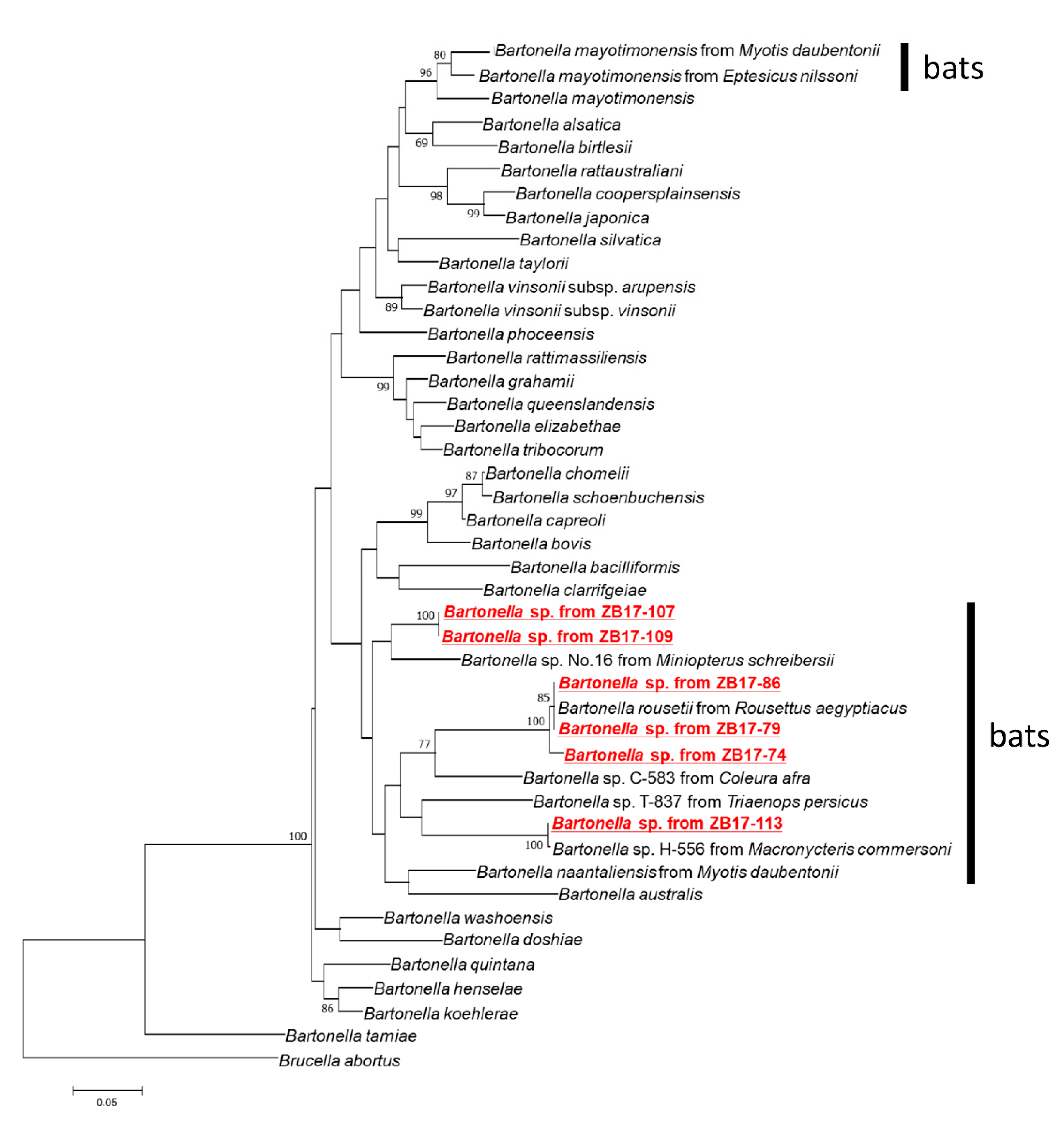
2.2. Molecular Characterization of Bartonella Isolates

Bartonella isolates were characterized via the sequencing of five housekeeping genes encoding citrate synthase (gltA), RNA polymerase beta subunit (rpoB), transfer messenger RNA (ssrA), cell division protein (ftsZ), and nicotinamide adenine dinucleotide dehydrogenase gamma subunit (nuoG). The sequences of the three isolates obtained from Rousettus aegyptiacus (ZB17-74, -79, and -86) were identical for all genes except for gltA, in which the isolate ZB17-74 harbored eight nucleotide mismatches. The sequences of all five genes showed the highest sequence identity with those of Candidatus Bartonella rousetti deposited in the database (Table 1). The sequences of five genes of two isolates (ZB17-107 and -109) obtained from Macronycteris vittatus were identical, which were distinct from those of the other isolate (ZB17-113). These three isolates showed the highest sequence identity with unclassified Bartonella spp. reported from various bat species, namely Rhinolophus ferrumequinum from Georgia (Bartonella sp. 44552) [14], Rh. ferrumequinum from China (Bartonella sp. SD-3/2015) [18], Eidolon helvum from Kenya (Bartonella sp. B23983) [19], and Macronycteris commersoni from Kenya (Bartonella sp. H-556) [15], and from a vole (Clethrionomys rutilus) from the United States of America (Bartonella sp. Cr28649) [20].
A maximum likelihood tree was reconstructed based on the concatenated sequences of ftsZ, gltA, nuoG, rpoB, ssrA, and the 16S rDNA. In the tree, Bartonella detected in this study belonged to a clade, including other Bartonella strains detected in bats (Figure 2). Briefly, the isolates from ZB17-74, -79, and -86 formed a cluster with Candidatus Bartonella rousetti, while the isolate from ZB17-113 clustered together with Bartonella sp. H-556 from Macronycteris commersoni from Kenya [15]. The isolates from ZB17-107 and -109 did not cluster with previously reported Bartonella spp., but were phylogenetically closest to Bartonella sp. no. 16 isolated from the common bent-wing bat, Miniopterus schreibersii, from Taiwan [21].

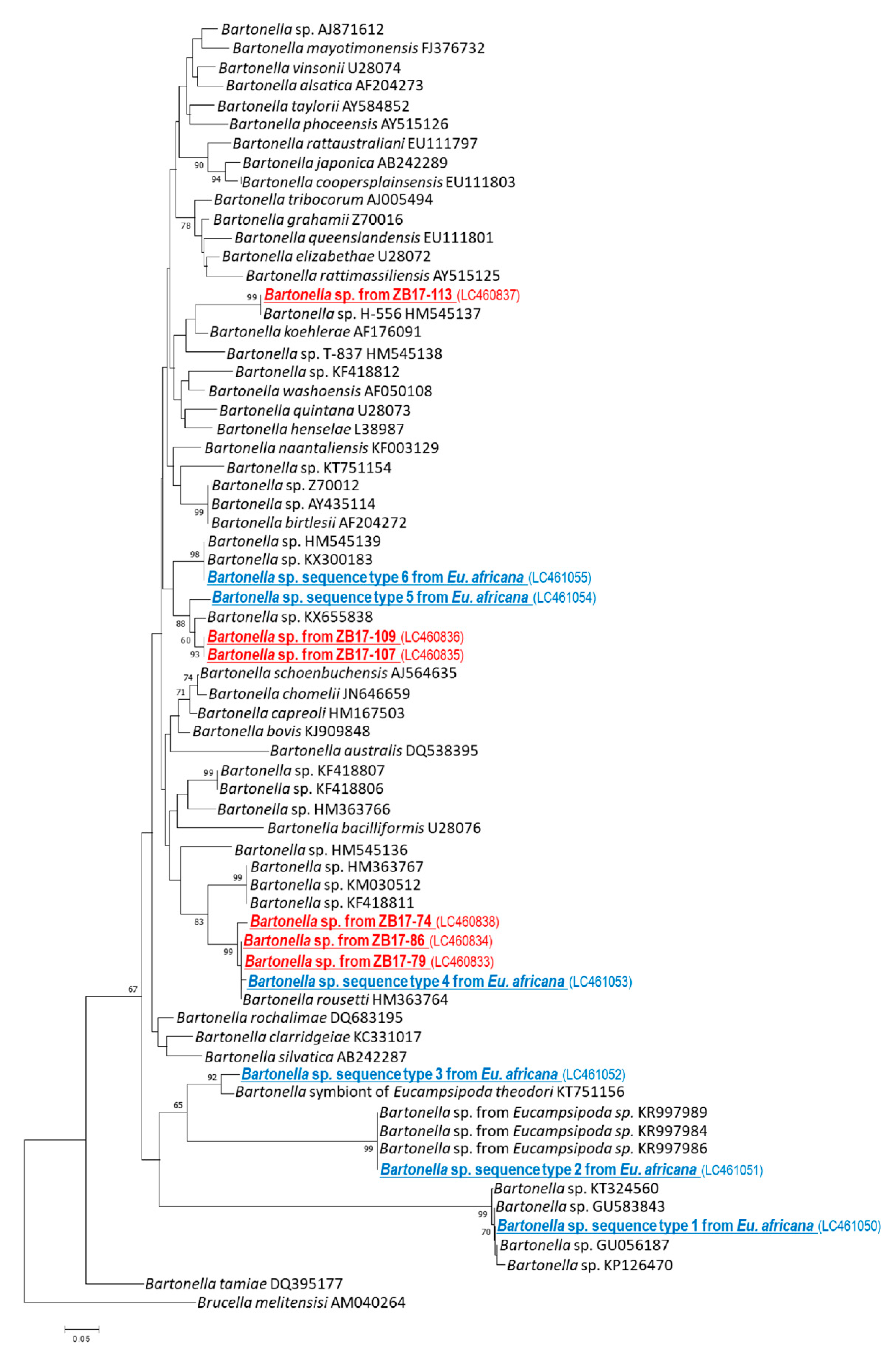
2.3. Detection and Characterization of Bartonella spp. from Bat Flies

All 19 bat flies were morphologically identified as Eucampsipoda africana, which is a common ectoparasite of the Egyptian fruit bat (Rousettus aegyptiacus) (Figure 3a). Further analysis of their cytochrome oxidase subunit I (COI) sequences divided them into three genotypes (COI sequence types 1, 2, and 3), all of which clustered together with Eu. africana collected in Kenya (KF021491) (Figure 3b) [22]. In total, nine bat flies were positive for gltA in a polymerase chain reaction (PCR). Sequence analysis of the amplicons revealed that the gltA sequences from bat flies were of six types. Sequence types 1 and 2 showed 100% (332/332 bp) identity to Bartonella sp. clone NG13-057 isolated from Eu. africana of Nigeria (MH151070) and Bartonella sp. from Eucampsipoda sp. of South Africa (KR997986), respectively [17,23]. Sequence type 3 showed 95.5% (315/330 bp) similarity to the Bartonella symbiont of Eu. theodori from Madagascar (KT751156) [24]. Sequence type 4 showed 99.7% (337/338 bp) similarity to Candidatus Bartonella rousetti (HM363764) [15]. Sequence type 5 showed 94.7% (320/338 bp) similarity to Bartonella sp. SD-3/2015 from Rhinolophus ferrumequinum of China (KX655838) [18]. The sequence type 6 showed 100% (338/338 bp) identity to Bartonella sp. from Miniopterus schreibersii of Georgia (KX300183) [14].
In the phylogenetic tree based on the partial sequences of gltA, sequence type 1 clustered together with Bartonella spp. from ectoparasites, such as flea and louse in Peru, Tunisia, and Thailand [25,26] (Figure 4). Sequence type 2 clustered together with Bartonella spp. isolated from Eucampsipoda sp. from South Africa [23]. Sequence type 3 clustered together with symbiotic Bartonella sp. from Eucampsipoda theodori of Madagascar [25]. Sequence type 4 was located in the same clade with the isolates from ZB17-74, -79, and -86 and Candidatus Bartonella rousetti, which was detected in Rousettus aegyptiacus from Kenya [15]. Sequence type 5 clustered together with the isolates from ZB17-107 and -109, and Bartonella sp. from Rhinolophus ferrumequinum of China. Sequence type 6 clustered together with Bartonella sp. from Miniopterus sp. of Kenya and Bartonella sp. from Miniopterus schreibersii of Georgia [14,15].

3. Discussion

This study investigated the presence and genetic diversity of Bartonella in bats and bat flies captured in Zambia in the Southern African region. To the best of our knowledge, this study is the first report on bat-associated bartonellae in Zambia, and the first to isolate Candidatus Bartonella rousetti from the Southern African region.
Candidatus Bartonella rousetti was first reported as a previously unclassified Bartonella strain detected from Rousettus aegyptiacus in Kenya in 2010 [15]. Thereafter, the same strain was detected during a survey on Bartonella species in bats and bat flies in a cave in southwestern Nigeria, where people entered and captured bats during a traditional bat festival [17]. The study also assessed human exposure to the bacterium using an indirect immunofluorescence assay and detected immunoglobulin G (IgG) against Ca. B. rousetti in the sera of several individuals from the surrounding communities without any cross-reaction to other Bartonella genotypes [17]. In the present study, Ca. B. rousetti was isolated from frugivorous bats (R. aegyptiacus) and detected from bat flies (Eucampsipoda africana) using PCR. These results indicated that Ca. B. rousetti is distributed widely in the African continent and is maintained between bats (R. aegyptiacus) and bat flies (Eu. aficana) in a cave in Zambia. Local people frequently enter the cave and collect bat feces to use it as manure for their vegetable farms, increasing the risk of Ca. B. rousetti infection in the local population, as is the case in Nigeria. Considering that individuals who entered the same cave developed a febrile illness caused by a novel Borrelia sp. [27], they should also be tested for bartonellosis for the differential diagnosis of febrile patients living in the vicinity of the cave.
In the present investigation, three isolates were obtained from insectivorous bats (Macronycteris vittatus). One isolate obtained from ZB17-113 showed high sequence identities with Bartonella sp. H-556 obtained from Macronycteris commersoni of Kenya (Table 1). These two isolates clustered together in a phylogenetic tree (Figure 2), suggesting that the same or closely related Bartonella species/genotype was infectious to both M. vittatus and M. commersoni. On the other hand, the sequences of isolates from ZB17-107 and -109 were identical to each other and were distinct from previously reported Bartonella spp. These results indicated that at least two distinct Bartonella species were prevalent in M. vittatus in Zambia. In the previous study, straw-colored fruit bats (Eidolon helvum) and long-fingered bats (Miniopterus spp.) were found to be positive for three and four different Bartonella genotypes, respectively [15]. It is also evident that the same Bartonella genotype can be detected from several different bat species [16]. Further investigations on the diversity of bat-associated bartonellae are required to understand the relationship between Bartonella spp. and their host bats. Our results provide information regarding the genetic diversity and geographic distribution of bat-associated bartonellae in the African continent.
Bat flies are specially adapted for a nearly permanent ectoparasitic relationship with their host bats. In the present study, we detected six different sequence types of the Bartonella gltA from one bat fly species (Eucampsipoda africana). A phylogenetic analysis revealed that the sequence types 4, 5, and 6 belonged to the same clades as bat-associated bartonellae, including Candidatus Bartonella rousetti (Figure 3). In contrast, the sequence types 1, 2, and 3 clustered together with bartonellae, which were only detected from ectoparasites. Similar findings were also obtained from a previous study conducted in Ghana and in the islands in the Gulf of Guinea, where bat flies harbored not only bat-associated bartonellae, but also other Bartonella spp. which were detected only in bat flies [28]. In another study of insect microbiota, arthropod-associated Bartonella spp. was reported elsewhere [29,30,31,32]. For example, Bartonella apis was isolated from the gut of the honeybee (Apis mellifera), which might provide honeybees with resistance to diseases [30,31]. Uncultured Bartonella spp. were also detected from Ponerine ants (Hymenoptera: Formicidae: Ponerinae) [32]. These arthropod-associated bartonellae were considered a symbiotic bacterium. A recent study regarding the evolutionary origin of the pathogenic bartonellae suggested that the ancestor of pathogenic bartonellae was a symbiotic bacterium of insects, and that the adaptation to blood sucking arthropods facilitated the colonization of bartonellae in the mammalian blood [33]. This hypothesis may explain why bat flies had more diverse groups of Bartonella spp. than bats.
This present study used a random sampling procedure and samples were collected in one location, which are the limitations of this study. Therefore, the study is not enough to describe the whole picture of bat-associated bartonellae in Zambia. However, the present study revealed the presence of diverse groups of Bartonella in bats and their ectoparasites in Zambia. Although cases of human bartonellosis caused by bat-associated Bartonella have not been reported so far, further studies such as continuous surveillance of Bartonella spp. in bats and serological surveys in humans are warranted to evaluate their potential as zoonotic agents in Africa.

4. Materials and Methods

4.1. Ethics

The capturing of bats was approved by the Department of National Parks and Wildlife (DNPW) and the Ministry of Tourism and Arts of the Republic of Zambia (Act No. 14 of 2015). The approval for placing traps and entering the cave was obtained from the village head and residents.

4.2. Isolation of Bartonella spp.

In 2017 and 2018, bats were captured in the Leopard’s Hill cave (15.44° S, 28.51° E), Chongwe district, Lusaka province, using a harp trap as a part of a surveillance program of filovirus infection in bats in Zambia, as described in a previous study [34]. Whole blood samples were collected from 36 randomly selected bats (31 Rousettus aegyptiacus, four Macronycteris vittatus, and one Rhinolophus sp.) in vacutainer ethylenediaminetetraacetic acid (EDTA) tubes. Individually, 25 µL of uncoagulated whole blood were spread onto sheep blood agar plates. The plates were incubated at 34 °C with 5.0% CO2.

4.3. Characterization of Bartonella spp.

After two weeks of incubation, bacterial colonies morphologically identified as Bartonella were selected and used for DNA extraction using the alkali–heat lysis method. Briefly, a colony was resuspended in 17 µL of 50 mM NaOH and the suspension was heated at 99 °C for 10 min. To adjust pH, 3 µL of 1 M Tris-HCl (pH 7.0) was added. The solutions were used as DNA templates for subsequent genetic characterization.
To confirm the identity of bacterial species, we amplified a fragment of 16S rDNA via PCR using the primers fD1 and Rp2 as described previously [35]. PCRs were conducted in a 20-µL reaction mixture containing 10 µL of Go Taq master mix (Promega, Madison, WI, USA), 100 nM of each primer, and 1 µL of template DNA. The PCR conditions were 95 °C for 2 min and 35 cycles of 95 °C for 30 s, 55 °C for 30 s, and 72 °C for 90 s, followed by a final extension at 72 °C for 5 min.
For further molecular characterization, PCRs targeting gltA, nuoG, ftsZ, ssrA, and rpoB were performed as described previously [36,37,38,39,40]. Information regarding the primers used in this study is listed in Table 2. All PCRs were conducted in a 20 µL reaction mixture as described above. The PCR conditions were as described above, with the exception of the annealing temperature (Table 2). The PCR products were visualized after electrophoresis on a 1.2% agarose gel stained with ethidium bromide.

4.4. Species Identification of Bat Flies and Detection of Bartonella spp.

To identify vector arthropods, 19 bat flies were collected from the surface of bat guano in the cave in 2018. The bat flies were morphologically identified to the genus level according to the keys described previously [41], and species were identified by sequencing a DNA fragment of COI [42]. DNA was extracted from bat flies using DNAzol (Molecular Research Center, Cincinnati, OH, USA) according to the manufacturer’s instructions. PCR amplification of gltA was used to screen Bartonella. PCR was performed and the products were analyzed as described above.

4.5. Sequencing and Phylogenetic Analysis

All amplicons were sequenced using the BigDye Terminator version 3.1 cycle sequencing kit (Applied Biosystems, Foster City, CA, USA) and an ABI Prism 3130x genetic analyzer (Applied Biosystems) according to the manufacturer’s instructions. The sequences were analyzed using GENETYX version 9.1 (GENETYX Corporation, Tokyo, Japan) and trimmed on both the 5’ and 3’ ends. The resulting sequences were compared to those in public databases using BLASTn (http://blast.ncbi.nlm.nih.gov/Blast.cgi). A phylogenetic analysis was conducted using MEGA version 6.05 [43]. ClustalW was used to align the sequences to those of closely related organisms deposited in the database. The sequences of multiple loci were concatenated and used for phylogenetic inference of Bartonella spp. isolated from bats using the maximum likelihood method. The neighbor-joining method was used to construct a phylogenetic tree based on the gltA sequence for the analysis of Bartonella spp. from bat flies and bats. The reference sequences used in the phylogenetic inference are listed in Supplementary Table S1. The DNA sequences obtained in this study are available in the GenBank database (accession numbers: LC460827-LC460832 for 16S rDNA, LC460833-LC460838 and LC461050-LC461055 for gltA, LC460839-LC460844 for rpoB, LC460845-LC460850 for ssrA, LC460851-LC460856 for ftsZ, LC460857-LC4608622 for nuoG, and LC536586- LC536588 for COI of Eucampsipoda africana).

Supplementary Materials

The following are available online at https://www.mdpi.com/2076-0817/9/6/469/s1, Table S1: GenBank accession numbers for ftsZ, gltA, nuoG, rpoB, and ssrA, and 16S rDNA sequences for reference.

Author Contributions

Conceptualization, Y.Q.; methodology, Y.Q.; formal analysis, Y.Q.; investigation, Y.Q., H.H.( Hayato Harima), E.M., M.K., B.M.H., Y.E., K.C., D.M., and A.T.; writing—original draft preparation, Y.Q.; writing—review and editing, Y.Q., H.H.(Hayato Harima), E.M., M.K., B.M.H., Y.E., K.C., D.M., R.N., H.S., H.H.( Hideaki Higashi), A.M., A.T., M.S., and C.S.; supervision, R.N. and C.S.; funding acquisition, H.S., A.T., Y.Q. All authors have read and agreed to the published version of the manuscript.

Funding

This work was supported by the Japan Initiative for the Global Research Network on Infectious Diseases (J-GRID) (JP15fm0108008) and the Japan Program for Infectious Diseases Research and Infrastructure (JP20wm0125008) from the Japan Agency for Medical Research and Development (AMED) and Grant-in-Aid for Young Scientists (JP16K19112, JP19K15992) from the Ministry of Education, Culture, Sports, Science and Technology, Japan (MEXT)/Japan Society for the Promotion of Science (JSPS), as well as by the Science and Technology Research Partnership for Sustainable Development (SATREPS) (JP18jm0110019) through the Japan International Cooperation Agency (JICA) and AMED.

Acknowledgments

We thank Kenji Yokoi (JICA) for coordinating the sampling activity. We thank the staff from Department of National Parks and Wildlife (DNPW)and the University of Zambia (UNZA) veterinary schools for their help during field activities and lab work. We would like to thank Editage (www.editage.com) for English language editing.

Conflicts of Interest

The authors declare no conflict of interest.

References

  1. Chomel, B.B.; Boulouis, H.J.; Breitschwerdt, E.B.; Kasten, R.W.; Vayssier-Taussat, M.; Birtles, R.J.; Koehler, J.E.; Dehio, C. Ecological fitness and strategies of adaptation of Bartonella species to their hosts and vectors. Vet. Res. 2009, 40, 29. [Google Scholar] [CrossRef] [Green Version]
  2. Boulouis, H.J.; Chang, C.C.; Henn, J.B.; Kasten, R.W.; Chomel, B.B. Factors associated with the rapid emergence of zoonotic Bartonella infections. Vet. Res. 2005, 36, 383–410. [Google Scholar] [CrossRef] [PubMed] [Green Version]
  3. Mühldorfer, K. Bats and bacterial pathogens: A review. Zoonoses Public Health 2013, 60, 93–103. [Google Scholar] [CrossRef] [PubMed]
  4. Koehler, J.E.; Quinn, F.D.; Berger, T.G.; LeBoit, P.E.; Tappero, J.W. Isolation of Rochalimaea species from cutaneous and osseous lesions of bacillary angiomatosis. N. Engl. J. Med. 1992, 327, 1625–1631. [Google Scholar] [CrossRef] [PubMed]
  5. Santos, R.; Cardoso, O.; Rodrigues, P.; Cardoso, J.; Machado, J.; Afonso, A.; Bacellar, F.; Marston, E.; Proença, R. Bacillary angiomatosis by Bartonella quintana in an HIV-infected patient. J. Am. Acad. Dermatol. 2000, 42, 299–301. [Google Scholar] [CrossRef]
  6. Zangwill, K.M.; Hamilton, D.H.; Perkins, B.A.; Regnery, R.L.; Plikaytis, B.D.; Hadler, J.L.; Cartter, M.L.; Wenger, J.D. Cat scratch disease in Connecticut. Epidemiology, risk factors, and evaluation of a new diagnostic test. N. Engl. J. Med. 1993, 329, 8–13. [Google Scholar] [CrossRef]
  7. Kerkhoff, F.T.; Bergmans, A.M.; van Der Zee, A.; Rothova, A. Demonstration of Bartonella grahamii DNA in ocular fluids of a patient with neuroretinitis. J. Clin. Microbiol. 1999, 37, 4034–4038. [Google Scholar] [CrossRef] [PubMed] [Green Version]
  8. Kosoy, M.; Morway, C.; Sheff, K.W.; Bai, Y.; Colborn, J.; Chalcraft, L.; Dowell, S.F.; Peruski, L.F.; Maloney, S.A.; Baggett, H.; et al. Bartonella tamiae sp. nov., a newly recognized pathogen isolated from three human patients from Thailand. J. Clin. Microbiol. 2008, 46, 772–775. [Google Scholar] [CrossRef] [PubMed] [Green Version]
  9. Eremeeva, M.E.; Gerns, H.L.; Lydy, S.L.; Goo, J.S.; Ryan, E.T.; Mathew, S.S.; Ferraro, M.J.; Holden, J.M.; Nicholson, W.L.; Dasch, G.A.; et al. Bacteremia, fever, and splenomegaly caused by a newly recognized bartonella species. N. Engl. J. Med. 2007, 356, 2381–2387. [Google Scholar] [CrossRef] [PubMed]
  10. Raoult, D.; Roblot, F.; Rolain, J.M.; Besnier, J.M.; Loulergue, J.; Bastides, F.; Choutet, P. First isolation of Bartonella alsatica from a valve of a patient with endocarditis. J. Clin. Microbiol. 2006, 44, 278–279. [Google Scholar] [CrossRef] [PubMed] [Green Version]
  11. McKee, C.D.; Kosoy, M.Y.; Bai, Y.; Osikowicz, L.M.; Franka, R.; Gilbert, A.T.; Boonmar, S.; Rupprecht, C.E.; Peruski, L.F. Diversity and phylogenetic relationships among Bartonella strains from Thai bats. PLoS ONE 2017, 12, e0181696. [Google Scholar] [CrossRef] [PubMed] [Green Version]
  12. Lin, E.Y.; Tsigrelis, C.; Baddour, L.M.; Lepidi, H.; Rolain, J.M.; Patel, R.; Raoult, D. Candidatus Bartonella mayotimonensis and endocarditis. Emerg. Infect. Dis. 2010, 16, 500–503. [Google Scholar] [CrossRef] [PubMed]
  13. Veikkolainen, V.; Vesterinen, E.J.; Lilley, T.M.; Pulliainen, A.T. Bats as reservoir hosts of human bacterial pathogen, Bartonella mayotimonensis. Emerg. Infect. Dis. 2014, 20, 960–967. [Google Scholar] [CrossRef] [PubMed]
  14. Urushadze, L.; Bai, Y.; Osikowicz, L.; McKee, C.; Sidamonidze, K.; Putkaradze, D.; Imnadze, P.; Kandaurov, A.; Kuzmin, I.; Kosoy, M. Prevalence, diversity, and host associations of Bartonella strains in bats from Georgia (Caucasus). PLoS Negl. Trop. Dis. 2017, 11, e0005428. [Google Scholar] [CrossRef] [Green Version]
  15. Kosoy, M.; Bai, Y.; Lynch, T.; Kuzmin, I.V.; Niezgoda, M.; Franka, R.; Agwanda, B.; Breiman, R.F.; Rupprecht, C.E. Bartonella spp. in bats, Kenya. Emerg. Infect. Dis. 2010, 16, 1875–1881. [Google Scholar] [CrossRef]
  16. Kamani, J.; Baneth, G.; Mitchell, M.; Mumcuoglu, K.Y.; Gutiérrez, R.; Harrus, S. Bartonella species in bats (Chiroptera) and bat flies (Nycteribiidae) from Nigeria, West Africa. Vector Borne Zoonotic Dis. 2014, 14, 625–632. [Google Scholar] [CrossRef] [Green Version]
  17. Bai, Y.; Osinubi, M.O.V.; Osikowicz, L.; McKee, C.; Vora, N.M.; Rizzo, M.R.; Recuenco, S.; Davis, L.; Niezgoda, M.; Ehimiyein, A.M.; et al. Human exposure to novel Bartonella species from contact with fruit bats. Emerg. Infect. Dis. 2018, 24, 2317–2323. [Google Scholar] [CrossRef] [Green Version]
  18. Han, H.J.; Wen, H.L.; Zhao, L.; Liu, J.W.; Luo, L.M.; Zhou, C.M.; Qin, X.R.; Zhu, Y.L.; Zheng, X.X.; Yu, X.J. Novel Bartonella species in insectivorous bats, Northern China. PLoS ONE 2017, 12, e0167915. [Google Scholar] [CrossRef]
  19. Bai, Y.; Hayman, D.T.; McKee, C.D.; Kosoy, M.Y. Classification of Bartonella strains associated with straw-colored fruit bats (Eidolon helvum) across Africa using a multi-locus sequence typing platform. PLoS Negl. Trop. Dis. 2015, 9, e0003478. [Google Scholar] [CrossRef] [Green Version]
  20. Matsumoto, K.; Cook, J.A.; Goethert, H.K.; Telford, S.R. Bartonella sp. Infection of voles trapped from an interior Alaskan site where ticks are absent. J. Wildl. Dis. 2010, 46, 173–178. [Google Scholar] [CrossRef] [Green Version]
  21. Lin, J.W.; Hsu, Y.M.; Chomel, B.B.; Lin, L.K.; Pei, J.C.; Wu, S.H.; Chang, C.C. Identification of novel Bartonella spp. in bats and evidence of Asian gray shrew as a new potential reservoir of Bartonella. Veter Microbiol. 2012, 156, 119–126. [Google Scholar] [CrossRef] [PubMed]
  22. Tortosa, P.; Dsouli, N.; Gomard, Y.; Ramasindrazana, B.; Dick, C.W.; Goodman, S.M. Evolutionary history of Indian Ocean nycteribiid bat flies mirroring the ecology of their hosts. PLoS ONE 2013, 8, e75215. [Google Scholar] [CrossRef] [PubMed]
  23. Dietrich, M.; Kearney, T.; Seamark, E.C.; Markotter, W. The excreted microbiota of bats: Evidence of niche specialisation based on multiple body habitats. FEMS Microbiol. Lett. 2017, 364, 364. [Google Scholar] [CrossRef] [PubMed]
  24. Wilkinson, D.A.; Duron, O.; Cordonin, C.; Gomard, Y.; Ramasindrazana, B.; Mavingui, P.; Goodman, S.M.; Tortosa, P. The bacteriome of bat flies (Nycteribiidae) from the Malagasy region: A community shaped by host ecology, bacterial transmission mode, and host-vector specificity. Appl. Environ. Microbiol. 2016, 82, 1778–1788. [Google Scholar] [CrossRef] [PubMed] [Green Version]
  25. Klangthong, K.; Promsthaporn, S.; Leepitakrat, S.; Schuster, A.L.; McCardle, P.W.; Kosoy, M.; Takhampunya, R. The distribution and diversity of Bartonella species in rodents and their ectoparasites across Thailand. PLoS ONE 2015, 10, e0140856. [Google Scholar] [CrossRef] [Green Version]
  26. Zouari, S.; Khrouf, F.; M’ghirbi, Y.; Bouattour, A. First molecular detection and characterization of zoonotic Bartonella species in fleas infesting domestic animals in Tunisia. Parasites Vectors 2017, 10, 436. [Google Scholar] [CrossRef] [PubMed] [Green Version]
  27. Qiu, Y.; Nakao, R.; Hang’ombe, B.M.; Sato, K.; Kajihara, M.; Kanchela, S.; Changula, K.; Eto, Y.; Ndebe, J.; Sasaki, M.; et al. Human borreliosis caused by a new world relapsing fever Borrelia-like organism in the old world. Clin. Infect. Dis. 2019, 69, 107–112. [Google Scholar] [CrossRef]
  28. Billeter, S.A.; Hayman, D.T.; Peel, A.J.; Baker, K.; Wood, J.L.; Cunningham, A.; Suu-Ire, R.; Dittmar, K.; Kosoy, M.Y. Bartonella species in bat flies (Diptera: Nycteribiidae) from western Africa. Parasitology 2012, 139, 324–329. [Google Scholar] [CrossRef]
  29. Hubert, J.; Erban, T.; Kopecky, J.; Sopko, B.; Nesvorna, M.; Lichovnikova, M.; Schicht, S.; Strube, C.; Sparagano, O. Comparison of microbiomes between red poultry mite populations (Dermanyssus gallinae): Predominance of Bartonella-like bacteria. Microb. Ecol. 2017, 74, 947–960. [Google Scholar] [CrossRef]
  30. Kešnerová, L.; Moritz, R.; Engel, P. Bartonella apis sp. nov., a honey bee gut symbiont of the class Alphaproteobacteria. Int. J. Syst. Evol. Microbiol. 2016, 66, 414–421. [Google Scholar] [CrossRef]
  31. Raymann, K.; Moran, N.A. The role of the gut microbiome in health and disease of adult honey bee workers. Curr. Opin. Insect Sci. 2018, 26, 97–104. [Google Scholar] [CrossRef] [PubMed]
  32. De Oliveira, T.B.; Ferro, M.; Bacci, M.; De Souza, D.J.; Fontana, R.; Delabie, J.H.C.; Silva, A. Bacterial communities in the midgut of Ponerine ants (Hymenoptera: Formicidae: Ponerinae). Sociobiology 2016, 63, 637–644. [Google Scholar] [CrossRef] [Green Version]
  33. Segers, F.H.; Kešnerová, L.; Kosoy, M.; Engel, P. Genomic changes associated with the evolutionary transition of an insect gut symbiont into a blood-borne pathogen. ISME J. 2017, 11, 1232–1244. [Google Scholar] [CrossRef] [PubMed] [Green Version]
  34. Changula, K.; Kajihara, M.; Mori-Kajihara, A.; Eto, Y.; Miyamoto, H.; Yoshida, R.; Shigeno, A.; Hang’ombe, B.; Qiu, Y.; Mwizabi, D.; et al. Seroprevalence of filovirus infection of Rousettus aegyptiacus bats in Zambia. J. Infect. Dis. 2018, 218, S312–S317. [Google Scholar] [CrossRef]
  35. Weisburg, W.G.; Barns, S.M.; Pelletier, D.A.; Lane, D.J. 16S ribosomal DNA amplification for phylogenetic study. J. Bacteriol. 1991, 173, 697–703. [Google Scholar] [CrossRef] [Green Version]
  36. Norman, A.F.; Regnery, R.; Jameson, P.; Greene, C.; Krause, D.C. Differentiation of Bartonella-like isolates at the species level by PCR-restriction fragment length polymorphism in the citrate synthase gene. J. Clin. Microbiol. 1995, 33, 1797–1803. [Google Scholar] [CrossRef] [Green Version]
  37. Colborn, J.M.; Kosoy, M.Y.; Motin, V.L.; Telepnev, M.V.; Valbuena, G.; Myint, K.S.; Fofanov, Y.; Putonti, C.; Feng, C.; Peruski, L. Improved detection of Bartonella DNA in mammalian hosts and arthropod vectors by real-time PCR using the NADH dehydrogenase gamma subunit (nuoG). J. Clin. Microbiol. 2010, 48, 4630–4633. [Google Scholar] [CrossRef] [Green Version]
  38. Zeaiter, Z.; Liang, Z.; Raoult, D. Genetic classification and differentiation of Bartonella species based on comparison of partial ftsZ gene sequences. J. Clin. Microbiol. 2002, 40, 3641–3647. [Google Scholar] [CrossRef] [Green Version]
  39. Diaz, M.H.; Bai, Y.; Malania, L.; Winchell, J.M.; Kosoy, M.Y. Development of a novel genus-specific real-time PCR assay for detection and differentiation of Bartonella species and genotypes. J. Clin. Microbiol. 2012, 50, 1645–1649. [Google Scholar] [CrossRef] [Green Version]
  40. Renesto, P.; Gouvernet, J.; Drancourt, M.; Roux, V.; Raoult, D. Use of rpoB gene analysis for detection and identification of Bartonella species. J. Clin. Microbiol. 2001, 39, 430–437. [Google Scholar] [CrossRef] [Green Version]
  41. Theodor, O. An Illustrated Catalogue of the Rothschild Collection of Nycteridiidae (Diptera) in the British Museum (Natural History); Whitefriars Press Ltd: London, UK, 1967. [Google Scholar]
  42. Folmer, O.; Black, M.; Hoeh, W.; Lutz, R.; Vrijenhoek, R. DNA primers for amplification of mitochondrial cytochrome c oxidase subunit I from diverse metazoan invertebrates. Mol. Mar. Biol. Biotechnol. 1994, 3, 294–299. [Google Scholar] [PubMed]
  43. Tamura, K.; Stecher, G.; Peterson, D.; Filipski, A.; Kumar, S. MEGA6: Molecular Evolutionary Genetics Analysis version 6.0. Mol. Biol. Evol. 2013, 30, 2725–2729. [Google Scholar] [CrossRef] [PubMed] [Green Version]
Figure 1. Colonies of Bartonella sp. on a blood agar plate. Bartonella sp. was isolated from Rousettus aegyptiacus (ZB17-79) and cultured on a blood agar plate.
Pathogens 09 00469 g001
Figure 2. Phylogenetic inference of Bartonella spp. A phylogenetic inference from the concatenated sequences of six loci (ftsZ, gltA, nuoG, rpoB, ssrA, and 16S rDNA) of Bartonella species is shown. For phylogenetic reconstruction, the maximum likelihood model proposed in MEGA 6.06 was used with 1000 bootstrap iterations.
Pathogens 09 00469 g002
Figure 3. Bat fly and its molecular identification. (a) Photograph of a bat fly under an optical microscope. (b) A phylogenetic tree based on the partial sequence of a fragment of cytochrome oxidase subunit I (COI).
Pathogens 09 00469 g003
Figure 4. Neighbor-joining phylogeny of citrate synthase (gltA) gene. This tree is based on a partial sequence of gltA and was rooted with Brucella melitensis. Bootstrap values > 60% based on 1000 replications are shown on the interior branch nodes. The sequences from bats and bat flies obtained in this study are shown in red and blue, respectively.
Pathogens 09 00469 g004
Table 1. Sequence identity of the six isolates to the most closely related Bartonella species.
IDBat speciesftsZgltAunoGrpoBssrA
Accession NumberBLAST ResultAccession NumberBLAST ResultAccession NumberBLAST ResultAccession NumberBLAST ResultAccession NumberBLAST Result
ZB17-74Rousettus aegyptiacusHM363769Bartonella rousetti 99%MH069695Bartonella rousetti B32137 100%KM387321Bartonella rousetti 99%HM363774Bartonella rousetti 96%KM382247Bartonella rousetti 99%
ZB17-79Rousettus aegyptiacusHM363769Bartonella rousetti 100%HM363764Bartonella rousetti 100%KM387321Bartonella rousetti 100%HM363774Bartonella rousetti 100%KM382247Bartonella rousetti 100%
ZB17-86Rousettus aegyptiacusHM363769Bartonella rousetti 100%HM363764Bartonella rousetti 100%KM387321Bartonella rousetti 100%HM363774Bartonella rousetti 100%KM382247Bartonella rousetti 100%
ZB17-107Macronycteris vittatusKX300182Bartonella sp. isolate 44552 96%KX655838Uncultured Bartonella sp. SD-3/2015 98%KX300163Bartonella sp. isolate 44552 97%KX300164Bartonella sp. isolate 44552 96%KM233461Bartonella sp. B23983 97%
ZB17-109Macronycteris vittatusKX300182Bartonella sp. isolate 44552 96%KX655838Uncultured Bartonella sp. SD-3/2015 98%KX300163Bartonella sp. isolate 44552 97%KX300164Bartonella sp. isolate 44552 95%KM233461Bartonella sp. B23983 96%
ZB17-113Macronycteris vittatusKM382254Bartonella sp. H-556 99%HM545137Bartonella sp. H-556 100%KM382252Bartonella sp. H-556 100%EU979536Bartonella sp. Cr28649 88%KM382250Bartonella sp. H-556 99%
Table 2. Primers used in this study.
Primer NameSequence 5’-3’Target OrganismTarget GeneAnnealing (°C)Size Expected (bp)Reference
fD1AGAGTTTGATCCTGGCTCAGBartonella spp.16S ribosomal DNA55140035
Rp2ACGGCTACCTTGTTACGACTT
BhCS781.pGGGGACCAGCTCATGGTGGgltA4538036
BhCS1137.nAATGCAAAAAGAACAGTAAACA
1400FCGCATTGGCTTACTTCGTATGrpoB5386040
2300RGTAGACTGATTAGAACGCTG
nuoG FGGCGTGATTGTTCTCGTTAnuoG5536037
nuoG RCACGACCACGGCTATCAAT
Bfp1ATTAATCTGCAYCGGCCAGAftsZ5590038
Bfp2ACVGADACACGAATAACACC
ssrA-FGCTATGGTAATAAATGGACAATGAAATAAssrA6030039
ssrA-RGCTTCTGTTGCCAGGTG
LCO1490GGTCAACAAATCATAAAGATATTGGBat flyCOI5771042
HCO2198TAAACTTCAGGGTGACCAAAAAATCA

