Article

Innovative Alternatives for Continuous In Vitro Culture of Babesia bigemina in Medium Free of Components of Animal Origin

by
Jesús A. Álvarez Martínez
1,
Julio V. Figueroa Millán
1,
Massaro W. Ueti
2,* and
Carmen Rojas-Martínez
1,*
1
Babesia Unit-CENID-Salud Animal e Inocuidad, INIFAP, Carr. Fed. Cuernavaca-Cuautla No. 8534, Col. Progreso, Jiutepec, Morelos C.P. 62550, Mexico
2
Animal Disease Research Unit, Agricultural Research Service, US Department of Agriculture, Pullman, WA 99164-7040, USA
*
Authors to whom correspondence should be addressed.
Pathogens 2020, 9(5), 343; https://doi.org/10.3390/pathogens9050343
Submission received: 4 April 2020 / Revised: 29 April 2020 / Accepted: 29 April 2020 / Published: 1 May 2020
(This article belongs to the Special Issue Animal Parasitic Diseases)

Abstract

:
In this study, we report Babesia bigemina proliferation in culture medium free of components of animal origin supplemented with a lipid mixture. Babesia bigemina continuously proliferated in VP-SFM with a higher percent parasitized erythrocyte as compare to using other animal component-free culture media. Compared with Advanced DMEM/F12 (ADMEM/F12), VP-SFM had a similar percent parasitized erythrocyte (PPE). Supplementation of VP-SF with a lipid acid mixture improved B. bigemina proliferation in vitro culture, with a maximum PPE of 11.3%. Growth of B. bigemina in a perfusion bioreactor using VP-SFM medium supplemented with lipid mixture resulted in a PPE above 28%. In conclusion, we demonstrated that B. bigemina proliferated in an animal component-free medium supplemented with the fatty acid mixture. This innovation to B. bigemina in vitro culture method presented herein is an important source of biological material for live vaccine production and understanding the mechanisms and molecules involved in parasite attachment and invasion of bovine erythrocytes.

1. Introduction

Bovine babesiosis is a tick-borne disease caused by Babesia bigemina and Babesia bovis. This disease affects bovines in tropical and subtropical areas and creates a negative economic impact on the cattle industry [1]. Severe adverse effects occur during clinical disease, including weight loss, decrease in meat and milk production, and, in some cases, death [2,3]. Rhipicephalus microplus and R. annulatus are the vectors for B. bigemina and B. bovis. There is no biological animal model to study these parasites other than bovines. The success of Babesia in vitro growth in erythrocytes using the microaerophilic stationary phase system (MASP) facilitated the study of gene regulation necessary for parasite development, drug efficiency evaluation, and Babesia parasite attenuation for vaccines [4,5,6,7]. Also, due to the advance of engineering technology, the in vitro culture of B. bigemina and B. bovis allowed transfection of these parasites, which has aided the understanding of parasite biology [8,9]. Importantly, more than 100 species of Babesia have been isolated. However, few Babesia species have been successfully cultivated in vitro system [10]. The primary concern is that to maintain continuous in vitro Babesia growth is the requirement of high sera concentrations in the growth medium.
Like other Babesia spp., B. bigemina was cultured in media that contained animal-derived components including 40% of bovine sera. This parasite was first cultivated in vitro more than 30 years ago [11]. A recent report in our laboratory using Dulbecco’s modified Eagle medium/F-12 (Advanced DMEM/F12) that contains components of animal origin, supplemented with insulin-transferrin-selenite and putrescine showed that B. bigemina successfully proliferated in culture media without bovine sera supplementation [12]. Culture media without components of animal origin or supplementation with animal products to propagate Babesia spp. are required to standardize in vitro culture methods among laboratories. Reports showed that culture media formulated without animal products are available and used to proliferate protozoan parasites and virus under in vitro conditions. A study showed that culture media without animal products successfully maintained the growth of Toxoplasma gondii in in vitro culture [13]. Another study showed culture media-free of the animal product was used to grow mammalian cell lines for virus production [14,15,16,17,18]. A chemically defined mixture of lipids as a supplement added in culture media-free of animal products resulted in superior growth of CHO cells to produce recombinant proteins [19]. Similarly, lipids added to culture medium increased lentiviral vector productivity and infectivity of the HEK 293 cell line [20].
In this study, we investigated whether culture media, free of animal components, supplemented with a lipid mixture would support the growth of B. bigemina. We determined that B. bigemina proliferated in VP-SFM medium supplemented with a lipid mixture. In contrast, the other three animal component-free culture media supplemented with or without lipid mixture failed to maintain B. bigemina growth. Using a perfusion bioreactor system, VP-SFM medium supplemented with lipid mixture improved the parasitemia to over 29% of parasitized erythrocytes. These larger numbers of parasites harvested from in vitro culture can be an important source of biological material for the development of strategies to control B. bigemina.

2. Materials and Methods

2.1. Ethical Statement

A five-year-old Bos taurus (Holstein Friesian) bovine was the source of erythrocytes to maintain in vitro B. bigemina growth. This bovine was certified free of Babesia spp., Anaplasma marginale, brucellosis, tuberculosis, leucosis, IBR, and BDV. Animal care and use protocol number were NOM-062-ZOO-1999 “Technical specifications for production, care and use of laboratory animals” (http://www.senasica.gob.mx/?doc¼743).

2.2. Bovine Erythrocytes

Whole blood was collected from the jugular by venipuncture into a flask containing glass beads [21]. Defibrinated blood was centrifuged at 450× g, 4 °C for 30 min. Serum was removed, and the buffy coat discarded. The erythrocytes were washed five times as follows: the pellet of erythrocyte was suspended 1/2 with VYM solution + antioxidant mixture (v/v) and centrifuged at 450× g, 4 °C for 30 min. Erythrocytes were suspended in the VYM solution at a PCV of 30% and stored at 4 °C [12].

2.3. Culture Media

Four culture media free of animal components were used in this study, including VP-SFM (Gibco®), CD-CHO (Gibco®), CD-Hydrolyzed (Sigma-Aldrich, St. Louis, MO, USA), and CD-CHO (Sigma-Aldrich St. Louis, MO, USA). The Advanced-DMEM/F12 (Gibco®) that contains animal components was used as the control for B. bigemina growth (Table 1).
All culture media were buffered with 25 mM 2-[(2-hydroxy-1,1-bis(hydroxymethyl)ethyl)amino] ethane sulfonic, N-[Tris(hydroxymethyl)methyl]-2-aminoethanosulphonic (TES) (Sigma-Aldrich, St. Louis, MO, USA). An antioxidant mixture was added to 2 mM of L-glutamine (Sigma-Aldrich, St. Louis, MO, USA) (GIBCO®). The pH was adjusted (6.8) for all culture media and sterilized by filtration with a 0.22 µm membrane (Millipore). All media were supplemented with different concentrations of CD-Lipid (CORNING®) (Table 2).

2.4. Parasite and In Vitro Culture

The B. bigemina obtained and proliferated in vitro was used in this study [12]. Also, B. bigemina was adapted to continuous proliferation in ADMEM/F12. Babesia bigemina cultures were maintained at 37 °C in a saturated atmosphere of 90% N2, 5% CO2, and 5% O2 [22]. Fresh culture ADMEM/F12 medium was replaced every 24 h to maintain the growth of the parasites [12].

2.5. Selection of an Animal Component-Free Culture Medium

The effect of four animal component-free culture media without supplementation was evaluated on the proliferation of B. bigemina. The percentage of parasitized erythrocytes (PPE) was daily determined by Giemsa-stained blood smears. All assays began with a 1% parasitemia, and when PPE reached 4%, subcultures were carried out. Subcultures were adjusted to 1% parasitemia by adding uninfected erythrocytes [23].

2.6. Effect of Lipids on In Vitro Proliferation of B. bigemina

The effect of a chemically defined lipid mixture on growth of B. bigemina was also evaluated. VP-SFM medium was supplemented with five different concentrations of a commercial lipid mixture to determine the optimum concentration of lipid mixture for the growth of B. bigemina (Table 2). Dilutions of the lipids were performed in VP-SFM medium.

2.7. In Vitro Proliferation of B. bigemina in a Perfusion Bioreactor

Babesia bigemina was cultured with a VP-SFM medium supplemented with lipid mixture in a 75 cm2 culture flask. As a control, B. bigemina was cultured in another culture flask with ADMEM/F12. Briefly, harvested parasites from each flask were applied into a perfusion bioreactor system (FiberCell® System, New Market, MD, USA) as previously described [12,23] for large-scale production of parasites. The bioreactors were kept at 37 °C under a saturated atmosphere of 90% N2, 5% CO2, and 5% O2. The initial parasitemia was 5% in a package of 10 mL erythrocytes. Then, 40 mL of uninfected erythrocytes was added and mixed before introducing them to the bioreactor cartridge. The culture medium was supported by a peristaltic pump to continuously circulate through the system, and the medium was replaced every 24 h with fresh medium. Parasite proliferation was verified at 24 h intervals and subcultures performed every 24–48 h.

2.8. Statistical Analysis

The data were analyzed as differences of percent parasitized erythrocytes for the B. bigemina in vitro cultures using one-way ANOVA followed by a Dunnett’s test to compare means from different experimental groups against a control group. A Student’s t-test was used to assess continuous culture with VP-SFM compared to ADMEM/F12 and to compare VP-SFM supplemented with lipid mixture vs. ADMEM/F12.

3. Results

3.1. Selection of an Animal Component-Free Culture Medium

The VP-SFM medium performed the best for B. bigemina proliferation as compared to other culture media free of animal components. There was a significant difference based on the PPE values between VP-SFM and other media (p < 0.05). Overall, the maximum PPE values observed for VP-SFM was 6.0%, CD-CHO (Gibco) was 3.44%, CD-Hydrolyzed (Sigma) was 4.01%, CD-CHO (Sigma) was 2.57% and the control medium, ADMEM/F12, was 5.0% (Figure 1).
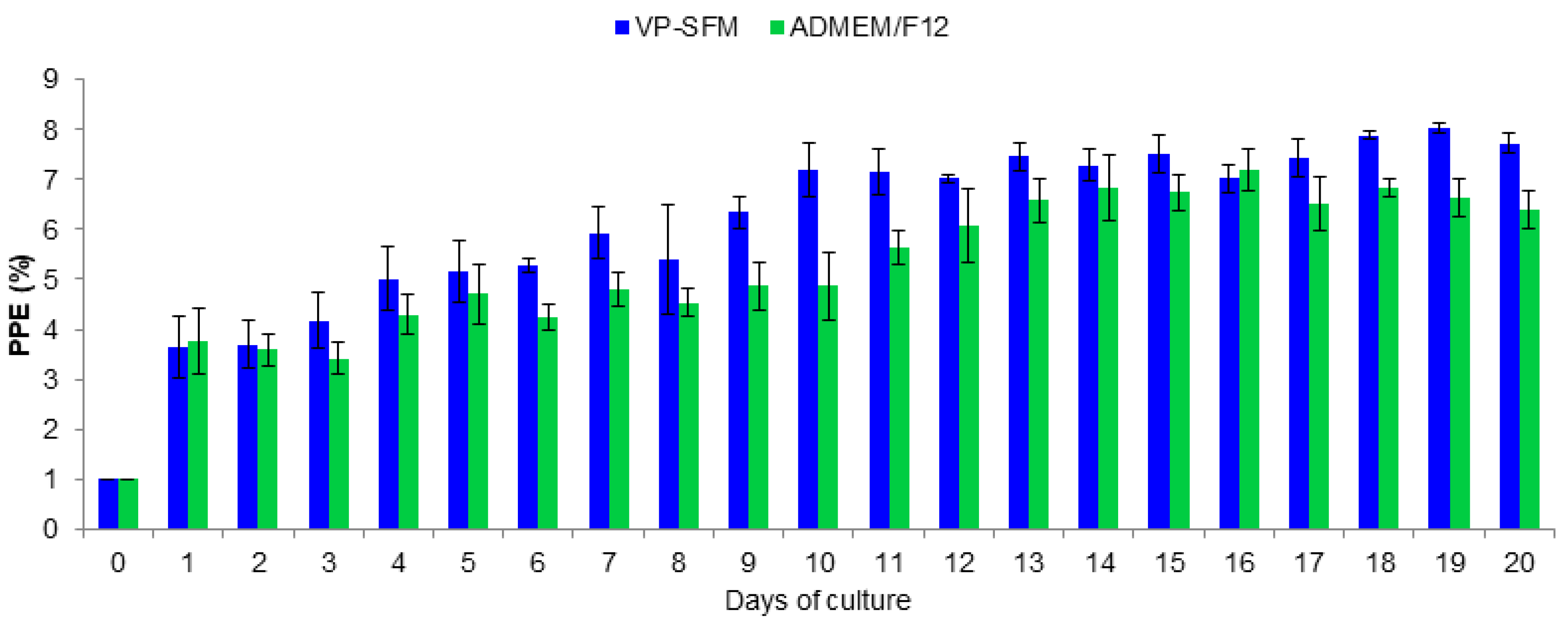
The growth of B. bigemina was similar in culture with VP-SFM or ADMEM/F12. At 14 days of cultures, the PPE values remained greater than 4% for VP-SFM or ADMEM/F12. In contrast, the PPE values using CD-CHO, CD-hydrolyzed, and CD-CHO as media started declining on day two. In these media, parasites were not detected after day nine. Comparing proliferation of B. bigemina between VP-SFM and ADMEM/F12 revealed that the maximum PPE mean values were 8.0% and 7.2%, respectively (Figure 2).
Based on the percentage of parasitized erythrocytes (PPE), there was no significant difference between these two media (p > 0.05). The continuous proliferation of B. bovis using the VP-SFM culture medium was successfully performed during more than 350 subcultures carried out every 24–48 h (data not shown).

3.2. Effect of the Supplementation with Chemically Defined Lipid on Proliferation of B. bigemina

The proliferation of B. bigemina was improved by adding a chemically defined lipid mixture. Supplementation of VP-SFM medium with a lipid mixture at a concentration of 0.1 mg/L of myristic acid, palmitic acid, palmitoleic acid, stearic acid, oleic acid, linoleic acid, acid stearic, 0.02 mg/L of arachidonic acid, and 2.2 mg/L of cholesterol resulted in improvement of B. bigemina growth in vitro culture. There was a significant difference between treatments (p < 0.05). In this lipid concentration, the percentage of parasitemia increased from 1.0% to the highest mean value of 11.35%. Other concentrations of lipid mixture treatments showed parasitemia values below 9% (Table 3).
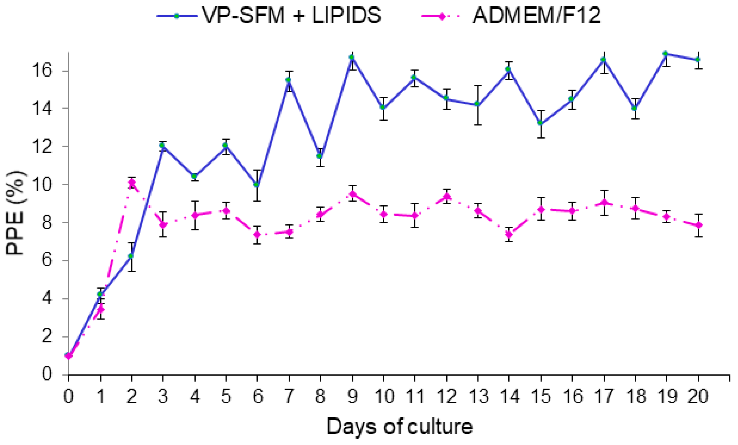
Continuous B. bigemina culture for 20 cycles in lipid supplemented VP-SFM improved the parasitemia values above 12%. At day 19, the maximum PPE was 16.85%. In contrast, continuous B. bigemina culture using ADEM/F12 showed PPE values below 10% (Figure 3). Based on parasitemia values, there was a significant difference between treatments (p < 0.05).

3.3. In Vitro Proliferation of B. bigemina in a Perfusion Bioreactor System

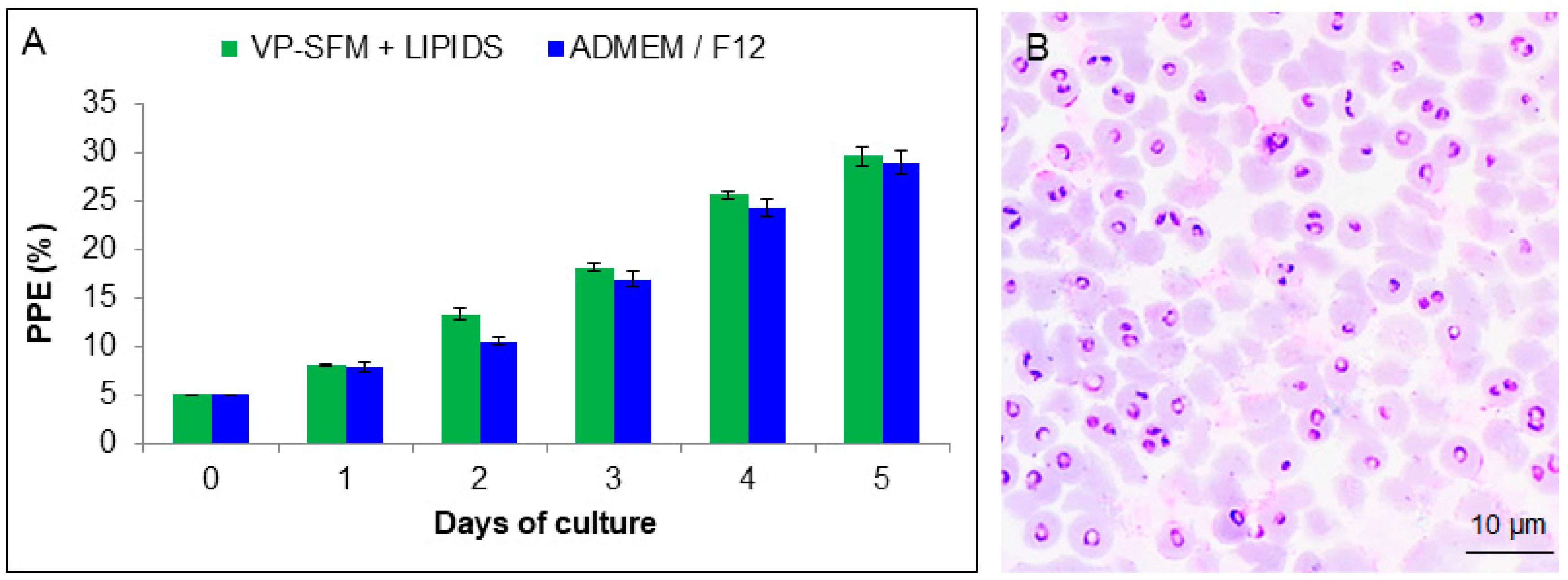
In vitro proliferation of B. bigemina using a perfusion bioreactor system showed a parasitemia of 29.63% using the new lipid supplemented VP-SFM culture medium. Babesia bigemina growth using ADMEM/F12 had a parasitemia of 28.95%, (Figure 4A). There was no significant difference between the two treatments (p > 0.05). Detectable morphological changes were not observed in smears by light microscopy (Figure 4B).

4. Discussion

This is the first report of the continuous in vitro growth of B. bigemina in a medium free of components of animal origin supplemented with a lipid mixture. Among four different animal component-free culture media, B. bigemina proliferated continuously in VP-SFM medium with and without lipid mixture supplementation.
The previous study in our laboratory demonstrated that B. bigemina successfully proliferated in ADMEM/F12 serum-free supplemented with insulin-transferrin-selenite and putrescine. Importantly, putrescine may be critical to achieving the growth of B. bigemina in a serum-free medium. Putrescine is a polyamine previously reported as essential for the proliferation of B. bigemina and B. bovis [12,23] and necessary for other protozoa such P. falciparum [24], Toxoplasma, and Leishmania. These parasites can regulate endogenous levels of putrescine [25]. However, in silico analysis of the B. bovis genome sequence revealed that this parasite does not have the ability for de novo polyamine biosynthesis [26]. It has been suggested that parasites acquire polyamines from the external environment. Other studies have demonstrated that polyamines such as putrescine, spermidine, and spermine facilitate the rapid proliferation of P. falciparum within erythrocytes [27].
In this study, we demonstrated that a relevant factor of the achievement by the use of VP-SFM probably is because one of its components is putrescine for the growth of B. bigemina in vitro culture using VP-SFM medium, a medium free of components of animal origin. Also, the supplementation of VP-SFM with a lipid mixture improved B. bigemina growth. There is no report showing a combination of animal component-free culture medium and lipids for the proliferation of Babesia spp., or other protozoan parasites. The lipid mixture is a source of energy, structural components for cell membranes, transport, and signaling systems [28]. A previous report suggested that lipid metabolism is vital in pathogen replication [29], as well as necessary for P. falciparum merozoite stage survival during the intraerythrocytic phase of infection [30]. It has been proposed that exogenous incorporation of fatty acids for the growth and maturation of the parasite in the trophozoite stage is required [31,32]. Plasmodium falciparum can modify and control the phospholipids composition of the erythrocyte membrane [33]. Plasmodium falciparum increases intracellular levels of fatty acids, such as oleic acid. However, the mechanism by which parasites acquire the fatty acids is unknown [34]. Additionally, P. falciparum is capable of synthesizing small quantities of de novo fatty acids, and those that are required are obtained from the external environment [26,30]. Another species, Plasmodium knowlesi requires large amounts of phospholipids for its development. Nevertheless, these parasites are not able to produce those lipids de novo. Thus, the sources of those lipids are from the erythrocyte membrane or the external medium [35].
We showed that VP-SFM combined with a lipid mixture achieved high efficiency of B. bigemina proliferation in a perfusion bioreactor reaching a maximum PPE of 29.63%. These values were essentially equal to a previous study using ADEM/F12 medium containing components of animal origin supplemented with insulin-transferrin-selenite and putrescine [12].

5. Conclusions

In this study, we demonstrated that VP-SFM medium, free of animal components, supplemented with a lipid mixture supported B. bigemina proliferation in an in vitro culture system. The absence of animal components in the culture medium is a novel method to maintain B. bigemina growth under in vitro conditions. This method will allow harvesting of a high number of B. bigemina infected erythrocytes that can be an excellent source of parasites for vaccine development or understanding the mechanisms and molecules involved in parasite attachment and invasion of erythrocytes. Future studies are needed to evaluate the B. bigemina genome and transcriptome of these parasites adapted to the new animal component-free medium supplemented with a lipid mixture.

Author Contributions

Conceptualization, C.R.-M. and J.A.Á.M.; methodology, C.R.-M., J.V.F.M., M.W.U.; validation, C.R.-M. and M.W.U.; formal analysis, J.A.Á.M. and C.R.-M., investigation, C.R.-M. and J.A.Á.M.; resources, C.R.-M. and M.W.U.; writing—original draft preparation, C.R.-M. and J.A.Á.M.; writing—review and editing, C.R.-M., J.V.F.M. and M.W.U.; visualization, M.W.U. and C.R.-M.; supervision, C.R.-M. and J.A.Á.M.; project administration, C.R.-M. and J.A.Á.M.; funding acquisition, C.R.-M., M.W.U. and J.A.Á.M. All authors have read and agree to the published version of the manuscript.

Funding

This research was funded by INIFAP projects 14354234751 and 21314134828, the United States Department of Agriculture-Agricultural Research Service project 2090-32000-039-00D, and USDA-ARS Offshore project 2090-32000-039-19-A.

Conflicts of Interest

The authors declare no conflict of interest. The funders had no role in the design of the study; in the collection, analyses, or interpretation of data; in the writing of the manuscript, or in the decision to publish the results.

References

  1. Bock, R.; Jackson, L.; De Vos, A.; Jorgensen, W. Bovine babesiosis. Parasitology 2004, 129, 247–269. [Google Scholar] [CrossRef] [PubMed]
  2. Grisi, L.; Leite, R.C.; Martins, J.R.; Barros, A.T.; Andreotti, R.; Cançado, P.H.; Pérez de León, A.A.; Pereira, J.B.; Villela, H.S. Reassessment of the potential economic impact of cattle parasites in Brazil. Rev. Bras. Parasitol. Vet. 2014, 23, 150–156. [Google Scholar] [CrossRef] [PubMed] [Green Version]
  3. Florín-Christensen, M.; Suarez, C.E.; Rodriguez, A.E.; Flores, D.A.; Schnittger, L. Vaccines against bovine babesiosis: Where we are now and possible roads ahead. Parasitology 2014, 141, 1563–1592. [Google Scholar] [CrossRef] [PubMed]
  4. Levy, M.G.; Ristic, M. Babesia bovis: Continuous cultivation in a microaerophilus stationary phase culture. Science 1980, 207, 1218–1220. [Google Scholar] [CrossRef]
  5. Rodríguez-Vivas, R.I.; Trees, J.A. Utilización del cultivo in vitro de Babesia bovis para evaluar la efectividad de babesicidas. Rev. Biomed. 1994, 5, 133–139. [Google Scholar]
  6. Jackson, L.A.; Waldron, S.J.; Weier, H.M.; Nicoll, C.L.; Cooke, B.M. Babesia bovis: Culture of laboratory-adapted parasite lines and clinical isolates in a chemically defined medium. Exp. Parasitol. 2001, 99, 168–174. [Google Scholar] [CrossRef]
  7. Mosqueda, J.; McElwain, T.F.; Stiller, D.; Palmer, G.H. Babesia bovis merozoite surface antigen 1 and rhoptry-associated protein 1 are expressed in sporozoites, and specific antibodies inhibit sporozoite attachment to erythrocytes. Infect. Immun. 2002, 70, 1599–1603. [Google Scholar] [CrossRef] [Green Version]
  8. Silva, M.G.; Knowles, D.P.; Mazuz, M.L.; Cooke, B.M.; Suarez, C.E. Stable transformation of Babesia bigemina and Babesia bovis using a single transfection plasmid. Sci. Rep. 2018, 1, 1–9. [Google Scholar] [CrossRef] [Green Version]
  9. Suarez, C.E.; McElwain, T.F. Transfection systems for Babesia bovis: A review of methods for the transient and stable expression of exogenous genes. Vet. Parasitol. 2010, 4, 205–215. [Google Scholar] [CrossRef]
  10. Schuster, F.L. Cultivation of Babesia and Babesia-like blood parasites: Agents of an emerging zoonotic disease. Clin. Microbiol. Rev. 2012, 15, 365–373. [Google Scholar] [CrossRef] [Green Version]
  11. Vega, C.A.; Buening, G.M.; Green, T.J.; Carson, C.A. In vitro cultivation of Babesia bigemina. Am. J. Vet. Res. 1985, 46, 416–420. [Google Scholar] [PubMed]
  12. Rojas-Martínez, C.; Rodríguez-Vivas, R.I.; Figueroa, M.J.V.; Acosta, V.K.Y.; Gutiérrez, R.E.J.; Bautista-Garfias, C.R.; Lira-Amaya, J.J.; Polanco-Martínez, D.J.; Álvarez, M.J.A. Babesia bigemina: Advances in continuous in vitro culture using serum-free medium supplemented with insulin, transferrin, selenite, and putrescine. Parasitol. Int. 2018, 6, 294–301. [Google Scholar] [CrossRef] [PubMed]
  13. da Costa-Silva, T.A.; da Silva, M.C.; Frazzatti-Gallina, N.; Pereira-Chioccola, V.L. Toxoplasma gondii antigens: Recovery analysis of tachyzoites cultivated in Vero cell maintained in serum free medium. Exp. Parasitol. 2012, 4, 463–469. [Google Scholar] [CrossRef] [PubMed] [Green Version]
  14. Mochizuki, M. Growth characteristics of canine pathogenic viruses in MDCK cells cultured in RPMI 1640 medium without animal protein. Vaccine 2006, 11, 1744–1748. [Google Scholar] [CrossRef]
  15. Trabelsi, K.; Ben Zakour, M.; Kallel, H. Purification of rabies virus produced in Vero cells grown in serum free medium. Vaccine 2019, 47, 7052–7060. [Google Scholar] [CrossRef]
  16. Frazzati-Gallina, N.M.; Paoli, R.L.; Mourão-Fuches, R.M.; Jorge, S.A.; Pereira, C.A. Higher production of rabies virus in serum-free medium cell cultures on microcarriers. J. Biotechnol. 2001, 1, 67–72. [Google Scholar] [CrossRef]
  17. Kallel, H.; Jouini, A.; Majoul, S.; Rourou, S. Evaluation of various serum and animal protein free media for the production of a veterinary rabies vaccine in BHK-21 cells. J. Biotechnol. 2002, 3, 195–204. [Google Scholar] [CrossRef]
  18. Merten, O.W. Development of serum-free media for cell growth and production of viruses/viral vaccines-safety issues of animal products used in serum-free media. Dev. Biol. 2002, 111, 233–257. [Google Scholar]
  19. Kim, D.K.; Choi, H.; Nishida, H.; Oh, J.Y.; Gregory, C.; Lee, R.H.; Yu, J.M.; Watanabe, J.; An, S.Y.; Bartosh, T.J.; et al. Scalable Production of a Multifunctional Protein (TSG-6). That Aggregates with Itself and the CHO Cells That Synthesize It. PLoS ONE 2016, 1, e0147553. [Google Scholar] [CrossRef]
  20. Gélinas, J.F.; Davies, L.A.; Gill, D.R.; Hyde, S.C. Assessment of selected media supplements to improve F/HN lentiviral vector production yields. Sci. Rep. 2017, 1, 10198. [Google Scholar] [CrossRef] [Green Version]
  21. Rojas, M.C.; Rodríguez-Vivas, R.I.; Figueroa, M.J.V.; Acosta, V.K.Y.; Gutiérrez, R.E.J.; Álvarez, M.J.A. In vitro culture of Babesia bovis in a bovine serum-free culture medium supplemented with insulin, transferrin, and selenite. Exp. Parasitol. 2016, 170, 214–219. [Google Scholar] [CrossRef] [PubMed]
  22. Figueroa, M.J.V.; Canto, A.G.J.; Juarez, F.J.; Ruiz, L.F. Cultivo in vitro de Babesia bovis: Establecimiento y condiciones óptimas de multiplicación. Rev. Mex. Cienc. Pecu. 1984, 46, 46–52. [Google Scholar]
  23. Rojas-Martínez, C.; Rodríguez-Vivas, R.I.; Figueroa, M.J.V.; Acosta, V.K.Y.; Gutiérrez, R.E.J.; Álvarez, M.J.A. Putrescine: Essential factor for in vitro proliferation of Babesia bovis. Exp. Parasitol. 2017, 175, 79–84. [Google Scholar] [CrossRef]
  24. Müller, I.B.; Das Gupta, R.; Lüersen, K.; Wrenger, C.; Walter, R.D. Assessing the polyamine metabolism of Plasmodium falciparum as chemotherapeutic target. Mol. Biochem. Parasitol. 2008, 1, 1–7. [Google Scholar] [CrossRef]
  25. Cook, T.; Roos, D.; Morada, M.; Zhu, G.; Keithly, J.; Feagin, J.; Wu, G.; Yarlett, N. Divergent polyamine metabolism in the Apicomplexa. Microbiology 2007, 153, 1123–1130. [Google Scholar] [CrossRef] [Green Version]
  26. Brayton, K.; Lau, A.; Herndon, D.; Hannick, L.; Kappmeyer, L.; Berens, S.; Bidwell, S.; Brown, W.; Crabtree, J.; Fadrosh, D.; et al. Genome sequence of Babesia bovis and comparative analysis of apicomplexan hemoprotozoa. PLoS Pathog. 2007, 10, 1401–1413. [Google Scholar] [CrossRef]
  27. Müller, S.; Coombs, G.; Walter, R. Targeting polyamines of parasitic protozoa in chemotherapy. Trends Parasitol. 2001, 17, 242–249. [Google Scholar] [CrossRef]
  28. van der Valk, J.; Brunner, D.; de Smet, K.; Fex Svenningsen, A.; Honegger, P.; Knudsen, L.E.; Lindl, T.; Noraberg, J.; Price, A.; Scarino, M.; et al. Optimization of chemically defined cell culture media replacing fetal bovine serum in mammalian in vitro. Toxicol. In Vitro 2010, 24, 1053–1063. [Google Scholar] [CrossRef] [Green Version]
  29. Ramakrishnan, S.; Serricchio, M.; Striepen, B.; Bütikofer, P. Lipid synthesis in protozoan parasites: A comparison between kinetoplastids and apicomplexans. Prog. Lipid. Res. 2013, 4, 488–512. [Google Scholar] [CrossRef] [Green Version]
  30. Mi-Ichi, F.; Kita, K.; Mitamura, T. Intraerythrocytic Plasmodium falciparum utilize a broad range of serum-derived fatty acids with limited modification for their growth. Parasitology 2006, 133, 399–410. [Google Scholar] [CrossRef]
  31. Vial, H.J.; Thuet, M.J.; Philippot, J.R. Phospholipid biosynthesis in synchronous Plasmodium falciparum cultures. J. Protozool. 1982, 29, 258–263. [Google Scholar] [CrossRef]
  32. Asahi, H.; Kanazawa, T.; Hirayama, N.; Kajihara, Y. Investigating serum factors promoting erythrocytic growth of Plasmodium falciparum. Exp. Parasitol. 2005, 109, 7–15. [Google Scholar] [CrossRef] [PubMed]
  33. Hsiao, L.L.; Howard, R.J.; Aikawa, M.; Taraschi, T.F. Modification of host cell membrane lipid composition by the intra-erythrocytic human malaria parasite Plasmodium falciparum. Biochem. J. 1991, 274, 121–132. [Google Scholar] [CrossRef] [PubMed] [Green Version]
  34. Gratraud, P.; Huws, E.; Falkard, B.; Adjalley, S.; Fidock, D.A.; Berry, L.; Jacobs, W.R., Jr.; Baird, M.S.; Vial, H.; Kremer, L. Oleic acid biosynthesis in Plasmodium falciparum: Characterization of the stearoyl-CoA desaturase and investigation as a potential therapeutic target. PLoS ONE 2009, 9, e6889. [Google Scholar] [CrossRef] [PubMed] [Green Version]
  35. Vial, H.J.; Thuet, M.J.; Broussal, J.L.; Philippot, J.R. Phospholipid biosynthesis by Plasmodium knowlesi-infected erythrocytes: The incorporation of phospohlipid precursors and the identification of previously undetected metabolic pathways. J. Parasitol. 1982, 68, 379–391. [Google Scholar] [CrossRef]
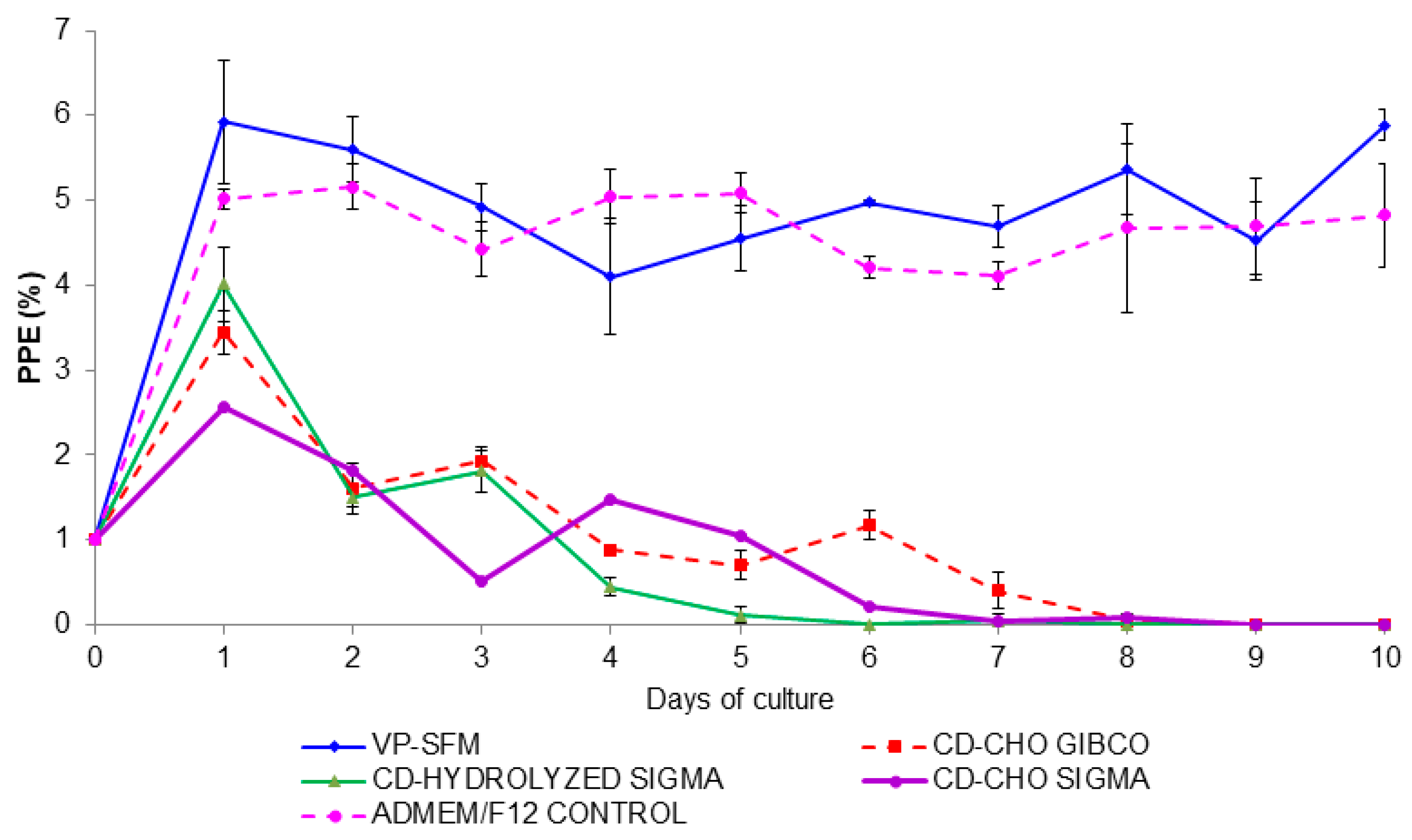
Figure 1. Babesia bigemina growth curve using animal component-free media. The percent of parasitized erythrocyte (PPE) was adjusted to 1% when PPE reached 4%. If the PPE was ≤1 then a 1:2 subculture was carried out. Subcultures are not shown.
Pathogens 09 00343 g001
Figure 2. Comparison of Babesia bigemina proliferation between VP-SFM and ADMEM/F12 control medium. The percentage of parasitized erythrocytes (PPE) was adjusted to 1% when reached 4%. If the PPE was ≤1 then a 1:2 subculture was carried out. Subcultures are not shown.
Pathogens 09 00343 g002
Figure 3. Comparison of growth curve of Babesia bigemina proliferation using VP-SFM + lipid with ADMEM/F12 control medium. The parasitemia was adjusted to 1% when the percentage of parasitized erythrocytes (PPE) reached 4. If the PPE was ≤1 then a 1:2 subculture was carried out. Subcultures are not shown.
Pathogens 09 00343 g003
Figure 4. Babesia bigemina proliferation in a perfusion bioreactor. (A) Growth comparison of B. bigemina between VP-SFM + lipid mixture and ADMEM/F12 control medium. (B) Giemsa-stained blood smear showing B. bigemina parasitized erythrocytes.
Pathogens 09 00343 g004
Table 1. Growth of B. bigemina in media free of animal products.
MediumSerum-FreeAnimal-Free ComponentsProtein-Free
ADMEM/F12YesNoNo
CD-CHO (Gibco®),YesYesYes
CD-CHO (Sigma-Aldrich)YesYesYes
CD-Hydrolyzed (Sigma-Aldrich)YesYesYes
VP-SFM (Gibco®)YesYesNo
Table 2. Lipid concentration used for in vitro B. bigemina proliferation.
TreatmentLipids Mixture Concentration (mg/mL)
MyristicPalmiticPalmitoleicStearicOleicLinolenicStearicArachidonicCholesterol
I0.20.20.20.20.20.20.20.044.4
II0.10.10.10.10.10.10.10.022.2
III0.050.050.050.050.050.050.050.011.1
IV0.0250.0250.0250.0250.0250.0250.0250.0050.55
V0.01250.01250.01250.01250.01250.01250.01250.00250.275
Lipid concentration: half-dilution factor.
Table 3. Effect of lipid concentration on in vitro B. bigemina proliferation.
Treatment
Lipid
Days of Culture
0123456
I1.0%2.43%5.71%8.00%7.92%8.96%8.03%
II1.0%4.93%7.19% *9.85% *10.33% *10.43% *11.35% *
II1.0%4.77%5.03%7.91%8.70%8.60%9.04%
IV1.0%4.20%5.08%7.06%7.77%5.25%8.10%
V1.0%3.12%3.74%5.49%5.61%8.60%7.06%
VP-SFM1.0%1.77%4.40%4.99%6.07%7.42%7.43%
Treatment lipid: one-half dilution factor as described in Table 2. Asterisks indicate significant difference between treatments.