Review

The Diverse Roles of the Global Transcriptional Regulator PhoP in the Lifecycle of Yersinia pestis

1
Clinical Laboratory Sciences Department, School of Health Technology and Management, Stony Brook University, Stony Brook, NY 11794, USA
2
Paul G. Allen School for Global Animal Health, Washington State University, Pullman, WA 99164, USA
*
Author to whom correspondence should be addressed.
Pathogens 2020, 9(12), 1039; https://doi.org/10.3390/pathogens9121039
Submission received: 29 October 2020 / Revised: 30 November 2020 / Accepted: 4 December 2020 / Published: 11 December 2020

Abstract

:
Yersinia pestis, the causative agent of plague, has a complex infectious cycle that alternates between mammalian hosts (rodents and humans) and insect vectors (fleas). Consequently, it must adapt to a wide range of host environments to achieve successful propagation. Y. pestis PhoP is a response regulator of the PhoP/PhoQ two-component signal transduction system that plays a critical role in the pathogen’s adaptation to hostile conditions. PhoP is activated in response to various host-associated stress signals detected by the sensor kinase PhoQ and mediates changes in global gene expression profiles that lead to cellular responses. Y. pestis PhoP is required for resistance to antimicrobial peptides, as well as growth under low Mg2+ and other stress conditions, and controls a number of metabolic pathways, including an alternate carbon catabolism. Loss of phoP function in Y. pestis causes severe defects in survival inside mammalian macrophages and neutrophils in vitro, and a mild attenuation in murine plague models in vivo, suggesting its role in pathogenesis. A Y. pestis phoP mutant also exhibits reduced ability to form biofilm and to block fleas in vivo, indicating that the gene is also important for establishing a transmissible infection in this vector. Additionally, phoP promotes the survival of Y. pestis inside the soil-dwelling amoeba Acanthamoeba castellanii, a potential reservoir while the pathogen is quiescent. In this review, we summarize our current knowledge on the mechanisms of PhoP-mediated gene regulation in Y. pestis and examine the significance of the roles played by the PhoP regulon at each stage of the Y. pestis life cycle.

1. Introduction

Yersinia pestis, a gram-negative bacterium that causes plague, emerged as a subclone of Yersinia pseudotuberculosis as recently as 5000–6000 years ago [1,2,3,4]. Despite the genetic similarity [5], Y. pestis has evolved a distinct and more complex life cycle compared to its ancestor. While Y. pseudotuberculosis normally persists in the soil and is transmitted through contaminated food and water, Y. pestis primarily infects rodents and is transmitted to other mammals by fleas [6,7], and has poor survivability in the soil by itself [8,9,10]. When the flea vector takes a blood meal from an infected animal, the bacteria enters the digestive system and forms a biofilm that blocks the flea foregut. As the blocked flea tries to feed on a new mammalian host, it transmits the bacteria by regurgitating into the bite site [6]. Y. pestis occasionally infects humans through flea bites, causing bubonic plague. The bacteria can also be transmitted through an aerosol route to cause more life-threatening pneumonic plague [7]. If untreated, both bubonic and primary pneumonic plague can lead to septicemic plague and death. While Y. pestis causes high mortality in mammalian hosts during the epizootic phase, it can also persist for a long time without causing fatal outbreaks during the inter-epizootic/quiescent phase [10]. It is commonly thought that the bacteria survive by circulating between resistant populations of rodents and fleas. However, it has also been suggested that the bacteria may survive by residing inside free-living amoeba in the soil [10]. Thus, Y. pestis encounters a wide range of conditions during its life cycle and needs to respond rapidly to the changes in order to achieve successful maintenance and propagation.
Bacterial two-component systems are transcriptional regulators that play critical roles in the adaptation of the bacteria to changing habitats. They can sense various environmental cues and orchestrate complex regulatory networks to facilitate transcriptional responses. The analysis of the Y. pestis CO92 genome has revealed up to 29 predicted two-component systems, including four pseudogenes [11,12]. Of these, the PhoP/PhoQ (PhoPQ) two-component system has been most extensively studied as a potential virulence determinant. Studies have shown that the function of PhoP, the response regulator in the PhoPQ system, is important for various stages of Y. pestis life cycle, including survival inside the mammalian phagocytes, biofilm formation inside the flea gut, and survival inside the soil-dwelling amoeba. In this review, we summarize our current knowledge on the signals that activate Y. pestis PhoP in different host environments, the genes and molecular pathways that are regulated by PhoP, and different ways by which the PhoP regulon contributes to the adaptation of Y. pestis to various stages of its life cycle.

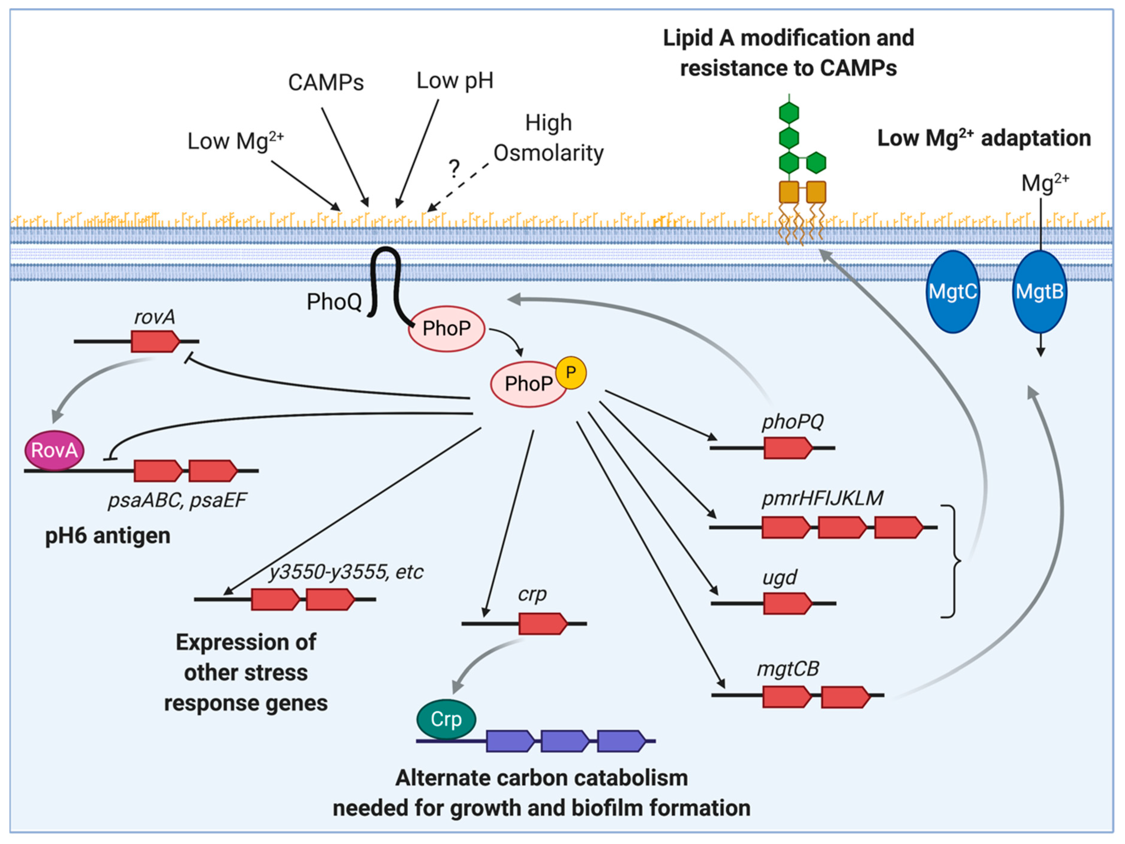
2. Y. pestis PhoP Regulatory Networks

PhoPQ is a two-component system that plays a crucial role in virulence and the adaptation to harsh conditions in many Gram-negative bacteria. PhoPQ consists of the sensor histidine kinase/phosphatase PhoQ and the response regulator PhoP. PhoQ detects changes in the periplasmic environment and transduces a signal that promotes kinase activity in its cytoplasmic domain. The phosphoryl group is then transferred to activate PhoP in the cytoplasm. Phosphorylated PhoP binds to sequences containing two direct (T/G)GTTTA repeats separated by five nucleotides in the promoter region of a number of genes, activating or repressing their transcription [13].
During infection, the PhoPQ system can sense different environmental stresses, such as low magnesium (Mg2+), the presence of cationic antimicrobial peptides (CAMPs) and low pH (Figure 1). Positively charged Mg2+, CAMPs, and H+ interact with highly acidic residues in PhoQ’s periplasmic sensor domain. Under physiological concentrations, Mg2+ is bound to inactive PhoQ, and in Mg2+ limiting conditions, the cations dissociate and PhoQ gets activated [13]. Similarly, CAMPs activate the system by displacing Mg2+ from PhoQ metal binding sites [14]. Hydrogen ions also bind to the PhoQ sensor domain, but do not directly compete for binding to divalent cation-binding sites [15]. Additional stimuli associated with the host, such as hyperosmotic stress and disruption of the oxidizing environment of the periplasm, have been proposed to stimulate PhoQ in E. coli [16,17].
PhoP regulates the activity of a number of genes and operons that govern a wide variety of cell processes in Y. pestis. Transcriptome analysis revealed that 0.5–2.0% of the whole Y. pestis genome is either activated or repressed by PhoP [18,19]. However, PhoP only binds to the promoters of a limited number of genes or operons, suggesting that most PhoP targets are indirectly regulated [20]. The direct targets of PhoP include those involved in adaptation to a low-Mg2+ environment (mgtCB), protection against reactive oxygen species (sodA, sodB, sodC, katA, and ahpC) and other stresses, and resistance to antimicrobial peptides (arnBCADTEF/pmrHFIJKLM, ugd/pmrE, and pagP) [20] (Figure 1). Antimicrobial peptides are critical components of the host innate immune response that bind to and destabilize the outer membrane of Gram-negative bacteria. To decrease the binding of the positively-charged CAMPs, Y. pestis uses the gene products of the PhoP-regulated pmrHFIJKLM (also termed arnBCADTEF or pbgP operon) and ugd (also termed pmrE) genes to add a 4-aminoarabinose to the lipid A, thereby neutralizing the negative charge of the outer membrane [21,22]. Another phoP-regulated gene, pagP, encodes a lipopolysaccharide (LPS) modifying enzyme that mediates the transfer of a palmitate group to the lipid A in Y. pseudotuberculosis and Yersinia enterocolitica. This modification increases the Toll-like receptor 4(TLR4)/myeloid differentiation factor 2 (MD-2)-mediated innate immune recognition of these pathogens during infection. However, Y. pestis lacks the palmitoylated lipid A species [23]. A recent study shows that Y. pestis has a single nucleotide polymorphism (SNP) that renders the enzyme inactive [24]. It is proposed that the loss of a functional PagP during Y. pestis evolution resulted in increased survival of the bacteria in the mammalian hosts by evasion of the innate immune response.
Y. pestis PhoP governs various cellular pathways indirectly by acting on a set of regulators [20,25]. Beside regulating its own expression, PhoP activates the transcription of the global regulator, cyclic AMP receptor protein (crp), which controls sugar uptake and catabolism, and modulates a wide variety of Y. pestis genes, including those involved in quorum sensing, iron acquisition, biofilm formation, and the expression of several virulence factors [25,26]. PhoP also controls the expression of the global virulence regulator RovA, which activates expression of the antiphagocytic pH6 antigen in Y. pestis, and the adhesin invasin in Y. pseudotuberculosis and Y. enterocolitica [27,28]. While rovA expression is self-activated, it is repressed by PhoP binding to its promoter region [27].
The evolution of gene regulation in bacteria results in major differences between related organisms. Although Y. pestis and Salmonella PhoP are highly homologous, their individual regulons are considerably different. One example is the highly conserved type III secretion system (T3SS), which is essential for virulence in the two pathogens. The T3SSs encoded in the Salmonella pathogenicity island (SPI)-1 and SPI-2 are highly regulated by PhoP. In Salmonella, induction of endocytosis by epithelial cells governed by SPI-1 is repressed by PhoP’s negative effect on the regulator HilA, and SPI-2-mediated intramacrophage survival is activated by the action of PhoP on SsrB, a regulator that binds to the promoters of all SPI-2 functional gene clusters [29,30]. Notably, activation of the T3SS in Yersinia does not appear to be regulated by PhoP.
While PhoP proteins from Yersinia and Salmonella both regulate a common set of core ancestral genes, they differentially transcribe species-specific genes in the two pathogens [31]. Similarly, both conserved and unique PhoP binding sites are found in Y. pestis and Salmonella. The conserved promoters, called prototypical class II, regulate Salmonella ancestral genes, such as Mg2+ transport gene mgtA, but not genes acquired horizontally. On the other hand, Salmonella’s horizontally acquired ugtL gene, which is required for low pH resistance, has a PhoP box with an inverted orientation that is not present in Yersinia [31]. Most PhoP boxes in Yersinia resemble the organization of the class II promoter with respect to its orientation and its distance from the -10 region. Other Yersinia PhoP DNA binding sites, such as that of mgtC, are located further upstream of the transcription initiation site.
In addition to different promoter structures, Yersinia and Salmonella use different PhoP regulatory pathways to control the genes encoding conserved proteins. The arabinose modification of Lipid A in Salmonella is regulated by the PhoP-activated PmrD protein. PmrD post-translationally activates PmrA, which binds to the promoter of the 4-aminoarabinose biosynthetic genes. Because Yersinia lack PmrD, regulation of the pmr operon and ugd in a low-Mg2+ environment occurs by direct binding of PhoP to a second promoter in those genes [21]. It reflects how evolutionary changes in the PhoP regulon allows the pathogen to transcribe newly acquired genes and maintain control of ancestral ones.

3. Role of phoP in Intracellular Replication in Mammalian Hosts

Y. pestis is a facultative intracellular pathogen. During Y. pestis infection, bacteria are mainly found extracellularly within the tissues of an infected host. To inhibit phagocytosis and suppress proinflammatory cytokine production by host immune cells, Y. pestis expresses an array of virulence factors, including T3SS and Yop effector proteins encoded on a virulence plasmid (pCD1), as well as a capsule composed of F1 protein [32,33]. However, the expression of these factors is suppressed when Y. pestis is inside the flea [34], so at the initial stage of infection, the bacteria are more readily taken into the professional phagocytes. Therefore, it has been proposed that the host phagocytes may provide a replicative niche and a vehicle for dissemination of Y. pestis before the pathogen can replicate more robustly in the extracellular environment [35]. Consistent with this hypothesis, Y. pestis is found inside host macrophages in experimentally infected animals [36,37,38,39,40]. More recently, in vivo tracking studies using intravital confocal microscopy and flow cytometry in murine intradermal models of bubonic plague have shown that neutrophils, and later resident macrophages and dendritic cells (DCs), to a lesser extent, are recruited to the initial site of infection within several hours of the pathogen inoculation [41,42,43]. In these studies, the bacteria are often found to be associated with or inside macrophages and neutrophils [41,42].
Various in vitro studies have also demonstrated that Y. pestis can survive and replicate within macrophage phagosomes [19,39,44,45], and that phoP is essential for this ability. The phoP deletion mutants of Y. pestis exhibit a severe defect in growth inside murine macrophages, as well as growth under in vitro conditions that mimic the intracellular environment: low pH, oxidative stress, low Mg2+, high osmolarity, and the presence of antimicrobial peptides [19,44,46]. Transcriptome analysis has shown that many PhoP-regulated genes, including the members of the pmr operon, ugd, and mgtC, are more highly expressed in Y. pestis, replicating inside macrophages compared to the bacteria replicating in the tissue culture media [47]. Inactivation of these PhoP-regulated genes reduces the ability of the bacteria to replicate inside murine macrophages [19,48], confirming that they are critical for the adaptation of Y. pestis to the intracellular environment. Surprisingly, deletion of mgtB, the PhoP-regulated gene encoding for an Mg2+ transporter, does not affect intracellular replication of Y. pestis, although the mutant is attenuated in murine infection models, suggesting that MgtB may have an additional role in Y. pestis pathogenesis outside of intracellular survival [49]. Inside macrophages, Y. pestis resides in spacious phagosomes, some of which have acquired double membranes and the characteristics of autophagosome [50]. Like other intracellular pathogens that survive inside macrophages by inhibiting phagosomal maturation, Y. pestis blocks acidification and remodels its phagosome by recruiting components of the host endocytic pathway Rab1b, Rab4, and Rab11 [50,51,52]. However, there is no evidence that phoP plays a part in this process [50].
Y. pestis is also able to survive inside neutrophils, the first immune cells to be recruited to the site of infection. Though less permissive than macrophages, about 10–15% of the bacteria internalized by human neutrophils survive and replicate intracellularly [22]. Deletion of phoP leads to defective survival inside human neutrophils and increased sensitivity to the components of α-granules produced by these cells, including several CAMPS in vitro [22]. Y. pestis-containing neutrophils can be internalized into macrophages in a process called efferocytosis, thereby suppressing the secretion of inflammatory cytokines while promoting the expression of anti-inflammatory cytokine 1L-1RA [53]. Therefore, it has been proposed that neutrophils may provide a path for Y. pestis to infect macrophages while minimizing inflammatory responses [53].
Despite the well-established role of phoP in the intracellular replication of Y. pestis, the extent of its contribution to the overall virulence in mammalian hosts is less clear. In one study, the Y. pestis strain GB ΔphoP showed a 75-fold increase in the LD50 compared to the wild type in a murine bubonic plague model via subcutaneous infection [44]. The strain KIM5+ ΔphoP was also attenuated in a murine pneumonic plague (aerosol) model (Bliska, J. B., unpublished results [54]), suggesting that phoP is important for the virulence of Y. pestis. However, in another study, there was no difference in the LD50 between a CO92 wild-type strain and the corresponding ΔphoP mutant in murine bubonic (subcutaneous inoculation) and pneumonic (aerosol) plague models [55]. Only an increase in time to death was observed in the bubonic plague model at lower dosages, indicating that the role of phoP in Y. pestis virulence may be minor. One contributing factor for the difference in observed phenotypes could be that these studies used different strains of Y. pestis (GB vs. CO92) and mice (BALB/c vs. Swiss Webster mice) [44,55]. Moreover, these studies used subcutaneous injection, while the natural fleabites target the intradermal layer of skin. To mimic flea-mediated infection more closely, murine intradermal infection models have been established in the last several years [41,43]. It would be interesting to know whether testing phoP mutants using the latter model would reveal a role of this gene in bacterial dissemination from the initial site of infection that was not obvious in the earlier studies of this mutant.
The relatively mild attenuation of Y. pestis phoP mutants is similar to the effect of phoP deletion on virulence of Y. pseudotuberculosis (40–100× increase in LD50) [55,56], but is in contrast to the severe attenuation of phoP mutants in Salmonella [57,58,59]. This is probably because Salmonella PhoP controls a wider range of critical virulence genes than Y. pestis PhoP, including the SPI-1 and SPI-2 genes [29,30]. In addition, the requirement for the intracellular phase of Y. pestis in its pathogenesis is controversial. Although neutrophils are recruited to the site of Y. pestis injection and the bacteria are found nearby or inside these cells, neutrophil depletion did not alter dissemination or the virulence of Y. pestis in murine intradermal [41,43] and intranasal [60] infection models using fully virulent strains. Tracking studies have shown that Y. pestis reaches draining lymph nodes as quickly as 10 min after intradermal inoculation, without using macrophages and neutrophils as carriers [43], suggesting that these cells are not required for trafficking or the pathogenesis of Y. pestis. However, in another murine subcutaneous infection model, using an attenuated strain of Y. pestis KIM5-, lacking the pigmentation (pgm) locus necessary for infection via peripheral routes, massive infiltrates of DCs and macrophages were observed at the site of injection, many harboring intracellular bacteria [61]. In this study, the migration of Y. pestis-containing phagocytes via lymphatic vasculature was found to be critical for bacterial dissemination and virulence [61]. In yet another murine intravenous infection study, depletion of macrophages and DCs led to the reduced colonization of spleen and liver by Y. pestis [62]. These results support the idea that macrophages and DCs are important for the early replication and dissemination of Y. pestis. Further investigation is necessary to determine the exact role of the intracellular stage in Y. pestis pathogenesis, as well as the contribution of phoP to the overall virulence of Y. pestis in mammalian hosts. Also, of interest is whether phoP plays any additional role outside of intracellular replication during infection of mammalian hosts.

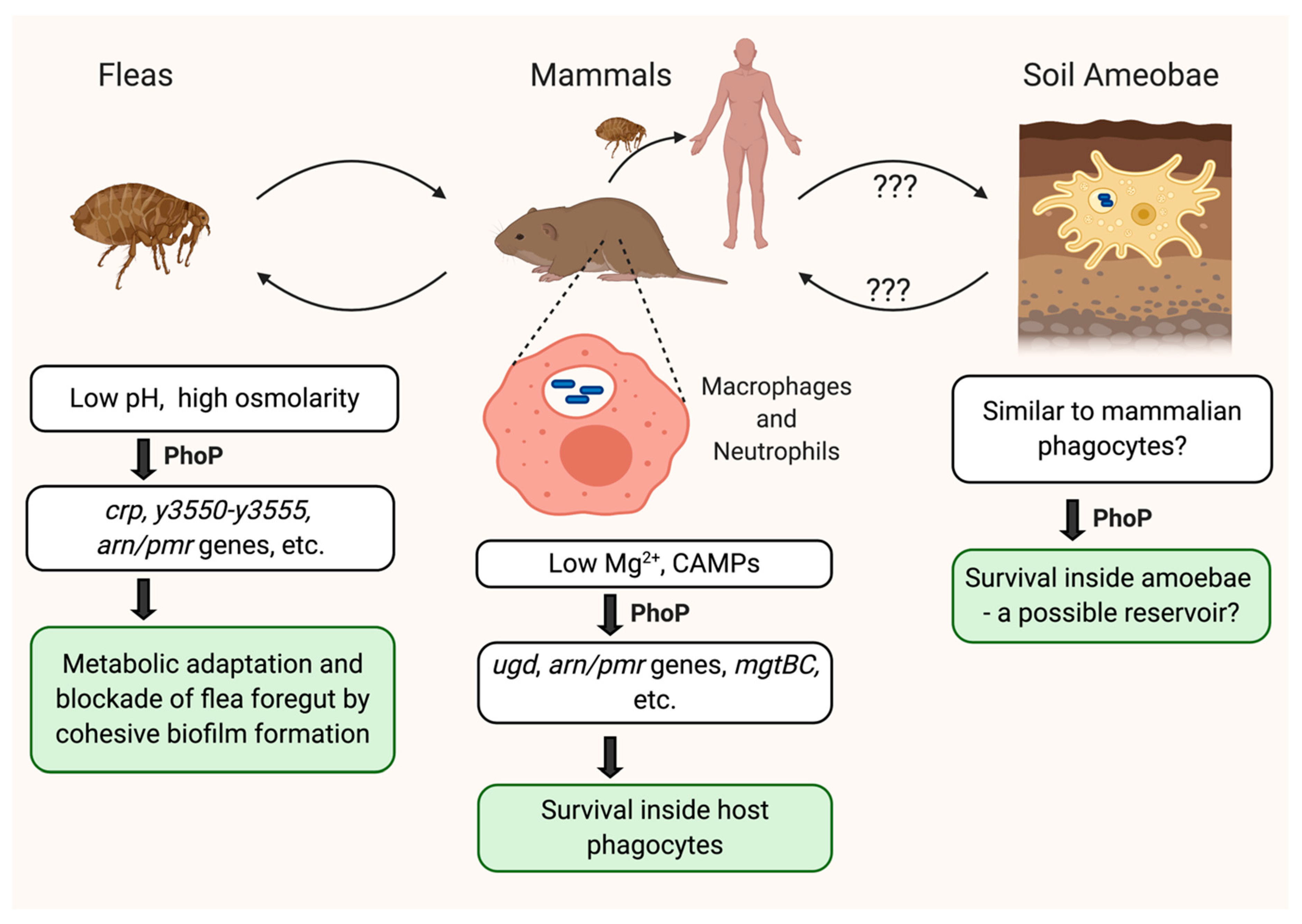
4. Biofilm and Flea Colonization

Unlike its seemingly uncertain contribution to development of infection in the mammalian host, PhoP appears to be important for coordinating the flea-associated stage of Y. pestis to enable transmission [34,63,64]. Inside fleas, Y. pestis forms an adherent biofilm in the foregut to prevent bloodmeal passage to the midgut, and this blockage enhances Y. pestis transmission by inducing the flea’s feeding attempts and the subsequent regurgitation of the bacteria from the biofilm [6]. PhoP function is required for the formation of sufficiently cohesive biofilm, which robustly attaches to the flea foregut to maintain normal blockage. A phoP mutant instead forms aggregates of fragile biofilm localized to the midgut, and is therefore less likely to be transmitted [63]. Although PhoP does not directly regulate genes that control biofilm production, it directly activates the expression of the carbon catabolite regulator crp [25], which coordinates utilization of secondary carbon nutrient sources to support a marked growth spurt and strong biofilm formation [25,26,65]. Genes involved in the uptake and catabolism of secondary sugars like galactose, as well as the pentose sugars, ribose, arabinose, and xylose, are expressed in fleas blocked with the designated wild-type, Y. pestis KIM (pCD1- pgm+) strain [34]. In addition, genes encoding ribose ABC transporter proteins (y3243/rbsA, y3345, and y3346 in KIM strain) are down-regulated in the phoP mutant infecting fleas [64]. These results are consistent with a role of PhoP in mediating Crp-dependent regulation of alternate sugar use during flea infection.
Mildly acidic pH (~6.6) and hyperosmolarity (~500 mOsm) of the flea gut milieu are considered to be the stress signals encountered by Y. pestis during insect infection [66,67]. PhoP expression is closely linked to management of these stresses in the flea gut, as demonstrated in comparative transcriptomic studies between a phoP mutant and wild-type strain during flea infection [64]. A number of stress-response genes are differentially regulated in the phoP mutant, including a polycistronic operon comprised of six low-GC-content genes (y3555/aspB-3554/nhaC-y3553-y3552-y3551/ridA-y3550 in KIM). [64]. These genes have putative roles in aromatic amino acid metabolism and are stimulated by hyperosmotic salinity [67]. Together with phoP and y3555, y3553 and y3550 rank among the mostly highly expressed genes in wild-type Y. pestis during flea blockage [34]. Another example is that of the YhcN family genes (y0666 and y1667 in KIM), which normally display induced expression and promote bacterial aggregation at low pH conditions in vitro [64]. These genes are highly expressed in fleas blocked from a wild-type infection [34], and in a phoP mutant their expression is further induced, presumably to manage the acidity in the flea gut in the absence of PhoP. PhoP binding motifs have been identified by bioinformatic analysis in the promoter regions of y0666 and y3555/aspB [68], predicting that these genes are directly regulated by PhoP.
Known PhoP-repressed genes psaABC and psaEF, which are involved in production of the pH 6 antigen [28,69], also have increased expression in the phoP mutant during flea infection, suggesting that they are derepressed by the absence of PhoP. In addition to direct repression, PhoP indirectly represses psa gene expression by downregulating the transcription of rovA, whose gene product positively regulates psa gene expression [27,28]. However, while wild-type bacteria in the blocked fleas show significant down-regulation of rovA [27,28] only a small increase in rovA expression occurs in the phoP mutant during flea infection. The expression of psa genes is also stimulated by acidic conditions in vitro [27,28]. Thus, the activation of psa transcription in the phoP mutant is likely a combined effect of direct derepression by PhoP, induction through de-repression of the activator RovA, and low pH. It is proposed that the fimbrial structure that constitutes the pH 6 antigen may interfere with the cohesiveness of the Y. pestis phoP mutant flea biofilm [64]. Whether this is the reason that the pH 6 antigen is generally repressed in wild-type blocked fleas by significant down-regulation of rovA and high expression of phoP [34] is unknown.
Other conserved PhoP-regulated genes, pmrHFIJKLM and ugd, are downregulated in the phoP mutant colonizing fleas. However, their roles in PhoP-mediated flea infection are elusive. This is because the deletion of ugd and pmrA, a response regulator that is thought to control the pmr operon, along with PhoP in Y. pestis, does not affect flea infection dynamics during single infection [63], but mutants defective in lipid A modification appear to be required for early flea infection when competing with a wild-type strain [70]. It is unknown whether Y. pestis elicits a response to flea antibacterial CAMPs. Further to this is that decreased expression of mgtC observed during phoP mutant flea infection appears to be innocuous, as the flea Mg2+ concentration range is not considered to be low [63].
Multiple stress adaptation genes, not confirmed to be directly regulated by PhoP, are induced in the phoP mutant in fleas [64]. This suggests that a compensatory stress response is activated in order to counter unresolved stress due to the loss of PhoP. General stress response genes involved in osmotic stress (kdpA, kdpB) and heat shock response (ipbA, ipbB, rpoH, htpX, and dnaK) are examples of such induced genes. Additionally, the expression of L-glutamate group amino acid utilization genes (e.g., glnHPQ, gltJKL, astBD, argD, and hutHU) is significantly decreased in phoP mutant infecting fleas. These are metabolic genes that are implicated in the alleviation of osmotic stress and are predominantly expressed in wild-type blocked fleas [34]. A plethora of other metabolic genes that are normally induced in these blocked fleas are also down-regulated during phoP mutant flea infection. Interestingly, a few toxin–antitoxin module encoding genes (mqsR, mqsA, higB2, y1074, and y1075) are generally found to be induced under stress conditions like glucose and amino acid starvation, hyperosmolarity, and antibiotic exposure also show notably increased expression in the phoP mutant in fleas [71]. Expression of stress-related toxin–antitoxin modules is often associated with the formation of persister cells. [72,73]. Persister cells usually comprise 1% of biofilms and are non-growing, metabolically inactive cells that are recalcitrant to environmentally stressful conditions. This supports the notion that the reduced expression of metabolic genes may be a consequence of the phoP mutant bacteria attempting to maintain viability in a harsh environment by minimizing metabolic processes. Accordingly, a phoP mutant exhibits a severe defect in competitive fitness compared to the wild type in a flea co-infection assay, indicating that this mutant is physiologically impaired [74,75]. Together, in fleas, PhoP plays a critical role in coordinating physiological adaptation to low pH and osmotic stress, in a manner that preserves active metabolic processes that support vigorous biofilm-mediated flea blockage.
Much of the current PhoP transcriptional response in fleas represents a snapshot at two weeks post-Y. pestis infection, when bacteria are presumably not actively replicating. Immediately after fleas acquire a Y. pestis infection from feeding on infected blood, the bacteria begin replicating throughout the period of a week to 10 days [76,77]. Fleas start to block 3–5 days post-infection, with the numbers of blocked fleas peaking around 12 days post-infection [78]. Afterwards, fewer fleas are blocked, in accordance with the absence of bacterial replication [76]. One surprising observation in the comparative transcriptomic studies between the phoP mutant and wild-type infected fleas is that the expression of the PhoP target, crp, is not significantly altered. Given that Crp dictates optimal growth and production of bacterial biofilm, this is unexpected. However, Crp may then only function in the initial two weeks post-infection, in synchrony with active bacterial growth and biofilm blockage development. As such, investigations into the role of PhoP during the first week of active bacterial replication in fleas may provide more comprehensive insight into its role in Y. pestis flea infection and biofilm blockage.

5. Amoeba as a Potential Host While the Pathogen Is Quiescent

While the infectious cycle of Y. pestis is well-studied, it is not known where and how this pathogen persists naturally during inter-epizootic periods. It has been hypothesized that amoebae serve as reservoir hosts for Y. pestis during this quiescent phase [10]. Recent experimental studies show that Y. pestis can survive for up to a week within spacious phagosomal compartments in the free-living, soil-dwelling amoeba Acanthamoeba castellani [79]. In other bacterial pathogen–amoeba interactions, the composition of the intracellular niche and the factors required for intracellular survival of bacteria are often conserved in both mammalian phagocytes and their phagocytic amoeba counterparts. This is no different for Y. pestis, as PhoP is required for intracellular survival in A. castellani, with no viable phoP mutant cells recovered beyond 24 h post-invasion of amoeba [79]. However, because the Yersinia-containing amoebal phagosome does not appear to fuse with the lysosome, it remains unknown if this vacuole has sufficiently low pH to require a PhoP-mediated response. It is also unknown if PhoP-triggering conditions, such as low Mg2+ and CAMP expression, are present in the Y. pestis-containing amoeba. The mechanism by which Y. pestis interacts with amoebae and exploits this potential reservoir niche remains a fascinating question worthy of further exploration.

6. The Effects of a phoP SNP on the PhoP Function and the Evolution of Y. pestis Virulence

During its emergence as a highly virulent, flea-borne pathogen, Y. pestis is thought to have acquired unique characteristics critical for the infection of mammalian hosts and flea vectors through gains and losses of numerous genes, genomic rearrangements, and the incorporation of two plasmids (pMT1 and pPCP1) [5,80]. Additionally, the comparative analysis of both the extant and the ancient Y. pestis genomes recovered from archaeological sites suggests that small sequence changes (such as SNPs) in key genes also contributed to the increased virulence and transmissibility of Y. pestis. Consistent with this idea, it has been shown that a single amino acid change in the plasminogen activator protease Pla led to the increased ability of Y. pestis to disseminate within its mammalian hosts and cause bubonic plague [81], while inactivating mutations in the three genes controlling biofilm formation enhanced the transmissibility of Y. pestis by fleas [82].
Interestingly, the phoP gene also acquired a SNP during Y. pestis evolution [83]. Y. pestis strains are classified into several phylogenetic branches based on their genome sequences. Branch 0 is rooted in Y. pseudotuberculosis, and includes the ancestral/basal strains such as Angola and Microtus. Branches 1 through 4 diverged from branch 0, and include strains associated with the major pandemics of the past, as well as KIM and CO92 [84]. The SNP in phoP was acquired near the polytomy where branches 1-4 diverged from branch 0, and causes an amino acid substitution at position 215 of the PhoP protein [83], a residue that is predicted to contact the target DNA [74]. Whereas glycine is encoded at this position (PhoP-G215) in Y. pseudotuberculosis, and the ancestral strain of Y. pestis, serine (PhoP-S215), is encoded in the strains belonging to branches 1 and 2. A mutational study showed that the wild-type Y. pestis KIM strain carrying the phoP-S215 allele has an increased ability to grow under a low-Mg2+ condition, induce the PhoP-regulated gene ugd, and resist antimicrobial peptide activity compared to an isogenic strain carrying the ancestral phoP-G215 allele [74]. The results suggest that this amino acid substitution causes a subtle alteration in PhoP activity and its regulatory network. The evolutionary significance of this change is unknown, as it does not appear to affect the ability of Y. pestis to survive inside macrophages or to colonize the flea gut [74]. However, these results raise a possibility that the acquisition of the modern phoP allele may have been part of many small genetic changes that contributed to the increased virulence and/or the host adaptability of Y. pestis.

7. Summary and Future Perspective

PhoP is a versatile transcriptional regulator that is involved in the response to various stresses Y. pestis encounters during its life (Figure 2). In mammalian hosts, PhoP plays a crucial role in survival inside host phagocytes by promoting resistance to antimicrobial peptides and other harsh conditions. In fleas, PhoP responds to osmotic and acidic stresses, and coordinates changes in bacterial metabolism and the formation of a cohesive biofilm required for flea blockage. The roles of PhoP during interaction with amoebae need further elucidation, but they may be similar to those observed within mammalian phagocytes. The PhoP regulon in Y. pestis appears to have evolved dynamically, incorporating newly acquired genes into its network while keeping the ancestral genes as part of the core regulon, and these evolutionary changes may have facilitated adaptation of the pathogen to different host environments [31]. For example, the low-GC content y3550–y3555 operon has been predicted to be a direct target of Y. pestis PhoP [68], but this operon is not commonly found in bacteria other than Y. pestis and Y. pseudotuberculosis; among its gene products, only Y3555 is found to have homologs in rare Salmonella food-borne isolates [67]. An increased expression of this operon during flea infection suggests that perhaps its incorporation into the Y. pestis PhoP regulon may have improved the pathogen’s ability to cope with physiological challenges specific to the flea gut. On the other hand, the core PhoP regulon, including pmrHFIJKLM, ugd, and mgtCB, may encode evolutionarily conserved mechanisms that promote Y. pestis intracellular survival in both the soil amoebae and the mammalian phagocytes. Finally, a number of small changes in the PhoP regulon during Y. pestis evolution, such as SNPs in phoP and the PhoP-target pagP, which resulted in a subtle increase in the PhoP transcriptional activity and the alteration of lipid A modification to a less immunostimulatory form, respectively, also may have contributed to the enhanced pathogenicity of the bacteria [24,74]. Further analysis of the PhoP regulon should provide insights into how the dynamic nature of the bacterial regulatory network may contribute to the adaptation of a pathogen to different hosts during evolution.

Author Contributions

H.S.F. conceptualized and developed the outline of the manuscript. G.I.V., V.V. and H.S.F. wrote, revised, and edited the manuscript. H.S.F. created the figures, and G.I.V. and V.V. reviewed them. All authors have read and agreed to the published version of the manuscript.

Funding

This research received no external funding.

Acknowledgments

We thank James Bliska for sharing the unpublished data and providing critical comments on the manuscript.

Conflicts of Interest

The authors declare no conflict of interest.

References

  1. Achtman, M.; Zurth, K.; Morelli, G.; Torrea, G.; Guiyoule, A.; Carniel, E. Yersinia pestis, the cause of plague, is a recently emerged clone of Yersinia pseudotuberculosis. Proc. Natl. Acad. Sci. USA 1999, 96, 14043–14048. [Google Scholar] [CrossRef] [Green Version]
  2. Rasmussen, S.; Allentoft, M.E.; Nielsen, K.; Orlando, L.; Sikora, M.; Sjogren, K.G.; Pedersen, A.G.; Schubert, M.; Van Dam, A.; Kapel, C.M.; et al. Early divergent strains of Yersinia pestis in Eurasia 5000 years ago. Cell 2015, 163, 571–582. [Google Scholar] [CrossRef]
  3. Rascovan, N.; Sjogren, K.G.; Kristiansen, K.; Nielsen, R.; Willerslev, E.; Desnues, C.; Rasmussen, S. Emergence and spread of basal lineages of Yersinia pestis during the Neolithic decline. Cell 2019, 176, 295–305. [Google Scholar] [CrossRef] [Green Version]
  4. Spyrou, M.A.; Tukhbatova, R.I.; Wang, C.C.; Valtuena, A.A.; Lankapalli, A.K.; Kondrashin, V.V.; Tsybin, V.A.; Khokhlov, A.; Kuhnert, D.; Herbig, A.; et al. Analysis of 3800-year-old Yersinia pestis genomes suggests Bronze Age origin for bubonic plague. Nat. Commun. 2018, 9, 2234. [Google Scholar] [CrossRef] [Green Version]
  5. Chain, P.S.; Carniel, E.; Larimer, F.W.; Lamerdin, J.; Stoutland, P.O.; Regala, W.M.; Georgescu, A.M.; Vergez, L.M.; Land, M.L.; Motin, V.L.; et al. Insights into the evolution of Yersinia pestis through whole-genome comparison with Yersinia pseudotuberculosis. Proc. Natl. Acad. Sci. USA 2004, 101, 13826–13831. [Google Scholar] [CrossRef] [Green Version]
  6. Chouikha, I.; Hinnebusch, B.J. Yersinia-flea interactions and the evolution of the arthropod-borne transmission route of plague. Curr. Opin. Microbiol. 2012, 15, 239–246. [Google Scholar] [CrossRef] [Green Version]
  7. Perry, R.D.; Fetherston, J.D. Yersinia pestis—Etiologic agent of plague. Clin. Microbiol. Rev. 1997, 10, 35–66. [Google Scholar] [CrossRef]
  8. Ayyadurai, S.; Houhamdi, L.; Lepidi, H.; Nappez, C.; Raoult, D.; Drancourt, M. Long-term persistence of virulent Yersinia pestis in soil. Microbiology 2008, 154, 2865–2871. [Google Scholar] [CrossRef] [Green Version]
  9. Boegler, K.A.; Graham, C.B.; Montenieri, J.A.; MacMillan, K.; Holmes, J.L.; Petersen, J.M.; Gage, K.L.; Eisen, R.J. Evaluation of the infectiousness to mice of soil contaminated with Yersinia pestis-infected blood. Vector Borne Zoonotic Dis. 2012, 12, 948–952. [Google Scholar] [CrossRef] [Green Version]
  10. Eisen, R.J.; Gage, K.L. Adaptive strategies of Yersinia pestis to persist during inter-epizootic and epizootic periods. Vet. Res. 2009, 40, 1. [Google Scholar] [CrossRef] [Green Version]
  11. Marceau, M. Transcriptional regulation in Yersinia: An update. Curr. Issues Mol. Biol. 2005, 7, 151–177. [Google Scholar]
  12. Sun, Y.C.; Hinnebusch, B.J.; Darby, C. Experimental evidence for negative selection in the evolution of a Yersinia pestis pseudogene. Proc. Natl. Acad. Sci. USA 2008, 105, 8097–8101. [Google Scholar] [CrossRef] [Green Version]
  13. Groisman, E.A. The pleiotropic two-component regulatory system PhoP-PhoQ. J. Bacteriol. 2001, 183, 1835–1842. [Google Scholar] [CrossRef] [Green Version]
  14. Bader, M.W.; Sanowar, S.; Daley, M.E.; Schneider, A.R.; Cho, U.; Xu, W.; Klevit, R.E.; Le Moual, H.; Miller, S.I. Recognition of antimicrobial peptides by a bacterial sensor kinase. Cell 2005, 122, 461–472. [Google Scholar] [CrossRef] [Green Version]
  15. Prost, L.R.; Daley, M.E.; Le Sage, V.; Bader, M.W.; Le Moual, H.; Klevit, R.E.; Miller, S.I. Activation of the bacterial sensor kinase PhoQ by acidic pH. Mol. Cell 2007, 26, 165–174. [Google Scholar] [CrossRef]
  16. Yuan, J.; Jin, F.; Glatter, T.; Sourjik, V. Osmosensing by the bacterial PhoQ/PhoP two-component system. Proc. Natl. Acad. Sci. USA 2017, 114, E10792–E10798. [Google Scholar] [CrossRef] [Green Version]
  17. Lippa, A.M.; Goulian, M. Perturbation of the oxidizing environment of the periplasm stimulates the PhoQ/PhoP system in Escherichia coli. J. Bacteriol. 2012, 194, 1457–1463. [Google Scholar] [CrossRef] [Green Version]
  18. Zhou, D.; Han, Y.; Qin, L.; Chen, Z.; Qiu, J.; Song, Y.; Li, B.; Wang, J.; Guo, Z.; Du, Z.; et al. Transcriptome analysis of the Mg2+-responsive PhoP regulator in Yersinia pestis. FEMS Microbiol. Lett. 2005, 250, 85–95. [Google Scholar] [CrossRef] [Green Version]
  19. Grabenstein, J.P.; Fukuto, H.S.; Palmer, L.E.; Bliska, J.B. Characterization of phagosome trafficking and identification of PhoP-regulated genes important for survival of Yersinia pestis in macrophages. Infect. Immun. 2006, 74, 3727–3741. [Google Scholar] [CrossRef] [Green Version]
  20. Li, Y.; Gao, H.; Qin, L.; Li, B.; Han, Y.; Guo, Z.; Song, Y.; Zhai, J.; Du, Z.; Wang, X.; et al. Identification and characterization of PhoP regulon members in Yersinia pestis biovar Microtus. BMC Genom. 2008, 9, 143. [Google Scholar] [CrossRef] [Green Version]
  21. Winfield, M.D.; Latifi, T.; Groisman, E.A. Transcriptional regulation of the 4-amino-4-deoxy-L-arabinose biosynthetic genes in Yersinia pestis. J. Biol. Chem. 2005, 280, 14765–14772. [Google Scholar] [CrossRef] [PubMed] [Green Version]
  22. O’Loughlin, J.L.; Spinner, J.L.; Minnich, S.A.; Kobayashi, S.D. Yersinia pestis two-component gene regulatory systems promote survival in human neutrophils. Infect. Immun. 2010, 78, 773–782. [Google Scholar] [CrossRef] [PubMed] [Green Version]
  23. Bishop, R.E. The lipid A palmitoyltransferase PagP: Molecular mechanisms and role in bacterial pathogenesis. Mol. Microbiol. 2005, 57, 900–912. [Google Scholar] [CrossRef]
  24. Chandler, C.E.; Harberts, E.M.; Pelletier, M.R.; Thaipisuttikul, I.; Jones, J.W.; Hajjar, A.M.; Sahl, J.W.; Goodlett, D.R.; Pride, A.C.; Rasko, D.A.; et al. Early evolutionary loss of the lipid A modifying enzyme PagP resulting in innate immune evasion in Yersinia pestis. Proc. Natl. Acad. Sci. USA 2020, 117, 22984–22991. [Google Scholar] [CrossRef] [PubMed]
  25. Zhang, Y.; Wang, L.; Han, Y.; Yan, Y.; Tan, Y.; Zhou, L.; Cui, Y.; Du, Z.; Wang, X.; Bi, Y.; et al. Autoregulation of PhoP/PhoQ and positive regulation of the cyclic AMP receptor protein-cyclic AMP complex by PhoP in Yersinia pestis. J. Bacteriol. 2013, 195, 1022–1030. [Google Scholar] [CrossRef] [PubMed] [Green Version]
  26. Ritzert, J.T.; Minasov, G.; Embry, R.; Schipma, M.J.; Satchell, K.J.F. The cyclic AMP receptor protein regulates quorum sensing and global gene expression in Yersinia pestis during planktonic growth and growth in biofilms. mBio 2019, 10. [Google Scholar] [CrossRef] [Green Version]
  27. Zhang, Y.; Gao, H.; Wang, L.; Xiao, X.; Tan, Y.; Guo, Z.; Zhou, D.; Yang, R. Molecular characterization of transcriptional regulation of rovA by PhoP and RovA in Yersinia pestis. PLoS ONE 2011, 6, e25484. [Google Scholar] [CrossRef] [Green Version]
  28. Zhang, Y.; Wang, L.; Fang, N.; Qu, S.; Tan, Y.; Guo, Z.; Qiu, J.; Zhou, D.; Yang, R. Reciprocal regulation of pH 6 antigen gene loci by PhoP and RovA in Yersinia pestis biovar Microtus. Future Microbiol. 2013, 8, 271–280. [Google Scholar] [CrossRef]
  29. Bijlsma, J.J.; Groisman, E.A. The PhoP/PhoQ system controls the intramacrophage type three secretion system of Salmonella enterica. Mol. Microbiol. 2005, 57, 85–96. [Google Scholar] [CrossRef]
  30. Palmer, A.D.; Kim, K.; Slauch, J.M. PhoP-mediated repression of the SPI1 type 3 secretion system in Salmonella enterica serovar Typhimurium. J. Bacteriol. 2019, 201, e00264-19. [Google Scholar] [CrossRef] [Green Version]
  31. Perez, J.C.; Groisman, E.A. Transcription factor function and promoter architecture govern the evolution of bacterial regulons. Proc. Natl. Acad. Sci. USA 2009, 106, 4319–4324. [Google Scholar] [CrossRef] [PubMed] [Green Version]
  32. Viboud, G.I.; Bliska, J.B. Yersinia outer proteins: Role in modulation of host cell signaling responses and pathogenesis. Annu. Rev. Microbiol. 2005, 59, 69–89. [Google Scholar] [CrossRef]
  33. Prentice, M.B.; Rahalison, L. Plague. Lancet 2007, 369, 1196–1207. [Google Scholar] [CrossRef]
  34. Vadyvaloo, V.; Jarrett, C.; Sturdevant, D.E.; Sebbane, F.; Hinnebusch, B.J. Transit through the flea vector induces a pretransmission innate immunity resistance phenotype in Yersinia pestis. PLoS Pathog. 2010, 6, e1000783. [Google Scholar] [CrossRef] [PubMed] [Green Version]
  35. Pujol, C.; Bliska, J.B. Turning Yersinia pathogenesis outside in: Subversion of macrophage function by intracellular yersiniae. Clin. Immunol. 2005, 114, 216–226. [Google Scholar] [CrossRef] [PubMed]
  36. Finegold, M.J. Pneumonic plague in monkeys. An. electron microscopic study. Am. J. Pathol. 1969, 54, 167–185. [Google Scholar]
  37. Lukaszewski, R.A.; Kenny, D.J.; Taylor, R.; Rees, D.G.; Hartley, M.G.; Oyston, P.C. Pathogenesis of Yersinia pestis infection in BALB/c mice: Effects on host macrophages and neutrophils. Infect. Immun. 2005, 73, 7142–7150. [Google Scholar] [CrossRef] [Green Version]
  38. Bosio, C.M.; Goodyear, A.W.; Dow, S.W. Early interaction of Yersinia pestis with APCs in the lung. J. Immunol. 2005, 175, 6750–6756. [Google Scholar] [CrossRef] [Green Version]
  39. Cavanaugh, D.C.; Randall, R. The role of multiplication of Pasteurella pestis in mononuclear phagocytes in the pathogenesis of flea-borne plague. J. Immunol. 1959, 83, 348–363. [Google Scholar]
  40. Charnetzky, W.T.; Shuford, W.W. Survival and growth of Yersinia pestis within macrophages and an effect of the loss of the 47-megadalton plasmid on growth in macrophages. Infect. Immun. 1985, 47, 234–241. [Google Scholar] [CrossRef] [Green Version]
  41. Shannon, J.G.; Hasenkrug, A.M.; Dorward, D.W.; Nair, V.; Carmody, A.B.; Hinnebusch, B.J. Yersinia pestis subverts the dermal neutrophil response in a mouse model of bubonic plague. mBio 2013, 4, e00170-13. [Google Scholar] [CrossRef] [PubMed] [Green Version]
  42. Shannon, J.G.; Bosio, C.F.; Hinnebusch, B.J. Dermal neutrophil, macrophage and dendritic cell responses to Yersinia pestis transmitted by fleas. PLoS Pathog. 2015, 11, e1004734. [Google Scholar] [CrossRef] [PubMed] [Green Version]
  43. Gonzalez, R.J.; Lane, M.C.; Wagner, N.J.; Weening, E.H.; Miller, V.L. Dissemination of a highly virulent pathogen: Tracking the early events that define infection. PLoS Pathog. 2015, 11, e1004587. [Google Scholar] [CrossRef] [PubMed] [Green Version]
  44. Oyston, P.C.; Dorrell, N.; Williams, K.; Li, S.R.; Green, M.; Titball, R.W.; Wren, B.W. The response regulator PhoP is important for survival under conditions of macrophage-induced stress and virulence in Yersinia pestis. Infect. Immun. 2000, 68, 3419–3425. [Google Scholar] [CrossRef] [Green Version]
  45. Straley, S.C.; Harmon, P.A. Growth in mouse peritoneal macrophages of Yersinia pestis lacking established virulence determinants. Infect. Immun. 1984, 45, 649–654. [Google Scholar] [CrossRef] [Green Version]
  46. Hitchen, P.G.; Prior, J.L.; Oyston, P.C.; Panico, M.; Wren, B.W.; Titball, R.W.; Morris, H.R.; Dell, A. Structural characterization of lipo-oligosaccharide (LOS) from Yersinia pestis: Regulation of LOS structure by the PhoPQ system. Mol. Microbiol. 2002, 44, 1637–1650. [Google Scholar] [CrossRef]
  47. Fukuto, H.S.; Svetlanov, A.; Palmer, L.E.; Karzai, A.W.; Bliska, J.B. Global gene expression profiling of Yersinia pestis replicating inside macrophages reveals the roles of a putative stress-induced operon in regulating type III secretion and intracellular cell division. Infect. Immun. 2010, 78, 3700–3715. [Google Scholar] [CrossRef] [Green Version]
  48. Klein, K.A.; Fukuto, H.S.; Pelletier, M.; Romanov, G.; Grabenstein, J.P.; Palmer, L.E.; Ernst, R.; Bliska, J.B. A transposon site hybridization screen identifies galU and wecBC as important for survival of Yersinia pestis in murine macrophages. J. Bacteriol. 2012, 194, 653–662. [Google Scholar] [CrossRef] [Green Version]
  49. Ford, D.C.; Joshua, G.W.P.; Wren, B.W.; Oyston, P.C.F. The importance of the magnesium transporter MgtB for virulence of Yersinia pseudotuberculosis and Yersinia pestis. Microbiology 2014, 160 Pt 12, 2710–2717. [Google Scholar] [CrossRef]
  50. Pujol, C.; Klein, K.A.; Romanov, G.A.; Palmer, L.E.; Cirota, C.; Zhao, Z.; Bliska, J.B. Yersinia pestis can reside in autophagosomes and avoid xenophagy in murine macrophages by preventing vacuole acidification. Infect. Immun. 2009, 77, 2251–2261. [Google Scholar] [CrossRef] [Green Version]
  51. Connor, M.G.; Pulsifer, A.R.; Chung, D.; Rouchka, E.C.; Ceresa, B.K.; Lawrenz, M.B. Yersinia pestis targets the host endosome recycling pathway during the biogenesis of the Yersinia-containing vacuole to avoid killing by macrophages. mBio 2018, 9. [Google Scholar] [CrossRef] [PubMed] [Green Version]
  52. Connor, M.G.; Pulsifer, A.R.; Price, C.T.; Abu Kwaik, Y.; Lawrenz, M.B. Yersinia pestis requires host Rab1b for survival in macrophages. PLoS Pathog. 2015, 11, e1005241. [Google Scholar] [CrossRef] [PubMed] [Green Version]
  53. Spinner, J.L.; Winfree, S.; Starr, T.; Shannon, J.G.; Nair, V.; Steele-Mortimer, O.; Hinnebusch, B.J. Yersinia pestis survival and replication within human neutrophil phagosomes and uptake of infected neutrophils by macrophages. J. Leukoc. Biol. 2014, 95, 389–398. [Google Scholar] [CrossRef] [PubMed] [Green Version]
  54. Bliska, J.B.; Geisel School of Medicine at Dartmouth, Hanover, NH, USA. Personal communication, 2020.
  55. Bozue, J.; Mou, S.; Moody, K.L.; Cote, C.K.; Trevino, S.; Fritz, D.; Worsham, P. The role of the phoPQ operon in the pathogenesis of the fully virulent CO92 strain of Yersinia pestis and the IP32953 strain of Yersinia pseudotuberculosis. Microb. Pathog. 2011, 50, 314–321. [Google Scholar] [CrossRef] [PubMed]
  56. Grabenstein, J.P.; Marceau, M.; Pujol, C.; Simonet, M.; Bliska, J.B. The response regulator PhoP of Yersinia pseudotuberculosis is important for replication in macrophages and for virulence. Infect. Immun. 2004, 72, 4973–4984. [Google Scholar] [CrossRef] [PubMed] [Green Version]
  57. Fields, P.I.; Groisman, E.A.; Heffron, F. A Salmonella locus that controls resistance to microbicidal proteins from phagocytic cells. Science 1989, 243 Pt 1, 1059–1062. [Google Scholar] [CrossRef]
  58. Galan, J.E.; Curtiss, R., 3rd. Virulence and vaccine potential of phoP mutants of Salmonella typhimurium. Microb. Pathog. 1989, 6, 433–443. [Google Scholar]
  59. Miller, S.I.; Kukral, A.M.; Mekalanos, J.J. A two-component regulatory system (phoP phoQ) controls Salmonella typhimurium virulence. Proc. Natl. Acad. Sci. USA 1989, 86, 5054–5058. [Google Scholar] [CrossRef] [Green Version]
  60. Pechous, R.D.; Sivaraman, V.; Price, P.A.; Stasulli, N.M.; Goldman, W.E. Early host cell targets of Yersinia pestis during primary pneumonic plague. PLoS Pathog. 2013, 9, e1003679. [Google Scholar] [CrossRef]
  61. St John, A.L.; Ang, W.X.G.; Huang, M.N.; Kunder, C.A.; Chan, E.W.; Gunn, M.D.; Abraham, S.N. S1P-Dependent trafficking of intracellular Yersinia pestis through lymph nodes establishes Buboes and systemic infection. Immunity 2014, 41, 440–450. [Google Scholar] [CrossRef] [Green Version]
  62. Ye, Z.; Kerschen, E.J.; Cohen, D.A.; Kaplan, A.M.; van Rooijen, N.; Straley, S.C. Gr1+ cells control growth of YopM-negative Yersinia pestis during systemic plague. Infect. Immun. 2009, 77, 3791–3806. [Google Scholar] [CrossRef] [PubMed] [Green Version]
  63. Rebeil, R.; Jarrett, C.O.; Driver, J.D.; Ernst, R.K.; Oyston, P.C.; Hinnebusch, B.J. Induction of the Yersinia pestis PhoP-PhoQ regulatory system in the flea and its role in producing a transmissible infection. J. Bacteriol. 2013, 195, 1920–1930. [Google Scholar] [CrossRef] [PubMed] [Green Version]
  64. Vadyvaloo, V.; Viall, A.K.; Jarrett, C.O.; Hinz, A.K.; Sturdevant, D.E.; Joseph Hinnebusch, B. Role of the PhoP-PhoQ gene regulatory system in adaptation of Yersinia pestis to environmental stress in the flea digestive tract. Microbiology 2015, 161, 1198–1210. [Google Scholar] [CrossRef] [PubMed]
  65. Willias, S.P.; Chauhan, S.; Lo, C.C.; Chain, P.S.; Motin, V.L. CRP-mediated carbon catabolite regulation of Yersinia pestis biofilm formation is enhanced by the carbon storage regulator protein, CsrA. PLoS ONE 2015, 10, e0135481. [Google Scholar] [CrossRef]
  66. Bontemps-Gallo, S.; Fernandez, M.; Dewitte, A.; Raphael, E.; Gherardini, F.C.; Elizabeth, P.; Koch, L.; Biot, F.; Reboul, A.; Sebbane, F. Nutrient depletion may trigger the Yersinia pestis OmpR-EnvZ regulatory system to promote flea-borne plague transmission. Mol. Microbiol. 2019, 112, 1471–1482. [Google Scholar] [CrossRef]
  67. Martinez-Chavarria, L.C.; Sagawa, J.; Irons, J.; Hinz, A.K.; Lemon, A.; Graca, T.; Downs, D.M.; Vadyvaloo, V. Putative horizontally acquired genes, highly transcribed during Yersinia pestis flea infection, are induced by hyperosmotic stress and function in aromatic amino acid metabolism. J. Bacteriol. 2020, 202. [Google Scholar] [CrossRef] [Green Version]
  68. Harari, O.; Park, S.Y.; Huang, H.; Groisman, E.A.; Zwir, I. Defining the plasticity of transcription factor binding sites by deconstructing DNA consensus sequences: The PhoP-binding sites among gamma/enterobacteria. PLoS Comput. Biol. 2010, 6, e1000862. [Google Scholar] [CrossRef] [Green Version]
  69. Ben-Efraim, S.; Aronson, M.; Bichowsky-Slomnicki, L. New antigenic component of Pasteurella pestis formed under specified conditions of pH and temperature. J. Bacteriol. 1961, 81, 704–714. [Google Scholar] [CrossRef] [Green Version]
  70. Aoyagi, K.L.; Brooks, B.D.; Bearden, S.W.; Montenieri, J.A.; Gage, K.L.; Fisher, M.A. LPS modification promotes maintenance of Yersinia pestis in fleas. Microbiology 2015, 161 Pt 3, 628–638. [Google Scholar] [CrossRef] [Green Version]
  71. Gerdes, K.; Christensen, S.K.; Løbner-Olesen, A. Prokaryotic toxin-antitoxin stress response loci. Nat. Rev. Microbiol. 2005, 3, 371–382. [Google Scholar] [CrossRef]
  72. Wood, T.K.; Knabel, S.J.; Kwan, B.W. Bacterial persister cell formation and dormancy. Appl. Environ. Microbiol. 2013, 79, 7116–7121. [Google Scholar] [CrossRef] [Green Version]
  73. Gollan, B.; Grabe, G.; Michaux, C.; Helaine, S. Bacterial persisters and infection: Past, present, and progressing. Annu. Rev. Microbiol. 2019, 73, 359–385. [Google Scholar] [CrossRef]
  74. Fukuto, H.S.; Vadyvaloo, V.; McPhee, J.B.; Poinar, H.N.; Holmes, E.C.; Bliska, J.B. A single amino acid change in the response regulator PhoP, acquired during Yersinia pestis evolution, affects PhoP target. Gene transcription and polymyxin B susceptibility. J. Bacteriol. 2018, 200. [Google Scholar] [CrossRef] [PubMed] [Green Version]
  75. Lemon, A.; Silva-Rohwer, A.; Sagawa, J.; Vadyvaloo, V. Co-infection assay to determine Yersinia pestis competitive fitness in fleas. Methods Mol. Biol. 2019, 2010, 153–166. [Google Scholar] [PubMed]
  76. Lemon, A.; Cherzan, N.; Vadyvaloo, V. Influence of temperature on development of Yersinia pestis foregut blockage in Xenopsylla cheopis (Siphonaptera: Pulicidae) and Oropsylla montana (Siphonaptera: Ceratophyllidae). J. Med. Entomol. 2020, 57, 1997–2007. [Google Scholar] [CrossRef] [PubMed]
  77. Hinnebusch, B.J. The evolution of flea-borne transmission in Yersinia pestis. Curr. Issues Mol. Biol. 2005, 7, 197–212. [Google Scholar]
  78. Hinnebusch, B.J.; Bland, D.M.; Bosio, C.F.; Jarrett, C.O. Comparative ability of Oropsylla montana and Xenopsylla cheopis fleas to transmit Yersinia pestis by two different mechanisms. PLoS Negl. Trop. Dis. 2017, 11, e0005276. [Google Scholar] [CrossRef]
  79. Benavides-Montano, J.A.; Vadyvaloo, V. Yersinia pestis resists predation by Acanthamoeba castellanii and exhibits prolonged intracellular survival. Appl. Environ. Microbiol. 2017, 83. [Google Scholar] [CrossRef] [Green Version]
  80. Wren, B.W. The Yersiniae—A model genus to study the rapid evolution of bacterial pathogens. Nat. Rev. Microbiol. 2003, 1, 55–64. [Google Scholar] [CrossRef]
  81. Zimbler, D.L.; Schroeder, J.A.; Eddy, J.L.; Lathem, W.W. Early emergence of Yersinia pestis as a severe respiratory pathogen. Nat. Commun. 2015, 6, 7487. [Google Scholar] [CrossRef]
  82. Sun, Y.C.; Jarrett, C.O.; Bosio, C.F.; Hinnebusch, B.J. Retracing the evolutionary path that led to flea-borne transmission of Yersinia pestis. Cell Host Microbe 2014, 15, 578–586. [Google Scholar] [CrossRef] [PubMed] [Green Version]
  83. Bos, K.I.; Schuenemann, V.J.; Golding, G.B.; Burbano, H.A.; Waglechner, N.; Coombes, B.K.; McPhee, J.B.; DeWitte, S.N.; Meyer, M.; Schmedes, S.; et al. A draft genome of Yersinia pestis from victims of the Black Death. Nature 2011, 478, 506–510. [Google Scholar] [CrossRef] [PubMed]
  84. Cui, Y.; Yu, C.; Yan, Y.; Li, D.; Li, Y.; Jombart, T.; Weinert, L.A.; Wang, Z.; Guo, Z.; Xu, L.; et al. Historical variations in mutation rate in an epidemic pathogen, Yersinia pestis. Proc. Natl. Acad. Sci. USA 2013, 110, 577–582. [Google Scholar] [CrossRef] [PubMed] [Green Version]
Figure 1. The Yersinia pestis PhoPQ regulatory network. The sensor kinase PhoQ detects changes in the periplasmic environment, such as low Mg2+, cationic antimicrobial peptides (CAMPs), low pH and possibly high osmolarity, and phosphorylates the response regulator PhoP in the cytoplasm. Phosphorylated PhoP binds to the promoter region of a number of genes, activating or repressing their transcription. PhoP-regulated genes include pmrHFIJKLM/arnBCADTEF and ugd/pmrE, which are necessary for the modification of the lipid A moiety of Y. pestis lipooligosaccharide and resistance to CAMPs; mgtBC genes that are involved in adaptation to low Mg2+ environment; a global regulator crp that coordinates genes necessary for carbon uptake and catabolism, as well as biofilm formation; and other stress response genes, such as the y3550–y3555 operon (locus tags based on KIM strain). PhoP also regulates its own expression and represses pH6 antigen-encoding psaABC and psaEF genes either directly or indirectly by repressing RovA. This figure was created with BioRender.com.
Pathogens 09 01039 g001
Figure 2. The role of PhoP during different stages of the Y. pestis life cycle. PhoP plays a diverse role in the Y. pestis life cycle by responding to various stresses the bacteria encounters. Inside the phagosomes of mammalian host macrophages and neutrophils, PhoP is likely activated by the low-Mg2+ signal and the presence of CAMPs, and promotes resistance to antimicrobial peptides and other intracellular stresses. Inside the flea gut, PhoP responds to osmotic and acidic stresses, and coordinates changes in bacterial metabolism and formation of a cohesive biofilm necessary for the blockage of flea foregut. The PhoP function is also required for the survival of Y. pestis inside the soil-dwelling amoeba Acanthamoeba castellanii, suggesting the possibility that the environment inside the phagosomes of the amoeba might be similar to that of mammalian phagocytes. Further studies are needed to define the role of amoeba during the quiescent stage of the Y. pestis life cycle. This figure was created with BioRender.com.
Pathogens 09 01039 g002
Publisher’s Note: MDPI stays neutral with regard to jurisdictional claims in published maps and institutional affiliations.

