Article

Bartonella Associated Cutaneous Lesions (BACL) in People with Neuropsychiatric Symptoms

by
Edward B. Breitschwerdt
1,*,
Julie M. Bradley
1,
Ricardo G. Maggi
1,
Erin Lashnits
1 and
Paul Reicherter
2
1
Intracellular Pathogens Research Laboratory, Comparative Medicine Institute, College of Veterinary Medicine, North Carolina State University, Raleigh, NC 27607, USA
2
Dermatology Clinic, Truman Medical Center, University of Missouri Kansas City, Kansas City, MO 64108, USA
*
Author to whom correspondence should be addressed.
Pathogens 2020, 9(12), 1023; https://doi.org/10.3390/pathogens9121023
Submission received: 29 October 2020 / Revised: 24 November 2020 / Accepted: 30 November 2020 / Published: 4 December 2020
(This article belongs to the Special Issue The Evolving Biomedical Importance of Bartonella Species Infections)

Abstract

:
Bartonella species are globally important emerging pathogens that were not known to infect animals or humans in North America prior to the human immunodeficiency virus (HIV) epidemic. Ongoing improvements in diagnostic testing modalities have allowed for the discovery of Bartonella species (spp.) DNA in blood; cerebrospinal fluid; and the skin of patients with cutaneous lesions, fatigue, myalgia, and neurological symptoms. We describe Bartonella spp. test results for participants reporting neuropsychiatric symptoms, the majority of whom reported the concurrent development of cutaneous lesions. Study participants completed a medical history, a risk factor questionnaire, and provided cutaneous lesion photographs. Bartonella spp. serology and Bartonella alpha proteobacteria enrichment blood culture/PCR were assessed. Within a 14-month period, 33 participants enrolled; 29/33 had serological and/or PCR evidence supporting Bartonella spp. infection, of whom 24 reported concurrent cutaneous lesions since neuropsychiatric symptom onset. We conclude that cutaneous lesions were common among people reporting neuropsychiatric symptoms and Bartonella spp. infection or exposure. Additional studies, using sensitive microbiological and imaging techniques, are needed to determine if, or to what extent, Bartonella spp. might contribute to cutaneous lesions and neuropsychiatric symptoms in patients.

1. Introduction

Fever, lymphadenopathy, and a history of a cat scratch/bite are the prototypical manifestations of Cat Scratch Disease (CSD), an acute onset illness caused by Bartonella henselae [1,2]. A cutaneous primary inoculation papule is also reported in 60–90% of CSD cases [3]. Although rare, a spectrum of other dermatological lesions, including maculopapular and urticarial eruptions, granuloma annulare, erythema nodosum, erythema marginatum, thrombocytopenic purpura, leukocytoclastic vasculitis, multiple granulomatous lesions, and erythema annulare, have been reported in CSD patients [3]. Thus, a spectrum of cutaneous lesions has been historically associated with CSD.
With the advent of increasingly sensitive diagnostic modalities, chronic blood stream infection with Bartonella spp. has been reported in patients with cardiovascular, neurological, and rheumatological diseases [1,2,4]. If, or to what extent, persistent Bartonella spp. infection might result in cutaneous manifestations in patients with neuropsychiatric symptoms has not been determined. To our knowledge, linear cutaneous lesions were first clinically associated with B. henselae infection in 2002 in 10 pediatric patients with a history of abdominal pain, skin rash, mesenteric adenitis, gastritis, and duodenitis following a cat scratch or tick bite [5]. These cutaneous lesions were described as purpuric, serpiginous, nodular rashes that did not blanche when pressure was applied. Subsequently, cutaneous linear lesions, referred to as “striae”, were described as “prototypical for bartonellosis” in a book published in 2008 [6]. In 2013, we described B. henselae infection in a boy whose symptoms included linear cutaneous lesions—at the time, also referred to as “striae”—as well as headaches, memory loss, disorientation, and peripheral neuropathic pain; B. henselae deoxyribonucleic acid (DNA) was amplified and sequenced from a biopsy of one cutaneous lesion, as well as several blood and serum specimens [7]. In 2019, we described B. henselae blood stream infection in another boy diagnosed with Pediatric Acute-onset Neuropsychiatric Syndrome (PANS) who had skin lesions described as “Bartonella-associated cutaneous lesions (BACL)”. In this patient, the cutaneous lesions and neuropsychiatric symptoms resolved with antimicrobial therapy [8]. In 2020, three additional children with neuropsychiatric symptoms and concurrent cutaneous lesions, referred to as “Bartonella tracks”, were described [9]. Due to variability in the cutaneous lesions in patients with acute-onset CSD and in those with chronic symptoms and blood stream infections, we propose the more inclusive designation encompassing historical terms (striae and tracks) to describe the overall spectrum of lesions as Bartonella-Associated Cutaneous Lesions (BACL).
In retrospect, it is unfortunate that the term “striae” was initially applied to lesions reported in association with clinically suspected or microbiologically confirmed cases of Bartonellosis. In a 2018 retrospective case series of 12 adolescent boys with striae distensae, the authors concluded that horizontal striae distensae of the lower back in adolescent boys is associated with a rapid growth spurt, tall stature, and family (genetic) history of striae distensae [10]. As only one boy was tested for CSD, the authors stated that the possibility of infection as a trigger for striae distensae could not be excluded based on their study. Although genetics can influence patterns of disease expression in family units, common environmental and vector exposures can also result in familial disease patterns. As summarized in association with a recent familial cluster, familial infection with Bartonella spp. has been reported in families residing in Australia, Canada, Denmark, the Netherlands, and the United States [11].
The purpose of this investigation was to describe the largest case series to date involving microbiological testing for Bartonelloses in individuals with self-reported neuropsychiatric symptoms, the majority of whom developed concurrent cutaneous lesions. Dermatologists, neurologists, and psychiatrists should be aware that Bartonella exposure/infection occurs in patients reporting neuropsychiatric symptoms and a spectrum of cutaneous lesions.

2. Results

2.1. Study Design and Subject Enrollment

Between August 2018 and October 2019, participants experiencing neurological or neuropsychiatric symptoms were enrolled after contacting the investigators requesting inclusion in a research study entitled “Detection of Bartonella Species in the blood of healthy and sick people”. A cross-sectional study was performed to determine the seroprevalence to six Bartonella species/genotypes. Bacteremia was concurrently assessed by means of a Bartonella alpha-Proteobacteria growth medium (BAPGM) enrichment blood culture platform. Twenty-nine of 33 participants with self-reported neuropsychiatric symptoms and serological and/or PCR evidence supporting Bartonella spp. infection were included in the study. There were 12 males and 17 females, ranging in age from 12 to 58 years. The mean age for males and females was 23.3 years and 28.1 years, respectively. Fourteen participants were students (two veterinary students). Nine students were adolescents (14 to 17 years old, three of which were medically unable to attend school); the remaining five students were 21 to 29 years old. Eight participants were veterinary workers (veterinarians or technicians). One participant each was a landscaper, teacher, and psychologist. The remaining four participants, all in their 20s, were medically disabled. Twenty-eight participants were from the United States, residing in sixteen states. One participant only lived in Australia. Only 10/24 participants reported consulting a dermatologist, whereas 18/29 and 21/29 reported consultation with a neurologist or psychiatrist, respectively. Participants reported median illness durations of 57.7 months (range four months to 13 years).

2.2. Reported Neuropsychiatric Symptoms

The frequencies of self-reported neuropsychiatric symptoms are summarized in Table 1. Sleep disorders, mental confusion, irritability/rage, anxiety, depression, and headache/migraine were predominant symptoms. Other symptoms, not attributable to the skin or nervous system, are also listed in Table 1.

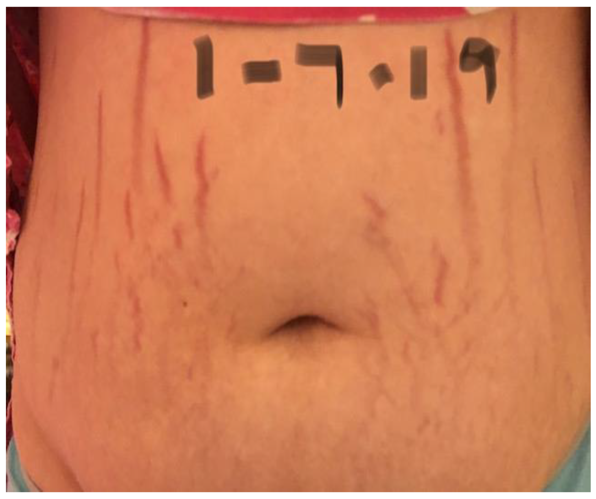
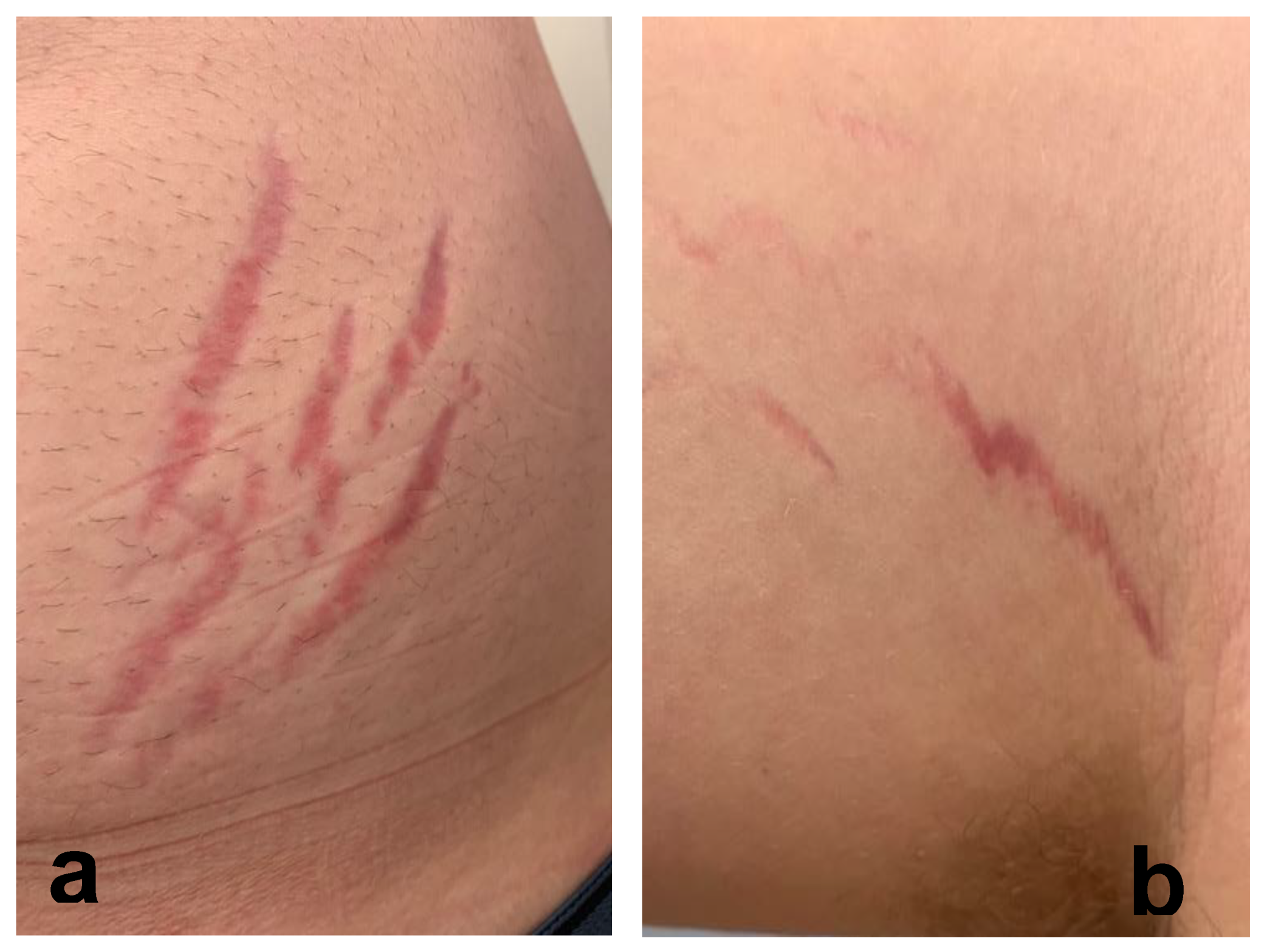
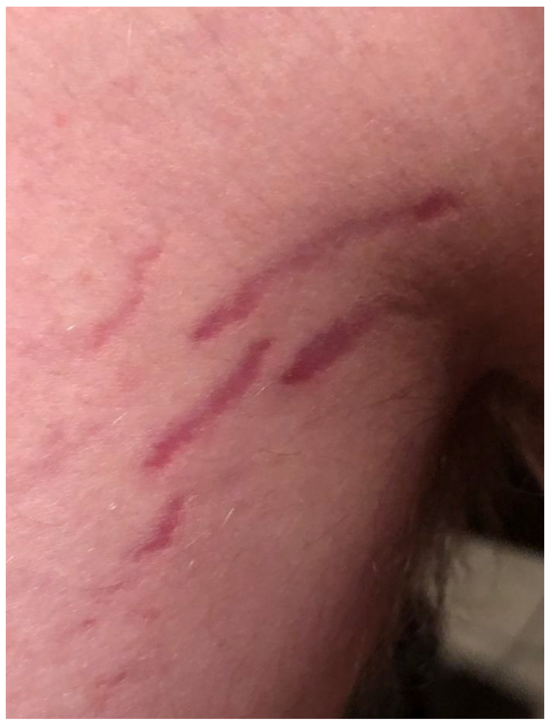
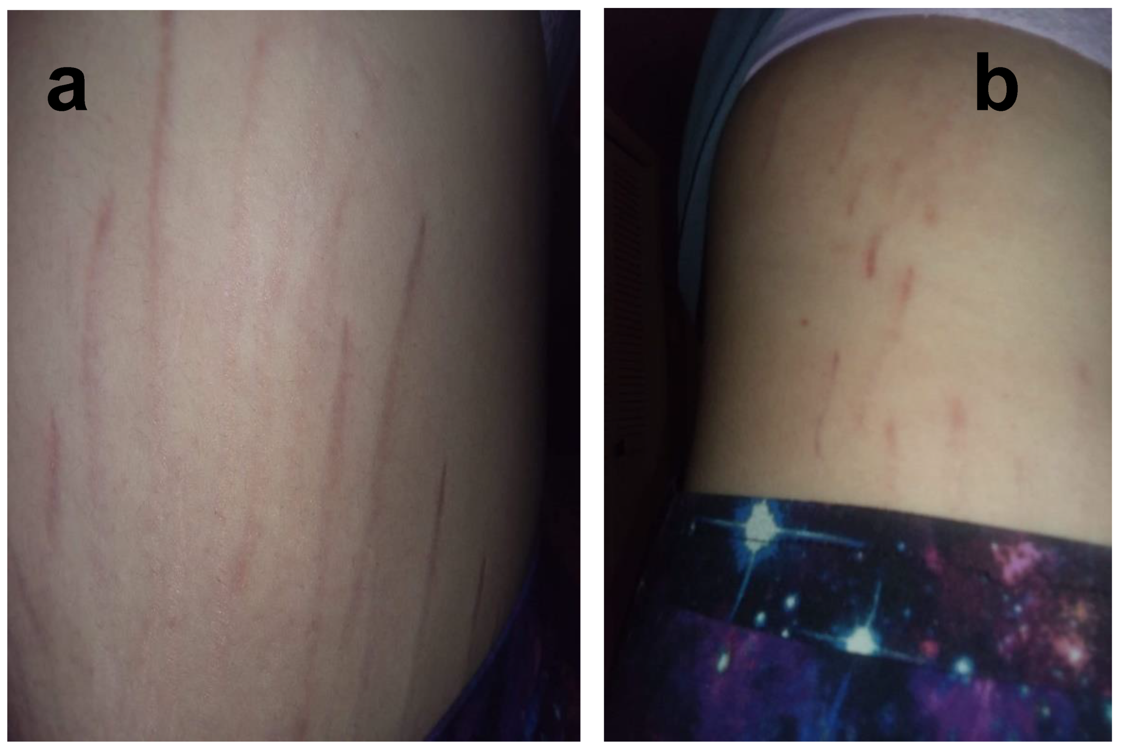
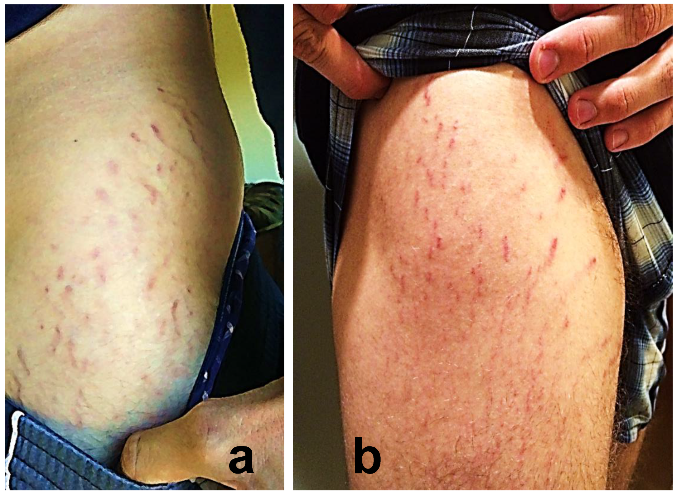
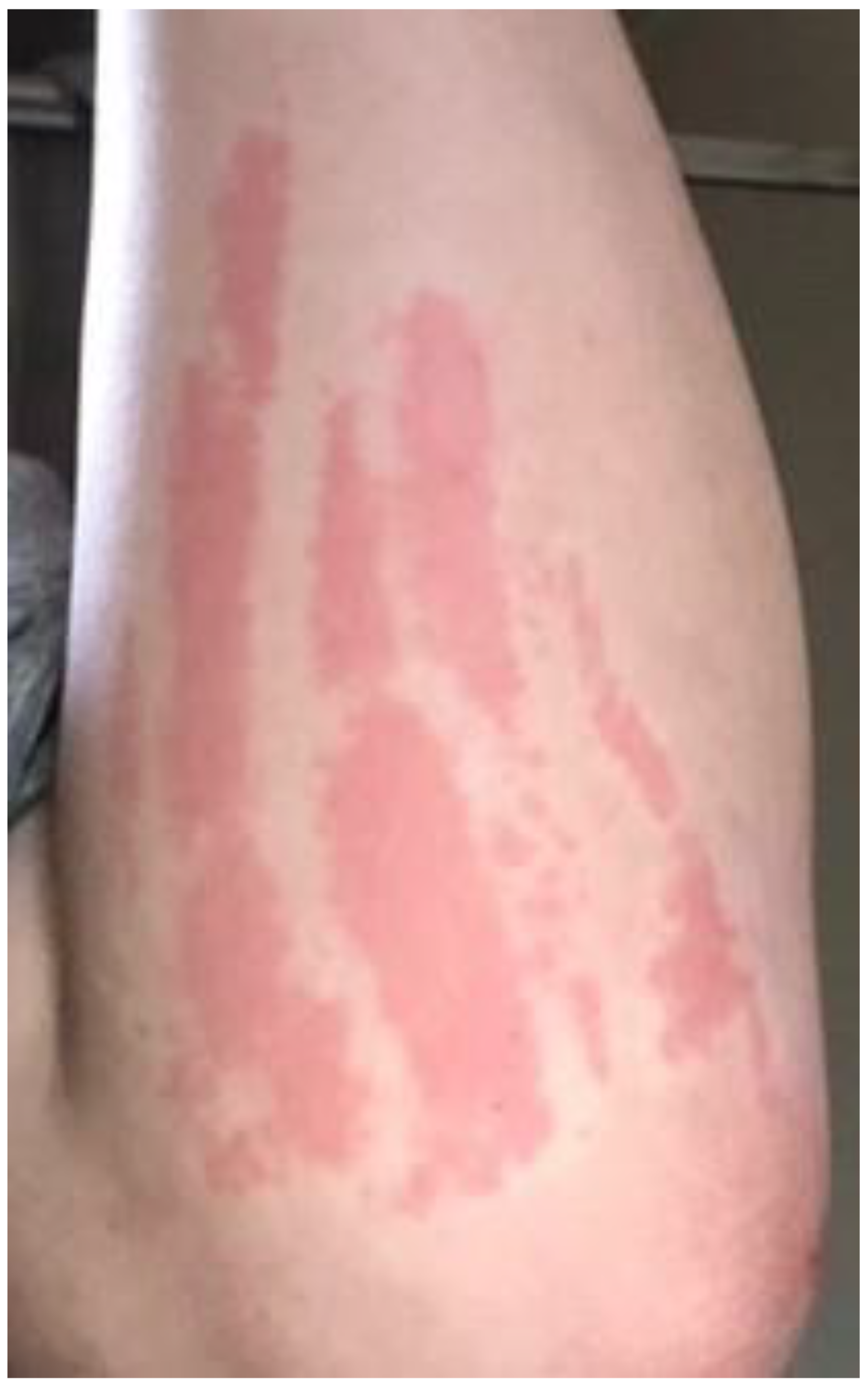
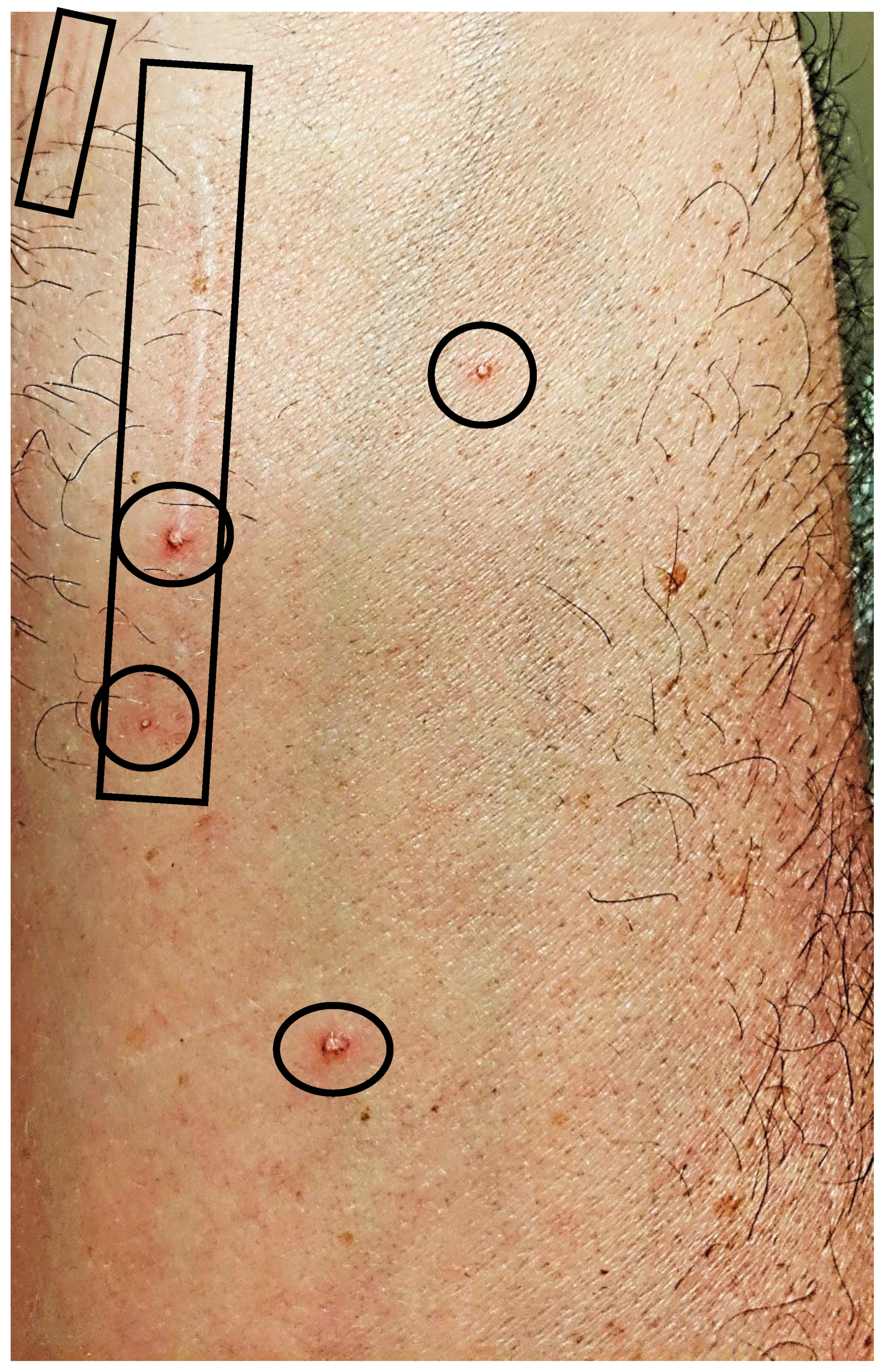
2.3. Reported Cutaneous Lesions

Twenty-four of 29 participants (83%) reported cutaneous lesions during their illness. Ten participants had only diffuse skin eruptions, 2 only linear lesions, and 12 had both diffuse skin eruptions and linear lesions. Linear lesions were characteristically serpiginous pink-to-red patches and/or depressed or slightly elevated plaques (Figure 1, Figure 2, Figure 3, Figure 4, Figure 5, Figure 6 and Figure 7). Seven participants had urticarial papules and plaques with dermatographism (Figure 8). A veterinarian, scratched by a cat, developed recurrent, small, discrete pustules around the scar (Figure 9). Linear lesions occurred in five males, all of whom were 14 or 15 years old, and in four females after puberty, who ranged in age from 14 to 17 years old. When questioned about risk factors for striae distensae, 22 of 24 successfully contacted participants reported no bodybuilding activities; one male reported a rapid weight gain, and five participants reported prior treatment with prednisone, four of whom were treated for one month or less. Marfans disease and Cushing’s syndrome were not listed as diagnoses on the questionnaire; however, one woman reported testing negative for Cushing’s syndrome. Two women reported a diagnosis of Hashimoto’s thyroiditis. Linear lesions were not visualized in the photographs provided from three previously pregnant woman, all of whom reported rashes.

2.4. Bartonella spp. Seroprevalence and Blood Stream Infection

Based on the case definition, all 29 participants were Bartonella spp. indirect fluorescent assay (IFA) seroreactive and/or polymerase chain reaction (PCR)-positive (Table 2). Sixteen participants were both PCR-positive and IFA seroreactive to one or more Bartonella spp. antigens. Seven participants were Bartonella spp. seroreactive but PCR-negative, and six participants were PCR-positive but not seroreactive. For the 23 seroreactors, IFA titers were 1:64, 1:128, or 1:256 for 13, 4, and 6 participants, respectively.

2.5. ddPCR Results on Direct Blood Testing versus BAPGM Enrichment Blood Culture

Of the 21/29 ddPCR-positive participants, one was positive only from blood; 13 were positive only after BAPGM enrichment culture (7, 14, or 21 days); and 7 were positive from both blood and BAPGM enrichment culture (Table 3). The participant infected with B. koehlerae (Figure 9) was blood DNA extraction qPCR-positive and ddPCR-positive from one and two collection dates, respectively, but BAPGM enrichment blood culture DNA extractions were qPCR and ddPCR-negative for all three blood collection dates and all three incubation testing time points.
Overall, 22 participants were PCR-positive, four by quantitative polymerase chain reaction (qPCR) and 21 by droplet digital polymerase chain reaction (ddPCR). Samples that were qPCR/DNA sequence-positive exhibited a Ct value (calculated cycle number at which the PCR product crosses a threshold of detection) ranging from 35.8 to 41.2, indicating a very low bacteria gene load in blood or enrichment blood cultures for the four qPCR+ samples (these values represent between one and 0.05 bacteria gene target copy per microliter of sample). Samples positive by ddPCR also showed a very low bacterial gene load. The number of Bartonella-positive ddPCR droplets varied between one and three per sample. All qPCR and ddPCR (no droplets visualized)-negative controls remained negative throughout the study.
Based upon DNA sequencing, three participants were infected with B. henselae and one with Bartonella koehlerae (Table 2). Three of four qPCR-positive patients were ddPCR-positive from blood or BAPGM enrichment blood culture. qPCR DNA sequences were analyzed by BLAST. Three amplicons shared sequence identities of 109/110 bp (99.1%) with the B. henselae Houston I strain (GenBank CP020742) or 110/110 bp (100%) with the B. henselae San Antonio 2 (SA2) strain (GenBank AF369529). A longer sequence could be amplified from one of the three B. henselae qPCR-positives, which were 549/549-bp (100%) homologous with B. henselae SA2 (GenBank AF369529). The B. koehlerae amplicon shared 100% sequence identity (110/110 bp) with B. koehlerae (GenBank accession AF312490).

2.6. Animal and Vector Exposures

As reported in their medical surveys, the 29 participants included in the study described the following animal exposures: dogs n = 27, cats n = 22, pet birds n = 8, horses n = 7, pet reptiles n = 9, pet rabbits n = 11, pet rodents n = 16, cattle n = 5, goats n = 3, poultry n = 6, and swine n = 4. Pets slept in beds with 19/29 participants, and 23/29 participants reported allowing pets to lick their faces and hands. None of the animals to whom exposure was reported were tested for Bartonella spp. Reported known vector exposures included bed bugs n = 3, biting flies n = 17, fleas n = 23, lice n = 15, mosquitos n = 29, scabies mites n = 10, spiders n = 20, and ticks n = 26. None of the vectors to whom exposure was reported were tested for Bartonella spp.

3. Discussion

This case series describes 29 people with self-reported neuropsychiatric symptoms and evidence of Bartonella spp. exposure or infection, the majority of whom (83%) had cutaneous lesions accompanying their illness onset. Combining an enrichment blood culture with ddPCR substantially improved the detection of Bartonella spp. DNA in this study. Diagnostically, ddPCR is a powerful molecular technique that uses a water–oil emulsion technology driven by microfluidics and surfactant chemistry to massively partition samples into 15,000 to 20,000 1-ηL-sized droplets prior to performing DNA amplification [12]. DNA targets within the original sample are randomly localized within these droplets—after which, the amplification of DNA within each drop is recorded using fluorescently labeled probe detection. The number and fluorescent output for each droplet is read in a manner similar to flow cytometry, where each individual droplet is then identified as being positive or negative for the template (pathogen gene target) of interest.
The frequency and extent to which B. henselae or other Bartonella spp. can induce cutaneous lesions or neuropsychiatric symptoms remains unclear. However, there is increasing case-based data to support an association between Bartonella spp. and cutaneous lesions, including urticarial papules and plaques along with erythematous, serpiginous patches, and plaques, reported as BACL in this case series. Maculopapular rash [13,14], septal panniculitis [14,15], and vasculitis [16,17] have been reported previously in association with Bartonella spp. infections and cutaneous lesions.
In the context of chronicity, histopathology confirmed mild, nonspecific lymphoplasmacytic infiltrates in lesional cutaneous biopsies, consistent with a chronic antigenic immune response in two B. henselae case reports [7,8]. As depicted in Figure 1, Figure 2, Figure 3, Figure 4, Figure 5, Figure 6 and Figure 7, the cutaneous lesions we refer to as linear BACL are often vertical, can be narrow or very wide, and do not occur in areas such as the back, where rapid growth would typically contribute to horizontal striae. No participant with linear BACL in this case series reported an association with conditions with known associations with striae distensae, including pregnancy, rapid growth, weightlifting, Cushing’s syndrome, or Marfans disease. Linear BACL lesions did not develop in a 36-year-old veterinarian infected with B. henselae, despite treatment for environmental allergies/rash with prednisone for one year prior to study entry. However, in the context of medical complexity, she was examined by 11 medical specialists, including an allergist, dermatologist, and neurologist.
Previously, we proposed that dogs may serve as sentinels for human exposure to Bartonella spp. [18,19]. Additionally, dogs develop similar, if not identical, pathology when persistently infected with a Bartonella spp. [20]. In the context of a potential common exposure source, infection with B. henselae was documented in an elderly man with ulcerated nodular skin lesions and in his dog with histopathologically confirmed panniculitis [21]. Identical SA2 strain types of B. henselae DNA were amplified and sequenced from the man’s enrichment blood culture and from the dog’s panniculitis biopsy. Ulcerative skin lesions and B. henselae infection were also documented by PCR amplification and DNA sequencing in a beagle with vasculitis and superficial erosions of the ear tip [22]. These and other disease associations shared by dogs and humans suggest that a comparative infectious disease approach might enhance the clinical understanding of these stealth bacteria in a timelier manner [1,23].
Based upon questionnaire responses, animal and vector exposures were reported frequently by study participants. The primary mode of Bartonella spp. transmission is via arthropod vectors, including, but not limited to, fleas, lice, sandflies, and ticks [1,2,4]. Other modes of transmission include animal bites and scratches, blood transfusions, and needle sticks. As further illustrated by this study, veterinary workers and others with frequent exposure to arthropods and animals may be occupationally at risk for acquiring infection and, thus, a sentinel population to enhance disease understanding [24]. To facilitate the subsequent acquisition by blood-sucking arthropods, vector-borne organisms evolved to avoid immune elimination, to localize in cutaneous tissues, and to downregulate the local inflammatory response, thereby avoiding the induction of obvious skin lesions [24]. Despite evolutionary adaptations by vectors and vector-borne organisms, skin lesions occur in association with a variety of vector-borne diseases. Three somewhat prototypical examples of acute-onset cutaneous lesions induced by tick-transmitted vector-borne pathogens are: erythema chronicum migrans (ECM) associated with Borrelia burgdorferi infection [25], the maculopapular rash with Rocky Mountain spotted fever [26] caused by Rickettsia rickettsii and ehrlichiosis [26] caused by Ehrlichia chaffeensis and Ehrlichia ewingii. In contrast, acrodermatitis chronica atrophica (ACA), most commonly caused by Borrelia afzelii, occurs months to years after infection [27]. Since B. henselae and B. koehlerae are predominantly flea-borne pathogens, flea (and other arthropod) exposures should be a component of each patient’s dermatological and neuropsychiatric histories. In addition, when dermatologists consult with families experiencing flea infestations, Bartonella spp. testing should be considered if symptoms subsequently develop during the ensuing months or years.
Blood stream infection with Bartonella spp. has been recently reported in blood donors and other healthy individuals [28,29]. Thus, despite PCR documentation of Bartonella spp. DNA in blood or enrichment blood cultures from the majority (22/29) of the study participants, it is not possible to confirm causation for the neuropsychiatric symptoms or the cutaneous lesions. Controlled treatment trials are likely necessary to determine if or to what extent infections with Bartonella spp. contribute to the clinical presentations described in this case series. The resolution of skin lesions and neuropsychiatric symptoms with antimicrobial therapy, as described in the boy diagnosed with PANS [8], provided potential support for causation. Similar to previous studies [28,29,30], a subset (6/29 in this study) of blood stream PCR-positive individuals were not Bartonella spp. seroreactive. The reason for this discrepancy remains unclear. The detection of antibody reactivity only indicates exposure to one or more Bartonella spp.; however, seronegative infection is not unusual. Thus, serology, enrichment blood culture, and PCR should be used concurrently prior to and during the treatment of suspected Bartonelloses patients.
The limitations of this study included participant self-selection and self-reporting, as these factors may have positively biased the findings. Participant age, duration and severity of illness, and prior treatment regimens likely influenced the microbiological serology and enrichment blood culture/PCR results. Due to the individual’s concerns over neuropsychiatric symptoms, most participants did not consult a dermatologist; nonprofessional photographs were provided by participants or parents, and skin biopsies were not obtained for histopathology, Bartonella PCR, or immunohistochemical imaging of Bartonella organisms.
Based upon the limited case reports and this case series, it is imperative to design studies that will determine if, or to what extent, Bartonella spp. might contribute to concurrent cutaneous lesions in patients with neuropsychiatric symptoms. Mechanistically, could long-standing microvascular injury induced by this endotheliotropic bacteria result in chronic vascular inflammation, local mast cell activation, collagen injury, and the development of a spectrum of BACL [31,32]? As Bartonella spp. are predominantly vector-borne pathogens that can localize to the skin, dermatologists should assess vector and animal exposures in patients with unusual or unexplained cutaneous lesions and consider testing patients with BACL and neuropsychiatric symptoms for Bartonella spp. infection.

4. Materials and Methods

4.1. Study Enrollment

Between August 2018 and October 2019, participants experiencing neurological or neuropsychiatric symptoms were enrolled after contacting investigators requesting inclusion in a research study entitled “Detection of Bartonella Species in the blood of healthy and sick people.” A cross-sectional study was performed to determine the seroprevalence to six Bartonella species/genotypes. Bacteremia was concurrently assessed by means of a Bartonella alpha-Proteobacteria growth medium (BAPGM) (IPRL, NCSU, Raleigh, NC, USA) enrichment blood culture platform. Participants were considered to have Bartonella spp. exposure or infection if they were Bartonella spp. seroreactive at an IFA titer of ≥1:64, qPCR or ddPCR positive, or positive by more than one testing modality.

4.2. Questionnaire Data and Blood Specimen Collection

A standardized questionnaire, including demographic information, symptoms experienced, domestic and wild animal bites, scratches or exposures, and travel history, was completed. Exposure to, or bites by, different arthropods (lice, fleas, ticks, mites, bed bugs, and others) was recorded. This study was carried out in accordance with all relevant guidelines applying to human study participation. Most participants were enrolled after publication of the case report involving the boy diagnosed with PANS [8]. In the case of children, the parents requested enrollment and provided signed permission for testing. All participants (or parents) completed a detailed questionnaire used in previous collaborative studies from our laboratory [30].
Only participants with positive Bartonella spp. serological or molecular test results were included in the study. Participants/parents provided cutaneous lesion photographs for dermatologist review and written permission for publication. The dermatological description of cutaneous lesions was determined by the dermatologist author. Striae distensae risk factors were determined retrospectively via an email questionnaire provided to all participants reporting cutaneous lesions.
Blood was collected aseptically into ethylenediaminetetraacetic acid (EDTA) anti-coagulated and serum separator tubes for shipment to the Intracellular Pathogens Research Laboratory (IPRL), Comparative Medicine Institute, College of Veterinary Medicine, North Carolina State University (NCSU) for Bartonella testing. With one exception (two collection dates), each participant provided three blood and serum specimens collected on alternate days to increase the Bartonella spp. enrichment blood culture/PCR sensitivity [33]. Approximately 10–12 mL of blood (5–6 mL in EDTA anti-coagulant tubes and 5–6 mL in serum separator tubes) was collected every other day for three time points at the time of enrollment. Aseptic blood collection, including chlorhexidine decontamination of the skin, was performed by an experienced nurse.

4.3. Bartonella IFA Serological Testing

As described in previous studies from our research group [11,30,34], each participant was tested using six IFA assays, each representing a unique Bartonella species or subspecies. Bartonella vinsonii subsp. berkhoffii, B. henselae, B. koehlerae, and B. quintana antibodies were determined in the IPRL at NCSU (Raleigh, NC, USA) using cell culture-grown bacteria as antigens and following standard IFA techniques [30,34]. Canine isolates of B. vinsonii subsp. berkhoffii genotype I (NCSU 93CO-01 Tumbleweed, ATCC type strain #51672), B. vinsonii subsp. berkhoffii genotype II (NCSU 95CO-08, Winnie), and B. vinsonii subsp. berkhoffii genotype III (NCSU 06CO-01 Klara); feline isolates of B. henselae SA2 strain (NCSU 95FO-099, Missy), B. koehlerae (NCSU 09FO-01, Trillium), and B. quintana (NCU11-MO-01 Monkey origin) were passed from agar plate-grown cultures into Bartonella-permissive cell lines, i.e., the DH82 (a canine monocytoid) cell line for strains B. henselae SA2, B. quintana, B. vinsonii subsp. berkhoffii I, and B. koehlerae and Vero cells (a mammalian fibroblast cell line) for B. vinsonii subsp. berkhoffii II and III to obtain antigens for IFA testing. For each antigen, heavily infected cell cultures were spotted onto 30-well Teflon-coated slides (Cell-Line/Thermo Scientific, Waltham, MA, USA), air-dried, acetone-fixed, and stored frozen. Fluorescein-conjugated goat anti-human immunoglobulin G (IgG) (Cappel, ICN, Costa Mesa, CA, USA) was used to detect bacteria within cells using a fluorescent microscope (Carl Zeiss Microscopy, LLC, Thornwood, NY, USA). Serum samples diluted in a phosphate-buffered saline (PBS) solution containing normal goat serum, Tween-20, and powdered nonfat dry milk to block nonspecific antigen-binding sites were tested with two-fold dilutions out to 1:8192. To avoid confusion with possible nonspecific binding found at low dilutions, a cutoff of 1:64 was selected as a seroreactive antibody titer.

4.4. Bartonella Enrichment Blood Culture/PCR Testing

Bartonella alpha-Proteobacteria growth medium (BAPGM) enrichment blood culture/PCR was performed as previously described. Briefly, qPCR and ddPCR amplifications were performed by targeting the 16S-23S intergenic transcribed spacer (ITS) region [30,34]. For qPCR products, amplicon identity was confirmed by DNA sequencing in a commercial laboratory (Genewiz, Inc, Research Triangle Park, NC, USA). Due to instrument design limitations, digital PCR droplets are not able to be sequenced [12]. Positive and negative control samples were consistently included when testing using both qPCR and ddPCR amplification modalities. Positive samples had bacterial loads equivalent to 1, 0.5, and 0.05 gene target copies per microliter. Negative controls consisted of DNA extracted from naïve human blood and the same molecular grade water used for all PCR preparations.

4.5. Statistics

Summary statistics were calculated for participant age and illness duration; distributions for each of these measures were examined visually for normality.

4.6. Ethics Approval and Consent to Participate

In accordance with the 1964 Helsinki Declaration, written informed consent to be a participant and to be part of a publication if printed was obtained from the study participant or parent (in the case of a minor). This study was carried out in accordance with all relevant guidelines applying to human study participation. No nonhuman animals were tested. The protocol was approved by the North Carolina State University (NCSU) Sponsored Programs and Regulatory Compliance, Institutional Review Board (IRB) (Approval IRB#1960). Specimen handling was performed following regional and national guidelines and regulations.

Author Contributions

Conceptualization: E.B.B. and R.G.M.; methodology: R.G.M. and J.M.B.; validation: E.B.B. and R.G.M.; formal analysis: E.L. and R.G.M.; investigation: J.M.B. and R.G.M.; resources: E.B.B. and R.G.M.; data curation: J.M.B., E.L., and R.G.M.; writing—original draft preparation: E.B.B., J.M.B., R.M., and P.R.; writing—review and editing: E.B.B., J.M.B., and R.G.M.; visualization: E.B.B.; supervision: E.B.B.; project administration: E.B.B.; funding acquisition: E.B.B.; and dermatology review: P.R. All authors have read and agree to the published version of the manuscript.

Funding

This research was supported in part by the State of North Carolina and donations to the Bartonella/Vector-Borne Diseases Research Fund at the College of Veterinary Medicine, North Carolina State University.

Conflicts of Interest

In conjunction with S. Sontakke and North Carolina State University, Edward B. Breitschwerdt holds US Patent No. 7115385: Media and Methods for Cultivation of Microorganisms, which was issued on 3 October 2006. He is a cofounder, shareholder, and Chief Scientific Officer for Galaxy Diagnostics, a company that provides advanced diagnostic testing for the detection of Bartonella spp. infections. R. Maggi is the Chief Technical Officer for Galaxy Diagnostics. The remaining authors declare no conflicts of interest. The funders had no role in the design of the study; in the collection, analyses, or data interpretation; in the writing of the manuscript; or in the decision to publish the results. This study was presented in part at the International Lyme and Associated Diseases (ILADS) Virtual Conference, Washington DC, 10–13 September 2020 and at the Companion Animal Vector Borne Disease (CVBD) Annual World Forum, Virtual International Conference, 10–12 November 2020.

Availability of Data and Materials

Data supporting the conclusions of the authors are included in the article. To assure participant confidentiality, please contact E.B.B. for questions relative to the raw data.

References

  1. Regier, Y.; Rourke, F.O.; Kempf, V.A. Bartonella spp.—A chance to establish one health concepts in veterinary and human medicine. Parasit Vectors 2016, 9, 261. [Google Scholar] [CrossRef] [Green Version]
  2. Cheslock, M.A.; Embers, M.E. Human bartonellosis: An underappreciated public health problem? Trop. Med. Infect. Dis. 2019, 19, 2. [Google Scholar] [CrossRef] [Green Version]
  3. Landau, M.; Kletter, Y.; Avidor, B.; Ephrat, G.; Ephros, M.; Brenner, S.; Giladi, M. Unusual eruption as a presenting symptom of cat scratch disease. J. Am. Acad. Dermatol. 1999, 41, 833–836. [Google Scholar] [CrossRef]
  4. Álvarez-Fernández, A.; Breitschwerdt, E.B.; Solano-Gallego, L. Bartonella infections in cats and dogs including zoonotic aspects. Parasit Vectors 2018, 11, 624. [Google Scholar] [CrossRef] [PubMed]
  5. Fried, M.D. Abstract Bartonella henselae is associated with heartburn, abdominal pain, skin rash, mesenteric adenitis, gastritis and duodenitis. In Proceedings of the North American Society of Pediatric Gastroenterology, Hepatology, and Nutrition Annual Meeting, San Antonio, TX, USA, 24–27 October 2002. [Google Scholar]
  6. Schaller, J.L. The Diagnosis Treatment and Prevention of Bartonella; Hope Academic Press: Tampa, FL, USA, 2008. [Google Scholar]
  7. Maggi, R.G.; Ericson, M.; Mascarelli, P.E.; Bradley, J.M.; Breitschwerdt, E.B. Bartonella henselae bacteremia in a mother and son exposed to ticks in The Netherlands. Parasites Vectors 2013, 6, 101. [Google Scholar] [CrossRef] [PubMed] [Green Version]
  8. Breitschwerdt, E.B.; Greenberg, R.; Maggi, R.G.; Lewis, A.; Bradley, J.M. Bartonella henselae infection in a boy with Pediatric Acute-onset Neuropsychiatric Syndrome (PANS). J. Nerv. Syst. Dis. 2019, 11, 1–8. [Google Scholar]
  9. Azar, M.; Breitschwerdt, E.; Bemis, L.; Greenberg, R.; Mozayeni, R.; Dencklau, J.; Ericson, M. Imaging analysis of Bartonella species in the skin using single-photon and multi-photon (second harmonic generation) laser scanning microscopy. Clin. Case Rep. 2020, 1–7. [Google Scholar] [CrossRef]
  10. Boozalis, E.; Grossberg, A.L.; Puttgen, K.B.; Heath, C.R.; Cohen, B.A. Demographic characteristics of teenage boys with horizontal striae distensae of the lower back. Pediatr. Dermatol. 2018, 35, 59–63. [Google Scholar] [CrossRef]
  11. Breitschwerdt, E.B.; Maggi, R.G.; Quach, C.; Bradley, J.M. Bartonella spp. bloodstream infection in a canadian family. Vector Borne Zoonotic Dis. 2019, 19, 234–241. [Google Scholar] [CrossRef]
  12. Maggi, R.G.; Richardson, T.; Breitschwerdt, E.B.; Miller, J.C. Development and validation of a droplet digital PCR assay for the detection and quantification of Bartonella species in human clinical samples. J. Microbiol. Methods 2020, 176, e106022. [Google Scholar] [CrossRef]
  13. Maritsi, D.N.; Zarganis, D.; Metaxa, Z.; Papaioannou, G.; Vartzelis, G. Bartonella henselae infection: An uncommon mimicker of autoimmune disease. Case Rep. Pediatr. 2013, 2013, 726826. [Google Scholar] [CrossRef] [PubMed]
  14. Im, J.H.; Kwon, H.Y.; Baek, J.; Durey, A.; Lee, S.M.; Park, Y.K.; Kang, J.S.; Chung, M.H.; Lee, J.S. Serologic study of Bartonella henselae in patients with acute undifferentiated febrile illness in Korea. Vector Borne Zoonotic Dis. 2018, 18, 291–296. [Google Scholar] [CrossRef] [PubMed] [Green Version]
  15. Safdar, A.; McEvoy, P.L.; Burns, R.G.; Perfect, J.R. Clinical microbiological case: Severe relapsing septal panniculitis in a healthy man from the southeastern USA. Clin. Microbiol. Infect. 2002, 8, 801–802. [Google Scholar] [CrossRef] [PubMed] [Green Version]
  16. Schmoor, P.; Darie, H.; Maccari, F.; Gros, P.; Millet, P. Cutaneous vasculitis disclosing cat-scratch disease. Ann. Dermatol. Venereol. 1998, 125, 894–896. [Google Scholar] [PubMed]
  17. Cozzani, E.; Cinotti, E.; Ameri, P.; Sofia, A.; Murialdo, G.; Parodi, A. Onset of cutaneous vasculitis and exacerbation of IgA nephropathy after Bartonella henselae infection. Clin. Dermatol. 2011, 37, 238–240. [Google Scholar] [CrossRef] [PubMed]
  18. Lashnits, E.; Correa, M.; Hegarty, B.C.; Birkenheuer, A.; Breitschwerdt, E.B. Bartonella seroepidemiology in dogs from North America, 2008–2014. J. Vet. Intern. Med. 2018, 32, 222–231. [Google Scholar] [CrossRef] [PubMed] [Green Version]
  19. Lashnits, E.W.; Dawson, D.E.; Breitschwerdt, E.; Lanzas, C. Ecological and socioeconomic factors associated with Bartonella henselae exposure in dogs tested for vector-borne diseases in North Carolina. Vector Borne Zoonotic Dis. 2019, 19, 582–595. [Google Scholar] [CrossRef]
  20. Breitschwerdt, E.B.; Linder, K.L.; Day, M.J.; Maggi, R.G.; Chomel, B.B.; Kempf, V.A.J. Koch’s postulates and the pathogenesis of comparative infectious disease causation associated with Bartonella species. J. Comp. Pathol. 2013, 1, 1–11. [Google Scholar] [CrossRef]
  21. Rossi, M.A.; Balakrishnan, N.; Linder, K.E.; Messa, J.B.; Breitschwerdt, E.B. Concurrent Bartonella henselae infection in a dog with panniculitis and owner with ulcerated nodular skin lesions. Vet. Dermatol. 2015, 26, 60–63. [Google Scholar] [CrossRef]
  22. Southern, B.L.; Neupane, P.; Ericson, M.E.; Dencklau, J.C.; Linder, K.E.; Bradley, J.M.; McKeon, G.P.; Long, C.T.; Breitschwerdt, E.B. Bartonella henselae in a dog with ear tip vasculitis. Vet. Dermatol. 2018, 29, 537-e180. [Google Scholar] [CrossRef]
  23. Breitschwerdt, E.B. Bartonellosis: One health perspectives for an emerging infectious disease. ILAR J. 2014, 55, 46–58. [Google Scholar] [CrossRef] [PubMed]
  24. Mosbacher, M.E.; Klotz, S.; Klotz, J.; Pinnas, J.L. Bartonella henselae and the potential for arthropod vector borne transmission. Vector Borne Zoonotic Dis. 2011, 11, 471–477. [Google Scholar] [CrossRef] [PubMed]
  25. Wormser, G.P.; Brady, K.C.; Cho, M.S.; Scavarda, C.A.; McKenna, D. Efficacy of a 14-day course of amoxicillin for patients with erythema migrans. Diagn. Microbiol. Infect. Dis. 2019, 94, 192–194. [Google Scholar] [CrossRef] [PubMed]
  26. Muzumdar, S.; Rothe, M.J.; Grant-Kels, J.M. The rash with maculopapules and fever in adults. Clin. Dermatol. 2019, 37, 109–118. [Google Scholar] [CrossRef] [PubMed]
  27. Maraspin, V.; Mrvič, T.; Ružić-Sabljić, E.; Jurčić, V.; Strle, F. Acrodermatitis chronica atrophicans in children: Report on two cases and review of the literature. Ticks Tick Borne Dis. 2019, 10, 180–185. [Google Scholar] [CrossRef] [PubMed]
  28. Maggi, R.G.; Mascarelli, P.E.; Pultorak, E.L.; Hegarty, B.C.; Bradley, J.M.; Mozayeni, R.B.; Breitschwerdt, E.B. Bartonella spp. bacteremia in high-risk immunocompetent patients. Diagn. Microbiol. Infect. Dis. 2011, 71, 430–437. [Google Scholar] [CrossRef]
  29. Diniz, P.P.; Velho, P.E.; Pitassi, L.H.; Drummond, M.R.; Lania, B.G.; Barjas-Castro, M.L.; Sowy, S.; Breitschwerdt, E.B.; Scorpio, D.G. Risk factors for Bartonella species infection in blood donors from Southeast Brazil. PLoS Negl. Trop. Dis. 2016, 10, e0004509. [Google Scholar] [CrossRef] [Green Version]
  30. Oteo, J.A.; Maggi, R.; Portillo, A.; Bradley, J.; García-Álvarez, L.; San-Martín, M.; Roura, X.; Breitschwerdt, E. Prevalence of Bartonella spp. by culture, PCR and serology, in veterinary personnel from Spain. Parasit Vectors 2017, 10, 553. [Google Scholar] [CrossRef] [Green Version]
  31. Vaca, D.J.; Thibau, A.; Schutz, M.; Kraiczy, P.; Happonen, L.; Malmstrom, J.; Kempf, V.A.J. Interaction with the host: The role of fibronectin and extracellular matrix proteins in the adhesion of Gram-negative bacteria. Med. Microbiol. Immunol. 2020, 209, 277–299. [Google Scholar] [CrossRef] [Green Version]
  32. Da Silva, E.Z.M.; Jamur, M.C.; Oliver, C. Mast cell function: A new vision of an old cell. J. Histochem. Cytochem. 2014, 62, 698–738. [Google Scholar] [CrossRef]
  33. Pultorak, E.L.; Maggi, R.G.; Mascarelli, P.E.; Breitschwerdt, E.B. Serial testing from a three-day collection period using the BAPGM platform may enhance the sensitivity of Bartonella spp. detection in bacteremic human patients. J. Clin. Microbiol 2013, 57, 1673–1677. [Google Scholar] [CrossRef] [PubMed] [Green Version]
  34. Lantos, P.M.; Maggi, R.G.; Ferguson, B.; Varkey, J.; Park, L.P.; Breitschwerdt, E.B.; Woods, C.W. Detection of Bartonella species in the blood of veterinarians and veterinary technicians: A newly recognized occupational hazard. Vector Borne Zoonotic Dis. 2015, 14, 563–570. [Google Scholar] [CrossRef] [PubMed] [Green Version]
Figure 1. Bartonella-Associated Cutaneous Lesions. Fifteen-year-old male medically disabled student from North Carolina, with neuropsychiatric symptoms of six months’ duration. Serpiginous, vertical red lesions on the left hip and buttocks (a) and the right hip and upper thigh (b). The participant was positive for Bartonella infection by droplet digital PCR (ddPCR) enrichment blood culture (Bartonella henselae indirect fluorescent assay (IFA) serology titer 1:64). He reported no history of steroid use, bodybuilding, or rapid weight gain. Photograph of lesions provided by participant’s parents.
Pathogens 09 01023 g001
Figure 2. Bartonella-Associated Cutaneous Lesions. Sixteen-year-old female student from North Carolina, with neuropsychiatric symptoms of six years’ duration. Similar, but thinner and paler, but more serpiginous, vertical red lesions were photographed in November 2017. Positive for Bartonella infection by ddPCR enrichment blood culture (Bartonella henselae and Bartonella koehlerae IFA titers of 1:128 and 1:256, respectively). She reported no history of pregnancy or rapid weight gain. Photograph of abdominal lesions provided by participant’s parents.
Pathogens 09 01023 g002
Figure 3. Bartonella-Associated Cutaneous Lesions. Seventeen-year-old male medically disabled student from North Carolina, with neuropsychiatric symptoms of one-year duration. Serpiginous, vertical red lesions on the left lower abdomen/upper groin (a) and the right axilla (b). Positive for Bartonella infection by ddPCR enrichment blood culture (Bartonella IFA serology-negative). He reported no history of steroid use, bodybuilding, or rapid weight gain. Photograph of lower abdomen, upper groin area and right axilla provided by participant’s parents.
Pathogens 09 01023 g003
Figure 4. Bartonella-Associated Cutaneous Lesions. Twenty-year-old male medically disabled student from Virginia, with neuropsychiatric symptoms of four years’ duration. Serpiginous, vertical red lesions on the left hip (a) and on the left axilla (b). Positive for Bartonella infection by ddPCR enrichment blood culture (Bartonella IFA serology-negative). He reported no history of steroid use, bodybuilding, or rapid weight gain. Photographs of the hip and axilla provided by the study participant.
Pathogens 09 01023 g004
Figure 5. Bartonella-Associated Cutaneous Lesions. Twenty-one-year-old male landscaper from Connecticut, with neuropsychiatric symptoms of three years’ duration. Serpiginous, vertical red lesions on the right axilla. Positive for Bartonella infection by ddPCR enrichment blood culture (Bartonella henselae IFA titer 1:64). He reported no history of steroid use, bodybuilding, or rapid weight gain. Photograph of left axilla provided by the study participant.
Pathogens 09 01023 g005
Figure 6. Bartonella-Associated Cutaneous Lesions. Twenty-one-year-old female veterinary assistant from Louisiana, with neuropsychiatric symptoms of 2 years’ duration. Serpiginous, vertical red lesions on the left abdomen (a) and the right upper side of the abdomen (b). Positive for Bartonella infection by ddPCR from direct EDTA blood sample and enrichment blood culture (Bartonella henselae and Bartonella koehlerae IFA titers of 1:256 and 1:64, respectively). Photo provided by the study participant.
Pathogens 09 01023 g006
Figure 7. Bartonella-Associated Cutaneous Lesions. Twenty-two-year-old male medically disabled student from Minnesota, with neuropsychiatric symptoms of four years’ duration. Serpiginous, vertical red lesions on the left hip and buttocks (a) and the right upper thigh (b). Participant was positive for Bartonella infection by ddPCR enrichment blood culture (Bartonella IFA serology-negative). He reported no history of steroid use, bodybuilding, or rapid weight gain. Photographs of lesions provided by participant’s parents.
Pathogens 09 01023 g007
Figure 8. Bartonella-Associated Cutaneous Lesions. Twenty-one-year-old female medically disabled student from Mississippi, with neuropsychiatric symptoms of 13 years’ duration. Photograph of left forearm showing urticarial papules and plaques with dermatographism. Positive for Bartonella infection by ddPCR enrichment blood culture (Bartonella IFA serology-negative). Photo provided by the study participant.
Pathogens 09 01023 g008
Figure 9. Bartonella-Associated Cutaneous Lesions. Thirty-four-year-old male veterinarian/rancher from Texas, with neuropsychiatric symptoms of six months’ duration. Photograph of forearm showing healed cat scratches (rectangles) (one year previously) and adjacent, recurrent pustular lesions (circles) that are not perifollicular. Positive for Bartonella koehlerae infection by qPCR and ddPCR (Bartonella koehlerae IFA titer 1:64). Photo provided by the study participant.
Pathogens 09 01023 g009
Table 1. Symptoms checked off by participants on the Institutional Review Board-approved questionnaire, and self-reported conditions recorded by participants under the categories of prior diagnoses or treatments. Reported symptoms/conditions were divided into neuropsychological versus non-neuropsychological by the authors.
Neuropsychological SymptomsParticipants ReportingNon-Neuropsychological SymptomsParticipants Reporting
Sleep Disorders ^27Fatigue ^26
Mental Confusion ^24Ocular Involvement20
Irritability/Rage ^23 Eye Pain ^15
Anxiety/Panic Attacks ^22 Blurred Vision ^15
Depression ^21 Light Sensitivity *4
Headache/Migraine ^21 Floaters/Flashes *2
Tremors ^12Joint/Back/Neck Pain ^20
Hallucinations ^7GI Issues *17
Institutionalized (Psych) *2 Bowel Dysfunction ^11
Postural Orthostatic Tachycardia (POTS) *5 Diarrhea ^8
Seizures ^4 Abdominal Pain * 3
Schizophrenia *2 Vomiting/Nausea ^4
Suicidal *2 IBS *1
Tourettes *2 Bloating *1
Paralysis ^2Muscle Pain ^15
Mast Cell Activation Syndrome *2Respiratory ^15
MS-Like diagnosis *1Muscle Weakness ^14
Sudden Onset OCD *1Numbness ^13
PANS *1Tachycardia (Not POTS) ^10
Psychosis *1Tendonitis ^8
Stuttering *1Syncope ^6
Dental Issues ^6
 Jaw Pain *5
 Gingival Recession *2
 Mouth Ulcers *1
Fever ^ of Unknown Origin *5
Ehrlos Danlos Syndrome *3
Cardiac (not reported elsewhere) *2
^ Symptoms checked off by participants on the Institutional Review Board-approved questionnaire. * Participants provided written responses in the medical survey questionnaire indicating prior diagnoses and/or conditions for which treatments were administered. GI: gastrointestinal, IBS: irritable bowel syndrome, OCD: obsessive-compulsive disorder, PANS: Pediatric Acute-onset Neuropsychiatric Syndrome, and MS: multiple sclerosis.
Table 2. Bartonella spp. serology and Bartonella alpha proteobacteria enrichment blood culture qPCR and ddPCR results for 29 individuals with self-reported neuropsychiatric illnesses.
Reciprocal IFA Antibody Titers to:Direct Blood PCRBAPGM Enrichment Blood Culture/PCR
Study No:Bartonella vinsonii berkhoffii Type IBartonella vinsonii berkhoffii Type IIBartonella vinsonii berkhoffii Type IIIBartonella henselae San Antonio 2Bartonella koehleraeBartonella quintanaqPCR *ddPCRqPCR *ddPCR
1326432323232NegNegNegNeg
23232643264128NegPosNegPos
3323232643216NegNegNegPos
4<1632326416<16NegNegNegNeg
525664321283216NegNegNegPos
61281286464128128NegNegNegPos
7<16646464<1664NegNegNegNeg
816<16<16<16<16<16NegNegNegPos
9<16<1616<16<1616NegPosNegPos
10<16<16<1616<16<16NegNegNegPos
11<1664642566432NegPosNegPos
12<16163264<1632NegNegNegPos
13<16<16<1632<16<16NegNegNegPos
14<16<16<16<16<16<16NegNegNegPos
156464641286464NegNegBhNeg
1625612825612864128NegNegNegPos
17<16323264<1632BhPosNegPos
18163232646464NegNegNegPos
19323264326432BkPosNegNeg
2016163232<1664NegNegNegNeg
21321632643232NegNegNegPos
22<1616<1616<1616NegPosNegPos
23163216641616NegPosBhPos
242566464646464NegPosNegPos
25<16<16326416<16NegNegNegNeg
26<16<1632643216NegNegNegNeg
27<16321612832<16NegNegNegNeg
28128128641286464NegNegNegPos
29<16161616<1616NegNegNegPos
* Bartonella species confirmed by DNA sequence analyses. IFA: indirect fluorescent assay, ddPCR: droplet digital PCR, and BAPGM: Bartonella alpha-Proteobacteria growth medium. Bold fonts were used to emphasize positive reults.
Table 3. Bartonella ddPCR results generated using the Bartonella alpha-Proteobacteria growth medium (BAPGM) enrichment blood culture platform. All but one participant provided three blood samples obtained on alternate days for enrichment blood culture PCR testing. DNA was extracted from blood and from BAPGM enrichment blood cultures at 7, 14, and 21 days.
ParticipantSource of DNA Tested by ddPCR
BloodBAPGM Incubation Day 7BAPGM Incubation Day 14BAPGM Incubation Day 21
1NegNegNegNeg
2PosNegNegPos
3NegNegPosNeg
4NegNegNegNeg
5NegNegNegPos
6NegNegNegPos
7NegNegNegNeg
8NegPosNegNeg
9PosPosPosPos
10NegNegNegPos
11PosNegPosNeg
12NegNegNegPos
13NegNegNegPos
14NegNegNegPos
15NegNegNegNeg
16NegPosNegNeg
17PosPosPosNeg
18NegNegNegPos
19PosNegNegNeg
20NegNegNegNeg
21NegPosNegNeg
22PosNegNegPos
23PosPosPosPos
24PosPosNegNeg
25NegNegNegNeg
26NegNegNegNeg
27NegNegNegNeg
28NegNegPosPos
29NegNegNegPos
Bold fonts were used to emphasize positive reults.
Publisher’s Note: MDPI stays neutral with regard to jurisdictional claims in published maps and institutional affiliations.

