Article

Multilocus Genotyping Reveals New Molecular Markers for Differentiating Distinct Genetic Lineages among “Candidatus Phytoplasma Solani” Strains Associated with Grapevine Bois Noir

1
Department of Agricultural and Environmental Sciences, Production, Landscape, Agroenergy, University of Milan, 20133 Milano, Italy
2
Molecular Plant Pathology Laboratory, USDA-Agriculture Research Service, Beltsville, MD 20705, USA
3
Department of Agricultural, Food and Environmental Sciences, Marche Polytechnic University, 60131 Ancona, Italy
4
Department of Agriculture, Food and Environment, University of Pisa, 56124 Pisa, Italy
5
Department for Plant and Environment Protection, Faculty of Agriculture, Goce Delcev University, Štip 2000, North Macedonia
6
Department of Biotechnology, University of Verona, 37134 Verona, Italy
7
National Research Centre, Pests and Plant Protection Department, Agricultural & Biological Research Division, Giza 12622, Egypt
8
Centro di Ricerca e Sperimentazione in Agricoltura Basile Caramia, 70010 Locorotondo, Italy
9
Department of Biological and Environmental Sciences and Technologies, University of Salento, 73100 Lecce, Italy
10
Institute for Sustainable Plant Protection, National Research Council (IPSP-CNR), Strada delle Cacce 73, 10135 Turin, Italy
*
Author to whom correspondence should be addressed.
Deceased.
Pathogens 2020, 9(11), 970; https://doi.org/10.3390/pathogens9110970
Submission received: 30 October 2020 / Revised: 18 November 2020 / Accepted: 19 November 2020 / Published: 21 November 2020

Abstract

:
Grapevine Bois noir (BN) is associated with infection by “Candidatus Phytoplasma solani” (CaPsol). In this study, an array of CaPsol strains was identified from 142 symptomatic grapevines in vineyards of northern, central, and southern Italy and North Macedonia. Molecular typing of the CaPsol strains was carried out by analysis of genes encoding 16S rRNA and translation elongation factor EF-Tu, as well as eight other previously uncharacterized genomic fragments. Strains of tuf-type a and b were found to be differentially distributed in the examined geographic regions in correlation with the prevalence of nettle and bindweed. Two sequence variants were identified in each of the four genomic segments harboring hlyC, cbiQ-glyA, trxA-truB-rsuA, and rplS-tyrS-csdB, respectively. Fifteen CaPsol lineages were identified based on distinct combinations of sequence variations within these genetic loci. Each CaPsol lineage exhibited a unique collective restriction fragment length polymorphism (RFLP) pattern and differed from each other in geographic distribution, probably in relation to the diverse ecological complexity of vineyards and their surroundings. This RFLP-based typing method could be a useful tool for investigating the ecology of CaPsol and the epidemiology of its associated diseases. Phylogenetic analyses highlighted that the sequence variants of the gene hlyC, which encodes a hemolysin III-like protein, separated into two clusters consistent with the separation of two distinct lineages on the basis of tufB gene sequences. Alignments of deduced full protein sequences of elongation factor-Tu (tufB gene) and hemolysin III-like protein (hlyC gene) revealed the presence of critical amino acid substitutions distinguishing CaPsol strains of tuf-type a and b. Findings from the present study provide new insights into the genetic diversity and ecology of CaPsol populations in vineyards.

1. Introduction

Bois noir (BN), a grapevine disease associated with “Candidatus Phytoplasma solani” (CaPsol) infection, causes typical grapevine yellows (GY) symptoms and results in important crop losses in the majority of vine-growing European countries, in the Middle East, and in South America [1,2]. Due to the involvement of multiple insect vectors and plant hosts, the biological cycle of CaPsol is extremely complex [3,4,5,6,7,8,9,10], hindering the development of control strategies for effective control of BN epidemics [11]. Molecular markers of genetic diversity among grapevine-affecting phytoplasmas set a solid foundation to improve knowledge of BN epidemiology. In Europe, sequence analysis of translation elongation factor EF-Tu gene tufB revealed two main tuf-types of CaPsol (tuf-type a and tuf-type b) present in diseased grapevines, as well as in alternative plant hosts nearby [3], suggesting that ecological differences could be associated with molecular diversification of CaPsol populations and their differential distributions. Numerous studies on molecular typing, based on anaylses of nucleotide sequences of more variable genes (e.g., secY, stamp, vmp1), showed large variability among CaPsol strain populations, shedding light on differences in virulence, origin, and host range of different strains [10,12,13,14,15]. Multiple gene typing analysis was applied to investigate genetic diversity in various bacterial taxa [16,17,18,19]. Such molecular typing was also used to improve knowledge in phytoplasma classification [1,20,21,22,23] and improve knowledge of the epidemiology of phytoplasmal diseases [4,6,7,8,24]. In the present study, molecular characterization of CaPsol phytoplasma strains from Italian and North Macedonian vineyards was carried out by analyses of 16S rRNA and tufB genes and eight other previously uncharacterized genomic fragments. The study identified new molecular markers useful for fine differentiation of CaPsol genetic lineages associated with different biological and geographic features.

2. Results and Discussion

2.1. CaPsol Identification

The primer pair R16F1/R16R1, which is known to prime amplification of 16S rDNA from phytoplasmas classified in groups 16SrI and 16SrXII by PCR [25], was used to characterize 142 DNA samples from grapevines. All symptomatic samples yielded amplicons of approximately 1.1 kb. As expected, positive-control PCRs containing template DNA derived from periwinkle plants infected by the phytoplasma reference strain STOL also produced an amplicon of the same size, whereas negative-control PCRs containing healthy periwinkle DNA or water instead of DNA showed no observable DNA amplification following electrophoresis on agarose gels. All 142 amplicons from symptomatic field samples yielded MseI-RFLP patterns, visualized by electrophoresis on agarose gels, that were indistinguishable from one another and from the pattern typical of the reference strain STOL, indicating that the strains detected in diseased grapevines and other hosts were members of the subgroup 16SrXII-A. The MseI restriction patterns of 15 representative samples are shown in Figure 1a.

2.2. Characterization and Distribution of CaPsol tuf-Types

Fragments of tufB genes were amplified from all the grapevine samples in nested PCRs using the primer pair fTufAY/rTufAY. Two different HpaII-RFLP patterns were found among digested amplicons (Figure 1b). These two patterns were identical to those previously reported for CaPsol tuf-type a and tuf-type b, respectively [3]. These results were consistent with previous findings of two tuf-types present in vineyards of northern [5,8], central [10,12], and southern [26] Italy. In the present work, CaPsol tuf-type a and tuf-type b were detected in 49.3% and 50.7% of the 142 symptomatic grapevine samples tested, respectively, but the two CaPsol tuf-types were differentially distributed (Table 1). In northern Italy, 63 out of 85 (74.1%) symptomatic vines carried CaPsol tuf-type a, and in southern Italy, 30 out of 32 (91.7%) symptomatic vines carried CaPsol tuf-type b. In central Italy, the prevalent CaPsol population was also tuf-type b, as 14 out of 16 symptomatic vines carried CaPsol strains of this lineage. It is worth noting that central Italy has a similar latitude to North Macedonia. A previous study revealed that the dominant CaPsol type in North Macedonia was also tuf-type b [27]. Such differential distributions of CaPsol tuf-type a and tuf-type b in northern and central/southern Italy is conceivably linked to ecological differences, particularly the potential CaPsol reservoirs in the respective regions. The polyphagous planthopper Hyalesthes obsoletus is a known vector of CaPsol. It was reported that nettle (Urtica dioica L.) is the main host plant of H. obsoletus in northern Italy [28], while bindweed (Convolvulus arvensis L.) is a major host of the planthopper in central and southern Italy [26]. Since nettle and bindweed are likely reservoirs of CaPsol inoculum, it would be interesting to learn whether they also play a role as niches for the differentiation and adaptation of these two distinct CaPsol types.
In the present study, a close examination of the CaPsol populations within Northern Italy unveiled a striking difference in distribution of the two CaPsol tuf-types in vineyards of Lombardy vs Veneto regions: while there was a high prevalence of tuf-type a (92.3%) in Veneto, an almost even distribution of CaPsol tuf-type a (58.7%) and tuf-type b (41.3%) was found in Lombardy. These differential distribution patterns within northern Italy could be explained by the differences in weeds present within the vineyards. In fact, in Europe, CaPsol tuf-type a and tuf-type b are mainly associated with nettle and bindweed, respectively [3]. Previous studies indicated that nettle is highly present within vineyards in Veneto [5], while in Lombardy it is found mainly in the surroundings rather than in the vineyards [8]. Likewise, in central and southern Italy where rainfall is limited in the summer, nettle is less frequent and bindweed prevails [29]. These considerations reinforce the idea that, even within a given geographic area, the variation in the prevalence of weed species among vineyards influences the composition of CaPsol strain populations in nearby grapevine plants [15].

2.3. Possible Role of Protein Encoded by tufB Gene (EF-Tu) in Host Selection

Forty symptomatic vine samples, representing seven geographic regions, were selected for further analyses. Half of the vine samples were infected with tuf-type a CaPsol strains and the other half were infected with tuf-type b CaPsol strains (Table 2). Nested PCR conducted with primer pairs fusAF2/tufBR1 allowed the amplification of a 1399 bp DNA segment from the 40 samples. The nucleotide sequences of the amplicons were determined, with each amplicon containing a partial fusA gene (1–92 bp) and a full-length tufB gene (215–1399 bp). Alignment of the sequences confirmed the presence of two tufB sequence variants characteristic of tuf-type a (Acc. No. MW175420) and b (Acc. No. MW175421), respectively, distinguished on the basis of four single nucleotide polymorphisms (SNPs), positioned in the tufB gene at nucleotides 277, 880, 941, and 1304 relative to the annealing site of the primer fusAF2 (Figure 2). The SNP at nucleotide 880 (C/T) differentiated between tuf-type a and b; this SNP accounts for the difference in HpaII-RFLP patterns, as previously reported [3] (Figure 1b and Figure 2). Full protein sequences (344 amino acids) of elongation factor Tu obtained from in silico translation of tufB nucleotide sequences related to tuf-type a and b were aligned. The alignment revealed differences in amino acid composition at positions 243 (Val/Ile, corresponding to an SNP at nucleotide position 941) and 364 (Asp/Asn, corresponding to an SNP at nucleotide position 1304) (Figure 2). Previous studies on plants and humans indicated that a single substitution between Val and Ile or between Asp and Asn could modify receptor binding activity [30] or enzymatic catalytic activity [31,32]. Thus, key amino acid substitutions in CaPsol tufB genes could also change EF-Tu activity and/or modify interactions with its binding protein(s). Although EF-Tu is well known as a cytoplasmic protein involved in translation [33], it was reported that EF-Tu can (i) be localized at the cell surface and act as a virulence factor [34], and (ii) interact with virulence factors inside the bacterial cytoplasm [35]. Previous studies reported differences in EF-Tu protein sequences of CaPsol strains infecting grapevine in Austria and Iran [36,37], and suggested that these differences in EF-Tu primary structure may act as CaPsol fitness factors in host selection.

2.4. Genetic Lineages in CaPsol Strains Determined by Multiple Gene Sequence Typing

Nested PCR reactions, using different specific primer pairs (Table 3), allowed amplification of the eight genomic fragments (cbiQ-glyA, rplS-tyrS-csdB, trxA-truB-rsuA, hlyC, potC-potD, pnp, gyrA-gyrB, aspS-mesJ) from all 40 selected vines (Table 2). The analyzed genes encode proteins involved in different cell functions, including: (i) replication, transcription, and translation processes (aspS, encoding an aspartyl-tRNA synthetase; csdB, encoding nifS-like protein; gyrA, encoding DNA gyrase subunit A, gyrB, encoding DNA gyrase subunit B; glyA, encoding glycine hydroxymethyltransferase; mesJ, encoding tRNA(Ile)-lysidine/2-thiocytidine synthetase; pnp, encoding polyribonucleotide nucleotidyltransferase; rplS, encoding 50S ribosomal protein L19; rsuA, encoding 16S rRNA pseudouridylate synthase; tyrS, encoding tyrosyl-tRNA synthetase; and truB, encoding tRNA pseudouridine synthase B); (ii) antioxidant activities (trxA, encoding thioredoxin); (iii) transport systems (cbiQ, encoding an ABC-type cobalt transport protein CbiQ; potC, encoding a spermidine/putrescine ABC transporter, permease protein; potD, encoding spermidine/putrescine ABC transporter, periplasmic protein); and (iv) toxicity (hlyC, encoding hemolysin III-like protein). Obtained amplicons were sequenced and analyzed. Nucleotide sequence alignments revealed that (i) two sequence variants were identified in each of four genomic fragments (hlyC, cbiQ-glyA, trxA-truB-rsuA, and rplS-tyrS-csdB), and (ii) no sequence variation was identified in the remaining genomic fragments (potC-potD, pnp, gyrA-gyrB, and aspS-mesJ). One representative sequence for each of these four fragments was deposited into NCBI GenBank under Acc. No. MW175430-MW175433.
Figure 2 shows the SNP position, differential restriction sites, and the corresponding amino acid variations in deduced protein sequences for each sequence variant. In the genomic fragment hlyC, the two sequence variants hlyC-A (Acc. No. MW175422) and -B (Acc. No. MW175423) were distinguished by five SNPs and a nine-nucleotide indel, differentiating the predicted protein sequences by seven amino acid substitutions. In the genomic fragment cbiQ-glyA, the two sequence variants cbiQ-glyA-A (Acc. No. MW175424) and -B (Acc. No. MW175425) were distinguished by two nonsynonymous SNPs. In the genomic fragment trxA-truB-rsuA, the two sequence variants trxA-truB-rsuA-A (Acc. No. MW175428) and -B (Acc. No. MW175429) were distinguished by four SNPs and a three-nucleotide indel, differentiating the predicted protein sequences by two amino acid substitutions. In the genomic fragment rplS-tyrS-csdB, the two sequence variants rplS-tyrS-csdB-A (Acc. No. MW175426) and -B (Acc. No. MW175427) were distinguished by two synonymous SNPs.
From the CaPsol strains identified in the 40 selected vines, 15 multiple gene profiles associated with distinct CaPsol lineages (named as CaPsol lineage 1 to 15) were determined by the combination of genomic fragment sequence variants (Table 2). The result from a phylogenetic analysis of hlyC gene sequences demonstrated that the two hlyC sequence variants grouped into two clusters (hlyC-1 and -2) were consistent with those identified on the basis of tufB gene sequences (Figure 3a and Figure S1a). In phytoplasmas, hlyC gene encodes a hemolysin III-like protein. It was reported that in humans [38] and other plant pathogens [39], hemolysins act as virulence factors. While it remains unknown whether hemolysin III-like proteins are involved in phytoplasma virulence and/or fitness in different hosts, our finding that the separation of the two CaPsol hlyC gene sequence variants (lineages) was parallel to the separation of the two CaPsol tuf types delineated previously certainly raises this possibility.
Two distinct clusters of sequence variants were also identified based on the alignment of the truB, glyA, and tyrS genes. While the sequence alignment alone did not provide a clear picture as to whether such clustering was parallel to the separation of the two CaPsol tuf types, results from a phylogenetic analysis on the concatenated hlyC, truB, glyA, and tyrS gene sequences showed that the 15 CaPsol lineages were grouped into two distinct clusters (cluster-1 and -2) (Figure 3b and Figure S1a–c), consistent with the separation of the two lineages delineated on the basis of tufB genes (Figure 3c). Further studies should be conducted to investigate whether EF-Tu and/or hemolysin III-like protein are involved in interactions of CaPsol with host plants and/or insect vectors, thereby driving adaptation to varied vineyard ecosystems.

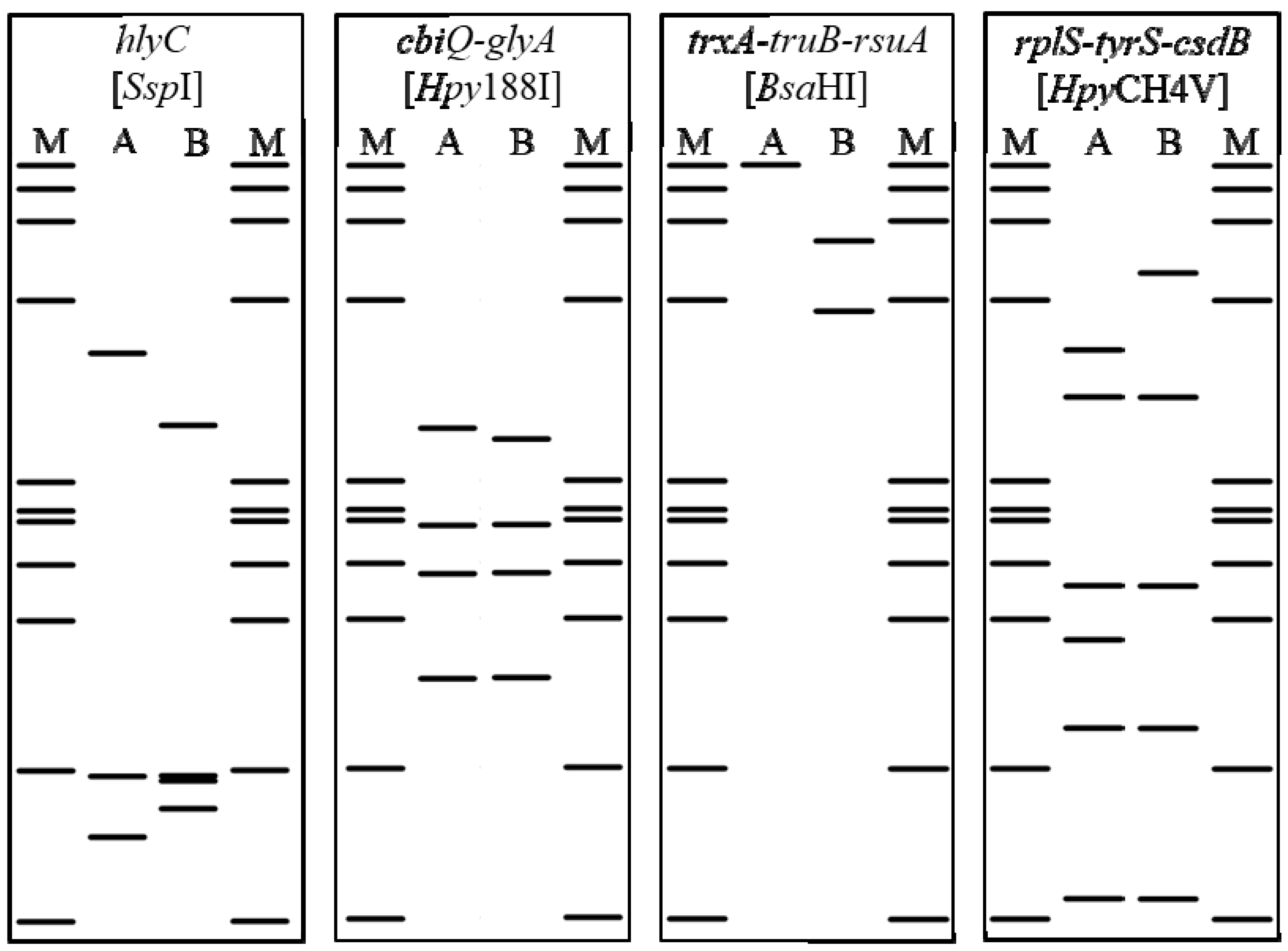
2.5. RFLP Analyses: Prevalence of CaPsol Lineages

In silico digestion, carried out on the representative nucleotide sequences of CaPsol sequence variants of the hlyC, cbiQ-glyA, trxA-truB-rsuA, and rplS-tyrS-csdB genomic fragments, allowed generation of virtual RFLP profiles for the restriction enzymes SspI, Hpy188I, BsaHI, and HpyCH4V, respectively (Figure 4). For each genetic locus analyzed, the obtained virtual RFLP profiles consistently differentiated the two sequence variants identified in the present study (Figure 2). In vitro RFLP assays, conducted on nested PCR products amplified from the 40 selected vines, produced the predicted restriction profiles for each sequence variant of each genomic fragment (data not shown). Having determined the resolution power of these RFLP assays, the CaPsol strains identified in the remaining 102 vines were attributed to lineages using in vitro digestion (Table 1). Based on the obtained collective RFLP patterns, each CaPsol strain was attributed to one of the 15 previously determined lineages (Table 1). Molecular markers distinguishing such lineages can be exploited to study different aspects of BN disease, such as CaPsol strain population structure determination and epidemiology in different agroecosystems. In particular, the major contribution to the variability among these lineages is due to SNPs within the cbiQ-glyA, trxA-truB-rsuA, and rplS-tyrS-csdB genomic fragments. In fact, numerous studies reported the usefulness of genes not directly related to phytoplasma virulence (e.g., rplV-rpsC, groEL, map) as valuable markers for phytoplasma strain typing related to their ecology [40,41,42].
The prevalence of CaPsol lineages was evaluated in the different geographic areas under study, leading to new insights into CaPsol populations in Italinan vineyards: (i) three lineages (13, 14, 15) were found exclusively in North Macedonia and not in Italy; (ii) a single lineage (6) was found in southern Italy; (iii) four lineages were found in central Italy, with two in Marche (10, 12) and two in Tuscany (5, 11); (iv) six lineages (1, 2, 4, 6, 7, 10) were found in Veneto, three of which (1, 4, 10) represented 87% of CaPsol strains; (v) twelve lineages (1 to 12) were found in Lombardy, three of which (1, 4, 7) represented 65% of CaPsol strains, with three (3, 8, 9) found exclusively in this region (Table 1). Notably, a low number of CaPsol lineages, reflecting low genetic diversity within strain populations, was found in the geographic areas (Veneto, Marche, Tuscany, Apulia, Sicily, North Macedonia) where high prevalence of a single tuf-type was reported. In contrast, numerous CaPsol lineages, reflecting an elevated genetic diversity within strain populations, were found in Lombardy where the two tuf-types (a and b) were equally present. Such genetic heterogeneity within the CaPsol population in Lombardy was also noted in a recent study based on CaPsol molecular characterization using the hypervariable gene stamp [8]. It is reasonable to hypothesize that this variability in CaPsol strains could be related to the ecological complexity of vineyards and their surroundings, including the presence of multiple insect vectors and alternative plant hosts [8]. The RFLP-based typing method used in the present study could be considered to be a valuable tool for research on the ecology of CaPsol and the epidemiology of its associated diseases.

3. Materials and Methods

3.1. Sample Collection

From 2015 to 2018, leaf samples were collected from 142 symptomatic grapevine plants in BN-affected vineyards in Lombardy and Veneto regions (northern Italy), Marche, and Tuscany regions (central Italy), Apulia and Sicily regions (southern Italy), and the Republic of North Macedonia (Table 1). For each plant sample, 1 g of leaf petioles was stored at −30 °C until molecular analysis.

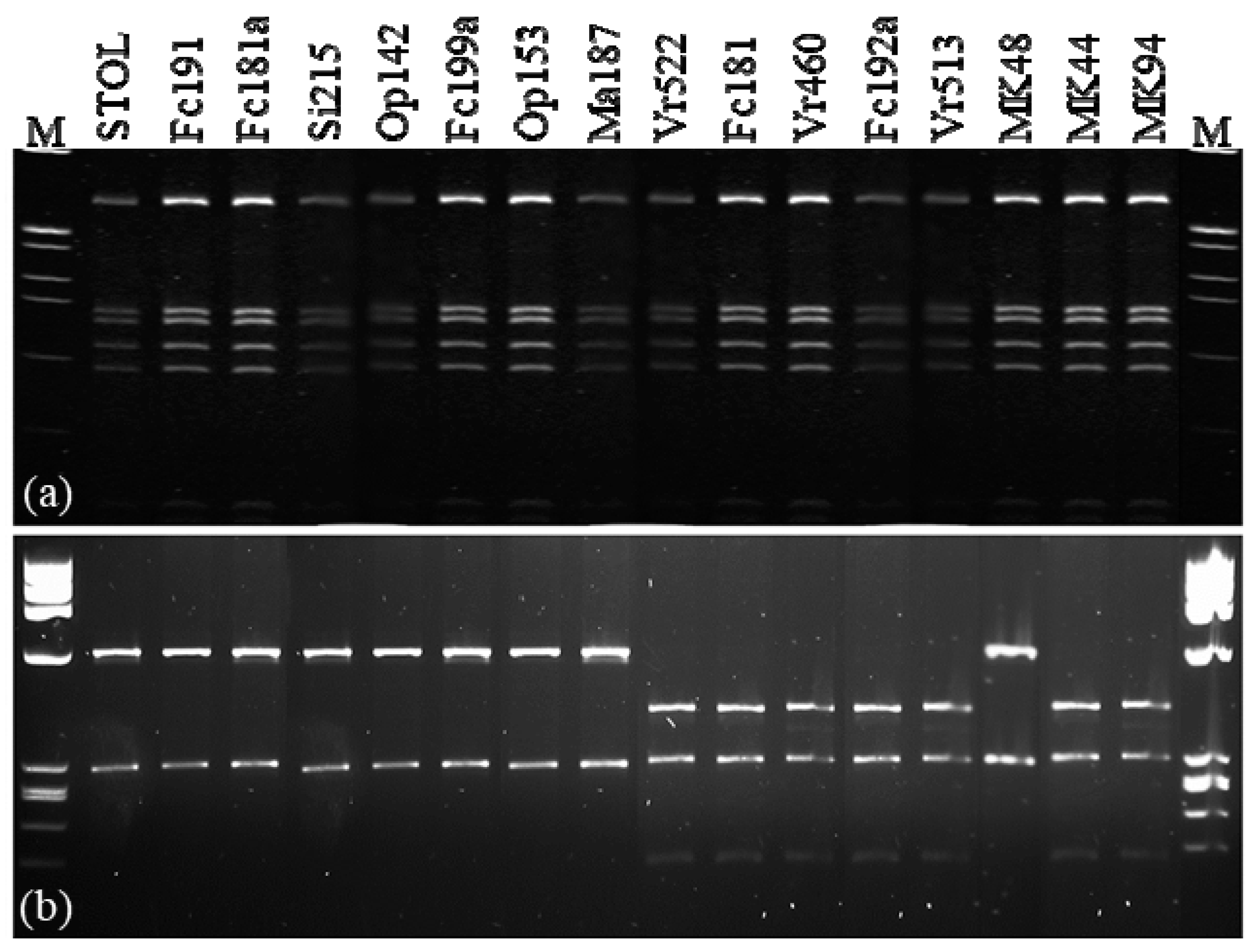
3.2. CaPsol Molecular Identification

Total nucleic acids were extracted from the leaf petioles of the examined plants, as previously described [43]. Detection and identification of CaPsol were carried out by means of nested PCR amplification of 16S rDNA primed by the universal primer pairs P1/P7 [44] and followed by the 16SrI group-specific primer pair R16F1/R16R1 [25], with a subsequent MseI-RFLP assay performed on the obtained amplicons. PCR and RFLP reaction conditions were as previously described [45]. PCRs were performed using Taq polymerase (Promega, Milan, Italy) in an automated thermal cycler (MasterCycler Gradient, Eppendorf, Milan, Italy). PCR and enzymatic digestion products were electrophoresed through 1% and 3% agarose gel, respectively, in Tris-Borate- Ethylenediaminetetraacetic acid (TBE) buffer, stained with Midori Green Advance (Biosigma, Venice, Italy), and visualized under a UV transilluminator. Total nucleic acids from periwinkle (Catharanthus roseus (L.) G. Don) infected by the phytoplasma strain STOL (CaPsol, subgroup 16SrXII-A) was used as the reference control. Total nucleic acids extracted from the healthy periwinkle and PCR mixture devoid of nucleic acids was used as the negative control.

3.3. Molecular Characterization of CaPsol Strains through Multilocus Genotyping Analysis

The tufB genotyping of CaPsol strains identified in infected samples was performed by nested PCR amplification using the primer pair fTuf1/rTuf1 followed by fTufAY/rTufAY, with subsequent HpaII-RFLP assays performed on the obtained amplicons [3]. The experimental controls, PCR conditions, and PCR-RFLP analysis were same as the above 16S rRNA gene analysis.
Multilocus genotyping analysis was carried out by employing 40 CaPsol strains representing distinct tuf-types (20 tuf-type a and 20 tuf-type b) and different geographic origins. Based on the genome sequences of CaPsol strain 284/09 (FO393427) and 231/09 (FO393428) [46], primer pairs were designed to amplify the complete tufB gene sequence and 8 genomic fragments (cbiQ-glyA, rplS-tyrS-csdB, trxA-truB-rsuA, hlyC, potC-potD, pnp, gyrA-gyrB, aspS-mesJ) containing 16 previously uncharacterized genes encoding proteins (Table 3). The experimental controls, PCR conditions, and PCR-RFLP analysis were same as the above 16S rRNA gene analysis. Obtained amplicons of the full tufB gene and the 8 genomic fragments were sequenced in both senses (5X coverage per base position) by a commercial service (Eurofins Genomics, Germany). Nucleotide sequences were compiled in FASTA format, assembled by employing the Contig Assembling Program of the software BioEdit version 7.2 [47], and trimmed to the annealing sites of the related primers utilized by the nested PCRs. For each genomic fragment under study, nucleotide sequences were aligned using the ClustalW Multiple Alignment application within the software BioEdit, and single nucleotide polymorphisms (SNPs), restriction enzymatic sites, and deduced amino acid substitution were searched for.

3.4. Phylogenetic Analyses

Phylogenetic trees were established by aligning the nucleotide sequences of tufB, hlyC, glyA, truB, and tyrS genes of CaPsol strains representing genetic lineages identified in this study and those of available phytoplasma strains in National Center for Biotechnology Information (NCBI) GenBank. Moreover, phylogenetic analysis was also performed on hlyC, glyA, truB, and tyrS concatenated nucleotide sequences. The evolutionary history was inferred using the Maximum Likelihood method and the Tamura–Nei model [48]. Initial trees for the heuristic search were obtained automatically by applying Neighbor-Join and BioNJ algorithms to a matrix of pairwise distances estimated using the Maximum Composite Likelihood (MCL) approach, and then selecting the topology with the superior log likelihood value. Evolutionary analyses were conducted in MEGA X [49].

3.5. Survey on CaPsol Genetic Lineages by Restriction Fragment Length Polymorphism Analyis

SNPs distinguishing CaPsol genetic lineages were checked for their position in recognition sites for restriction enzymes by in silico restriction fragment length polymorphism (RFLP) assays using the software pDRAW32 (AcaClone Software, http://acaclone.com). The obtained virtual RFLP profiles were confirmed by actual digestion of genomic fragments amplified from the 40 grapevines selected for multiple gene sequencing. Nested PCR amplification of the genomic fragments (hlyC, cbiQ-glyA, trxA-truB-rsuA, and rplS-tyrS-csdB) was conducted on the remaining 102 CaPsol-infected grapevines (not included in the multilocus genotyping). Actual RFLP analyses were performed using the enzyme SspI on hlyC amplicons, Hpy188I on cbiQ-glyA amplicons, BsaHI on trxA-truB-rsuA amplicons, and HpyCH4V on rplS-tyrS-csdB amplicons, respectively. Digestion reactions were carried out as indicated by the enzyme manufacturer’s instructions (New England Biolabs, Ipswich, MA, USA). RFLP profiles were visualized by electrophoresis on 3% agarose gel.

Supplementary Materials

The following are available online at https://www.mdpi.com/2076-0817/9/11/970/s1, Figure S1: Unrooted phylogenetic trees constructed based on the nucleotide sequence alignment of the genes tufB (a), glyA (b), and tyrS (c).

Author Contributions

Conceptualization: A.P. (Alessandro Passera), F.Q., Y.Z., R.E.D., G.R., P.A.B., A.M. (Alberto Materazzi), S.M. (Sasa Mitrev), and A.L.; methodology: F.Q., Y.Z., R.E.D., and W.W.; formal analysis: F.Q., A.P. (Alessandro Passera), A.P. (Alessandra Panattoni), R.P., E.A., S.M. (Sergio Murolo), A.M. (Alberto Materazzi), A.M. (Abdelhameed Moussa), and M.R.S.; data curation: F.Q., A.P. (Alessandro Passera), R.P., E.A., N.M., and P.C.; writing—original draft preparation: F.Q. and A.P. (Alessandro Passera); writing—review and editing: All authors. All authors read and agreed to the published version of the manuscript.

Funding

This research received no external funding.

Conflicts of Interest

The authors declare no conflict of interest.

References

  1. Quaglino, F.; Zhao, Y.; Casati, P.; Bulgari, D.; Bianco, P.A.; Wei, W.; Davis, R.E. ‘Candidatus Phytoplasma solani’, a novel taxon associated with stolbur and Bois Noir related diseases of plants. Int. J. Syst. Evol. Microbiol. 2013, 63, 2879–2894. [Google Scholar] [CrossRef] [PubMed] [Green Version]
  2. Romanazzi, G.; Murolo, S.; Feliziani, E. Effects of an innovative strategy to contain grapevine Bois noir: Field treatment with resistance inducers. Phytopathology 2013, 103, 785–791. [Google Scholar] [CrossRef] [PubMed] [Green Version]
  3. Langer, M.; Maixner, M. Molecular characterisation of Grapevine yellows associated phytoplasmas of the stolbur-group based on RFLP-analysis of non-ribosomal DNA. Vitis 2004, 43, 191–200. [Google Scholar]
  4. Cvrković, T.; Jović, J.; Mitrović, M.; Krstić, Q.; Toševski, I. Experimental and molecular evidence of Reptalus panzeri as a natural vector of bois noir. Plant Pathol. 2014, 63, 42–53. [Google Scholar] [CrossRef] [Green Version]
  5. Mori, N.; Quaglino, F.; Tessari, F.; Pozzebon, A.; Bulgari, D.; Casati, P.; Bianco, P.A. Investigation on ‘bois noir’ epidemiology in north-eastern Italian vineyards through a multidisciplinary approach. Ann. Appl. Biol. 2015, 166, 75–89. [Google Scholar] [CrossRef]
  6. Kosovac, A.; Radonjić, S.; Hrnčić, S.; Krstić, O.; Toševski, I.; Jović, J. Molecular tracing of the transmission routes of bois noir in Mediterranean Vineyards of Montenegro and experimental evidence for the epidemiological role of Vitex agnus-castus (Lamiaceae) and associated Hyalesthes obsoletus (Cixiidae). Plant Pathol. 2016, 65, 285–298. [Google Scholar] [CrossRef] [Green Version]
  7. Kosovac, A.; Jakovljević, M.; Krstić, O.; Cvrković, T.; Mitrović, M.; Toševski, I.; Jović, J. Role of plant-specialized Hyalesthes obsoletus associated with Convolvulus arvensis and Crepis foetida in the transmission of ‘Candidatus Phytoplasma solani’-inflicted Bois noir disease of grapevine in Serbia. Eur. J. Plant Pathol. 2019, 153, 183–195. [Google Scholar] [CrossRef]
  8. Quaglino, F.; Sanna, F.; Moussa, A.; Faccincani, M.; Passera, A.; Casati, P.; Bianco, P.A.; Mori, N. Identification and ecology of alternative insect vectors of ‘Candidatus Phytoplasma solani’ to grapevine. Sci. Rep. 2019, 9, 19522. [Google Scholar] [CrossRef]
  9. Moussa, A.; Mori, N.; Faccincani, M.; Pavan, F.; Bianco, P.A.; Quaglino, F. Vitex agnus-castus cannot be used as trap plant for the vector Hyalesthes obsoletus to prevent infections by ‘Candidatus Phytoplasma solani’ in northern Italian vineyards: Experimental evidence. Ann. Appl. Biol. 2019, 175, 302–312. [Google Scholar] [CrossRef]
  10. Pierro, R.; Panattoni, A.; Passera, A.; Materazzi, A.; Luvisi, A.; Loni, A.; Ginanni, M.; Lucchi, A.; Bianco, P.A.; Quaglino, F. Proposal of a new Bois noir epidemiological pattern related to ‘Candidatus Phytoplasma solani’ strains characterized by a possible moderate virulence in Tuscany. Pathogens 2020, 9, 268. [Google Scholar] [CrossRef] [Green Version]
  11. Bianco, P.A.; Romanazzi, G.; Mori, N.; Myrie, W.; Bertaccini, A. Integrated management of phytoplasma diseases. In Transmission and Management of Phytoplasma—Associated Diseases. Phytoplasmas: Plant Pathogenic Bacteria–II; Bertaccini, A., Weintraub, G.P., Rao, G.P., Mori, N., Eds.; Springer Nature: Singapore, 2019; pp. 208–237. [Google Scholar]
  12. Murolo, S.; Romanazzi, G. In-vineyard population structure of ‘Candidatus Phytoplasma solani’ using multilocus sequence typing analysis. Infect. Gen. Evol. 2015, 31, 221–230. [Google Scholar] [CrossRef] [PubMed]
  13. Quaglino, F.; Maghradze, D.; Casati, P.; Chkhaidze, N.; Lobjanidze, M.; Ravasio, A.; Passera, A.; Venturini, G.; Failla, O.; Bianco, P.A. Identification and characterization of new ‘Candidatus Phytoplasma solani’ strains associated with bois noir disease in Vitis vinifera L. cultivars showing a range of symptoms severity in Georgia, the Caucasus region. Plant Dis. 2016, 100, 904–915. [Google Scholar] [CrossRef] [PubMed] [Green Version]
  14. Pierro, R.; Passera, A.; Panattoni, A.; Casati, P.; Luvisi, A.; Rizzo, D.; Bianco, P.A.; Quaglino, F.; Materazzi, A. Molecular typing of ‘Bois Noir’ phytoplasma strains in the Chianti Classico area (Tuscany, Central Italy) and their association with symptom severity in Vitis vinifera L. cv. Sangiovese. Phytopathology 2018, 108, 362–373. [Google Scholar] [CrossRef] [PubMed] [Green Version]
  15. Pierro, R.; Passera, A.; Panattoni, A.; Rizzo, D.; Stefani, L.; Bartolini, L.; Casati, P.; Luvisi, A.; Quaglino, F.; Materazzi, A. Prevalence of a ‘Candidatus Phytoplasma solani’ strain, so far associated only with other hosts, in Bois Noir-affected grapevines within Tuscan vineyards. Ann. Appl. Biol. 2018, 173, 202–212. [Google Scholar] [CrossRef]
  16. Naser, S.M.; Vancanneyt, M.; Hoste, B.; Snauwaert, C.; Swings, J. Lactobacillus cypricasei Lawson et al. 2001 is a later heterotypic synonym of Lactobacillus acidipiscis Tanasupawat et al. 2000. Int. J. Syst. Evol. Microbiol. 2006, 56, 1681–1683. [Google Scholar] [CrossRef] [Green Version]
  17. Mignard, S.; Flandrois, J.P. A seven-gene, multilocus, genus-wide approach to the phylogeny of mycobacteria using supertrees. Int. J. Syst. Evol. Microbiol. 2008, 58, 1432–1441. [Google Scholar] [CrossRef]
  18. Pascual, J.; Macián, M.C.; Arahal, D.R.; Garay, E.; Pujalte, M.J. Multilocus sequence analysis of the central clade of the genus Vibrio by using the 16S rRNA, recA, pyrH, rpoD, gyrB, rctB and toxR genes. Int. J. Syst. Evol. Microbiol. 2010, 60, 154–165. [Google Scholar] [CrossRef] [Green Version]
  19. Adkar-Purushothama, C.R.; Quaglino, F.; Casati, P.; Bianco, P.A. Molecular typing of Coorg black pepper yellows phytoplasma by multiple gene analysis. Ann. Appl. Biol. 2011, 159, 58–68. [Google Scholar] [CrossRef]
  20. Malembic-Maher, S.; Salar, P.; Filippin, L.; Carle, P.; Angelini, E.; Foissac, X. Genetic diversity of European phytoplasmas of the 16SrV taxonomic group and proposal of ‘Candidatus Phytoplasma rubi’. Int. J. Syst. Evol. Microbiol. 2011, 61, 2129–2134. [Google Scholar] [CrossRef]
  21. Lee, I.M.; Bottner-Parker, K.D.; Zhao, Y.; Bertaccini, A.; Davis, R.E. Differentiation and classification of phytoplasmas in the pigeon pea witches’-broom group (16SrIX): An update based on multiple gene sequence analysis. Int. J. Syst. Evol. Microbiol. 2012, 62, 2279–2285. [Google Scholar] [CrossRef]
  22. Durante, G.; Casati, P.; Clair, D.; Quaglino, F.; Bulgari, D.; Boudon-Padieu, E.; Bianco, P.A. Sequence analyses of S10-spc operon among 16SrV group phytoplasmas: Phylogenetic relationships and identification of discriminating single nucleotide polymorphisms. Ann. Appl. Biol. 2012, 161, 234–246. [Google Scholar] [CrossRef]
  23. Davis, R.E.; Zhao, Y.; Dally, E.L.; Lee, I.M.; Jomantiene, R.; Douglas, S.M. ‘Candidatus Phytoplasma pruni’, a novel taxon associated with X-disease of stone fruits, Prunus spp.: Multilocus characterization based on 16S rRNA, secY, and ribosomal protein genes. Int. J. Syst. Evol. Microbiol. 2013, 63, 766–776. [Google Scholar] [CrossRef] [PubMed]
  24. Casati, P.; Quaglino, F.; Stern, A.R.; Tedeschi, R.; Alma, A.; Bianco, P.A. Multiple gene analyses reveal extensive genetic diversity among ‘Candidatus Phytoplasma mali’ populations. Ann. Appl. Biol. 2011, 158, 257–266. [Google Scholar] [CrossRef]
  25. Lee, I.-M.; Gundersen, D.E.; Hammond, R.W.; Davis, R.E. Use of mycoplasmalike organism (MLO) group-specific oligonucleotide primers for nested-PCR assays to detect mixed-MLO infections in a single host plant. Phytopathology 1994, 84, 559–566. [Google Scholar] [CrossRef]
  26. Oliveri, C.; Pacifico, D.; D’Urso, V.; La Rosa, R.; Marzachi, C.; Tessitori, M. Bois noir phytoplasma variability in a Mediterranean vineyard system: New plant host and putative vectors. Australas. Plant Pathol. 2015, 44, 235–244. [Google Scholar] [CrossRef]
  27. Kostadinovska, E.; Quaglino, F.; Mitrev, S.; Casati, P.; Bulgari, D.; Bianco, P.A. Multiple gene analyses identify distinct “bois noir” phytoplasma genotypes in the Republic of Macedonia. Phytopathol. Mediterr. 2014, 53, 491–501. [Google Scholar]
  28. Mori, N.; Pavan, F.; Reggiani, N.; Bacchiavini, M.; Mazzon, L.; Paltrinieri, S.; Bertaccini, A. Correlation of bois noir disease with nettle and vector abundance in northern Italy vineyards. J. Pest Sci. 2012, 85, 23–28. [Google Scholar] [CrossRef]
  29. Murolo, S.; Garbarino, M.; Mancini, V.; Romanazzi, G. Spatial pattern of Bois noir: Case study of a delicate balance between disease progression and recovery. Sci. Rep. 2020, 10, 9801. [Google Scholar] [CrossRef]
  30. Berg, K.A.; Dunlop, J.; Sanchez, T.; Silva, M.; Clarke, W.P. A conservative, single-amino acid substitution in the second cytoplasmic domain of the human serotonin2C receptor alters both ligand-dependent and -independent receptor signalling. J. Pharm. Exp. Therap. 2008, 324, 1084–1092. [Google Scholar] [CrossRef] [Green Version]
  31. Bruner, A.C.; Jung, S.; Abbott, A.G.; Powell, G.L. The naturally occurring high oleate oil character in some peanut varieties results from reduced oleoyl-PC desaturase activity from mutation of aspartate 150 to asparagine. Crop Sci. 2001, 4, 522–526. [Google Scholar] [CrossRef]
  32. Kawachi, T.; Sunaga, Y.; Ebato, M.; Hatanaka, T.; Harada, H. Repression of nitrate uptake by replacement of Asp105 by asparagine in AtNTR3.1 in Arabidopsis thaliana L. Plant Cell Physiol. 2006, 47, 1437–1441. [Google Scholar] [CrossRef] [PubMed]
  33. Kavaliauskas, D.; Nissen, P.; Knudsen, C.R. The busiest of all ribosomal assistants: Elongation factor Tu. Biochemistry 2012, 51, 2642–2651. [Google Scholar] [CrossRef] [PubMed]
  34. Kunert, A.; Losse, J.; Gruszin, C.; Hiihn, M.; Kraendler, K.; Mikkat, S.; Volke, D.; Hoffmann, R.; Jokiranta, T.S.; Seeberger, H.; et al. Immune evasion of the human pathogen Pseudomonas aeruginosa: Elongation factor Tuf is a factor H and plasminogen binding protein. J. Immunol. 2007, 179, 2979–2988. [Google Scholar] [CrossRef] [PubMed] [Green Version]
  35. Archambaud, C.; Gouin, E.; Pizarro-Cerda, J.; Cossart, P.; Dussurget, O. Translation elongation factor EF-Tu is a target for Stp, a serine-threonine phosphatase involved in virulence of Listeria monocytogenes. Mol. Microbiol. 2005, 56, 383–396. [Google Scholar] [CrossRef]
  36. Aryan, A.; Brader, G.; Mörtel, J.; Pastar, M.; Riedle-Bauer, M. An abundant ‘Candidatus Phytoplasma solani’ tuf b strain is associated with grapevine, stinging nettle and Hyalesthes obsoletus. Eur. J. Plant Pathol. 2014, 140, 213–227. [Google Scholar] [CrossRef] [Green Version]
  37. Jamshidi, E.; Murolo, S.; Salehi, M.; Romanazzi, G. Sequence analysis of new tuf molecular types of ‘Candidatus Phytoplasma solani’ in Iranian vineyards. Pathogens 2020, 9, 508. [Google Scholar] [CrossRef]
  38. Goebel, W.; Chakraborty, T.; Kreft, J. Bacterial hemolysins as virulence factors. Ant. Van Leeuw. 1998, 54, 453–463. [Google Scholar] [CrossRef] [Green Version]
  39. Gambetta, G.A.; Matthews, M.A.; Syvanen, M. The Xylella fastidosa RTX operons: Evidence for the evolution of protein mosaics through novel genetic exchanges. BMC Genom. 2018, 19, 329. [Google Scholar] [CrossRef] [Green Version]
  40. Quaglino, F.; Casati, P.; Bianco, P.A. Distinct rpsC single nucleotide polymorphism lineages of Flavescence dorée subgroup 16SrV-D phytoplasma co-infect Vitis vinifera L. Folia Microbiol. 2010, 55, 251–257. [Google Scholar] [CrossRef]
  41. Mitrovic, J.; Kakizawa, S.; Duduk, B.; Oshima, K.; Namba, S.; Bertaccini, A. The groEL gene as an additional marker for finer differentiation of ‘Candidatus Phytoplasma asteris’-related strains. Ann. Appl. Biol. 2011, 159, 41–48. [Google Scholar] [CrossRef]
  42. Casati, P.; Jermini, M.; Quaglino, F.; Corbani, G.; Schaerer, S.; Passera, A.; Bianco, P.A.; Rigamonti, I.E. New insights on Flavescence dorée phytoplasma ecology in the vineyard agro-ecosystem in southern Switzerland. Ann. Appl. Biol. 2017, 171, 37–51. [Google Scholar] [CrossRef]
  43. Angelini, E.; Clair, D.; Borgo, M.; Bertaccini, A.; Boudon-Padieu, E. Flavescence dorée in France and Italy: Occurrence of closely related phytoplasma isolates and their near relationships to palatinate grapevine yellows and an alder yellows phytoplasma. Vitis 2001, 40, 79–86. [Google Scholar]
  44. Deng, S.; Hiruki, C. Genetic relatedness between two nonculturable mycoplasmalike organisms revealed by nucleic acid hybridization and polymerase chain reaction. Phytopathology 1991, 81, 1475–1479. [Google Scholar] [CrossRef]
  45. Quaglino, F.; Zhao, Y.; Bianco, P.A.; Wei, W.; Casati, P.; Durante, G.; Davis, R.E. New 16Sr subgroups and distinct single nucleotide polymorphism lineages among grapevine Bois noir phytoplasma populations. Ann. Appl. Biol. 2009, 154, 279–289. [Google Scholar] [CrossRef]
  46. Mitrović, J.; Siewert, C.; Duduk, B.; Hecht, J.; Mölling, K.; Broeker, F.; Beyerlein, P.; Büttner, C.; Bertaccini, A.; Kube, M. Generation and analysis of draft sequences of ‘stolbur’ phytoplasma from multiple displacement amplification templates. J. Mol. Microbiol. Biotechnol. 2014, 24, 1–11. [Google Scholar] [CrossRef]
  47. Hall, T.A. BioEdit: A user-friendly biological sequence alignment editor and analysis program for Windows 95/98/NT. Nucleic Acids Symp. Ser. 1999, 41, 95–98. [Google Scholar]
  48. Tamura, K.; Nei, M. Estimation of the number of nucleotide substitutions in the control region of mitochondrial DNA in humans and chimpanzees. Mol. Biol. Evol. 1993, 10, 512–526. [Google Scholar]
  49. Kumar, S.; Stecher, G.; Li, M.; Knyaz, C.; Tamura, K. MEGA X: Molecular Evolutionary Genetics Analysis across computing platforms. Mol. Biol. Evol. 2018, 35, 1547–1549. [Google Scholar] [CrossRef]
Figure 1. Restriction profiles obtained from the enzyme digestions of nested PCR amplicons of (a) 16S rRNA gene with the enzyme MseI, and (b) tufB gene with the enzyme HpaII.
Pathogens 09 00970 g001
Figure 2. Single nucleotide polymorphism (SNP) positions, predicted enzymatic restriction sites, and amino acid substitutions distinguish sequence variance in each CaPsol genomic fragment, fusA-tufB, hlyC, cbiQ-glyA, trxA-truB-rsuA, and rplS-tyrS-csdB.
Pathogens 09 00970 g002
Figure 3. Unrooted phylogenetic trees constructed based on the nucleotide sequence alignment of hlyC (a) and truB (b) genes, and the hlyC, glyA, truB, and tyrS concatenated gene sequences (c). The highest log likelihood values of trees a, b, and c are −3001.71, −3427.33, and −5020.67, respectively. The percentages of trees in which the associated taxa clustered together are shown next to the branches. This analysis involved 24 sequences with a total of 748 positions (a), 24 sequences with a total of 892 positions (b), and 17 sequences with a total of 3778 positions (c). Strains representing the CaPsol lineages identified in this study are in bold. The number (1–15) in parentheses represents the CaPsol strain’s lineage; CaPsol strains clustered into tuf-type a and b are indicated in blue and black, respectively.
Pathogens 09 00970 g003
Figure 4. In silico restriction profiles reveal sequence variance of CaPsol strains by analyzing the genomic fragments hlyC, cbiQ-glyA, trxA-truB-rsuA, and rplS, tyrS-csdB.
Pathogens 09 00970 g004
Table 1. Prevalence of tufB types and CaPsol lineages in considered geographic areas.
Country RegionNo. of Samplestuf-Type atuf-Type bNo. of CaPsol Strains in Each Lineage
123456789101112131415
Italy (North)Lombardy46271911419321011211
Veneto39363132 11 12 10
Italy (Center)Marche16214 2 14
Tuscany16016 6 10
Italy (South)Apulia606 6
Sicily303 3
Macedonia (North) 16214 263 1 121
Overall142677524612011181511141215121
Table 2. Fifteen CaPsol lineages determined by collective sequence variance/RFLP profiles.
OriginCaPsol StrainSequence Variant/RFLP ProfileCaPsol
fusA-tufBhlyCcbiQ-glyAtrxA-truB-rsuArplS-tyrS-csdBLineage
LombardyFc180A (tuf-type a) AAAA1
(North Italy)Fc181AAABA2
Fc182AAAAA1
Fc183AAABA2
Fc185AAAAA1
Fc191B (tuf-type b)BAAB3
Fc181aBBBBA5
Fc192aAABBB8
Fc199aBBBAB9
Op142BBABB7
Op153BBBBB11
Op280AABBA4
Op282AABBA4
Op304AABBA4
VenetoVr460AABBA4
(North Italy)Vr462BBABA6
Vr501BBABB7
Vr509BBABB7
Vr513AABAA10
Vr517AABAA10
Vr522AAAAA1
MarcheMa187BBBAA12
(Center Italy)Ma189BBBAA12
Ma190AABAA10
Ma191AABAA10
TuscanySan11/15BBBBB11
(Center Italy)San15/15BBBBB11
San17/15BBBBB11
San5/16BBBBB11
ApuliaPu123BBABA6
(South Italy)Pu124BBABA6
Pu125BBABA6
SicilySi212BBABA6
(South Italy)Si214BBABA6
Si215BBABA6
North MK44AAAAB13
MacedoniaMK48BBAAA14
MK61BBABA6
MK62BBABA6
MK94AAABB15
Table 3. Primer sequences designed for molecular typing and expected PCR product sizes.
GenePrimerSequence (5’–3’)PCRNested PCR
Product Size (nt)
fusA-tufBfusAF1CTTTCTGARATGTTTGGMTATGCTACd1399
fusAF2GCGTTCCAATACYCAAGGAAGAGn
tufBR1ACAAAGCTCCAACGTTATCGCCTGCd/n
cbiQ-glyAcbiQF1AGAGGTTATGTATTGGGAGCGd/n1024
glyAR1CAAAGAACTTGCAAGAGTTTGGGCd
glyAR2TGTTGATAATCTTTAAAGGCAGG n
rplS-tyrS-csdBrplSF1CCTGTGCACTCCCCTAATAACGAd1529
csdBR1ACCTTCTTGGAGTGTTTCGCCTAGACd
rplSF2CGTCGTGCTAAGTCGCATTACGn
csdBR2GTTTCAAAGAGGTAGCCGCATTATCGn
trxA-truB-rsuAtrxAF1TGCCAATTGGTGTGGTCCATGTCd1363
truBR3GCCTCTATGATCAAATCAAGGACAGd
trxAF2GAATTATCACAATCAGAACAGGGTGn
truBR4TCTTTGGCGGTCGAAAGGTAGCCn
hlyChlysF1ATKATTVTGAAATGKBCTACd914
ackAR2GAAATTTTAAAGAAGARCTACd
hlysF2ATKATTVTGAAATGKBCTACYAAAMMAACn
ackAR3AGAAGARCTACCWGAATTWACCGACn
potDpotF1ACGATTAATCCAACTGTTAATGCd953
potR1TACTTGGATAAGCAATGATGTCd
potF2AGGGTGAATATTTAGACCCTCAAACn
potR2TGGATAAGCAATGATGTCATTCCn
pnppnpF1TGCTAGAAATGTGGATGCTTCTGd1448
pnpR1TGACATTTCTTGGCGTGGAGTGd
pnpF2GATACAGTAGTTTTATCGGCTACn
pnpR2GTAATACCATCTTTGCTGCCAGC n
gyrA-gyrBgyrAF2TGGGCTTCTTTGATGTCTGCTGd1705
gyrBR2TGACCGATGCTGACGTTGATGGTGd
gyrAF3TCTAATTGCAGTATCGATGTCn
gyrBR3TGCTGACGTTGATGGTGCTCACn
aspS-mesJaspSF1GTAGTTGAGATCAAGGGGTTAGTTG d1333
mesJR1TGAATCAACGCCGCCGCTAACAGd
aspSF2TCGCAGCCAAGATAGTCTTGAAGn
mesJR2CTTTTCGACTGTTCCGGGGGAATCn
Publisher’s Note: MDPI stays neutral with regard to jurisdictional claims in published maps and institutional affiliations.

