Article

Trypanosoma evansi: Molecular Characterization and Validation of a Cathepsin-L-like Peptidase as a Potential Therapeutic Target


1
Grupo de Bioquímica e Inmunología de Hemoparásitos, Departamento de Biología Celular, Universidad Simón Bolívar, Sartenejas 1080, Estado Miranda, Venezuela
2
Laboratorio de Bioquímica, Programa de Ciencias Veterinarias, Universidad Nacional Experimental Francisco de Miranda, Coro 4101, Estado Falcón, Venezuela
3
Tropical Disease Research Unit, Department of Pathology, University of California San Francisco, VAMC, San Francisco, CA 94121, USA
4
Pathology Department, Stanford University, Stanford, CA 94305, USA
*
Author to whom correspondence should be addressed.
Current Address: Department of Surgery, College of Medicine, University of Kentucky, Lexington, KY 40506, USA.
Current Address: Center for Discovery and Innovation in Parasitic Diseases, Skaggs School of Pharmacy and Pharmaceutical Sciences, University of California San Diego, La Jolla, CA 92093, USA.
Pathogens 2026, 15(9), 954; https://doi.org/10.3390/pathogens15090954
Submission received: 22 July 2026 / Revised: 28 August 2026 / Accepted: 28 August 2026 / Published: 8 September 2026
(This article belongs to the Special Issue Veterinary Parasitology: Advances in Diagnosis and Control)

Abstract

C1A cysteine peptidases (CPs) are major contributors to host–parasite crosstalk and have been proposed as anti-trypanosomal chemotherapeutic targets. Trypanosoma evansi is the most widespread animal trypanosome of African origin causing the trypanosomosis known as “Surra” or “derrengadera”. Gelatin zymography combined with peptidase class-specific inhibitors identified four CPs in T. evansi lysates with relative masses of 28, 31, 36 and 40 kDa (p28, p31, p36 and p40). The CPs present in the T. evansi lysate preferentially hydrolyzed the Z-Phe-Arg-AMC fluorogenic substrate over Z-Gly-Arg-AMC and Z-Ala-Arg-AMC, and the hydrolysis of Z-Phe-Arg-AMC was inhibited by the K11777 (also known as SLV213) vinyl sulfone inhibitor with an apparent IC50 of 3.06 nM. Active-site labeling using the 125I-LHVS-PhOH vinyl sulfone inhibitor confirmed the identity of p28, p31, p36 and p40 as cathepsin-like peptidases. Partial purification of the T. evansi p40, analysis of its N-terminal peptide sequence and cross-reactivity with an anti-T. brucei (Tbr)CATL antiserum revealed its orthology to the C1A CPs present in T. b. brucei, T. cruzi, T. dionisii and T. rangeli. Treatment of infected mice with the pan-cathepsin irreversible inhibitor, K11777 (10 mg/day), for 14 (n = 5) or 17 (n = 3) consecutive days was trypanocidal and significantly increased mice survival. These results validate TevCATL as a potential therapeutic target for the treatment of animal trypanosomosis caused by T. evansi.

1. Introduction

Trypanosoma evansi is an African kinetoplastid pathogen within the Trypanozoon subgenus that causes the trypanosomosis known as “Surra” in the Old World and “mal de caderas”, “derrengadera” or “murrina” in Latin America [1,2]. In Northern Africa, the Middle East and Asia, the disease affects domesticated and wild mammals, mainly camelids, equids and dogs, while in Central and South America the main reservoirs are equids, buffaloes, cattle, dogs, capybaras and other wildlife [2,3,4,5]. Imported animals with T. evansi infections have been reported in Europe [6,7]. In South America, the parasite is mechanically transmitted by biting flies (tabanids and Stomoxys spp.), by vampire bats, Desmodus rotundus, as a biological vector, and by iatrogenic procedures [2,3,4,5]. The first documented case of human T. evansi infection was reported in an immunocompromised Indian patient who lacked the trypanolytic factor associated with ApoL1 [8,9]. T. evansi infection was also described in an individual in Vietnam with no genetic Apo-L1 deficiency [10].
The Trypanozoon subgenus also includes T. brucei brucei, which causes “nagana” in cattle, T. b. rhodesiense and T. b. gambiense which cause Human African trypanosomiasis (HAT or sleeping sickness) and Trypanosoma equiperdum, the etiological agent of “Dourine” that affects equids [1,4,11]. Genetic studies support the reclassification of T. evansi and T. equiperdum as subspecies of T. brucei, i.e., T. b. evansi and T. b. equiperdum, since they evolved in multiple independent events from genetically distinct T. brucei strains and share a high degree of genomic similarity despite significant biological differences [12,13,14]. Phylogenetic and clustering analysis based on 168 single-copy orthologous genes demonstrated a close relationship between T. b. gambiense DAL972 and two T. evansi strains, STIB805 and YNB [13,15], which segregate from the T. b. brucei TREU 927/4 [16] and T.b. equiperdum OVI clades [14]. Whole-genomic studies showed the emergence of four different clades of related pathogenic trypanosomes, with distinct evolutionary origins: T. b. evansi type A and type B evolved from T. brucei in Western and Central Africa, whereas T. b. equiperdum type OVI and type BoTat evolved from T. brucei in Eastern Africa [14,17]. Lukeš et al. (2022) [18] proposed that the Trypanozoon (sub)species, resulting from a monophyletic origin, be designated as T. brucei ecotypes resulting from independent evolutionary paths (polyphyly).
Kinetoplastid peptidases participate in parasite nutrition, lysosomal protein turnover, host cell invasion and signaling, and are essential for infectivity and survival in the host [19,20,21,22]. In addition, the immunogenicity of trypanosomatid peptidases has encouraged their use as diagnostic tools and experimental vaccines [23,24,25]. Cysteine peptidases (CPs) of the Clan CA, family C1 [26] degrade host immunoglobulins and promote the turnover of the variant surface glycoproteins (VSGs) that cover the surface of T. brucei [19,27]. CPs are also major factors in the pathogenesis of HAT, one of the most important neglected tropical diseases (NTDs) affecting humans in sub-Saharan Africa [19,20,21]. The T. brucei rhodesiense cathepsin L-related CP (TbrCATL) [28,29] facilitates parasite entry into the central nervous system, an essential step for the neurological stage of the disease [30,31], while the TbCATL has been implicated in extravasation in endothelial cells, which contributes to cardiac dysfunction [32].
Chemical validation using CP inhibitors has demonstrated that the TbrCATL lysosomal peptidase is essential for parasite survival and is therefore a potential drug target [33,34]. Several CP inhibitors, including synthetic vinyl sulfones that engage TbrCATL are effective in vitro and in vivo against T. brucei [29,35,36,37,38]. This includes the CP inhibitor, K11777, or SLV213 (N-methyl-piperazine-Phe-homoPhe-vinyl sulfone-phenyl), which has been validated as a broad-spectrum anti-trypanosomal agent [20,29,37,38,39,40,41,42,43].
Here, we characterize the major TevCATL peptidases and demonstrate that their inhibition by K11777 kills T. evansi and extends host survival in a mouse infection model.

2. Materials and Methods

2.1. Parasites

The T. evansi capybara strain from Venezuela (TeAp-ElFrío01) was expanded in Sprague-Dawley rats as previously described [44,45]. Parasites were purified by anion-exchange chromatography using DEAE-cellulose [46], quantitated in a Neubauer chamber and stored in liquid nitrogen until use.

2.2. Peptidase Identification and Characterization

Purified trypanosomes (6 × 10⁸ parasites) were resuspended in 1 mL Tris saline hypotonic buffer (TSH; Tris-HCl 10 mM, pH 7.4, NaCl 20 mM) and lysed with three cycles of freezing and thawing. The extract was clarified by centrifugation at 5000× g for 30 s and the protein concentration determined [47]. The soluble lysate (30 μg/lane) was analyzed by 0.1% gelatin zymography using 12.5% polyacrylamide gels [48]. Zymograms were washed three times each for 5 min with distilled water at room temperature (RT), once in 100 mM Tris-HCl, 1%Triton X-100, pH 8.8, for 30 min at RT and then incubated in 100 mM Tris-HCl, 2.5 mM CaCl2, pH 7.2, with or without 25 mM cysteine for 30 min at 37 °C. Peptidase activity was identified as clear bands after staining with 3% Coomassie Brilliant Blue R-250 in 40% methanol, 10% acetic acid, and then destaining in the same solution minus the Coomassie Blue [48].
The pH profile of T. evansi peptidases was analyzed by 0.1% gelatin zymography of the trypanosome lysate (20 μg/lane). The resolved peptidase activity was activated at 37 °C for 30 min using the following buffers supplemented with 25 mM cysteine, 100 mM sodium citrate (pH 3.0–6.0), 100 mM Tris-HCl (pH 7.0–8.0) or 100 mM sodium bicarbonate (pH 9.0–10.0).
Susceptibility to various peptidase class-specific inhibitors (Sigma-Aldrich, St. Louis, MO, USA) was tested by 0.1% gelatin zymography of parasite lysates (20 μg/lane). Zymograms were incubated in 100 mM sodium citrate buffer, pH 4.0, with 25 mM cysteine at 37 °C for 30 min in the presence of one of the following inhibitors: 1 mM PMSF that inhibits serine peptidases, 10 mM EDTA (a metallo-peptidase inhibitor), 1 µM pepstatin (an aspartic peptidase inhibitor), 1 mM iodoacetic acid (IAA—a CP inhibitor), 20 µM leupeptin (inhibits serine peptidases and CPs) or 10 µM E-64 (a CP inhibitor). Control assays were incubated under the same conditions with no inhibitor or with 10% DMSO.
Four acrylamide-conjugated equine proteins: hemoglobin, fibrinogen, albumin and immunoglobulin A (IgA) (Sigma-Aldrich, St. Louis, MO, USA) were tested as substrates for the T. evansi lysates (20 μg/lane) using 0.03% zymography [49,50]. Degradation of hemoglobin, fibrinogen, albumin and IgA was examined at pH 7.0, to mimic the pH of the cytosolic and extracellular compartments. Activation of all zymograms was carried out in 100 mM Tris-HCl buffer, pH 7.0, supplemented with 25 mM cysteine and 2.5 mM CaCl2, at 37 °C for 30 min. Hemoglobin degradation was also assessed by incubation in 100 mM sodium citrate buffer, pH 5.0, supplemented with 25 mM cysteine and 2.5 mM CaCl2, at 37 °C for 30 min to simulate the pH of the lysosomal compartment.

2.3. Measuring T. evansi CP Activity with Fluorogenic Peptidyl Substrates

The trypanosome lysate (2 × 10⁶ parasites/mL) was tested with three fluorogenic dipeptidyl substrates (4 µM): Z(benzyloxycarbonyl)-Phe-Arg-AMC (7-amido-4-methyl-coumarin), Z-Arg-Arg-AMC and Z-Gly-Arg-AMC (Sigma-Aldrich, St. Louis, MO, USA), as described in [51] with minor modifications. The assay contained 100 mM sodium phosphate buffer, pH 7.0, 10% DMSO and 2 mM DTT in a final volume of 2 mL and was incubated at 30 °C for 10 min. Fluorescence was measured every minute using a Shimadzu Plus fluorimeter (RP-5301 PC) (Schimadzu Scientific Instruments, Columbia, MD, USA) at 380 nm excitation and 460 nm emission wavelengths. Fluorometric calibration was performed with 7-amino-4-methyl-coumarin (AMC) as a reference standard, in the same reaction buffer used for the fluorogenic peptidyl substrates. The resulting AMC calibration curve showed a strong correlation coefficient (r = 0.9987).

2.4. Effect of K11777 on the T. evansi CPs In Vitro

Parasite lysates (100 µg each) were pre-incubated with K11777 (0, 1, 10, 100 and 1000 nM in 10% DMSO) for 5 min at RT in 100 µL 50 mM sodium acetate buffer with 10 mM DTT, pH 5.5. The K11777 vinyl sulfone was custom synthesized by Seres Laboratories Inc. (Santa Rosa, CA, USA). Assays were carried out in duplicate and initiated by addition of an equal volume of 40 µM Z-Phe-Arg-AMC in the same buffer. Fluorescence was measured for 5 min at RT in a Multiskan fluorimeter (Thermo Fisher Scientific, Walthham, MA, USA) (380/460 excitation/emission wavelengths) and the data analyzed by non-linear regression (curve fit) of dose–response inhibition, using GraphPad Prism version 11.0.0 for Windows, (GraphPad Software, Boston, MA, USA, www.graphpad.com).

2.5. Radiolabeling of the T. evansi CPs

Parasite lysates were prepared by sonication in 50 mM sodium acetate buffer, pH 6.8, followed by clarification and re-extraction, as previously described [51,52]. To assess radiolabeling specificity, the combined parasite supernatants (100 µg per assay) were preincubated with 10% DMSO or 10 µM of the unlabeled CP inhibitors, benzyloxycarbonyl-phenylalanyl-alanine diazomethane (Z-Phe-Ala-CHN2) (Bachem, Torrance, CA, USA) or K11777, for 30 min at RT. After the addition of iodinated morpholinourea-leucinyl-homophenylalanine vinyl sulfone phenol (125I-LHVS-PhOH; 106 cpm per sample) in 100 µL 50 mM Tris, 5 mM MgCl2, 2 mM DTT, pH 5.5, [51,52], the mixture was incubated on ice for 2 h. The reaction was terminated by addition of 4 × SDS sample buffer and analyzed by 12.5% SDS-PAGE [53], followed by autoradiography. The autoradiogram image was saved as a bmp file (350 × 1260 pixels) and analyzed with the Image J software (National Institutes of Health, Bethesda, MD, USA; nih.gov bundled with 64-bit Java Image J 8 (https://imagej.net/ij/download.html, accessed on 21 February 2024).

2.6. Purification of the T. evansi Cathepsin L by FPLC and N-Terminal Sequencing

A T. evansi lysate (2 × 10⁹ trypanosomes per mL) was prepared in 50 mM sodium acetate buffer, pH 6.8, as previously described. The parasite pellet was reextracted with the same buffer and the supernatants were combined and applied to a MonoQ HR5/5 column (Amersham; Cytiva, Marlborough, MA, USA) that had been previously equilibrated with 20 mM Tris-HCl, pH 8.0. An acetonitrile gradient (0.04–0.3 M) was applied, and 0.5 mL fractions were collected at a flow rate of 0.5 mL/min. The protein concentration of the fractions was determined using the BCA Reagent (Pierce, Rockford, IL, USA) and hydrolysis of the preferred Z-Phe-Arg-AMC substrate (20 µM) was performed, as previously described for the K11777 in vitro assay. For immunoblotting, aliquots of the starting material, the wash and the collected fractions (5 µg each) were subjected to SDS-PAGE (10% gel) electrophoresis, blotted onto PVDF (Millipore, Burlington, MA, USA) and incubated with rabbit anti-TbrCAT L antiserum at a 1/4000 dilution [51]. The secondary antibody, horseradish peroxidase-conjugated goat anti-rabbit IgG (Santa Cruz Biotechnology, Dallas, TX, USA), was used at a 1/5000 dilution, and the blot was developed with the ECL reagent (Amersham; Cytiva, Marlborough, MA, USA).
To sequence the amino-terminal of the CATL peptidase, an aliquot of MonoQ column fraction #10 (200 µL) containing the p40 polypeptide band was transferred to a PVDF membrane (Immobilon-P Millipore) at 23 V for 50 min and sequenced by Edman degradation (UCSF Biomolecular Resource Center). The resulting pentapeptide sequence was analyzed by blastp using the https://blast.ncbi.nlm.nih.gov/Blast.cgi (accessed on 29 July 2026) server [54], against the UniProtKB/Swiss-Prot database. Search parameters were adjusted for a short input sequence, and the search was limited to Trypanosomatidae (taxid:5654) records.

2.7. Effect of K11777 on Parasite Survival In Vivo

The experimental T. evansi mice infections were performed with three-week-old, female BALB/c mice that were intraperitoneally (IP) injected with the Venezuelan TeAp-ElFrío strain (5 × 10⁴ parasites) [45,55,56]. Controls for these experiments were T. evansi-infected mice that received either no treatment (n = 6) or 100 µL 10% DMSO in PBS (n = 5), also at 12 hpi and then twice daily, as described below for the two K11777 experiments.
The dosing regimens used in the in vivo experiments with K11777 were based on previous assays using a mouse model of T. cruzi infection [57]. Parasitemia was estimated every 2–4 days by wet blood film examination, using the “matching method” described by Herbert and Lumsden [58]. This method provides a rapid microscopic estimate of patent parasitemia in the blood, i.e., above antilog 5.4 (250,000) parasites/mL. Mouse survival and signs of disease (e.g., lethargy, lack of social behavior and trans-grooming) were recorded daily for 80 days, at which time the surviving animals were euthanized.
For K11777 Experiment 1, mice (n = 5) received 100 µL K11777 (50 mg/mL in 10% DMSO) intra-peritoneally (IP) 12 hpi and twice daily for the following 14 days. Four out of these five K11777-treated mice received additional doses (50 mg/mL IP) on days 16, 18 and 20 dpi (Group 1) or 30 and 34 dpi (Group 2).
For K11777 Experiment 2, mice (n = 3) were treated with the same dose regimen of inhibitor, 100 µL of 50 mg/mL IP, beginning 12 hpi, and then twice daily for 17 consecutive days. Survival and signs of disease, (e.g., lethargy, lack of social behavior and trans-grooming) were monitored daily for 52 days, at which point the surviving animals were euthanized. Animals that had parasitemia between 5 × 10⁸ and 2 × 10⁹ parasites/mL or those that displayed signs of disease, (e.g., lethargy, lack of social behavior and trans-grooming) were euthanized. Data from the survival curves were analyzed with Logrank (Mantel–Cox test) using the GraphPad Prism version 11.0.0 for Windows: (GraphPad Software, Boston, MA, USA, www.graphpad.com).

3. Results

3.1. Identification of CPs in T. evansi Lysates

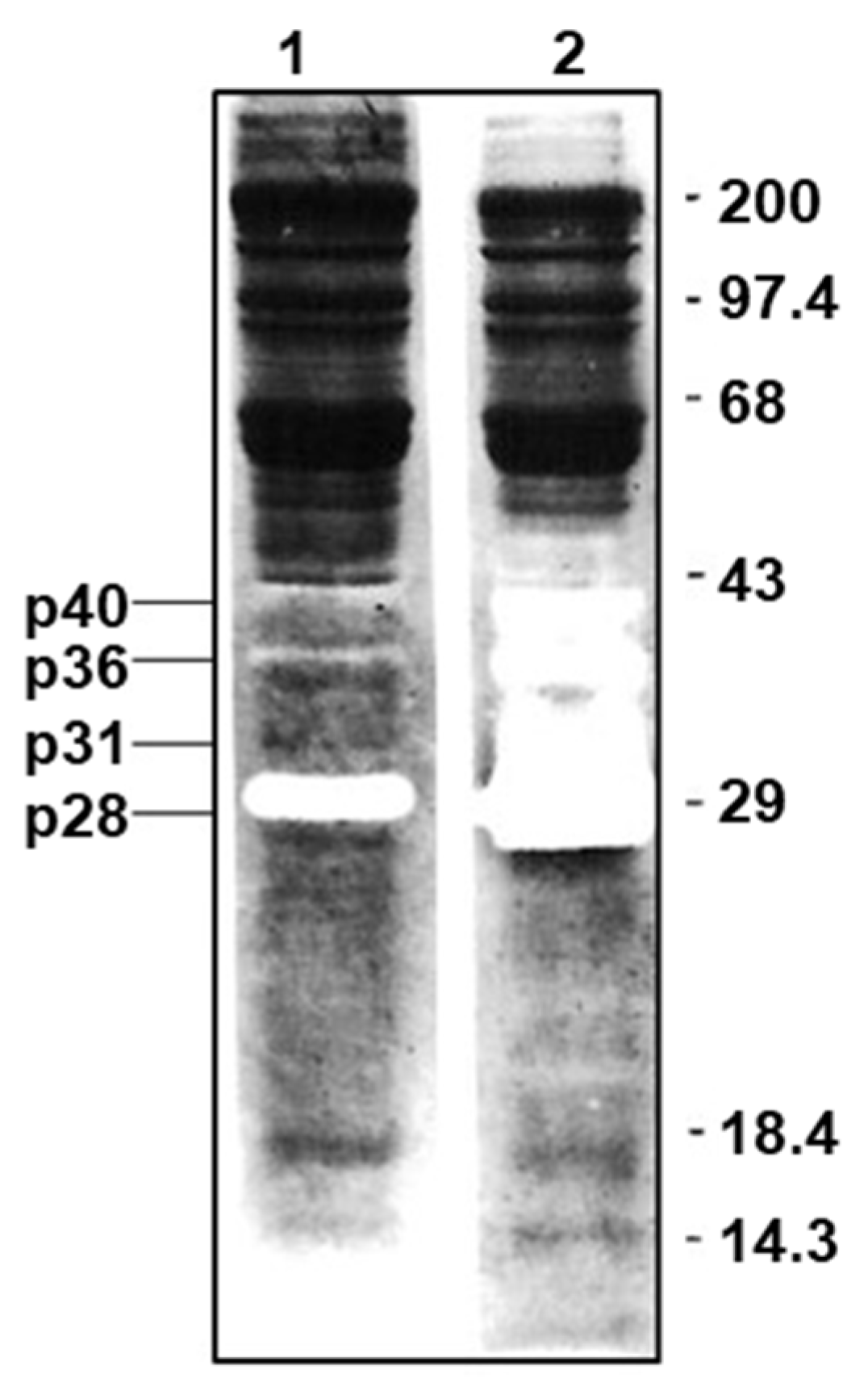
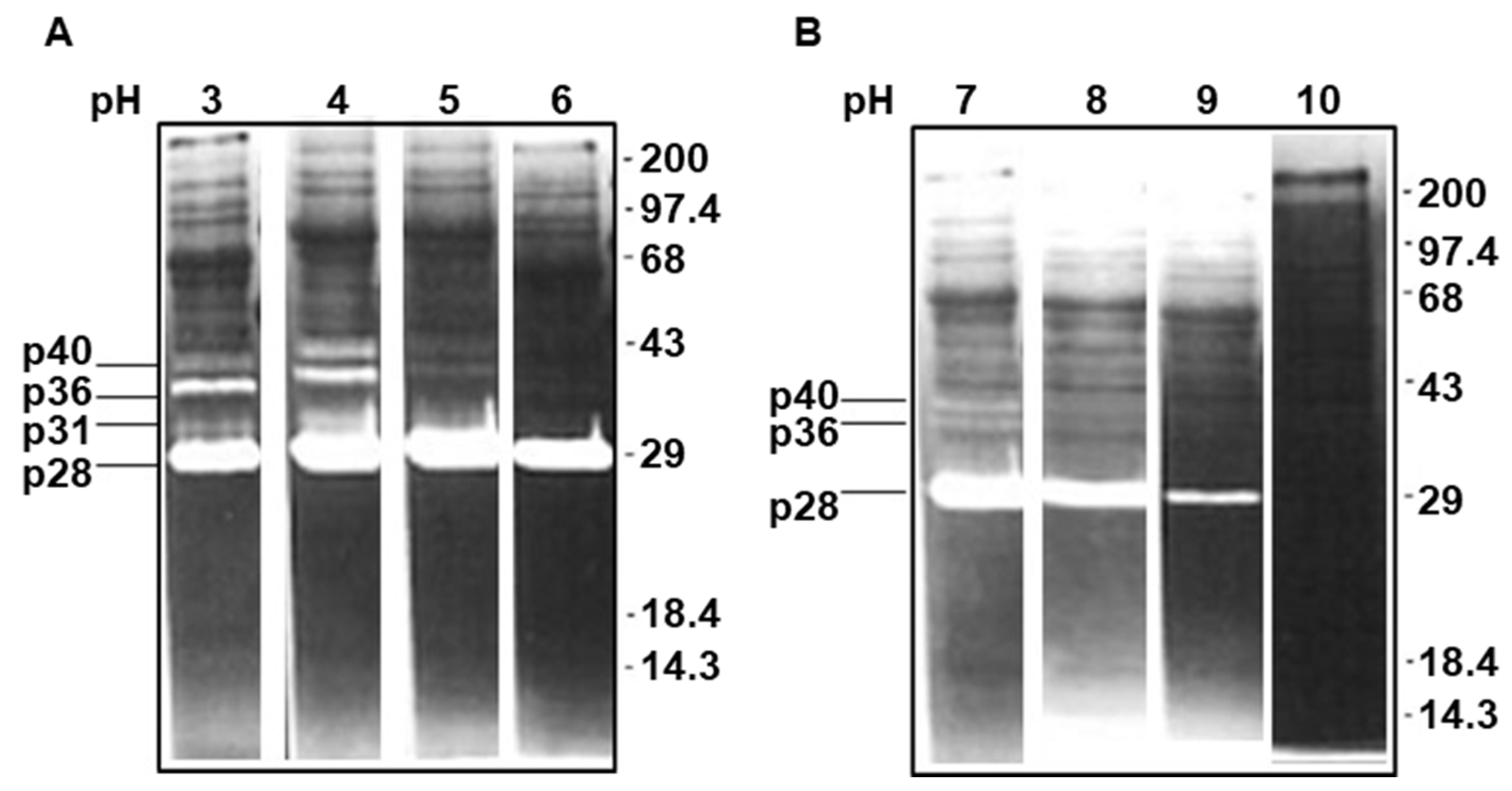
In the presence of 25 mM cysteine, four peptidase bands, with relative mobilities of 28, 31, 36 and 40 kDa (p28, p31, p36 and p40), were identified in soluble T. evansi lysates using gelatin zymography at neutral pH (Figure 1, lane 2). In the absence of cysteine, only the p40, p36 and p28 bands were seen, all with decreased intensity (Figure 1, lane 1), suggesting that they correspond to CPs. The pH-dependence of these peptidases was analyzed in four buffers ranging from pH 3.0 to 10.0 (Figure 2). The p28 peptidase was active across a broad pH range, between 3 and 9, with maximal degradation at pH 4 and 5 (Figure 2A). The p31, p36 and p40 gelatinase activities were detected in the pH 3−5 interval (optimal between pH 3.0 and 4.0) and at pH 7.0. A significant inhibition of all the CPs was observed between pH 8.0 and 10.0 (Figure 2B).
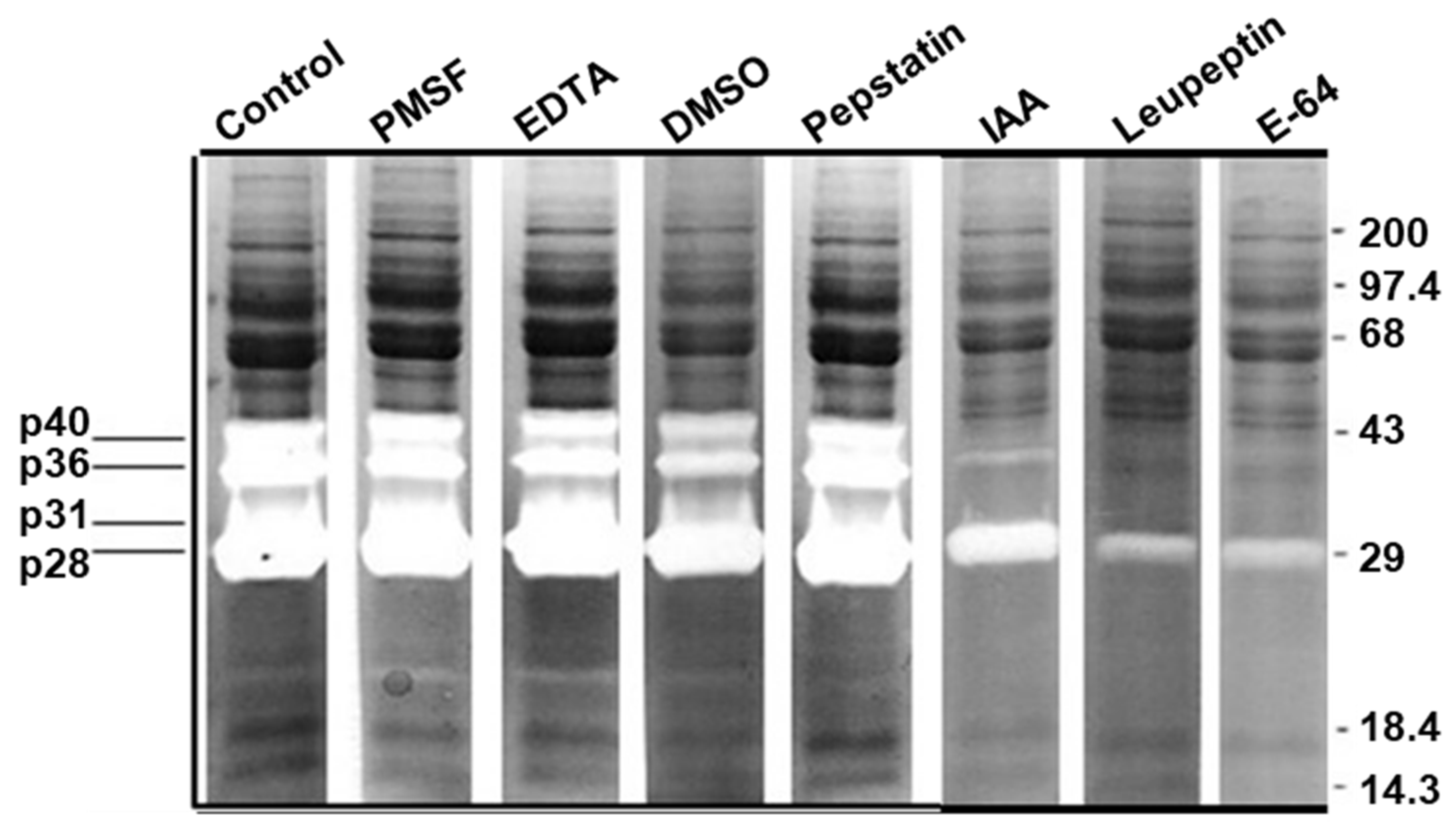
Various peptidase class-specific inhibitors were used to further characterize the enzymatic activities in the T. evansi lysate (Figure 3). Four bands, p28, p31, p36 and p40, were observed in the control assays with no inhibitors or with the 10% DMSO solvent (Figure 3). The peptidases were strongly inhibited by the two CP inhibitors, IAA and E-64, and by leupeptin, which targets both CPs and serine peptidases (Figure 3). In contrast, three inhibitors had no effect: PMSF, which inhibits serine peptidases; EDTA, a metallo-peptidase inhibitor; and pepstatin, which is selective for aspartyl peptidases (Figure 3). Based on their activation by cysteine, patterns of inhibition in the presence of CP inhibitors, as well as their pH profiles, the four T. evansi bands with peptidase activities were designated as CPs.

3.2. Degradation of Equine Proteins and Fluorogenic Peptides by T. evansi Peptidases

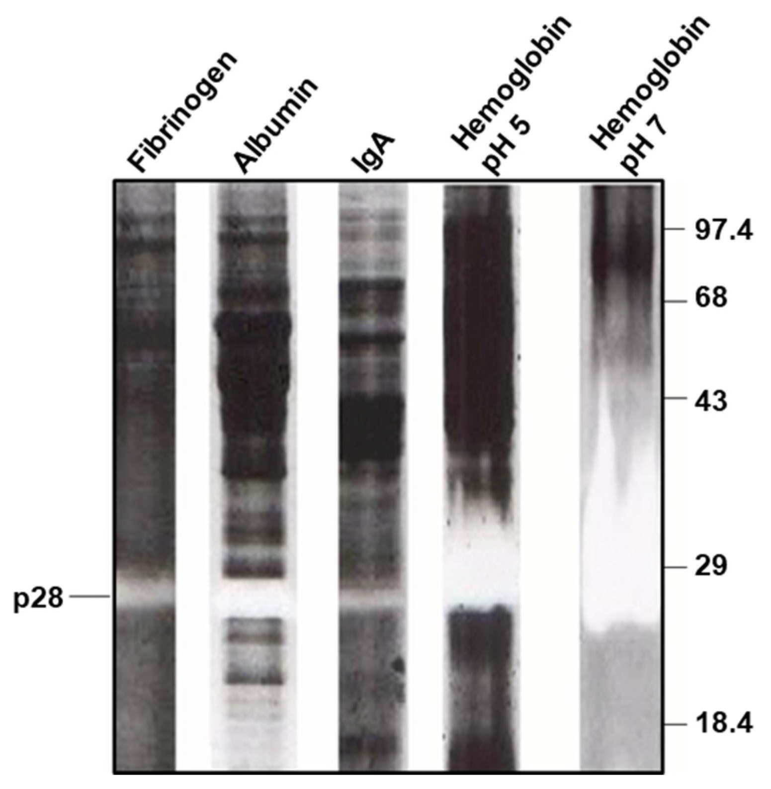
Because T. evansi is a hemotropic parasite and equids are one of its preferred hosts, we evaluated the ability of the parasite CPs to degrade the major equine proteins, hemoglobin, fibrinogen, albumin, and IgA, the dominant immunoglobulin isotype present in mucosal membranes, body secretions and blood [59]. One of the previously identified CPs, p28, degraded fibrinogen, albumin, and IgA in zymograms at pH 7.0 (Figure 4), and showed strong hemoglobinolytic activity at pH 7.0 and 5.0 (Figure 4).
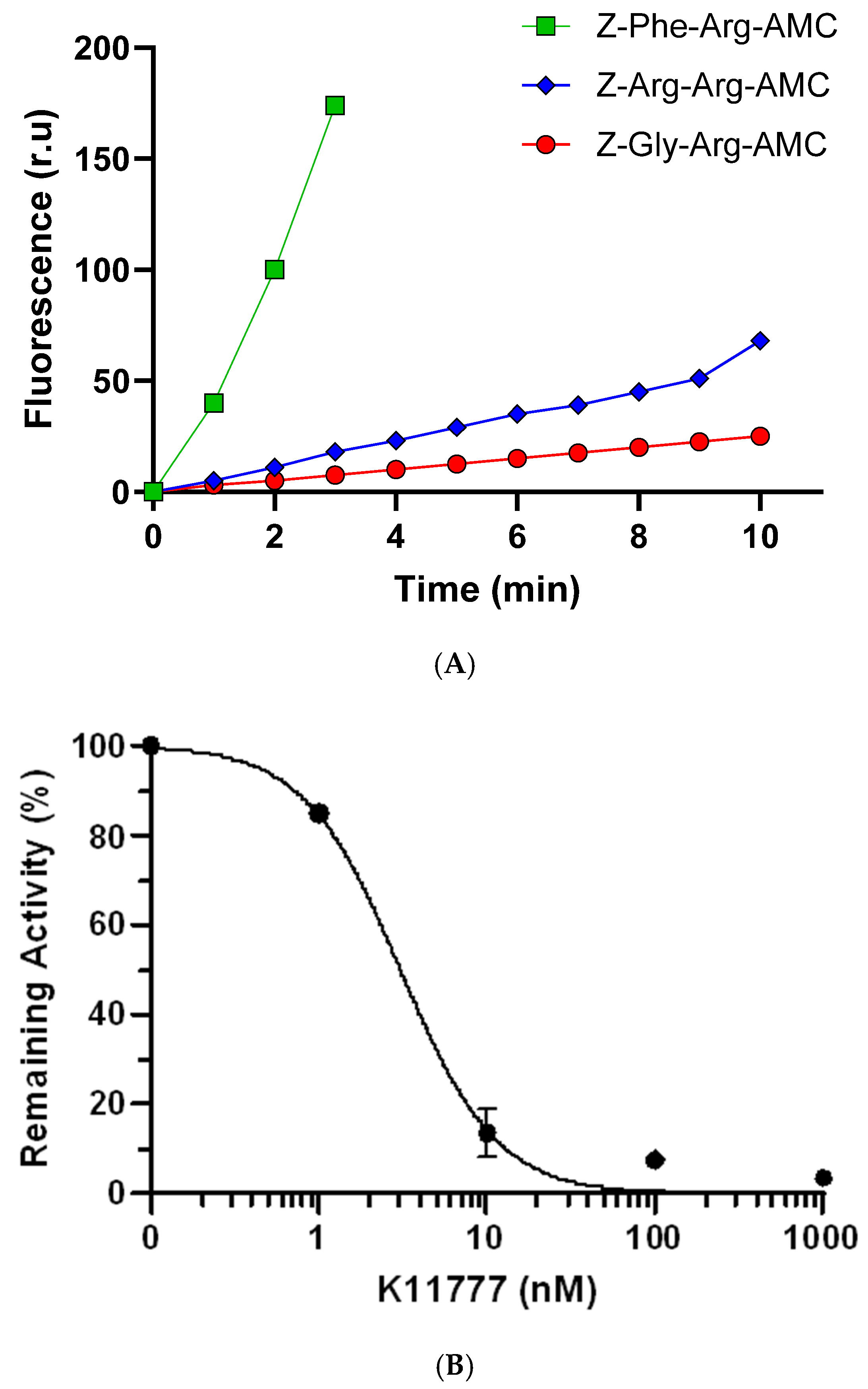
The CP activities present in the T. evansi lysate were further examined in solution assays with three peptidyl-AMC substrates (Figure 5A). Z-Phe-Arg-AMC was the preferred substrate with 7× and 17× higher hydrolysis rates relative to Z-Arg-Arg-AMC and Z-Gly-Arg-AMC, respectively (Figure 5A). The cleavage of Z-Phe-Arg-AMC by the T. evansi lysate was completely inhibited by K11777 in a concentration-dependent fashion with an apparent IC50 of 3.06 nM (95% CI: 2.15–4.47 nM, Rsquared = 0.9893) (Figure 5B). These results indicate that the CPs in the T. evansi lysate belong to the papain-superfamily and can be classified as cathepsin-like enzymes.

3.3. Active Site Radiolabeling Identifies Major CPs in T. evansi Lysates

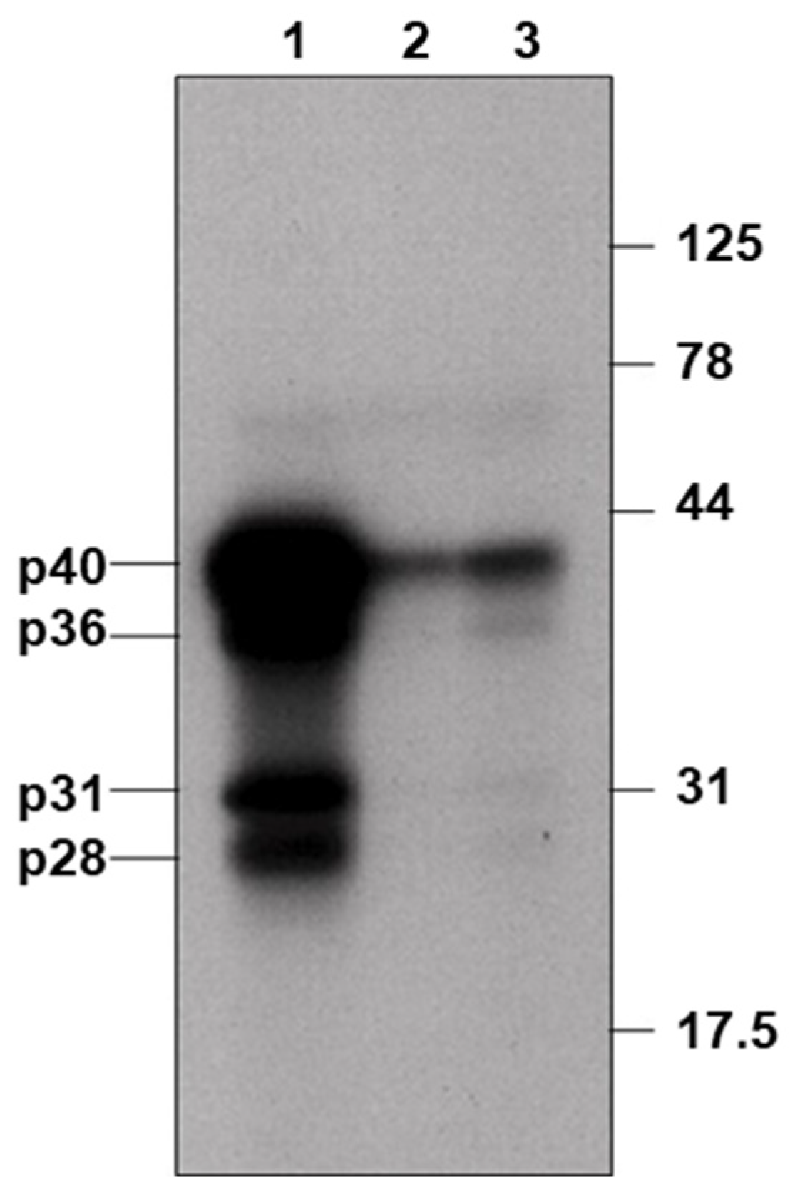
The active-site-directed peptidyl inhibitor 125I-LHVS-PhOH identified CPs with relative mobilities of 28, 31, 36 and 40 kDa (Figure 6, lane 1). Densitometric analysis showed that binding of 125I-LHVS-PhOH to these CPs was reduced by approximately 80%, after a 30 min preincubation with 10 µM of either of two CP inhibitors, Z-Phe-Ala-CHN2 (lane 2) or K11777 (lane 3).

3.4. N-Terminal Sequencing Confirms the Identity of the Cathepsin L Peptidase

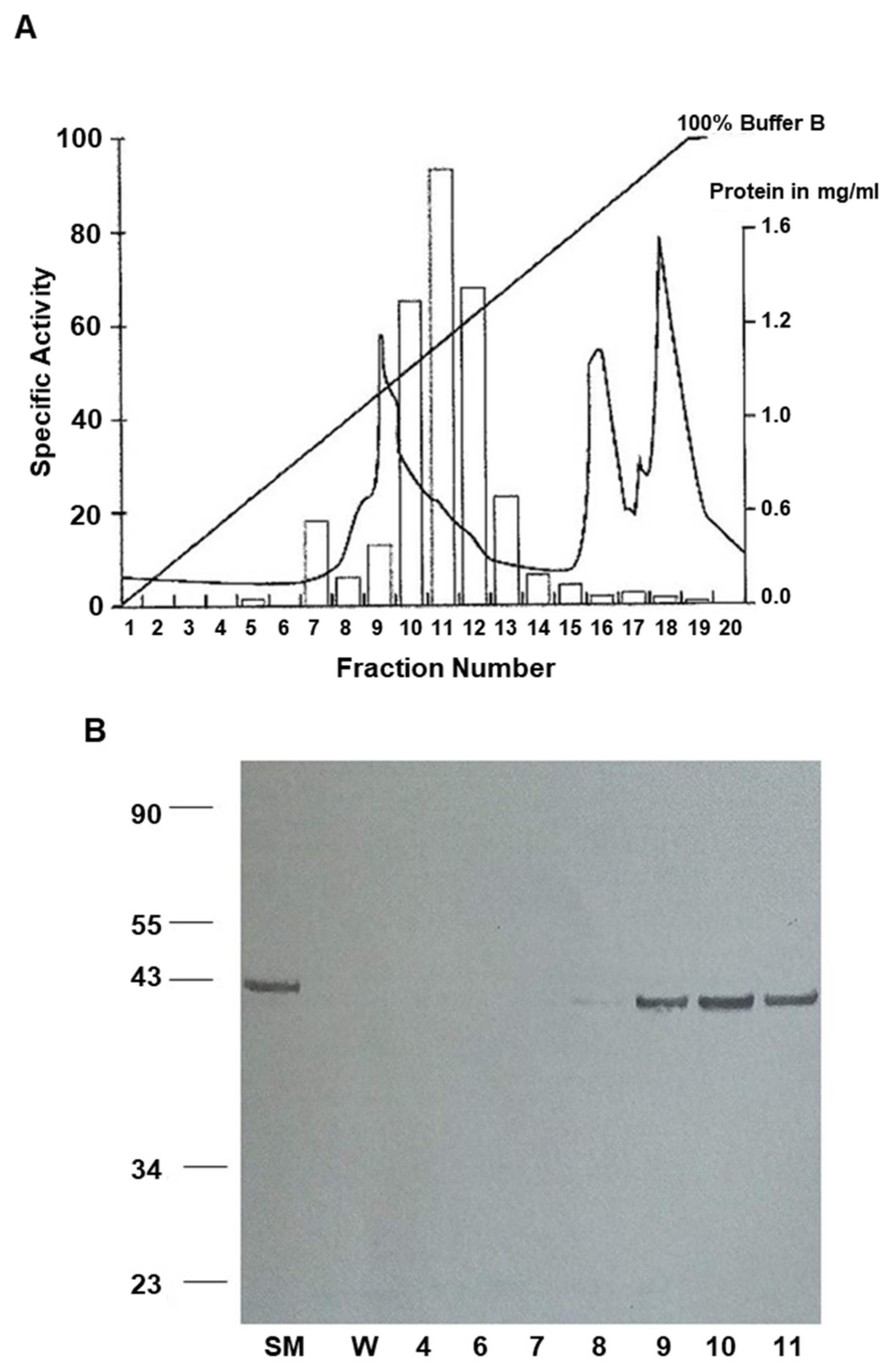
The 40 kDa T. evansi CP was purified in a single chromatographic step, using MonoQ FPLC and an acetonitrile gradient (Figure 7). Figure 7A shows that the cathepsin activity, measured by the hydrolysis of the Z-Phe-Arg-AMC (fractions 9–13), was eluted at the tail of the first broad protein peak. Analysis by SDS-PAGE and immunoblotting with rabbit monospecific antibodies to the recombinant TbrCATL revealed the 40 kDa cross-reacting species in the starting material and in fractions 8–11 (Figure 7B). Partial Edman sequencing of the 40 kDa cathepsin polypeptide in fraction 10 returned an N-terminal pentapeptide sequence: APAAV. The APAAV pentapeptide has been recognized as an autoproteolytic cleavage signal and the N-terminal sequence of Cruzipain, the orthologous cathepsin L peptidase from T. cruzi, following removal of the proregion [60,61]. This motif has also been found in other papain-like cysteine peptidases. Our PSI-BLAST search of the UniProtKB/SwissProt database [54,62] showed that the pentapeptide sequence APAAV is identical, with a perfect match (5/5) to the N-terminus of the mature CATL peptidases from Trypanosoma cruzi and T. b. brucei [60,61,63] confirming the p40 species as TevCATL.

3.5. K11777 Is Parasiticidal In Vivo and Extends the Survival of Mice Infected with T. evansi

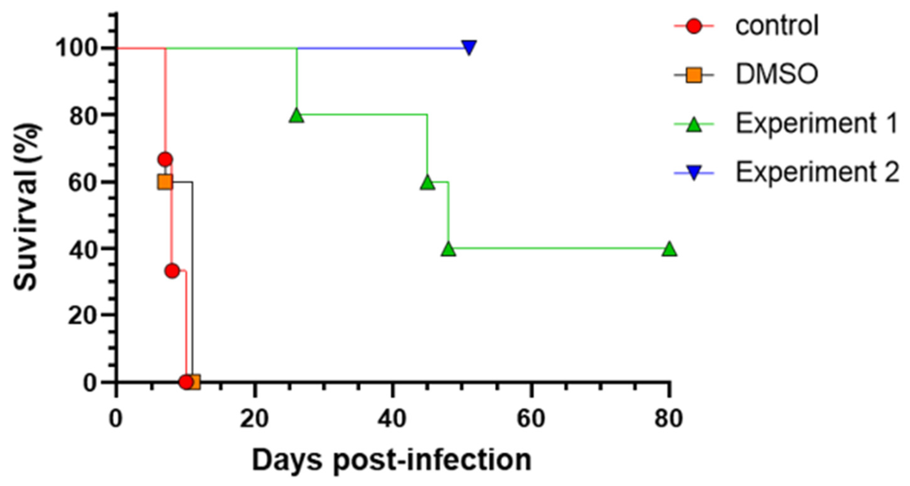
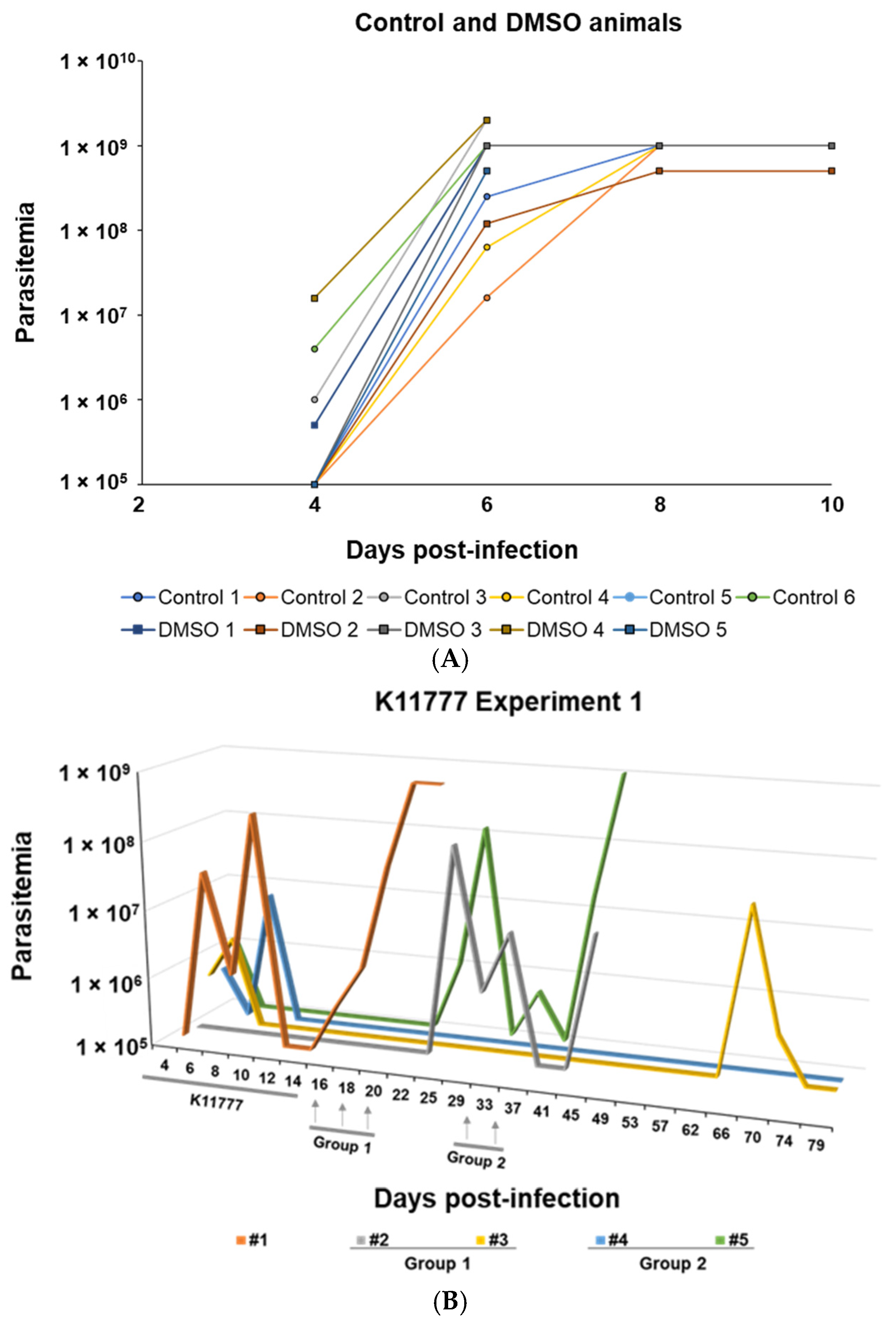
We then examined the effect of the CP inhibitor K11777 on the course of parasitemia and survival of T. evansi-infected BALB/c mice, compared to control mice that received no treatment or vehicle (10% DMSO) alone (Figure 8 and Figure 9). Vehicle control mice were euthanized 7–10 dpi (Figure 8) after showing signs of lethargy, non-social behavior and/or parasitemia values between 5 × 10⁸ and 2 × 10⁹ parasites/mL (Figure 9A). The parasitemia of the non-treated mice reached maximum values from 5 × 10⁸ to 1 × 10⁹ par/mL between 7 to 10 pi, at which time they were euthanized (Figure 8 and Figure 9A).
In comparison to the 10 dpi survival of the experimental control animals, treatment with K11777 for 14 consecutive days (Figure 8 and Figure 9B, Expt. 1, n = 5) with no further treatment (Figure 9B, #1) resulted in a 26 dpi survival. With additional doses of K11777 to Group 1 (#2 and # 3) (Figure 9B), mice survival was extended to 45 and 80 dpi, at which point the experiment was terminated. In Group 2 (#4 and #5) (Figure 9B), that also received additional doses of K11777, mouse #5 survived to 48 dpi, while mouse #4 survived to 80 dpi, at which point Experiment 1 was terminated. Despite the small number of mice in the subgroups, administration of additional doses of K1177 on 16, 18 and 20 dpi (Group 1), or on days 30 and 34 dpi (Group 2), had a beneficial effect on mice survival.
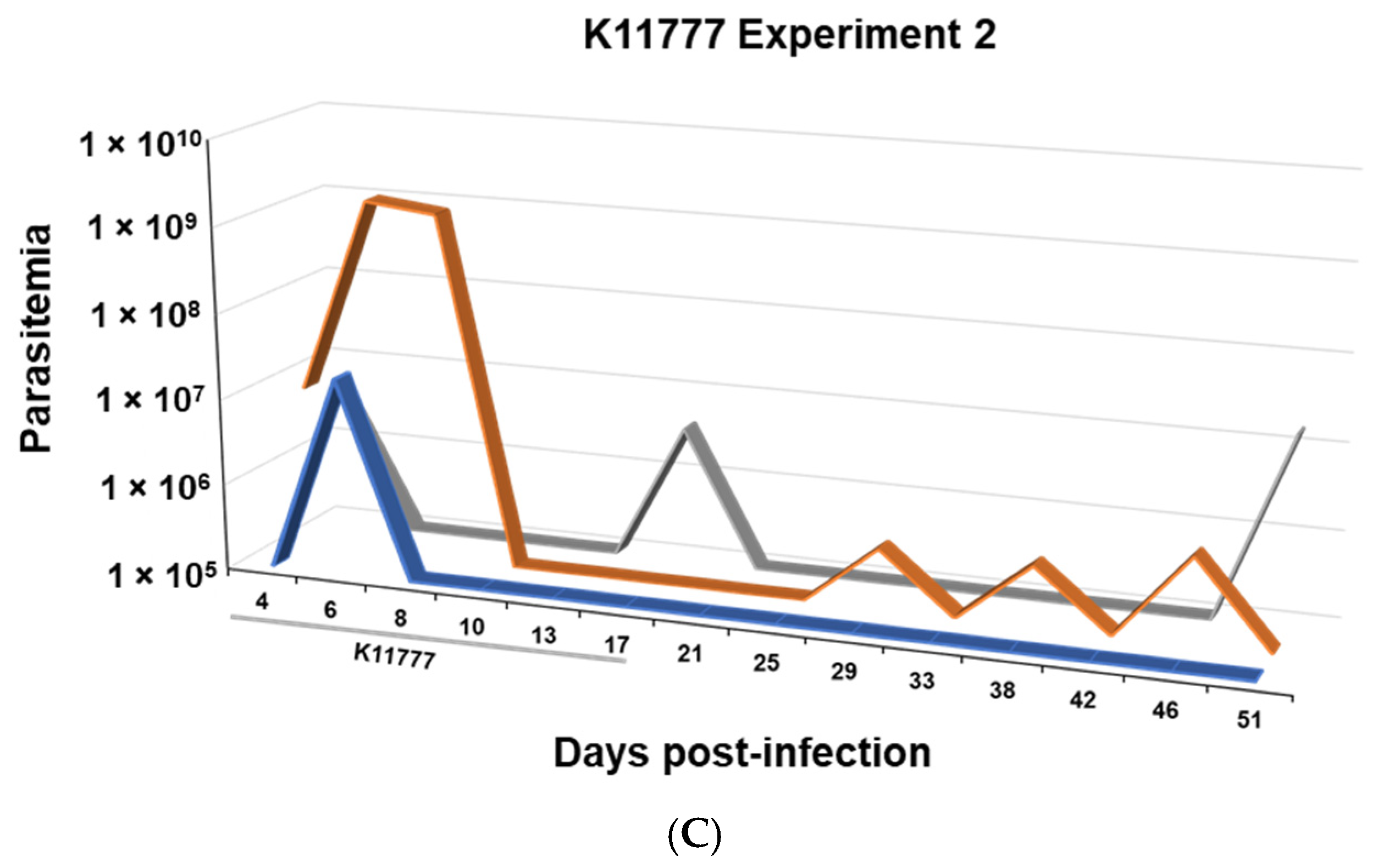
Treatment with K11777 for 17 consecutive days (Expt. 2) (Figure 8 and Figure 9C) also extended mice survival (n = 3) from the 10 dpi observed for the control T. evansi-infected mice to 52 dpi, at which point Experiment 2 was terminated. Statistical analyses (Kaplan–Meier) and Logrank test (*Mantel–Cox method) demonstrated the significant differences (p < 0.0001) in survival between the controls and the K11777-treated mice (Figure 8).
The parasitemia of the non-treated and DMSO-treated mice increased exponentially, reaching maximum values ranging from 5 × 10⁸ to 2 × 10⁹ parasites/mL between days 6 and 10 pi (Figure 9A). In contrast, the K11777-treated mice were able to at least partially control parasite proliferation (Figure 9B,C). In Experiment 1 (Figure 9B), two of the five K11777-treated mice (#3 and #4) survived for the duration of the study (80 dpi), whereas the remaining three mice showed partial parasitemia control during the first three to six wpi, with undulating parasitemia waves, until euthanasia on days 26, 45 and 48 pi, after exhibiting clinical signs of disease, non-social behavior and/or high parasitemia. In Experiment 2 (Figure 9C), parasite proliferation was well controlled, and the three mice survived for the duration of the study, 52 dpi. The twice-daily treatment with K11777, for 17 days, during the initial phase of the infection, resulted in partial parasitemia control and a significant increase in survival. Overall, the administration of the K11777 inhibitor provided a significant therapeutic benefit to T. evansi-infected mice.

4. Discussion

In this study, we identified and characterized p28, p31, 36 and p40 CP activities in a Venezuelan T. evansi strain using gelatin-gel zymography, peptidase class-specific inhibitors and peptidyl substrates. We also demonstrated a clear therapeutic benefit in T. evansi-infected mice treated with the CP inhibitor, K11777.
Acidic, thiol-dependent hemoglobinolytic activities have been documented in several kinetoplastid species, including T. brucei, T. evansi, T. equiperdum, C. fasciculata and Leishmania tarentolae [64]. In keeping with the nomenclature for papain and other thiol-dependent enzymes, the term “trypanopains” was coined to refer to trypanosome CPs [65]. The Venezuelan T. evansi CPs share common characteristics with the p42 and p28 trypanopains present in an African T. evansi camel isolate, and with CPs found in T. b. brucei and T. b. gambiense [65], including their size, pH profile and ability to degrade fibrinogen. Similarly, gelatinase activities (29, 32, 35 and 40 kDa) have been reported in T. evansi and T. equiperdum lysates [66], while a more complex set of eight peptidases (28, 32, 35, 41, 69, 95, 105 and 150 kDa) was described in an Indian T. evansi camel isolate [67].
The p31, p36 and p40 T. evansi CP activities identified here exhibit a narrow pH optimum of ~4.0, consistent with a lysosomal location. In contrast, the p28 activity showed a much broader activity range, between pH 3.0 and 9.0, and hydrolyzed all four equine protein substrates tested at pH 5.0, as well as hemoglobin at pH 7.0, suggesting a possible role in parasite nutrition and avoidance of host defense mechanisms. Other trypanosome CPs, including the T. b. brucei CP [68], the T. cruzi CP, cruzain [69] and the recombinant TbrCATL [51,70], have been localized to lysosomes or lysosome-like organelles, where they participate in the degradation of parasite and host proteins. Several peptidases, including the T. b. gambiense cathepsins and TbrCATL, are released or secreted into the extracellular milieu and have been linked to signal transduction cascades promoting T. brucei passing the blood–brain barrier into the CNS, as well as cardiac dysfunction via perturbation of intracellular calcium levels [31,32,71,72]. Future studies are needed to elucidate the role of T. evansi CPs in parasite survival and host–parasite interaction.
The T. evansi CPs preferentially hydrolyzed Z-Phe-Arg-AMC relative to Z-Arg-Arg-AMC AMC and Gly-Arg-AMC at neutral pH, and were inhibited by K11777, a potent and irreversible pan-cathepsin inhibitor [20,29,37,38,39,40,41,42,43]. Gly-Arg-AMC is a specific substrate for trypsin-like peptidases [73], while the T. evansi CPs preference for a bulky, hydrophobic Phe residue at the substrate P2 position is consistent with previous data for the recombinant cathepsins, TbrCATL and TbCATB [51,74]. Furthermore, the K11777 apparent IC50 value of 3.06 nM that we determined, using the crude T. evansi lysate, closely mirrors those reported for the purified recombinant orthologous peptidases, Cruzain and Rhodesain (1.5–8 nM) [75].
The T. evansi cathepsins, p28, p31, 36 and p40, resolved by gelatin zymography, were also labeled with the 125I-LHVS- PhOH active-site probe, which was originally designed to identify mammalian CPs. The same probe previously identified cathepsins with similar relative motilities (29, 33 and 40 KDa) in T. b. brucei lysates [51]. In addition, the purified p40 T. evansi CP was recognized by the monospecific antibodies developed to recombinant TbrCATL [51], providing further evidence of the close relationship between these two CPs. Edman degradation determined the N-terminal sequence of p40 as APAAV, which is identical to the N-termini of the catalytic domains of other trypanosome C1A orthologs, including the T. brucei CP [63], the T. cruzi, cruzain [60,61] and the CATL peptidases present in T. dionisii and T. rangeli [76]. Both the N-terminal sequence of the purified p40 T. evansi cathepsin and its cross-reactivity with the anti-TbrCATL antiserum are consistent with its classification as a CATL. Based on the post-translational processing reported for TbrCATL [51,70] and cruzain [77], we propose that p40 corresponds to the mature glycosylated peptidase, after autoproteolysis of its N-terminus, but bound to its 11.1 kDa C-terminal domain, whereas p28, p31 and p36 are active isoforms generated by further self-proteolysis and glycosylation. In keeping with suggested nomenclature [19,78], we use the term TevCATL to describe p40 and its differentially processed p28, p31 and p36 products.
Kinetoplastid CPs contribute to nutrient-acquisition and are involved with signaling pathways that subvert host defense mechanisms to cause pathology and disease [19,20,21,72,79]. Our study shows, for the first time, that the pan-cathepsin irreversible inhibitor, K11777, also known as SLV213, targets the TevCATL both in vitro and in vivo, and extends host survival in a mouse infection model in treated mice. Our results suggest that extending the K11777 treatment from 14 to 17 days leads to a more favorable control of parasite proliferation by the host, as evidenced by the undulating, sub-lethal levels of parasitemia and increased survival. The initial phase of parasite infection in this mouse model appears to be a crucial “window of opportunity” for inhibitor action and activation of host defense mechanisms that ultimately determine parasite multiplication and host survival.
These results underscore the importance of identifying TevCATL as a potential drug target for T. evansi. Surra affects domestic and wild animals and T. evansi, its causative agent, is the most widely distributed animal trypanosome through Latin America and Northern Africa, above the tsetse belt, the Middle East and Asia [3,4]. Previous reports have shown that Trypanosomatid CPs are essential for infectivity, parasite survival in the host, and have been implicated in pathogenicity [19,20,21,22]. Our findings extend promising drug discovery reports of K11777, which is considered to be a broad spectrum anti-trypanosomal agent, for the treatment of Chagas disease caused by Trypanosoma cruzi, a human and zoonotic pathogen [61], and HAT, caused by T. b. rhodesiense and T. brucei gambiense. In line with the One Health vision [80], the identification of novel and effective therapeutic targets against surra could lead to the development of new drugs to treat or cure other animal trypanosomosis, including nagana, caused by T. vivax, T. congolense, T. brucei brucei, and T. simiae, and dourine, a sexually transmitted disease in Equidae, caused by T. equiperdum.
The inhibition of TevCATL in vivo, following administration of K11777, resulted in parasite attenuation, perhaps due to a reduced ability of the parasite to evade the host immune response. The orthologous TbrCATL plays a crucial role in virulence and pathogenicity, facilitating parasite entry and trans-endothelial passage through the blood–brain barrier [30,31,79,81]. Our in vivo efficacy data are consistent with those reported by Scory et al. [35] showing that another irreversible CP inhibitor, Z-Phe-Ala-CHN2, effectively decreased parasitemia and prolonged host survival in a T. b. brucei mouse model. Other studies, both in vitro and in vivo, have amply demonstrated the trypanocidal activity of small molecule CP inhibitors [36,37,38,39,69,82]. Steverding et al. [33,34] showed that inhibition of the TbCATL alone is sufficient to kill the bloodstream form of T. brucei. They demonstrated that the trypanocidal effect of the peptidyl vinyl sulfone LU-102 is due to its selective inhibition of TbCATL over TbCATB, thus confirming TbCATL as an essential drug target.
Significant progress in the treatment of HAT was achieved in 2018 with the approval by the European Medicines Agency (EMA) of fexinidazole, a DNA synthesis inhibitor developed by the Drugs for Neglected Diseases initiative (DNDi) in collaboration with Sanofi [83,84]. Fexinidazole is the first all-oral treatment for the hemo-lymphatic and meningo-encephalitic stages of HAT [85,86]. Due to the high levels of toxicity, undesirable side effects of the current drugs, and the ability of the parasite to develop drug (cross)resistance, the development of novel drugs for animal trypanosomosis and HAT remains a high priority [29,42,43,82,87]. Our results open new opportunities for drug discovery to treat animal trypanosomosis caused by T. evansi and other related trypanosomes.

5. Conclusions

Animal trypanosomoses, caused by Trypanosoma brucei brucei, T. evansi and T. equiperdum, are widely distributed throughout the globe and are responsible for substantial direct and indirect costs associated with animal protein production. Trypanosoma evansi, the most widespread animal trypanosome of African origin, causes the disease known as “surra” [2,3]. In this work, we presented the molecular characterization of four CPs present in a Venezuelan strain of T. evansi: p28, p31, p36 and p40. Enzymatic assays with fluorogenic peptidyl substrates and active-site labeling with the 125I-LHVS-PhOH vinyl sulfone inhibitor showed that the T. evansi CPs belong to the Cathepsin L peptidase C1 family, thus their denomination as TevCATL. The irreversible pan-cathepsin vinyl sulfone inhibitor, K11777, targets the TevCATL both in vitro and in vivo, and it extended host survival in a mouse infection model. The potential of TevCATL as therapeutic target for the treatment of surra extends the promising drug discovery studies with several CP inhibitors against Chagas disease due to Trypanosoma cruzi, a human and zoonotic pathogen and HTA, caused by T. b. rhodesiense and T. brucei gambiense and transmitted by the tsetse in sub-Saharan Africa [20,29,33,34,37,38,39,40,41,42,43]. Consequently, and considering the One Health perspective [80], the exploitation of novel drug targets has potential for the development of effective treatments against various pathogenic trypanosomes that affect both humans and animals.

Author Contributions

Conceptualization: A.B., M.G., Y.G.P., C.R.C. and J.H.M. Funding acquisition: M.G. and Y.G.P. Investigation: A.B., Y.G.P., L.M.T., M.G., C.R.C., M.B. and J.H.M. Methodology: A.B., Y.G.P., L.M.T., M.G., C.R.C. and M.B. Data curation: A.B., M.G., Y.G.P., L.M.T. and C.R.C. Writing—original draft: M.G., A.B., Y.G.P. and C.R.C. Writing—review and editing: M.G., C.R.C., L.M.T. and J.H.M. All authors have read and agreed to the published version of the manuscript.

Funding

This research was funded by the GID-21 Program from Decanato de Investigación y Desarrollo-Universidad Simón Bolívar, Project G-98003462 from CONICIT and INCO-DEV: Trypadvac2 No. 003716 to MG and YGP.

Institutional Review Board Statement

The animal study protocol was approved by the Institutional Animal Care and Use Committee of the University of California, San Francisco, U.S.A. (Guide for the Care and Use of Laboratory Animals of the National Institutes of Health, AN079928-02A, 19 March 1999).

Data Availability Statement

The raw data supporting the conclusions of this article will be made available by the authors on request.

Acknowledgments

Thanks to Lennys González (Universidad Simón Bolivar) and Christopher Franklin, Elizabeth Hansell and KC Lim (University of California, San Francisco) for their contributions and technical support, and to Alberto Herrera (Universidad Experimental Francisco de Miranda) for the gift of equine proteins conjugated to polyacrylamide. Special thanks to Howard E. Takiff for proofreading the manuscript and providing critical editing support.

Conflicts of Interest

The authors declare no conflicts of interest. The funders had no role in the design of this study; in the collection, analyses, or interpretation of data; in the writing of the manuscript; or in the decision to publish the results.

Abbreviations

The following abbreviations are used in this manuscript:
CPsCysteine peptidases: also known as cysteine proteases, these are proteolytic enzymes that contain a cysteine residue in their catalytic triad.
CATLCathepsin L: the name cathepsin originates from the Greek word kathepsein, which means to digest. Cathepsins L and B belong to the Clan CA, C1 family of papain-like cysteine peptidases.
CATBCathepsin B: it has dual endo- and exopeptidase activity and plays an essential role in endocytic nutrient processing and parasite survival.
IAAIodoacetic acid: an irreversible alkylating inhibitor that binds covalently to the active-site cysteine residue of peptidases.
K11777 (SLV13)N-methyl-piperazine-phenyl-alanyl-homo-phenylalanyl-vinyl sulfone-phenyl: a potent, broad-range irreversible cysteine protease inhibitor.
PMSFPhenylmethyl sulfonyl fluoride: an irreversible inhibitor of serine proteases and some cysteine peptidases.
NTDsNeglected Tropical Diseases: include a diverse group of conditions caused by various pathogens, including viruses, bacteria, parasites, fungi and toxins that are associated with devastating health, social and economic consequences.
HATHuman African Trypanosomiasis: an NTD caused by Trypanosoma brucei gambiense and Trypanosoma brucei rhodesiense and transmitted through infected tsetse flies in sub-Saharan Africa.

References

  1. Hoare, C.A. The Trypanosomes of Mammals: A Zoological Monograph; Blackwell Scientific Publications: Oxford, UK, 1972. [Google Scholar]
  2. WOAH. Terrestrial Animal Health Code. Chapter 8.23. Infection with Trypanosoma evansi (Surra). Available online: www.woah.org/fileadmin/Home/eng/Health_standards/tahc/2024/en_chapitre_tryp_evansi.htm (accessed on 1 May 2026).
  3. Sazmand, A.; Desquesnes, M.; Otranto, D. Trypanosoma evansi. Trends Parasitol. 2022, 38, 489–490. [Google Scholar] [CrossRef] [Scilit] [PubMed]
  4. Desquesnes, M.; Gonzatti, M.; Sazmand, A.; Thevenon, S.; Bossard, G.; Boulange, A.; Gimonneau, G.; Truc, P.; Herder, S.; Ravel, S.; et al. A review on the diagnosis of animal trypanosomoses. Parasites Vectors 2022, 15, 64. [Google Scholar] [CrossRef] [Scilit] [PubMed]
  5. Kim, J.; Alvarez-Rodriguez, A.; Li, Z.; Radwanska, M.; Magez, S. Recent Progress in the Detection of Surra, a Neglected Disease Caused by Trypanosoma evansi with a One Health Impact in Large Parts of the Tropic and Sub-Tropic World. Microorganisms 2023, 12, 44. [Google Scholar] [CrossRef] [Scilit] [PubMed]
  6. Gutierrez, C.; Desquesnes, M.; Touratier, L.; Buscher, P. Trypanosoma evansi: Recent outbreaks in Europe. Vet. Parasitol. 2010, 174, 26–29. [Google Scholar] [CrossRef] [Scilit] [PubMed]
  7. Desquesnes, M.; Bossard, G.; Patrel, D.; Herder, S.; Patout, O.; Lepetitcolin, E.; Thevenon, S.; Berthier, D.; Pavlovic, D.; Brugidou, R.; et al. First outbreak of Trypanosoma evansi in camels in metropolitan France. Vet. Rec. 2008, 162, 750–752. [Google Scholar] [CrossRef] [Scilit] [PubMed]
  8. Joshi, P.P.; Shegokar, V.R.; Powar, R.M.; Herder, S.; Katti, R.; Salkar, H.R.; Dani, V.S.; Bhargava, A.; Jannin, J.; Truc, P. Human trypanosomiasis caused by Trypanosoma evansi in India: The first case report. Am. J. Trop. Med. Hyg. 2005, 73, 491–495. [Google Scholar] [CrossRef] [Scilit]
  9. Truc, P.; Buscher, P.; Cuny, G.; Gonzatti, M.I.; Jannin, J.; Joshi, P.; Juyal, P.; Lun, Z.R.; Mattioli, R.; Pays, E.; et al. Atypical human infections by animal trypanosomes. PLoS Negl. Trop. Dis. 2013, 7, e2256. [Google Scholar] [CrossRef] [Scilit] [PubMed]
  10. Van Vinh Chau, N.; Buu Chau, L.; Desquesnes, M.; Herder, S.; Phu Huong Lan, N.; Campbell, J.I.; Van Cuong, N.; Yimming, B.; Chalermwong, P.; Jittapalapong, S.; et al. A Clinical and Epidemiological Investigation of the First Reported Human Infection with the Zoonotic Parasite Trypanosoma evansi in Southeast Asia. Clin. Infect. Dis. 2016, 62, 1002–1008. [Google Scholar] [CrossRef] [Scilit] [PubMed]
  11. Buscher, P.; Gonzatti, M.I.; Hebert, L.; Inoue, N.; Pascucci, I.; Schnaufer, A.; Suganuma, K.; Touratier, L.; Van Reet, N. Equine trypanosomosis: Enigmas and diagnostic challenges. Parasites Vectors 2019, 12, 234. [Google Scholar] [CrossRef] [Scilit] [PubMed]
  12. Lai, D.H.; Hashimi, H.; Lun, Z.R.; Ayala, F.J.; Lukes, J. Adaptations of Trypanosoma brucei to gradual loss of kinetoplast DNA: Trypanosoma equiperdum and Trypanosoma evansi are petite mutants of T. brucei. Proc. Natl. Acad. Sci. USA 2008, 105, 1999–2004. [Google Scholar] [CrossRef] [Scilit] [PubMed]
  13. Carnes, J.; Anupama, A.; Balmer, O.; Jackson, A.; Lewis, M.; Brown, R.; Cestari, I.; Desquesnes, M.; Gendrin, C.; Hertz-Fowler, C.; et al. Genome and phylogenetic analyses of Trypanosoma evansi reveal extensive similarity to T. brucei and multiple independent origins for dyskinetoplasty. PLoS Negl. Trop. Dis. 2015, 9, e3404. [Google Scholar] [CrossRef] [Scilit] [PubMed]
  14. Cuypers, B.; Van den Broeck, F.; Van Reet, N.; Meehan, C.J.; Cauchard, J.; Wilkes, J.M.; Claes, F.; Goddeeris, B.; Birhanu, H.; Dujardin, J.C.; et al. Genome-wide SNP analysis reveals distinct origins of Trypanosoma evansi and Trypanosoma equiperdum. Genome Biol. Evol. 2017, 9, 1990–1997. [Google Scholar] [CrossRef] [Scilit] [PubMed]
  15. Zheng, L.; Jiang, N.; Sang, X.; Zhang, N.; Zhang, K.; Chen, H.; Yang, N.; Feng, Y.; Chen, R.; Suo, X.; et al. In-depth analysis of the genome of Trypanosoma evansi, an etiologic agent of surra. Sci. China Life Sci. 2019, 62, 406–419. [Google Scholar] [CrossRef] [Scilit] [PubMed]
  16. Berriman, M.; Ghedin, E.; Hertz-Fowler, C.; Blandin, G.; Renauld, H.; Bartholomeu, D.C.; Lennard, N.J.; Caler, E.; Hamlin, N.E.; Haas, B.; et al. The genome of the African trypanosome Trypanosoma brucei. Science 2005, 309, 416–422. [Google Scholar] [CrossRef] [Scilit] [PubMed]
  17. Oldrieve, G.; Verney, M.; Jaron, K.S.; Hebert, L.; Matthews, K.R. Monomorphic Trypanozoon: Towards reconciling phylogeny and pathologies. Microb. Genom. 2021, 7, 000632. [Google Scholar] [CrossRef] [Scilit] [PubMed]
  18. Lukes, J.; Kachale, A.; Votypka, J.; Butenko, A.; Field, M.C. African trypanosome strategies for conquering new hosts and territories: The end of monophyly? Trends Parasitol. 2022, 38, 724–736. [Google Scholar] [CrossRef] [Scilit] [PubMed]
  19. Caffrey, C.R.; Lima, A.P.; Steverding, D. Cysteine peptidases of kinetoplastid parasites. Adv. Exp. Med. Biol. 2011, 712, 84–99. [Google Scholar] [CrossRef] [Scilit] [PubMed]
  20. Ferreira, L.G.; Andricopulo, A.D. Targeting cysteine proteases in trypanosomatid disease drug discovery. Pharmacol. Ther. 2017, 180, 49–61. [Google Scholar] [CrossRef] [Scilit] [PubMed]
  21. Siqueira-Neto, J.L.; Debnath, A.; McCall, L.I.; Bernatchez, J.A.; Ndao, M.; Reed, S.L.; Rosenthal, P.J. Cysteine proteases in protozoan parasites. PLoS Negl. Trop. Dis. 2018, 12, e0006512. [Google Scholar] [CrossRef] [Scilit] [PubMed]
  22. McKerrow, J.H. The diverse roles of cysteine proteases in parasites and their suitability as drug targets. PLoS Negl. Trop. Dis. 2018, 12, e0005639. [Google Scholar] [CrossRef] [Scilit] [PubMed]
  23. Alvarez, V.E.; Iribarren, P.A.; Niemirowicz, G.T.; Cazzulo, J.J. Update on relevant trypanosome peptidases: Validated targets and future challenges. Biochim. Biophys. Acta Proteins Proteom. 2021, 1869, 140577. [Google Scholar] [CrossRef] [Scilit] [PubMed]
  24. Agbo, E.C.; Majiwa, P.A.; Buscher, P.; Claassen, E.; te Pas, M.F. Trypanosoma brucei genomics and the challenge of identifying drug and vaccine targets. Trends Microbiol. 2003, 11, 322–329. [Google Scholar] [CrossRef] [Scilit] [PubMed]
  25. Kateregga, J.; Lubega, G.W.; Lindblad, E.B.; Authie, E.; Coetzer, T.H.; Boulange, A.F. Effect of adjuvants on the humoral immune response to congopain in mice and cattle. BMC Vet. Res. 2012, 8, 63. [Google Scholar] [CrossRef] [Scilit] [PubMed]
  26. Rawlings, N.D. Twenty-five years of nomenclature and classification of proteolytic enzymes. Biochim. Biophys. Acta Proteins Proteom. 2020, 1868, 140345. [Google Scholar] [CrossRef] [Scilit] [PubMed]
  27. Sajid, M.; McKerrow, J.H. Cysteine proteases of parasitic organisms. Mol. Biochem. Parasitol. 2002, 120, 1–21. [Google Scholar] [CrossRef] [Scilit] [PubMed]
  28. Caffrey, C.R.; Steverding, D. Kinetoplastid papain-like cysteine peptidases. Mol. Biochem. Parasitol. 2009, 167, 12–19. [Google Scholar] [CrossRef] [Scilit] [PubMed]
  29. Caffrey, C.R.; Steverding, D.; Ferreira, R.S.; de Oliveira, R.B.; O’Donoghue, A.J.; Monti, L.; Ballatore, C.; Bachovchin, K.A.; Ferrins, L.; Pollastri, M.P.; et al. Drug Discovery and Development for Kinetoplastid Diseases. In Burger’s Medicinal Chemistry, Drug Discovery and Development; Abraham, D.J., Myers, M., Eds.; Wiley: Hoboken, NJ, USA, 2021; Volume 7, pp. 255–344. [Google Scholar]
  30. Abdulla, M.H.; O’Brien, T.; Mackey, Z.B.; Sajid, M.; Grab, D.J.; McKerrow, J.H. RNA interference of Trypanosoma brucei cathepsin B and L affects disease progression in a mouse model. PLoS Negl. Trop. Dis. 2008, 2, e298. [Google Scholar] [CrossRef] [Scilit] [PubMed]
  31. Nikolskaia, O.V.; de A. Lima, A.P.C.; Kim, Y.V.; Lonsdale-Eccles, J.D.; Fukuma, T.; Scharfstein, J.; Grab, D.J. Blood-brain barrier traversal by African trypanosomes requires calcium signaling induced by parasite cysteine protease. J. Clin. Investig. 2006, 116, 2739–2747. [Google Scholar] [CrossRef] [Scilit] [PubMed]
  32. Elliott, E.B.; McCarroll, D.; Hasumi, H.; Welsh, C.E.; Panissidi, A.A.; Jones, N.G.; Rossor, C.L.; Tait, A.; Smith, G.L.; Mottram, J.C.; et al. Trypanosoma brucei cathepsin-L increases arrhythmogenic sarcoplasmic reticulum-mediated calcium release in rat cardiomyocytes. Cardiovasc. Res. 2013, 100, 325–335. [Google Scholar] [CrossRef] [Scilit] [PubMed]
  33. Steverding, D.; Rushworth, S.A.; Florea, B.I.; Overkleeft, H.S. Trypanosoma brucei: Inhibition of cathepsin L is sufficient to kill bloodstream forms. Mol. Biochem. Parasitol. 2020, 235, 111246. [Google Scholar] [CrossRef] [Scilit] [PubMed]
  34. Steverding, D.; Sexton, D.W.; Wang, X.; Gehrke, S.S.; Wagner, G.K.; Caffrey, C.R. Trypanosoma brucei: Chemical evidence that cathepsin L is essential for survival and a relevant drug target. Int. J. Parasitol. 2012, 42, 481–488. [Google Scholar] [CrossRef] [Scilit] [PubMed]
  35. Scory, S.; Caffrey, C.R.; Stierhof, Y.D.; Ruppel, A.; Steverding, D. Trypanosoma brucei: Killing of bloodstream forms in vitro and in vivo by the cysteine proteinase inhibitor Z-phe-ala-CHN2. Exp. Parasitol. 1999, 91, 327–333. [Google Scholar] [CrossRef] [Scilit] [PubMed]
  36. Du, X.; Hansell, E.; Engel, J.C.; Caffrey, C.R.; Cohen, F.E.; McKerrow, J.H. Aryl ureas represent a new class of anti-trypanosomal agents. Chem. Biol. 2000, 7, 733–742. [Google Scholar] [CrossRef] [Scilit] [PubMed]
  37. Caffrey, C.R.; Scory, S.; Steverding, D. Cysteine proteinases of trypanosome parasites: Novel targets for chemotherapy. Curr. Drug Targets 2000, 1, 155–162. [Google Scholar] [CrossRef] [Scilit] [PubMed]
  38. Dunny, E.; Doherty, W.; Evans, P.; Malthouse, J.P.; Nolan, D.; Knox, A.J. Vinyl sulfone-based peptidomimetics as anti-trypanosomal agents: Design, synthesis, biological and computational evaluation. J. Med. Chem. 2013, 56, 6638–6650. [Google Scholar] [CrossRef] [Scilit] [PubMed]
  39. Doyle, P.S.; Zhou, Y.M.; Engel, J.C.; McKerrow, J.H. A cysteine protease inhibitor cures Chagas’ disease in an immunodeficient-mouse model of infection. Antimicrob. Agents Chemother. 2007, 51, 3932–3939. [Google Scholar] [CrossRef] [Scilit] [PubMed]
  40. Sales Junior, P.A.; Molina, I.; Fonseca Murta, S.M.; Sanchez-Montalva, A.; Salvador, F.; Correa-Oliveira, R.; Carneiro, C.M. Experimental and Clinical Treatment of Chagas Disease: A Review. Am. J. Trop. Med. Hyg. 2017, 97, 1289–1303. [Google Scholar] [CrossRef] [Scilit] [PubMed]
  41. Ettari, R.; Previti, S.; Tamborini, L.; Cullia, G.; Grasso, S.; Zappala, M. The Inhibition of Cysteine Proteases Rhodesain and TbCatB: A Valuable Approach to Treat Human African Trypanosomiasis. Mini Rev. Med. Chem. 2016, 16, 1374–1391. [Google Scholar] [CrossRef] [Scilit] [PubMed]
  42. Previti, S.; Ettari, R.; Cosconati, S.; Amendola, G.; Chouchene, K.; Wagner, A.; Hellmich, U.A.; Ulrich, K.; Krauth-Siegel, R.L.; Wich, P.R.; et al. Development of Novel Peptide-Based Michael Acceptors Targeting Rhodesain and Falcipain-2 for the Treatment of Neglected Tropical Diseases (NTDs). J. Med. Chem. 2017, 60, 6911–6923. [Google Scholar] [CrossRef] [Scilit] [PubMed]
  43. McKerrow, J.H. Update on drug development targeting parasite cysteine proteases. PLoS Negl. Trop. Dis. 2018, 12, e0005850. [Google Scholar] [CrossRef] [Scilit] [PubMed]
  44. Perrone, T.M.; Gonzatti, M.I.; Villamizar, G.; Escalante, A.; Aso, P.M. Molecular profiles of Venezuelan isolates of Trypanosoma sp. by random amplified polymorphic DNA method. Vet. Parasitol. 2009, 161, 194–200. [Google Scholar] [CrossRef] [Scilit] [PubMed]
  45. Sanchez, E.; Perrone, T.; Recchimuzzi, G.; Cardozo, I.; Biteau, N.; Aso, P.M.; Mijares, A.; Baltz, T.; Berthier, D.; Balzano-Nogueira, L.; et al. Molecular characterization and classification of Trypanosoma spp. Venezuelan isolates based on microsatellite markers and kinetoplast maxicircle genes. Parasites Vectors 2015, 8, 536, Erratum in Parasites Vectors 2015, 8, 566. https://doi.org/10.1186/s13071-015-1177-7.. [Google Scholar] [CrossRef] [Scilit] [PubMed]
  46. Lanham, S.M.; Godfrey, D.G. Isolation of salivarian trypanosomes from man and other mammals using DEAE-cellulose. Exp. Parasitol. 1970, 28, 521–534. [Google Scholar] [CrossRef] [Scilit] [PubMed]
  47. Lowry, O.H.; Rosebrough, N.J.; Farr, A.L.; Randall, R.J. Protein measurement with the Folin phenol reagent. J. Biol. Chem. 1951, 193, 265–275. [Google Scholar] [CrossRef] [Scilit]
  48. Heussen, C.; Joubert, F.; Dowdle, E.B. Purification of human tissue plasminogen activator with Erythrina trypsin inhibitor. J. Biol. Chem. 1984, 259, 11635–11638. [Google Scholar] [CrossRef] [Scilit]
  49. Grenier, D.; Chao, G.; McBride, B.C. Characterization of sodium dodecyl sulfate-stable Bacteroides gingivalis proteases by polyacrylamide gel electrophoresis. Infect. Immun. 1989, 57, 95–99. [Google Scholar] [CrossRef] [Scilit] [PubMed]
  50. Kelleher, P.J.; Juliano, R.L. Detection of proteases in polyacrylamide gels containing covalently bound substrates. Anal. Biochem. 1984, 136, 470–475. [Google Scholar] [CrossRef] [Scilit] [PubMed]
  51. Caffrey, C.R.; Hansell, E.; Lucas, K.D.; Brinen, L.S.; Alvarez Hernandez, A.; Cheng, J.; Gwaltney, S.L., 2nd; Roush, W.R.; Stierhof, Y.D.; Bogyo, M.; et al. Active site mapping, biochemical properties and subcellular localization of rhodesain, the major cysteine protease of Trypanosoma brucei rhodesiense. Mol. Biochem. Parasitol. 2001, 118, 61–73. [Google Scholar] [CrossRef] [Scilit] [PubMed]
  52. Bogyo, M.; Shin, S.; McMaster, J.S.; Ploegh, H.L. Substrate binding and sequence preference of the proteasome revealed by active-site-directed affinity probes. Chem. Biol. 1998, 5, 307–320. [Google Scholar] [CrossRef] [Scilit] [PubMed]
  53. Laemmli, U.K. Cleavage of structural proteins during the assembly of the head of bacteriophage T4. Nature 1970, 227, 680–685. [Google Scholar] [CrossRef] [Scilit] [PubMed]
  54. Altschul, S.F.; Gish, W.; Miller, W.; Myers, E.W.; Lipman, D.J. Basic local alignment search tool. J. Mol. Biol. 1990, 215, 403–410. [Google Scholar] [CrossRef] [PubMed]
  55. Perrone, T.; Sanchez, E.; Hidalgo, L.; Mijares, A.; Balzano-Nogueira, L.; Gonzatti, M.I.; Aso, P.M. Infectivity and virulence of Trypanosoma evansi and Trypanosoma equiperdum Venezuelan strains from three different host species. Vet. Parasitol. Reg. Stud. Rep. 2018, 13, 205–211. [Google Scholar] [CrossRef] [Scilit] [PubMed]
  56. Perrone, T.P.; Aso, P.M.; Mijares, A.; Holzmuller, P.; Gonzatti, M.; Parra, N. Comparison of infectivity and virulence of clones of Trypanosoma evansi and Trypanosoma equiperdum Venezuelan strains in mice. Vet. Parasitol. 2018, 253, 60–64. [Google Scholar] [CrossRef] [Scilit] [PubMed]
  57. Engel, J.C.; Doyle, P.S.; Hsieh, I.; McKerrow, J.H. Cysteine protease inhibitors cure an experimental Trypanosoma cruzi infection. J. Exp. Med. 1998, 188, 725–734. [Google Scholar] [CrossRef] [Scilit] [PubMed]
  58. Herbert, W.J.; Lumsden, W.H. Trypanosoma brucei: A rapid “matching” method for estimating the host’s parasitemia. Exp. Parasitol. 1976, 40, 427–431. [Google Scholar] [CrossRef] [Scilit] [PubMed]
  59. Macpherson, A.J.; Yilmaz, B.; Limenitakis, J.P.; Ganal-Vonarburg, S.C. IgA Function in Relation to the Intestinal Microbiota. Annu. Rev. Immunol. 2018, 36, 359–381. [Google Scholar] [CrossRef] [Scilit] [PubMed]
  60. Eakin, A.E.; Mills, A.A.; Harth, G.; McKerrow, J.H.; Craik, C.S. The sequence, organization, and expression of the major cysteine protease (cruzain) from Trypanosoma cruzi. J. Biol. Chem. 1992, 267, 7411–7420. [Google Scholar] [CrossRef] [Scilit]
  61. Cazzulo, J.J.; Couso, R.; Raimondi, A.; Wernstedt, C.; Hellman, U. Further characterization and partial amino acid sequence of a cysteine proteinase from Trypanosoma cruzi. Mol. Biochem. Parasitol. 1989, 33, 33–41. [Google Scholar] [CrossRef] [Scilit] [PubMed]
  62. UniProt. UniProt: The Universal Protein Knowledgebase in 2025. Nucleic Acids Res. 2025, 53, D609–D617. [Google Scholar] [CrossRef] [Scilit] [PubMed]
  63. Mottram, J.C.; North, M.J.; Barry, J.D.; Coombs, G.H. A cysteine proteinase cDNA from Trypanosoma brucei predicts an enzyme with an unusual C-terminal extension. FEBS Lett. 1989, 258, 211–215. [Google Scholar] [CrossRef] [Scilit] [PubMed]
  64. North, M.J.; Coombs, G.H.; Barry, J.D. A comparative study of the proteolytic enzymes of Trypanosoma brucei, T. equiperdum, T. evansi, T. vivax, Leishmania tarentolae and Crithidia fasciculata. Mol. Biochem. Parasitol. 1983, 9, 161–180. [Google Scholar] [CrossRef] [Scilit] [PubMed]
  65. Lonsdale-Eccles, J.D.; Mpimbaza, G.W. Thiol-dependent proteases of African trypanosomes. Analysis by electrophoresis in sodium dodecyl sulphate/polyacrylamide gels co-polymerized with fibrinogen. Eur. J. Biochem. 1986, 155, 469–473. [Google Scholar] [CrossRef] [Scilit] [PubMed]
  66. Giardina, S.; Paganico, G.; Urbani, G.; Rossi, M. A biochemical and immunological comparative study on Trypanosoma equiperdum and Trypanosoma evansi. Vet. Res. Commun. 2003, 27, 289–300. [Google Scholar] [CrossRef] [Scilit] [PubMed]
  67. Yadav, S.C.; Kumar, R.; Kumar, S.; Tatu, U.; Singh, R.K.; Gupta, A.K. Identification and characterization of cysteine proteinases of Trypanosoma evansi. Parasitol. Res. 2011, 109, 559–565. [Google Scholar] [CrossRef] [Scilit] [PubMed]
  68. Lonsdale-Eccles, J.D.; Grab, D.J. Lysosomal and non-lysosomal peptidyl hydrolases of the bloodstream forms of Trypanosoma brucei brucei. Eur. J. Biochem. 1987, 169, 467–475. [Google Scholar] [CrossRef] [Scilit] [PubMed]
  69. Engel, J.C.; Torres, C.; Hsieh, I.; Doyle, P.S.; McKerrow, J.H. Upregulation of the secretory pathway in cysteine protease inhibitor-resistant Trypanosoma cruzi. J. Cell Sci. 2000, 113, 1345–1354. [Google Scholar] [CrossRef] [Scilit] [PubMed]
  70. Koeller, C.M.; Bangs, J.D. Processing and targeting of cathepsin L (TbCatL) to the lysosome in Trypanosoma brucei. Cell Microbiol. 2019, 21, e12980. [Google Scholar] [CrossRef] [Scilit] [PubMed]
  71. McCarroll, C.S.; Rossor, C.L.; Morrison, L.R.; Morrison, L.J.; Loughrey, C.M. A Pre-clinical Animal Model of Trypanosoma brucei Infection Demonstrating Cardiac Dysfunction. PLoS Negl. Trop. Dis. 2015, 9, e0003811. [Google Scholar] [CrossRef] [Scilit] [PubMed]
  72. Morrison, L.J.; Steketee, P.C.; Tettey, M.D.; Matthews, K.R. Pathogenicity and virulence of African trypanosomes: From laboratory models to clinically relevant hosts. Virulence 2023, 14, 2150445. [Google Scholar] [CrossRef] [Scilit] [PubMed]
  73. Mbawa, Z.R.; Gumm, I.D.; Fish, W.R.; Lonsdale-Eccles, J.D. Endopeptidase variations among different life-cycle stages of African trypanosomes. Eur. J. Biochem. 1991, 195, 183–190. [Google Scholar] [CrossRef] [Scilit] [PubMed]
  74. O’Brien, T.C.; Mackey, Z.B.; Fetter, R.D.; Choe, Y.; O’Donoghue, A.J.; Zhou, M.; Craik, C.S.; Caffrey, C.R.; McKerrow, J.H. A parasite cysteine protease is key to host protein degradation and iron acquisition. J. Biol. Chem. 2008, 283, 28934–28943. [Google Scholar] [CrossRef] [Scilit] [PubMed]
  75. Kerr, I.D.; Lee, J.H.; Farady, C.J.; Marion, R.; Rickert, M.; Sajid, M.; Pandey, K.C.; Caffrey, C.R.; Legac, J.; Hansell, E.; et al. Vinyl sulfones as antiparasitic agents and a structural basis for drug design. J. Biol. Chem. 2009, 284, 25697–25703. [Google Scholar] [CrossRef] [Scilit] [PubMed]
  76. Lima, L.; Ortiz, P.A.; da Silva, F.M.; Alves, J.M.; Serrano, M.G.; Cortez, A.P.; Alfieri, S.C.; Buck, G.A.; Teixeira, M.M. Repertoire, genealogy and genomic organization of cruzipain and homologous genes in Trypanosoma cruzi, T. cruzi-like and other trypanosome species. PLoS ONE 2012, 7, e38385. [Google Scholar] [CrossRef] [Scilit] [PubMed]
  77. Hellman, U.; Wernstedt, C.; Cazzulo, J.J. Self-proteolysis of the cysteine proteinase, cruzipain, from Trypanosoma cruzi gives a major fragment corresponding to its carboxy-terminal domain. Mol. Biochem. Parasitol. 1991, 44, 15–21. [Google Scholar] [CrossRef] [Scilit] [PubMed]
  78. Clayton, C.; Adams, M.; Almeida, R.; Baltz, T.; Barrett, M.; Bastien, P.; Belli, S.; Beverley, S.; Biteau, N.; Blackwell, J.; et al. Genetic nomenclature for Trypanosoma and Leishmania. Mol. Biochem. Parasitol. 1998, 97, 221–224. [Google Scholar] [CrossRef] [Scilit] [PubMed]
  79. Lonsdale-Eccles, J.D.; Grab, D.J. Trypanosome hydrolases and the blood-brain barrier. Trends Parasitol. 2002, 18, 17–19. [Google Scholar] [CrossRef] [Scilit] [PubMed]
  80. One Health High-Level Expert, P.; Adisasmito, W.B.; Almuhairi, S.; Behravesh, C.B.; Bilivogui, P.; Bukachi, S.A.; Casas, N.; Cediel Becerra, N.; Charron, D.F.; Chaudhary, A.; et al. One Health: A new definition for a sustainable and healthy future. PLoS Pathog. 2022, 18, e1010537. [Google Scholar] [CrossRef] [Scilit] [PubMed]
  81. Grab, D.J.; Garcia-Garcia, J.C.; Nikolskaia, O.V.; Kim, Y.V.; Brown, A.; Pardo, C.A.; Zhang, Y.; Becker, K.G.; Wilson, B.A.; de A. Lima, A.P.C.; et al. Protease activated receptor signaling is required for African trypanosome traversal of human brain microvascular endothelial cells. PLoS Negl. Trop. Dis. 2009, 3, e479. [Google Scholar] [CrossRef] [Scilit] [PubMed]
  82. Agost-Beltran, L.; Zimmer, C.; Rader, H.J.; Kersten, C.; Schirmeister, T.; Rodriguez, S.; Gonzalez, F.V. Rhodesain inhibitors on the edge of reversibility-irreversibility. Bioorg. Chem. 2024, 153, 107830. [Google Scholar] [CrossRef] [Scilit] [PubMed]
  83. Deeks, E.D. Fexinidazole: First Global Approval. Drugs 2019, 79, 215–220. [Google Scholar] [CrossRef] [Scilit] [PubMed]
  84. Neau, P.; Hanel, H.; Lameyre, V.; Strub-Wourgaft, N.; Kuykens, L. Innovative Partnerships for the Elimination of Human African Trypanosomiasis and the Development of Fexinidazole. Trop. Med. Infect. Dis. 2020, 5, 17. [Google Scholar] [CrossRef] [Scilit] [PubMed]
  85. Matovu, E.; Nyirenda, W.; Eriatu, A.; Alves, D.; Perdrieu, C.; Lemerani, M.; Wamboga, C.; Lejon, V.; Seixas, J.; Signorell, A.; et al. Fexinidazole as a new oral treatment for human African trypanosomiasis due to Trypanosoma brucei rhodesiense: A prospective, open-label, single-arm, phase 2–3, non-randomised study. Lancet Glob. Health 2025, 13, e910–e919. [Google Scholar] [CrossRef] [Scilit] [PubMed]
  86. Kumeso, V.K.B.; Perdrieu, C.; Menetrey, C.; Kyhi, M.I.W.; Tete, D.N.; Camara, M.; Tampwo, J.; Kavunga, P.; Camara, M.L.; Kourouma, A.; et al. Effectiveness and safety of fexinidazole for gambiense human African trypanosomiasis and exploration of adherence in outpatients: A phase 3b, prospective, open-label, non-randomised, cohort study. Lancet Glob. Health 2025, 13, e900–e909. [Google Scholar] [CrossRef] [Scilit] [PubMed]
  87. Kasozi, K.I.; MacLeod, E.T.; Welburn, S.C. African animal trypanocide resistance: A systematic review and meta-analysis. Front. Vet. Sci. 2022, 9, 950248. [Google Scholar] [CrossRef] [Scilit] [PubMed]
Figure 1. Activation of T. evansi peptidases by cysteine. T. evansi lysates (20 μg/lane) were electrophoresed through 12.5% polyacrylamide gels containing 0.1% gelatin. After SDS-PAGE, peptidase activation was carried out in 100 mM Tris-HCl buffer, 2.5 mM CaCl2, pH 7.2, at 37 °C for 30 min, in the absence (lane 1) or in the presence (lane 2) of 25 mM cysteine. Peptidase bands were revealed by staining with 3% Coomassie Brilliant Blue R-250 in water: methanol: acetic acid (50:40:10) followed by destaining in the same solution without dye until the appearance of clear bands. Molecular weight markers (GIBCO/BRL) are indicated on the right.
Pathogens 15 00954 g001
Figure 2. Effect of pH on T. evansi peptidase activity. T. evansi lysates (20 μg/lane) were electrophoresed through 12.5% polyacrylamide gels containing 0.1% gelatin. Activation was performed in 100 mM sodium citrate buffer, pH 3.0–6.0 (panel (A)), or 100 mM Tris-HCl, pH 7.0–8.0, or 100 mM sodium bicarbonate, pH 9.0–10.0 (panel (B)), in the presence of 25 mM cysteine at 37 °C for 30 min. Peptidase bands were revealed by staining with 3% Coomassie brilliant blue R-250 in water: methanol: acetic acid (50:40:10) followed by destaining in the same solution without dye until the appearance of clear bands. Molecular weight markers (GIBCO/BRL, Bethesda, MD, USA) are indicated on the right.
Pathogens 15 00954 g002
Figure 3. Effect of protease class-specific inhibitors on T. evansi peptidases. T. evansi lysates (20 μg/lane) were electrophoresed through 12.5% polyacrylamide gels containing 0.1% gelatin. Gels were incubated in 100 mM sodium citrate, 25 mM cysteine, pH 4.0, for 30 min at 37 °C with 1 mM PMSF, 10 mM EDTA, 1 μM pepstatin,1 mM IAA, 20 μM leupeptin, 10 μM E-64 or 10% DMSO as an inhibitor control. Peptidase bands were revealed by staining with 3% Coomassie Brilliant Blue R-250 in water: methanol: acetic acid (50:40:10) followed by destaining in the same solution without dye until the appearance of clear bands. Molecular weight markers (GIBCO/BRL, Bethesda, MD, USA) are indicated on the right.
Pathogens 15 00954 g003
Figure 4. Degradation of equine proteins by T. evansi lysate. Equine fibrinogen, albumin and IgA (0.03%) were conjugated in 12.5% polyacrylamide gels and evaluated as substrates for T. evansi lysates (20 μg/lane). Zymograms were activated for 30 min at 37 °C in 100 mM Tris-HCl, 25 mM cysteine, 2.5 mM CaCl2, pH 7.0, whereas hemoglobin was tested under two different pH conditions: 100 mM sodium citrate, pH 5.0, and 100 mM Tris-HCl buffer, pH 7.0, containing the same final concentrations of cysteine and CaCl2. Peptidase bands were revealed by staining with 3% Coomassie Brilliant Blue R-250 in water: methanol: acetic acid (50:40:10) followed by destaining in the same solution without dye until the appearance of clear bands. Molecular weight markers are indicated on the right.
Pathogens 15 00954 g004
Figure 5. (A) Hydrolysis of fluorogenic peptidyl substrates by T. evansi lysate. Lysate from 2 × 10⁶ trypanosomes/mL was tested with 4 μM Z-Phe-Arg-AMC, Z-Arg-Arg-AMC or Z-Gly-Arg-AMC in 2 mL 100 mM sodium phosphate, pH 7.0, containing 10% DMSO and 2 mM DTT, at 30 °C. Fluorescence was measured every 60 s for 10 min in a Shimadzu Plus fluorimeter (λexc = 380 nm; λemission = 460 nm). (B) Inhibition of T. evansi CP activity by K11777. CP assays (duplicates) were initiated by pre-incubating parasite lysate (100 µg protein) with K11777 at 0, 1, 10, 100 and 1000 nM in 10% DMSO for 5 min at RT in 0.1 mL 50 mM sodium acetate, 10 mM DTT, pH 5.5. Z-Phe-Arg-AMC (20 µM in 0.1 mL) was added and incubated for 5 min at RT. Fluorescence was measured in a Multiskan fluorimeter (λexc = 380 nm; λemission = 460 nm). An apparent IC50 = 3.06 nM (95% CI: 2.15–4.47 nM, Rsquare = 0.9893) was calculated using a non-linear regression (best fit values) log10 inhibitor vs. normalized response-variable slope with the GraphPad Prism 11.02 statistics software.
Pathogens 15 00954 g005
Figure 6. Active site radiolabeling of T. evansi extracts. Parasite lysate (100 μg) was incubated with the radiolabeled active-site probe, 125I-LHVS- PhOH, on ice for 2 h, followed by SDS-PAGE (12.5%) and autoradiography. To evaluate the specificity of binding by 125I-LHVS- PhOH, lysates were preincubated for 30 min at RT with 10% DMSO (control, lane 1), or 10 µM Z-Phe-Ala-CHN2 (lane 2) or K11777 (lane 3), prior to radiolabeling. The image was analyzed with the NIH Image J software bundled with 64-bit Java Image J 8 (https://imagej.net/ij/download.html, accessed on 21 February 2024). Molecular weight markers (GIBCO/BRL) are indicated on the right.
Pathogens 15 00954 g006
Figure 7. Purification of the TevCATL by Mono Q-FPLC Chromatography and Western blotting with anti-TbrCATL antibodies. (A) T. evansi lysate (2 × 10⁹ trypanosomes/mL) was separated using a MonoQ HR5/5 column equilibrated with 20 mM Tris-HCl, pH 8.0, and a 0.04–0.3 M acetonitrile gradient, as indicated (straight line). Fractions (0.5 mL each) were collected at a flow rate of 0.5 mL/min, quantitated by the BCA reagent (---) and tested for CATL activity with the Z-Phe-Arg-AMC substrate (20 μM) in 50 mM sodium acetate, 10 mM DTT, pH 5.5 (bars). (B) Aliquots of the starting material (SM), wash (W, fractions 1–3) and fractions 4–11 (5 µg) were subjected to 10% SDS-PAGE, blotted onto PVDF, and reacted with monospecific rabbit antibodies raised to the recombinant TbrCATL (1/4000 dilution). Horseradish peroxidase-conjugated goat anti-rabbit IgG was employed as the secondary antibody (1/5000 dilution), and the blot was developed using the ECL detection reagent (Amersham, Cytiva, MA, USA).
Pathogens 15 00954 g007
Figure 8. Effect of K11777 on the survival of mice infected with T. evansi. Three-week-old, female BALB/c mice were infected IP with 5 × 10⁴ parasites. Control groups either received no treatment (n = 6) or vehicle 10% DMSO (n = 5). In Experiment 1, mice (n = 5) received 100 µL of K11777 (50 mg/mL, IP in 10% DMSO) 12 hpi and twice daily for the subsequent 14 days. Four of the five treated mice received equal doses on days 16, 18 and 20 pi (Group 1: # 2 and #3), or on days 30 and 34 pi (Group 2: # 4 and #5). In Experiment 2, mice (n = 3) were treated with 100 µL of K11777 (50 mg/mL, IP in 10% DMSO), beginning 12 hpi, and then twice daily for 17 days. Mice survival was monitored for 80 days (Expt. 1) or for 52 days (Expt. 2), at which time the surviving animals were euthanized. Animals with parasitemia between 5 × 10⁸–2 × 10⁹ parasites per mL or displaying signs of severe disease, including lethargy or lack of social behavior and trans-grooming, were euthanized. Survival analyses (Kaplan–Meier) using GraphPad Prism 11.02 showed statistically significant differences between the control, the DMSO control and the K11777-treated infected mice with p values < 0.0001 (Logrank test for trend).
Pathogens 15 00954 g008
Figure 9. Effect of the K11777 on blood parasitemia and survival of T. evansi-infected mice. Three-week-old, female BALB/c mice were infected IP with 5 × 10⁴ parasites. Parasitemia was estimated every 2 days by wet blood film examination, using the Herbert and Lumsden “matching method” [58]. Animals with parasitemia reaching between 5 × 10⁸ and 2 × 10⁹ parasites/mL and/or displaying lethargy, or lack of social behavior and trans-grooming, were euthanized. (A) Non-treated and DMSO vehicle control experiments. The control T. evansi-infected mice received no treatment (n = 6) and the vehicle control mice (n = 5) received 100 µL 10% DMSO starting 12 hpi and twice daily through 10 dpi. (B) In K11777 Experiment 1, the T. evansi-infected mice (n = 5) received a single IP dose of 100 µL K11777 (50 mg/mL in 10% DMSO) administered 12 h pi and then twice daily for the next 14 days. Two of the five mice then received additional 50 mg/mL IP doses on days 16, 18 and 20 pi (Group 1), whereas two other mice received additional K11777 IP (100 μL of 50 mg/mL) doses on 30 and 34 dpi (Group 2), as indicated by the arrows. (C) In K11777 Experiment 2, the T. evansi-infected mice (n = 3) were treated with a single IP dose of 100 µL K11777 (50 mg/mL in 10% DMSO) 12 hpi and then twice daily for the next 17 days.
Pathogens 15 00954 g009aPathogens 15 00954 g009b
Disclaimer/Publisher’s Note: The statements, opinions and data contained in all publications are solely those of the individual author(s) and contributor(s) and not of MDPI and/or the editor(s). MDPI and/or the editor(s) disclaim responsibility for any injury to people or property resulting from any ideas, methods, instructions or products referred to in the content.