Review

Study on the Pathogenic Mechanism of Pasteurella multocida

1
College of Veterinary Medicine, Inner Mongolia Agricultural University, Hohhot 010018, China
2
College of Pharmacy, Baotou Medical College, Baotou 014040, China
3
College of Pharmacy, Jinan University, Guangzhou 511443, China
4
The First Affiliated Hospital of Baotou Medical College, Baotou 014010, China
*
Author to whom correspondence should be addressed.
These authors contributed equally to this work.
Pathogens 2026, 15(9), 881; https://doi.org/10.3390/pathogens15090881
Submission received: 3 July 2026 / Revised: 12 August 2026 / Accepted: 17 August 2026 / Published: 22 August 2026
(This article belongs to the Section Bacterial Pathogens)

Abstract

Pasteurella multocida is highly prevalent in animal populations. It is usually a component of the normal microbiome of the oral, nasopharyngeal and upper respiratory tracts of animals and can cause varying degrees of infection and even death in animals. P. multocida poses a threat to public health safety because humans can be infected by being bitten by infected animals or coming into contact with their nasal secretions. This article summarises the morphological and culture characteristics of P. multocida, in addition to serotyping, virulence factors and pathogenesis, aiming to explore the mechanism by which the pathogen establishes acute and chronic infections. The article also summarises the mechanism underlying the activation of inflammasomes after P. multocida infection, providing a reference for exploring the epidemiological laws and pathogenesis of the bacterium and for formulating prevention and control strategies.

1. Introduction

Pasteurella multocida is one of the most common commensal and opportunistic pathogens found in domestic and wild animals worldwide and is highly prevalent in animal populations. It is often found as part of the microbial community in the oral, nasopharyngeal and upper respiratory tracts of animals [1]. P. multocida can cause serious infectious diseases in livestock, including bovine respiratory disease (BRD) [2], bovine haemorrhagic septicaemia and progressive atrophic rhinitis in pigs and fowl cholera, rabbit snuffles and pneumonia in small ruminants and pigs [3,4] (Figure 1). These diseases often have acute, febrile and highly contagious characteristics, causing considerable economic losses to the livestock industry. Furthermore, P. multocida can interact with other pathogens, contributing to the formation of respiratory disease complexes or endemic pneumonia in cattle and pigs [5]. P. multocida can be transmitted to humans through animal bites or contact with infected animals or carcasses, causing pulmonary infections, arthritis, encephalitis and other serious diseases [6,7,8].
Cats and dogs, as the most common companion animals worldwide, are the primary reservoirs and sources of P. multocida infection for humans [9,10]. The bacterium is a commensal of the oral and nasopharyngeal cavities of these animals, with carriage rates of 70–90% in cats and 20–50% in dogs [10,11,12]. Consequently, P. multocida can be isolated from approximately 75% and 50% of cat and dog bite wounds, respectively [10,13]. A recent retrospective study of 482 human cases further confirmed that animal exposure was the primary source of infection, with cats accounting for 54.1% and dogs for 29% of cases [10]. Zoonotic pathogens are usually transmitted to humans through animal bites or nasal secretions, and P. multocida is the most commonly isolated bacterium in human infections [14,15,16].
In recent years, rapid developments in molecular biology and immunology have enhanced our understanding of the pathogenic mechanism of P. multocida [17]. The pathogenicity of P. multocida is closely related to its complex virulence factors, including capsules, lipopolysaccharides (LPSs), outer membrane proteins, iron carriers, sialidases and toxins, which play a crucial role in bacterial colonisation, evasion of host immunity, invasion of cells and tissue damage [18,19]. Moreover, P. multocida infection is not a simple interaction between bacteria and host cells; rather, it involves complex host immune responses [20]. The activation of inflammasomes is one of the important mechanisms by which a host responds to P. multocida infection. Inflammasomes are multi-protein complexes that can rapidly assemble and activate in response to pathogen-associated molecular patterns (PAMPs) or damage-associated molecular patterns (DAMPs), triggering a series of inflammatory responses, which include the release of pro-inflammatory cytokines and pyroptosis in cells [21,22]. These responses help clear pathogens but may lead to tissue damage and the progression of diseases.
Although previous reviews have comprehensively summarised the virulence factors, epidemiology and clinical manifestations of P. multocida infections, a systematic overview of the host inflammatory response, particularly the role of inflammasome activation in this bacterium’s pathogenesis, remains lacking. Most existing reviews have focused on the bacterial side of the host–pathogen interaction, with limited attention to how the host immune system recognises and responds to P. multocida infection at the molecular level. The present review fills this gap by providing a focused and up-to-date synthesis of the molecular mechanisms by which P. multocida virulence factors trigger inflammasome activation and how this inflammatory response contributes to both bacterial clearance and tissue pathology. By integrating recent findings from in vitro and in vivo studies, this review presents a novel perspective that bridges bacterial pathogenesis and host immunology and may inform future research directions and therapeutic strategies.
The aim of this review is to explore the pathogenic mechanism of P. multocida, especially its relationship with inflammasome activation. By elucidating how P. multocida evades host immunity, invades cells and causes tissue damage through its virulence factors, as well as how the host responds to infection via inflammasome activation, we hope to provide new insights and strategies for the prevention, diagnosis and treatment of P. multocida-related diseases. This review will help us better understand the role of inflammasomes in anti-infection immunity, laying the foundation for the development of novel immunotherapies and vaccines.

2. Morphology and Serotypes of Pasteurella multocida

P. multocida is a small, rod-shaped or short bacillus with blunt ends and a nearly elliptical shape, measuring approximately 0.2–0.4 μm in width and 0.5–2.5 μm in length. It is Gram-negative and has no spores or flagella [23]. Infected diseased tissues or body fluids stained with Wright’s stain or methylene blue show typical bipolar staining. Freshly isolated virulent strains possess a mucoid capsule, which rapidly disappears upon cultivation. P. multocida is not highly fastidious and grows well on common media such as nutrient agar and blood agar, forming small circular colonies within 24 h at 37 °C; P. multocida does not grow on MacConkey agar, a feature that aids in its differentiation from other Enterobacteriaceae. When cultivated on blood agar plates for 24 h, it forms small, pale greyish-white, smooth and shiny dewdrop-like colonies with neat edges without haemolysis. It is an aerobic or facultatively anaerobic bacterium with an optimum temperature of 37 °C and a pH range of 7.2–7.4 [24]. Most strains can decompose glucose, fructose, sucrose, mannose and galactose, producing acid but not gas and can ferment mannitol but generally do not ferment lactose.
The serotypes of P. multocida are primarily classified according to the different characteristics of their capsule and somatic antigens [25]. Capsule antigen typing divides P. multocida into five capsule serotypes: A, B, D, E and F [26] (Table 1). Type A is the most common and widely distributed serotype, associated with fowl cholera, bovine respiratory disease (BRD), and pneumonia in pigs, among other hosts [27]. Type B (with arabinose, mannose and galactose as the main components of the capsule) is the primary cause of haemorrhagic septicaemia in cattle and buffaloes, and has also been occasionally reported in pigs [28]. Type D P. multocida (whose capsule is composed primarily of heparosan, a heparin precursor) is a major causative agent of progressive atrophic rhinitis in pigs. However, only toxigenic strains that produce the Pasteurella multocida toxin (PMT) are pathogenic, and type A strains, often in combination with Bordetella bronchiseptica, can also contribute to the disease [29]. Type E strains are rarely isolated and appear to be largely restricted to specific regions in Africa [30]. Although serotype E was historically associated with haemorrhagic septicaemia in cattle, subsequent studies have established that serotype B:2 is the primary etiological agent of this disease in most endemic regions, particularly in Asia [31]. Their isolation frequency varies considerably by region and host species, but they remain uncommon even within endemic areas. Type F (with chondroitin as the main component of the capsule) can be isolated from animals [32,33,34]. For somatic antigen typing, the classification of P. multocida’s somatic antigen (O antigen) is relatively complex. P. multocida can be divided into 12 serotypes through agglutination reactions, but this method has issues, such as cross-reactions and autoagglutination, which limits its application [35,36]. Another historical typing method is the Heddleston scheme, which uses a gel diffusion precipitin test to classify P. multocida into 16 somatic serotypes (types 1–16) based on the antigenicity of its lipopolysaccharide (LPS) [37]. However, this serological method suffers from significant limitations, including poor reproducibility, cross-reactivity, and the inability to type many isolates. Consequently, it has been largely superseded by molecular methods that assign strains to eight LPS genotypes (L1–L8) based on the genetic structure of the LPS outer core locus, which offer improved resolution and reproducibility. Isolated strains are typically designated by their capsular serotype and LPS type (e.g., A:1, D:L6) [38]. While L1, L3, L4, and L6 are the most frequently reported genotypes, primarily associated with capsular types A and D, other genotypes such as L2 (often linked to serotype B) and L5, L7, L8 have also been identified, albeit with less well-defined host and disease associations.
Table 1. Comparative table for P. multocida serotypes.
Capsular SerotypeLPS Genotype (L-Type)Primary HostsAssociated Diseases
A1, 3, 4Cattle, pigs, poultry, rabbits, cats, dogsPneumonia, fowl cholera, bovine respiratory disease (BRD), rabbit snuffles, wound infections (bites)
B2, 5Cattle, buffalo, pigsHaemorrhagic septicaemia (in cattle/buffalo), progressive atrophic rhinitis (in pigs)
D3, 4, 6Pigs, cattle, rabbitsProgressive atrophic rhinitis (toxigenic strains), pneumonia
ECattle, buffaloRare; sporadic cases of haemorrhagic septicaemia reported mainly in Africa (note: serotype B is the primary etiological agent worldwide)
FPoultry (turkeys, chickens), pigs (occasionally)Pneumonia, fowl cholera (less common) (other L-types (e.g., L5, L7, L8) have also been reported in various isolates, but their host associations remain unclear; notably, L2 is predominantly associated with serotype B and is not typically found in serotypes A or D)

3. Epidemiology of Pasteurella multocida

P. multocida can infect a variety of animals, including domestic animals (such as pigs, cows and sheep), poultry (such as chickens, ducks and geese), wildlife and humans [39,40,41]. This wide host range makes the bacterium easily transmittable and prevalent in animal populations. In addition, P. multocida has diverse transmission routes, including direct contact (such as bites and scratches), indirect contact (such as contact with contaminated objects or environments), airborne transmission (such as droplet transmission) and digestive tract transmission (such as consuming contaminated food or water) [42]. These diverse transmission routes increase the risk of the bacterium spreading in animal populations and humans. The onset of P. multocida infection is mostly detected in young animals, and the infection has a high mortality rate [14]. The clinical manifestations of P. multocida infection are highly diverse and host-dependent. In rabbits and pigs, the infection can present primarily as upper respiratory tract disease, with rhinitis as a predominant symptom, or as lower respiratory tract infection leading to pneumonia. In cattle and other ungulates, P. multocida is a major contributor to respiratory disease complexes, such as bovine respiratory disease (BRD), often manifesting as chronic and fibrous bronchopneumonia [43,44]. Additionally, in cattle and buffalo, certain serotypes, particularly B:2, can cause acute, fatal haemorrhagic septicaemia, a systemic infection distinct from respiratory disease. In poultry, P. multocida subsp. multocida is the causative agent of fowl cholera, which often presents as an acute septicaemic disease with high mortality, rather than a localized respiratory infection. In wildlife, outbreaks frequently manifest as acute septicaemia (e.g., in saiga antelope), though chronic respiratory forms can also occur [45]. Serotypes F and D are rarely observed in poultry. In chronic or subclinical forms of fowl cholera, the respiratory tract can serve as a primary site of infection, with clinical signs including asymptomatic or mild chronic sinusitis, conjunctivitis or pneumonia; however, acute infections can progress rapidly and cause high mortality.
In livestock and wildlife, environmental and management factors play crucial roles in triggering P. multocida outbreaks. As an opportunistic commensal, P. multocida typically resides harmlessly in the upper respiratory tract; however, environmental stressors, including transportation, weaning, overcrowding, poor ventilation and dietary changes, can precipitate transition to an invasive infection [43]. Climatic factors also contribute substantially: in rabbits, elevated ammonia concentrations, suboptimal temperatures and high relative humidity (exceeding recommended 60–70%) increase pasteurellosis prevalence and severity [46,47]; in zoo animals, outbreaks have been linked to changes in rainfall, humidity and temperature [48]; and in saiga antelope, unusually high humidity and temperature preceded a mass die-off of approximately 200,000 animals from haemorrhagic septicaemia [49,50]. In addition, physiological states such as pregnancy and lactation have been associated with increased mortality in rabbits, likely due to immune modulation [47]. Collectively, these observations highlight that P. multocida disease emerges from a complex interplay between the pathogen, host, and environment.
P. multocida infections in humans primarily occur via contact with animals through bites, scratches or mucous secretions. Among these, cats and dogs are the most frequently implicated sources, reflecting their high rates of carriage and close physical proximity to humans [51]. Approximately 300,000 patients are seen annually in emergency departments in the United States alone for animal scratches or bites, and P. multocida is the most common pathogen associated with infections in these patients. The infection rate for cat bites (20–80%) is considerably higher than that for dog bites (3–18%) [52], and cats are involved in 60–80% of human P. multocida infections [53].
Following an animal bite or scratch, local symptoms progress rapidly. Common manifestations include swelling, cellulitis, and purulent exudate at the wound site [54,55]. In most cases, P. multocida infection remains localized to the skin and soft tissues around the bite wound. However, in severe cases—particularly in immunocompromised individuals or those with underlying comorbidities—the infection can progress to bacteraemia, sepsis, osteomyelitis, endocarditis, or meningitis [54]. The mortality rate varies considerably by infection type: it is very low for localized wound infections, approximately 8–31% for bacteraemia [56,57], and up to 30% for meningitis [58]. The risk of severe disease and death is considerably higher in immunocompromised individuals than in the general population [59,60]. The clinical outcome of P. multocida infection is profoundly influenced by the host’s immune status. Immunocompromised individuals, including those with malignancies, diabetes mellitus, chronic liver disease (particularly cirrhosis), end-stage renal disease or those undergoing chemotherapy or immunosuppressive therapy, are at substantially heightened risk for developing severe and invasive infections [61,62,63]. In such patients, P. multocida can cause fulminant infections, including bacteraemia, sepsis, endocarditis, peritonitis and septic shock, even in the absence of an overt animal bite or scratch [64,65]. Multiple risk factors have been associated with increased risk for developing septicaemia, including advanced age, chronic liver disease and diabetes mellitus. In particular, liver cirrhosis has been frequently reported in association with P. multocida septicaemia. Conversely, infections in immunocompetent hosts are typically milder and more localised, although severe cases have occasionally been reported [66]. These observations highlight that P. multocida functions as a classic opportunistic pathogen, whose pathogenic potential is magnified in the setting of compromised host defences. Furthermore, co-infections with other pathogens can predispose an individual to P. multocida infection. For example, SARS-CoV-2 infection has been reported to promote P. multocida pulmonary coinfection through alteration of the epithelial–endothelial barrier and virus-induced immunosuppression [67]. Similarly, bovine herpesvirus-1 (BHV-1) infection enhances the adherence and invasion of P. multocida to host cells, thus illustrating how viral co-infections can increase susceptibility to bacterial superinfection [68].
In addition to bite-related infections, P. multocida can also be transmitted via the respiratory route, primarily through inhalation of contaminated aerosols or droplets, or through close contact with the respiratory secretions of infected animals. Human respiratory tract infections occur frequently, mainly in patients with underlying chronic lung diseases (such as bronchiectasis or chronic obstructive pulmonary disease), manifesting as severe pneumonia, lymphadenopathy, and epiglottitis [6,69,70,71].

4. Virulence Factors and Pathogenesis of Pasteurella multocida

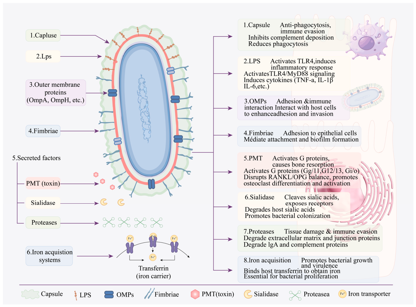
In addition to capsules and LPSs, the virulence factors of P. multocida are adhesins (PfhA and PtfA), iron acquisition systems (ExbB-TonB, HgbA and HgbB), sialidases (NanH and NanB), outer membrane protein (OMP; OmpH and Oma87) and P. multocida toxin (PMT) [72,73,74] (Figure 2) (Table 2). Several important virulence factors are described below. The initial step of infection requires bacterial colonization and adhesion to host cells, mediated by adhesins and fimbriae. Subsequently, the capsular polysaccharide, which mimics host glycosaminoglycans, enables the bacteria to evade innate immune recognition and phagocytosis [75,76,77]. The second obstacle is a host’s inherent immune system, which involves phagocytosis, bactericidal effect of complements and isolation of free iron. However, the structure of the pathogen’s capsular polysaccharide is similar to that of the vertebrate glycosaminoglycan and provides bacteria with conducive conditions, such as anti-phagocytosis and molecular simulation, thus enabling the bacterium to escape a host’s immune system [78]. When P. multocida breaks through the immune barrier and enters blood vessels, it proliferates, increases its virulence and causes bacteraemia [79]. Then, it is transported to various organs, causing tissue degeneration and necrosis, organ dysfunction, body sepsis and death. The virulence factor of P. multocida becomes the key factor for bacteria to invade the body and cause diseases and is the entry point for the development of vaccines and disease resistance at the immune level.

4.1. Capsule

P. multocida is a typical capsular bacterium, and the composition and structure of the capsular materials found in serotypes A, D and F are extremely similar to mammalian glycosaminoglycans, consisting mainly of hyaluronic acid (HA) [80], heparin and chondroitin. HA is a polymer of D-glucuronic acid and N-acetyl-D-glucosamine [81]. Glycosaminoglycan capsular polysaccharide is considered an essential virulence factor for P. multocida, providing protection against other surface antigens from the host’s immune system, preventing phagocytosis, and complementing bactericidal activity [82,83,84], which can cause haemorrhagic septicaemia and avian cholera [75,85]. Capsular hyaluronic acid is thought to contribute to immune evasion by mimicking host glycosaminoglycans, thereby disguising the bacterium from host immune recognition [75,82]. Al-Haddawi et al. [86] observed that P. multocida serotype A:3 strains adhered to and colonised lung explants, possibly because of the high affinity of HA capsular binding to specific glycoproteins in epithelial cells. Beyond its structural and functional characterisation, recent studies have revealed regulatory mechanisms governing capsule synthesis. Fis, a nucleoid-associated protein, positively regulates the transcription of capsular glycosaminoglycan genes in P. multocida. Fis’s expression is upregulated in vivo and correlates positively with capsular production and virulence. The deletion of Fis reduces capsular synthesis, biofilm formation, and resistance to macrophage phagocytosis [87].

4.2. Lipopolysaccharides

LPS is a complex molecule composed of lipid A, core oligosaccharides, and, in many Gram-negative bacteria, O-antigens. However, P. multocida produces a lipooligosaccharide (LOS) that lacks the highly variable O-antigen polysaccharide repeats found in enteric bacteria like E. coli or Salmonella. The structural diversity and serotypic specificity of P. multocida LOS are instead determined by variations in the structure of the outer core oligosaccharide, not by an O-antigen [88,89]. Lipid A is the innermost and most toxic part of LPSs. It is a scaffold of two glucosamine units connected by a β-1, 6-glucoside bond, on which multiple fatty acid chains are attached. The number and length of these fatty acid chains vary by bacterial species [90,91]. LPSs play a critical role in pathogenesis and are the major stimulators of a host’s immune response, which is a key determinant of the protective effect of bacteria [92]. LPSs induce a strong innate immune response primarily through toll-like receptor (TLR) 4, whereas other bacterial components such as lipoproteins are recognized by TLR2 [88]. Upon invasion by Gram-negative bacteria, immature dendritic cells near the site of inflammation (induced by an innate immune response) bind to LPS/LBP complexes (via CD14/MD2 receptors), triggering TLR4 activation and transforming into mature antigen-presenting cells, which then migrate to local lymph nodes. A series of antigen-specific T lymphocytes are then activated, which in turn initiates the proliferation of B cells and subsequently produces antibodies that are specific to LPSs and other bacterial components. Depending on the properties of invading bacteria, a cell-mediated immune response may be activated, and CD4 helper T cell subpopulations are recruited to the site of infection [93,94,95]. Zhao et al. [96] constructed a new attenuated live vaccine strain PMZ2 of P. multocida, which deleted the entire gatA gene and a part of the hptB gene that produced a truncated LPS structure. The oral or intranasal inoculation of mutant strains induces high levels of serum IgG, potent bactericidal action and considerable mucosal IgA response. In the PMZ2 oral and intranasal vaccination groups, considerable reduction in bacterial loads in the blood, spleen, liver, and lungs alleviates spleen and liver lesions. P. multocida LPS can induce the release of pro-inflammatory and immunomodulatory cytokines, such as interleukin (IL)-1α, IL-6, TNF-α, interferon α and IL-12 from mouse spleen cells [97]. Yap et al. [98] found that LPSs play an important role in increasing the ability of P. multocida B:2 to invade cells by regulating the actin microfilaments of infected cells. This type of regulation is a key step in the entry of bacteria into the bloodstream and subsequent sepsis.

4.3. Filamentous Hemagglutinin

P. multocida possesses two filamentous hemagglutinins, PfhB1 and PfhB2 [99,100]. The C-terminal contains a YopT-like cysteine protease domain. The YopT domain of PfhB2 contains a conserved Cys-His-Asp catalytic triplet that defines a member of the YopT family and has a high sequence similarity to the prototype YopT from the genus Yersinia [101]. These proteins are similar to the LspA1 and LspA2 filamentous hemagglutinins from H. ducreyi [102]. PfhB2 has been shown to play a key role in the pathogenesis of diseases associated with colonization or invasion of respiratory mucosal surfaces. Tatum et al. [103] found that the PfhB2 mutant of P. multocida was highly attenuated by nasal administration and less attenuated by intravenous administration in turkey, and the PfhB2 mutant showed no increased sensitivity to the bactericidal activity of the serum complement system. Recent studies have shown that inoculation with recombinant PfhB2 peptide P-1059 (serotype A:3) derived from P. multocida can protect turkeys against P-1059 attack [104].

4.4. OMP

Constituting the main structure of the outer membrane of Gram-negative bacteria, outer membrane proteins are related to nutrient acquisition and ion transport [72]. OmpH and OmpA are the most typical virulence factors of P. multocida and play an important role in bacterial infection and disease [74,105], particularly in adhesion and invasion [106]. DNA sequence analysis shows that most of the OmpA sequences of P. multocida can be divided into two major alleles, namely, OmpA allele (I) and allele (II). P. multocida strains with OmpA (I) are more aggressive and virulent than P. multocida strains with OmpA (II). Pm0442, which is a lipoprotein located in the outer membrane of P. multocida [107], was significantly upregulated in the lung tissues of mice infected with P. multocida, and the toxicity of Pm0442 gene deletion strain PmCQ2Δ0442 was significantly reduced [108]. After OmpH gene deletion, bacterial adhesion ability and virulence were significantly reduced [109]. Recombinant PmOmpA induced a strong Th2 type immune response in mice and produced high immunoglobulin G1 antibodies [110]. Okay et al. [111] demonstrated the immunogenicity and protective efficacy of recombinant lipoprotein E and OmpH of P. multocida A:3 in mice.

4.5. Iron Carrier

Iron is an essential growth factor for almost all bacteria, and low concentrations of free iron on mucous membranes and tissues are a host’s first lines of defence against bacterial infection; access to iron may be a major determining factor for pathogens to maintain themselves in animal hosts [112]. Iron regulatory outer membrane proteins belong to a class of proteins embedded in the outer membrane of P. multocida and can participate in bacterial adhesion, invasion and immune escape, thus affecting virulence [113,114,115]. P. multocida produces siderophores and non-classical transferrin receptors that promote iron absorption [116,117]. Haemoglobin-binding receptors have been characterized [118,119]. Luo et al. [120] expressed and purified aspartate amino lyase (AspA), glycerol diester kinase (DgK) and 30S ribosomal protein S6 (RpsF) to characterise three novel proteins identified under iron-restricted conditions and used them to immunise chickens; the results showed that AspA, DgK and RpsF proteins induced 80.0%, 66.7% and 80.0% immunity, respectively. These data suggest that the three novel proteins identified in the supernatant of the medium play an important role in the survival of bacteria under iron-restricted conditions and thereby protect chickens from Pasteurella multocida. Kharb et al. [121] showed that the extraction of the outer membrane proteins of P. multocida B:2 cultured under normal conditions (OMP) and iron deficiency conditions (IROMP) induced a higher antibody response. Notably, Fis also determines whether P. multocida can utilise bound iron ions for survival, as Fis deletion severely restricts iron acquisition and attenuates pathogenicity [87]. This expands our understanding of iron homeostasis beyond conventional iron carriers.

4.6. Other Virulence Factors

P. multocida toxin (PMT) is another important virulence factor. It is a 146 kDa heat-labile toxin encoded by the toxA gene and is the primary causative agent of progressive atrophic rhinitis (PAR) in pigs [122]. PMT activates at least four G-protein families: Gq/11, G12/13, Gi/o, and Gs. Through its deamidase activity, PMT irreversibly activates Gq and Gα13, leading to RhoA inactivation in osteoblasts and subsequent suppression of bone formation. Concurrently, PMT upregulates RANKL expression, which promotes osteoclast differentiation and activity [123,124]. The net effect of these combined actions—reduced bone formation coupled with increased bone resorption—ultimately leads to turbinate atrophy [125,126]. Kim et al. [127] demonstrated that deletion of the toxA gene significantly reduced the toxicity of P. multocida, confirming the essential role of PMT in the toxigenesis of this bacterium. P. multocida uses sialases to hydrolyse sialic acid and obtain nutrients and has two genes encoding sialases: NanH and NanB. NanH tends to hydrolyse Neu5Acα2-3Gal, which is normally present in bird cells, whereas NanB, which is a broad-spectrum sialase, tends to hydrolyse Neu5Acα2–6Gal [128].
In addition to the well-characterised factors discussed above, recent epidemiological studies have systematically screened the prevalence of multiple virulence-associated genes in clinical isolates. A survey of respiratory cases revealed that plpB, tadD, gatG and hgbA were commonly detected in both serogroup B and E strains, whereas ompA, toxA, pcgD, latB, nctB, ppgB, natG, hgbB and exbB were absent in all isolates [129]. Notably, although serogroup B isolates carried fewer virulence genes, an inactivated vaccine derived from them induced a strong immune response in mice. However, it failed to provide cross-protection against the local serogroup E strain. This observation suggests that capsular serotype and the presence of individual virulence genes do not always correlate with cross-protective immunity, thereby emphasising the complexity of vaccine development against heterogeneous P. multocida populations.
Table 2. Major virulence factors of Pasteurella multocida, their biological functions, host targets, and associated diseases.
Virulence FactorCategoryBiological Function(s)Host Target(s)/MechanismAssociated Diseases/Outcomes
CapsulePolysaccharideAnti-phagocytosis, resistance to serum complement, biofilm formationPhysical barrier; inhibits macrophage uptakeSystemic infection, sepsis, pneumonia
LPSEndotoxinInduces pro-inflammatory cytokines (TNF-α, IL-1β, IL-6); activates TLR4/MD-2 complexTLR4 on macrophages; induces endotoxic shockEndotoxic shock, inflammation, tissue damage
AdhesinsSurface proteinsAdhesion to host epithelial cellsRespiratory epithelial cells; promotes colonisationRespiratory tract colonisation, pneumonia
Fimbriae (pili)Surface appendagesMediate adherence and biofilm formationHost–cell surface receptors; mediates initial attachmentPersistence, biofilm-associated infections
OMPs (OmpA, OmpH, Plp)Surface proteinsAdhesion, invasion, nutrient acquisition, immune evasionEpithelial cells; binds fibronectin and other extracellular matrix componentsRespiratory infection, systemic spread
Iron acquisition systemsTransport proteinsScavenging iron from host transferrin/lactoferrin via Tf receptors, heme receptors, and siderophoresTransferrin, lactoferrin in host serumIn vivo growth, systemic infection
Sialidase (neuraminidase)EnzymeCleaves host sialic acid, promotes colonisationMucosal glycoproteins; enhances biofilmRespiratory tract colonisation
PMT (toxin)Protein toxinActivates G-protein families (Gq/11, G12/13, Gi/o and Gs); stimulates osteoclast formation via deamidation of Gα subunitsOsteoblasts, osteoclasts; Rho GTPase activationProgressive atrophic rhinitis (PAR) in pigs; bone atrophy
ProteasesEnzymesDegrades host immunoglobulins and complement componentsImmunoglobulins, complement proteinsImmune evasion, tissue damage

5. Pasteurella multocida Infection Induces Inflammasome Activation

5.1. Pasteurella multocida and Inflammasomes

Inflammasomes are multi-protein complexes formed in cells and are activated in response to sensing pathogens or danger signals within the cells, leading to inflammatory responses specifically associated with the initiation of a form of cell death called pyroptosis [130]. Inflammasomes belong to the nucleotide-binding oligomerising domains (NOD)-like receptor (NLR) family and play an important role in innate immunity against microbial infections. To date, many protein receptors have been shown to assemble inflammasomes, such as NLR family members NLRP1, NLRP3, NLRP6, NLRP7, NLRC4, as well as AIM2 (absent in melanoma 2) [131]. In response to microbial infection, the host innate immune system is activated by the interaction of immune cells and pathogens. Initiation of innate immune responses depends on activation of pattern recognition receptors (PRRs), including TLRs and NLRs that sense PAMPs and DAMPs [132,133].
The pulmonary response after P. multocida infection mainly includes the expression of some inflammatory mediators and apoptosis [134]. For example, the inoculation of P. multocida through transduction induces the mRNA expression of pro-inflammatory cytokines such as TNF-α, IL-6, IL-1, IL-8 and IL-12, and increases the number of bovine lung neutrophils [135]. RNA-seq analysis was used to comprehensively explore the gene expression profile of the highly virulent P. multocida serotype A strain CQ2 (PmCQ2) in the lungs of infected and uninfected mice. Transcriptome sequencing results showed considerable changes in the lung expression profile during in vivo P. multocida infection. Most of the altered genes were associated with pattern recognition receptors (PRRs) and the downstream signalling of PRRs and cytokines, suggesting a complex immune response in the lungs during P. multocida infection [136]. Further transcriptomic analysis of the host response induced by PmCQ2 (P. multocida strain CQ2, serotype A) revealed differential expression of genes encoding putative virulence factors and components of the NLR signalling pathway [137,138]. These findings suggest that multiple virulence factors may contribute to infection and that NLR signalling might be activated following PmCQ2 infection. However, these transcriptomic observations require validation at the protein level and in functional studies.
While the above studies collectively establish that P. multocida infection triggers NLR signalling and inflammasome activation, several fundamental questions remain unresolved. First, most of the current evidence relies on transcriptomic profiling and in vitro macrophage infection models, with limited validation at the protein level or in physiologically relevant in vivo systems. Second, the specific pathogen-associated molecular pattern molecules (PAMPs) of P. multocida that are recognised by NLRP3 or NLRP6 have not been definitively identified. Whether lipopolysaccharide (LPS), capsule components or secreted toxins serve as the primary trigger remains speculative. Third, the cell-type specificity of inflammasome activation has not been systematically examined; it is unclear whether epithelial cells, alveolar macrophages or neutrophils are the primary responders during respiratory infection. These gaps underscore the need for future studies combining genetic knockout models, biochemical ligand–receptor characterisation, and spatial transcriptomics to delineate the spatiotemporal dynamics of inflammasome activation during P. multocida infection.
Host genetic factors are increasingly recognised as determinants of susceptibility to P. multocida infection. Comparative genomic analyses have revealed host-correlated phylogenetic clustering, with strains from cattle, pigs, poultry and cats forming distinct clades, and multi-locus sequence typing has identified strong host–sequence type associations (e.g., ST1 in cattle, ST11/ST10/ST3 in pigs and ST30 in cats) [25,26]. However, systematic studies on host genetic polymorphisms (e.g., in TLR, NLR or cytokine genes) remain scarce. In poultry, breed-specific differences in resistance have been documented: indigenous Ri chickens were more susceptible to P. multocida than commercial Luong Phuong chickens, with one MHC haplotype associated with pathological lesions and mortality [139]. Genetic variation in resistance has also been observed in turkeys [140]. These underlying genetic variants have not been definitively identified, representing a substantial knowledge gap that could inform selective breeding for disease-resistant livestock and the identification of high-risk human populations.

5.2. Pasteurella multocida Can Activate the NLRP3/NLRP6 Inflammasome

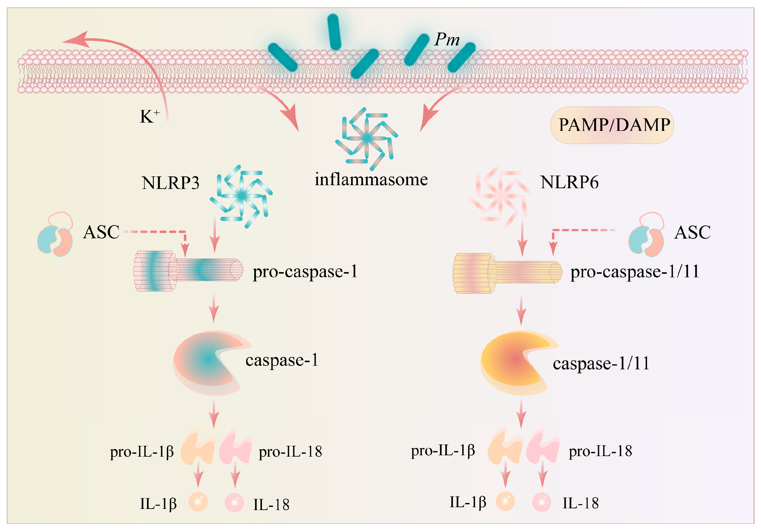
NLRP3 is a key protein in the inflammasome complex and is a member of the NLR family. It is the core component of inflammasomes and is one of the important PRRs during P. multocida infection. When NLRP3 is activated, ASC acts as a connector protein, connecting NLRP3 to downstream effector molecules. ASC then recruits and activates caspase-1, which is a cysteine protease that can shear and activate inflammatory cytokines, such as IL-1β and IL-18 [141]. After NLRP3 inflammasome assembly, the dormant procaspase-1 protein is hydrolysed and cleaved to active caspase-1 [142]. Subsequently, caspase-1 converts the cytokine precursors pro-IL-1β and pro-IL-18 into mature and biologically active IL-1β and IL-18 and induces a type of pro-inflammatory cell death called pyroptosis [143,144]. The role of NLRP3 inflammasome in response to microbial infection has been extensively studied. The NLRP6 inflammasome is a relatively new member of the NLR family. Similar to NLRP3 activation, NLRP6 activation triggered by PAMPs or DAMPs induces the recruitment of caspase-1 via the adaptor ASC, leading to an inflammatory response and the secretion of IL-1β and IL-18 [145,146] (Figure 3).
Several studies have provided evidence for the involvement of the NLRP3 inflammasome in P. multocida infection. Fang et al. [147] reported that NLRP3 inflammasome activation could be induced in cattle following PmCQ2 infection, thus providing one of the first in vivo observations in a natural host species. In a subsequent mechanistic study using macrophage cell lines, Ran et al. [148] found that the scaffold protein RACK1 was positively regulated with PmCQ2-induced NLRP3 activation; specifically, siRNA RACK1 knockdown attenuated caspase-1 activation and IL-1β production, whereas RACK1 overexpression enhanced these responses. While these loss- and gain-of-function experiments strongly suggest a regulatory role for RACK1, the findings are currently limited to immortalised cell lines and require confirmation in primary cells or in vivo.
A notable and counterintuitive observation was reported by Fang et al. [149], who compared NLRP3 inflammasome activation induced by high (PmCQ2) and low (PmCQ6) virulence strains. Surprisingly, the low-virulence strain induced higher levels of NLRP3 transcription, caspase-1 activation and mature IL-1β secretion than the highly virulent isolate. The authors attributed this difference to the thicker capsule of PmCQ2, which suggests that capsule thickness may influence bacterial colonisation and modulate NLRP3 activation. However, this interpretation remains correlative, and direct causal evidence linking capsule thickness to inflammasome modulation is currently lacking. Furthermore, it is worth noting that the identity of the specific P. multocida component(s) recognised by NLRP3 has not been established. In addition, Fang et al. [147] observed that NLRP3-deficient macrophages exhibited significantly reduced caspase-1 activation and IL-1β secretion in response to P. multocida and suggested that splenic tyrosine kinase may be involved in this process, although the underlying mechanism remains speculative.
Regarding the NLRP6 inflammasome, Wu et al. [146] investigated its role using an in vivo mouse model. They found that P. multocida induced severe pulmonary inflammation with extensive immune cell infiltration in both wild-type and Nlrp6-deficient mice. Interestingly, Nlrp6-deficient mice were more susceptible to infection, exhibiting higher bacterial loads and reduced numbers of immune cells in the lungs. The authors proposed that NLRP6 mediates caspase-1 activation and ASC oligomerisation, which leads to IL-1β secretion, and also regulates the expression of chemokines (CXCL1, CXCL2, and CXCR2) that promote neutrophil recruitment. Based on these observations, Wu et al. [146] proposed a possible synergistic effect between NLRP6 and NLRP3 during P. multocida infection. However, this hypothesis is currently based on single-gene knockout studies and requires formal validation using Nlrp3/Nlrp6 double-knockout models to confirm the interplay and relative contributions of each inflammasome pathway.
Although the involvement of NLRP3 and NLRP6 in P. multocida infection is supported by multiple studies, several inconsistencies and knowledge gaps warrant further attention. First, the relationship between bacterial virulence and inflammasome activation appears paradoxical. Fang et al. [149] reported that a low-virulence strain (PmCQ6) induced stronger NLRP3 activation than a highly virulent strain (PmCQ2), which suggests that robust inflammasome responses do not necessarily correlate with bacterial pathogenicity. This raises the important question of whether inflammasome activation is primarily a host protective response or, conversely, a pathological driver of tissue damage. Second, the interplay between NLRP3 and NLRP6 remains poorly understood. Wu et al. [146] suggested a possible synergistic effect, but the molecular basis of this synergy, whether through shared downstream signalling, reciprocal regulation or independent parallel pathways, has not been elucidated. Third, most studies have focused on acute infection models, leaving the role of inflammasomes in chronic or persistent P. multocida infections unexplored. In addition, the translational relevance of these findings to human infections and livestock disease management has not been adequately addressed. Future research should prioritise the use of cell-type-specific knockout models and longitudinal in vivo studies to resolve these uncertainties.
While our review focuses on the well-supported roles of NLRP3 and NLRP6, it remains to be determined whether other inflammasome sensors are also activated during P. multocida infection. Notably, recent studies have demonstrated that L-serine [145] and glycine [150] can reduce the production of inflammatory cytokines in macrophages by blocking the activation of multiple inflammasome pathways, including NLRP1, NLRP3, NLRC4, AIM2, and caspase-1, in PmCQ2-infected macrophages. These pharmacological observations raise the possibility that NLRC4 and AIM2 could participate in host defence against P. multocida infection, which still awaits validation via gene-deficient models.

5.3. K+ Outflow and Nek7 Play a Key Role in the Activation of NLRP3 Inflammasome Induced by Pasteurella multocida

K+ efflux has been identified as a critical upstream event for NLRP3 inflammasome activation in various infection models [151]. In the context of P. multocida, emerging evidence suggests that K+ outflow may similarly contribute to NLRP3-dependent pyroptosis. It has been proposed that P. multocida infection may trigger K+ efflux by disrupting potassium channels in the cell membrane or interfering with potassium transport systems. However, the precise molecular mechanism remains poorly elucidated. Furthermore, K+-efflux-mediated NLRP3 inflammasome activation requires Nek7, a member of the mammalian NIMA-associated kinase family, which has been reported to interact directly with NLRP3 [151,152].
Wang et al. [153] investigated the role of K+ efflux and Nek7 in PmCQ2-induced inflammasome activation using macrophage cell lines. They demonstrated that K+ efflux mediates PmCQ2-induced IL-1β secretion and that pharmacological blockade of K+ efflux attenuates caspase-1 activation and ASC oligomerisation. In addition, they observed that Nek7 is involved in PmCQ2-induced caspase-1 activation and IL-1β secretion, and that PmCQ2 infection promotes Nek7–NLRP3 interaction in a K+ efflux-dependent manner.
This demonstration that K+ efflux and Nek7 are essential for NLRP3 inflammasome activation by P. multocida represents an important mechanistic advance [151,152,153]. However, several critical questions remain. First, the upstream signal that triggers K+ efflux following P. multocida infection has not been identified. This includes whether it is mediated by a specific bacterial toxin, pore-forming protein or host-derived damage signals. Second, it is unclear whether Nek7 is exclusively required for NLRP3 activation induced by P. multocida, or whether it also participates in other inflammasome pathways (e.g., NLRP6 or NLRC4) during infection. Third, the physiological relevance of the Nek7–NLRP3 interaction needs to be validated in primary cells and in vivo models, as current evidence is predominantly derived from immortalised macrophage cell lines. Fourth, the temporal relationship between K+ efflux, Nek7 recruitment and ASC oligomerisation has not been resolved with sufficient kinetic resolution. Addressing these questions will be essential for translating these mechanistic insights into therapeutic strategies for P. multocida-associated diseases.

5.4. Knowledge Gaps and Future Directions in Inflammasome Research During P. multocida Infection

Despite the progress summarised above, the field currently suffers from several interconnected limitations that constrain the fundamental understanding and clinical translation: (i) Species- and strain-specific differences: Most mechanistic studies have been performed with a limited number of P. multocida strains (primarily PmCQ2 and PmCQ6) in murine or bovine models. Whether the observed inflammasome activation patterns are generalisable across different capsular serotypes (A, B and D) and host species remains unknown. (ii) Conflicting observations on bacterial virulence: As noted above, the observation that low-virulence strains may elicit stronger inflammasome responses than highly virulent ones challenges the assumption that inflammasome activation is a correlate of virulence. This paradox may be explained by differences in capsule thickness [149], potentially combined with variations in LPS structure or the expression of immunomodulatory virulence factors that actively suppress host sensing—although these latter possibilities remain speculative. This area warrants additional systematic comparative studies. (iii) Signalling cross-talk and redundancy: The potential interplay between NLRP3, NLRP6 and other inflammasome sensors (e.g., NLRP1, NLRC4) during P. multocida infection has not been explored. Multiple sensors may operate hierarchically or redundantly, depending on the infection stage or bacterial load. Multi-gene knockout approaches are required to dissect this complexity. (iv) Translational gap: While animal models have provided valuable insights, the relevance of these findings to human P. multocida infections, particularly in immunocompromised patients, the elderly and those with chronic lung diseases, remains largely unexamined. Furthermore, the potential for targeting inflammasome components as therapeutic interventions in veterinary medicine has not been sufficiently investigated.
Addressing these knowledge gaps will require a concerted effort involving standardised infection models, multi-omics approaches and collaborative research bridging basic immunology and veterinary clinical practice.

6. Conclusions and Prospects

A study of the virulence factors of P. multocida is helpful to the examination of its pathogenic mechanism to a host and the development of new vaccines. The in-depth analysis of pathogenic and immune mechanisms will provide insight into the molecular basis of host–microbe interactions in chronic infections and the transition from subclinical or chronic diseases to acute transmissible diseases. In this review, the pathogenic mechanism of P. multocida and its relationship with the activation of inflammasomes were discussed, and the complex mechanism of the pathogenic bacteria in causing diseases in livestock and humans was revealed. The pathogenesis of P. multocida and the activation of inflammasomes still have many directions worth exploring. First, the interaction between the various virulence factors of P. multocida and its specific contribution to the pathogenesis process needs to be further clarified. This information is essential to the development of novel drugs or vaccines targeting specific virulence factors. Second, the mechanism of inflammasome activation in anti-infection immunity still needs to be further studied, especially the activation mode of inflammasomes and its influence on disease outcomes in different infection stages and host contexts. In addition, modern biotechnological means, such as high-throughput sequencing, proteomics and metabolomics, will be used to fully analyse host–pathogen interactions during P. multocida infection, and identify new perspectives and strategies for disease prevention and control.

Author Contributions

M.Q.: Writing—review and editing, Writing—original draft, Visualization, Methodology, Funding acquisition, Data curation. J.Z.: Writing—review and editing, Writing—original draft, Data curation, Conceptualization. D.Z.: Data curation, Conceptualization. M.S.: Project administration, Methodology, Investigation. D.Y.: Writing—review and editing, Investigation. Z.Y.: Data curation, Conceptualization. R.W.: Writing—review and editing, Writing—original draft, Visualization, Supervision. All authors have read and agreed to the published version of the manuscript.

Funding

This work was supported by the National Natural Science Foundation of China grant no. 32160838; Special Project of Science and Technology Innovation Group Construction of B-Class Team (Class II Team) grant no. BR251303; Key R & D and Achievement Transformation Projects of Inner Mongolia, China grant no. 2023YFDZ0048; Project of Major Innovation Platform (Base) Construction grant no. KCX2024016; Inner Mongolia Autonomous Region First Class Discipline Research Special Project grant no. YLXKZX-NND-012; Natural Science Foundation of Inner Mongolia, China, under grant no. 2024MS08012, 2026LHMS0005 and 2024MS08061; Program for Young Talents of Science and Technology in Universities of Inner Mongolia Autonomous Region no. NJYT23112.

Data Availability Statement

No new data were created or analyzed in this study. Data sharing is not applicable to this article.

Conflicts of Interest

The authors declare no conflicts of interest.

References

  1. Pintér, K.; Domán, M.; Wehmann, E.; Makrai, L.; Gantelet, H.; Magyar, T. Correlations between virulence gene profiles and other genetic factors of Pasteurella multocida strains isolated from various host species. Vet. Microbiol. 2025, 308, 110657. [Google Scholar] [CrossRef] [Scilit] [PubMed]
  2. Garzon, A.; Miramontes, C.; Weimer, B.C.; Profeta, R.; Hoyos-Jaramillo, A.; Fritz, H.M.; Pereira, R.V. Comparison of virulence and resistance genes in Mannheimia haemolytica and Pasteurella multocida from dairy cattle with and without bovine respiratory disease. Microbiol. Spectr. 2025, 13, e0120025. [Google Scholar] [CrossRef] [Scilit] [PubMed]
  3. Harper, M.; Boyce, J.D.; Adler, B. Pasteurella multocida pathogenesis: 125 years after Pasteur. FEMS Microbiol. Lett. 2006, 265, 1–10. [Google Scholar] [CrossRef] [Scilit] [PubMed]
  4. Christensen, J.P.; Bisgaard, M. Fowl cholera. Sci. Tech. Rev. 2000, 19, 626–637. [Google Scholar] [CrossRef] [Scilit] [PubMed]
  5. Harper, M.; Boyce, J.D. The Myriad Properties of Pasteurella multocida Lipopolysaccharide. Toxins 2017, 9, 254. [Google Scholar] [CrossRef] [Scilit] [PubMed]
  6. Slehria, T.; Hendricks, S.; Honeycutt, T. Pasteurella multocida Bacterial Meningitis in a 33-Day-Old Infant. Pediatr. Infect. Dis. J. 2022, 41, e267–e268. [Google Scholar] [CrossRef] [Scilit] [PubMed]
  7. Piorunek, M.; Brajer-Luftmann, B.; Trafas, T.; Schneider, A.; Walkowiak, J. Lower respiratory infection in humans caused by Pasteurella multocida. Respir. Physiol. Neurobiol. 2023, 315, 104091. [Google Scholar] [CrossRef] [Scilit] [PubMed]
  8. Mogilner, L.; Katz, C. Pasteurella multocida . Pediatr. Rev. 2019, 40, 90–92. [Google Scholar] [CrossRef] [Scilit] [PubMed]
  9. Jan, L.; Boute, P.; Mouawad, F. Pasteurella multocida acute epiglottitis. Eur. Ann. Otorhinolaryngol. Head Neck Dis. 2021, 138, 100–102. [Google Scholar] [CrossRef] [Scilit] [PubMed]
  10. Wei, B.; Liu, C.; Zhu, J.; Zou, X.; Zhang, Z. Pasteurella multocida infection: A differential retrospective study of 482 cases of P. multocida infection in patient of different ages. BMC Infect. Dis. 2025, 25, 313. [Google Scholar] [CrossRef] [Scilit] [PubMed]
  11. Ziagham, A.; Gharibi, D.; Mosallanejad, B.; Avizeh, R. Molecular characterization of Pasteurella multocida from cats and antibiotic sensitivity of the isolates. Vet. Med. Sci. 2024, 10, e1424. [Google Scholar] [CrossRef] [Scilit] [PubMed]
  12. Costanzo, J.T., 2nd; Wojciechowski, A.L.; Bajwa, R.P.S. Urinary tract infection with Pasteurella multocida in a patient with cat exposure and abnormal urinary tract physiology: Case report and literature review. IDCases 2017, 9, 109–111. [Google Scholar] [CrossRef] [Scilit] [PubMed]
  13. El Amri, H.; Rafique, S.; Zubairi, A. Pasteurella Pneumonia With Complicated Parapneumonic Effusion in a Pet Owner. Cureus 2025, 17, e79887. [Google Scholar] [CrossRef] [Scilit] [PubMed]
  14. Wilson, B.A.; Ho, M. Pasteurella multocida: From zoonosis to cellular microbiology. Clin. Microbiol. Rev. 2013, 26, 631–655. [Google Scholar] [CrossRef] [Scilit] [PubMed]
  15. Klein, N.C.; Cunha, B.A. Pasteurella multocida pneumonia. Semin. Respir. Infect. 1997, 12, 54–56. [Google Scholar] [PubMed]
  16. Smallman, T.R.; Perlaza-Jiménez, L.; Wang, X.; Korman, T.M.; Kotsanas, D.; Gibson, J.S.; Turni, C.; Harper, M.; Boyce, J.D. Pathogenomic analysis and characterization of Pasteurella multocida strains recovered from human infections. Microbiol. Spectr. 2024, 12, e0380523. [Google Scholar] [CrossRef] [Scilit] [PubMed]
  17. Sugino, Y.; Kato, M.; Yagi, A.; Kawabata, A. Pasteurella multocida pneumonia with molecular evidence of zoonotic transmission. Kansenshogaku Zasshi 2007, 81, 726–730. [Google Scholar] [CrossRef] [Scilit] [PubMed]
  18. Nguyen, P.V.; Le, C.T.; Nguyen, X.H.; Nguyen, T.M.; Nguyen, K.C.T. First study on capsular serotypes and virulence factors of Pasteurella multocida isolates from Phan Rang sheep in Vietnam. Vet. World 2023, 16, 281–290. [Google Scholar] [CrossRef] [Scilit] [PubMed]
  19. Farahani, M.F.; Esmaelizad, M.; Jabbari, A.R. Investigation of iron uptake and virulence gene factors (fur, tonB, exbD, exbB, hgbA, hgbB1, hgbB2 and tbpA) among isolates of Pasteurella multocida from Iran. Iran. J. Microbiol. 2019, 11, 191–197. [Google Scholar] [CrossRef] [Scilit]
  20. Miyoshi, S.; Hamada, H.; Miyoshi, A.; Ito, R.; Hamaguchi, N.; Murakami, S.; Miyamoto, H.; Takeuchi, T.; Okura, T.; Higaki, J. Pasteurella multocida pneumonia: Zoonotic transmission confirmed by molecular epidemiological analysis. Geriatr. Gerontol. Int. 2012, 12, 159–163. [Google Scholar] [CrossRef] [Scilit] [PubMed]
  21. Cai, Q.; Li, Y.; Chang, Y.F.; Tang, Z.; Zhang, H.; Xie, Q. Pasteurella multocida causes liver injury in ducks by mediating inflammatory, apoptotic and autophagic pathways. Microb. Pathog. 2023, 184, 106336. [Google Scholar] [CrossRef] [Scilit] [PubMed]
  22. Broz, P.; Dixit, V.M. Inflammasomes: Mechanism of assembly, regulation and signalling. Nat. Rev. Immunol. 2016, 16, 407–420. [Google Scholar] [CrossRef] [Scilit] [PubMed]
  23. Heddleston, K.L.; Wessman, G. Characteristics of Pasteurella multocida of human origin. J. Clin. Microbiol. 1975, 1, 377–383. [Google Scholar] [CrossRef] [Scilit] [PubMed]
  24. Rimler, R.B.; Rhoades, K.R. Serogroup F, a new capsule serogroup of Pasteurella multocida. J. Clin. Microbiol. 1987, 25, 615–618. [Google Scholar] [CrossRef] [Scilit] [PubMed]
  25. Peng, Z.; Wang, X.; Zhou, R.; Chen, H.; Wilson, B.A.; Wu, B. Pasteurella multocida: Genotypes and Genomics. Microbiol. Mol. Biol. Rev. 2019, 83, e00014-19. [Google Scholar] [CrossRef] [Scilit] [PubMed]
  26. Smith, E.; Miller, E.; Aguayo, J.M.; Figueroa, C.F.; Nezworski, J.; Studniski, M.; Wileman, B.; Johnson, T. Genomic diversity and molecular epidemiology of Pasteurella multocida. PLoS ONE 2021, 16, e0249138. [Google Scholar] [CrossRef] [Scilit] [PubMed]
  27. Peng, Z.; Liang, W.; Liu, W.; Wu, B.; Tang, B.; Tan, C.; Zhou, R.; Chen, H. Genomic characterization of Pasteurella multocida HB01, a serotype A bovine isolate from China. Gene 2016, 581, 85–93. [Google Scholar] [CrossRef] [Scilit] [PubMed]
  28. Michael, F.S.; Cairns, C.M.; Fleming, P.; Vinogradov, E.V.; Boyce, J.D.; Harper, M.; Cox, A.D. The capsular polysaccharides of Pasteurella multocida serotypes B and E: Structural, genetic and serological comparisons. Glycobiology 2021, 31, 307–314. [Google Scholar] [CrossRef] [Scilit] [PubMed]
  29. Nguyen, T.V.; Trinh, T.T.H.; Nguyen, T.V.; Ng-Nguyen, D.; Nguyen, H.Q.; Vu-Khac, H. Comprehensive capsular, lipopolysaccharide, virulence, and antimicrobial resistance profiling of Pasteurella multocida isolated from buffaloes in Vietnam: First report of capsular type D and predominant L2 genotype. Vet. World 2025, 18, 4069–4081. [Google Scholar] [CrossRef] [Scilit] [PubMed]
  30. Richardson, N.I.; Ravenscroft, N.; Kuttel, M.M. Conformational comparisons of Pasteurella multocida types B and E and structurally related capsular polysaccharides. Glycobiology 2023, 33, 745–754. [Google Scholar] [CrossRef] [Scilit] [PubMed]
  31. Prajapati, A.; Yogisharadhya, R.; Mohanty, N.N.; Mendem, S.K.; Nizamuddin, A.; Chanda, M.M.; Shivachandra, S.B. Comparative genome analysis of Pasteurella multocida serogroup B:2 strains causing haemorrhagic septicaemia (HS) in bovines. Gene 2022, 826, 146452. [Google Scholar] [CrossRef] [Scilit] [PubMed]
  32. Peng, Z.; Liang, W.; Wang, Y.; Liu, W.; Zhang, H.; Yu, T.; Zhang, A.; Chen, H.; Wu, B. Experimental pathogenicity and complete genome characterization of a pig origin Pasteurella multocida serogroup F isolate HN07. Vet. Microbiol. 2017, 198, 23–33. [Google Scholar] [CrossRef] [Scilit] [PubMed]
  33. Wang, J.; Sun, S.; Chen, Y.; Chen, D.; Sang, L.; Xie, X. Pathogenic and genomic characterisation of a rabbit sourced Pasteurella multocida serogroup F isolate s4. BMC Vet. Res. 2022, 18, 288. [Google Scholar] [CrossRef] [Scilit] [PubMed]
  34. Wang, J.; Sun, S.; Chen, D.; Gao, C.; Sang, L.; Xie, X. Pathogenic and genomic characterization of rabbit-sourced Pasteurella multocida serogroup F isolates recovered from dead rabbits with respiratory disease. Microbiol. Spectr. 2024, 12, e0365423. [Google Scholar] [CrossRef] [Scilit] [PubMed]
  35. Furian, T.Q.; Borges, K.A.; Laviniki, V.; Rocha, S.L.; de Almeida, C.N.; do Nascimento, V.P.; Salle, C.T.; Moraes, H.L. Virulence genes and antimicrobial resistance of Pasteurella multocida isolated from poultry and swine. Braz. J. Microbiol. 2016, 47, 210–216. [Google Scholar] [CrossRef] [Scilit] [PubMed]
  36. Peng, Z.; Wang, H.; Liang, W.; Chen, Y.; Tang, X.; Chen, H.; Wu, B. A capsule/lipopolysaccharide/MLST genotype D/L6/ST11 of Pasteurella multocida is likely to be strongly associated with swine respiratory disease in China. Arch. Microbiol. 2018, 200, 107–118. [Google Scholar] [CrossRef] [Scilit] [PubMed]
  37. Christensen, H.; Sajid, S.M.; Bisgaard, M.; Magistrali, C.F.; Massacci, F.R.; Liman, M.; Menke, T.; Bischoff, H.; Olsen, J.E. Prediction of Pasteurella multocida serotypes based on whole genomic sequences. Vet. Microbiol. 2022, 271, 109492. [Google Scholar] [CrossRef] [Scilit] [PubMed]
  38. Hashish, A.; Johnson, T.J.; Ghanem, M.; Sato, Y.; Macedo, N.R.; LeCount, K.J.; El-Gazzar, M. Complete genome sequences of eight Pasteurella multocida isolates representing all lipopolysaccharide outer core loci. Microbiol. Resour. Announc. 2024, 13, e0060424. [Google Scholar] [CrossRef] [Scilit] [PubMed]
  39. Ujvári, B.; Weiczner, R.; Deim, Z.; Terhes, G.; Urbán, E.; Tóth, A.R.; Magyar, T. Characterization of Pasteurella multocida strains isolated from human infections. Comp. Immunol. Microbiol. Infect. Dis. 2019, 63, 37–43. [Google Scholar] [CrossRef] [Scilit] [PubMed]
  40. Mo, Q.; Nawaz, S.; Kulyar, M.F.; Li, K.; Li, Y.; Zhang, Z.; Rahim, M.F.; Ahmed, A.E.; Ijaz, F.; Li, J. Exploring the intricacies of Pasteurella multocida dynamics in high-altitude livestock and its consequences for bovine health: A personal exploration of the yak paradox. Microb. Pathog. 2024, 194, 106799. [Google Scholar] [CrossRef] [Scilit] [PubMed]
  41. Cid, D.; Fernández-Garayzábal, J.F.; Pinto, C.; Domínguez, L.; Vela, A.I. Antimicrobial susceptibility of Pasteurella multocida isolated from sheep and pigs in Spain—Short communication. Acta Vet. Hung. 2019, 67, 489–498. [Google Scholar] [CrossRef] [Scilit] [PubMed]
  42. Abdulrahman, R.F.; Davies, R.L. Diversity and characterization of temperate bacteriophages induced in Pasteurella multocida from different host species. BMC Microbiol. 2021, 21, 97. [Google Scholar] [CrossRef] [Scilit] [PubMed]
  43. Dabo, S.M.; Taylor, J.D.; Confer, A.W. Pasteurella multocida and bovine respiratory disease. Anim. Health Res. Rev. 2007, 8, 129–150. [Google Scholar] [CrossRef] [Scilit] [PubMed]
  44. Snyder, E.; Credille, B. Mannheimia haemolytica and Pasteurella multocida in Bovine Respiratory Disease: How Are They Changing in Response to Efforts to Control Them? Vet. Clin. N. Am. Food Anim. Pract. 2020, 36, 253–268. [Google Scholar]
  45. Amirgazin, A.; Yessembekova, G.; Akhmetova, A.; Karibayev, T.; Mukanov, K.; Shevtsova, E.; Abdigulov, B.; Abdrakhmanov, S. Genomic Insights into Pasteurella multocida Serotype B:2 from Hemorrhagic Septicemia Outbreaks in Wildlife and Livestock in Kazakhstan. Pathogens 2025, 14, 1273. [Google Scholar] [CrossRef] [Scilit] [PubMed]
  46. Webster, L.T.; Burn, C.G. Biology of Bacterium Lepisepticum: IV. Virulence of Diffuse and Mucoid Types and Their Variants. J. Exp. Med. 1926, 44, 359–386. [Google Scholar] [PubMed]
  47. Chongrattanameteekul, P.; Rattanaworapanit, N.; Wongsawan, K.; Chuammitri, P.; Anuntakulnatee, T.; Veerathong, S.; Mektrirat, R. Antimicrobial resistance and etiological dynamics affected by tropical climate variability on year-round diagnosis of upper respiratory infections in companion rabbits with snuffles. Sci. Rep. 2025, 15, 13994. [Google Scholar] [CrossRef] [Scilit] [PubMed]
  48. Okoh, A.E. An outbreak of pasteurellosis in Kano Zoo. J. Wildl. Dis. 1980, 16, 3–5. [Google Scholar] [CrossRef] [Scilit] [PubMed]
  49. Kock, R.A.; Orynbayev, M.; Robinson, S.; Zuther, S.; Singh, N.J.; Beauvais, W.; Morgan, E.R.; Kerimbayev, A.; Khomenko, S.; Martineau, H.M.; et al. Saigas on the brink: Multidisciplinary analysis of the factors influencing mass mortality events. Sci. Adv. 2018, 4, eaao2314. [Google Scholar] [CrossRef] [Scilit] [PubMed]
  50. Fereidouni, S.; Freimanis, G.L.; Orynbayev, M.; Ribeca, P.; Flannery, J.; King, D.P.; Zuther, S.; Beer, M.; Höper, D.; Kydyrmanov, A.; et al. Mass Die-Off of Saiga Antelopes, Kazakhstan, 2015. Emerg. Infect. Dis. 2019, 25, 1169–1176. [Google Scholar] [CrossRef] [Scilit] [PubMed]
  51. Rybolt, L.E.; Sabunwala, S.; Greene, J.N. Zoonotic Bacterial Respiratory Infections Associated With Cats and Dogs: A Case Series and Literature Review. Cureus 2022, 14, e24414. [Google Scholar] [CrossRef] [Scilit] [PubMed]
  52. Körmöndi, S.; Terhes, G.; Pál, Z.; Varga, E.; Harmati, M.; Buzás, K.; Urbán, E. Human Pasteurellosis Health Risk for Elderly Persons Living with Companion Animals. Emerg. Infect. Dis. 2019, 25, 229–235. [Google Scholar] [CrossRef] [Scilit] [PubMed]
  53. Jin, M.; Osman, M.; Green, B.A.; Yang, Y.; Ahuja, A.; Lu, Z.; Cazer, C.L. Evidence for the transmission of antimicrobial resistant bacteria between humans and companion animals: A scoping review. One Health 2023, 17, 100593. [Google Scholar] [CrossRef] [Scilit] [PubMed]
  54. Guilbart, M.; Zogheib, E.; Hchikat, A.H.; Kirat, K.; Ferraz, L.; Guerin-Robardey, A.M.; Trojette, F.; Moubarak-Daher, M.; Dupont, H. Fatal multifocal Pasteurella multocida infection: A case report. BMC Res. Notes 2015, 8, 287. [Google Scholar] [CrossRef] [Scilit] [PubMed]
  55. Abreu, F.; Rodríguez-Lucas, C.; Rodicio, M.R.; Vela, A.I.; Fernández-Garayzábal, J.F.; Leiva, P.S.; Cuesta, F.; Cid, D.; Fernández, J. Human Pasteurella multocida Infection with Likely Zoonotic Transmission from a Pet Dog, Spain. Emerg. Infect. Dis. 2018, 24, 1145–1146. [Google Scholar] [CrossRef] [Scilit] [PubMed]
  56. Jeong, S.; Chang, E.; Lee, N.; Kim, H.S.; Kim, H.S.; Kim, J.S.; Kim, Y.A.; Kim, C.K.; Lee, K.; Lee, H.; et al. Pasteurella Infections in South Korea and Systematic Review and Meta-analysis of Pasteurella Bacteremia. Emerg. Infect. Dis. 2024, 30, 1987–1997. [Google Scholar] [CrossRef] [Scilit] [PubMed]
  57. Pena, D.; Santana, Y.; Perez Lara, J.; Gonzalez, E.; Khaja, M. Multiorgan Failure and Refractory Lactic Acidosis due to Pasteurella multocida Septicemia in a Patient with No Animal Exposure. Case Rep. Infect. Dis. 2018, 2018, 2574184. [Google Scholar] [CrossRef] [Scilit] [PubMed]
  58. Kumar, A.; Devlin, H.R.; Vellend, H. Pasteurella multocida meningitis in an adult: Case report and review. Rev. Infect. Dis. 1990, 12, 440–448. [Google Scholar] [CrossRef] [Scilit] [PubMed]
  59. He, J.; Yang, Z.; Wang, M.; Jia, R.; Chen, S.; Liu, M.; Zhao, X.; Yang, Q.; Wu, Y.; Zhang, S.; et al. Integrative and conjugative elements of Pasteurella multocida: Prevalence and signatures in population evolution. Virulence 2024, 15, 2359467. [Google Scholar] [CrossRef] [Scilit] [PubMed]
  60. Giordano, A.; Dincman, T.; Clyburn, B.E.; Steed, L.L.; Rockey, D.C. Clinical Features and Outcomes of Pasteurella multocida Infection. Medicine 2015, 94, e1285. [Google Scholar] [CrossRef] [Scilit] [PubMed]
  61. Kukrety, S.; Parekh, J.; Townley, T. Pasteurella multocida Bacteremia in an Immunocompromised Patient. Case Rep. Med. 2016, 2016, 7392847. [Google Scholar] [CrossRef] [Scilit] [PubMed]
  62. Lnu, K.; Orozco, D.; Cream, C. Pasteurella multocida Bacteremia Due to Obstructive Pneumonia in an Immunocompromised Patient. J. Community Hosp. Intern. Med. Perspect. 2022, 12, 50–52. [Google Scholar] [CrossRef] [Scilit] [PubMed]
  63. Boadu, C.; Hernandez, A.; Zeidan, B., Jr.; Young, J.T.; Frunzi, J. Pasteurella multocida Bacteremia in an Immunocompromised Patient After Multiple Cat Scratches. Cureus 2021, 13, e12938. [Google Scholar] [CrossRef] [Scilit] [PubMed]
  64. Lopes Freitas, R.; Reis, F.; Ruiz Pena, E. Pasteurella multocida Bacteremia in an Immunocompromised Patient With Esophageal Squamous Cell Carcinoma: A Case Report. Cureus 2024, 16, e75924. [Google Scholar] [CrossRef] [Scilit] [PubMed]
  65. Leonhardt, L.P.; Pervez, A.; Tang, W. Pasteurella Multocida Bacteremia and Septic Arthritis in a Patient With Non-Bite Animal Exposure. Cureus 2021, 13, e14162. [Google Scholar] [CrossRef] [Scilit] [PubMed]
  66. Rueda Prada, L.; Cardozo, M.; Hudson, A.; McDermott, M.; Urbina Verjel, D.C.; Dumic, I. Disseminated Pasteurella multocida in a patient with liver cirrhosis and spontaneous bacterial peritonitis—The role of cirrhosis-associated immune dysfunction. IDCases 2022, 29, e01542. [Google Scholar] [CrossRef] [Scilit] [PubMed]
  67. Cabras, O.; Turmel, J.M.; Olive, C.; Bigeard, B.; Lehoux, M.; Pierre-Francois, S.; Guitteaud, K.; Abel, S.; Cuzin, L.; Cabié, A. COVID-19 and Pasteurella multocida Pulmonary Coinfection: A Case Series. Trop. Med. Infect. Dis. 2022, 7, 429. [Google Scholar] [CrossRef] [Scilit] [PubMed]
  68. Galdiero, M.; Pisciotta, M.G.; Marinelli, A.; Petrillo, G.; Galdiero, E. Coinfection with BHV-1 modulates cell adhesion and invasion by P. multocida and Mannheimia (Pasteurella) haemolytica. New Microbiol. 2002, 25, 427–436. [Google Scholar] [PubMed]
  69. Hombal, S.M.; Dincsoy, H.P. Pasteurella multocida endocarditis. Am. J. Clin. Pathol. 1992, 98, 565–568. [Google Scholar] [CrossRef] [Scilit] [PubMed]
  70. Tharmaseelan, K.; Morgan, M.S. Pasteurella multocida conjunctivitis. Br. J. Ophthalmol. 1993, 77, 815. [Google Scholar] [CrossRef] [Scilit] [PubMed]
  71. Zebeede, E.; Levinger, U.; Weinberger, A. Pasteurella multocida infectious arthritis. Isr. Med. Assoc. J. 2004, 6, 778–779. [Google Scholar] [PubMed]
  72. Hatfaludi, T.; Al-Hasani, K.; Boyce, J.D.; Adler, B. Outer membrane proteins of Pasteurella multocida. Vet. Microbiol. 2010, 144, 1–17. [Google Scholar] [CrossRef] [Scilit] [PubMed]
  73. Gharib Mombeni, E.; Gharibi, D.; Ghorbanpoor, M.; Jabbari, A.R.; Cid, D. Toxigenic and non-toxigenic Pasteurella multocida genotypes, based on capsular, LPS, and virulence profile typing, associated with pneumonic pasteurellosis in Iran. Vet. Microbiol. 2021, 257, 109077. [Google Scholar] [CrossRef] [Scilit] [PubMed]
  74. Katoch, S.; Sharma, M.; Patil, R.D.; Kumar, S.; Verma, S. In vitro and in vivo pathogenicity studies of Pasteurella multocida strains harbouring different ompA. Vet. Res. Commun. 2014, 38, 183–191. [Google Scholar] [CrossRef] [Scilit] [PubMed]
  75. Wilkie, I.W.; Harper, M.; Boyce, J.D.; Adler, B. Pasteurella multocida: Diseases and pathogenesis. In Pasteurella Multocida; Current Topics in Microbiology and Immunology; Springer: Berlin/Heidelberg, Germany, 2012; Volume 361, pp. 1–22. [Google Scholar]
  76. Kubatzky, K.F. Pasteurella multocida and immune cells. In Pasteurella Multocida; Current Topics in Microbiology and Immunology; Springer: Berlin/Heidelberg, Germany, 2012; Volume 361, pp. 53–72. [Google Scholar]
  77. Pruimboom, I.M.; Rimler, R.B.; Ackermann, M.R.; Brogden, K.A. Capsular hyaluronic acid-mediated adhesion of Pasteurella multocida to turkey air sac macrophages. Avian Dis. 1996, 40, 887–893. [Google Scholar] [CrossRef] [Scilit] [PubMed]
  78. Smith, D.G. Adherence and pathogenesis of pasteurella multocida—A sticky problem. Vet. J. 2000, 159, 215–216. [Google Scholar] [CrossRef] [Scilit] [PubMed]
  79. Christenson, E.S.; Ahmed, H.M.; Durand, C.M. Pasteurella multocida infection in solid organ transplantation. Lancet Infect. Dis. 2015, 15, 235–240. [Google Scholar] [CrossRef] [Scilit] [PubMed]
  80. DeAngelis, P.L. Enzymological characterization of the Pasteurella multocida hyaluronic acid synthase. Biochemistry 1996, 35, 9768–9771. [Google Scholar] [CrossRef] [Scilit] [PubMed]
  81. Pandit, K.K.; Smith, J.E. Capsular hyaluronic acid in Pasteurella multocida type A and its counterpart in type D. Res. Vet. Sci. 1993, 54, 20–24. [Google Scholar] [CrossRef] [Scilit] [PubMed]
  82. Boyce, J.D.; Chung, J.Y.; Adler, B. Pasteurella multocida capsule: Composition, function and genetics. J. Biotechnol. 2000, 83, 153–160. [Google Scholar] [CrossRef] [Scilit] [PubMed]
  83. Guan, L.; Zhang, L.; Xue, Y.; Yang, J.; Zhao, Z. Molecular pathogenesis of the hyaluronic acid capsule of Pasteurella multocida. Microb. Pathog. 2020, 149, 104380. [Google Scholar] [CrossRef] [Scilit] [PubMed]
  84. Chung, J.Y.; Wilkie, I.; Boyce, J.D.; Townsend, K.M.; Frost, A.J.; Ghoddusi, M.; Adler, B. Role of capsule in the pathogenesis of fowl cholera caused by Pasteurella multocida serogroup A. Infect. Immun. 2001, 69, 2487–2492. [Google Scholar] [CrossRef] [Scilit] [PubMed]
  85. Harper, M.; Boyce, J.D.; Adler, B. The key surface components of Pasteurella multocida: Capsule and lipopolysaccharide. In Pasteurella Multocida; Current Topics in Microbiology and Immunology; Springer: Berlin/Heidelberg, Germany, 2012; Volume 361, pp. 39–51. [Google Scholar]
  86. Al-Haddawi, M.H.; Jasni, S.; Zamri-Saad, M.; Mutalib, A.R.; Zulkifli, I.; Son, R.; Sheikh-Omar, A.R. In vitro study of Pasteurella multocida adhesion to trachea, lung and aorta of rabbits. Vet. J. 2000, 159, 274–281. [Google Scholar] [CrossRef] [Scilit] [PubMed]
  87. Wang, Z.; Liu, S.; Xie, M.; Lang, Z.; Zhang, X.; Luo, L.; Zhao, G.; Li, N.; Peng, Y. Deleting fis downregulates virulence and effectively protects Pasteurella multocida infection in mice. BMC Vet. Res. 2025, 21, 323. [Google Scholar] [CrossRef] [Scilit] [PubMed]
  88. Wang, X.; Quinn, P.J. Lipopolysaccharide: Biosynthetic pathway and structure modification. Prog. Lipid Res. 2010, 49, 97–107. [Google Scholar] [CrossRef] [Scilit] [PubMed]
  89. Harper, M.; Cox, A.D.; Adler, B.; Boyce, J.D. Pasteurella multocida lipopolysaccharide: The long and the short of it. Vet. Microbiol. 2011, 153, 109–115. [Google Scholar] [CrossRef] [Scilit] [PubMed]
  90. Maldonado, R.F.; Sá-Correia, I.; Valvano, M.A. Lipopolysaccharide modification in Gram-negative bacteria during chronic infection. FEMS Microbiol. Rev. 2016, 40, 480–493. [Google Scholar] [CrossRef] [Scilit] [PubMed]
  91. Gorman, A.; Golovanov, A.P. Lipopolysaccharide Structure and the Phenomenon of Low Endotoxin Recovery. Eur. J. Pharm. Biopharm. 2022, 180, 289–307. [Google Scholar] [CrossRef] [Scilit] [PubMed]
  92. Harper, M.; Cox, A.; St Michael, F.; Parnas, H.; Wilkie, I.; Blackall, P.J.; Adler, B.; Boyce, J.D. Decoration of Pasteurella multocida lipopolysaccharide with phosphocholine is important for virulence. J. Bacteriol. 2007, 189, 7384–7391. [Google Scholar] [CrossRef] [Scilit] [PubMed]
  93. Mogensen, T.H. Pathogen recognition and inflammatory signaling in innate immune defenses. Clin. Microbiol. Rev. 2009, 22, 240–273. [Google Scholar] [CrossRef] [Scilit] [PubMed]
  94. da Silva Correia, J.; Soldau, K.; Christen, U.; Tobias, P.S.; Ulevitch, R.J. Lipopolysaccharide is in close proximity to each of the proteins in its membrane receptor complex. transfer from CD14 to TLR4 and MD-2. J. Biol. Chem. 2001, 276, 21129–21135. [Google Scholar] [PubMed]
  95. Akashi, S.; Saitoh, S.; Wakabayashi, Y.; Kikuchi, T.; Takamura, N.; Nagai, Y.; Kusumoto, Y.; Fukase, K.; Kusumoto, S.; Adachi, Y.; et al. Lipopolysaccharide interaction with cell surface Toll-like receptor 4-MD-2: Higher affinity than that with MD-2 or CD14. J. Exp. Med. 2003, 198, 1035–1042. [Google Scholar] [PubMed]
  96. Zhao, X.; Yang, F.; Shen, H.; Liao, Y.; Zhu, D.; Wang, M.; Jia, R.; Chen, S.; Liu, M.; Yang, Q.; et al. Immunogenicity and protection of a Pasteurella multocida strain with a truncated lipopolysaccharide outer core in ducks. Vet. Res. 2022, 53, 17. [Google Scholar] [CrossRef] [Scilit] [PubMed]
  97. Iovane, G.; Pagnini, P.; Galdiero, M.; Cipollaro de l’Ero, G.; Vitiello, M.; D’Isanto, M.; Marcatili, A. Role of Pasteurella multocida porin on cytokine expression and release by murine splenocytes. Vet. Immunol. Immunopathol. 1998, 66, 391–404. [Google Scholar] [CrossRef] [Scilit] [PubMed]
  98. Yap, S.K.; Zakaria, Z.; Othman, S.S.; Omar, A.R. In vitro treatment of lipopolysaccharide increases invasion of Pasteurella multocida serotype B:2 into bovine aortic endothelial cells. J. Vet. Sci. 2018, 19, 207–215. [Google Scholar] [PubMed]
  99. Fuller, T.E.; Kennedy, M.J.; Lowery, D.E. Identification of Pasteurella multocida virulence genes in a septicemic mouse model using signature-tagged mutagenesis. Microb. Pathog. 2000, 29, 25–38. [Google Scholar] [CrossRef] [Scilit] [PubMed]
  100. May, B.J.; Zhang, Q.; Li, L.L.; Paustian, M.L.; Whittam, T.S.; Kapur, V. Complete genomic sequence of Pasteurella multocida, Pm70. Proc. Natl. Acad. Sci. USA 2001, 98, 3460–3465. [Google Scholar] [CrossRef] [Scilit] [PubMed]
  101. Kumar, S.; Hedrick, V.; Mattoo, S. YopT domain of the PfhB2 toxin from Pasteurella multocida: Protein expression, characterization, crystallization and crystallographic analysis. Acta Crystallogr. Sect. F Struct. Biol. Commun. 2018, 74, 128–134. [Google Scholar] [CrossRef] [Scilit] [PubMed]
  102. Ward, C.K.; Lumbley, S.R.; Latimer, J.L.; Cope, L.D.; Hansen, E.J. Haemophilus ducreyi secretes a filamentous hemagglutinin-like protein. J. Bacteriol. 1998, 180, 6013–6022. [Google Scholar] [CrossRef] [Scilit] [PubMed]
  103. Tatum, F.M.; Yersin, A.G.; Briggs, R.E. Construction and virulence of a Pasteurella multocida fhaB2 mutant in turkeys. Microb. Pathog. 2005, 39, 9–17. [Google Scholar] [CrossRef] [Scilit] [PubMed]
  104. Tatum, F.M.; Tabatabai, L.B.; Briggs, R.E. Protection against fowl cholera conferred by vaccination with recombinant Pasteurella multocida filamentous hemagglutinin peptides. Avian Dis. 2009, 53, 169–174. [Google Scholar] [CrossRef] [Scilit] [PubMed]
  105. Dabo, S.M.; Confer, A.W.; Quijano-Blas, R.A. Molecular and immunological characterization of Pasteurella multocida serotype A:3 OmpA: Evidence of its role in P. multocida interaction with extracellular matrix molecules. Microb. Pathog. 2003, 35, 147–157. [Google Scholar] [CrossRef] [Scilit] [PubMed]
  106. Li, Y.; Xiao, J.; Chang, Y.F.; Zhang, H.; Teng, Y.; Lin, W.; Li, H.; Chen, W.; Zhang, X.; Xie, Q. Immunogenicity and protective efficacy of the recombinant Pasteurella multocida lipoproteins VacJ and PlpE, and outer membrane protein H from P. multocida A:1 in ducks. Front. Immunol. 2022, 13, 985993. [Google Scholar] [CrossRef] [Scilit] [PubMed]
  107. Li, N.; Long, Q.; Du, H.; Zhang, J.; Pan, T.; Wu, C.; Lei, G.; Peng, Y.; Hardwidge, P.R. High and low-virulent bovine Pasteurella multocida capsular type A isolates exhibit different virulence gene expression patterns in vitro and in vivo. Vet. Microbiol. 2016, 196, 44–49. [Google Scholar] [CrossRef] [Scilit] [PubMed]
  108. He, F.; Qin, X.; Xu, N.; Li, P.; Wu, X.; Duan, L.; Du, Y.; Fang, R.; Hardwidge, P.R.; Li, N.; et al. Pasteurella multocida Pm0442 Affects Virulence Gene Expression and Targets TLR2 to Induce Inflammatory Responses. Front. Microbiol. 2020, 11, 1972. [Google Scholar] [CrossRef] [Scilit] [PubMed]
  109. Nefedchenko, A.V.; Glotova, T.I.; Glotov, A.G.; Ternovoy, V.A.; Sementsova, A.O. Prevalence of different OmpH-types among Pasteurella multocida isolated from lungs of calves with respiratory problems. Microb. Pathog. 2017, 104, 184–189. [Google Scholar] [CrossRef] [Scilit] [PubMed]
  110. Dabo, S.M.; Confer, A.; Montelongo, M.; York, P.; Wyckoff, J.H., 3rd. Vaccination with Pasteurella multocida recombinant OmpA induces strong but non-protective and deleterious Th2-type immune response in mice. Vaccine 2008, 26, 4345–4351. [Google Scholar] [CrossRef] [Scilit] [PubMed]
  111. Okay, S.; Özcengiz, E.; Gürsel, I.; Özcengiz, G. Immunogenicity and protective efficacy of the recombinant Pasteurella lipoprotein E and outer membrane protein H from Pasteurella multocida A:3 in mice. Res. Vet. Sci. 2012, 93, 1261–1265. [Google Scholar] [CrossRef] [Scilit] [PubMed]
  112. Bosch, M.; Garrido, M.E.; Pérez de Rozas, A.M.; Badiola, I.; Barbé, J.; Llagostera, M. Pasteurella multocida contains multiple immunogenic haemin- and haemoglobin-binding proteins. Vet. Microbiol. 2004, 99, 103–112. [Google Scholar] [CrossRef] [Scilit] [PubMed]
  113. Zhao, G.; Pijoan, C.; Choi, K.; Maheswaran, S.K.; Trigo, E. Expression of iron-regulated outer membrane proteins by porcine strains of Pasteurella multocida. Can. J. Vet. Res. 1995, 59, 46–50. [Google Scholar] [PubMed]
  114. Prado, M.E.; Dabo, S.M.; Confer, A.W. Immunogenicity of iron-regulated outer membrane proteins of Pasteurella multocida A:3 in cattle: Molecular characterization of the immunodominant heme acquisition system receptor (HasR) protein. Vet. Microbiol. 2005, 105, 269–280. [Google Scholar] [CrossRef] [Scilit] [PubMed]
  115. Choi-Kim, K.; Maheswaran, S.K.; Felice, L.J.; Molitor, T.W. Relationship between the iron regulated outer membrane proteins and the outer membrane proteins of in vivo grown Pasteurella multocida. Vet. Microbiol. 1991, 28, 75–92. [Google Scholar] [CrossRef] [Scilit] [PubMed]
  116. Hu, S.P.; Felice, L.J.; Sivanandan, V.; Maheswaran, S.K. Siderophore production by Pasteurella multocida. Infect. Immun. 1986, 54, 804–810. [Google Scholar] [CrossRef] [Scilit] [PubMed]
  117. Ogunnariwo, J.A.; Schryvers, A.B. Characterization of a novel transferrin receptor in bovine strains of Pasteurella multocida. J. Bacteriol. 2001, 183, 890–896. [Google Scholar] [CrossRef] [Scilit] [PubMed]
  118. Bosch, M.; Garrido, M.E.; Llagostera, M.; Pérez De Rozas, A.M.; Badiola, I.; Barbé, J. Characterization of the Pasteurella multocida hgbA gene encoding a hemoglobin-binding protein. Infect. Immun. 2002, 70, 5955–5964. [Google Scholar] [CrossRef] [Scilit] [PubMed]
  119. Cox, A.J.; Hunt, M.L.; Boyce, J.D.; Adler, B. Functional characterization of HgbB, a new hemoglobin binding protein of Pasteurella multocida. Microb. Pathog. 2003, 34, 287–296. [Google Scholar] [CrossRef] [Scilit] [PubMed]
  120. Luo, Q.; Kong, L.; Dong, J.; Zhang, T.; Wang, H.; Zhang, R.; Lu, Q.; Chen, H.; Shao, H.; Jin, M. Protection of chickens against fowl cholera by supernatant proteins of Pasteurella multocida cultured in an iron-restricted medium. Avian Pathol. 2019, 48, 221–229. [Google Scholar] [CrossRef] [Scilit] [PubMed]
  121. Kharb, S.; Charan, S. Immunogenicity of iron-regulated outer membrane proteins of Pasteurella multocida B:2 in mice model. Indian J. Exp. Biol. 2010, 48, 1181–1187. [Google Scholar] [PubMed]
  122. Siddaramappa, S. Comparative genomics of the Pasteurella multocida toxin. Genome 2021, 64, 679–692. [Google Scholar] [CrossRef] [Scilit] [PubMed]
  123. Banu, A.; Lax, A.J.; Grigoriadis, A.E. In Vivo Targets of Pasteurella Multocida Toxin. Int. J. Mol. Sci. 2020, 21, 2739. [Google Scholar] [CrossRef] [Scilit] [PubMed]
  124. Soni, S.; Priyanka Chugh, P.; Ghanghas, N.; Chahar, M.; Kumar, S.; Gupta, M.K.; Mohan, H. Unravelling Pasteurella multocida Toxin (PMT): Structural insights, antibiotic assessment, and identification of potential inhibitors. Comput. Biol. Chem. 2025, 119, 108567. [Google Scholar] [CrossRef] [Scilit] [PubMed]
  125. Kim, T.J.; Toan, N.T.; Jang, E.J.; Jung, B.G.; Lee, J.I.; Lee, B.J. Immunological roles of Pasteurella multocida toxin (PMT) using a PMT mutant strain. J. Microbiol. 2007, 45, 364–366. [Google Scholar] [PubMed]
  126. Kubatzky, K.F.; Kloos, B.; Hildebrand, D. Signaling cascades of Pasteurella multocida toxin in immune evasion. Toxins 2013, 5, 1664–1681. [Google Scholar] [CrossRef] [Scilit] [PubMed]
  127. Kim, T.J.; Lee, J.I.; Lee, B.J. Development of a toxA gene knock-out mutant of Pasteurella multocida and evaluation of its protective effects. J. Microbiol. 2006, 44, 320–326. [Google Scholar] [PubMed]
  128. Mizan, S.; Henk, A.; Stallings, A.; Maier, M.; Lee, M.D. Cloning and characterization of sialidases with 2-6′ and 2-3′ sialyl lactose specificity from Pasteurella multocida. J. Bacteriol. 2000, 182, 6874–6883. [Google Scholar] [CrossRef] [Scilit] [PubMed]
  129. Tawor, A.B.; Erganiş, O.; Balevi, A. Virulence genes of Pasteurella multocida cap B and its potential cross protection in mice. Int. Microbiol. 2025, 28, 1895–1903. [Google Scholar] [CrossRef] [Scilit] [PubMed]
  130. Fu, J.; Wu, H. Structural Mechanisms of NLRP3 Inflammasome Assembly and Activation. Annu. Rev. Immunol. 2023, 41, 301–316. [Google Scholar] [CrossRef] [Scilit] [PubMed]
  131. Zhou, H.; Coveney, A.P.; Wu, M.; Huang, J.; Blankson, S.; Zhao, H.; O’Leary, D.P.; Bai, Z.; Li, Y.; Redmond, H.P.; et al. Activation of Both TLR and NOD Signaling Confers Host Innate Immunity-Mediated Protection Against Microbial Infection. Front. Immunol. 2018, 9, 3082. [Google Scholar] [CrossRef] [Scilit] [PubMed]
  132. Coll, R.C.; Schroder, K.; Pelegrín, P. NLRP3 and pyroptosis blockers for treating inflammatory diseases. Trends Pharmacol. Sci. 2022, 43, 653–668. [Google Scholar] [CrossRef] [Scilit] [PubMed]
  133. Sharma, B.R.; Kanneganti, T.D. NLRP3 inflammasome in cancer and metabolic diseases. Nat. Immunol. 2021, 22, 550–559. [Google Scholar] [CrossRef] [Scilit] [PubMed]
  134. Zhao, G.; Tang, Y.; Dan, R.; Xie, M.; Zhang, T.; Li, P.; He, F.; Li, N.; Peng, Y. Pasteurella multocida activates apoptosis via the FAK-AKT-FOXO1 axis to cause pulmonary integrity loss, bacteremia, and eventually a cytokine storm. Vet. Res. 2024, 55, 46. [Google Scholar] [CrossRef] [Scilit] [PubMed]
  135. Mathy, N.L.; Mathy, J.P.; Lee, R.P.; Walker, J.; Lofthouse, S.; Meeusen, E.N. Pathological and immunological changes after challenge infection with Pasteurella multocida in naive and immunized calves. Vet. Immunol. Immunopathol. 2002, 85, 179–188. [Google Scholar] [CrossRef] [Scilit] [PubMed]
  136. Wu, C.; Qin, X.; Li, P.; Pan, T.; Ren, W.; Li, N.; Peng, Y. Transcriptomic Analysis on Responses of Murine Lungs to Pasteurella multocida Infection. Front. Cell. Infect. Microbiol. 2017, 7, 251. [Google Scholar] [CrossRef] [Scilit] [PubMed]
  137. Li, P.; He, F.; Wu, C.; Zhao, G.; Hardwidge, P.R.; Li, N.; Peng, Y. Transcriptomic Analysis of Chicken Lungs Infected With Avian and Bovine Pasteurella multocida Serotype A. Front. Vet. Sci. 2020, 7, 452. [Google Scholar] [CrossRef] [Scilit] [PubMed]
  138. He, F.; Zhao, Z.; Wu, X.; Duan, L.; Li, N.; Fang, R.; Li, P.; Peng, Y. Transcriptomic Analysis of High- and Low-Virulence Bovine Pasteurella multocida in vitro and in vivo. Front. Vet. Sci. 2021, 8, 616774. [Google Scholar] [CrossRef] [Scilit] [PubMed]
  139. Schou, T.W.; Labouriau, R.; Permin, A.; Christensen, J.P.; Sørensen, P.; Cu, H.P.; Nguyen, V.K.; Juul-Madsen, H.R. MHC haplotype and susceptibility to experimental infections (Salmonella enteritidis, Pasteurella multocida or Ascaridia galli) in a commercial and an indigenous chicken breed. Vet. Immunol. Immunopathol. 2010, 135, 52–63. [Google Scholar] [CrossRef] [Scilit] [PubMed]
  140. Sacco, R.E.; Saif, Y.M.; Nestor, K.E.; Anthony, N.B.; Emmerson, D.A.; Dearth, R.N. Genetic variation in resistance of turkeys to experimental challenge with Pasteurella multocida. Avian Dis. 1991, 35, 950–954. [Google Scholar] [CrossRef] [Scilit] [PubMed]
  141. Davis, B.K.; Wen, H.; Ting, J.P. The inflammasome NLRs in immunity, inflammation, and associated diseases. Annu. Rev. Immunol. 2011, 29, 707–735. [Google Scholar] [CrossRef] [Scilit] [PubMed]
  142. Martinon, F.; Burns, K.; Tschopp, J. The inflammasome: A molecular platform triggering activation of inflammatory caspases and processing of proIL-beta. Mol. Cell 2002, 10, 417–426. [Google Scholar] [PubMed]
  143. Yan, D.; Xu, G.; Cheng, Y.; Wang, Z.; Sun, Y.; Wang, L.; Ma, F.; Yan, K.; He, S. Pasteurella multocida causes liver pyroptosis in broilers through the MAPK-NLRP3-GSDMD signaling pathway. Vet. Microbiol. 2026, 319, 111071. [Google Scholar] [CrossRef] [Scilit] [PubMed]
  144. Shi, J.; Gao, W.; Shao, F. Pyroptosis: Gasdermin-Mediated Programmed Necrotic Cell Death. Trends Biochem. Sci. 2017, 42, 245–254. [Google Scholar] [CrossRef] [Scilit] [PubMed]
  145. He, F.; Lang, Z.; Huang, Y.; Qiu, Y.; Xiong, P.; Li, N.; Zhao, G.; Peng, Y. Exogenous L-Serine Alleviates Pasteurella multocida-Induced Inflammation by Reprogramming the Transcription and Metabolism of Macrophages. Vet. Sci. 2025, 12, 254. [Google Scholar] [CrossRef] [Scilit] [PubMed]
  146. Wu, X.; Zeng, Z.; Tian, H.; Peng, L.; Xu, D.; Wang, Y.; Ye, C.; Peng, Y.; Fang, R. The important role of NLRP6 inflammasome in Pasteurella multocida infection. Vet. Res. 2022, 53, 81. [Google Scholar] [CrossRef] [Scilit] [PubMed]
  147. Fang, R.; Du, H.; Lei, G.; Liu, Y.; Feng, S.; Ye, C.; Li, N.; Peng, Y. NLRP3 inflammasome plays an important role in caspase-1 activation and IL-1β secretion in macrophages infected with Pasteurella multocida. Vet. Microbiol. 2019, 231, 207–213. [Google Scholar] [CrossRef] [Scilit] [PubMed]
  148. Ran, J.; Yin, H.; Xu, Y.; Wang, Y.; Li, G.; Wu, X.; Peng, L.; Peng, Y.; Fang, R. RACK1 mediates NLRP3 inflammasome activation during Pasteurella multocida infection. Vet. Res. 2023, 54, 73. [Google Scholar] [CrossRef] [Scilit] [PubMed]
  149. Fang, R.; Lei, G.; Jiang, J.; Du, H.; Liu, Y.; Lei, Z.; Ye, C.; Li, N.; Peng, Y. High- and low-virulent bovine Pasteurella multocida induced differential NLRP3 inflammasome activation and subsequent IL-1β secretion. Vet. Microbiol. 2020, 243, 108646. [Google Scholar] [CrossRef] [Scilit] [PubMed]
  150. He, F.; Qiu, Y.; Wu, X.; Xia, Y.; Yang, L.; Wu, C.; Li, P.; Zhang, R.; Fang, R.; Li, N.; et al. Slc6a13 Deficiency Attenuates Pasteurella multocida Infection-Induced Inflammation via Glycine-Inflammasome Signaling. J. Innate Immun. 2023, 15, 107–121. [Google Scholar] [CrossRef] [Scilit] [PubMed]
  151. He, Y.; Zeng, M.Y.; Yang, D.; Motro, B.; Núñez, G. NEK7 is an essential mediator of NLRP3 activation downstream of potassium efflux. Nature 2016, 530, 354–357. [Google Scholar] [CrossRef] [Scilit] [PubMed]
  152. Liu, G.; Chen, X.; Wang, Q.; Yuan, L. NEK7: A potential therapy target for NLRP3-related diseases. Biosci. Trends 2020, 14, 74–82. [Google Scholar] [CrossRef] [Scilit] [PubMed]
  153. Wang, Y.; Zeng, Z.; Ran, J.; Peng, L.; Wu, X.; Ye, C.; Dong, C.; Peng, Y.; Fang, R. The Critical Role of Potassium Efflux and Nek7 in Pasteurella multocida-Induced NLRP3 Inflammasome Activation. Front. Microbiol. 2022, 13, 849482. [Google Scholar] [CrossRef] [Scilit] [PubMed]
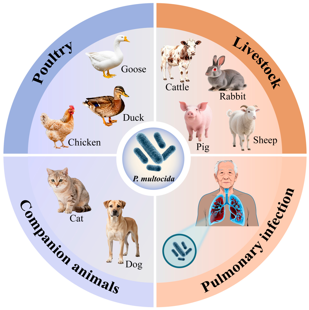
Figure 1. Host range of Pasteurella multocida.
Pathogens 15 00881 g001
Figure 2. Overview of P. multocida virulence factors and host interaction. The arrow indicates the position of the target structure.
Pathogens 15 00881 g002
Figure 3. Mechanism of activation of NLRP3/NLRP6 inflammasome by Pasteurella multocida. The arrows indicate the direction of progression to the subsequent step/process. Different colors are used solely for visual distinction between the components and have no specific scientific meaning.
Pathogens 15 00881 g003
Disclaimer/Publisher’s Note: The statements, opinions and data contained in all publications are solely those of the individual author(s) and contributor(s) and not of MDPI and/or the editor(s). MDPI and/or the editor(s) disclaim responsibility for any injury to people or property resulting from any ideas, methods, instructions or products referred to in the content.