


Article

Comorbidity Burden and Radiological Features Associated with Legionella pneumophila Pneumonia: A Retrospective Case–Control Study

by
Nicola Serra
1,*,
Teresa Maria Assunta Fasciana
2,
Paola Di Carlo
2,3,*,
Anna Giammanco
4,
Antonio Cascio
2,3,
Luca Pipitò
2,3,
Dario De Luca
5,
Caterina Carollo
6,
Valentina Caputo
2,
Martina Maria Federico
2,
Consolato Maria Sergi
7,
Emanuele Grassedonio
8 and
Tommaso Vincenzo Bartolotta
9
1
Audiology Section, Department of Neuroscience, Reproductive Sciences and Dentistry, University of Naples Federico II, 80131 Naples, Italy
2
Department of Health Promotion, Maternal-Childhood, Internal Medicine of Excellence G. D’Alessandro, University of Palermo, 90127 Palermo, Italy
3
Infectious and Tropical Disease Unit and Sicilian Regional Reference Center for the Fight Against AIDS, AOU Policlinico “P. Giaccone”, 90127 Palermo, Italy
4
Legionella Reference Laboratory, University of Palermo, 90127 Palermo, Italy
5
Spinal Unit at the P.O. “Villa delle Ginestre” of the ASP Palermo, Via Castellana, 145, 90135 Palermo, Italy
6
Section of Nephrology, Department of Health Promotion, Maternal-Childhood, Internal Medicine of Excellence G. D’Alessandro, University of Palermo, 90127 Palermo, Italy
7
Department of Laboratory Medicine and Pathology, University of Alberta, Edmonton, AB T6G 2R3, Canada
8
Section of Radiology, Department of Biomedicine, Neuroscience and Advanced Diagnostics (BiND), University of Palermo, 90127 Palermo, Italy
9
Section of Emergency Radiology Unit of Policlinico University Hospital “Paolo Giaccone”, Department of Biomedicine, Neuroscience and Advanced Diagnostics (BiND), University of Palermo, 90127 Palermo, Italy
*
Authors to whom correspondence should be addressed.
Pathogens 2026, 15(8), 866; https://doi.org/10.3390/pathogens15080866
Submission received: 16 July 2026 / Revised: 11 August 2026 / Accepted: 17 August 2026 / Published: 19 August 2026

Abstract

Background: Legionella pneumophila is a leading cause of community- and hospital-acquired pneumonia (HAP/CAP), although the clinical and radiological features associated with infection remain incompletely defined. This study aimed to identify clinical, comorbidity-related, and chest computed tomography (CT) features associated with L. pneumophila pneumonia in hospitalized adults with community-acquired pneumonia (CAP). Methods: A retrospective case–control study was conducted at a tertiary-care university hospital in Italy between January 2019 and August 2025. Eighty patients positive for L. pneumophila were compared with 79 controls negative for L. pneumophila. Demographic, clinical, microbiological, and radiological data were collected, and factors independently associated with infection were evaluated using multivariable logistic regression analysis. Results: Patients with L. pneumophila infection exhibited a greater comorbidity burden, particularly heart failure, and were more frequently smokers. Bilateral lung involvement and hilar/mediastinal lymphadenopathy were significantly more common among Legionella-positive patients, whereas overall pulmonary complications and bilateral pleural effusion were more frequently observed in controls. Multivariable analysis identified comorbidity burden as an independent positive predictor of L. pneumophila infection, while intensive care unit admission, oxygen therapy, and pulmonary complication score were independently associated with a lower likelihood of infection. Conclusions: The integration of comorbidity assessment with readily available chest CT findings may help raise clinical suspicion of L. pneumophila pneumonia and support consideration of appropriate diagnostic testing.

1. Introduction

Legionella pneumophila is an important cause of severe pneumonia acquired in both community and healthcare settings (CAP and HAP) and is associated with substantial morbidity and mortality, particularly among older adults, patients with chronic comorbidities, and immunocompromised individuals [1,2].
Although L. pneumophila accounts for the majority of reported cases of Legionnaires’ disease worldwide, other Legionella species may also cause clinically significant infection. In particular, Legionella longbeachae has emerged as an important human pathogen in specific geographic regions and exhibits epidemiological characteristics that differ from those of L. pneumophila. Nevertheless, L. pneumophila remains the predominant species responsible for Legionella infections in Europe and was the focus of the present investigation.
Although Legionella species account for only a minority of CAP cases, they represent one of the most important causes of atypical pneumonia requiring hospitalization and intensive care, making early recognition essential for appropriate antimicrobial treatment and improved clinical outcomes [2,3,4].
Over the past decade, the epidemiology of Legionnaires’ disease has changed considerably. Italy recently reported approximately 4627 cases of legionellosis, representing an 18–20% increase compared with the previous year [5]. This confirms Italy as the European country with the highest absolute number of reported cases. This trend is likely attributable to multiple factors, including an aging population, increased clinician awareness, wider use of microbiological diagnostic tests, and climate-related environmental changes that promote Legionella proliferation in natural and artificial water systems [6,7]. These epidemiological changes have renewed interest in strategies that facilitate earlier recognition of Legionella infection in patients presenting with CAP [3,4,8,9,10].
Despite important advances in laboratory diagnostics, the early diagnosis of Legionella pneumonia remains challenging. Urinary antigen testing and molecular assays have substantially improved diagnostic accuracy; however, these investigations are generally performed only after Legionella infection is clinically suspected [11,12]. Because the initial clinical presentation frequently overlaps with that of other bacterial pneumonias, delayed diagnostic suspicion remains one of the principal barriers to timely initiation of appropriate anti-Legionella therapy [11,13,14]. Consequently, the identification of readily available clinical and radiological features that can increase the pre-test probability of Legionella infection remains highly relevant in routine clinical practice [11,15].
L. pneumophila is a facultative intracellular microorganism that replicates within alveolar macrophages and monocytes, inducing a complex inflammatory response that frequently extends beyond the respiratory tract in pulmonary infections [16,17]. This distinctive pathophysiological behavior explains the multisystem manifestations that characterize Legionnaires’ disease and suggests that its thoracic imaging phenotype may also differ from that observed in conventional bacterial pneumonia. Previous computed tomography (CT) studies have described bilateral consolidations, ground-glass opacities, and multifocal pulmonary involvement in patients with Legionella pneumonia; however, the specificity and clinical utility of these findings remain incompletely defined, particularly in comparison with other causes of CAP [18,19,20].
In Italian emergency departments, chest CT is frequently performed during the initial evaluation of patients with severe community-acquired pneumonia because of their clinical presentation and the need to rapidly identify pulmonary complications. Consequently, radiological information is often available before microbiological confirmation. The integration of chest CT findings with clinical characteristics and host-related risk factors may provide additional clinical context when evaluating patients with community-acquired pneumonia and may contribute to clinical suspicion of L. pneumophila infection before microbiological confirmation [5,17,19,21].
In addition to imaging findings, host-related characteristics may play an important role in determining susceptibility to Legionella infection. Advanced age, smoking, chronic respiratory disease, cardiovascular disease, diabetes mellitus, malignancy, immunosuppression, and other chronic conditions have all been associated with an increased risk of Legionnaires’ disease [3,8,18,19,22,23]. Nevertheless, relatively few studies have evaluated whether overall comorbidity burden, beyond individual chronic conditions, contributes to the identification and clinical characterization of patients with Legionella pneumonia.
Therefore, the aim of the present study was to investigate the contribution of comorbidity burden and chest CT findings to the early recognition of L. pneumophila pneumonia among adults hospitalized with community-acquired pneumonia.

2. Materials and Methods

2.1. Study Design

This retrospective case-control study was conducted between January 2019 and August 2025 at the Policlinico University Hospital Paolo Giaccone (A.U.O.P.), University of Palermo, Italy, a tertiary-care academic teaching hospital designated as a second-level Emergency Department (DEA Level II). The institution serves as a major referral center for Western Sicily, has more than 500 hospital beds, and manages approximately 15,000 inpatient admissions and more than 50,000 Emergency Department visits annually [24]. The study was conducted in accordance with the Strengthening the Reporting of Observational Studies in Epidemiology (STROBE) statement [25]. The STROBE checklist and flowchart are provided in the Supplementary Materials (Table S1 and Figure S1, respectively).
Patients presenting with community-acquired pneumonia were initially evaluated in the Emergency Department and subsequently admitted to the most appropriate hospital ward according to disease severity, comorbidities, and bed availability. Clinical management was provided by a multidisciplinary team including emergency physicians, internal medicine physicians, pulmonologists, intensivists when required, and infectious disease specialists.
In this study, the patients were stratified into control and study groups (CG and SG, respectively). Patients aged ≥18 years who were hospitalized for community-acquired pneumonia (CAP) were classified according to microbiological testing for Legionella pneumophila performed within 48 h of emergency department admission.
CAP was defined according to the latest ATS/IDSA and ATS Clinical Practice Guidelines [26,27].
The control and study groups were composed of 79 and 80 hospitalized patients, respectively, who underwent urinary antigen testing, respiratory panels, and BioFire FilmArray™ PNplus Panels (BioFire Diagnostics, Salt Lake City, UT, USA), all performed within 48 h of admission [4].
To minimize selection bias, controls were selected among hospitalized patients with community-acquired pneumonia during the same study period and were chosen to achieve overall comparability with cases regarding age, sex, calendar year, season of admission, and sample size. No formal matching procedure was performed. Patients with HIV infection, SARS-CoV-2 infection, active tuberculosis, or hematological disorders were excluded. Patients who had received guideline-concordant anti-Legionella antimicrobial therapy for more than 2 days before presentation to the Emergency Department were excluded from the study [4,26,27].
All patients were admitted to the emergency radiology unit of A.U.O.P. and defined as having a new or progressive existing chest computed tomography infiltrate, in addition to a clinical manifestation suggesting a respiratory infection (fever and respiratory symptoms). A chest CT scan was performed according to Policlinico Universitario Hospital “Paolo Giaccone” guidelines [28,29]. Chest CT examinations were independently reviewed by experienced radiologists who were blinded to microbiological results.
Data on demographics, underlying diseases, pathogens, and ICU admission were retrospectively collected from the electronic records.
ICU admission and oxygen therapy variables referred to the initial management in the Emergency Department or emergency critical care area at presentation and not to events occurring later during hospitalization.
Chronic lung disease comorbidities included chronic obstructive pulmonary disease, interstitial lung disease, bronchiectasis, emphysema, asthma, and chronic bronchitis. Additionally, heart failure comorbidity included all patients, according to Bozkurt B. et al. [30]. The authors classify the medical setting in three categories—medicine, surgery, and intensive care unit (ICU)—reflecting differences in procedures and patient populations. Particularly, the medicine area included cardiology, endocrinology, gastroenterology, geriatrics, hematology, infectious diseases, internal medicine, nephrology, neurology, oncology, and pulmonology wards. The surgery area included cardiovascular surgery, general surgery, neurosurgery, oncological surgery, otolaryngology, orthopedic surgery, plastic surgery, urology, vascular surgery, and obstetrics and gynecology. Finally, the intensive care unit included general intensive care, postoperative care, and specialized care.
Urine samples were collected to determine the presence of L. pneumophila Sg 1–15 L. longbeachae antigens in urine by DiaSorin S.p.A. (Saluggia, Italy), as previously reported [4,31]. Bronchoalveolar Lavage (BAL)/mini-BAL/Protected Specimen Brush (PSB) samples were collected from patients hospitalized in the ICU and in other medical intensive care units and underwent microbiological investigation as previously reported [4,31,32].
Diagnostic testing was performed in accordance with current international recommendations for Legionella diagnosis [26,27]. Cases were defined as patients with microbiologically confirmed L. pneumophila infection based on a positive urinary antigen test and/or molecular detection of L. pneumophila in respiratory specimens using the BioFire FilmArray™ Pneumonia Panel Plus, (BioFire Diagnostics, Salt Lake City, UT, USA). Although diagnostic testing also allowed the evaluation of other clinically relevant Legionella species, including L. longbeachae, no cases attributable to non-pneumophila Legionella species were identified during the study period. Therefore, all Legionella-positive patients included in the present study had microbiologically confirmed L. pneumophila infection. We acknowledge that urinary antigen testing alone may have limited sensitivity and primarily detects L. pneumophila serogroup 1; therefore, molecular testing was also performed whenever respiratory specimens were available. Bacterial and fungal pathogens were identified using standard sputum culture and/or BioFire FilmArray Pneumonia Panel Plus, (BioFire Diagnostics, Salt Lake City, UT, USA), which can identify 15 typical bacteria, three atypical bacteria, and seven antimicrobial resistance (AMR) genes, including Chlamydia pneumoniae, Mycoplasma pneumoniae, and L. pneumophila [4,31,32].
Antibiotic therapy was initiated in the emergency department in accordance with the hospital guidelines [33].

2.2. Statistical Analysis

Categorical variables are expressed as absolute numbers and percentages, while continuous variables are reported as mean and standard deviation (SD) or median and interquartile intervals (IQRs), as appropriate.
Normality of continuous variables was assessed using the Shapiro–Wilk test. For comparisons between two groups, the unpaired t-test was used for normally distributed variables, while the Mann–Whitney U test was applied for non-normally distributed variables.
Differences in proportions between groups were evaluated using the chi-square test or Fisher’s exact test, as appropriate. When comparing more than two categories, post hoc analysis was performed using adjusted standardized residuals and Z-tests.
A multivariable logistic regression analysis was conducted to identify factors independently associated with L. pneumophila infection. Variables included in the model were selected based on statistical significance in univariate analysis (p < 0.05) and clinical relevance.
For this analysis, the following variables were considered dichotomous: Intensive Care Unit (ICU) admission (0 = no, 1 = yes), oxygen therapy (0 = no, 1 = yes), smoking status (0 = no, 1 = yes), and lung involvement (0 = unilateral, 1 = bilateral). ICU admission and oxygen therapy referred to patient status and management decisions made during the initial Emergency Department evaluation.
Comorbidities were quantified using the Charlson Comorbidity Index (CCI), a validated scoring system that assigns weighted points to specific conditions based on their impact on mortality risk [34,35]. The total CCI score for each patient was calculated as the sum of all weighted conditions. Higher scores indicate a greater comorbidity burden. In our study, the CCI was calculated as follows: liver cirrhosis (moderate–severe) = 3, dialysis = 2, diabetes = 1, heart failure = 1, dementia = 1, chronic lung diseases = 1, and hypertension = 0. Regarding lung complications, they included pneumothorax, cavitation, pulmonary embolism, hilar/mediastinal lymphadenopathy, unilateral pleural effusion, bilateral pleural effusion, and acute respiratory distress syndrome (ARDS).
As no validated scoring system is available for these conditions, a lung complication score was constructed based on clinical severity. Higher weights were assigned to conditions associated with greater respiratory impairment. The score was defined as follows: ARDS = 3; pneumothorax, pulmonary embolism, and bilateral pleural effusion = 2; cavitation, lymphadenopathy, and unilateral pleural effusion = 1.
In patients with multiple lung complications, the total score was calculated as the sum of all weighted conditions. This score was used for exploratory analysis. Patients with incomplete clinical, microbiological, or radiological data were excluded from the analysis.
To assess the adequacy of the sample size for multivariable modeling, the events-per-variable (EPV) approach was considered. The final logistic regression model included five covariates (ICU admission, oxygen therapy, smoking status, Charlson Comorbidity Index, and lung complication score) and was based on 80 patients with microbiologically confirmed L. pneumophila infection (events). Therefore, the ratio between events and covariates was 16 (EPV = 80/5 = 16), exceeding commonly recommended thresholds for logistic regression analyses and supporting the stability of the exploratory multivariable model [36,37].
All tests with a p-value < 0.05 were considered statistically significant. Statistical analysis was performed using MATLAB (Matrix Laboratory) analytical toolbox R2008b (MathWorks, Natick, MA, USA).

3. Results

The control group (CG) was composed of 72.2% (57) males and 27.8% (22) females, with ages ranging from 25 to 94 years, a mean of 65.2 years, and a standard deviation of 17.2 years. The study group (SG) comprised 66.3% (53) males and 33.8% (27) females, with ages ranging from 26 to 95 years, a mean of 67.2 years, and a standard deviation of 15.8 years. Among Legionella-negative controls, the microbiological investigations identified a heterogeneous spectrum of pathogens. The most frequently detected Gram-negative bacteria included Pseudomonas aeruginosa, Haemophilus influenzae, Klebsiella pneumoniae, and Moraxella catarrhalis, whereas Gram-positive isolates included Streptococcus pneumoniae and Staphylococcus aureus. Respiratory viruses were also identified, predominantly influenza A virus, parainfluenza viruses, and human rhinovirus/enterovirus. In addition, a small number of cases of Mycoplasma pneumoniae infection were detected during the study period.
Table 1 summarizes the demographic, clinical, microbiological, and comorbidity-related characteristics of both groups.
Table 1 shows significant differences between the control group (CG) and the study group (SG) in ICU admission, oxygen therapy, smoking status, and comorbidity-related characteristics.
ICU admission and oxygen therapy were more frequently observed in the CG than in the SG (79.7% vs. 63.8%, p = 0.0256, and 78.5% vs. 63.8%, p = 0.0412, respectively).
Conversely, smoking was more prevalent in the SG than in the CG (83.8% vs. 67.1%, p = 0.0149). Regarding underlying conditions, patients with L. pneumophila infection exhibited a greater burden of comorbidities, with heart failure being the condition most strongly represented in the SG (41.3%, p = 0.0004).
Table 2 summarizes the radiological findings observed during the acute phase of L. pneumophila pneumonia. Lung complications included pneumothorax, cavitation, pulmonary embolism, hilar and/or mediastinal lymphadenopathy, unilateral pleural effusion, bilateral pleural effusion, and acute respiratory distress syndrome (ARDS).
Table 2 shows that bilateral lung involvement was more frequent in the SG than in the CG (77.5% vs. 62.0%, p = 0.0336). Overall pulmonary complications were more frequently observed in the CG than in the SG (83.5% vs. 62.5%, p = 0.0029), although the distribution of specific radiological abnormalities differed between groups. Analysis of individual abnormalities revealed significant differences in bilateral pleural effusion and hilar/mediastinal lymphadenopathy.
Variables significantly associated with L. pneumophila infection in univariate analyses were entered into a multivariable logistic regression model (Table 3). The final model included five covariates (ICU admission, oxygen therapy, smoking status, Charlson Comorbidity Index, and lung complication score) and was based on 80 microbiologically confirmed L. pneumophila cases (events). The adequacy of the sample size for multivariable modeling was assessed using the events-per-variable (EPV) criterion. The EPV was calculated as the number of outcome events divided by the number of covariates included in the model (EPV = E/nC), where E represents the number of events and nC the number of covariates. Accordingly, the present model yielded an EPV of 16 (EPV = 80/5 = 16), exceeding the commonly recommended minimum threshold of 10 events per variable and supporting the stability of the exploratory multivariable analysis [36,37].
The multivariable logistic regression model was statistically significant (p < 0.0001), indicating that the included variables were collectively associated with L. pneumophila infection.
ICU admission, oxygen therapy, and lung complication score were independently associated with a lower probability of L. pneumophila infection (OR = 0.17, p = 0.0006; OR = 0.28, p = 0.0047; OR = 0.57, p = 0.0078, respectively).
Conversely, comorbidity burden, assessed by the Charlson Comorbidity Index (CCI), was identified as an independent positive predictor, with each unit increase associated with a 61% higher likelihood of infection (OR = 1.61, p = 0.0008).
Smoking status showed a positive trend but did not reach statistical significance (OR = 2.12, p = 0.09).
The odds ratios and corresponding 95% confidence intervals derived from the multivariable logistic regression model are presented in Figure 1.
Figure 1 shows the magnitude and direction of the associations identified in the multivariable model. The Charlson Comorbidity Index (CCI) was the only independent factor positively associated with L. pneumophila infection, whereas ICU admission, oxygen therapy, and lung complication score showed significant inverse associations. Smoking did not retain statistical significance.

4. Discussion

The rationale for the present study stems from the growing epidemiological relevance of Legionnaires’ disease in Italy, where incidence has increased in recent years. This trend likely reflects the combined effects of climate change, population aging, increased environmental exposure to Legionella spp., greater clinical awareness, and the more widespread use of molecular diagnostic techniques that improve the detection of Legionella infections. Warmer temperatures and altered rainfall patterns may promote bacterial proliferation in both natural and artificial water systems, raising concerns that the burden of infection will continue to increase [2,3,4].
In Italian emergency departments, patients presenting with severe community-acquired pneumonia (CAP) routinely undergo chest computed tomography (CT) due to clinical severity. This widespread use of thoracic imaging provides an opportunity to identify radiological patterns that, although not specific, may increase clinical suspicion for Legionella pneumonia at presentation. Accordingly, we investigated whether host characteristics, comorbidity burden, and imaging findings were associated with L. pneumophila infection and could help increase clinical suspicion in patients presenting with community-acquired pneumonia, supporting timely microbiological testing and appropriate antimicrobial therapy [8,20,38].
The term ‘atypical pneumonia’ has historically been applied to pathogens such as Legionella pneumophila, Mycoplasma pneumoniae, and Chlamydia pneumoniae because of their microbiological characteristics and the difficulty of detection using conventional diagnostic methods. Our findings do not seek to redefine this classification. Rather, they suggest that certain host-related and radiological features may increase clinical suspicion of L. pneumophila infection among patients presenting with community-acquired pneumonia.
The Charlson Comorbidity Index (CCI) was independently associated with L. pneumophila infection, supporting the hypothesis that cumulative comorbidity burden may contribute to host susceptibility to Legionella pneumonia, particularly among older adults [39,40,41].
Another important finding of our study concerns the radiological phenotype of Legionella pneumophila pneumonia. Patients with Legionella infection more frequently exhibited bilateral pulmonary involvement and hilar or mediastinal lymphadenopathy than patients with non-Legionella community-acquired pneumonia. Our findings are consistent with previous studies describing bilateral pulmonary involvement and hilar or mediastinal lymphadenopathy among patients with Legionella pneumonia. Previous CT studies have shown that initially focal infiltrates may extend to adjacent lobes and become bilateral, even despite appropriate antimicrobial therapy and clinical improvement [17,42,43,44]. In contrast, Rémi Poirier et al. reported that radiographic and tomographic manifestations of LD are nonspecific and like those found in community-acquired pneumonia of other bacterial origin [45]. The distinctive pathogenic mechanism of the higher frequency of bilateral pulmonary involvement, as well as the significantly greater prevalence of hilar and mediastinal lymphadenopathy observed in our cohort, may be due to the higher prevalence of heart failure in our enrolled population [46,47]. As reported by Di Carlo et al., heart failure was more frequent among Legionella cases and may increase susceptibility to L. pneumophila infection [4]. Therefore, the interpretation of mediastinal lymphadenopathy as a radiological feature associated with Legionella pneumonia should be considered with caution, given the potential confounding effect of heart failure.
Conversely, pulmonary complications were more frequently observed in the control group. This finding suggests that radiological complexity alone may not be a distinguishing feature of Legionella pneumonia. Rather, the higher frequency of complications in controls may reflect differences in disease severity, underlying clinical characteristics, or etiological heterogeneity between the study groups [32,48]. Furthermore, pathogens known to cause pulmonary disease, as well as other opportunistic pulmonary microorganisms, are associated with seasonality in our geographical area [49,50,51,52]. In summary, this analysis suggests that comorbidity burden may represent a major factor associated with L. pneumophila infection in patients with community-acquired pneumonia. Acute clinical severity and radiological complexity played a lesser role in risk stratification. These data suggest that a careful assessment of chronic conditions should guide clinical suspicion and subsequent diagnostic decisions, thereby optimizing the management of this population. Importantly, ICU admission and oxygen therapy in this study referred to clinical status and management decisions made during the initial Emergency Department evaluation rather than to events occurring later during hospitalization. Therefore, their inverse association with L. pneumophila infection reflects differences observed at presentation and should not be interpreted as evidence of a causal relationship.
Moreover, our findings support the hypothesis that bilateral pulmonary involvement and hilar or mediastinal lymphadenopathy, when interpreted together with a higher burden of comorbidities, define a clinical profile associated with L. pneumophila infection. These readily available clinical and imaging features may contribute to clinical suspicion of L. pneumophila infection and support consideration of appropriate microbiological testing when clinically indicated. Rather than replacing microbiological confirmation, their integration into routine clinical assessment may support earlier diagnostic investigation for L. pneumophila infection and consequently contribute to reducing diagnostic delay, particularly in countries such as Italy, where the incidence of Legionnaires’ disease is increasing [6,32,52,53].

Limitations

This study has several limitations that should be considered when interpreting the results.
First, the retrospective design may introduce selection bias and limits the ability to establish causal relationships between the identified predictors and L. pneumophila infection.
Second, the study was conducted at a single center, which may limit the generalizability of the findings to other settings or populations. Furthermore, the study was conducted in a tertiary-care university hospital and primarily involved patients evaluated in an emergency-care setting. Therefore, the findings may not be fully generalizable to lower-risk populations, primary care settings, or healthcare systems characterized by different referral pathways and access to specialized care. However, studies specifically investigating L. pneumophila pneumonia during the initial emergency assessment remain relatively limited, and this context should be considered when interpreting the present findings.
Third, although comorbidities were quantified using the Charlson Comorbidity Index (CCI), an adapted approach was used to include multiple conditions within the same category, which may differ from the standard Charlson methodology. While this approach better reflects the overall burden of disease, it may reduce comparability with other studies.
Fourth, the lung complication score was not based on a validated scoring system but was instead derived from clinical severity. Although this approach allowed a more comprehensive assessment of pulmonary involvement, it should be considered exploratory and requires external validation.
Finally, although the study included a relatively large series of microbiologically confirmed L. pneumophila cases compared with previous single-center studies, the overall sample size remained relatively small. This may have limited statistical power, increased the possibility of type II error, and reduced the generalizability of the findings to other populations and healthcare settings. Nevertheless, the number of events available for the multivariable analysis was considered adequate in relation to the number of covariates included in the final model. Therefore, the present findings should be interpreted as exploratory and hypothesis-generating and require confirmation in larger multicenter cohorts.

5. Conclusions

In conclusion, this study shows that L. pneumophila community-acquired pneumonia is associated with a characteristic clinical and radiological profile compared with other causes of CAP. A higher comorbidity burden, particularly as assessed by the Charlson Comorbidity Index, emerged as the primary independent factor associated with infection, highlighting the importance of host susceptibility in the pathogenesis of Legionella disease. On chest CT, bilateral pulmonary involvement and hilar or mediastinal lymphadenopathy were more frequently observed in patients with L. pneumophila infection and may help raise diagnostic suspicion when interpreted within the appropriate clinical context. The integration of comorbidity assessment with readily available radiological findings may contribute to raising clinical suspicion of L. pneumophila infection and may support consideration of Legionella-specific diagnostic testing in appropriate clinical settings. However, these findings should be interpreted as associations observed within a retrospective case-control study rather than as validated diagnostic predictors and therefore require prospective confirmation before any clinical implementation.
Future multicenter prospective studies are warranted to validate these findings and assess their applicability in broader populations of patients with community-acquired pneumonia.
Future prospective studies may further investigate whether the clinical and radiological factors identified in this study have utility in broader clinical settings and whether their potential integration into clinical decision-support tools warrants evaluation. However, any such application would require rigorous prospective validation before implementation in clinical practice [54,55].

Supplementary Materials

The following supporting information can be downloaded at: https://www.mdpi.com/article/10.3390/pathogens15080866/s1, Figure S1: Study flowchart; Table S1: STROBE checklist.

Author Contributions

Conceptualization: P.D.C., T.M.A.F. and N.S.; formal analysis: N.S.; methodology: N.S. and P.D.C.; investigation and data curation: D.D.L., M.M.F., T.M.A.F., E.G. and T.V.B.; writing—original draft preparation: P.D.C., N.S., D.D.L., E.G., C.M.S. and A.C.; writing—review and editing: A.G., C.C., L.P., V.C., N.S. and A.C.; supervision: P.D.C., C.M.S., A.G. and T.V.B. All authors have read and agreed to the published version of the manuscript.

Funding

This research received no external funding.

Institutional Review Board Statement

The study protocol was approved by the Ethics Committee Palermo 1 of the University Hospital, Policlinico Paolo Giaccone of Palermo (protocol code: No. 08/2026, date of approval: 9 April 2026).

Informed Consent Statement

Informed consent was obtained from all subjects involved in the study.

Data Availability Statement

The research data were available from Paola Di Carlo and Tommaso Vincenzo Bartolotta.

Conflicts of Interest

The authors declare no conflicts of interest.

Abbreviations

The following abbreviations are used in this manuscript:
CTComputer Tomography
ICUIntensive Care Unit
HIVHuman Immunodeficiency Virus
CAPCommunity-Acquired Pneumonia
CGControl Group
SGStudy Group

References

  1. Centers for Disease Control and Prevention (CDC). Clinical Overview of Legionnaires’ Disease. 2025. Available online: https://www.cdc.gov/legionella/hcp/clinical-overview/index.html (accessed on 1 July 2026).
  2. Wang, S.; Tang, J.; Tan, Y.; Song, Z.; Qin, L. Prevalence of atypical pathogens in patients with severe pneumonia: A systematic review and meta-analysis. BMJ Open 2023, 13, e066721. [Google Scholar] [CrossRef] [Scilit] [PubMed]
  3. Chahin, A.; Opal, S.M. Severe Pneumonia Caused by Legionella pneumophila: Differential Diagnosis and Therapeutic Considerations. Infect. Dis. Clin. N. Am. 2017, 31, 111–121. [Google Scholar] [CrossRef] [Scilit] [PubMed]
  4. Di Carlo, P.; Serra, N.; Fasciana, T.M.A.; Fasciana, F.; Pipitò, L.; Giammanco, A.; Capuano, A.; Carollo, C.; Caputo, V.; Bartolotta, T.V.; et al. Community-Acquired Legionella pneumophila Pneumonia: A Case-Control Study in Adult Inpatients from 2019 to 2024. Pathogens 2025, 15, 13. [Google Scholar] [CrossRef] [Scilit] [PubMed]
  5. Bellini, B.; Cannone, A.; Fadda, G.; Scaturro, M.; Mancini, F.; Bella, A.; Giannitelli, S.; Urcioli, R.; Mimmo, R.; Pezzotti, P.; et al. I casi di legionellosi segnalati al sistema di sorveglianza nazionale. Italia, 2024. Boll. Epidemiol. Naz. 2025, 6, 9–18. [Google Scholar] [CrossRef]
  6. Sciurti, A.; Iera, J.; Cannone, A.; Mateo-Urdiales, A.; De Angelis, L.; Baccolini, V.; Urciuoli, R.; Giannitelli, S.; Fadda, G.; Bellini, B.; et al. Seasonality and effects of climatic exposures on community-acquired Legionnaires’ disease incidence, Italy, 2005–2023. Eurosurveillance 2026, 31, 2500712. [Google Scholar] [CrossRef] [Scilit] [PubMed]
  7. Ha, R.; Heilmann, A.; Lother, S.A.; Turenne, C.; Alexander, D.; Keynan, Y.; Rueda, Z.V. The Adequacy of Current Legionnaires’ Disease Diagnostic Practices in Capturing the Epidemiology of Clinically Relevant Legionella: A Scoping Review. Pathogens 2024, 13, 857. [Google Scholar] [CrossRef] [Scilit] [PubMed]
  8. Gattuso, G.; Rizzo, R.; Lavoro, A.; Spoto, V.; Porciello, G.; Montagnese, C.; Cinà, D.; Cosentino, A.; Lombardo, C.; Mezzatesta, M.L.; et al. Overview of the Clinical and Molecular Features of Legionella pneumophila: Focus on Novel Surveillance and Diagnostic Strategies. Antibiotics 2022, 11, 370. [Google Scholar] [CrossRef] [Scilit] [PubMed]
  9. Cunha, B.A.; Cunha, C.B. Legionnaire’s Disease: A Clinical Diagnostic Approach. Infect. Dis. Clin. N. Am. 2017, 31, 81–93. [Google Scholar]
  10. Cheng, R.; Deng, Z.; Lin, F.; Zhang, B.; Liang, J.; Lu, M. Clinical, laboratory, and radiological features of community-acquired pneumonia due to Chlamydia psittaci and Legionella pneumophila confirmed using next-generation sequencing. Ann. Med. 2026, 58, 2627122. [Google Scholar] [CrossRef] [Scilit] [PubMed]
  11. Viasus, D.; Gaia, V.; Manzur-Barbur, C.; Carratalà, J. Legionnaires’ Disease: Update on Diagnosis and Treatment. Infect. Dis. Ther. 2022, 11, 973–986. [Google Scholar] [CrossRef] [Scilit] [PubMed]
  12. Miller, J.M.; Binnicker, M.J.; Campbell, S.; Carroll, K.C.; Chapin, K.C.; Gonzalez, M.D.; Harrington, A.; Jerris, R.C.; Kehl, S.C.; Leal, S.M.; et al. Guide to Utilization of the Microbiology Laboratory for Diagnosis of Infectious Diseases: 2024 Update. Clin. Infect. Dis. 2024. [Google Scholar] [CrossRef] [Scilit] [PubMed]
  13. Robesyn, E.; Uldum, S.A.; Bjerre, K.D.; Kjelsø, C.; Struelens, M.; Lundborg, C.S.; Ethelberg, S.; Faes, C. Diagnostic testing intensity for Legionnaires’ disease: Spatio-temporal assessment and its effect on surveillance case reporting, Denmark, 2014–2022. PLoS ONE 2026, 21, e0346580. [Google Scholar] [CrossRef] [Scilit] [PubMed]
  14. European Society of Clinical Microbiology and Infectious Diseases Study Group for Legionella Infections (ESGLI). ESGLI guidance for the diagnosis of Legionella infections. Clin. Microbiol. Infect. 2017, 23, 1–8. [Google Scholar]
  15. Bai, L.; Yang, W.; Li, Y. Clinical and Laboratory Diagnosis of Legionella Pneumonia. Diagnostics 2023, 13, 280. [Google Scholar] [CrossRef] [Scilit] [PubMed]
  16. Guillemot, J.; Ginevra, C.; Allam, C.; Kay, E.; Gilbert, C.; Doublet, P.; Jarraud, S.; Chapalain, A. TNF-α Response in Macrophages Depends on Clinical Legionella pneumophila Isolates Genotypes. Virulence 2022, 13, 160–173. [Google Scholar] [CrossRef] [Scilit] [PubMed]
  17. Zhang, Z.; Liu, X.; Chen, L.; Qiu, J. Chest radiographic characteristics of community-acquired Legionella pneumonia in the elderly. Chin. Med. J. 2014, 127, 2270–2274. [Google Scholar] [CrossRef] [Scilit]
  18. Sopena, N.; Sabrià, M. Multicenter study of Legionella pneumonia: Clinical and radiological findings. Chest 2007, 131, 1617–1625. [Google Scholar]
  19. Kim, K.W.; Goo, J.M.; Lee, H.Y.; Park, C.M.; Lee, C.H.; Im, J.G. Chest Computed Tomographic Findings and Clinical Features of Legionella Pneumonia. J. Comput. Assist. Tomogr. 2007, 31, 950–955. [Google Scholar] [CrossRef] [Scilit]
  20. Phin, N.; Parry-Ford, F.; Harrison, T.; Stagg, H.R.; Zhang, N.; Kumar, K.; Lortholary, O.; Zumla, A.; Abubakar, I. Epidemiology and clinical management of Legionnaires’ disease. Lancet Infect. Dis. 2014, 14, 1011–1021. [Google Scholar] [CrossRef] [Scilit] [PubMed]
  21. Pulsipher, A.M.; Khattar, G.; VanDolah, H.; Gotway, M.; Ham, K.; Alqalyoobi, S.; Cartin-Ceba, R.; Patel, R.; Simner, P.J.; Sen, A.; et al. Legionella Pneumonia in the Modern Era: Clinical Features and Predictors of Mortality. Clin. Infect. Dis. 2026, ciag085. [Google Scholar] [CrossRef] [Scilit] [PubMed]
  22. Chidiac, C.; Che, D.; Pires-Cronenberger, S.; Jarraud, S.; Campèse, C.; Bissery, A.; Weinbreck, P.; Brun-Buisson, C.; Sollet, J.P.; Ecochard, R.; et al. Factors associated with hospital mortality in community-acquired legionellosis in France. Eur. Respir. J. 2012, 39, 963–970. [Google Scholar] [CrossRef] [Scilit] [PubMed]
  23. Serra, N.; Andriolo, M.; Butera, I.; Mazzola, G.; Sergi, C.M.; Fasciana, T.M.A.; Giammanco, A.; Gagliano, M.C.; Cascio, A.; Di Carlo, P. A Serological Analysis of the Humoral Immune Responses of Anti-RBD IgG, Anti-S1 IgG, and Anti-S2 IgG Levels Correlated to Anti-N IgG Positivity and Negativity in Sicilian Healthcare Workers (HCWs) with Third Doses of the mRNA-Based SARS-CoV-2 Vaccine: A Retrospective Cohort Study. Vaccines 2023, 11, 1136. [Google Scholar] [CrossRef] [Scilit] [PubMed]
  24. Azienda Ospedaliera Universitaria Policlinico “Paolo Giaccone”. Piano Integrato di Attività e Organizzazione (PIAO) 2023–2025; PIAO 2023–2025; Azienda Ospedaliera Universitaria Policlinico “Paolo Giaccone”: Palermo, Italy, 2023; Available online: https://www.esteri.it/en/trasparenza_comunicazioni_legali/performance/piano-integrato-di-attivita-e-organizzazione-piao/ (accessed on 7 August 2026).
  25. von Elm, E.; Altman, D.G.; Egger, M.; Pocock, S.J.; Gøtzsche, P.C.; Vandenbroucke, J.P. Strengthening the reporting of observational studies in epidemiology (STROBE) statement: Guidelines for reporting observational studies. BMJ 2007, 335, 806–808. [Google Scholar] [CrossRef] [Scilit] [PubMed]
  26. Metlay, J.P.; Waterer, G.W.; Long, A.C.; Anzueto, A.; Brozek, J.; Crothers, K.; Cooley, L.A.; Dean, N.C.; Fine, M.J.; Flanders, S.A.; et al. Diagnosis and treatment of adults with community-acquired pneumonia. An official clinical practice guideline of the American Thoracic Society and Infectious Diseases Society of America. Am. J. Respir. Crit. Care Med. 2019, 200, e45–e67. [Google Scholar] [CrossRef] [Scilit] [PubMed]
  27. Jones, B.E.; Ramirez, J.A.; Oren, E.; Soni, N.J.; Sullivan, L.R.; Restrepo, M.I.; Musher, D.M.; Erstad, B.L.; Pickens, C.; Vaughn, V.M.; et al. Diagnosis and Management of Community-acquired Pneumonia: An Official American Thoracic Society Clinical Practice Guideline. Am. J. Respir. Crit. Care Med. 2026, 212, 24–44. [Google Scholar] [CrossRef] [Scilit] [PubMed]
  28. Agnello, F.; Rabiolo, L.; Grassedonio, E.; Toia, P.; Midiri, F.; Spatafora, L.; Matteini, F.; Tesè, L.; La Grutta, L.; Galia, M. Imaging the COVID-19: A practical guide. Monaldi Arch. Chest Dis. 2021, 91, 1–8. [Google Scholar] [CrossRef] [Scilit] [PubMed]
  29. Percorso Diagnostico Terapeutico Assistenziale (PDTA) Polmone. A.U.O.P. Paolo Giaccone. Available online: https://policlinico.pa.it/documents/d/guest/PDTA%20n15%20-%20Tumore%20del%20Polmone (accessed on 5 July 2026).
  30. Bozkurt, B.; Hershberger, R.E.; Butler, J.; Grady, K.L.; Heidenreich, P.A.; Isler, M.L.; Kirklin, J.K.; Weintraub, W.S. 2021 ACC/AHA key data elements and definitions for heart failure: A report of the American College of Cardiology/American Heart Association Task Force on Clinical Data Standards (Writing Committee to Develop Clinical Data Standards for Heart Failure). J. Am. Coll. Cardiol. 2021, 77, 2053–2150. [Google Scholar] [CrossRef] [Scilit] [PubMed]
  31. Fasciana, T.; Mascarella, C.; Distefano, S.A.; Calà, C.; Capra, G.; Rampulla, A.; Di Carlo, P.; Palermo, M.; Giammanco, A. Cluster of Legionnaires’ Disease in an Italian Prison. Int. J. Environ. Res. Public Health 2019, 16, 2062. [Google Scholar] [CrossRef] [Scilit] [PubMed]
  32. Ippolito, M.; Catalisano, G.; Velardo, M.; Campione, M.; Damiano, M.; Bocci, M.G.; Camilloni, B.; Cocuzza, C.; Comar, M.; De Robertis, E.; et al. The SIS NET ICU study: Characteristics of patients with severe community acquired pneumonia admitted to Italian ICUs-a multicenter prospective observational study. J. Anesth. Analg. Crit. Care 2026, 6, 14. [Google Scholar] [CrossRef] [Scilit] [PubMed] [PubMed Central]
  33. Antimicrobial Stewardship Team,- Schemi di Terapia Empirica. A.U.O.P. Paolo Giaccone. Available online: https://policlinico.pa.it/comitato-infezioni-ospedaliere (accessed on 5 July 2026).
  34. Charlson, M.E.; Pompei, P.; Ales, K.L.; MacKenzie, C.R. A new method of classifying prognostic comorbidity in longitudinal studies: Development and validation. J. Chronic Dis. 1987, 40, 373–383. [Google Scholar] [CrossRef] [Scilit] [PubMed]
  35. Quan, H.; Li, B.; Couris, C.M.; Fushimi, K.; Graham, P.; Hider, P.; Januel, J.M.; Sundararajan, V. Updating and validating the Charlson comorbidity index and score for risk adjustment in hospital discharge abstracts using data from 6 countries. Am. J. Epidemiol. 2011, 173, 676–682. [Google Scholar] [CrossRef] [Scilit] [PubMed]
  36. Peduzzi, P.; Concato, J.; Kemper, E.; Holford, T.R.; Feinstein, A.R. A simulation study of the number of events per variable in logistic regression analysis. J. Clin. Epidemiol. 1996, 49, 1373–1379. [Google Scholar] [CrossRef] [Scilit] [PubMed]
  37. Vittinghoff, E.; McCulloch, C.E. Relaxing the rule of ten events per variable in logistic and Cox regression. Am. J. Epidemiol. 2007, 165, 710–718. [Google Scholar] [CrossRef] [Scilit] [PubMed]
  38. Moran, G.J.; Talan, D.A.; Abrahamian, F.M. Diagnosis and management of pneumonia in the emergency department. Infect. Dis. Clin. N. Am. 2008, 22, 53–72. [Google Scholar] [CrossRef] [Scilit] [PubMed] [PubMed Central]
  39. Bahlis, L.F.; Diogo, L.P.; Fuchs, S.C. Charlson Comorbidity Index and other predictors of in-hospital mortality among adults with community-acquired pneumonia. J. Bras. Pneumol. 2021, 47, e20200257. [Google Scholar] [CrossRef] [Scilit] [PubMed]
  40. Acuña-Rocha, V.D.; Zuñiga-Hernández, J.A. Impact of exercise interventions on hospitalized patients with community-acquired pneumonia: The role of comorbidities in clinical outcomes. Clin. Infect. Dis. 2024, 80, 681. [Google Scholar] [CrossRef] [Scilit] [PubMed]
  41. Liao, J.; Hong, J.; Miao, L.; Shen, X.; Xie, C. A retrospective analysis of mental well-being, nutritional status, and comorbidity burden in elderly patients with community-acquired pneumonia. BMC Public Health 2025, 25, 667. [Google Scholar] [CrossRef] [Scilit] [PubMed] [PubMed Central]
  42. Coletta, F.S.; Fein, A.M. Radiological manifestations of Legionella/Legionella-like organisms. Semin. Respir. Infect. 1998, 13, 109–115. [Google Scholar] [PubMed]
  43. Taguchi, Y.; Nakahama, C.; Inamatsu, T.; Arakawa, M.; Saito, A.; Hara, K.; Ezaki, T.; Yabuuchi, E.; Ueda, Y. Analysis of chest radiographs of culture positive Legionella pneumonia in Japan, 1980-1990. Working Party for Legionellosis, Japanese Ministry of Health and Welfare. Kansenshogaku Zasshi 1992, 66, 1580–1586. [Google Scholar] [CrossRef] [Scilit] [PubMed][Green Version]
  44. Cunha, B.A.; Varantsova, A.; Jimada, I. Study of the radiologic features of Legionnaires’ disease with mediastinal adenopathy: Legionella or lymphoma? Eur. J. Clin. Microbiol. Infect. Dis. 2018, 37, 463–468. [Google Scholar] [CrossRef] [Scilit] [PubMed]
  45. Poirier, R.; Rodrigue, J.; Villeneuve, J.; Lacasse, Y. Early Radiographic and Tomographic Manifestations of Legionnaires’ Disease. Can. Assoc. Radiol. J. 2017, 68, 328–333. [Google Scholar] [CrossRef] [Scilit] [PubMed]
  46. Ngom, A.; Dumont, P.; Diot, P.; Lemarié, E. Benign mediastinal lymphadenopathy in congestive heart failure. Chest 2001, 119, 653–656. [Google Scholar] [CrossRef] [Scilit] [PubMed]
  47. Erly, W.K.; Borders, R.J.; Outwater, E.K.; Zaetta, J.M.; Borders, G.T. Location, size, and distribution of mediastinal lymph node enlargement in chronic congestive heart failure. J. Comput. Assist. Tomogr. 2003, 27, 485–489. [Google Scholar] [CrossRef] [Scilit] [PubMed]
  48. Cocchio, S.; Cozzolino, C.; Furlan, P.; Cozza, A.; Tonon, M.; Russo, F.; Saia, M.; Baldo, V. Pneumonia-Related Hospitalizations among the Elderly: A Retrospective Study in Northeast Italy. Diseases 2024, 12, 254. [Google Scholar] [CrossRef] [Scilit] [PubMed]
  49. Di Carlo, P.; Serra, N.; Collotta, O.; Colomba, C.; Firenze, A.; Aprea, L.; Distefano, S.A.; Cortegiani, A.; Giammanco, G.; Fasciana, T.M.A.; et al. The Influence of the Seasonal Variability of Candida spp. Bloodstream Infections and Antifungal Treatment: A Mediterranean Pilot Study. Antibiotics 2025, 14, 452. [Google Scholar] [CrossRef] [Scilit] [PubMed]
  50. Di Carlo, P.; Serra, N.; Sergi, C.M.; Toia, F.; Battaglia, E.; Fasciana, T.M.A.; Rodolico, V.; Giammanco, A.; Salamone, G.; Cordova, A.; et al. Seasonal Change in Microbial Diversity: Bile Microbiota and Antibiotics Resistance in Patients with Bilio-Pancreatic Tumors: A Retrospective Monocentric Study (2010–2020). Antibiotics 2025, 14, 283. [Google Scholar] [CrossRef] [Scilit] [PubMed]
  51. Malta, G.; Serra, N.; Spatola, G.F.; Maida, C.M.; Graziano, G.; Di Raimondo, D.; Fasciana, T.M.A.; Caputo, V.; Giammanco, A.; Capuano, A.; et al. The Impact of the Seasonal and Geographical Distribution of Tuberculosis in Sicily: A 6-Year Retrospective Study (2018–2023). J. Clin. Med. 2024, 13, 3546. [Google Scholar] [CrossRef] [Scilit] [PubMed]
  52. Arrigo, I.; Galia, E.; Fasciana, T.; Diquattro, O.; Tricoli, M.R.; Serra, N.; Palermo, M.; Giammanco, A. Four-Year Environmental Surveillance Program of Legionella spp. in One of Palermo’s Largest Hospitals. Microorganisms 2022, 10, 764. [Google Scholar] [CrossRef] [Scilit] [PubMed] [PubMed Central]
  53. Riccò, M.; Peruzzi, S.; Ranzieri, S.; Giuri, P.G. Epidemiology of Legionnaires’ Disease in Italy, 2004-2019: A Summary of Available Evidence. Microorganisms 2021, 9, 2180. [Google Scholar] [CrossRef] [Scilit] [PubMed] [PubMed Central]
  54. Rossi, M.; Rehman, S. Integrating Artificial Intelligence Into Telemedicine: Evidence, Challenges, and Future Directions. Cureus 2025, 17, e90829. [Google Scholar] [CrossRef] [Scilit] [PubMed] [PubMed Central]
  55. Bartolotta, T.V.; Militello, C.; Prinzi, F.; Ferraro, F.; Rundo, L.; Zarcaro, C.; Dimarco, M.; Orlando, A.A.M.; Matranga, D.; Vitabile, S. Artificial intelligence-based, semi-automated segmentation for the extraction of ultrasound-derived radiomics features in breast cancer: A prospective multicenter study. Radiol. Med. 2024, 129, 977–988. [Google Scholar] [CrossRef] [Scilit] [PubMed] [PubMed Central]
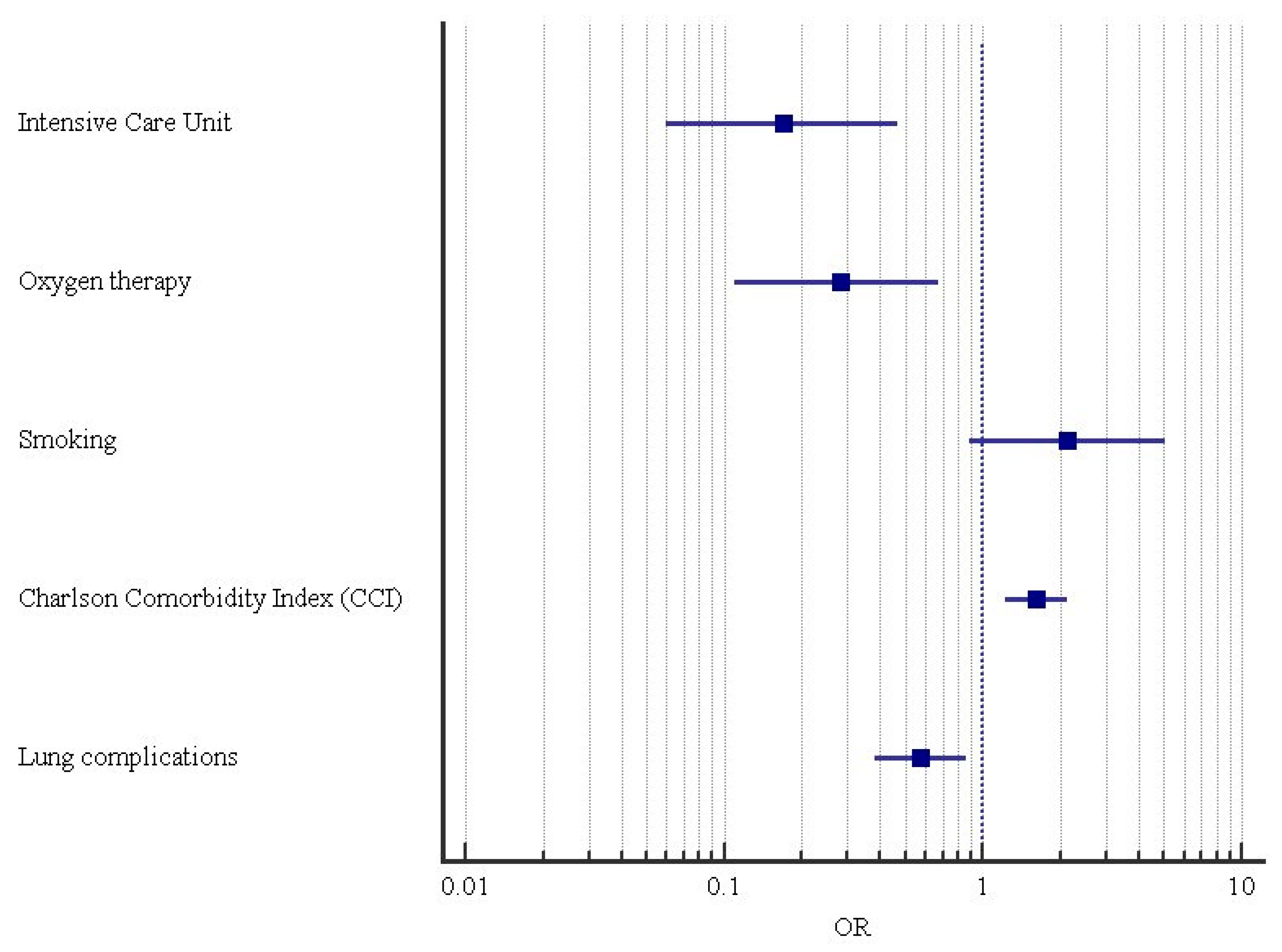
Figure 1. Forest plot of multivariable logistic regression analysis identifying factors associated with Legionella pneumophila infection.
Pathogens 15 00866 g001
Table 1. Comparison between negative (control group) and positive patients for L. pneumophila (SG).
ParametersCG
(Control Group)
SG
(L. pneumophila Group)
CG vs. SG
p-Value (Test)
Patients7980
Age
   Mean (SD)65.2 (17.2)67.2 (15.8)
   Median (IQR)70 (52, 79)70 (58.5, 79)0.5 (MW)
Gender
   Males72.2% (57)66.3% (53)
   Females27.8% (22)33.8% (27)0.42 (C)
Hospitalization (days)
   Mean (SD)16.1 (13.6)20.4 (17.0)
   Median (IQR)12 (8.0, 20.75)14 (9.0, 28.0)0.12 (MW)
Intensive Care Unit (ICU)79.7% (63)63.8% (51)0.0256 * (C)
Oxygen therapy78.5% (62)63.8% (51)0.0412 * (C)
Non-invasive mechanical ventilation60.8% (48)57.5% (46)0.68 (C)
Mortality30.4% (24)21.2 (17)0.19 (C)
Smoking67.1% (53)83.8% (67)0.0149 * (C)
Patients with comorbidities82.3% (65)86.3% (69)0.49 (C)
Comorbidities type
   Diabetes15.2% (12)26.3% (21)0.0047 * (F)
   Heart failure (HF)6.3% (5)41.3% (33) **
   Chronic lung diseases58.2% (46)62.5% (50)SG-HF: 41.3%, p = 0.0004 * (Z)
   Hypertension44.3% (35)46.3% (37)
   Dialysis10.1% (8)26.3% (21)
   Liver cirrhosis3.8% (3)3.8% (3)
   Dementia16.5% (13)33.8% (27)
Sepsis19.0% (15)27.5% (22)0.21 (C)
Cancer31.6% (25)33.8% (27)0.59 (C)
Isolates ††
   Gram−19.0% (15)31.3% (25)
   Gram+8.9% (7)10.0% (8)0.11 (C)
   Virus13.9% (11)7.5% (6)
Note: † = some patients had more comorbidities; †† = some patients had more isolates; CG: negative for L. pneumophila; SG: positive for L. pneumophila; * = significant test; MW = Mann–Whitney test; C = chi-square test; F = Fisher’s exact test; ** = more frequent modality; Z = post hoc Z-test. Italicized terms indicate study variables; non-italicized terms indicate the corresponding categories, levels, or values.
Table 2. Radiological pulmonary findings of patients with L. pneumophila.
ParametersCG
(Negative for L. pneumophila)
SG
(Positive for L. pneumophila)
CG vs. SG
p-Value (Test)
Lung involvement 0.0336 * (C)
Unilateral38.0% (30)22.5% (18)
Bilateral62.0% (49)77.5% (62)
Patients with lung complications83.5% (66)62.5% (50)0.0029 * (C)
Lung complications type 0.0008 * (F)
   Unilateral effusion26.6% (21)13.8% (11)
   Bilateral effusion (be)21.5% (17) **5.0% (4)CG-be, 21.5%, p = 0.0137 * (Z)
   Cavitation5.1% (4)2.5% (2)
   Pneumothorax1.3% (1)1.3% (1)
   ARDS ***16.5% (13)21.3% (17)
   Pulmonary embolism8.9% (7)1.3% (1)
   Hilar and/or mediastinal
lymphadenopathy > 10 mm (lin)
5.1% (4)18.8% (15) **SG-lin, 18.8%, p = 0.0006 * (Z)
Radiological findings
   Interstitial pneumoniae16.5% (13)15.0% (12)
   Lobar pneumoniae20.3% (16)20.0% (16)0.65 (C)
   Interstitial plus lobar pneumoniae20.3% (16)13.8% (11)
   Bronchopneumonia or lobular pneumonia43.0% (34)51.3% (41)
Note: *** = Acute respiratory distress syndrome; † = some patients had more complications; C = chi-square test; F = Fisher’s exact test; * = significant test; ** = more frequent modality; Z = post hoc Z-test. Italicized terms indicate study variables; non-italicized terms indicate the corresponding categories, levels, or values.
Table 3. Multiple logistic regression analysis among Legionella_var and significant variables described in Table 1 and Table 2.
Logistic RegressionCoefficientStandard
Error
ORCI at 95%p-Value
Null model vs. full model <0.0001 * (C)
Legionella_var/Intensive Care Unit−1.770.510.17(0.06; 0.47)0.0006 *
Legionella_var/Oxygen therapy−1.290.460.28(0.11; 0.67)0.0047 *
Legionella_var/Smoking0.750.442.12(0.89; 5.03)0.09
Legionella_var/Charlson Comorbidity Index (CCI)0.480.141.61(1.22; 2.12)0.0008 *
Legionella_var/Lung complication score−0.560.210.57(0.38; 0.86)0.0078 *
Constant1.540.760.0431 *
Note: * = significant test; OR = odds ratios; CI = odds ratios confidence interval at 95%. The null model = −2ln(L0), where L0 was the likelihood of obtaining the observations if the independent variables did not affect the outcome; the full model: −2ln(L0), where L0 was the likelihood of obtaining the observations with all independent variables incorporated in the model; C = chi-square test. Italicized terms indicate variables formalized as numerical variables for statistical analyses.
Disclaimer/Publisher’s Note: The statements, opinions and data contained in all publications are solely those of the individual author(s) and contributor(s) and not of MDPI and/or the editor(s). MDPI and/or the editor(s) disclaim responsibility for any injury to people or property resulting from any ideas, methods, instructions or products referred to in the content.


