Review

Advances and Challenges in the Diagnosis of Vector-Borne Protozoal Infections in Veterinary Medicine

by
Ana María Cevallos
*,
Tomas Meraz-Tay
and
Roberto Hernández
Departamento de Biología Molecular y Biotecnología, Instituto de Investigaciones Biomédicas, Universidad Nacional Autónoma de México, Ciudad de México 04510, Mexico
*
Author to whom correspondence should be addressed.
Pathogens 2026, 15(6), 561; https://doi.org/10.3390/pathogens15060561
Submission received: 16 April 2026 / Revised: 12 May 2026 / Accepted: 18 May 2026 / Published: 22 May 2026

Abstract

Vector-borne protozoal infections—including babesiosis, theileriosis, hepatozoonosis, trypanosomosis, and leishmaniosis—impose a substantial burden on livestock and companion animal health worldwide and carry important zoonotic and public health implications. Accurate diagnosis is essential yet challenging, given the diversity of parasite genera, their markedly different tissue tropisms, and the uneven distribution of diagnostic resources across veterinary settings. This review provides an integrated overview of the principal diagnostic approaches available, structured around the biological logic that guides test selection in practice. Microscopic examination remains the first-line method; its strengths and limitations are discussed for intraerythrocytic parasites (Plasmodium spp., Babesia spp., Theileria spp., Cytauxzoon spp.—the latter two with additional extra-erythrocytic schizont stages in leukocytes and tissue macrophages, respectively), leukocyte-associated forms (Hepatozoon spp.), extracellular trypanosomes, and tissue-stage parasites, including emerging applications of artificial intelligence. Serological methods—enzyme-linked immunosorbent assay (ELISA), indirect fluorescence antibody test (IFAT), and point-of-care lateral flow assays—are evaluated for their role in exposure detection, population screening, and international trade certification, with attention to cross-reactivity and the active-versus-past-infection distinction. Molecular diagnostics, encompassing conventional PCR, qPCR, droplet digital PCR, isothermal amplification, and next-generation sequencing, are reviewed with respect to target selection, sensitivity, and point-of-care applicability. Finally, diagnostic challenges are contextualised within a One Health framework, highlighting the fragmentation of veterinary surveillance and the need for integrated, cross-sector approaches to detect emerging threats.

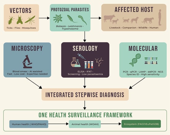
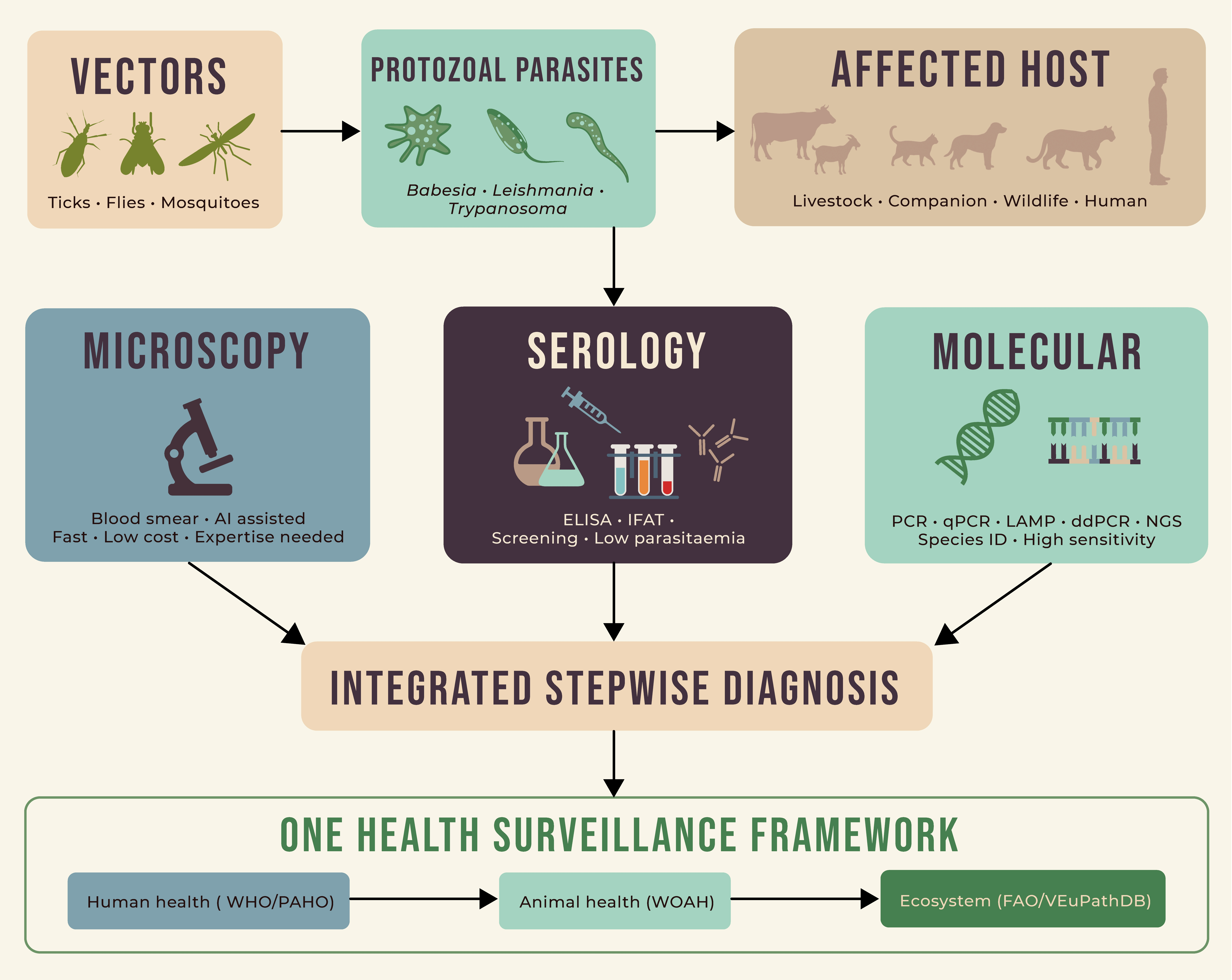
Graphical Abstract

1. Introduction

Protozoal infections transmitted by arthropod vectors such as ticks, flies, and mosquitoes—including babesiosis, theileriosis, hepatozoonosis, trypanosomosis, and leishmaniosis—affect both livestock and companion animals worldwide, causing significant levels of morbidity and mortality. Beyond their direct impact on animal welfare, these infections impose a substantial economic burden, reducing livestock productivity through decreased milk yield, weight loss, reproductive failure, and mortality, and generating considerable costs associated with treatment, prevention, and vector control programmes. Several of these parasites are also zoonotic or have wildlife reservoir hosts that place them at the human–animal–ecosystem interface, making their accurate diagnosis a priority not only for individual animal health but also for public health surveillance.
Among protozoa, vector-borne parasites—those that require an arthropod for biological development as part of a two-host life cycle—have evolved within a limited number of specific lineages across two distinct eukaryotic groups: the informal clade Discoba (incertae sedis) and the domain Diaphoretickes (Adl et al. [1]). Within Discoba, the relevant lineages belong to the order Trypanosomatida, family Trypanosomatidae; among the subfamilies of this family, the vector-borne genera fall into two: Leishmaniinae (genus Leishmania) and Trypanosomatinae (genus Trypanosoma). Among the seven subgenera of Trypanosoma, four contain vector-borne species of veterinary importance: Schizotrypanum (e.g., Trypanosoma cruzi), Trypanozoon (e.g., Trypanosoma brucei), Duttonella (Trypanosoma vivax), and Nannomonas (Trypanosoma simiae, Trypanosoma congolense) (Figure 1). Beyond their taxonomic placement, the Trypanosoma subgenera differ in features that have direct clinical and management implications. Most notably, they are transmitted by different routes: Schizotrypanum parasites are stercorarian, transmitted through the faeces of triatomine bugs, whereas Trypanozoon, Duttonella, and Nannomonas are salivarian, transmitted through the saliva of biting flies. Within Trypanozoon, Trypanosoma equiperdum and Trypanosoma evansi are considered derived forms of T. brucei that have undergone reductive evolution, particularly affecting mitochondrial function and the kinetoplast (kDNA), which is essential for development in the insect vector [2,3,4]. Consistent with this, T. evansi does not develop within an insect vector and is transmitted mechanically by biting flies, while T. equiperdum has lost vector transmission entirely and is now spread venereally.
Within the domain Diaphoretickes, the relevant lineages belong to the phylum Apicomplexa, mainly within the class Aconoidasida (orders Haemosporida and Piroplasmorida). To the order Haemosporida belong the genera Plasmodium and Leucocytozoon. Species of the genus Plasmodium are capable of infecting a wide range of vertebrate hosts, though individual species are typically restricted to a limited host range: for example, Plasmodium falciparum, Plasmodium malariae, and Plasmodium vivax infect humans, while Plasmodium gallinaceum and Plasmodium juxtanuclare are avian parasites; the systematics of the order as a whole has been comprehensively reviewed by Perkins [5]. Leucocytozoon spp. infect a broad diversity of bird taxa, although individual species are typically restricted to relatively narrow host ranges. To the order Piroplasmorida belong Babesia spp. and Theileria spp., whose individual species are also restricted to certain hosts, with many being important pathogens of livestock and companion animals. A non-aconoidasidian digenetic pathogen is Hepatozoon spp., which belongs to the class Conoidasida of the phylum Apicomplexa (order Eucoccidiorida). The genus has a broad vertebrate host range that includes reptiles, amphibians, and rodents; in the veterinary setting, however, the species of clinical relevance—such as Hepatozoon canis, Hepatozoon americanum, and Hepatozoon felis—primarily affect the order Carnivora, including dogs, cats, and wild carnivores [6]. Hepatozoon is acquired by the vertebrate host through ingestion of an infected tick or other arthropod—most notably Hepatozoon canis in dogs—a transmission route that applies broadly across the genus and constitutes an important diagnostic and epidemiological consideration [6].
The geographical distribution of the infections caused by these organisms is not arbitrary, being limited by the distribution of arthropod vectors and the availability of competent vertebrate hosts. Some parasites circulate in complex ecological networks involving numerous reservoir hosts and vector species. For example, T. cruzi, the causative agent of Chagas disease, infects more than 100 mammalian species and is transmitted by multiple triatomine bug genera, allowing the parasite to persist in sylvatic, peridomestic, and domestic cycles across the Americas [7]. Similarly, Leishmania infantum is transmitted by several phlebotomine sand fly species and maintained primarily in dogs but also in wildlife reservoirs [8,9]. In contrast, other protozoa exhibit more constrained transmission systems. Piroplasms such as Babesia bovis and Babesia canis depend on specific tick vectors and relatively limited host ranges [10,11], while H. canis and Hepatozoon americanum require ingestion of infected ticks and are therefore closely linked to particular host–vector ecological interactions [6]. These differences in host and vector diversity determine that, for a correct diagnosis, it is essential to evaluate the species of the animal affected and the specific geographic location where the animal lives or has come from (Supplementary Table S1). For example, B. bovis and Babesia bigemina primarily affect cattle, Theileria equi affects equids, while L. infantum principally parasitises dogs (though it can also infect cats, humans, and various wildlife species), and Cytauxzoon felis is restricted to felids [12,13]. In a similar fashion, a trypomastigote detected in a cow in sub-Saharan Africa is most likely an African animal trypanosome—most commonly Trypanosoma congolense or Trypanosoma vivax, but potentially Trypanosoma brucei—with species-level discrimination requiring molecular confirmation.
Within the following sections, the principal microscopic features used for detection and identification of these parasites are discussed, together with the serological and molecular tests that can confirm and refine the diagnosis. Finally, the importance of accurate vector-borne disease diagnosis within the framework of One Health is discussed.

2. Microscopic Diagnosis

Microscopic examination is the first-line approach for diagnosing vector-borne protozoal infections and can provide accurate identification, especially at the genus level, although determining the specific species can require additional tests. Among its advantages are low cost, rapid turnaround, and direct visualisation of the parasite within host cells, providing both diagnostic confirmation and morphological information unavailable from indirect methods. However, accuracy is highly dependent on the examiner’s expertise, particularly their knowledge of parasite morphology across different developmental stages and tissue distributions. Diagnosis is especially challenging when the parasitic load is low and can be easily missed if parasitic disease is not high on the differential diagnosis or if too few microscopic fields are examined. When the diagnosis is suspected but there are few parasites, concentration assays such as quantitative buffy coat (QBC) should be employed [14]. QBC is particularly useful for leukocyte-associated parasites (e.g., Hepatozoon spp.) and extracellular trypanosomes; it is not suited for intraerythrocytic organisms such as Babesia spp., Theileria spp., or Cytauxzoon spp., for which thick and thin blood smears remain the method of choice.
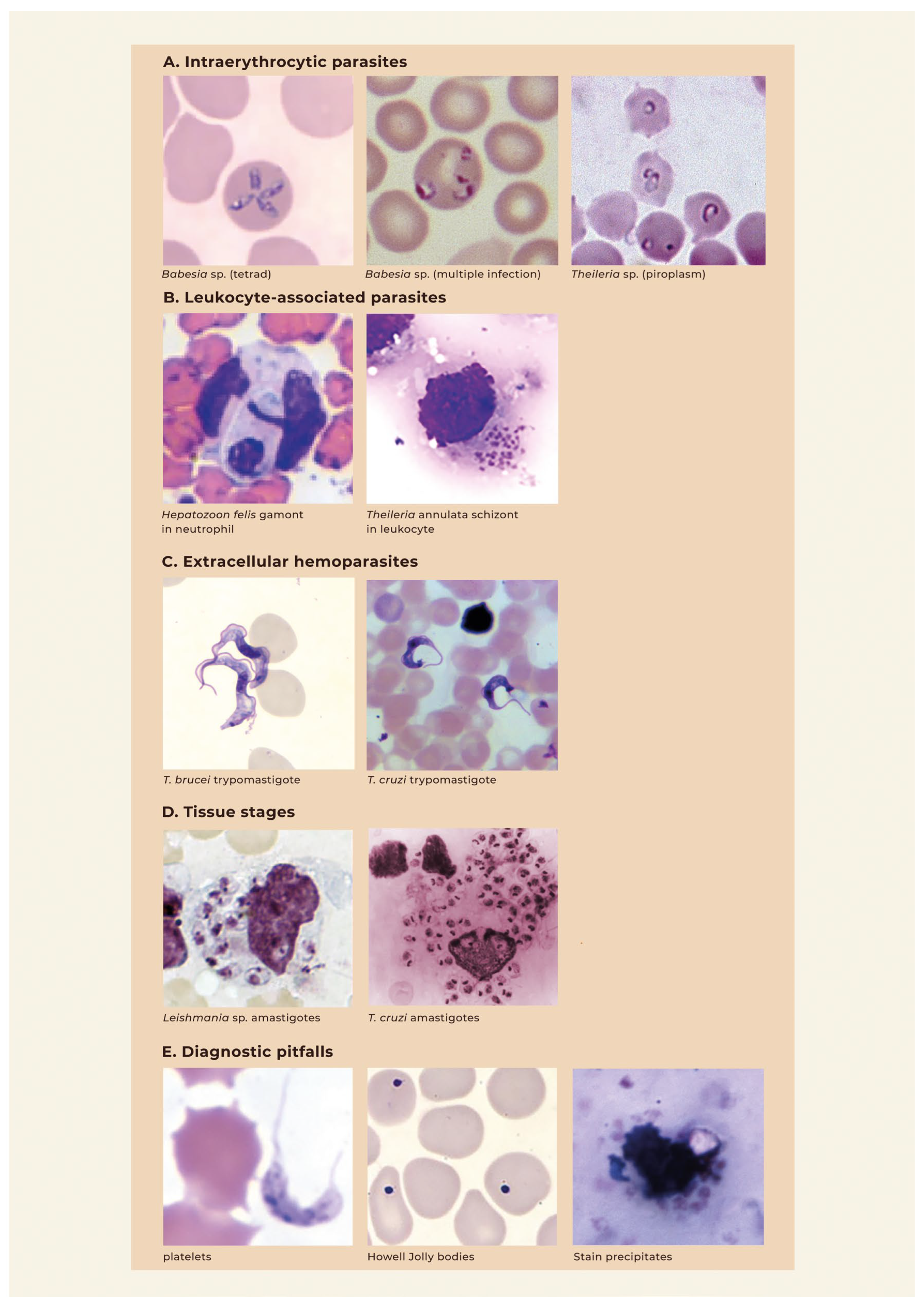
The choice of specimen is guided by the biology of the suspected parasite. Blood samples are used to identify Plasmodium spp., Babesia spp., Theileria spp., Cytauxzoon spp., Hepatozoon spp. and Trypanosoma spp. For infections in which the parasite preferentially resides in tissues rather than blood, such as Leishmania spp. and T. equiperdum, examination of biopsy material is necessary (Figure 2).

2.1. Blood Smear Examination

Thick and thin blood smears are the primary diagnostic tests done when a blood parasitic infection is suspected. In a recent study conducted in a low-prevalence human hospital setting, it was demonstrated that routine haematology thin smears can also be used to detect parasites, suggesting that careful examination of the routine haematology smear could yield a diagnosis even when infection is not initially suspected [17]; whether this finding translates to veterinary practice—where parasite species, prevalence baselines, host blood-cell morphology, and laboratory workflows differ markedly from human medicine—warrants systematic evaluation in veterinary-relevant settings before routine adoption can be recommended. Among the pathogens detectable in blood, Plasmodium spp., Babesia spp., Theileria spp., and Cytauxzoon spp. have intraerythrocytic stages. Theileria spp. and Cytauxzoon spp. also have stages that affect white blood cells, whereas Hepatozoon spp. are intra-leukocytic (gamonts within neutrophils and monocytes). In contrast, trypanosomes are extracellular. Correct identification requires familiarity with the morphological features of each genus at the stages present in peripheral blood. In this section some of the main characteristics that give information about the identification of specific species are detailed, with data centred on species that affect veterinary companion and livestock animals rather than humans.
The erythrocytic stages of avian Plasmodium species show marked variation in developmental patterns across hosts, requiring an experienced observer for correct identification. A key diagnostic feature is the presence of sexually dimorphic gametocytes (macro- and microgametocytes), which are readily identifiable under the light microscope and clearly distinguish avian malarial parasites from other intracellular apicomplexans, such as Babesia spp., Theileria spp., and Cytauxzoon spp. [18]. An additional finding is the accumulation of hemozoin—residual pigment granules resulting from incomplete haemoglobin digestion—which is birefringent under polarised light and serves as a reliable feature distinguishing haemosporidian parasites from piroplasms, which lack hemozoin.
In contrast to Plasmodium spp., Babesia spp. are obligate intraerythrocytic parasites with very limited extra-erythrocytic development, confining their diagnostic stages almost entirely to red blood cells. Based on their morphology, Babesia spp. are classified as small babesias (1.0–2.5 μm long), such as B. bovis, Babesia gibsoni, Babesia silvestris and Babesia rodhaini, and large babesias (2.5–5.0 μm long), such as B. bigemina, Babesia caballi and B. canis. Although pleomorphic, characteristic morphological features include paired pyriform (pear-shaped) merozoites and, in some species, the tetrad form (“Maltese cross”). Among veterinary piroplasms, this form is most characteristic of T. equi (formerly Babesia equi); it is also well described in Babesia microti, a rodent parasite and zoonotic agent, where it represents a key diagnostic feature. Unlike Plasmodium spp., Babesia spp. lack hemozoin and do not form gametocytes in the vertebrate host, which helps distinguish them in blood smear examination [19]. Although morphological features remain the foundation of microscopic Babesia identification, overlapping appearances between closely related species—and between Babesia and Theileria piroplasms—can limit species-level resolution on blood smears, and molecular confirmation is recommended where species discrimination is clinically or epidemiologically important.
In peripheral blood smears, the diagnostic stages of Theileria spp. are intraerythrocytic piroplasms that appear as small rings which form morphologically very similarly to those of Babesia spp. Unlike Babesia spp., however, Theileria spp. additionally undergo a schizogonous stage within leukocytes, and this leukocyte-stage schizont—when identifiable in lymph node aspirates or impression preparations—is a useful differential feature. Different species exhibit characteristic schizont host–cell tropisms: Theileria parva preferentially infects T lymphocytes (including CD4+, CD8+, and γδ T-cell subsets), whereas T. annulata primarily infects B cells and monocytes/macrophages [20,21,22,23]; T. equi has a broader tropism, infecting B lymphocytes, T lymphocytes, and monocytes/macrophages in vitro, although experimental evidence indicates that monocytes/macrophages are the functionally critical host cell in vivo [24].
Closely related to Theileria, Cytauxzoon belongs to the family Theileriidae, and their piroplasm stages in the erythrocytes of mammalian hosts are morphologically very similar. The two differ, however, in the location of schizogony: in Cytauxzoon spp., it occurs in macrophages, whereas in Theileria spp. it takes place in lymphocytes (e.g., T. parva) or macrophages (e.g., T. annulata). Cytauxzoon has a schizogonous tissue phase in addition to an intraerythrocytic piroplasm phase, characterised by massive proliferation of schizonts within macrophages lining the vascular endothelium of multiple organs. When tissue involvement is suspected, aspiration cytology or impression smears of affected organs and histological sections are required [13].
Unlike the apicomplexans discussed above, Hepatozoon is not intraerythrocytic at any blood stage. The diagnostic stage in peripheral blood is the gamont—a large, elongated organism enveloped in thick membranes found within the cytoplasm of neutrophils and occasionally monocytes—which is morphologically distinct from the intraerythrocytic ring forms of Babesia spp., Theileria spp., and Cytauxzoon spp. and, therefore, unlikely to cause confusion in a well-prepared blood smear. Gamonts of H. americanum, H. felis and Hepatozoon silvestris can also be identified, but parasitemia is low and buffy coat smears are needed to increase sensitivity [25].
Turning to the extracellular trypanosomes, the detection rate varies considerably across species due to differences in vascular localisation. The species T. congolense sequesters to the vascular endothelium, and T. vivax has recently been shown to display similar behaviour in experimental mouse models [26]; whether this is a species-wide feature or a strain-specific phenomenon in veterinary-relevant hosts remains to be determined. For T. congolense, bioengineered microvascular models have demonstrated that the vast majority of parasites adhere to the endothelium of multiple organs rather than circulating freely [27]. When peripheral parasitemia is low, concentration methods such as the miniature anion-exchange centrifugation technique (mAECT) or the quantitative buffy coat (QBC) method substantially improve detection sensitivity for trypanosomes [28]. By contrast, T. evansi circulates freely and yields high peripheral parasitaemia, making smear detection straightforward, though ocular and cerebrospinal fluid localization in late disease means blood smears do not reflect total parasite distribution. The species T. brucei circulates in blood but additionally establishes extravascular reservoirs in tissues such as skin, adipose tissue, and muscle by crossing the vascular endothelium; these reservoirs contribute to relapsing parasitaemia and may reduce smear sensitivity at low-burden time points. Among the salivarians, T. equiperdum presents the greatest diagnostic challenge: its primary localization in genital mucosa, skin, peripheral nerves, and the distal spinal cord means peripheral parasitemia is consistently and profoundly low. Blood smear examination was negative in all naturally infected horses despite confirmed infection by PCR and histopathology [29]. Blood smear examination therefore has no meaningful negative predictive value for T. equiperdum and cannot be relied upon to exclude infection. Finally, T. cruzi, a stercorarian trypanosome, differs from the salivarian species in that during the acute phase free-circulating parasites are detectable; in the chronic phase, however, the parasite load in blood becomes negligible, and therefore blood smears will not be diagnostic.
Across all of these parasite groups, a negative microscopy result does not exclude infection in low-parasitaemia, chronic, tissue-localised, or carrier states; clinical suspicion supported by host species, geographic origin, and clinical presentation should therefore trigger serological or molecular follow-up regardless of an unremarkable smear.

2.2. Tissue-Stage Parasites and Exo-Erythrocytic Development

Although the majority of these pathogens can be detected in blood, they can also present developmental stages in other tissues that require tissue examination for detection. Others, such as Leishmania spp., are not present in blood.
In avian malaria, erythrocytic merozoites can trigger secondary tissue merogony—a process of exo-erythrocytic replication occurring in endothelial cells, macrophages, and erythrocyte precursors, depending on the parasite lineage, and detectable in impression smears or histological sections of affected organs.
Hepatozoon species also exhibit diagnostically relevant tissue stages. Extravascular meronts of H. canis are detectable in cytological preparations from lymph nodes, spleen, and bone marrow, appearing round to oval with merozoites arranged peripherally around a central core, producing a characteristic “wheel-spoke” pattern [25]. In contrast, meronts of H. americanum and H. felis localise primarily to striated muscle (skeletal and cardiac), as does H. silvestris. Similarly, C. felis develops a macrophage-associated schizogonous stage in organs such as the spleen, liver, lungs, and lymph nodes, where enlarged parasitized macrophages obstruct small vessels and sinusoids, causing ischaemia and severe systemic inflammation. These stages are best identified by fine-needle aspiration cytology of affected organs, which reveals schizont-laden macrophages, in impression smears or histological sections rather than in peripheral blood [30].
In Leishmania infections, tissue tropism reflects the clinical form of disease. Cutaneous leishmaniosis involves dermal macrophages, while visceral disease disseminates to the spleen, liver, and bone marrow. In all forms, the diagnostic stage is the amastigote—a small, oval, non-flagellated organism residing within the parasitophorous vacuole of the macrophage, visible on Giemsa-stained tissue impressions or histological sections. Examination of affected tissues reveals macrophages packed with intracellular amastigotes, the so-called Leishman–Donovan bodies, which are the hallmark microscopic finding in parasitological diagnosis [31,32].
Similarly, requiring tissue examination in its chronic form, T. cruzi infection is characterised during the acute phase by detectable parasitemia, but in the chronic phase, the parasite localises in target organs such as the heart and intestines. A biopsy of cardiac or skeletal muscle in chronic Chagas disease may reveal intracellular amastigote nests, though tissue sampling is invasive and rarely performed in routine diagnosis.

2.3. Advances in Artificial Intelligence for Diagnosis of Parasitoses in Clinical Samples

With the development of strong algorithms for the analysis of medical images to improve diagnosis, efforts are being made to develop algorithms for the detection and classification of parasites, with the majority of studies being done for the diagnosis of human malaria infection [33,34]. In veterinary practice, there is a commercially available platform for the detection of intestinal parasites in the faeces of cats and dogs that performs with a sensitivity and specificity of greater than 90%, with the exception of the diagnosis of Ancylostoma in cats, which is approximately 80% [35]. For the diagnosis of vector-borne parasites, there are only a few studies that have shown the potential of artificial intelligence (AI)-assisted microscopy for use in veterinary practice, with algorithms for trypanosomes less developed and based either on experimental infection or on mixed data from humans and animals (Table 1).
However, the accuracy values reported in Table 1 reflect performance on curated research datasets and have not yet been validated in routine clinical or field practice. Current datasets are small, typically built from single-erythrocyte images rather than whole-smear samples, and the impact of variability in staining protocols, slide preparation, and microscope optics on accuracy has not been systematically evaluated. Existing tools are also assistive rather than autonomous—an initial clinical suspicion is still required to trigger their use—and ideally, they would be integrated into routine smear analysis rather than reserved for suspected cases. Image acquisition in low-resource settings often depends on smartphone adapters, and coordinated efforts to expand curated public repositories would help address data scarcity. Making validated algorithms freely available as downloadable software or apps would further accelerate uptake [43].

3. Serological Diagnosis of Vector-Borne Protozoal Infections

3.1. The Role of Serology in Vector-Borne Protozoal Diagnosis

Serology occupies a central place in the diagnosis and surveillance of vector-borne protozoal infections of veterinary importance. The reasons are practical: parasitaemia in many of these infections is low, intermittent, or below the detection limit of direct microscopy—the chronic phase of Trypanosoma cruzi (very low and intermittent peripheral parasitaemia), T. equiperdum (parasites confined to genital and neural tissues), Leishmania spp. infections (amastigotes generally absent or only rarely detectable in peripheral blood), and the carrier state in bovine babesiosis and theileriosis (low parasitaemia maintained by partial immune control of replication)—making direct microscopic detection insensitive. Serological assays detect host antibodies raised against parasite antigens and remain informative in precisely those situations where the parasite itself is not readily found [44]. They are easy to use, scalable, and capable of detecting prior exposure in asymptomatic or subclinical animals [45], and combined formats are increasingly available for the simultaneous detection of multiple co-circulating pathogens, reflecting the frequent occurrence of co-infections [46,47]. Serology is also widely available across the four parasite groups reviewed here, with mature reference standards: World Organisation for Animal Health (WOAH)-prescribed competitive ELISA (cELISA) for international trade certification of equine piroplasmosis [48]; WOAH manual standards for bovine babesiosis and theileriosis; and the LeishVet guidelines for clinical staging of canine leishmaniosis [49]. These assays exist in both laboratory-based and point-of-care (POC) formats, which serve genuinely different use-cases: clinical confirmation in the individual animal, herd-level screening, and population-level epidemiological surveillance. The remainder of this section is organised around these three roles, followed by a structured account of the principal limitations of serological testing and how to interpret a serological result in light of them.
A practical implication of the quantitative outputs of laboratory-based assays—titres or optical density values—is that a paired or serial sera can, under certain conditions, address a limitation that no single time point can: the inability to distinguish active recent infection from previous exposure. The diagnostic value of titre dynamics has been most clearly demonstrated for canine leishmaniosis, where longitudinal IFAT and ELISA studies show that rising titres track progression and clinical relapse, while falling titres accompany successful treatment response [50,51]; fluctuating titres in subclinically infected dogs reflect the complex host–parasite dynamics characteristic of chronic carriage. In canine T. cruzi infection, longitudinal cohort studies document seroconversion as a sensitive marker of new infection [52], although once the carrier state is established, antibody titres remain stable for years to decades, and the active-versus-past distinction by serology is not informative on practically useful timescales [53]. For bovine babesiosis and theileriosis, persistent carrier states with antigenic variation mean that antibodies are detectable for many months to years after acute infection and titre dynamics are correspondingly uninformative [54]. For African and South American animal trypanosomiases, longitudinal antibody-titre data in natural infections are notably scarce, and the diagnostic value of paired sera in this group remains to be established. Where validated molecular assays are unavailable, serial or paired-serum serological testing may provide a practical complementary approach for the diagnosis of canine leishmaniosis and acute Trypanosoma cruzi infection, although these strategies remain insufficiently standardised and warrant further systematic evaluation in veterinary practice.

3.2. Serology in Clinical Practice

At the level of the individual animal, serology is most useful for confirming infection in clinical contexts where direct parasite detection is unreliable. This includes chronic T. cruzi infection (the indeterminate and determinate phases, where parasitaemia is low and intermittent), T. equiperdum infection (where parasites are confined to genital and neural tissues), and Leishmania spp. infections (where amastigotes are generally absent or only rarely detectable in peripheral blood). For these and for any infection in which parasitaemia is low or intermittent, serological testing supports clinical suspicion and contributes to diagnosis when interpreted alongside host species, clinical signs, and geographic origin (Supplementary Table S1).
Two formats serve clinical practice. Laboratory-based assays—principally ELISA and IFAT—provide quantitative or semi-quantitative outputs (titres, optical density values) essential for clinical staging and longitudinal monitoring [55]. The IFAT method is commonly regarded as a reference for validation of new diagnostic tools and is the reference method for canine leishmaniosis [55,56]. ELISA is preferred over IFAT for bovine theileriosis and babesiosis when throughput matters, principally because the platform is more amenable to automation and large-scale screening [57]. Point-of-care (POC) tests—lateral flow assays and rapid diagnostic tests—detect antibodies, or occasionally antigens, using colloidal gold visible to the naked eye within 10–20 min without the need for laboratory equipment or trained personnel [58]. They are useful for sampling animals in remote locations and as rapid tools in veterinary clinics [59], can be performed on-site or later by elution of dried blood spots from filter paper [60], and are available in formats that detect antibodies against multiple co-circulating pathogens simultaneously—addressing the frequent occurrence of co-infections in vector-borne disease.
The rK39-based lateral flow test for canine visceral leishmaniosis shows high sensitivity and specificity approaching 100% in some validation studies [55,61], although performance is not uniform across endemic settings: in Colombia, two immunochromatographic tests showed sensitivity of 82.9–85.7% and specificity of 79.6–92.6% [62], with both missing Leishmania braziliensis and L. amazonensis cases—illustrating the risk of poor performance when local species differ from the test antigen’s target. In equids, combined POC tests permit simultaneous serological detection of antibodies to T. equi and B. caballi, a relevant capability given that the two agents differ in drug susceptibility [63]; however, reliable discrimination between concurrent infections depends on platform performance and cross-reactivity, which vary across products. The Card Agglutination Test for Trypanosomiasis (CATT/T. evansi) targets the RoTat 1.2 variant surface glycoprotein and shows high sensitivity in horses, camels, goats, sheep, buffaloes, and dogs but lower sensitivity in cattle and pigs. CATT performance is constrained by parasite biology because the assay detects primarily Trypanosoma evansi Type A strains expressing the RoTat 1.2 antigen. Type B isolates described to date lack the RoTat 1.2 gene and therefore escape detection. Similarly, certain dyskinetoplastic lineages—which have partially or completely lost their kinetoplast DNA (kDNA; discussed in Section 4)—have been associated with the absence of RoTat 1.2 expression and may yield false-negative results. Importantly, this limitation does not reflect recognition of a kDNA-encoded antigen, as RoTat 1.2 is nuclear-encoded, but rather the empirical association between certain dyskinetoplastic lineages and absence of RoTat 1.2 expression. Consequently, a negative CATT result does not exclude T. evansi infection in regions where Type B strains circulate or where dyskinetoplastic stocks are suspected. For bovine babesiosis there are also rapid diagnostic tests used for quick screening of the herd. Rapid tests, while convenient, often exhibit lower analytical sensitivity than laboratory-based assays, and their utility for international certification remains limited, with ELISA and IFAT being the current standards [48]. Examples of serological tests available for key vector-borne protozoal parasites, including the antigens used, platforms, and test types, are summarised in Supplementary Table S2; the list is illustrative rather than exhaustive.

3.3. Serology for Herd Management and Surveillance

Beyond the individual case, serology is the practical workhorse of herd-level screening and population-level surveillance. The reasons are operational: laboratory-based serology can be standardised, scaled, and applied to large numbers of samples at modest cost per sample, and quantitative outputs allow tracking of seroprevalence over time and across regions. For equine piroplasmosis, WOAH-prescribed cELISA is the regulatory standard for international trade movement of horses, and its use harmonises serological reporting across countries [48]. Surveillance of canine leishmaniosis in endemic Mediterranean and Latin American regions has historically relied on serological methods, particularly ELISA and IFAT, frequently based on whole-promastigote or soluble promastigote antigens, to estimate seroprevalence and monitor changes associated with control interventions [64].
An important but still underdeveloped contribution of herd-level and surveillance serology is the cross-geographical evaluation of diagnostic assay performance. Applying validated serological assays across multiple endemic settings through coordinated surveillance networks would allow assessment of how test sensitivity and specificity vary under differing transmission intensities, host assemblages, and circulating parasite populations. Such approaches would provide more realistic estimates of field performance than single-region validation studies, whose findings may not generalise across epidemiological contexts, as demonstrated for several protozoal serological assays [65,66,67]. Coordinated evaluation of this type aligns naturally with the One Health surveillance frameworks discussed in Section 5.
The need for cross-geographical assay evaluation is particularly acute for parasites with substantial genetic and antigenic diversity. The species T. cruzi is organised into seven Discrete Typing Units (DTUs: TcI–TcVI and TcBat), and the sensitivity of serological diagnostic tests varies considerably across DTUs, while specificity is frequently affected by cross-reactivity with other trypanosomes or with Leishmania spp. TcI is the most widely distributed in wildlife and domestic animals across Latin America; TcII, TcV, and TcVI predominate in domestic transmission cycles in the Southern Cone; and TcIV is associated with wildlife reservoirs in both North and South America. DTU typification is not possible through serological methods, and Chagas disease serodiagnosis is therefore conventionally based on a combination of at least two different tests; however, the choice of which tests to combine is not globally standardised and depends on the platforms validated for each region [68]. This is a worked example of why surveillance-grade assay performance for any one parasite group cannot be assumed to generalise across the parasite’s geographic range.

3.4. Interpreting a Serological Result: Four Limitations

The interpretation of any serological result is constrained by limitations that vary in importance by parasite, host, and epidemiological setting. Four limitations are particularly consequential for vector-borne protozoal infections.
Persistent antibodies after treatment, apparent parasite suppression, or carrier states. In several of the parasites reviewed here, antibodies remain detectable long after the acute phase has resolved or peripheral parasitaemia has been controlled. For Trypanosoma cruzi, infection is essentially lifelong in the absence of effective treatment, antibodies persist accordingly, and even after antitrypanosomal treatment, the time to negative seroconversion—which is itself an imperfect surrogate for parasitological cure—is measured in years to decades and is inversely proportional to the pretreatment duration of infection [53]. For bovine babesiosis, recovered cattle remain symptomless carriers for months to years (longer for B. bovis than for B. bigemina), and antibody titres are not a reliable indicator of current infection status: cattle that have become serologically negative on indirect haemagglutination testing have been shown to remain immune four years after initial vaccination or exposure, while cattle that have eliminated the parasite may continue to test positive [54]. Carrier states in bovine babesiosis and theileriosis are characterised by low parasitaemia resulting from partial immune control of parasite replication; the balance between parasite persistence mechanisms—including antigenic variation—and host splenic and adaptive immune responses permits long-term subclinical infections that serve as epidemiologically important reservoirs [69]. The general principle is that the presence or absence of detectable antibodies at a single time point reflects past immunological exposure, not necessarily ongoing parasite replication or current parasitaemia.
High background seroprevalence in endemic settings. Where transmission has been sustained for years, a substantial fraction of apparently healthy animals carry antibodies to the locally circulating parasites. This is the operational reality of endemic stability for bovine babesiosis and theileriosis: classically associated with high early-life exposure rates (often >70–75% of calves exposed to B. bovis before nine months of age) and a low incidence of clinical disease in older cattle, the dynamic equilibrium produces high herd-level seroprevalence in clinically unaffected populations [54]. The same dynamic occurs in canine leishmaniosis in the Mediterranean basin and in Brazilian foci, where seroprevalence in apparently healthy dogs can be substantial. In such settings, a positive serological result on a single sample carries limited specificity for clinically relevant infection, and interpretation must explicitly account for the local prevalence baseline. In settings where Leishmania spp. and Trypanosoma cruzi co-circulate, limited but measurable serological overlap has been documented: in rural Panamanian dogs, agreement between Leishmania panamensis IFAT and T. cruzi IFAT or multiplex microsphere immunoassay was low but detectable (κ = 0.18–0.20), illustrating the diagnostic complexity of interpreting serology in regions with sympatric trypanosomatid transmission [67].
Vaccine-induced seroconversion. Where vaccines have been deployed, vaccine-induced antibodies can interfere with serological diagnosis in ways that depend critically on the assay platform and the vaccine in use. For canine leishmaniosis, CaniLeish® induces antibodies that cross-react with whole-promastigote–based assays such as IFAT, DAT, and crude soluble-antigen ELISA: in a controlled longitudinal study, 74.1% of CaniLeish-vaccinated dogs were classified as seropositive by a whole-promastigote ELISA one month after completion of the primary vaccination course [70]. Recombinant rK39- and rK28-based ELISAs are less affected, with vaccinated non-infected dogs in a 25-month longitudinal cohort showing serological profiles statistically indistinguishable from non-vaccinated non-infected controls, although the cohort was small and individual false-positive results were observed [71]. Letifend®, a recombinant-protein vaccine designed under the Differentiating Infected from Vaccinated Animals (DIVA) principle, has shown reduced interference with several commonly used serological assays in vaccinated non-infected dogs, although the extent of non-interference is platform-dependent and independent peer-reviewed replication remains limited [72]. The practical consequence is that, in CaniLeish-vaccinated populations, whole-antigen serology cannot reliably distinguish vaccinated from infected animals, and confirmation of infection generally requires molecular detection of parasite DNA. A parallel issue arises for cattle babesiosis and theileriosis, where live, attenuated, and soluble-antigen vaccines have been in use for decades: vaccinated cattle seroconvert against the same antigens used in diagnostic assays, and serological assays cannot distinguish vaccinated from naturally infected animals in a single sample [73]. The DIVA principle has therefore become an explicit design objective for next-generation veterinary protozoal vaccines, and vaccination history must be a routine part of the clinical and epidemiological information accompanying any serological request in regions where these vaccines are deployed.
Antibody titre does not reliably correlate with parasitaemia or infectivity. Even where serology unambiguously confirms infection, the antibody titre itself is an unreliable proxy for parasite burden, ongoing transmission risk, or infectivity to the vector. Outside specific contexts such as the clinical staging of canine leishmaniosis—where titre magnitude does correlate moderately with disease severity, though not necessarily with infectivity—titres should not be interpreted as a quantitative readout of parasitaemia. This limitation underlies the need for adjunct molecular or parasitological testing whenever questions of active parasite replication, treatment response, or transmissibility are clinically or epidemiologically relevant—and it is the reason that surveillance programmes increasingly combine serological screening with molecular confirmation rather than relying on serology alone.
Together, these four limitations mean that a serological result is best interpreted not in isolation but within a structured framework that integrates host species and breed, geographic origin, clinical presentation, vaccination history, regional seroprevalence baseline, and, where available, paired-sera dynamics or molecular confirmation.

3.5. Implications for Surveillance in Vaccinated Populations

The growing use of canine leishmaniosis vaccines in endemic regions raises a structural question for surveillance. Where whole-antigen ELISA or IFAT has historically been the workhorse of population-level seroprevalence monitoring—across the Mediterranean basin and in Latin American foci—increasing CaniLeish® coverage will introduce a growing fraction of vaccinated animals whose serology is indistinguishable from naturally infected animals on these platforms [70]. The implication is that traditional seroprevalence-based surveillance becomes progressively less informative about Leishmania infection dynamics as vaccination coverage rises and that molecular surveillance, recombinant-antigen-based serology with documented absence of vaccine cross-reactivity, or DIVA-compatible vaccines (such as Letifend®, where independent validation continues to accrue) become correspondingly more important. A parallel issue arises for any vector-borne protozoal infection where vaccination is or becomes available—including bovine babesiosis and theileriosis, where the lack of DIVA-compatible vaccines limits the diagnostic resolution of post-vaccination surveillance: the reliability of seroprevalence as a surveillance indicator depends on the assay platform, the vaccine antigen, and the regional vaccination coverage.
A parallel surveillance gap exists for T. cruzi in veterinary settings. Commercially available serological tests were originally developed for human use and are widely implemented in hospital blood transfusion and organ transplant screening programmes in endemic regions [74]. The limited application of serodiagnosis in livestock is partly explained by the prolonged indeterminate phase of Chagas disease, characterised by low parasitaemia and the absence of clinical signs: production animals usually have relatively short productive lifespans and are unlikely to develop chronic disease. Despite this, the recognised role of dogs as important reservoirs and sentinels in the domestic transmission cycle of T. cruzi [75] underscores the need for diagnostic tools and integrated screening programmes that include both humans and companion animals within a One Health framework. These considerations are taken up further in Section 5.

4. Molecular Diagnosis

4.1. Target Selection

The detection of the DNA of the pathogen is ideal, as it demonstrates the presence of an active infection rather than the demonstration of antibodies, which indicate exposure to the parasite. The selection of an assay for the molecular identification of any infectious agent is based on the choice of a specific target to detect, the availability and cost of the method needed to detect it, and the reasons behind testing (individual case diagnosis vs. surveillance). Target selection is a balance between the sensitivity, specificity and epidemiological utility of the test. The most widely used targets are highly conserved sequences present in multiple copies in the pathogen genome, such as ribosomal RNA (rRNA) genes, mitochondrial DNA and repetitive elements. The greater the number of copies of a given target, the greater the sensitivity of the assay. For example, differences in copy number determine that in C. felis, PCR amplification of the cytochrome c oxidase subunit III gene (cox3) is more sensitive than amplification of the 18S gene [76].
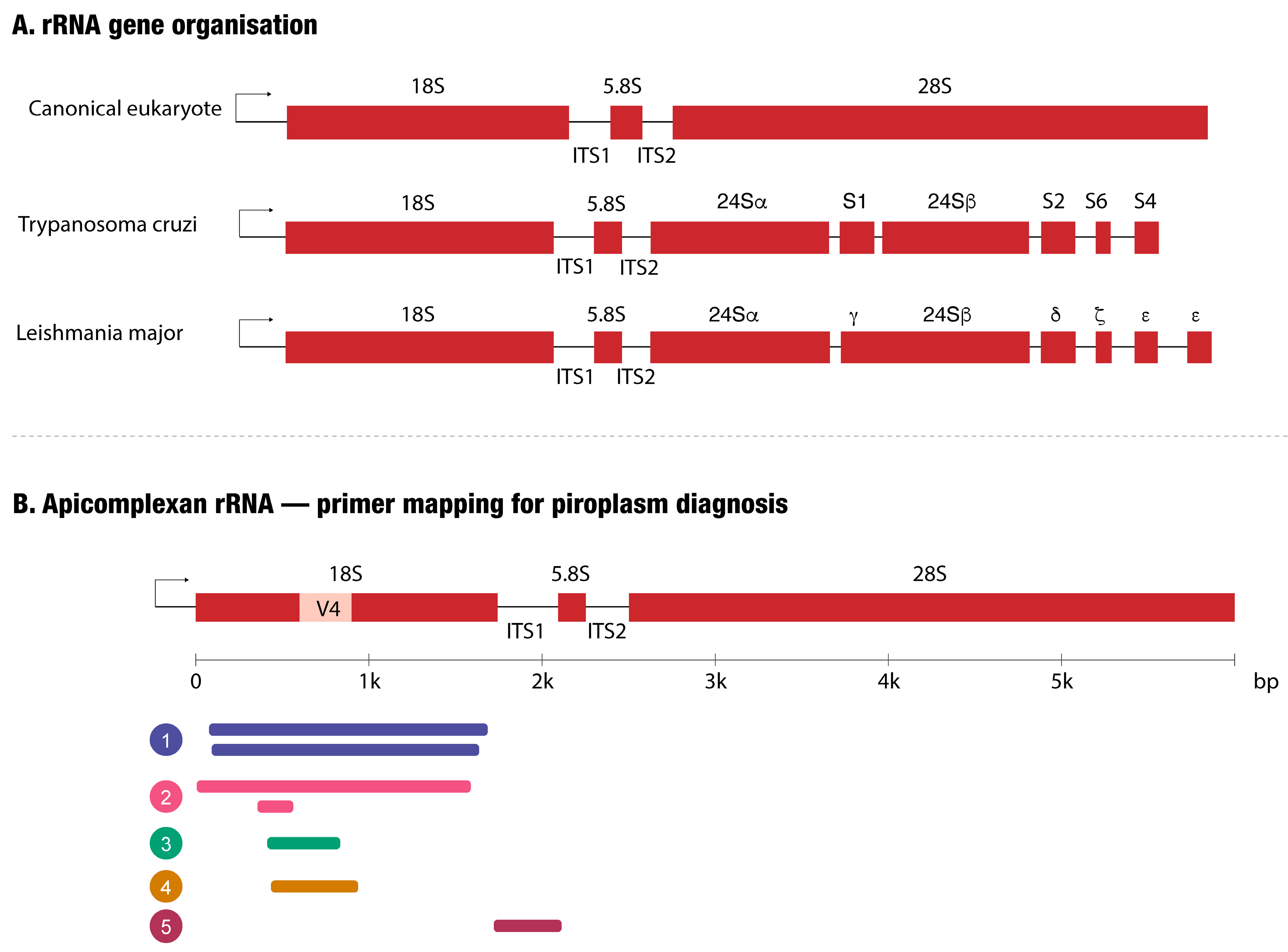
The rRNA genes encode ribosomal RNAs, which constitute the structural and catalytic core of the ribosome. They are usually present in tandem clusters that encode the 18S, 5.8S, and 28S rRNA subunits, whereas the 5S rRNA is encoded at a separate locus. In trypanosomatids, it is the large ribosomal subunit (28S-type) that is atypical: it is encoded by multiple discrete fragments (such as 24Sα, 24Sβ, and several small subunit RNAs in T. brucei) that assemble into the mature large subunit, whereas the 18S subunit is structurally equivalent to that of other eukaryotes (Figure 3) [77]. The rRNA gene copy number varies widely across genera—from only 2–4 units in Theileria and Babesia spp. to over 100 in T. cruzi—directly influencing assay sensitivity [77]. The coding regions of all rRNA genes are highly conserved; their sequences are therefore frequently used to evaluate evolutionary relationships between organisms. The most conserved regions serve as annealing sites for universal primers, while the more variable flanking regions are exploited for the design of taxon-specific primers [78,79,80,81,82]. The internal transcribed spacer (ITS) regions evolve approximately 100 times more rapidly and are consequently more variable [83]. A combination of primers (18S-based and ITS-based) have been exploited both for the design of primers enabling simultaneous detection of several species of piroplasms, plasmodia or trypanosomes and for species identification using the more variable ITS1 and ITS2 regions. A critical limitation of 18S rRNA-based assays in Piroplasmida, however, is that diagnostic utility depends strongly on the length of the amplified fragment and the variable regions it spans. The most widely used pan-piroplasmid primers generate amplicons of approximately 320–450 bp, spanning only one or two variable regions (primarily V4) of the 18S gene (Figure 3B). Sequence divergence within V4 among closely related piroplasm species is insufficient for reliable species- or genotype-level discrimination, and similarity-based identification methods such as BLASTn may return ambiguous or misleading results when short fragments are queried.
The need for greater discriminatory power than 18S provides is exemplified by the Theileria orientalis complex: identification of the pathogenic Ikeda and Chitose genotypes, responsible for substantial outbreaks of bovine anaemia in Australia and New Zealand since 2006, was achieved through sequencing of the major piroplasm surface protein (MPSP) gene rather than 18S, and whole-genome analysis subsequently revealed sufficient divergence among Ikeda, Chitose, and Buffeli genotypes to warrant their reconsideration as separate species [84,85]. For applications requiring species- or genotype-level resolution in Piroplasmida, amplicons of at least 800–1000 bp incorporating the V4, V7, and V8 hypervariable regions have been used. For the detection of Babesia species, a multiplex PCR system targeting the ITS1 region of Babesia divergens, Babesia duncani, B. microti and Babesia odocoilei that demonstrated no cross-amplification among the Babesia species tested (Figure 3B, Supplementary Table S3) [86].
Figure 3. Ribosomal RNA gene organisation in trypanosomatid and apicomplexan vector-borne protozoa and primer mapping for piroplasm molecular diagnosis. (A) Schematic comparison of the rRNA transcription unit. In the canonical eukaryotic cassette, the 18S, 5.8S and 28S rRNA genes are co-transcribed and separated by the internal transcribed spacers ITS1 and ITS2. In trypanosomatids the large-subunit equivalent is fragmented post-transcriptionally into multiple small RNA species: 24Sα, S1, 24Sβ, S2, S6 and S4 in Trypanosoma cruzi and 24Sα, γ, 24Sβ, δ, ζ and two ε species in Leishmania major [77]; (B) Primer pairs from five published assays mapped onto the apicomplexan rRNA locus, drawn to scale from the Theileria parva Muguga 18S reference sequence (1744 bp; gene fragments to scale across the full ~6 kb cassette). The V4 hypervariable region of 18S is highlighted in amber. Numbered, colour-coded bars indicate amplicon position and size; bars stacked in the same colour represent the two rounds of a nested PCR (outer bar above, nested inner bar below). (1) A near-full-length 18S nested PCR [87]. (2) A full-length 18S outer round, followed by a short, nested fragment upstream of V4 [88]. (3) A single-round 18S amplifying V4 [89]. (4) A single-round 18S V4 PCR coupled to targeted amplicon deep sequencing [90]. (5) A multiplex PCR for detection of four Babesia spp. targeting the ITS1 region [86]. Detailed primer sequences, per-species amplicon sizes and additional references are provided in Supplementary Table S3.
Pathogens 15 00561 g003
Other multicopy targets are sequences within mitochondrial DNA. Some organisms, such as Plasmodium spp. and trypanosomatids, only have one mitochondrion [91,92]. However, each of these mitochondria contains multiple copies of mitochondrial DNA, making the encoded sequences multicopy. In trypanosomatids, the mitochondrial genome is organised as kinetoplast DNA (kDNA), which contains two classes of circular molecules with strikingly different copy numbers that have direct consequences for assay sensitivity [93,94]. Maxicircles are present in only 20–50 copies per cell and encode mitochondrial proteins, including cytochrome-related genes; their low copy number limits the sensitivity of maxicircle-based assays, but the specificity of maxicircle sequences makes them useful for species discrimination—for example, the maxicircle-encoded nad5 (NADH dehydrogenase subunit 5) gene distinguishes T. brucei/T. equiperdum from T. evansi [95]. Minicircles, by contrast, are present in thousands of copies per cell (typically 5000–10,000 in Leishmania spp.) and encode guide RNAs; this hundred-fold or greater copy number advantage over maxicircles is the direct basis for the superior sensitivity of minicircle-based assays. Conserved minicircle sequence regions are therefore the primary high-sensitivity molecular target for Leishmania spp. detection [96,97,98] and have also been applied to the diagnosis of Trypanosoma caninum [99]. In Apicomplexa, one of the more commonly targeted mitochondrial genes is cytochrome b [100,101,102,103,104]. Dyskinetoplastic T. evansi stocks, which lack kDNA entirely, cannot be detected by any kDNA-based assay and require nuclear targets.
For African trypanosomes of the brucei and congolense groups, as well as for T. cruzi, the equivalent high-copy diagnostic targets are nuclear repetitive sequences. In T. brucei spp. a 177 bp nuclear repeat and in T. congolense, a 369 bp nuclear repeat have been used for sensitive PCR-based detection [105], while in T. cruzi, a 195 bp satellite DNA repeat element present in approximately 105 copies per genome—representing approximately 10% of total DNA [106]—is the most frequently used target. In trypanosomatids all mRNAs are modified by trans-splicing; the gene encoding the spliced leader sequence has multiple copies and has also been used as a diagnostic target [107]. For species-level identification, parasite-specific proteins have been used as targets, such as the variant surface glycoprotein of the RoTat 1.2 of Trypanosoma evansi Type A [108,109,110,111], the Tams1 gene of T. annulata [112] and the Rhoptry protein gene of B. canis [113].

4.2. Amplification Methodologies

With regard to methodology, conventional PCR needs a basic laboratory setting, and results can be visualised with agarose gels. Certain protocols include restriction enzyme digestion of the amplified products [114,115], and multiplex PCR can detect one or more species in a single amplification [116]. With post-run high resolution melting analysis, specific species can be determined [80,82]. Quantitative PCR (qPCR) uses either an intercalating dye such as SYBR Green or hydrolysis probes (e.g., TaqMan) [117,118,119,120]. Droplet digital PCR (ddPCR) enables absolute quantification without a standard curve, improving sensitivity for low-parasitaemia samples, and has been applied to the detection of T. cruzi, C. felis, B. microti and B. duncani [121,122,123] and for simultaneous detection of protist and bacterial infections through multiplex ddPCR [124].
Beyond PCR-based methods, targeted amplicon deep sequencing has been applied to bovine piroplasms [90] and to characterise genetic diversity within T. orientalis populations [125]. The nested universal parasite diagnostic assay (nUPDx) uses nested amplification of the conserved 18S ribosomal DNA (rDNA) combined with restriction enzyme digestion of host-derived sequences prior to Illumina deep sequencing, achieving a limit of detection comparable to real-time PCR while enabling simultaneous detection of multiple blood-borne parasites, including Plasmodium spp., Babesia spp. and Trypanosoma spp. in a single reaction [126].
Isothermal amplification protocols have been designed to be applied at the point of care. The most common assay is loop-mediated isothermal amplification (LAMP) where detection can be achieved through SYBR Green or calcein fluorescence, turbidimetry, or specific probes such as lateral flow assay strips. Recombinase polymerase amplification (RPA) employs a protein complex including a recombinase, a single-strand binding protein and a strand-displacing DNA polymerase and is typically faster than LAMP and more tolerant of inhibitors [127]. This technology has also been coupled with CRISPR–Cas12a (extension of recombinase-aided amplification with CRISPR–Cas12a detection, ERA-Cas12a), combining high sensitivity with equipment-independent readout options [128].

4.3. Commercially Available Assays

Although many assays have been published for the detection of vector-borne protozoa, the assays that are commercially available and standardised are considerably fewer, and routine clinical use depends heavily on regional availability. In clinical practice, samples should be sent to a reference laboratory or, where appropriate, point-of-care formats should be used when standard microscopy is not diagnostic. Commercial real-time PCR (qPCR) kits are available for Babesia spp., Theileria spp., and Trypanosoma cruzi from multiple manufacturers and to a lesser extent for Cytauxzoon spp., Hepatozoon spp., and the Trypanozoon subgenus (T. brucei, T. evansi, T. equiperdum), where standalone commercial kits remain comparatively scarce and laboratory-developed PCR assays still dominate routine diagnosis. Isothermal amplification formats—including LAMP and recombinase-aided amplification—are also represented and are particularly attractive for point-of-care and field-surveillance applications because they require minimal instrumentation and short turnaround times. Local availability and regulatory status (Conformité Européenne—In Vitro Diagnostic, CE-IVD vs. Research Use Only, RUO vs. veterinary-use-only) vary widely by region; T. cruzi is the only target with multiple CE-IVD-marked human IVDs. For human babesiosis and Chagas disease in the United States, diagnosis still relies predominantly on laboratory-developed tests (LDTs) at reference laboratories. Commercial molecular kits for African animal trypanosomosis exist but are predominantly RUO and not widely deployed in routine veterinary practice, and stand-alone CE-IVD or U.S. Food and Drug Administration (FDA)-cleared commercial qPCR for T. brucei in humans is essentially absent: HAT diagnosis still relies primarily on parasitology, CATT/Rapid Diagnostic Test (RDT) serology, and laboratory-developed PCR or LAMP performed at World Health Organization (WHO)/Foundation for Innovative New Diagnostics (FIND)-supported reference centres. A comprehensive list of commercial molecular kits is provided in Supplementary Table S2, and a reference table of published primer sets and molecular assay designs is provided in Supplementary Table S3.

5. Surveillance of Vector-Borne Protozoal Infections: Challenges and a One Health Perspective

One Health is a framework that recognises the interdependence of human, animal, and ecosystem health. Despite the WHO’s longstanding emphasis on preventive and primary health care, investment in prevention remains disproportionately low, accounting for roughly 3% of total health expenditure in many human health systems [129]; comparable data for veterinary systems are lacking, but the gap between surveillance investment and disease burden is similarly stark. This imbalance is particularly consequential for vector-borne and zoonotic diseases, where upstream ecological and animal health dynamics directly shape human disease risk.
At the institutional level, the Food and Agriculture Organization (FAO), WOAH, and WHO—collectively the Tripartite—have jointly addressed public health, animal health, and environmental challenges through multisectoral and transnational cooperation, most notably through the Tripartite Zoonoses Guide [130,131]. This collaborative architecture provides an important foundation, yet translating its principles into functional, integrated surveillance systems at the national and regional levels remains an ongoing challenge.
Vector-borne protozoal infections represent a particularly complex challenge at the human–animal–ecosystem interface. Surveillance data for these pathogens are accessible through several platforms: WOAH’s World Animal Health Information System (WAHIS) provides official country-level disease data; FAO’s Emergency Prevention System for Animal Health Global Information System (EMPRES-i) integrates field reports and risk mapping [132]; the Programme Against African Animal Trypanosomosis (PAAT) Atlas covers the distribution of African trypanosomiasis; and the Pan American Health Organization (PAHO) contributes regional data on zoonotic trypanosomes in the Americas. The Eukaryotic Pathogen, Vector and Host Informatics Resource Database (VEuPathDB)’s VEuMAP platform integrates epidemiological and population-level data relevant to these pathogens [133]. Despite the breadth of these resources, veterinary surveillance remains considerably more fragmented than its human health counterpart, limiting the ability to detect emerging signals before they reach human populations.
Anthropogenic changes—including land-use modification, deforestation, agricultural intensification, and urbanisation—are expanding vector habitats and reshaping the transmission cycles of pathogens such as Theileria spp., Babesia spp., trypanosomes, and Leishmania spp. These dynamics intensify contact between wildlife, livestock, vectors, and humans, increasing the risk of pathogen emergence and spillover. In this context, expanding knowledge of parasite diversity and host specificity in wildlife populations is central to anticipating where and how transmission will occur.
Although the presence of many of these parasites in wildlife has been documented, characterisation frequently remains at the genus level. The bat-associated trypanosomes offer a compelling illustration: bats carry at least two T. cruzi lineages—TcBat, which appears primarily bat-specific with only a single reported case of human infection attributed to this genotype [134], and T. cruzi I, which is pathogenic to humans, domestic animals, and wildlife. Reliable discrimination requires multiple molecular approaches, including small subunit (SSU) rDNA, cytochrome b, and histone H2B gene sequence analyses, ITS1 rDNA-based genotyping, and nuclear multilocus sequence typing (nMLST) [135,136]. This reflects a systemic limitation in wildlife surveillance, where resource constraints routinely prevent the depth of characterisation needed to distinguish epidemiologically distinct lineages.
Although humans are not natural hosts for Babesia spp. and Theileria spp., their close proximity to domestic animals has resulted in occasional human infections. This is illustrated by two recent investigations. Breitschwerdt et al. [137] documented a One Health family outbreak in which all five members of a single household and one of their dogs were infected with Babesia divergens-like MO-1—a zoonotic species not previously reported in dogs—with concurrent Bartonella and Borrelia co-infection in several family members; the affected individuals presented with neurological and neuropsychiatric symptoms. In a separate cohort study, Breitschwerdt et al. [138] used enrichment blood culture and droplet digital PCR to detect Babesia DNA, Bartonella DNA, or both in 23 of 50 patients presenting with chronic fatigue and concurrent neurological symptoms. Together, these studies highlight the importance of detecting mixed infections with related stealth pathogens (Babesia, Bartonella, Borrelia), which standard single-target diagnostic workflows readily miss.
The consequences of inadequate molecular resolution are well illustrated by studies of Theileria orientalis, in which standard PCR and consensus sequencing fail to resolve mixed-genotype infections. Targeted amplicon deep sequencing has revealed substantial within-host and within-herd genotype diversity, indicating that conventional methods can markedly underestimate the complexity of parasite populations [125]. This example underscores that the choice of diagnostic method is not merely a clinical question but a surveillance design decision with direct epidemiological consequences.
Addressing these interconnected challenges requires integrating molecular diagnostics, ecological surveillance, and cross-sector data—spanning human clinical records, veterinary findings, vector monitoring, and environmental information—within a coordinated One Health framework [139]. Such integration supports early detection of emerging threats, rational use of antiparasitic treatments, resistance monitoring, and the design of targeted interventions, including vector control and vaccination strategies.
Looking ahead, closing the diagnostic gap for vector-borne protozoal infections will require coordinated investment across several fronts: development and validation of standardised, accessible molecular tools for neglected livestock pathogens; expansion of cross-sector surveillance networks that link veterinary, wildlife, and human health data; and greater harmonisation of reporting frameworks at the national and international levels. Advances in portable sequencing, multiplex isothermal amplification, and AI-assisted image analysis hold genuine promise for field-deployable diagnostics in resource-limited settings. Realising this potential will depend not only on technological development but also on political and institutional commitment to One Health as an operational—rather than merely aspirational—principle.

6. Conclusions

The framework presented in this review is best illustrated through concrete examples. Trypanosoma equiperdum and T. evansi are closely related at the genome level, and their trypomastigote stages are morphologically indistinguishable; what separates them in practice is where in the host the parasite is found—T. equiperdum is predominantly localised to genital and neural tissues, T. evansi is typically detectable in peripheral blood, especially during active parasitaemia—making sample selection (genital swab vs. blood) the primary diagnostic decision and serology a particular challenge given the antigenic overlap. Likewise, Leishmania amastigotes and intracellular Trypanosoma cruzi amastigote nests can be confused on tissue smears or histology, with the differential resting on host species, geographic origin, lesion location, and tissue tropism (cutaneous vs. cardiac/intestinal); molecular confirmation is often decisive. These examples make concrete the dependencies—between host species, anatomical site, parasite biology, and assay choice—that the following priorities are designed to address.
Building on this framework, four priorities emerge for advancing veterinary diagnosis of vector-borne protozoal infections:
1. Validated, regionally appropriate molecular assays. Routine clinical use of well-characterised PCR and qPCR assays—with documented analytical and clinical performance in the target region—would substantially improve diagnostic accuracy beyond what serology alone can achieve, particularly for active infection, treatment monitoring, and discrimination of closely related parasites, and especially for parasites with substantial geographic genetic diversity—such as T. cruzi DTUs, Leishmania species complexes, and Babesia/Theileria genotypes—where regional variation may affect primer binding, assay sensitivity, and specificity. Where vaccination is in use (notably canine leishmaniosis), molecular confirmation has become essential for distinguishing infection from vaccine-induced serology.
2. One Health integration. Companion animals (especially dogs) are sentinels and reservoirs for several zoonotic vector-borne protozoa, including T. cruzi and Leishmania spp. Integrated veterinary–human surveillance—sharing diagnostic platforms, sample banks, and reporting frameworks—would strengthen early detection of geographic expansion, emerging genotypes, and spillover events. This integration is particularly important for parasites for which human commercial assays exist, but veterinary equivalents do not.
Equally important is the reverse flow of information: surveillance data on circulating species, genotypes, and resistance patterns should be made accessible to practising clinicians through regularly updated, openly available networks, so that test selection, interpretation, and case management reflect current local epidemiology rather than outdated regional assumptions.
3. Cross-geographical evaluation of diagnostic assays. As emphasised in Section 3.3, single-region validation of serological and molecular assays can yield performance estimates that do not generalise. Coordinated multi-site evaluation across endemic settings—covering the full range of circulating parasite genotypes, host species mixes, and transmission intensities—would provide more realistic field-performance estimates and reduce diagnostic mismatch when assays are deployed outside their original validation context.
4. Diagnostic strategies tailored to endemicity and resource setting. No single diagnostic configuration fits every veterinary setting. In high-endemicity regions with stable transmission, where seroprevalence baselines are high and a positive single-time-point result has limited specificity for active disease, surveillance design must prioritise molecular confirmation, paired-sera dynamics, or integrated clinical and exposure data over seroprevalence alone. Importantly, the optimal diagnostic configuration also differs according to whether the objective is clinical case confirmation, herd or population surveillance, or regulatory certification for trade.
In low-endemicity or outbreak-prone settings, by contrast, a positive serological result carries higher predictive value, but local laboratory expertise may be more limited; here, point-of-care lateral flow assays and dried-blood-spot strategies that can be processed centrally have a particular role, complemented by molecular confirmation at reference laboratories.
In resource-limited field settings, the practical case for AI-assisted microscopy depends on the availability of veterinary-relevant curated image repositories, smartphone-compatible acquisition workflows, and validated free-to-use algorithms—none of which has yet been validated and deployed at the scale required for routine veterinary protozoal diagnostics, and all of which warrant coordinated investment.
A specific challenge that will intensify in the next decade is the surveillance interference introduced by canine leishmaniosis vaccination. As vaccination coverage rises in endemic Mediterranean and Latin American regions, traditional whole-antigen serological surveillance becomes progressively less informative, and the field will increasingly need to rely on molecular surveillance, recombinant-antigen serology validated against vaccine cross-reactivity, or DIVA-compatible vaccines whose use does not interfere with infection diagnosis.
Accurate diagnosis of vector-borne protozoal infections in veterinary medicine requires an integrated approach that matches the selected method to the biological characteristics of the suspected pathogen, the clinical context, and the available resources. Microscopy provides rapid, direct parasite identification but demands expertise and is not sensitive enough for low-parasitaemia infections or for species such as T. equiperdum and Leishmania spp. that do not circulate in peripheral blood. Serological methods, for which more commercially available kits exist, extend diagnostic reach to subclinical and chronic infections and are indispensable for population screening and trade certification, but cannot distinguish active infection from prior exposure and are hampered by cross-reactivity among related taxa. Molecular diagnostics offer the highest sensitivity and resolution, enabling species- and genotype-level discrimination and quantitative estimation of parasite burden, yet their availability in veterinary settings remains severely limited—particularly for livestock trypanosomoses and emerging piroplasmid variants. Target selection critically determines assay performance: short 18S rRNA amplicons are often sufficient for broad screening or genus-level detection but may lack discriminatory power for closely related genotypes in Piroplasmida, where longer fragments or protein-coding gene targets are required. Although many protocols have proved specific and sensitive, few are commercially available; their adoption by reference laboratories and veterinary surveillance consortia would nevertheless be invaluable. Placing these diagnostic considerations within a One Health framework is not merely aspirational—it is operationally necessary. Animals serve as reservoirs, sentinels, and amplifying hosts for many of the pathogens reviewed here, and the fragmentation of veterinary surveillance substantially limits our capacity for early detection of zoonotic threats.
Coordinated investment in accessible, validated molecular tools, cross-sector data integration, and harmonised reporting standards is essential to close the diagnostic gap that currently impedes both animal health management and public health preparedness.
Ultimately, even the best-validated assays and most thoughtfully designed surveillance frameworks will remain wishful thinking unless national and regional authorities translate them into action. This requires three concrete commitments from local governments: incorporating evidence-based diagnostic and surveillance recommendations into national animal-health policy and veterinary regulatory frameworks; allocating sustained funding for laboratory infrastructure, reagent supply chains, personnel training, and field implementation; and establishing—or strengthening—mandatory reporting systems that capture veterinary case and surveillance data domestically and transmit it transparently to international bodies such as the World Organisation for Animal Health (WOAH) and the FAO. Without political commitment, dedicated investment, and a functioning reporting architecture that connects the practising veterinarian to the international surveillance system, the diagnostic advances reviewed here will deliver only a fraction of their potential public- and animal-health value.

Supplementary Materials

The following supporting information can be downloaded at: https://www.mdpi.com/article/10.3390/pathogens15060561/s1. Table S1: Distribution of vector-borne protozoal parasites of veterinary relevance by host species and geographic region; Table S2: Commercial diagnostic tests for vector-borne protozoal parasites of veterinary importance—Sheet 1: serological assays, Sheet 2: commercial molecular diagnostic kits; Table S3: Published primer sets and molecular assay designs for veterinary vector-borne protozoa, including target genes, amplification methods, and detection formats.

Author Contributions

Conceptualization, A.M.C.; investigation, A.M.C., T.M.-T. and R.H.; writing—original draft preparation, A.M.C., T.M.-T. and R.H.; writing—review and editing, A.M.C. All authors have read and agreed to the published version of the manuscript.

Funding

The research of A.M.C. is supported by a grant from the Secretaría de Ciencia, Humanidades, Tecnología e Innovación (SECIHTI), Mexico (grant number CBF2023-2024-664). T.M.-T. is the recipient of a fellowship from SECIHTI (CVU 1083505).

Institutional Review Board Statement

Not applicable.

Informed Consent Statement

Not applicable.

Data Availability Statement

No new data were created or analysed in this study.

Acknowledgments

Bibliographic material was searched using PubMed, Google Scholar, ChatGPT (GPT-5.5, OpenAI), Gemini (Gemini 3.5, Google), Claude (Claude Opus 4.7, Anthropic), and Perplexity AI (web version, powered by GPT-5 Mini). During the preparation of this manuscript, Claude (Claude Opus 4.7, Anthropic) was used for language management and editing. The authors have reviewed and edited the output and take full responsibility for the content of this publication.

Conflicts of Interest

The authors declare no conflicts of interest.

Abbreviations

The following abbreviations are used in this manuscript:
AIartificial intelligence
BYOLbootstrap your own latent (self-supervised learning method)
CATTcard agglutination test for trypanosomiasis
CBIRcontent-based image retrieval
CE-IVDConformité Européenne—In Vitro Diagnostic
cox3cytochrome c oxidase subunit III gene
CRISPRclustered regularly interspaced short palindromic repeats
ddPCRdroplet digital PCR
DATdirect agglutination test
DIVADifferentiating Infected from Vaccinated Animals
DNAdeoxyribonucleic acid
DTUdiscrete typing unit
ELISAenzyme-linked immunosorbent assay
EMPRES-i+Emergency Prevention System for Animal Health Global Information System
ERA-Cas12aextension of recombinase-aided amplification coupled with CRISPR-Cas12a detection
FAOFood and Agriculture Organization of the United Nations
FDAU.S. Food and Drug Administration
FINDFoundation for Innovative New Diagnostics
HAThuman African trypanosomiasis
IFATindirect fluorescence antibody test
ITSinternal transcribed spacer
kDNAkinetoplast DNA
KNNk-nearest neighbours
LAMPloop-mediated isothermal amplification
LDTlaboratory-developed test
mAECTminiature anion-exchange centrifugation technique
MLmachine learning
MPSPmajor piroplasm surface protein
mRNAmessenger RNA
nad5NADH dehydrogenase subunit 5 gene
NCBINational Center for Biotechnology Information
nMLSTnuclear multilocus sequence typing
nUPDxnested universal parasite diagnostic assay
PAATProgramme Against African Trypanosomosis
PAHOPan American Health Organization
PCRpolymerase chain reaction
POCpoint of care
QBCquantitative buffy coat
qPCRquantitative PCR
rDNAribosomal DNA
RDTrapid diagnostic test
RNAribonucleic acid
RUOResearch Use Only
RPArecombinase polymerase amplification
rRNAribosomal RNA
SARStramenopiles–Alveolates–Rhizaria (a major eukaryotic supergroup)
SSLself-supervised learning
SSUsmall subunit (ribosomal RNA)
SYBRSYBR Green intercalating fluorescent dye
VEuMAPVEuPathDB map (eukaryotic pathogen epidemiological and population-level data platform)
WAHISWorld Animal Health Information System
WHOWorld Health Organization
WOAHWorld Organisation for Animal Health

References

  1. Adl, S.M.; Bass, D.; Lane, C.E.; Lukeš, J.; Schoch, C.L.; Smirnov, A.; Agatha, S.; Berney, C.; Brown, M.W.; Burki, F.; et al. Revisions to the classification, nomenclature, and diversity of eukaryotes. J. Eukaryot. Microbiol. 2019, 66, 4–119. [Google Scholar] [CrossRef] [PubMed] [PubMed Central]
  2. Lai, D.H.; Hashimi, H.; Lun, Z.R.; Ayala, F.J.; Lukes, J. Adaptations of Trypanosoma brucei to gradual loss of kinetoplast DNA: Trypanosoma equiperdum and Trypanosoma evansi are petite mutants of T. brucei. Proc. Natl. Acad. Sci. USA 2008, 105, 1999–2004. [Google Scholar] [CrossRef] [PubMed] [PubMed Central]
  3. Desquesnes, M.; Dargantes, A.; Lai, D.H.; Lun, Z.R.; Holzmuller, P.; Jittapalapong, S. Trypanosoma evansi and surra: A review and perspectives on transmission, epidemiology and control, impact, and zoonotic aspects. BioMed Res. Int. 2013, 2013, 321237. [Google Scholar] [CrossRef] [PubMed] [PubMed Central]
  4. Carnes, J.; Anupama, A.; Balmer, O.; Jackson, A.; Lewis, M.; Brown, R.; Cestari, I.; Desquesnes, M.; Gendrin, C.; Hertz-Fowler, C.; et al. Genome and phylogenetic analyses of Trypanosoma evansi reveal extensive similarity to T. brucei and multiple independent origins for dyskinetoplasty. PLoS Negl. Trop. Dis. 2015, 9, e3404. [Google Scholar] [CrossRef] [PubMed] [PubMed Central]
  5. Perkins, S.L. Malaria’s many mates: Past, present and future of the systematics of the order Haemosporida. J. Parasitol. 2014, 100, 11–25. [Google Scholar] [CrossRef] [PubMed]
  6. Dubey, J.P.; Alić, A.; Hodžić, A.; Lopez-Flores, J.; Baneth, G. Hepatozoon infections in domestic and wild Carnivora: Etiology, prevalence, clinical disease, diagnosis and treatment, and redescription of Hepatozoon silvestris, H. martis, and H. ursi. Parasites Vectors 2025, 18, 391. [Google Scholar] [CrossRef] [PubMed] [PubMed Central]
  7. Zingales, B.; Bartholomeu, D.C. Trypanosoma cruzi genetic diversity: Impact on transmission cycles and Chagas disease. Mem. Inst. Oswaldo Cruz 2022, 117, e210193. [Google Scholar] [CrossRef] [PubMed] [PubMed Central]
  8. Dantas-Torres, F. The role of dogs as reservoirs of Leishmania parasites, with emphasis on Leishmania (Leishmania) infantum and Leishmania (Viannia) braziliensis. Vet. Parasitol. 2007, 149, 139–146. [Google Scholar] [CrossRef] [PubMed]
  9. Mohebali, M.; Moradi-Asl, E.; Rassi, Y. Geographic distribution and spatial analysis of Leishmania infantum infection in domestic and wild animal reservoir hosts of zoonotic visceral leishmaniasis in Iran: A systematic review. J. Vector Borne Dis. 2018, 55, 173–183. [Google Scholar] [CrossRef] [PubMed]
  10. Irwin, P.J. Canine babesiosis: From molecular taxonomy to control. Parasites Vectors 2009, 2, S4. [Google Scholar] [CrossRef] [PubMed]
  11. de la Fuente, J.; Estrada-Pena, A.; Venzal, J.M.; Kocan, K.M.; Sonenshine, D.E. Overview: Ticks as vectors of pathogens that cause disease in humans and animals. Front. Biosci. 2008, 13, 6938–6946. [Google Scholar] [CrossRef] [PubMed]
  12. WOAH (World Organisation for Animal Health). Bovine Babesiosis. Disease Card. 2023. Available online: https://www.woah.org/fileadmin/Home/eng/Animal_Health_in_the_World/docs/pdf/Disease_cards/BOVINE_BABESIOSIS.pdf (accessed on 15 March 2026).
  13. Wang, J.L.; Li, T.T.; Liu, G.H.; Zhu, X.Q.; Yao, C. Two Tales of Cytauxzoon felis Infections in Domestic Cats. Clin. Microbiol. Rev. 2017, 30, 861–885. [Google Scholar] [CrossRef] [PubMed] [PubMed Central]
  14. Moody, A.H.; Chiodini, P.L. Methods for the detection of blood parasites. Clin. Lab. Haematol. 2000, 22, 189–201. [Google Scholar] [CrossRef] [PubMed]
  15. Baneth, G.; Sheiner, A.; Eyal, O.; Hahn, S.; Beaufils, J.P.; Anug, Y.; Talmi-Frank, D. Redescription of Hepatozoon felis (Apicomplexa: Hepatozoidae) based on phylogenetic analysis, tissue and blood form morphology, and possible transplacental transmission. Parasites Vectors 2013, 6, 102. [Google Scholar] [CrossRef] [PubMed] [PubMed Central]
  16. Branco, S.; Orvalho, J.; Leitão, A.; Pereira, I.; Malta, M.; Mariano, I.; Carvalho, T.; Baptista, R.; Shiels, B.R.; Peleteiro, M.C. Fatal cases of Theileria annulata infection in calves in Portugal associated with neoplastic-like lymphoid cell proliferation. J. Vet. Sci. 2010, 11, 27–34. [Google Scholar] [CrossRef] [PubMed] [PubMed Central]
  17. Patel, J.; Schuett, J.; Chen, D.J. Hematology thin smears perform equally to parasitology thick and thin blood smears for the diagnosis of Plasmodium and Babesia infections in a low prevalence setting. J. Clin. Microbiol. 2025, 63, e0160124. [Google Scholar] [CrossRef] [PubMed] [PubMed Central]
  18. Valkiūnas, G.; Iezhova, T.A. Keys to the avian malaria parasites. Malar. J. 2018, 17, 212. [Google Scholar] [CrossRef] [PubMed] [PubMed Central]
  19. Laha, R.; Das, M.; Sen, A. Morphology, epidemiology, and phylogeny of Babesia: An overview. Trop. Parasitol. 2015, 5, 94–100. [Google Scholar] [CrossRef] [PubMed] [PubMed Central]
  20. Dobbelaere, D.; Heussler, V. Transformation of leukocytes by Theileria parva and T. annulata. Annu. Rev. Microbiol. 1999, 53, 1–42. [Google Scholar] [CrossRef] [PubMed]
  21. Spooner, R.L.; Innes, E.A.; Glass, E.J.; Brown, C.G. Theileria annulata and T. parva infect and transform different bovine mononuclear cells. Immunology 1989, 66, 284–288. [Google Scholar] [PubMed] [PubMed Central]
  22. Spooner, R.L.; Innes, E.A.; Glass, E.J.; Millar, P.; Brown, C.G. Bovine mononuclear cell lines transformed by Theileria parva or Theileria annulata express different subpopulation markers. Parasite Immunol. 1988, 10, 619–629. [Google Scholar] [CrossRef] [PubMed]
  23. Daubenberger, C.A.; Taracha, E.L.; Gaidulis, L.; Davis, W.C.; McKeever, D.J. Bovine gammadelta T-cell responses to the intracellular protozoan parasite Theileria parva. Infect. Immun. 1999, 67, 2241–2249. [Google Scholar] [CrossRef] [PubMed] [PubMed Central]
  24. Ramsay, J.D.; Ueti, M.W.; Johnson, W.C.; Scoles, G.A.; Knowles, D.P.; Mealey, R.H. Lymphocytes and macrophages are infected by Theileria equi, but T cells and B cells are not required to establish infection in vivo. PLoS ONE 2013, 8, e76996. [Google Scholar] [CrossRef] [PubMed] [PubMed Central]
  25. Baneth, G.; Allen, K. Hepatozoonosis of Dogs and Cats. Vet. Clin. N. Am. Small Anim. Pract. 2022, 52, 1341–1358. [Google Scholar] [CrossRef] [PubMed]
  26. Silva Pereira, S.; Brás, D.; Porqueddu, T.; Nascimento, A.M.; De Niz, M. Investigation of Trypanosoma-induced vascular damage sheds insights into Trypanosoma vivax sequestration. Cell Surf. 2023, 10, 100113. [Google Scholar] [CrossRef] [PubMed] [PubMed Central]
  27. Porqueddu, T.; Zorrinho-Almeida, M.; De Niz, M.; Casas-Sánchez, A.; Introini, V.; Sanz Sender, S.; Carrasqueira, D.; Figueiredo, L.M.; Bernabeu, M.; Silva Pereira, S. Bioengineered 3D microvessels and complementary animal models reveal mechanisms of Trypanosoma congolense sequestration. Commun. Biol. 2025, 8, 321. [Google Scholar] [CrossRef] [PubMed] [PubMed Central]
  28. Büscher, P.; Mumba Ngoyi, D.; Kaboré, J.; Lejon, V.; Robays, J.; Jamonneau, V.; Bebronne, N.; Van der Veken, W.; Biéler, S. Improved Models of Mini Anion Exchange Centrifugation Technique (mAECT) and Modified Single Centrifugation (MSC) for sleeping sickness diagnosis and staging. PLoS Negl. Trop. Dis. 2009, 3, e471. [Google Scholar] [CrossRef] [PubMed] [PubMed Central]
  29. Yasine, A.; Ashenafi, H.; Geldhof, P.; Van Brantegem, L.; Vercauteren, G.; Bekana, M.; Tola, A.; Van Soom, A.; Duchateau, L.; Goddeeris, B.; et al. Histopathological lesions in reproductive organs, distal spinal cord and peripheral nerves of horses naturally infected with Trypanosoma equiperdum. BMC Vet. Res. 2019, 15, 175. [Google Scholar] [CrossRef] [PubMed] [PubMed Central]
  30. Sherrill, M.K.; Cohn, L.A. Cytauxzoonosis: Diagnosis and treatment of an emerging disease. J. Feline Med. Surg. 2015, 17, 940–948. [Google Scholar] [CrossRef] [PubMed] [PubMed Central]
  31. Cosma, C.; Maia, C.; Khan, N.; Infantino, M.; Del Riccio, M. Leishmaniasis in humans and animals: A One Health Approach for surveillance, prevention and control in a changing world. Trop. Med. Infect. Dis. 2024, 9, 258. [Google Scholar] [CrossRef]
  32. Pal, M.; Gutama, K.P.; Steinmetz, C.H.; Dave, P. Leishmaniasis: An emerging and re-emerging disease of global public health concern. Am. J. Infect. Dis. 2022, 10, 22–25. [Google Scholar] [CrossRef]
  33. Dahiya, A.; Raghuvanshi, D.; Sharma, C.; Joshi, K.; Nehra, A.; Sharma, A.; Jangra, R.; Badhwar, P.; Tuteja, R.; Gill, S.S.; et al. Deep learning method for malaria parasite evaluation from microscopic blood smear. Artif. Intell. Med. 2025, 163, 103114. [Google Scholar] [CrossRef] [PubMed]
  34. Faratisha, I.F.D.; Yunita, K.C.; Rahmawati, H.R.; Fitri, L.E.; Winaris, N.; Muflikah, L. Diagnostic accuracy of utilizing artificial intelligence for malaria diagnostic: A systematic review and meta-analysis. Infect. Dis. Rep. 2026, 18, 11. [Google Scholar] [CrossRef] [PubMed] [PubMed Central]
  35. Nagamori, Y.; Scimeca, R.; Hall-Sedlak, R.; Blagburn, B.; Starkey, L.A.; Bowman, D.D.; Lucio-Forster, A.; Little, S.E.; Cree, T.; Loenser, M.; et al. Multicenter evaluation of the Vetscan Imagyst system using Ocus 40 and EasyScan One scanners to detect gastrointestinal parasites in feces of dogs and cats. J. Vet. Diagn. Investig. 2024, 36, 32–40. [Google Scholar] [CrossRef] [PubMed] [PubMed Central]
  36. Baruah, D.K.; Boruah, K. Early Detection Of Canine Babesia From Red blood cell images using deep ensemble learning. J. Electron. Electromed. Eng. Med. Inform. 2024, 6, 509–523. [Google Scholar] [CrossRef]
  37. Baruah, D.K.; Boruah, K.; Barman, N.N.; Deka, A.; Bharali, A.; Buragohain, L. Automated detection of canine Babesia parasite in blood smear images using deep learning and contrastive learning techniques. Parasitologia 2025, 5, 23. [Google Scholar] [CrossRef]
  38. Arif, R.; Nugraha, A.; Kedaton, F.; Erlangga, W.; Wibowo, B. Pengembangan Deteksi Parasit Darah Theileria equi dan Babesia caballi secara Otomatis menggunakan Algoritma YOLOv8. J. Sain Vet. 2025, 43, 456–462. [Google Scholar] [CrossRef]
  39. Kittichai, V.; Kaewthamasorn, M.; Thanee, S.; Jomtarak, R.; Klanboot, K.; Naing, K.M.; Tongloy, T.; Chuwongin, S.; Boonsang, S. Classification for avian malaria parasite Plasmodium gallinaceum blood stages by using deep convolutional neural networks. Sci. Rep. 2021, 11, 16919. [Google Scholar] [CrossRef] [PubMed] [PubMed Central]
  40. Morais, M.C.C.; Silva, D.; Milagre, M.M.; de Oliveira, M.T.; Pereira, T.; Silva, J.S.; Costa, L.D.F.; Minoprio, P.; Junior, R.M.C.; Gazzinelli, R.; et al. Automatic detection of the parasite Trypanosoma cruzi in blood smears using a machine learning approach applied to mobile phone images. PeerJ 2022, 10, e13470. [Google Scholar] [CrossRef] [PubMed] [PubMed Central]
  41. Kittichai, V.; Kaewthamasorn, M.; Thanee, S.; Jomtarak, R.; Klanboot, K.; Naing, K.M.; Tongloy, T.; Chuwongin, S.; Boonsang, S. Deep metric learning for blood-borne parasite identification in animal models and human samples. Sci. Rep. 2024, 14, 12089. [Google Scholar] [PubMed] [PubMed Central]
  42. Busayakanon, C.; Tongloy, T.; Chuwongin, S.; Boonsang, S. Blood parasite classification using bootstrap your own latent self-supervised learning (BYOL). Biomed. Signal Process. Control 2024, 87, 105448. [Google Scholar] [CrossRef]
  43. Anzaku, E.T.; Mohammed, M.A.; Ozbulak, U.; Won, J.; Hong, H.; Krishnamoorthy, J.; Van Hoecke, S.; Magez, S.; Van Messem, A.; De Neve, W. Tryp: A dataset of microscopy images of unstained thick blood smears for trypanosome detection. Sci. Data 2023, 10, 716. [Google Scholar] [CrossRef] [PubMed] [PubMed Central]
  44. Desquesnes, M.; Gonzatti, M.; Sazmand, A.; Thévenon, S.; Bossard, G.; Boulangé, A.; Gimonneau, G.; Truc, P.; Herder, S.; Ravel, S.; et al. A review on the diagnosis of animal trypanosomoses. Parasites Vectors 2022, 15, 64. [Google Scholar] [CrossRef]
  45. Arnold, B.F.; Scobie, H.M.; Priest, J.W.; Lammie, P.J. Integrated serologic surveillance of population immunity and disease transmission. Emerg. Infect. Dis. 2018, 24, 1188–1194. [Google Scholar] [CrossRef] [PubMed] [PubMed Central]
  46. Krishnamurthy, H.K.; Jayaraman, V.; Krishna, K.; Wang, T.; Bei, K.; Changalath, C.; Matilda, S.; Rai, A.J.; Welc-Falęciak, R.; Pawełczyk, A.; et al. A customizable multiplex protein microarray for antibody testing and its application for tick-borne and other infectious diseases. Sci. Rep. 2025, 15, 2527. [Google Scholar] [CrossRef]
  47. Rogier, E.W.; Giorgi, E.; Tetteh, K.; Sepúlveda, N. Editorial: Current research on serological analyses of infectious diseases. Front. Med. 2023, 10, 1154584. [Google Scholar] [CrossRef] [PubMed] [PubMed Central]
  48. Jongejan, F.; Du, C.; Papadopoulos, E.; Blanda, V.; Di Bella, S.; Cannella, V.; Guercio, A.; Vicari, D.; Tirosh-Levy, S.; Steinman, A.; et al. Diagnostic performance of a rapid immunochromatographic test for the simultaneous detection of antibodies to Theileria equi and Babesia caballi in horses and donkeys. Parasites Vectors 2024, 17, 160. [Google Scholar] [CrossRef] [PubMed] [PubMed Central]
  49. Solano-Gallego, L.; Miró, G.; Koutinas, A.; Cardoso, L.; Pennisi, M.G.; Ferrer, L.; Bourdeau, P.; Oliva, G.; Baneth, G. LeishVet guidelines for the practical management of canine leishmaniosis. Parasites Vectors 2011, 4, 86. [Google Scholar] [CrossRef]
  50. Costa, E.P.; Samoel, G.V.A.; da Rosa, G.; Osmari, V.; Souza, M.L.; Lopes, L.F.D.; Vogel, F.S.F.; Botton, S.A.; Sangioni, L.A. Antibody dynamics in dogs submitted to different canine visceral leishmaniasis treatment protocols. Rev. Bras. Parasitol. Vet. 2025, 34, e014824. [Google Scholar] [CrossRef] [PubMed Central]
  51. da Silva, A.V.M.; de Paula, A.A.; Pereira, D.P.; Brazil, R.P.; Carreira, J.C.A. Canine leishmaniasis in Brazil: Serological follow-up of a dog population in an endemic area of American visceral leishmaniasis. Interdiscip. Perspect. Infect. Dis. 2009, 2009, 680790. [Google Scholar] [CrossRef] [PubMed Central]
  52. Busselman, R.E.; Meyers, A.C.; Zecca, I.B.; Auckland, L.D.; Castro, A.H.; Dowd, R.E.; Curtis-Robles, R.; Hodo, C.L.; Saunders, A.B.; Hamer, S.A. High incidence of Trypanosoma cruzi infections in dogs directly detected through longitudinal tracking at 10 multi-dog kennels, Texas, USA. PLoS Negl. Trop. Dis. 2021, 15, e0009935. [Google Scholar] [CrossRef] [PubMed]
  53. Bern, C. Chagas’ Disease. N. Engl. J. Med. 2015, 373, 456–466. [Google Scholar] [CrossRef]
  54. Bock, R.; Jackson, L.; de Vos, A.; Jorgensen, W. Babesiosis of cattle. Parasitology 2004, 129, S247–S269. [Google Scholar] [CrossRef]
  55. Anfossi, L.; Di Nardo, F.; Profiti, M.; Nogarol, C.; Cavalera, S.; Baggiani, C.; Giovannoli, C.; Spano, G.; Ferroglio, E.; Mignone, W.; et al. A versatile and sensitive lateral flow immunoassay for the rapid diagnosis of visceral leishmaniasis. Anal. Bioanal. Chem. 2018, 410, 4123–4134. [Google Scholar] [CrossRef] [PubMed]
  56. Adel, A.; Berkvens, D.; Abatih, E.; Soukehal, A.; Bianchini, J.; Saegerman, C. Evaluation of immunofluorescence antibody test used for the diagnosis of canine leishmaniasis in the Mediterranean basin: A systematic review and meta-analysis. PLoS ONE 2016, 11, e0161051. [Google Scholar] [CrossRef] [PubMed]
  57. Lira-Amaya, J.J.; Martínez-García, G.; Santamaria-Espinosa, R.M.; Castañeda-Arriola, R.O.; Ojeda-Carrasco, J.J.; Ávila-Ramírez, G.; Figueroa-Millán, J.V. Comparative study of indirect fluorescent antibody, ELISA, and immunochromatography tests for serological diagnosis of bovine babesiosis caused by Babesia bovis. Animals 2021, 11, 3358. [Google Scholar] [CrossRef]
  58. Wang, J.; Liu, J.; Zhang, S.; Zhou, R.; Qiu, J.; Zhao, Y.; Ma, X.; Wu, X.; Li, X.; Mao, W.; et al. Development of a colloidal gold immunochromatography assay strip for the diagnosis of Babesia canis. Front. Vet. Sci. 2025, 12, 1626094. [Google Scholar] [CrossRef]
  59. Bora, M.; Manu, M.; Mathew, D.D.; Das, H.; Bora, D.P.; Barman, N.N. Point of care diagnostics and non-invasive sampling strategy: A review on major advances in veterinary diagnostics. Acta Vet. Brno 2022, 91, 17–34. [Google Scholar] [CrossRef]
  60. Samsonova, J.V.; Saushkin, N.Y.; Osipov, A.P. Dried Blood Spots technology for veterinary applications and biological investigations: Technical aspects, retrospective analysis, ongoing status and future perspectives. Vet. Res. Commun. 2022, 46, 655–698. [Google Scholar] [CrossRef] [PubMed] [PubMed Central]
  61. Lemos, E.; Carvalho, S.; Dietze, R. Avaliação do teste rápido utilizando o antígeno recombinante K39 no diagnóstico da leishmaniose visceral no Brasil. Rev. Soc. Bras. Med. Trop. 2003, 36, 36–38. [Google Scholar] [CrossRef][Green Version]
  62. Herrera, G.; Castillo, A.; Ayala, M.S.; Flórez, C.; Cantillo-Barraza, O.; Ramirez, J.D. Evaluation of four rapid diagnostic tests for canine and human visceral Leishmaniasis in Colombia. BMC Infect. Dis. 2019, 19, 747. [Google Scholar] [CrossRef]
  63. Suthar, A.; Maji, C.; Gopalkrishnan, A.; Raval, S.H.; Kumar, R.; Kumar, S. Anti-piroplasmic activity of novobiocin as heat shock protein 90 inhibitor against in vitro cultured Theileria equi and Babesia caballi parasites. Ticks Tick Borne Dis. 2021, 12, 101696. [Google Scholar] [CrossRef] [PubMed]
  64. Romero, G.A.S.; Boelaert, M. Control of visceral leishmaniasis in Latin America—A systematic review. PLoS Negl. Trop. Dis. 2010, 4, e584. [Google Scholar] [CrossRef] [PubMed] [PubMed Central]
  65. Truyens, C.; Dumonteil, E.; Alger, J.; Cafferata, M.L.; Ciganda, A.; Gibbons, L.; Herrera, C.; Sosa-Estani, S.; Buekens, P. Geographic variations in test reactivity for the serological diagnosis of Trypanosoma cruzi infection. J. Clin. Microbiol. 2021, 59, e01062-21. [Google Scholar] [CrossRef] [PubMed]
  66. Candia-Puma, M.A.; Roque-Pumahuanca, B.M.; Machaca-Luque, L.Y.; Pola-Romero, L.; Galdino, A.S.; Machado-de-Ávila, R.A.; Cordeiro Giunchetti, R.; Ferraz Coelho, E.A.; Adaui, V.; Chávez-Fumagalli, M.A. Global landscape of molecular and immunological diagnostic tests for human leishmaniasis: A systematic review and meta-analysis. Pathogens 2025, 14, 1123. [Google Scholar] [CrossRef] [PubMed] [PubMed Central]
  67. Archbold, K.; Pineda, V.; Calzada, J.E.; Saldaña, A.; Reina, A.; Cumbrera, A.; Rodríguez, A.; Rigg, C.; Hawkins, C.; Mendieta, D.; et al. Using Bayesian methods to evaluate the diagnostic performance of blood-based diagnostic tests for American cutaneous leishmaniasis and Chagas disease in rural Panamanian dogs in the absence of a gold standard. Vet. Parasitol. 2026, 344, 110767. [Google Scholar] [CrossRef] [PubMed]
  68. Lopez-Albizu, C.; Rivero, R.; Ballering, G.; Freilij, H.; Santini, M.S.; Bisio, M.M.C. Laboratory diagnosis of Trypanosoma cruzi infection: A narrative review. Front. Parasitol. 2023, 2, 1138375. [Google Scholar] [CrossRef]
  69. Mans, B.J.; Pienaar, R.; Latif, A.A. A review of Theileria diagnostics and epidemiology. Int. J. Parasitol. Parasites Wildl. 2015, 4, 104–118. [Google Scholar] [CrossRef] [PubMed] [PubMed Central]
  70. Velez, R.; Domenech, E.; Cairó, J.; Gállego, M. The impact of canine leishmaniosis vaccination with CaniLeish® in Leishmania infantum infection seroprevalence studies. Acta Trop. 2020, 202, 105259. [Google Scholar] [CrossRef] [PubMed]
  71. Lima, C.; Santarém, N.; Nieto, J.; Moreno, J.; Carrillo, E.; Bartholomeu, D.C.; Bueno, L.L.; Fujiwara, R.; Amorim, C.; Cordeiro-da-Silva, A. The use of specific serological biomarkers to detect CaniLeish vaccination in dogs. Front. Vet. Sci. 2019, 6, 373. [Google Scholar] [CrossRef] [PubMed Central]
  72. Velez, R.; Gállego, M. Commercially approved vaccines for canine leishmaniosis: A review of available data on their safety and efficacy. Trop. Med. Int. Health 2020, 25, 540–557. [Google Scholar] [CrossRef] [PubMed]
  73. Solano-Gallego, L.; Cardoso, L.; Pennisi, M.G.; Petersen, C.; Bourdeau, P.; Oliva, G.; Miró, G.; Ferrer, L.; Baneth, G. Diagnostic challenges in the era of canine Leishmania infantum vaccines. Trends Parasitol. 2017, 33, 706–717. [Google Scholar] [CrossRef] [PubMed]
  74. PAHO (Pan American Health Organization). Guidelines for the Diagnosis and Treatment of Chagas Disease; PAHO: Washington, DC, USA, 2019. [Google Scholar]
  75. Castillo-Neyra, R.; Chou Chu, L.; Quispe-Machaca, V.; Ancca-Juarez, J.; Malaga Chavez, F.S.; Bastos Mazuelos, M.; Naquira, C.; Bern, C.; Gilman, R.H.; Levy, M.Z. The potential of canine sentinels for reemerging Trypanosoma cruzi transmission. Prev. Vet. Med. 2015, 122, 219–226. [Google Scholar] [CrossRef] [PubMed] [PubMed Central]
  76. Schreeg, M.E.; Marr, H.S.; Griffith, E.H.; Tarigo, J.L.; Bird, D.M.; Reichard, M.V.; Cohn, L.A.; Levy, M.G.; Birkenheuer, A.J. PCR amplification of a multi-copy mitochondrial gene (cox3) improves detection of Cytauxzoon felis infection as compared to a ribosomal gene (18S). Vet. Parasitol. 2016, 225, 123–130. [Google Scholar] [CrossRef] [PubMed]
  77. Torres-Machorro, A.L.; Hernández, R.; Cevallos, A.M.; López-Villaseñor, I. Ribosomal RNA genes in eukaryotic microorganisms: Witnesses of phylogeny? FEMS Microbiol. Rev. 2010, 34, 59–86. [Google Scholar] [CrossRef] [PubMed]
  78. Yang, Y.; Mao, Y.; Kelly, P.; Yang, Z.; Luan, L.; Zhang, J.; Li, J.; El-Mahallawy, H.S.; Wang, C. A pan-Theileria FRET-qPCR survey for Theileria spp. in ruminants from nine provinces of China. Parasites Vectors 2014, 7, 413. [Google Scholar] [CrossRef] [PubMed] [PubMed Central][Green Version]
  79. Murthy, S.; Suresh, A.; Dandasena, D.; Singh, S.; Subudhi, M.; Bhandari, V.; Bhanot, V.; Arora, J.S.; Sharma, P. Multiplex ddPCR: A promising diagnostic assay for early detection and drug monitoring in Bovine theileriosis. Pathogens 2023, 12, 296. [Google Scholar] [CrossRef] [PubMed] [PubMed Central]
  80. Wang, J.; Liu, A.; Zhang, S.; Gao, S.; Rashid, M.; Li, Y.; Liu, J.; Ma, Q.; Li, Z.; Liu, Z.; et al. High resolution melting analysis of the 18S rRNA gene for the rapid diagnosis of bovine babesiosis. Parasites Vectors 2019, 12, 523. [Google Scholar] [CrossRef] [PubMed] [PubMed Central]
  81. Maggi, R.; Breitschwerdt, E.B.; Qurollo, B.; Miller, J.C. Development of a multiplex droplet digital PCR assay for the detection of Babesia, Bartonella, and Borrelia species. Pathogens 2021, 10, 1462. [Google Scholar] [CrossRef] [PubMed] [PubMed Central]
  82. Grillini, M.; Beraldo, P.; Frangipane di Regalbono, A.; Dotto, G.; Tessarin, C.; Franzo, G.; Marchiori, E.; Modrý, D.; Simonato, G. Molecular survey of Cytauxzoon spp. and Hepatozoon spp. in felids using a novel real-time PCR approach. Front. Vet. Sci. 2023, 10, 1113681. [Google Scholar] [CrossRef] [PubMed] [PubMed Central]
  83. Coleman, A.W. Nuclear rRNA transcript processing versus internal transcribed spacer secondary structure. Trends Genet. 2015, 31, 157–163. [Google Scholar] [CrossRef] [PubMed]
  84. Bogema, D.R.; Micallef, M.L.; Liu, M.; Padula, M.P.; Djordjevic, S.P.; Darling, A.E.; Jenkins, C. Analysis of Theileria orientalis draft genome sequences reveals potential species-level divergence of the Ikeda, Chitose and Buffeli genotypes. BMC Genom. 2018, 19, 298. [Google Scholar] [CrossRef]
  85. Lakew, B.T.; Eastwood, S.; Walkden-Brown, S.W. Epidemiology and transmission of Theileria orientalis in Australasia. Pathogens 2023, 12, 1187. [Google Scholar] [CrossRef]
  86. Calchi, A.C.; Moore, C.O.; Bartone, L.; Kingston, E.; André, M.R.; Breitschwerdt, E.B.; Maggi, R.G. Development of multiplex assays for the identification of zoonotic Babesia species. Pathogens 2024, 13, 1094. [Google Scholar] [CrossRef] [PubMed] [PubMed Central]
  87. Mahmoud, H.Y.A.H.; Rady, A.A.; Tanaka, T. Molecular detection and characterization of Theileria annulata, Babesia bovis, and Babesia bigemina infecting cattle and buffalo in southern Egypt. Parasite Epidemiol. Control 2024, 25, e00340. [Google Scholar] [CrossRef] [PubMed] [PubMed Central]
  88. Lane, M.; Kashani, M.; Barratt, J.L.N.; Qvarnstrom, Y.; Yabsley, M.J.; Garrett, K.B.; Bradbury, R.S. Application of a universal parasite diagnostic test to biological specimens collected from animals. Int. J. Parasitol. Parasites Wildl. 2023, 20, 20–30. [Google Scholar] [CrossRef] [PubMed] [PubMed Central]
  89. Kumar, B.; Maharana, B.R.; Thakre, B.; Brahmbhatt, N.N.; Joseph, J.P. 18S rRNA gene-based piroplasmid PCR: An assay for rapid and precise molecular screening of Theileria and Babesia species in animals. Acta Parasitol. 2022, 67, 1697–1707. [Google Scholar] [CrossRef] [PubMed] [PubMed Central]
  90. Galon, E.M.; Macalanda, A.M.; Sugi, T.; Hayashida, K.; Kawai, N.; Kidaka, T.; Ybañez, R.H.; Adjou Moumouni, P.F.; Ringo, A.E.; Li, H.; et al. Bovine piroplasma populations in the Philippines characterized using targeted amplicon deep sequencing. Microorganisms 2023, 11, 2584. [Google Scholar] [CrossRef] [PubMed] [PubMed Central]
  91. Vaidya, A.B.; Mather, M.W. Mitochondrial evolution and functions in malaria parasites. Annu. Rev. Microbiol. 2009, 63, 249–267. [Google Scholar] [CrossRef] [PubMed]
  92. Shapiro, T.A.; Englund, P.T. The structure and replication of kinetoplast DNA. Annu. Rev. Microbiol. 1995, 49, 117–143. [Google Scholar] [CrossRef] [PubMed]
  93. Lukes, J.; Guilbride, D.L.; Votýpka, J.; Zíková, A.; Benne, R.; Englund, P.T. Kinetoplast DNA network: Evolution of an improbable structure. Eukaryot. Cell 2002, 1, 495–502. [Google Scholar] [CrossRef] [PubMed] [PubMed Central]
  94. Ray, D.S. Conserved sequence blocks in kinetoplast minicircles from diverse species of trypanosomes. Mol. Cell. Biol. 1989, 9, 1365–1367. [Google Scholar] [CrossRef] [PubMed] [PubMed Central]
  95. Li, F.J.; Gasser, R.B.; Lai, D.H.; Claes, F.; Zhu, X.Q.; Lun, Z.R. PCR approach for the detection of Trypanosoma brucei and T. equiperdum and their differentiation from T. evansi based on maxicircle kinetoplast DNA. Mol. Cell. Probes 2007, 21, 1–7. [Google Scholar] [CrossRef] [PubMed]
  96. Castellanos-Gonzalez, A.; Saldarriaga, O.A.; Tartaglino, L.; Gacek, R.; Temple, E.; Sparks, H.; Melby, P.C.; Travi, B.L. A novel molecular test to diagnose Canine Visceral Leishmaniasis at the point of care. Am. J. Trop. Med. Hyg. 2015, 93, 970–975. [Google Scholar] [CrossRef] [PubMed] [PubMed Central]
  97. Gao, C.H.; Ding, D.; Wang, J.Y.; Steverding, D.; Wang, X.; Yang, Y.T.; Shi, F. Development of a LAMP assay for detection of Leishmania infantum infection in dogs using conjunctival swab samples. Parasites Vectors 2015, 8, 370. [Google Scholar] [CrossRef] [PubMed] [PubMed Central]
  98. Ferreira, A.S.; Moreira, J.M.A.R.; da Cruz Boa Sorte, E.; Sumiyoshi, M.H.; Nogueira, B.S.; Sousa, V.R.F.; Dutra, V.; Novais, A.A.; Nakazato, L.; Almeida, A.D.B.P.F. Standardization of real-time polymerase chain reaction (qPCR) and investigation of Trypanosoma caninum infection in dogs in Brazil. Vet. Parasitol. Reg. Stud. Rep. 2026, 67, 101409. [Google Scholar] [CrossRef] [PubMed]
  99. Barros, J.H.S.; Xavier, S.C.D.C.; Bilac, D.; Lima, V.S.; Dario, M.A.; Roque, A.L.R.; Jansen, A.M. Molecular study of Trypanosoma caninum isolates based on different genetic markers. Vet. Parasitol. 2015, 208, 270–276. [Google Scholar] [CrossRef] [PubMed]
  100. Hellgren, O.; Waldenström, J.; Bensch, S. A new PCR assay for simultaneous studies of Leucocytozoon, Plasmodium, and Haemoproteus from avian blood. J. Parasitol. 2004, 90, 797–802. [Google Scholar] [CrossRef] [PubMed]
  101. Pacheco, M.A.; Cepeda, A.S.; Bernotienė, R.; Lotta, I.A.; Matta, N.E.; Valkiūnas, G.; Escalante, A.A. Primers targeting mitochondrial genes of avian haemosporidians: PCR detection and differential DNA amplification of parasites belonging to different genera. Int. J. Parasitol. 2018, 48, 657–670. [Google Scholar] [CrossRef] [PubMed] [PubMed Central]
  102. Kyaw, M.T.; Janwan, P.; Thanchomnang, T.; Rodpai, R.; Tangkawanit, U.; Boonroumkaew, P.; Sadaow, L.; Intapan, P.M.; Maleewong, W.; Sanpool, O. Development and validation of a real-time SYBR green PCR method for the detection and differentiation of Babesia and Theileria species (Apicomplexa: Piroplasmida) in hard ticks and cattle blood from Thailand. Parasite 2025, 32, 54. [Google Scholar] [CrossRef] [PubMed] [PubMed Central]
  103. Zhang, B.; Sambono, J.L.; Morgan, J.A.T.; Venus, B.; Rolls, P.; Lew-Tabor, A.E. An evaluation of quantitative PCR assays (TaqMan® and SYBR Green) for the detection of Babesia bigemina and Babesia bovis, and a novel fluorescent-ITS1-PCR capillary electrophoresis method for genotyping B. bovis isolates. Vet. Sci. 2016, 3, 23. [Google Scholar] [CrossRef] [PubMed] [PubMed Central]
  104. Arnuphapprasert, A.; Nugraheni, Y.R.; Aung, A.; Asada, M.; Kaewthamasorn, M. Detection of Babesia bovis using loop-mediated isothermal amplification (LAMP) with improved thermostability, sensitivity and alternative visualization methods. Sci. Rep. 2023, 13, 1838. [Google Scholar] [CrossRef] [PubMed] [PubMed Central]
  105. Moser, D.R.; Cook, G.A.; Ochs, D.E.; Bailey, C.P.; McKane, M.R.; Donelson, J.E. Detection of Trypanosoma congolense and Trypanosoma brucei subspecies by DNA amplification using the polymerase chain reaction. Parasitology 1989, 99, 57–66. [Google Scholar] [CrossRef] [PubMed]
  106. Elias, M.C.; Vargas, N.S.; Zingales, B.; Schenkman, S. Organization of satellite DNA in the genome of Trypanosoma cruzi. Mol. Biochem. Parasitol. 2003, 129, 1–9. [Google Scholar] [CrossRef] [PubMed]
  107. Harris, E.; Kropp, G.; Belli, A.; Rodriguez, B.; Agabian, N. Single-step multiplex PCR assay for characterization of New World Leishmania complexes. J. Clin. Microbiol. 1998, 36, 1989–1995. [Google Scholar] [CrossRef] [PubMed] [PubMed Central]
  108. Njiru, Z.K.; Ouma, J.O.; Enyaru, J.C.; Dargantes, A.P. Loop-mediated isothermal amplification (LAMP) test for detection of Trypanosoma evansi strain B. Exp. Parasitol. 2010, 125, 196–201. [Google Scholar] [CrossRef] [PubMed]
  109. Salim, B.; Hayashida, K.; Mossaad, E.; Nakao, R.; Yamagishi, J.; Sugimoto, C. Development and validation of direct dry loop mediated isothermal amplification for diagnosis of Trypanosoma evansi. Vet. Parasitol. 2018, 260, 53–57. [Google Scholar] [CrossRef] [PubMed]
  110. Tong, Q.; Chen, R.; Kong, Q.; Goossens, J.; Radwanska, M.; Lou, D.; Ding, J.; Zheng, B.; Fu, Y.; Wang, T.; et al. DNA detection of Trypanosoma evansi: Diagnostic validity of a new assay based on loop-mediated isothermal amplification (LAMP). Vet. Parasitol. 2018, 250, 1–6. [Google Scholar] [CrossRef] [PubMed]
  111. Li, Z.; Pinto Torres, J.E.; Goossens, J.; Stijlemans, B.; Sterckx, Y.G.; Magez, S. Development of a recombinase polymerase amplification lateral flow assay for the detection of active Trypanosoma evansi infections. PLoS Negl. Trop. Dis. 2020, 14, e0008044. [Google Scholar] [CrossRef] [PubMed] [PubMed Central]
  112. Kundave, V.R.; Patel, A.K.; Patel, P.V.; Hasnani, J.J.; Joshi, C.G. Detection of theileriosis in cattle and buffaloes by polymerase chain reaction. J. Parasit. Dis. 2015, 39, 508–513. [Google Scholar] [CrossRef] [PubMed] [PubMed Central]
  113. Müller, H.; Aysul, N.; Liu, Z.; Salih, D.A.; Karagenc, T.; Beyer, D.; Kullmann, B.; Ahmed, J.S.; Seitzer, U. Development of a loop-mediated isothermal amplification (LAMP) assay for rapid diagnosis of Babesia canis infections. Transbound. Emerg. Dis. 2010, 57, 63–65. [Google Scholar] [CrossRef] [PubMed]
  114. Tian, Z.; Du, J.; Yang, J.; Liu, A.; Liu, X.; Liu, G.; Yin, H. A PCR-RFLP Assay targeting RPS8 gene for the discrimination between bovine Babesia and Theileria species in China. Parasites Vectors 2015, 8, 475. [Google Scholar] [CrossRef] [PubMed] [PubMed Central]
  115. Jalali, S.M.; Khaki, Z.; Kazemi, B.; Rahbari, S.; Shayan, P.; Bandehpour, M.; Yasini, S.P. Molecular detection and identification of Theileria Species by PCR-RFLP Method in sheep from Ahvaz, Southern Iran. Iran. J. Parasitol. 2014, 9, 99–106. [Google Scholar] [PubMed] [PubMed Central]
  116. Sharma, A.; Das Singla, L.; Tuli, A.; Kaur, P.; Batth, B.K.; Javed, M.; Juyal, P.D. Molecular prevalence of Babesia bigemina and Trypanosoma evansi in dairy animals from Punjab, India, by duplex PCR: A step forward to the detection and management of concurrent latent infections. Biomed. Res. Int. 2013, 2013, 893862. [Google Scholar] [CrossRef] [PubMed] [PubMed Central]
  117. Cao, T.; Liu, J.; Li, Z.; Shi, K.; Shi, M.; Li, Y.; Guan, G.; Yin, H.; Luo, J. Establishment and application of a qPCR diagnostic method for Theileria annulata. Parasitol. Res. 2022, 121, 973–980. [Google Scholar] [CrossRef] [PubMed]
  118. de Paiva Cavalcanti, M.; Dantas-Torres, F.; da Cunha Gonçalves de Albuquerque, S.; Silva de Morais, R.C.; de Brito, M.E.; Otranto, D.; Brandão-Filho, S.P. Quantitative real time PCR assays for the detection of Leishmania (Viannia) braziliensis in animals and humans. Mol. Cell. Probes 2013, 27, 122–128. [Google Scholar] [CrossRef]
  119. Su, S.; Zhao, S.; Liu, J.; Zhang, C.; Zhu, H.; Guan, G.; Yin, H.; Luo, J. Establishment and application of TaqMan real-time PCR method for detection of Theileria annulata resistant to buparvaquone. Vet. Parasitol. 2024, 328, 110183. [Google Scholar] [CrossRef] [PubMed]
  120. Latrofa, M.S.; Cereda, M.; Louzada-Flores, V.N.; Dantas-Torres, F.; Otranto, D. Q3 lab-on-chip real-time PCR for the diagnosis of Leishmania infantum infection in dogs. J. Clin. Microbiol. 2024, 62, e00104–e00124. [Google Scholar] [CrossRef] [PubMed]
  121. Ramírez, J.D.; Herrera, G.; Hernández, C.; Cruz-Saavedra, L.; Muñoz, M.; Flórez, C.; Butcher, R. Evaluation of the analytical and diagnostic performance of a digital droplet polymerase chain reaction (ddPCR) assay to detect Trypanosoma cruzi DNA in blood samples. PLoS Negl. Trop. Dis. 2018, 12, e0007063. [Google Scholar] [CrossRef] [PubMed] [PubMed Central]
  122. Kao, Y.F.; Peake, B.; Madden, R.; Cowan, S.R.; Scimeca, R.C.; Thomas, J.E.; Reichard, M.V.; Ramachandran, A.; Miller, C.A. A probe-based droplet digital polymerase chain reaction assay for early detection of feline acute cytauxzoonosis. Vet. Parasitol. 2021, 292, 109413. [Google Scholar] [CrossRef] [PubMed]
  123. Wilson, M.; Glaser, K.C.; Adams-Fish, D.; Boley, M.; Mayda, M.; Molestina, R.E. Development of droplet digital PCR for the detection of Babesia microti and Babesia duncani. Exp. Parasitol. 2015, 149, 24–31. [Google Scholar] [CrossRef] [PubMed] [PubMed Central]
  124. Calchi, A.C.; Mongruel, A.C.B.; Cavalcanti, F.B.P.; Bartone, L.; Duarte, J.M.B.; Medici, E.P.; Kluyber, D.; Caiaffa, M.G.; Alves, M.H.; Desbiez, A.L.J.; et al. Using digital pcr to unravel the occurrence of piroplasmids, Bartonella spp., and Borrelia spp. in wild animals from Brazil. Pathogens 2025, 14, 567. [Google Scholar] [CrossRef] [PubMed] [PubMed Central]
  125. Koehler, A.V.; Jabbar, A.; Hall, R.S.; Gasser, R.B. A targeted “Next-Generation” sequencing-informatic approach to define genetic diversity in Theileria orientalis populations within individual cattle: Proof-of-principle. Pathogens 2020, 9, 448. [Google Scholar] [CrossRef] [PubMed] [PubMed Central]
  126. Flaherty, B.R.; Barratt, J.; Lane, M.; Talundzic, E.; Bradbury, R.S. Sensitive universal detection of blood parasites by selective pathogen-DNA enrichment and deep amplicon sequencing. Microbiome 2021, 9, 1. [Google Scholar] [CrossRef] [PubMed] [PubMed Central]
  127. Daher, R.K.; Stewart, G.; Boissinot, M.; Bergeron, M.G. Recombinase polymerase amplification for diagnostic applications. Clin. Chem. 2016, 62, 947–958. [Google Scholar] [CrossRef] [PubMed] [PubMed Central]
  128. Paenkaew, S.; Poommouang, A.; Pradit, W.; Chomdej, S.; Nganvongpanit, K.; Siengdee, P.; Buddhachat, K. Feasibility of implementing RPA coupled with CRISPR-Cas12a (RPA-Cas12a) for Hepatozoon canis detection in dogs. Vet. Parasitol. 2024, 331, 110298. [Google Scholar] [CrossRef] [PubMed]
  129. OECD. Health at a Glance 2025. Organisation for Economic Co-Operation and Development. 2025. Available online: https://www.oecd.org/health/health-at-a-glance.htm (accessed on 15 March 2026).
  130. WHO; FAO; WOAH. Taking a Tripartite Approach to Zoonoses; World Health Organization: Geneva, Switzerland; Food and Agriculture Organization: Rome, Italy; World Organisation for Animal Health: Paris, France, 2019. [Google Scholar]
  131. WHO; FAO; WOAH. Tripartite Zoonoses Guide: Working at the Animal–Human–Environment Interface, 2nd ed.; World Health Organization: Geneva, Switzerland; Food and Agriculture Organization: Rome, Italy; World Organisation for Animal Health: Paris, France, 2024. [Google Scholar]
  132. FAO. EMPRES-i+: Global Animal Disease Information System. Food and Agriculture Organization of the United Nations. 2026. Available online: https://empres-i.apps.fao.org (accessed on 15 March 2026).
  133. Amos, B.; Aurrecoechea, C.; Barba, M.; Barreto, A.; Basenko, E.Y.; Bażant, W.; Belnap, R.; Blevins, A.S.; Böhme, U.; Brestelli, J.; et al. VEuPathDB: The eukaryotic pathogen, vector and host bioinformatics resource center. Nucleic Acids Res. 2022, 50, D898–D911. [Google Scholar] [CrossRef] [PubMed] [PubMed Central]
  134. Ramírez, J.D.; Hernández, C.; Montilla, M.; Zambrano, P.; Flórez, A.C.; Parra, E.; Cucunubá, Z.M. First report of human Trypanosoma cruzi infection attributed to TcBat genotype. Zoonoses Public Health 2014, 61, 477–479. [Google Scholar] [CrossRef] [PubMed]
  135. Marcili, A.; Lima, L.; Cavazzana, M.; Junqueira, A.C.; Veludo, H.H.; Maia Da Silva, F.; Campaner, M.; Paiva, F.; Nunes, V.L.; Teixeira, M.M. A new genotype of Trypanosoma cruzi associated with bats evidenced by phylogenetic analyses using SSU rDNA, cytochrome b and Histone H2B genes and genotyping based on ITS1 rDNA. Parasitology 2009, 136, 641–655. [Google Scholar] [CrossRef] [PubMed]
  136. Ramírez, J.D.; Tapia-Calle, G.; Guhl, F. Genetic structure of Trypanosoma cruzi in Colombia revealed by a high-throughput nuclear multilocus sequence typing (nMLST) approach. BMC Genet. 2013, 14, 96. [Google Scholar] [CrossRef] [PubMed] [PubMed Central]
  137. Breitschwerdt, E.B.; Maggi, R.G.; Moore, C.O.; Robveille, C.; Greenberg, R.; Kingston, E. A One Health zoonotic vector borne infectious disease family outbreak investigation. Pathogens 2025, 14, 110. [Google Scholar] [CrossRef] [PubMed] [PubMed Central]
  138. Breitschwerdt, E.B.; Maggi, R.G.; Bush, J.C.; Kingston, E. Babesia and Bartonella species DNA in blood and enrichment blood cultures from people with chronic fatigue and concurrent neurological symptoms. Pathogens 2025, 15, 2. [Google Scholar] [CrossRef] [PubMed] [PubMed Central]
  139. WHO. Primary Health Care. World Health Organization. 2023. Available online: https://www.who.int/health-topics/primary-health-care (accessed on 15 March 2026).
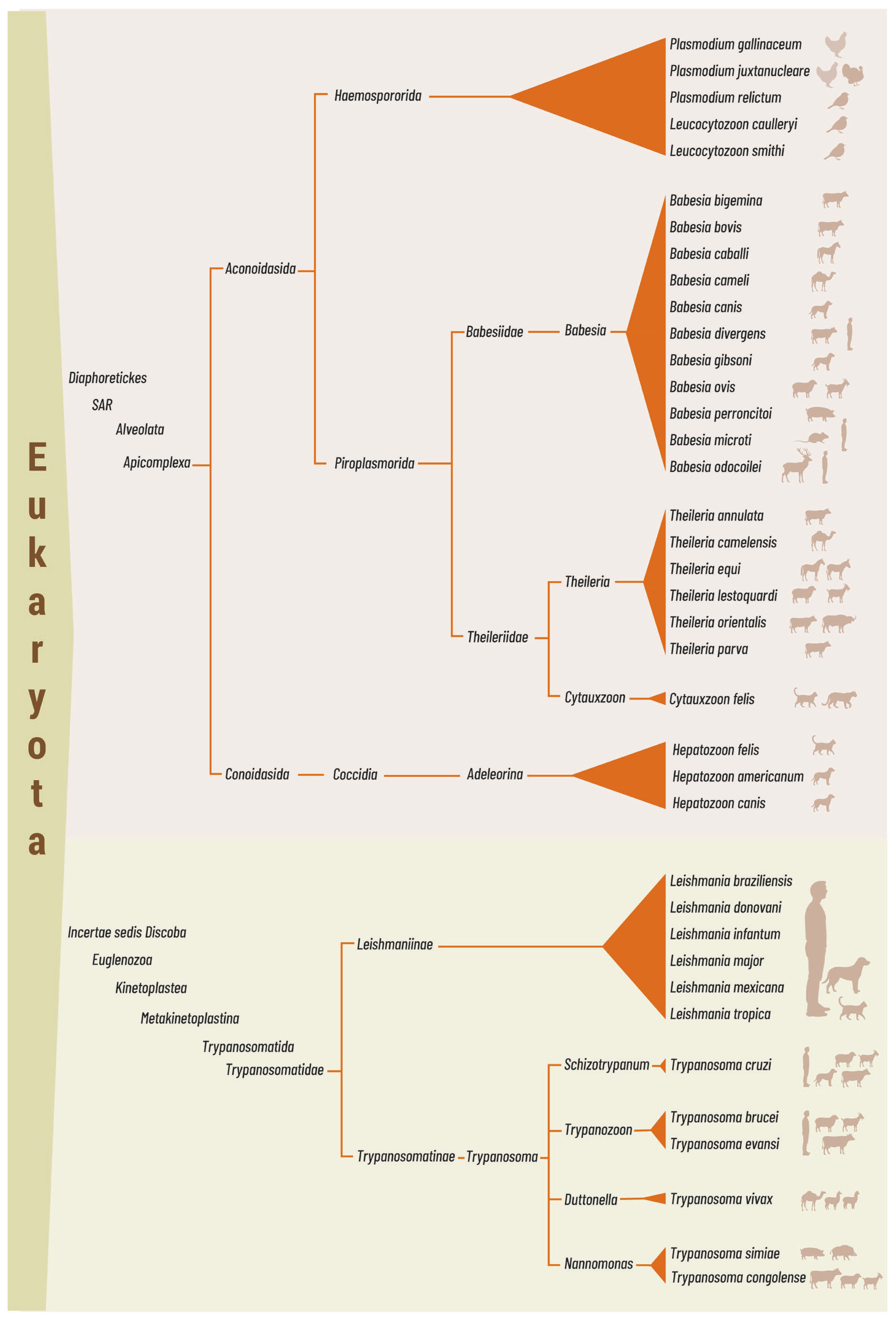
Figure 1. Taxonomic distribution of vector-borne protozoa of veterinary importance and their vertebrate hosts (indicated by silhouettes). The classification shown integrates two complementary frameworks. Higher-level clades—Diaphoretickes, SAR, Alveolata, Discoba, and Euglenozoa—follow Adl et al. [1], who propose them as a hierarchy of nested clades without formal Linnaean rank designations. Linnaean ranks (class, order, family, subfamily, genus, subgenus) follow the NCBI Taxonomy Browser, as Adl et al. [1] only informally suggest phylum- and class-level positions and do not formalise lower ranks. The placement of Discoba within the eukaryotic tree remains unresolved and is therefore indicated as incertae sedis; in contrast, Apicomplexa is securely nested within the SAR clade of Diaphoretickes. Within the family Trypanosomatidae, the vector-borne genera shown belong to two subfamilies: Leishmaniinae (containing Leishmania) and Trypanosomatinae (containing Trypanosoma).
Pathogens 15 00561 g001
Figure 2. Comparative microscopic features and diagnostic pitfalls of vector-borne protozoa relevant to veterinary diagnosis. (A) Intraerythrocytic parasites illustrating morphological overlap among piroplasms, including Babesia spp. (tetrad “Maltese cross” formation and multiple infections of erythrocytes) and Theileria spp. (piroplasms within erythrocytes); (B) Leukocyte-associated parasites demonstrating host cell tropism. Hepatozoon spp. gamonts (represented here by Hepatozoon felis) are observed within neutrophils, whereas Theileria annulata schizonts are present within leukocytes. (C) Extracellular hemoparasites showing the trypomastigote morphology of Trypanosoma spp., highlighting differences between Trypanosoma brucei (elongated form with a well-developed undulating membrane) and Trypanosoma cruzi (prominent kinetoplast and typically curved body); (D) Tissue-stage parasites showing intracellular amastigotes of Leishmania spp. within macrophages and Trypanosoma cruzi within a bladder cell; (E) Diagnostic artefacts in blood smears that may be misinterpreted as protozoal parasites. Examples include a platelet mimicking an extracellular parasite, erythrocytes containing Howell–Jolly bodies resembling intraerythrocytic organisms, and stain precipitate forming irregular, non-cellular deposits. Images in panels (A,CE) are courtesy of the Centers for Disease Control and Prevention (CDC), Public Health Image Library (PHIL), and DPDx and are in the public domain. The Hepatozoon felis image in panel (B) is reproduced from Baneth et al. (2013) [15], distributed under the Creative Commons Attribution (CC BY 2.0) licence. The T. annulata schizont image in panel (B) is reproduced from Branco et al. (2010) [16], with permission of the authors, under the Creative Commons Attribution Non-Commercial (CC BY-NC 4.0) licence.
Pathogens 15 00561 g002
Table 1. Published AI-assisted microscopy algorithms for vector-borne protozoal parasites of veterinary relevance.
HostParasite(s)AI Method *Accuracy (%)Precision (%)Recall (%)F1-Score (%)ReferenceDataset Size **
DogBabesia spp.WeightedEnsemble97.759897.598[36]4000
DogBabesia gibsoniSimCLR (EfficientNet_b2)97.0994.597.395.9[37]1578
HorseTheileria equi, Babesia caballiYOLOv8 ‡91989295[38] ‡2000
PoultryPlasmodium gallinaceumYOLOv3 + Darknet99.2— §99.2[39]12,761
MiceTrypanosoma cruziML—Random Forest89.587.689[40]2628
Human/Animal (archived)Trypanosoma brucei, T. cruzi, Trypanosoma evansiDeep Metric Learning (ResNet50 + CBIR/KNN)99.7193.596.694.9[41]32,276
Multiple (archived)Babesia spp., Leishmania spp., Plasmodium spp., Trypanosoma spp.BYOL SSL (ResNet50)99.298.998.298.7[42]33,694
When various models were tested, the model with the best results was included. * Total number of images used for training and evaluation (test and controls). ‡ Article in Indonesian; title and content translated using Google Translate. § The paper reports specificity (≥99%), not precision; these metrics are not equivalent, and the value has been omitted.
Disclaimer/Publisher’s Note: The statements, opinions and data contained in all publications are solely those of the individual author(s) and contributor(s) and not of MDPI and/or the editor(s). MDPI and/or the editor(s) disclaim responsibility for any injury to people or property resulting from any ideas, methods, instructions or products referred to in the content.