Molecular Pathways Driving Corneal Neovascularization in Herpes Simplex Keratitis



Abstract
1. Introduction
2. Angiogenic Privilege, Angiogenesis, and CNV
2.1. Cornea and Angiogenic Privilege
2.2. Angiogenesis Versus CNV
2.3. CNV Models
2.4. Cell-Type Specific Roles in CNV Pathogenesis
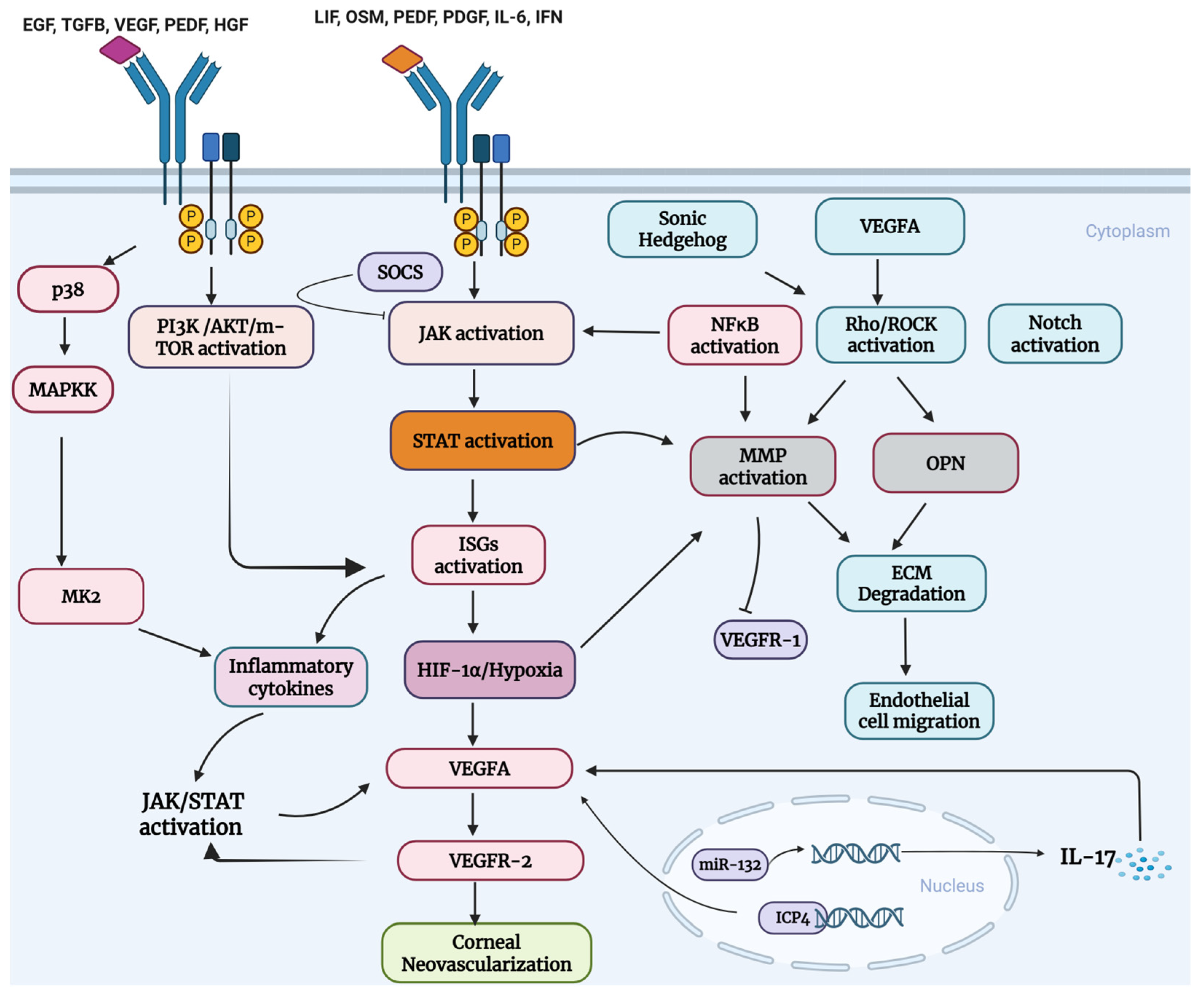
3. Overview of Key Host–Cell Molecular Pathway Drivers in the Pathogenesis of HSK-Induced CNV
3.1. VEGFA and sVEGFR-1 Balance as an Important Index of CNV
3.2. Matrix Metalloproteinases (MMPs) and TIMPs Ratio in CNV
3.3. JAK-STAT Signaling Pathway in HSV-1 Infection
3.4. JAK-STAT-VEGFA Signaling
3.5. JAK-STAT-MMPs Signaling
3.6. Rho/ROCK -MMPs Signaling
3.7. Notch-MMPs Signaling
3.8. MicroRNAs and JAK-STAT Signaling
3.9. Hypoxia-HIF-1α Signaling
3.10. PI3K-AKT-mTOR Pathway in CNV
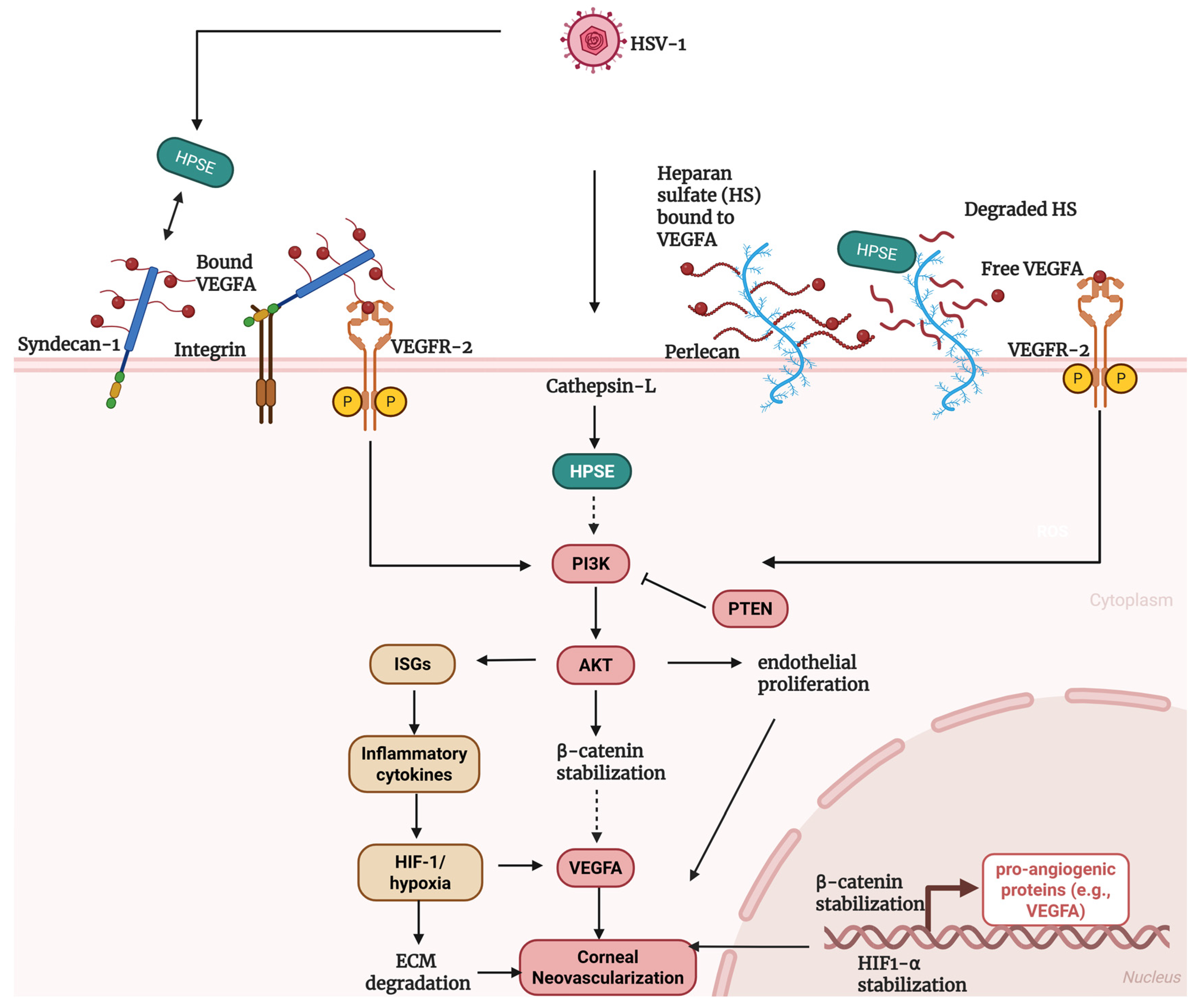
3.11. Heparanase in HSV-1 Pathology
3.12. Heparanase-AKT Signaling
3.13. Heparanase-VEGFA Signaling
3.14. Proposed Mechanism of HPSE-Regulation in HSK-Induced CNV
3.15. Osteopontin in Corneal Wound Healing
3.16. Syndecan-1 in CNV
4. Unanswered Questions and Future Directions
5. Conclusions
Author Contributions
Funding
Institutional Review Board Statement
Informed Consent Statement
Data Availability Statement
Acknowledgments
Conflicts of Interest
Abbreviations
| AKT | Protein kinase B |
| Ang-2 | Angiopoietin-2 |
| AP-1/AP-2 | Activator protein-1/Activator protein-2 |
| BBB | Blood–brain barrier |
| CNV | Corneal neovascularization |
| COX-2 | Cyclooxygenase-2 |
| CNTF | Ciliary neurotrophic factor |
| CXCL | C-X-C motif chemokine ligand |
| CCL | C-C motif chemokine ligand |
| ECM | Extracellular matrix |
| EGF | Epidermal growth factor |
| ERK | Extracellular signal-regulated kinase |
| FGF2 | Fibroblast growth factor-2 |
| FLT-1 | Fms-like tyrosine kinase-1 (VEGFR1) |
| GSK3β | Glycogen synthase kinase-3 beta |
| HGF | Hepatocyte growth factor |
| HIF-1α | Hypoxia-inducible factor-1 alpha |
| HPSE | Heparanase |
| HS | Heparan sulfate |
| HSK | Herpes simplex keratitis |
| HSV-1 | Herpes simplex virus type-1 |
| HUVECs | Human umbilical vein endothelial cells |
| ICP4 | Infected cell protein-4 |
| IFN | Interferon |
| IFNAR | Interferon-alpha receptor |
| IL | Interleukin |
| ISGs | Interferon-stimulated genes |
| JAK | Janus kinase |
| KDR | Kinase insert domain receptor (VEGFR2) |
| LIF | Leukemia inhibitory factor |
| MAPK | Mitogen-activated protein kinase |
| MCP-1 | Monocyte chemoattractant protein-1 |
| miRNA/miR | MicroRNA |
| MK2 | MAPK-activated protein kinase-2 |
| MMP | Matrix metalloproteinase |
| mTOR | Mechanistic target of rapamycin |
| NF-κB | Nuclear factor kappa-B |
| NO | Nitric oxide |
| NR-1 | Neuregulin-1 |
| OPN | Osteopontin |
| OSM | Oncostatin M |
| PDCD4 | Programmed cell death protein-4 |
| PDGF | Platelet-derived growth factor |
| PEDF | Pigment epithelium-derived factor |
| PI3K | Phosphatoinositide 3-kinase |
| PIAS | Protein inhibitor of activated STAT |
| PMN | Polymorphonuclear neutrophils |
| PTEN | Phosphatase and tensin homolog |
| PTP | Protein tyrosine phosphatase |
| Rho/ROCK | Rho-associated protein kinase |
| ROS | Reactive oxygen species |
| SFK/phosphoSFK | Src family kinases |
| SGK1 | Serum and glucocorticoid-regulated kinase-1 |
| Shh | Sonic hedgehog |
| sVEGFR1 | Soluble vascular endothelial growth factor receptor-1 |
| SOCS | Suppressor of cytokine signaling |
| STAT | Signal transducer and activator of transcription |
| TGFB/TGF-β | Transforming growth factor-beta |
| Th1 | T helper type-1 |
| Th17 | T helper type-17 |
| TIMP | Tissue inhibitor of metalloproteinases |
| TNF-α | Tumor necrosis factor-alpha |
| TSP-1/TSP-2 | Thrombospondin-1/Thrombospondin-2 |
| UL | Unique long (HSV gene region) |
| VEGFA | Vascular endothelial growth factor-A |
| VEGFR1/VEGFR2 | Vascular endothelial growth factor receptor-1/-2 |
| VECs | Vascular endothelial cells |
| WT | Wild type |
References
- James, C.; Harfouche, M.; Welton, N.J.; Turner, K.M.; Abu-Raddad, L.J.; Gottlieb, S.L.; Looker, K.J. Herpes simplex virus: Global infection prevalence and incidence estimates, 2016. Bull. World Health Organ. 2020, 98, 315–329. [Google Scholar] [CrossRef] [Scilit]
- Gupta, R.; Warren, T.; Wald, A. Genital herpes. Lancet 2007, 370, 2127–2137. [Google Scholar] [CrossRef] [Scilit]
- Stephen, K.; Anshoo, C. Herpes simplex keratitis. Prog. Retin. Eye Res. 2006, 25, 355–380. [Google Scholar] [CrossRef] [Scilit] [PubMed]
- Liesegang, T.J.; Melton, L.J., 3rd; Daly, P.J.; Ilstrup, D.M. Epidemiology of ocular herpes simplex. Incidence in Rochester, Minn, 1950 through 1982. Arch. Ophthalmol. 1989, 107, 1155–1159. [Google Scholar] [CrossRef] [Scilit] [PubMed]
- Asim, V.F.; Deepak, S. Herpes Simplex Epithelial and Stromal Keratitis: An Epidemiologic Update. Surv. Ophthalmol. 2012, 57, 448–462. [Google Scholar] [CrossRef] [Scilit] [PubMed]
- McCormick, I.; James, C.; Welton, N.J.; Mayaud, P.; Turner, K.M.E.; Gottlieb, S.L.; Foster, A.; Looker, K.J. Incidence of herpes simplex virus keratitis and other ocular disease: Global review and estimates. Ophthalmic Epidemiol. 2022, 29, 353–362. [Google Scholar] [CrossRef] [Scilit]
- Looker, K.J.; Magaret, A.S.; May, M.T.; Turner, K.M.; Vickerman, P.; Gottlieb, S.L.; Newman, L.M. Global and Regional Estimates of Prevalent and Incident Herpes Simplex Virus Type 1 Infections in 2012. PLoS ONE 2015, 10, e0140765. [Google Scholar] [CrossRef] [Scilit]
- Souza, P.M.; Holland, E.J.; Huang, A.J. Bilateral herpetic keratoconjunctivitis. Ophthalmology 2003, 110, 493–496. [Google Scholar] [CrossRef] [Scilit]
- Agarwal, R.; Maharana, P.K.; Titiyal, J.S.; Sharma, N. Bilateral herpes simplex keratitis: Lactation a trigger for recurrence! BMJ Case Rep. 2019, 12, e223713. [Google Scholar] [CrossRef] [Scilit]
- Rowe, A.M.; St Leger, A.J.; Jeon, S.; Dhaliwal, D.K.; Knickelbein, J.E.; Hendricks, R.L. Herpes keratitis. Prog. Retin. Eye Res. 2013, 32, 88–101. [Google Scholar] [CrossRef] [Scilit]
- Stephanie Hiu Ling, P.; William Ho Lam, W.; Amy Cheuk Yin, L.; Hao, Y.; Chien-Fu, C.; Vishal, J.; Yau Kei, C.; Kendrick Co, S. A systematic review on advances in diagnostics for herpes simplex keratitis. Surv. Ophthalmol. 2021, 66, 514–530. [Google Scholar] [CrossRef] [Scilit] [PubMed]
- Stuart, P.M.; Keadle, T.L. Recurrent herpetic stromal keratitis in mice: A model for studying human HSK. Clin. Dev. Immunol. 2012, 2012, 728480. [Google Scholar] [CrossRef] [Scilit] [PubMed]
- Azher, T.N.; Yin, X.-T.; Stuart, P.M. Understanding the Role of Chemokines and Cytokines in Experimental Models of Herpes Simplex Keratitis. J. Immunol. Res. 2017, 2017, 7261980. [Google Scholar] [CrossRef] [Scilit] [PubMed]
- Partha, S.B.; Barry, T.R. Early events in HSV keratitis—Setting the stage for a blinding disease. Microbes Infect. 2005, 7, 799–810. [Google Scholar] [CrossRef] [Scilit]
- Lee, D.H.; Jaggi, U.; Ghiasi, H. CCR2+ migratory macrophages with M1 status are the early-responders in the cornea of HSV-1 infected mice. PLoS ONE 2019, 14, e0215727. [Google Scholar] [CrossRef] [Scilit]
- Wang, L.; Wang, R.; Xu, C.; Zhou, H. Pathogenesis of Herpes Stromal Keratitis: Immune Inflammatory Response Mediated by Inflammatory Regulators. Front. Immunol. 2020, 11, 766. [Google Scholar] [CrossRef] [Scilit]
- Doymaz, M.Z.; Rouse, B.T. Herpetic stromal keratitis: An immunopathologic disease mediated by CD4+ T lymphocytes. Investig. Ophthalmol. Vis. Sci. 1992, 33, 2165–2173. [Google Scholar]
- Mott, K.R.; Gate, D.; Matundan, H.H.; Ghiasi, Y.N.; Town, T.; Ghiasi, H. CD8+ T Cells Play a Bystander Role in Mice Latently Infected with Herpes Simplex Virus 1. J. Virol. 2016, 90, 5059–5067. [Google Scholar] [CrossRef] [Scilit]
- Suryawanshi, A.; Veiga-Parga, T.; Rajasagi, N.K.; Reddy, P.B.; Sehrawat, S.; Sharma, S.; Rouse, B.T. Role of IL-17 and Th17 cells in herpes simplex virus-induced corneal immunopathology. J. Immunol. 2011, 187, 1919–1930. [Google Scholar] [CrossRef] [Scilit]
- Likun, X.; Shengnan, Z.; Zheyao, C.; Yuan, H.; Hongwei, Y.; Dan, W. Interleukin-17 enhanced immunoinflammatory lesions in a mouse model of recurrent herpetic keratitis. Microbes Infect. 2013, 15, 126–139. [Google Scholar] [CrossRef] [Scilit]
- Wuest, T.; Zheng, M.; Efstathiou, S.; Halford, W.P.; Carr, D.J. The herpes simplex virus-1 transactivator infected cell protein-4 drives VEGF-A dependent neovascularization. PLoS Pathog. 2011, 7, e1002278. [Google Scholar] [CrossRef] [Scilit]
- Wu, D.; Chan, K.E.; Lim, B.X.H.; Lim, D.K.; Wong, W.M.; Chai, C.; Manotosh, R.; Lim, C.H.L. Management of corneal neovascularization: Current and emerging therapeutic approaches. Indian J. Ophthalmol. 2024, 72, S354–S371. [Google Scholar] [CrossRef] [Scilit] [PubMed]
- Lobo, A.M.; Agelidis, A.M.; Shukla, D. Pathogenesis of herpes simplex keratitis: The host cell response and ocular surface sequelae to infection and inflammation. Ocul. Surf. 2019, 17, 40–49. [Google Scholar] [CrossRef] [Scilit] [PubMed]
- Shukla, S.D.; Valyi-Nagy, T. Host Molecules That Promote Pathophysiology of Ocular Herpes. Front. Microbiol. 2022, 13, 818658. [Google Scholar] [CrossRef] [Scilit] [PubMed]
- Jaggi, U.; Matundan, H.H.; Yu, J.; Hirose, S.; Mueller, M.; Wormley, F.L., Jr.; Ghiasi, H. Essential role of M1 macrophages in blocking cytokine storm and pathology associated with murine HSV-1 infection. PLoS Pathog. 2021, 17, e1009999. [Google Scholar] [CrossRef] [Scilit]
- Jaggi, U.; Yang, M.; Matundan, H.H.; Hirose, S.; Shah, P.K.; Sharifi, B.G.; Ghiasi, H. Increased phagocytosis in the presence of enhanced M2-like macrophage responses correlates with increased primary and latent HSV-1 infection. PLoS Pathog. 2020, 16, e1008971. [Google Scholar] [CrossRef] [Scilit]
- Wuest, T.R.; Carr, D.J. VEGF-A expression by HSV-1-infected cells drives corneal lymphangiogenesis. J. Exp. Med. 2010, 207, 101–115. [Google Scholar] [CrossRef] [Scilit]
- Suryawanshi, A.; Mulik, S.; Sharma, S.; Reddy, P.B.; Sehrawat, S.; Rouse, B.T. Ocular neovascularization caused by herpes simplex virus type 1 infection results from breakdown of binding between vascular endothelial growth factor A and its soluble receptor. J. Immunol. 2011, 186, 3653–3665. [Google Scholar] [CrossRef] [Scilit]
- Antony, F.; Kinha, D.; Nowińska, A.; Rouse, B.T.; Suryawanshi, A. The immunobiology of corneal HSV-1 infection and herpetic stromal keratitis. Clin. Microbiol. Rev. 2024, 37, e0000624. [Google Scholar] [CrossRef] [Scilit]
- Suryawanshi, A.; Veiga-Parga, T.; Reddy, P.B.; Rajasagi, N.K.; Rouse, B.T. IL-17A differentially regulates corneal vascular endothelial growth factor (VEGF)-A and soluble VEGF receptor 1 expression and promotes corneal angiogenesis after herpes simplex virus infection. J. Immunol. 2012, 188, 3434–3446. [Google Scholar] [CrossRef] [Scilit]
- Ezeonwumelu, I.J.; Garcia-Vidal, E.; Ballana, E. JAK-STAT Pathway: A Novel Target to Tackle Viral Infections. Viruses 2021, 13, 2379. [Google Scholar] [CrossRef] [Scilit] [PubMed]
- Chen, K.; Li, Y.; Zhang, X.; Ullah, R.; Tong, J.; Shen, Y. The role of the PI3K/AKT signalling pathway in the corneal epithelium: Recent updates. Cell Death Dis. 2022, 13, 513. [Google Scholar] [CrossRef] [Scilit] [PubMed]
- Ambati, B.K.; Nozaki, M.; Singh, N.; Takeda, A.; Jani, P.D.; Suthar, T.; Albuquerque, R.J.; Richter, E.; Sakurai, E.; Newcomb, M.T.; et al. Corneal avascularity is due to soluble VEGF receptor-1. Nature 2006, 443, 993–997. [Google Scholar] [CrossRef] [Scilit] [PubMed]
- Ellenberg, D.; Azar, D.T.; Hallak, J.A.; Tobaigy, F.; Han, K.Y.; Jain, S.; Zhou, Z.; Chang, J.H. Novel aspects of corneal angiogenic and lymphangiogenic privilege. Prog. Retin. Eye Res. 2010, 29, 208–248. [Google Scholar] [CrossRef] [Scilit]
- Azar, D.T. Corneal angiogenic privilege: Angiogenic and antiangiogenic factors in corneal avascularity, vasculogenesis, and wound healing (an American Ophthalmological Society thesis). Trans. Am. Ophthalmol. Soc. 2006, 104, 264–302. [Google Scholar]
- Michael, L.R.; Nicholas, E.F.; Ethan, M.S.; Timothy, P.F.; Partha, S.B.; Harris, E.M., Jr.; Christian, C.; Paulo, C.R.; Walter, J.L.; Donald, R.B.; et al. Corneal neovascularization: A review of the molecular biology and current therapies. Expert Rev. Ophthalmol. 2013, 8, 167–189. [Google Scholar] [CrossRef] [Scilit]
- Gurung, H.R.; Carr, M.M.; Bryant, K.; Chucair-Elliott, A.J.; Carr, D.J. Fibroblast growth factor-2 drives and maintains progressive corneal neovascularization following HSV-1 infection. Mucosal Immunol. 2018, 11, 172–185. [Google Scholar] [CrossRef] [Scilit]
- Muller, E.; Feinberg, L.; Woronkowicz, M.; Roberts, H.W. Corneal Neovascularization: Pathogenesis, Current Insights and Future Strategies. Biology 2026, 15, 136. [Google Scholar] [CrossRef] [Scilit]
- Thairu, N.; Kiriakidis, S.; Dawson, P.; Paleolog, E. Angiogenesis as a therapeutic target in arthritis in 2011: Learning the lessons of the colorectal cancer experience. Angiogenesis 2011, 14, 223–234. [Google Scholar] [CrossRef] [Scilit]
- Zaher, K.O.; Rami, A.R.M.; Jawad, A.M.; Ali, I.S. Understanding the biology of angiogenesis: Review of the most important molecular mechanisms. Blood Cells Mol. Dis. 2007, 39, 212–220. [Google Scholar]
- Chen, Z.; Han, Z.C. STAT3: A critical transcription activator in angiogenesis. Med. Res. Rev. 2008, 28, 185–200. [Google Scholar] [CrossRef] [Scilit] [PubMed]
- Giménez, F.; Suryawanshi, A.; Rouse, B.T. Pathogenesis of herpes stromal keratitis--a focus on corneal neovascularization. Prog. Retin. Eye Res. 2013, 33, 1–9. [Google Scholar] [CrossRef] [Scilit] [PubMed]
- Potente, M.; Gerhardt, H.; Carmeliet, P. Basic and therapeutic aspects of angiogenesis. Cell 2011, 146, 873–887. [Google Scholar] [CrossRef] [Scilit] [PubMed]
- Maruyama, K.; Ii, M.; Cursiefen, C.; Jackson, D.G.; Keino, H.; Tomita, M.; Van Rooijen, N.; Takenaka, H.; D’Amore, P.A.; Stein-Streilein, J.; et al. Inflammation-induced lymphangiogenesis in the cornea arises from CD11b-positive macrophages. J. Clin. Investig. 2005, 115, 2363–2372. [Google Scholar] [CrossRef] [Scilit]
- Patnam, M.; Dommaraju, S.R.; Masood, F.; Herbst, P.; Chang, J.H.; Hu, W.Y.; Rosenblatt, M.I.; Azar, D.T. Lymphangiogenesis Guidance Mechanisms and Therapeutic Implications in Pathological States of the Cornea. Cells 2023, 12, 319. [Google Scholar] [CrossRef] [Scilit]
- Nicholas, M.P.; Mysore, N. Corneal neovascularization. Exp. Eye Res. 2021, 202, 108363. [Google Scholar] [CrossRef] [Scilit]
- Shi, W.; Liu, J.; Li, M.; Gao, H.; Wang, T. Expression of MMP, HPSE, and FAP in stroma promoted corneal neovascularization induced by different etiological factors. Curr. Eye Res. 2010, 35, 967–977. [Google Scholar] [CrossRef] [Scilit]
- Tian, S.; Wang, S.; He, Y.; Liu, X.; Li, Y.; Su, G.; Zhang, Y. Review of the Progress in Corneal Neovascularization Animal Models. Am. J. Biochem. Biotechnol. 2015, 11, 221–227. [Google Scholar] [CrossRef] [Scilit]
- Sitong, S.; Yan, Z. Restoration of corneal epithelial barrier function: A possible target for corneal neovascularization. Ocul. Surf. 2024, 34, 38–49. [Google Scholar] [CrossRef] [Scilit]
- Yu, H.; Sun, L.; Cui, J.; Li, Y.; Yan, Y.; Wei, X.; Wang, C.; Song, F.; Jiang, W.; Liu, Y.; et al. Three kinds of corneal host cells contribute differently to corneal neovascularization. eBioMedicine 2019, 44, 542–553. [Google Scholar] [CrossRef] [Scilit]
- Roshandel, D.; Eslani, M.; Baradaran-Rafii, A.; Cheung, A.Y.; Kurji, K.; Jabbehdari, S.; Maiz, A.; Jalali, S.; Djalilian, A.R.; Holland, E.J. Current and emerging therapies for corneal neovascularization. Ocul. Surf. 2018, 16, 398–414. [Google Scholar] [CrossRef] [Scilit] [PubMed]
- Ma, J.; Zhou, D.; Fan, M.; Wang, H.; Huang, C.; Zhang, Z.; Wu, Y.; Li, W.; Chen, Y.; Liu, Z. Keratocytes create stromal spaces to promote corneal neovascularization via MMP13 expression. Investig. Ophthalmol. Vis. Sci. 2014, 55, 6691–6703. [Google Scholar] [CrossRef] [Scilit] [PubMed]
- ZHAO, W.; JIANG, A.-h.; LI, C.-y.; YANG, W.-z.; XU, C.-c.; LIU, Z.-g. Pericytes are correlated with the permeability of rat corneal neovascular vessels induced by alkali burn. Chin. Med. J. 2007, 120, 274–279. [Google Scholar] [CrossRef] [Scilit] [PubMed]
- Dente, C.J.; Steffes, C.P.; Speyer, C.; Tyburski, J.G. Pericytes augment the capillary barrier in in vitro cocultures. J. Surg. Res. 2001, 97, 85–91. [Google Scholar] [CrossRef] [Scilit]
- Do carmo, A.; Ramos, P.; Reis, A.; Proença, R.; Cunha-vaz, J.G. Breakdown of the inner and outer blood retinal barrier in streptozotocin-induced diabetes. Exp. Eye Res. 1998, 67, 569–575. [Google Scholar] [CrossRef] [Scilit]
- Drzyzga, Ł.; Śpiewak, D.; Dorecka, M.; Wyględowska-Promieńska, D. Available Therapeutic Options for Corneal Neovascularization: A Review. Int. J. Mol. Sci. 2024, 25, 5479. [Google Scholar] [CrossRef] [Scilit]
- Conway, E.M.; Collen, D.; Carmeliet, P. Molecular mechanisms of blood vessel growth. Cardiovasc. Res. 2001, 49, 507–521. [Google Scholar] [CrossRef] [Scilit]
- Zheng, M.; Deshpande, S.; Lee, S.; Ferrara, N.; Rouse, B.T. Contribution of vascular endothelial growth factor in the neovascularization process during the pathogenesis of herpetic stromal keratitis. J. Virol. 2001, 75, 9828–9835. [Google Scholar] [CrossRef] [Scilit]
- Stevenson, W.; Cheng, S.F.; Dastjerdi, M.H.; Ferrari, G.; Dana, R. Corneal neovascularization and the utility of topical VEGF inhibition: Ranibizumab (Lucentis) vs bevacizumab (Avastin). Ocul. Surf. 2012, 10, 67–83. [Google Scholar] [CrossRef] [Scilit]
- Ito, T.K.; Ishii, G.; Chiba, H.; Ochiai, A. The VEGF angiogenic switch of fibroblasts is regulated by MMP-7 from cancer cells. Oncogene 2007, 26, 7194–7203. [Google Scholar] [CrossRef] [Scilit]
- Huo, N.; Ichikawa, Y.; Kamiyama, M.; Ishikawa, T.; Hamaguchi, Y.; Hasegawa, S.; Nagashima, Y.; Miyazaki, K.; Shimada, H. MMP-7 (matrilysin) accelerated growth of human umbilical vein endothelial cells. Cancer Lett. 2002, 177, 95–100. [Google Scholar] [CrossRef] [Scilit] [PubMed]
- Zhang, J.; Wang, S.; He, Y.; Yao, B.; Zhang, Y. Regulation of matrix metalloproteinases 2 and 9 in corneal neovascularization. Chem. Biol. Drug Des. 2020, 95, 485–492. [Google Scholar] [CrossRef] [Scilit] [PubMed]
- Maddula, S.; Davis, D.K.; Maddula, S.; Burrow, M.K.; Ambati, B.K. Horizons in therapy for corneal angiogenesis. Ophthalmology 2011, 118, 591–599. [Google Scholar] [CrossRef] [Scilit] [PubMed]
- Lee, S.; Zheng, M.; Kim, B.; Rouse, B.T. Role of matrix metalloproteinase-9 in angiogenesis caused by ocular infection with herpes simplex virus. J. Clin. Investig. 2002, 110, 1105–1111. [Google Scholar] [CrossRef]
- Song, D.; Yang, Q.; Li, X.; Chen, K.; Tong, J.; Shen, Y. The role of the JAK/STAT3 signaling pathway in acquired corneal diseases. Exp. Eye Res. 2024, 238, 109748. [Google Scholar] [CrossRef] [Scilit]
- Masaaki, M.; Daisuke, K.; Toshio, H. Pleiotropy and Specificity: Insights from the Interleukin 6 Family of Cytokines. Immunity 2019, 50, 812–831. [Google Scholar] [CrossRef] [Scilit]
- Morris, R.; Kershaw, N.J.; Babon, J.J. The molecular details of cytokine signaling via the JAK/STAT pathway. Protein Sci. 2018, 27, 1984–2009. [Google Scholar] [CrossRef] [Scilit]
- Steven, E.W. The corneal fibroblast: The Dr. Jekyll underappreciated overseer of the responses to stromal injury. Ocul. Surf. 2023, 29, 53–62. [Google Scholar] [CrossRef] [Scilit]
- Chen, C.; Wang, J.; Liu, C.; Hu, J.; Liu, L. Pioneering therapies for post-infarction angiogenesis: Insight into molecular mechanisms and preclinical studies. Biomed. Pharmacother. 2023, 166, 115306. [Google Scholar] [CrossRef] [Scilit]
- Ren, J.; Antony, F.; Rouse, B.T.; Suryawanshi, A. Role of Innate Interferon Responses at the Ocular Surface in Herpes Simplex Virus-1-Induced Herpetic Stromal Keratitis. Pathogens 2023, 12, 437. [Google Scholar] [CrossRef] [Scilit]
- Schoggins, J.W. Interferon-stimulated genes: Roles in viral pathogenesis. Curr. Opin. Virol. 2014, 6, 40–46. [Google Scholar] [CrossRef] [Scilit]
- Yuan, H.; You, J.; You, H.; Zheng, C. Herpes Simplex Virus 1 UL36USP Antagonizes Type I Interferon-Mediated Antiviral Innate Immunity. J. Virol. 2018, 92, e01161-18. [Google Scholar] [CrossRef] [Scilit] [PubMed]
- Zhang, R.; Tang, J. Evasion of I Interferon-Mediated Innate Immunity by Pseudorabies Virus. Front. Microbiol. 2021, 12, 801257. [Google Scholar] [CrossRef] [Scilit] [PubMed]
- Sato, Y.; Koshizuka, T.; Ishibashi, K.; Hashimoto, K.; Ishioka, K.; Ikuta, K.; Yokota, S.I.; Fujii, N.; Suzutani, T. Involvement of herpes simplex virus type 1 UL13 protein kinase in induction of SOCS genes, the negative regulators of cytokine signaling. Microbiol. Immunol. 2017, 61, 159–167. [Google Scholar] [CrossRef] [Scilit] [PubMed]
- Mott, K.R.; UnderHill, D.; Wechsler, S.L.; Town, T.; Ghiasi, H. A role for the JAK-STAT1 pathway in blocking replication of HSV-1 in dendritic cells and macrophages. Virol. J. 2009, 6, 56. [Google Scholar] [CrossRef] [Scilit]
- Hsia, H.C.; Stopford, C.M.; Zhang, Z.; Damania, B.; Baldwin, A.S. Signal transducer and activator of transcription 3 (Stat3) regulates host defense and protects mice against herpes simplex virus-1 (HSV-1) infection. J. Leukoc. Biol. 2017, 101, 1053–1064. [Google Scholar] [CrossRef] [Scilit]
- D’Aiuto, L.; Williamson, K.; Dimitrion, P.; McNulty, J.; Brown, C.E.; Dokuburra, C.B.; Nielsen, A.J.; Lin, W.J.; Piazza, P.; Schurdak, M.E.; et al. Comparison of three cell-based drug screening platforms for HSV-1 infection. Antivir. Res. 2017, 142, 136–140. [Google Scholar] [CrossRef] [Scilit]
- Xue, C.; Xie, J.; Zhao, D.; Lin, S.; Zhou, T.; Shi, S.; Shao, X.; Lin, Y.; Zhu, B.; Cai, X. The JAK/STAT3 signalling pathway regulated angiogenesis in an endothelial cell/adipose-derived stromal cell co-culture, 3D gel model. Cell Prolif. 2017, 50, e12307. [Google Scholar] [CrossRef] [Scilit]
- Ebihara, N.; Matsuda, A.; Nakamura, S.; Matsuda, H.; Murakami, A. Role of the IL-6 classic- and trans-signaling pathways in corneal sterile inflammation and wound healing. Investig. Ophthalmol. Vis. Sci. 2011, 52, 8549–8557. [Google Scholar] [CrossRef] [Scilit]
- Tohru, S.; Akiko, I. Anti-inflammatory effect of topical administration of tofacitinib on corneal inflammation. Exp. Eye Res. 2016, 145, 110–117. [Google Scholar] [CrossRef] [Scilit]
- Wang, X.; Zhang, S.; Dong, M.; Li, Y.; Zhou, Q.; Yang, L. The proinflammatory cytokines IL-1β and TNF-α modulate corneal epithelial wound healing through p16(Ink4a) suppressing STAT3 activity. J. Cell. Physiol. 2020, 235, 10081–10093. [Google Scholar] [CrossRef] [Scilit] [PubMed]
- Yang, W.; Yuelan, G.; Yuqing, H.; Yumiao, P.; Yi, Y.; Yiwen, Z.; Shan-shan, W.; Yan-ning, Y. The potential protective effects of miR-497 on corneal neovascularization are mediated via macrophage through the IL-6/STAT3/VEGF signaling pathway. Int. Immunopharmacol. 2021, 96, 107745. [Google Scholar]
- Wei, L.; Yingnan, Z.; Liang, Z.; Xiaoqin, L.; Margaret, E.F.; Wei, W.; Hui, S.; Douglas, C.D.; Lijun, Z.; Yongqing, L. Zeb1 regulation of wound-healing-induced inflammation in alkali-damaged corneas. iScience 2022, 25, 104038. [Google Scholar]
- Tuli, H.S.; Sak, K.; Iqubal, A.; Garg, V.K.; Varol, M.; Sharma, U.; Chauhan, A.; Yerer, M.B.; Dhama, K.; Jain, M.; et al. STAT signaling as a target for intervention: From cancer inflammation and angiogenesis to non-coding RNAs modulation. Mol. Biol. Rep. 2022, 49, 8987–8999. [Google Scholar] [CrossRef] [Scilit]
- Miriam, F.-F.; Alicia, R.F.; Sandra, C.; Carlos, L.-O. Matrix metalloproteinases: Evolution, gene regulation and functional analysis in mouse models. Biochim. Biophys. Acta (BBA)—Mol. Cell Res. 2010, 1803, 3–19. [Google Scholar]
- Kim, J.W.; Jeong, H.; Yang, M.S.; Lim, C.W.; Kim, B. Therapeutic effects of zerumbone in an alkali-burned corneal wound healing model. Int. Immunopharmacol. 2017, 48, 126–134. [Google Scholar] [CrossRef] [Scilit]
- Barry, Z.; Park, B.; Corson, T.W. Pharmacological Potential of Small Molecules for Treating Corneal Neovascularization. Molecules 2020, 25, 3468. [Google Scholar] [CrossRef] [Scilit]
- Menager, C.; Vassy, J.; Doliger, C.; Legrand, Y.; Karniguian, A. Subcellular localization of RhoA and ezrin at membrane ruffles of human endothelial cells: Differential role of collagen and fibronectin. Exp. Cell Res. 1999, 249, 221–230. [Google Scholar] [CrossRef] [Scilit]
- Bryan, B.A.; Dennstedt, E.; Mitchell, D.C.; Walshe, T.E.; Noma, K.; Loureiro, R.; Saint-Geniez, M.; Campaigniac, J.P.; Liao, J.K.; D’Amore, P.A. RhoA/ROCK signaling is essential for multiple aspects of VEGF-mediated angiogenesis. Faseb J. 2010, 24, 3186–3195. [Google Scholar] [CrossRef] [Scilit]
- Renault, M.A.; Roncalli, J.; Tongers, J.; Thorne, T.; Klyachko, E.; Misener, S.; Volpert, O.V.; Mehta, S.; Burg, A.; Luedemann, C.; et al. Sonic hedgehog induces angiogenesis via Rho kinase-dependent signaling in endothelial cells. J. Mol. Cell. Cardiol. 2010, 49, 490–498. [Google Scholar] [CrossRef] [Scilit]
- Pola, R.; Ling, L.E.; Silver, M.; Corbley, M.J.; Kearney, M.; Blake Pepinsky, R.; Shapiro, R.; Taylor, F.R.; Baker, D.P.; Asahara, T.; et al. The morphogen Sonic hedgehog is an indirect angiogenic agent upregulating two families of angiogenic growth factors. Nat. Med. 2001, 7, 706–711. [Google Scholar] [CrossRef] [Scilit] [PubMed]
- Yaşar, M.; Çakmak, H.; Dündar, S.; Örenay Boyacıoğlu, S.; Çalışkan, M.; Ergin, K. The role of microRNAs in corneal neovascularization and its relation to VEGF. Cutan. Ocul. Toxicol. 2020, 39, 341–347. [Google Scholar] [CrossRef] [Scilit] [PubMed]
- Mulik, S.; Xu, J.; Reddy, P.B.; Rajasagi, N.K.; Gimenez, F.; Sharma, S.; Lu, P.Y.; Rouse, B.T. Role of miR-132 in angiogenesis after ocular infection with herpes simplex virus. Am. J. Pathol. 2012, 181, 525–534. [Google Scholar] [CrossRef] [Scilit] [PubMed]
- Mukwaya, A.; Jensen, L.; Peebo, B.; Lagali, N. MicroRNAs in the cornea: Role and implications for treatment of corneal neovascularization. Ocul. Surf. 2019, 17, 400–411. [Google Scholar] [CrossRef] [Scilit]
- Cao, Q.; Li, Y.; Li, Y.; Li, L. Loss of miR-673-5p expression in the cornea promotes rat corneal allograft rejection by promoting Th17 cell differentiation mediated by JAK2/STAT3. Ann. Transl. Med. 2021, 9, 1409. [Google Scholar] [CrossRef] [Scilit]
- Zhang, Y.; Zhang, T.; Ma, X.; Zou, J. Subconjunctival injection of antagomir-21 alleviates corneal neovascularization in a mouse model of alkali-burned cornea. Oncotarget 2017, 8, 11797–11808. [Google Scholar] [CrossRef] [Scilit]
- Gabriely, G.; Wurdinger, T.; Kesari, S.; Esau, C.C.; Burchard, J.; Linsley, P.S.; Krichevsky, A.M. MicroRNA 21 promotes glioma invasion by targeting matrix metalloproteinase regulators. Mol. Cell. Biol. 2008, 28, 5369–5380. [Google Scholar] [CrossRef] [Scilit]
- Sakai, E.; Miura, Y.; Suzuki-Kouyama, E.; Oka, K.; Tachibana, M.; Kawabata, K.; Sakurai, F.; Mizuguchi, H. A mammalian mirtron miR-1224 promotes tube-formation of human primary endothelial cells by targeting anti-angiogenic factor epsin2. Sci. Rep. 2017, 7, 5541. [Google Scholar] [CrossRef] [Scilit]
- Niu, Y.; Mo, D.; Qin, L.; Wang, C.; Li, A.; Zhao, X.; Wang, X.; Xiao, S.; Wang, Q.; Xie, Y.; et al. Lipopolysaccharide-induced miR-1224 negatively regulates tumour necrosis factor-α gene expression by modulating Sp1. Immunology 2011, 133, 8–20. [Google Scholar] [CrossRef] [Scilit]
- Ferrari, G.; Bignami, F.; Rama, P. Tumor necrosis factor-α inhibitors as a treatment of corneal hemangiogenesis and lymphangiogenesis. Eye Contact Lens 2015, 41, 72–76. [Google Scholar] [CrossRef] [Scilit]
- Siddheshvar, B.; Sachin, M.; Fernanda, G.; Pradeep, B.J.R.; Raphael, L.R.; Siva Karthik, V.; Ujjaldeep, J.; John, X.; Patrick, Y.L.; Barry, T.R. Role of miR-155 in the Pathogenesis of Herpetic Stromal Keratitis. Am. J. Pathol. 2015, 185, 1073–1084. [Google Scholar] [CrossRef] [Scilit] [PubMed]
- Ye, J.; Guo, R.; Shi, Y.; Qi, F.; Guo, C.; Yang, L. miR-155 Regulated Inflammation Response by the SOCS1-STAT3-PDCD4 Axis in Atherogenesis. Mediat. Inflamm. 2016, 2016, 8060182. [Google Scholar] [CrossRef] [Scilit] [PubMed]
- Huang, X.; Peng, A.; Sun, M.; Wu, M.; Zhang, P. MicroRNA-155 enhances ocular inflammation after Pseudomonas aeruginosa infection (158.11). J. Immunol. 2011, 186, 158.11. [Google Scholar] [CrossRef] [Scilit]
- Pang, K.; Lennikov, A.; Yang, M. Hypoxia adaptation in the cornea: Current animal models and underlying mechanisms. Anim. Models Exp. Med. 2021, 4, 300–310. [Google Scholar] [CrossRef] [Scilit]
- Chen, P.; Yin, H.; Wang, Y.; Wang, Y.; Xie, L. Inhibition of VEGF expression and corneal neovascularization by shRNA targeting HIF-1α in a mouse model of closed eye contact lens wear. Mol. Vis. 2012, 18, 864–873. [Google Scholar]
- Zidan, A.A.; Elbasiony, E.; Lin, Z.; Najafi, S.; Pate, K.; Yin, J. Supersaturated Oxygen Emulsion Mitigates Hypoxia-Driven Corneal Neovascularization after Alkali Burn. Ophthalmol. Sci. 2025, 5, 100898. [Google Scholar] [CrossRef] [Scilit]
- Pugh, C.W.; Ratcliffe, P.J. Regulation of angiogenesis by hypoxia: Role of the HIF system. Nat. Med. 2003, 9, 677–684. [Google Scholar] [CrossRef] [Scilit]
- Chandrasekher, G.; Pothula, S.; Maharaj, G.; Bazan, H.E. Differential effects of hepatocyte growth factor and keratinocyte growth factor on corneal epithelial cell cycle protein expression, cell survival, and growth. Mol. Vis. 2014, 20, 24–37. [Google Scholar]
- Hong, J.; Qian, T.; Le, Q.; Sun, X.; Wu, J.; Chen, J.; Yu, X.; Xu, J. NGF promotes cell cycle progression by regulating D-type cyclins via PI3K/Akt and MAPK/Erk activation in human corneal epithelial cells. Mol. Vis. 2012, 18, 758–764. [Google Scholar]
- Park, J.H.; Kang, S.S.; Kim, J.Y.; Tchah, H. Nerve Growth Factor Attenuates Apoptosis and Inflammation in the Diabetic Cornea. Investig. Ophthalmol. Vis. Sci. 2016, 57, 6767–6775. [Google Scholar] [CrossRef] [Scilit]
- Di, G.; Zhao, X.; Qi, X.; Zhang, S.; Feng, L.; Shi, W.; Zhou, Q. VEGF-B promotes recovery of corneal innervations and trophic functions in diabetic mice. Sci. Rep. 2017, 7, 40582. [Google Scholar] [CrossRef] [Scilit] [PubMed]
- Shen, M.; Zhou, X.Z.; Ye, L.; Yuan, Q.; Shi, C.; Zhu, P.W.; Jiang, N.; Ma, M.Y.; Yang, Q.C.; Shao, Y. Xanthatin inhibits corneal neovascularization by inhibiting the VEGFR2-mediated STAT3/PI3K/Akt signaling pathway. Int. J. Mol. Med. 2018, 42, 769–778. [Google Scholar] [CrossRef] [Scilit] [PubMed]
- Shin, Y.J.; Hyon, J.Y.; Choi, W.S.; Yi, K.; Chung, E.S.; Chung, T.Y.; Wee, W.R. Chemical injury-induced corneal opacity and neovascularization reduced by rapamycin via TGF-β1/ERK pathways regulation. Investig. Ophthalmol. Vis. Sci. 2013, 54, 4452–4458. [Google Scholar] [CrossRef] [Scilit] [PubMed]
- Li, J.; Han, J.; Shi, Y.; Liu, M. Rapamycin inhibits corneal inflammatory response and neovascularization in a mouse model of corneal alkali burn. Exp. Eye Res. 2023, 233, 109539. [Google Scholar] [CrossRef] [Scilit]
- Li, J.; Du, S.; Shi, Y.; Han, J.; Niu, Z.; Wei, L.; Yang, P.; Chen, L.; Tian, H.; Gao, L. Rapamycin ameliorates corneal injury after alkali burn through methylation modification in mouse TSC1 and mTOR genes. Exp. Eye Res. 2021, 203, 108399. [Google Scholar] [CrossRef] [Scilit]
- Xie, M.; Jie, Y. Transforming corneal alkali burn treatment: Unveiling mechanisms and pioneering therapies from bench to bedside. J. Transl. Med. 2025, 23, 1132. [Google Scholar] [CrossRef] [Scilit]
- Wei, Y.; Songyi, T.; Yue, W.; Wenzhe, W.; Yumeilan, L.; Huanhuan, X.; Hu, M.; Yan, L.; Zhenping, H.; Yirui, G. SGK1 mediates herpes simplex keratitis via the PI3K/SGK1/ Wnt signaling pathways. Cell. Signal. 2025, 127, 111566. [Google Scholar]
- Kaur, S.; Sassano, A.; Dolniak, B.; Joshi, S.; Majchrzak-Kita, B.; Baker, D.P.; Hay, N.; Fish, E.N.; Platanias, L.C. Role of the Akt pathway in mRNA translation of interferon-stimulated genes. Proc. Natl. Acad. Sci. USA 2008, 105, 4808–4813. [Google Scholar] [CrossRef] [Scilit]
- Kaur, S.; Sassano, A.; Joseph, A.M.; Majchrzak-Kita, B.; Eklund, E.A.; Verma, A.; Brachmann, S.M.; Fish, E.N.; Platanias, L.C. Dual regulatory roles of phosphatidylinositol 3-kinase in IFN signaling. J. Immunol. 2008, 181, 7316–7323. [Google Scholar] [CrossRef] [Scilit]
- Kaur, S.; Katsoulidis, E.; Platanias, L.C. Akt and mRNA translation by interferons. Cell Cycle 2008, 7, 2112–2116. [Google Scholar] [CrossRef] [Scilit]
- Tian, J.; Zhang, X.; Wu, H.; Liu, C.; Li, Z.; Hu, X.; Su, S.; Wang, L.F.; Qu, L. Blocking the PI3K/AKT pathway enhances mammalian reovirus replication by repressing IFN-stimulated genes. Front. Microbiol. 2015, 6, 886. [Google Scholar] [CrossRef] [Scilit]
- Suryawanshi, R.K.; Koujah, L.; Patil, C.D.; Ames, J.M.; Agelidis, A.; Yadavalli, T.; Patil, S.V.; Shukla, D. Bacterial Pigment Prodigiosin Demonstrates a Unique Antiherpesvirus Activity That Is Mediated through Inhibition of Prosurvival Signal Transducers. J. Virol. 2020, 94, 10–1128. [Google Scholar] [CrossRef] [Scilit] [PubMed]
- Koujah, L.; Madavaraju, K.; Agelidis, A.M.; Patil, C.D.; Shukla, D. Heparanase-Induced Activation of AKT Stabilizes β-Catenin and Modulates Wnt/β-Catenin Signaling during Herpes Simplex Virus 1 Infection. mBio 2021, 12, e02792–02721. [Google Scholar] [CrossRef] [Scilit]
- Jaishankar, D.; Yakoub, A.M.; Yadavalli, T.; Agelidis, A.; Thakkar, N.; Hadigal, S.; Ames, J.; Shukla, D. An off-target effect of BX795 blocks herpes simplex virus type 1 infection of the eye. Sci. Transl. Med. 2018, 10, eaan5861. [Google Scholar] [CrossRef] [Scilit] [PubMed]
- Jiang, L.; He, W.; Tang, F.; Tang, N.; Huang, G.; Huang, W.; Wu, X.; Guan, J.; Zeng, S.; Li, M.; et al. Epigenetic Landscape Analysis of the Long Non-Coding RNA and Messenger RNA in a Mouse Model of Corneal Alkali Burns. Investig. Ophthalmol. Vis. Sci. 2021, 62, 28. [Google Scholar] [CrossRef] [Scilit] [PubMed]
- Chen, Y.; Yang, W.; Zhang, X.; Yang, S.; Peng, G.; Wu, T.; Zhou, Y.; Huang, C.; Reinach, P.S.; Li, W.; et al. MK2 inhibitor reduces alkali burn-induced inflammation in rat cornea. Sci. Rep. 2016, 6, 28145. [Google Scholar] [CrossRef] [Scilit]
- Sun, J.; Huang, W.; Yang, S.F.; Zhang, X.P.; Yu, Q.; Zhang, Z.Q.; Yao, J.; Li, K.R.; Jiang, Q.; Cao, C. Gαi1 and Gαi3mediate VEGF-induced VEGFR2 endocytosis, signaling and angiogenesis. Theranostics 2018, 8, 4695–4709. [Google Scholar] [CrossRef] [Scilit]
- Koganti, R.; Suryawanshi, R.; Shukla, D. Heparanase, cell signaling, and viral infections. Cell. Mol. Life Sci. 2020, 77, 5059–5077. [Google Scholar] [CrossRef] [Scilit]
- Koujah, L.; Suryawanshi, R.K.; Shukla, D. Pathological processes activated by herpes simplex virus-1 (HSV-1) infection in the cornea. Cell. Mol. Life Sci. 2019, 76, 405–419. [Google Scholar] [CrossRef] [Scilit]
- Agelidis, A.M.; Hadigal, S.R.; Jaishankar, D.; Shukla, D. Viral Activation of Heparanase Drives Pathogenesis of Herpes Simplex Virus-1. Cell Rep. 2017, 20, 439–450. [Google Scholar] [CrossRef] [Scilit]
- Ilan, N.; Elkin, M.; Vlodavsky, I. Regulation, function and clinical significance of heparanase in cancer metastasis and angiogenesis. Int. J. Biochem. Cell Biol. 2006, 38, 2018–2039. [Google Scholar] [CrossRef] [Scilit] [PubMed]
- Blich, M.; Golan, A.; Arvatz, G.; Sebbag, A.; Shafat, I.; Sabo, E.; Cohen-Kaplan, V.; Petcherski, S.; Avniel-Polak, S.; Eitan, A.; et al. Macrophage Activation by Heparanase Is Mediated by TLR-2 and TLR-4 and Associates With Plaque Progression. Arterioscler. Thromb. Vasc. Biol. 2013, 33, e56–e65. [Google Scholar] [CrossRef] [Scilit] [PubMed]
- Gingis-Velitski, S.; Zetser, A.; Flugelman, M.Y.; Vlodavsky, I.; Ilan, N. Heparanase induces endothelial cell migration via protein kinase B/Akt activation. J. Biol. Chem. 2004, 279, 23536–23541. [Google Scholar] [CrossRef] [Scilit] [PubMed]
- Ben-Zaken, O.; Gingis-Velitski, S.; Vlodavsky, I.; Ilan, N. Heparanase induces Akt phosphorylation via a lipid raft receptor. Biochem. Biophys. Res. Commun. 2007, 361, 829–834. [Google Scholar] [CrossRef] [Scilit]
- Seghezzi, G.; Patel, S.; Ren, C.J.; Gualandris, A.; Pintucci, G.; Robbins, E.S.; Shapiro, R.L.; Galloway, A.C.; Rifkin, D.B.; Mignatti, P. Fibroblast growth factor-2 (FGF-2) induces vascular endothelial growth factor (VEGF) expression in the endothelial cells of forming capillaries: An autocrine mechanism contributing to angiogenesis. J. Cell Biol. 1998, 141, 1659–1673. [Google Scholar] [CrossRef] [Scilit]
- Ke, L.; Yang, Y.; Li, J.W.; Wang, B.; Wang, Y.; Yang, W.; Yan, J. Modulation of Corneal FAK/PI3K/Akt Signaling Expression and of Metalloproteinase-2 and Metalloproteinase-9 during the Development of Herpes Simplex Keratitis. Biomed Res. Int. 2019, 2019, 4143981. [Google Scholar] [CrossRef] [Scilit]
- Liu, J.F.; Crépin, M.; Liu, J.M.; Barritault, D.; Ledoux, D. FGF-2 and TPA induce matrix metalloproteinase-9 secretion in MCF-7 cells through PKC activation of the Ras/ERK pathway. Biochem. Biophys. Res. Commun. 2002, 293, 1174–1182. [Google Scholar] [CrossRef] [Scilit]
- Rapraeger, A.C.; Ell, B.J.; Roy, M.; Li, X.; Morrison, O.R.; Thomas, G.M.; Beauvais, D.M. Vascular endothelial-cadherin stimulates syndecan-1-coupled insulin-like growth factor-1 receptor and cross-talk between αVβ3 integrin and vascular endothelial growth factor receptor 2 at the onset of endothelial cell dissemination during angiogenesis. Febs J. 2013, 280, 2194–2206. [Google Scholar] [CrossRef] [Scilit]
- Shukla, D.; Agelidis, A.; Hopkins, J. Heparanase Activation by Cathepsin-L is a Trigger for Pro-Inflammatory and Pro-Angiogenic Conditions in the Cornea during Herpes Stromal Keratitis. Investig. Ophthalmol. Vis. Sci. 2019, 60, 4628. [Google Scholar]
- Miyazaki, K.-i.; Okada, Y.; Yamanaka, O.; Kitano, A.; Ikeda, K.; Kon, S.; Uede, T.; Rittling, S.R.; Denhardt, D.T.; Kao, W.W.-Y.; et al. Corneal Wound Healing in an Osteopontin-Deficient Mouse. Investig. Ophthalmol. Vis. Sci. 2008, 49, 1367–1375. [Google Scholar] [CrossRef] [Scilit]
- Shirakawa, K.; Sano, M. Osteopontin in Cardiovascular Diseases. Biomolecules 2021, 11, 1047. [Google Scholar] [CrossRef] [Scilit]
- Ashkar, S.; Weber, G.F.; Panoutsakopoulou, V.; Sanchirico, M.E.; Jansson, M.; Zawaideh, S.; Rittling, S.R.; Denhardt, D.T.; Glimcher, M.J.; Cantor, H. Eta-1 (Osteopontin): An Early Component of Type-1 (Cell-Mediated) Immunity. Science 2000, 287, 860–864. [Google Scholar] [CrossRef] [Scilit] [PubMed]
- Filiberti, A.; Gmyrek, G.B.; Montgomery, M.L.; Sallack, R.; Carr, D.J.J. Loss of Osteopontin Expression Reduces HSV-1-Induced Corneal Opacity. Investig. Ophthalmol. Vis. Sci. 2020, 61, 24. [Google Scholar] [CrossRef] [Scilit] [PubMed]
- Åström, P.; Pirilä, E.; Lithovius, R.; Heikkola, H.; Korpi, J.T.; Hernández, M.; Sorsa, T.; Salo, T. Matrix metalloproteinase-8 regulates transforming growth factor-β1 levels in mouse tongue wounds and fibroblasts in vitro. Exp. Cell Res. 2014, 328, 217–227. [Google Scholar] [CrossRef] [Scilit] [PubMed]
- Bian, F.; Pelegrino, F.S.; Henriksson, J.T.; Pflugfelder, S.C.; Volpe, E.A.; Li, D.Q.; de Paiva, C.S. Differential Effects of Dexamethasone and Doxycycline on Inflammation and MMP Production in Murine Alkali-Burned Corneas Associated with Dry Eye. Ocul. Surf. 2016, 14, 242–254. [Google Scholar] [CrossRef] [Scilit] [PubMed]
- Hadigal, S.; Koganti, R.; Yadavalli, T.; Agelidis, A.; Suryawanshi, R.; Shukla, D. Heparanase-Regulated Syndecan-1 Shedding Facilitates Herpes Simplex Virus 1 Egress. J. Virol. 2020, 94, e01672-19. [Google Scholar] [CrossRef] [Scilit]
- Götte, M.; Joussen, A.M.; Klein, C.; Andre, P.; Wagner, D.D.; Hinkes, M.T.; Kirchhof, B.; Adamis, A.P.; Bernfield, M. Role of Syndecan-1 in Leukocyte–Endothelial Interactions in the Ocular Vasculature. Investig. Ophthalmol. Vis. Sci. 2002, 43, 1135–1141. [Google Scholar]
- Chang, J.H.; Garg, N.K.; Lunde, E.; Han, K.Y.; Jain, S.; Azar, D.T. Corneal neovascularization: An anti-VEGF therapy review. Surv. Ophthalmol. 2012, 57, 415–429. [Google Scholar] [CrossRef] [Scilit]
- Alkharsah, K.R. VEGF Upregulation in Viral Infections and Its Possible Therapeutic Implications. Int. J. Mol. Sci. 2018, 19, 1642. [Google Scholar] [CrossRef] [Scilit]
- Mahjoor, M.; Mahmoudvand, G.; Farokhi, S.; Shadab, A.; Kashfi, M.; Afkhami, H. Double-edged sword of JAK/STAT signaling pathway in viral infections: Novel insights into virotherapy. Cell Commun. Signal. 2023, 21, 272. [Google Scholar] [CrossRef] [Scilit]
- Chhabra, M.; Ferro, V. PI-88 and Related Heparan Sulfate Mimetics. Adv. Exp. Med. Biol. 2020, 1221, 473–491. [Google Scholar]
- Galli, M.; Chatterjee, M.; Grasso, M.; Specchia, G.; Magen, H.; Einsele, H.; Celeghini, I.; Barbieri, P.; Paoletti, D.; Pace, S.; et al. Phase I study of the heparanase inhibitor roneparstat: An innovative approach for ultiple myeloma therapy. Haematologica 2018, 103, e469–e472. [Google Scholar] [CrossRef] [Scilit]
- Liang, X.J.; Yuan, L.; Hu, J.; Yu, H.H.; Li, T.; Lin, S.F.; Tang, S.B. Phosphomannopentaose sulfate (PI-88) suppresses angiogenesis by downregulating heparanase and vascular endothelial growth factor in an oxygen-induced retinal neovascularization animal model. Mol. Vis. 2012, 18, 1649–1657. [Google Scholar]
- Ahmed, A.; Berati, H.; Nalan, A.; Aylin, S. Effect of bevacizumab on corneal neovascularization in experimental rabbit model. Clin. Exp. Ophthalmol. 2009, 37, 730–736. [Google Scholar] [CrossRef] [Scilit]
- Dastjerdi, M.H.; Al-Arfaj, K.M.; Nallasamy, N.; Hamrah, P.; Jurkunas, U.V.; Pineda, R., 2nd; Pavan-Langston, D.; Dana, R. Topical bevacizumab in the treatment of corneal neovascularization: Results of a prospective, open-label, noncomparative study. Arch. Ophthalmol. 2009, 127, 381–389. [Google Scholar] [CrossRef] [Scilit]
- Mukwaya, A.; Mirabelli, P.; Lennikov, A.; Thangavelu, M.; Jensen, L.; Peebo, B.; Lagali, N. Repeat Corneal Neovascularization is Characterized by More Aggressive Inflammation and Vessel Invasion Than in the Initial Phase. Investig. Ophthalmol. Vis. Sci. 2019, 60, 2990–3001. [Google Scholar] [CrossRef] [Scilit]


| Feature | Physiological Angiogenesis | CNV |
|---|---|---|
| Tissue context | Vascularized tissues | Avascular, immune-privileged cornea |
| Primary trigger | Development, growth, tissue repair | Chronic inflammation, infection (e.g., HSV-1) |
| Role of hypoxia | Context-dependent or transient | Secondary, persistent, or modulatory |
| VEGF regulation | Tightly controlled | Sustained, dysregulated expression |
| Anti-angiogenic control | Preserved | Compromised (loss of sVEGFR-1 and TIMPs) |
| Inflammatory input | Context-dependent, resolving | Dominant and chronic |
| Cellular drivers | Endothelial-centric | Multicellular (epithelium, stroma, immune) |
| Vessel architecture | Organized and functional | Disorganized, leaky vessels |
| Corneal Cell Type | Major Angiogenic Outputs | Functional Role in CNV |
|---|---|---|
| Corneal epithelial cells | VEGFA, IL-6, pro-inflammatory cytokines | Initiation of angiogenic signaling |
| Stromal/keratocytes | MMP-2, MMP-9, ECM remodeling | Amplification and stromal invasion |
| Limbal vascular endothelial cells | Endothelial migration, tube formation | Execution of neovascular growth |
| Immune cells (CD4+T cells, macrophages, neutrophils) | VEGFA, IL-17, TNF-α | Modulation and inflammatory amplification |
| Corneal endothelial cells | Barrier dysfunction, metabolic stress | Permissive role in CNV progression |
| Pericytes/vascular support cells | Vessel stabilization and persistence | Maintenance and maturation of CNV |
| Disease Context | Pro-Angiogenic miRNAs | Anti-Angiogenic miRNAs | Unresolved/Context-Dependent miRNAs |
|---|---|---|---|
| HSk-induced CNV | miR-132 | miR-155 | |
| Bacterial-induced CNV | miR-155 | ||
| Injury-induced CNV (alkali burn, suture, and transplant) | miR-21; | miR-184 | miR-21; |
| miR-126; | miR-673-5p; | miR-122; | |
| miR-27a; | miR-1224 | ||
| miR-23/27 cluster miR-142 | |||
| Other angiogenic models (non-corneal) | miR-21; | miR-184 | miR-155; |
| miR-126; miR-27a | miR-1224 |
Disclaimer/Publisher’s Note: The statements, opinions and data contained in all publications are solely those of the individual author(s) and contributor(s) and not of MDPI and/or the editor(s). MDPI and/or the editor(s) disclaim responsibility for any injury to people or property resulting from any ideas, methods, instructions or products referred to in the content. |
© 2026 by the authors. Licensee MDPI, Basel, Switzerland. This article is an open access article distributed under the terms and conditions of the Creative Commons Attribution (CC BY) license.
Share and Cite
Akinsiku, S.; Shukla, D. Molecular Pathways Driving Corneal Neovascularization in Herpes Simplex Keratitis. Pathogens 2026, 15, 186. https://doi.org/10.3390/pathogens15020186
Akinsiku S, Shukla D. Molecular Pathways Driving Corneal Neovascularization in Herpes Simplex Keratitis. Pathogens. 2026; 15(2):186. https://doi.org/10.3390/pathogens15020186
Chicago/Turabian StyleAkinsiku, Soromidayo, and Deepak Shukla. 2026. "Molecular Pathways Driving Corneal Neovascularization in Herpes Simplex Keratitis" Pathogens 15, no. 2: 186. https://doi.org/10.3390/pathogens15020186
APA StyleAkinsiku, S., & Shukla, D. (2026). Molecular Pathways Driving Corneal Neovascularization in Herpes Simplex Keratitis. Pathogens, 15(2), 186. https://doi.org/10.3390/pathogens15020186