Article

Polymorphism of BoLA-DRB3 in Semen and Its Influence on Progeny Derived from Semen with Resistance and Susceptibility to Bovine Leukemia Virus Proviral Load

1
Laboratory of Global Infectious Diseases Control Science, Graduate School of Agricultural and Life Sciences, 1-1-1 Yayoi, Bunkyo-ku, Tokyo 113-8657, Japan
2
Laboratory of Global Animal Resource Science, Graduate School of Agricultural and Life Sciences, The University of Tokyo, 1-1-1 Yayoi, Bunkyo-ku, Tokyo 113-8657, Japan
3
Shandong Binzhou Animal Science and Veterinary Medicine Academy, Binzhou 256600, China
4
Kawata Animal Clinic, 724-1 Arai, Fukaya, Saitama 366-0016, Japan
*
Author to whom correspondence should be addressed.
Pathogens 2025, 14(9), 837; https://doi.org/10.3390/pathogens14090837
Submission received: 14 June 2025 / Revised: 31 July 2025 / Accepted: 15 August 2025 / Published: 22 August 2025
(This article belongs to the Special Issue Virus–Host Cell Interactions and Research of New Antivirals)

Abstract

Bovine leukemia virus (BLV) is widespread globally and causes economic losses in the cattle industry. BoLA-DRB3 is a polymorphic gene associated with the BLV proviral load (PVL), which correlates with disease progression and transmission risk. However, the distribution of BoLA-DRB3 alleles in semen and their potential impact on the PVL of progeny remain unclear. Here, we investigated whether BLV susceptibility linked to BoLA-DRB3 alleles in semen is inherited by progeny. We analyzed 178 commercial semen samples from Japanese Black sires and identified 20 BoLA-DRB3 alleles and 70 genotypes. The susceptible allele DRB3*016:01 was the most frequent (26.4%), whereas resistant alleles DRB3*011:01 (5.3%) and DRB3*009:02 (0.6%) were rare. Subsequently, we collected blood samples from 200 progeny produced by artificial insemination using 36 of the 178 semen samples. Progeny derived from semen carrying at least one susceptible allele and no resistant alleles had significantly higher PVL in the blood than those derived from semen containing at least one resistant allele. These findings demonstrate that BLV susceptibility is inherited via BoLA-DRB3 alleles in semen and highlight the potential of BoLA-DRB3 alleles as valuable markers in breeding strategies aimed at mitigating BLV infection and transmission.

1. Introduction

Bovine leukemia virus (BLV) is a retrovirus linked to enzootic bovine leukosis (EBL), which is characterized by the neoplastic proliferation of B cells and can lead to lymphoma in cattle in some cases [1]. The virus integrates into the host genome as a provirus and causes lifelong infection. BLV is transmitted through both horizontal and vertical routes. Horizontal transmission includes spread through blood-sucking insects and contact with wounds or mucous membranes [2,3]. Invasive procedures that may cause bleeding, such as castration, ear tagging, dehorning, rectal examination, and blood transfusion from infected cattle, can substantially facilitate the spread of BLV [4,5]. BLV could be transmitted vertically from the dam to the progeny via the placenta, colostrum, or milk [6,7,8,9,10,11,12,13]. Although some studies have suggested that semen is a potential source of vertical transmission [14,15], the associated risk of BLV infection remains controversial [16,17,18,19,20].
Despite its eradication in parts of Western Europe [21,22], BLV remains prevalent in many regions worldwide [23]. BLV epidemics have been identified worldwide owing to improved detection using antibody- and PCR-based methods [24,25,26,27,28]. In North America, 40.0% of cattle in the United States [29,30], 39.0% in Canada [31], and 58.7% in Mexico [32] are BLV-positive. In Asia, the reported rates are approximately 10.0% in China based on a nationwide meta-analysis [33], 39.8–47.7% in Korea [34], and 28.7–40.9% in Japan [35]. Southeast Asian countries also show variable prevalence rates, such as 5.3–100.0% in Thailand [36], 4.8–9.7% in the Philippines [37], and 37.0% in Myanmar [38]. Lower rates have been reported in Mongolia (3.9%) [39], whereas African regions, such as Egypt, show moderate levels (21.5–28.0%) [40]. In addition, because BLV causes chronic infection and effective vaccines or treatments are unavailable [41], it poses a serious threat to the dairy and beef industries. Its endemic presence has substantial economic consequences, including reduced milk production [42,43,44], increased premature culling and carcass condemnation rates [45,46,47,48,49], elevated veterinary and management costs related to immunosuppression [50,51], and trade restrictions [52,53,54]. BLV infection in the United States results in an estimated annual economic loss of USD 525 million from milk loss alone [42,55,56]. In Canada, the annual economic burden per infected cow is approximately CAD 635, with losses attributed to production impacts, such as carcass condemnation owing to EBL and other factors [57]. In Japan, BLV infection in culled dairy cows with high proviral loads (PVLs) resulted in reduced carcass weight, leading to an estimated economic loss of approximately USD 1.39 million in 2017 [58].
The major histocompatibility complex (MHC) plays a crucial role in immune regulation by presenting intracellular antigenic peptides to T lymphocytes [59]. In cattle, the MHC system is referred to as the bovine leukocyte antigen (BoLA), located on chromosome 23, and is known for its high polymorphism [60]. The BoLA-DRB3 locus has been studied among BoLA class II genes because of its significant allelic diversity and functional importance [60]. Over 386 BoLA-DRB3 alleles are registered in the Immuno Polymorphism Database (IPD)-MHC database (https://www.ebi.ac.uk/ipd/mhc/group/BoLA/) (accessed on 9 April 2025), highlighting its potential as a genetic marker for disease resistance studies. BoLA-DRB3 polymorphisms are associated with genetic resistance or susceptibility to various infectious diseases in cattle, including mastitis [61,62], tick-borne diseases [63], foot-and-mouth disease [64], and bovine herpesvirus 1 [65]. Recent studies have increasingly focused on the relationship between BoLA-DRB3 polymorphisms and BLV infection outcomes. Multiple reports have demonstrated that specific BoLA-DRB3 alleles are associated with various infection-related factors, including PVL, infectivity, persistent lymphocytosis, lymphoma development, and in utero transmission [9,66,67,68,69,70,71,72]. PVL is a major diagnostic index for determining disease progression and transmission risk [8,66,68,69,73,74,75,76,77,78,79]. Therefore, BoLA-DRB3’s resistant alleles associated with low BLV PVL and susceptible alleles associated with high PVL have been identified in Japanese Black and Holstein cattle [65,70,80,81]. In a large-scale study across four farms in Japan, an integrated BLV eradication program using resistant cattle as a biological barrier and preferentially eliminating susceptible cattle was established as an effective strategy to maximally reduce BLV prevalence and PVL, even in group-housed BLV-infected and -uninfected cattle in stall barns. Consequently, both BLV prevalence and mean PVL decreased on all four farms, with one farm achieving a BLV-free status [82]. This field effectiveness has been confirmed in studies in Argentina, where cattle with resistant alleles and low PVL disrupted the BLV transmission chain [74]. Furthermore, a kinetic study of the herd level of BLV infectivity in susceptible and resistant cattle in Japan from 2017 to 2019 demonstrated that the order of BLV infectivity intensity was susceptible cattle > neutral cattle > resistant cattle [83]. Moreover, a field study involving 120 dam–calf pairs across five farms in Japan revealed that the risk of vertical transmission was lower in pairs carrying BoLA-DRB3’s resistant alleles than in those with susceptible alleles [9]. These findings demonstrate the practicality and value of incorporating BoLA-DRB3 genotyping into herd-level disease-control programs. Therefore, the diversity and distribution of BoLA-DRB3 in different cattle breeds and geographic locations aid cattle breeders and veterinary geneticists in designing breeding strategies to increase the number of disease-resistant sires. This has been analyzed in various breeds worldwide [84].
The selection of sires during breeding is crucial. Since 1972, a two-stage selection has been implemented to improve carcass characteristics in Japanese Black cattle [85]. This system involves selecting bulls with satisfactory outcomes in performance tests, followed by evaluation through progeny tests. Therefore, sire selection is vital in the development of BLV-resistant herds of cattle. In addition, despite growing evidence linking BoLA-DRB3 polymorphism to BLV infection and transmission risk, it remains unclear whether the progeny conceived via artificial insemination (AI) using semen from resistant or susceptible sires inherit these resistance or susceptibility traits. In addition, whether these traits influence their PVL remains unknown. Thus, to demonstrate whether specific BoLA-DRB3 allele profiles in bull semen are associated with PVL in their progeny, we aimed to investigate BoLA-DRB3 polymorphisms in 178 commercial frozen semen samples from Japanese Black sires and their progeny. We focused on the inheritance of resistant phenotypes and their potential influence on PVL. Hence, this study provides scientific evidence for genetic selection strategies to control and eradicate BLV.

2. Materials and Methods

2.1. Sample Collection, DNA Extraction, and Plasma Isolation

A total of 178 commercial frozen semen samples from top-ranking Japanese Black sires were purchased from the AI Center of the Kawata Animal Clinic (Fukaya, Japan) in an annual market survey between 2000 and 2022. These widely used semen samples were carefully selected from among the semen collected from prefectural livestock breeding laboratories and suppliers in Japan. The non-random selection process was chosen to reflect the broad distribution of commercially available semen in Japan. Whole blood samples were collected from 200 progeny born via AI using 36 selected semen samples (3 resistant, 15 susceptible, and 18 neutral sires). The 36 bulls that produced progeny for analysis were initially selected to include resistant, neutral, and susceptible sires. The bulls were further selected based on the criteria that the progeny were alive and available for blood collection with the farmer’s permission.
The semen samples were sealed in 0.5 mL polyethylene straws. Genomic DNA was extracted from 200 μL of frozen semen samples using the phenol–chloroform extraction method [86], dissolved in 30 or 90 μL Tris-ethylenediaminetetraacetic acid (EDTA) (TE) buffer, and stored at −20 °C. Based on the typical sperm concentrations in commercial bull semen, approximately 200–300 million sperm cells were present in each 200 μL sample used for DNA extraction. Blood samples were collected from the progeny of the tubes containing EDTA. For genotyping and PVL calculations, genomic DNA was extracted using the Wizard Genomic DNA Purification Kit (Promega Corporation, Tokyo, Japan), according to the manufacturer’s instructions. Peripheral blood samples were used to separate the plasma to detect anti-BLV antibodies.

2.2. Ethics Approval

The Animal Experiments Committee of the University of Tokyo approved this study (Approval Number: p22–2–030).

2.3. Detection of Anti-BLV gp51 Antibodies

Anti-BLV gp51 antibodies were detected using an anti-BLV antibody enzyme-linked immunosorbent assay (ELISA) kit (Nippon Gene, Toyama, Japan) according to the manufacturer’s instructions.

2.4. Determination of the BLV PVL Using BLV-CoCoMo-qPCR-2

The BLV-CoCoMo-qPCR-2 assay (Nippon Gene) was performed to determine BLV PVLs using the THUNDERBIRD Probe qPCR Mix (Toyobo, Tokyo, Japan), as previously described [27]. Briefly, the BLV long terminal region (LTR) was amplified using a degenerate BLV CoCoMo primer mix and detected using a FAM-labeled BLV MGB probe in the CoCoMoTM-BLV Primer/Probe Kit (Nippon Gene). As an internal control, BoLA-DRA was amplified using a DRA primer mix and detected using a FAM-labeled DRA MGB probe with a CoCoMo kit (Nippon Gene). The PCR conditions were 95 °C for 1 min, followed by 45 cycles of 95 °C for 15 s, and 60 °C for 1 min. All the amplification steps were conducted using Light Cycler® 480 System II (Roche Diagnostics, Mannheim, Germany). Finally, PVL was calculated using the following formula: (number of BLV LTR copies per number of BoLA-DRA copies) × 105. BLV PVL is expressed as the number of copies per 105 cells. This method has a high sensitivity for BLV provirus detection because it detects proviruses in low-copy cows at one copy per 105 cells [87]. In addition, the assay demonstrated superior sensitivity compared with those of other real-time PCR assays for molecular clones and field samples [87,88,89].

2.5. BoLA-DRB3 Genotyping

BoLA-DRB3 alleles were determined using the PCR-sequence-based typing (SBT) method described by Takeshima et al. (2011) [90]. Briefly, BoLA-DRB3 exon 2 was amplified via single-step PCR using the DRB3 forward (5′-CGCTCCTGTGAYCAGATCTATCC-3′) and reverse (5′-CACCCCCGCGCTCACC-3′) primer sets. The PCR products were purified using a FastGene Gel/PCR Extraction Kit (NIPPON Genetics Co., Ltd., Tokyo, Japan) and sequenced using FASMAC (Atsugi, Japan). Sequence data were analyzed using the Assign 400ATF ver. 1.0.2.45 software (Gonexio Genomics, Fremantle, Australia) to determine the BoLA-DRB3 genotype. When one or two alleles were detected, the sample was considered to be homozygous or heterozygous, respectively.
Although spermatozoa are haploid cells carrying only one allele per locus, semen samples contain a heterogeneous mixture of sperm derived from both parental chromosomes. Therefore, genomic DNA extracted from whole semen reflects the diploid genotype of the individual, allowing the detection of two BoLA-DRB3 alleles in each sample. This principle has been demonstrated in cattle by Lewin et al. (1992) [91] through single sperm typing, confirming that semen-derived DNA reliably represents the individual’s genotype, similar to somatic cell DNA.

2.6. Classification of BoLA-DRB3 Alleles and Genotype Grouping

Based on previously established associations between specific BoLA-DRB3 alleles and PVL in Japanese black cattle [67,68], each allele was classified into one of three categories: susceptible, resistant, or neutral. The allele BoLA-DRB3*016:01, which has been consistently associated with elevated PVL, was classified as susceptible. The alleles BoLA-DRB3*009:02 and BoLA-DRB3*011:01, which have been associated with significantly lower PVL levels, were classified as resistant. The remaining alleles that have not shown statistically significant associations with PVL in previous studies were classified as neutral. Progeny genotypes were grouped based on the combinations of these allele categories to evaluate their association with PVL. Six genotype combinations were defined: resistant/resistant (R/R), resistant/susceptible (R/S), resistant/neutral (R/N), susceptible/neutral (S/N), neutral/neutral (N/N), and susceptible/susceptible (S/S) allele genotypes.

2.7. Statistical Analysis

A two-tailed Student’s t-test was used to compare PVL levels between the progeny carrying the susceptible and resistant BoLA-DRB3 alleles. The equality of variances was assessed using Levene’s test. Welch’s correction was applied when equal variance was not assumed. Genotype classification was based on previously reported associations between specific BoLA-DRB3 alleles and BLV PVL [92]. Statistical significance was set at p < 0.05. As only a single primary comparison was performed, no correction for multiple comparisons was applied. All the statistical analyses were performed using R version 4.4.2 with RStudio version 2024.12.1 Build 563 (Posit Software, Boston, MA, USA).

3. Results

3.1. Distribution of BoLA-DRB3 Alleles in Widely Used Commercial Frozen Semen

The distribution of BoLA-DRB3 alleles was determined in 178 widely used commercial frozen semen samples from Japanese Black cattle using PCR-SBT targeting exon 2 of the BoLA-DRB3 gene (Table 1). A total of 20 alleles were identified, which were reported in the IPD-MHC Database, which lists 386 known BoLA-DRB3 alleles. The alleles with frequencies >5% were BoLA-DRB3*002:01, *005:02, *005:03, *007:01, *010:01, *011:01, *012:01, *015:01, and *016:01. Two alleles (*001:01 and *013:02) had frequencies between 2% and 5%. Eight alleles (BoLA-DRB3*005:08, *006:01, *008:01, *009:02, *020:01:02, *027:03, *034:01, and *040:02) was present at a frequency of <2%. Of the 20 alleles detected in semen, the most frequent was BoLA-DRB3*016:01 (26.4%). The second most frequent allele was BoLA-DRB3*015:01 (13.2%). The other frequent alleles were BoLA-DRB3*010:01 (10.4%), BoLA-DRB3*002:01 (8.1%), and BoLA-DRB3*005:03 (7.0%). In contrast, several rare alleles were identified, including DRB3*006:01, *020:01:02, *034:01, and DRB3*040:02, each in only one animal (0.3%).

3.2. Distribution of Susceptible, Resistant, and Neutral BoLA-DRB3 Alleles in Widely Used Commercial Frozen Semen

Previous studies have shown that BoLA-DRB3*016:01 is associated with high BLV PVL and is, therefore, classified as a susceptible allele, whereas BoLA-DRB3*009:02 and BoLA-DRB3*0:11:01 are associated with low PVL and are considered resistant alleles in the Japanese Black breed [68]. Because none of the other alleles showed a significant association with PVL [80], they were classified as being neutral. In this study, we compared the frequencies of the BoLA-DRB3 alleles, including the resistant and susceptible alleles. As shown in Figure 1A, 5.9% of the resistant alleles (BoLA-DRB3*009:02 and BoLA-DRB3*0:11:01), 26.4% of the susceptible alleles (BoLA-DRB3*016:01), and 67.7% of the neutral alleles were detected in 178 widely used commercial frozen semen samples. Among the resistant alleles, BoLA-DRB3*011:01 was detected in 19 samples (allele frequency = 5.3%) and BoLA-DRB3*009:02 in 2 samples (allele frequency = 0.6%) (Table 1). In contrast, the most frequent allele, BoLA-DRB3*016:01 (26.4%), was associated with susceptibility to BLV PVL (Table 1). Thus, the rate of resistant BoLA-DRB3 alleles was lower than susceptible alleles.

3.3. Distribution of Susceptible, Resistant, and Neutral BoLA-DRB3 Genotypes in Widely Used Commercial Frozen Semen

We compared the frequency of BoLA-DRB3 genotypes, including resistant and susceptible alleles, in widely used commercial frozen semen samples. Semen carrying at least one susceptible BoLA-DRB3*016:01 but no resistant allele was defined as susceptible; those carrying at least one resistant allele (BoLA-DRB3*009:02 or BoLA-DRB3*011:01) were defined as resistant; and those that did not carry susceptible or resistant alleles were defined as neutral. As presented in Table 2, 178 cattle were divided into 70 BoLA-DRB3 genotypes among the 178 semen samples, which included 13 genotypes carrying resistant alleles, 14 carrying susceptible alleles without resistant alleles, and 43 comprising neutral alleles. As shown in Figure 1B, the results of genotype frequency showed that 11.8% resistant genotypes, 41.5% susceptible genotypes, and 46.7% neutral genotypes were detected in 178 widely used commercial frozen semen. Among the 178 semen samples, 16 (8.9%) were homozygous genotype for susceptible allele (BoLA-DRB3*016:01/*016:01), 58 (32.6%) were heterozygous genotype for susceptible and neutral allele (BoLA-DRB3*016:01/neutral), 3 (1.7%) were heterozygous for resistant and susceptible alleles (BoLA-DRB3*011:01/*016:01), 16 (8.9%) were heterozygous for resistant and neutral alleles (BoLA-DRB3*011:01/neutral), 1 (0.6%) were heterozygous for resistant and susceptible alleles (BoLA-DRB3*009:02/*016:01), 1 (0.6%) were heterozygous for resistant and neutral alleles (BoLA-DRB3*009:02/neutral), and 83 (46.7%) were homozygous for neutral alleles (neutral/neutral) (Table 3). No semen samples were homozygous for resistant alleles (BoLA-DRB3*009:02/*009:02 or BoLA-DRB3*011:01/*011:01) (Table 3). These distributions provide a foundation for further investigation of the relationship between paternal BoLA-DRB3 genotypes and BLV resistance/susceptibility in progeny.

3.4. Selection of Semen and Progeny for BLV Susceptibility Evaluation

To further investigate the potential impact of the BoLA-DRB3 genotypes of the fathers on the susceptibility or resistance of their progeny to BLV infection, we selected 36 representative semen samples from progeny subjected to an experiment with 70 BoLA-DRB3 genotypes (Table 2). The 36 selected semen samples covered typical genotypes from the resistant (3 semen samples), susceptible (15 semen samples), and neutral (18 semen samples) categories (Table 2).
Of the progeny born by AI from the 36 selected semen samples, we selected 200 progeny from the cattle we collected peripheral blood samples (Table S1). Among the 15 selected sires carrying susceptible genotypes, 93 progeny were produced, including 51 progeny from seven sires carrying BoLA-DRB3*016:01/*016:01, 30 progeny from two sires carrying BoLA-DRB3*016:01/*005:03, and 4 progeny from two sires carrying BoLA-DRB3*016:01/*007:01 (Table 2). Among the three selected sires carrying the resistant genotypes, 41 progeny were produced: 12 from one sire carrying BoLA-DRB3*011:01/*002:01, 16 from another sire carrying BoLA-DRB3*011:01/*001:01, and 13 from a third sire carrying BoLA-DRB3*011:01/*005:02 (Table 2). Among the 18 selected sires carrying neutral genotypes, 66 progeny were obtained, including 13 progeny from 2 sires carrying BoLA-DRB3*005:03/*015:01 and 3 progeny from 2 sires carrying BoLA-DRB3*005:02/*005:03. This selection provides insights into investigating the heritability of BLV resistance and susceptibility to specific paternal BoLA-DRB3 genotypes.

3.5. BLV Infection and PVL in Progeny Derived from Different Sire Genotypes

We extracted genomic DNA from peripheral blood samples of the selected 200 progeny, genotyped them to determine BoLA-DRB3 allele polymorphisms, and performed BLV-CoCoMo-qPCR-2 to calculate the BLV PVL. Plasma was separated from the blood of cattle using ELISA to detect anti-BLV antibodies (Table S1). Of the 200 progeny, 68 (34%) tested positive for BLV provirus and anti-BLV antibodies (Table 4). Nineteen (46.3%) of the 41 progeny from resistant semen, 26 (28.0%) of the 93 progeny from susceptible semen, and 23 (34.8%) of the 66 progeny from neutral semen were BLV-positive (Table 4 and Table S1).
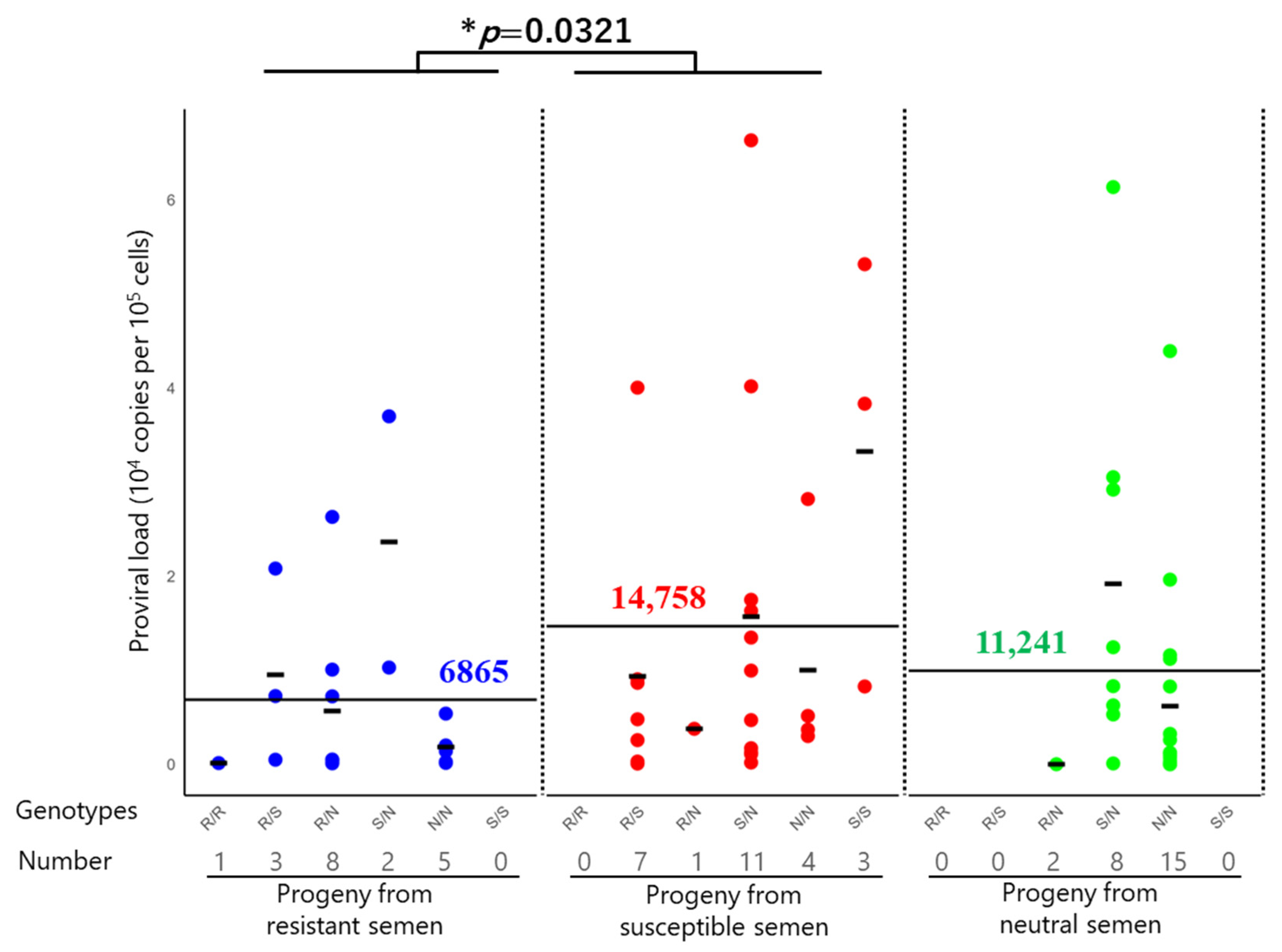
The progeny derived from semen carrying resistant, susceptible, or neutral genotypes exhibited notable differences in the BLV PVL (Table 4 and Table S1). Figure 2 summarizes the distribution of the PVL in these groups. The average PVL in the 19 BLV-positive progeny derived from the resistant semen was 6865 copies per 105 cells. Twenty-three BLV-positive progeny from neutral semen had an intermediate PVL (11,241 copies per 105 cells), whereas 26 BLV-positive progeny derived from susceptible semen had the highest average PVL of 14,758 copies per 105 cells. The order of the average PVL was as follows: progeny from susceptible semen > neutral progeny from neutral semen > progeny from resistant semen. Furthermore, to assess the statistical significance of PVL differences between groups, a two-tailed t-test following log10 transformation of PVL values revealed a significant difference between the resistant and susceptible semen groups (p = 0.032), indicating that paternal genotype may influence the PVL in the progeny (Figure 2).
The BLV-positive progeny were divided into several groups based on whether their genotypes were homozygous or heterozygous for the susceptible and resistant alleles (Figure 2 and Table 4). Further genotype-level analysis revealed that progeny carrying a homozygous genotype for resistant alleles had the lowest average PVL (124 copies per 105 cells), whereas those carrying a homozygous genotype for susceptible alleles had the highest (33,725 copies per 105 cells). Progeny carrying a heterozygous genotype for a susceptible allele with a resistant allele exhibited intermediate PVLs, with a mean value of 9515 copies per 105 cells. Although the PVL values in this group varied across individuals, they were lower than those observed for the homozygous genotype of the susceptible allele.

4. Discussion

Here, we investigated the distribution of BoLA-DRB3 polymorphisms in popular commercial semen from Japanese Black sires in Japan and the effect of paternal genotypes on PVL in their AI-derived progeny, leading to two major conclusions. First, by identifying the distribution of BoLA-DRB3 alleles in 178 widely used commercial frozen semen samples from Japanese Black sires, we identified 20 alleles and 70 genotypes of the BoLA-DRB3 gene and defined the distribution of susceptible, resistant, and neutral BoLA-DRB3 alleles for BLV PVL and their genotypes. The most frequent allele, BoLA-DRB3*016:01 (26.4%), in the semen samples was susceptible to BLV PVL, whereas the frequency of the resistant BoLA-DRB3 alleles, BoLA-DRB3*009:02 (0.6%) and BoLA-DRB3*011:01 (5.3%), was less than that of the susceptible and neutral alleles. The distribution of susceptible, resistant, and neutral alleles observed in the semen samples was consistent with that reported in previous studies based on blood samples collected from three Japanese Black cattle herds [92]. Thus, the results indicated that the distribution of resistant and susceptible alleles for BLV PVL in the blood of Japanese Black cattle was greatly affected by the frequency of BoLA-DRB3 alleles in the semen. To the best of our knowledge, this is the first study to investigate the distribution of BoLA-DRB3 polymorphisms in the semen of Japanese Black sires. Second, this study provides the first direct evidence that the paternal BoLA-DRB3 genotype significantly influences BLV PVL in the progeny. This is consistent with the findings of Benitez et al. (2019), who identified breeding bulls as a potential source of BLV transmission in beef herds [16]. We collected blood from 200 progeny derived from AI using 36 of 178 semen samples and observed an association between the BoLA-DRB3 genotype in semen and PVL in the blood samples collected from the progeny derived from their semen. The average PVL was highest in the progeny from the susceptible semen, followed by neutral, and lowest in the progeny from the resistant semen. There was a significant difference in the BLV PVL between the resistant and susceptible semen groups. This study revealed significant differences in BLV PVL in the blood of progeny derived from the susceptible and resistant semen, which concurred with the results of a previous study in which PVL in the blood was ranked in the order of susceptible, neutral, and resistant Japanese Black cattle [68]. Therefore, our investigation of the polymorphism of BoLA-DRB3 in semen and progeny derived from semen demonstrated that paternal genotype may influence the viral burden in the progeny.
As presented in Table S2, the frequency of BoLA-DRB3 alleles in the semen identified in this study partially aligns with that of previous studies based on blood samples from Japanese Black cattle [92]. To statistically evaluate the observed differences, we performed a chi-square test comparing the frequency of the BoLA-DRB3*1601 allele between the blood and semen samples. The results indicated no significant difference (χ2 = 1.91, df = 1, p = 0.167), suggesting that the variation is not pronounced. The most common and susceptible allele, DRB3*016:01 (allele frequency = 26.4%), was also the most frequent in blood samples (32.2%), as previously reported [92]. The frequency difference between the two sample types was within ±20%, indicating a comparable allele distribution. The population of the most frequent resistant DRB3*011:01 (allele frequency = 5.3%) in semen was like the previously reported frequency in the blood (allele frequency = 8.1%) (Table S2). These results suggest that BoLA-DRB3 present in frozen semen is a reliable source for BoLA-DRB3 genotyping at the population level and that the distribution status of the BoLA-DRB3 allele in semen is a critical factor in the distribution of resistant and susceptible alleles for BLV PVL in Japanese Black cattle. Specific BoLA-DRB3 alleles originating from common sires have been introduced into different farms of Japanese Black cattle [86]. Therefore, our principle is supported by a previous study showing that maternal genetics were somewhat fixed, implying that sire genetics were more likely to be affected in Japanese Black cattle [93]. In contrast, the difference between the current semen data and previous blood data was that the frequency of the resistant allele BoLA-DRB3*009:02 (allele frequency = 0.6% in semen) in our study was relatively low compared to that in the blood (11.1%) in the previous study (Table S2). In addition, the incidence rate of BoLA-DRB3*009:02 in Japanese Black cattle was 6.9% in Hokkaido prefecture [94], 6.8% in Miyazaki and Ooita prefecture [94], and 1.6% in Iwate [95], Japan. This allele has been identified as resistant in both Japanese Black and Holstein cattle, where it contributes to BLV control by suppressing viral replication [94]. None of the sires were homozygous for the resistant allele BoLA-DRB3*009:02, and two sires (3.2%) were heterozygous. In addition, two studies showed that all cattle carrying BoLA-DRB3*009:02 were heterozygous. In contrast, among the 178 semen samples with the susceptible allele BoLA-DRB3*016:01, a high proportion of 8.9% and 41.6% were homozygous and heterozygous, respectively. This suggests that elite AI sires may contribute to the high frequency of susceptible cattle in the Japanese Black cattle herd, resulting in an increased risk of horizontal and vertical transmission via cattle, which reduces the PVL of BLV and the development of BLV-induced lymphoma. Such allele frequency patterns in semen likely reflect past selection pressure for production traits and AI practices that involve using a few genetically similar sires. This distribution underscores the historical selection focus on production traits over disease resistance; therefore, future breeding strategies should incorporate disease susceptibility to prevent the spread of BLV infection. Notably, breeding programs may introduce selection bias in semen allele distributions, potentially influencing these patterns.
This study revealed a significant association between semen genotype and PVL in the progeny. We first collected blood from 200 progeny derived from AI, using 36 of 178 semen samples carrying 70 BoLA-DRB3 genotypes: three sires with at least one resistant allele, 15 sires with one susceptible allele without a resistant allele, and 18 sires with a neutral allele without a susceptible or resistant allele. Furthermore, BoLA-DRB3 typing revealed that 56% of the progeny from three resistant semen samples carried at least one resistant allele, whereas 83% of the progeny from 15 susceptible semen samples carried at least one susceptible allele. Finally, as estimated using the BLV-CoCoMo-qPCR-2 method, the average PVL was 6865 copies per 105 cells in progeny from resistant semen, 11,241 copies per 105 cells in progeny from neutral semen, and 14,758 copies per 105 cells in progeny from susceptible semen. The average PVL in the progeny was highest in susceptible semen, followed by neutral and resistant semen. These data are consistent with previous reports that the average PVL of blood from susceptible cows is higher than resistant cows [68]. Our results indicate that disease susceptibility to BLV in semen affects BLV PVL control in the progeny. Similarly to other breeds besides the Japanese Black cattle, several results from Holstein cattle support our semen results. For example, Bai et al. (2021) [83] showed that the mean PVL of blood from 49 resistant cattle was 4216 copies per 105 cells and that of 62 susceptible cattle was 19,206 copies per 105 cells, indicating that the PVLs of blood from susceptible cattle were significantly higher than those from resistant cattle. In addition, an evaluation of whether the BoLA-DRB3 polymorphism affected PVL in milk from BLV-infected dams [69] showed the same trend: the level of BLV PVL was significantly higher in milk from susceptible dams than in milk from resistant dams. Similarly to our previous study showing that BoLA-DRB3 alleles control PVL, the current study demonstrated that progeny regulate PVL based on the BoLA-DRB3 alleles of sires, providing evidence that resistance to BLV is inherited from sires.
Further division of susceptible and resistant progeny based on homozygous or heterozygous genotypes for susceptible or resistant alleles revealed that progeny with homozygous susceptible genotypes had the highest average PVL in blood (mean: 33,725 copies per 105 cells), whereas progeny possessing homozygous resistant genotypes had the lowest average PVL in blood (mean: 124 copies per 105 cells). A previous study reported that the BLV PVL in the blood or milk of cattle with homozygous susceptible genotypes was significantly higher than that in cattle with heterozygous resistant and neutral genotypes [9,68,69]. In addition, intermediate PVL levels in the blood (mean: 9362–9515 copies per 105 cells) were observed in progeny that were heterozygous for resistant and susceptible genotypes, suggesting a dose-dependent effect and supporting the hypothesis that resistance is partially dominant [68]. These findings were statistically significant (p = 0.032) and provide strong evidence of the paternal immunogenetic influence on BLV susceptibility in the progeny. However, these results were observational, and further studies are warranted to confirm causality.
Our results are consistent with previous findings, highlighting the role of BoLA-DRB3 polymorphisms as key genetic factors affecting BLV PVL and transmission risk [96]. For example, Borjigin et al. (2021) [9] demonstrated that Holstein dams carrying resistant alleles (DRB3*009:02, *014:01:01) had significantly lower PVLs and perinatal transmission rates than those with susceptible genotypes (DRB3*015:01, *012:01). All calves carrying resistant alleles in that study remained BLV-negative, even when born to infected dams. Although the aforementioned studies focused on maternal genetic contributions, the current findings extend this understanding by demonstrating that paternal BoLA-DRB3 alleles also influence PVL in calves. The consistent dose–response trend observed in both studies (resistant/resistant < resistant/susceptible < susceptible/susceptible) further reinforced the immunogenic role of BoLA-DRB3 in controlling BLV.
Although a low frequency of resistant BoLA-DRB3 alleles may appear, this is likely to reflect a historical emphasis on economically important traits, such as growth rate, carcass quality, and reproductive efficiency in breeding programs. However, the high frequency of susceptibility-associated BoLA-DRB3 alleles may be linked to the desirable production characteristics that contribute to their persistence in the population. BoLA-DRB3 exerts positive and negative effects on dairy cattle productivity [62,97,98,99,100,101]. In addition, we previously reported that some resistant Holstein and Japanese Black cattle were less likely to develop EBL, whereas susceptible Holstein and Japanese Black cattle were more likely to develop high PVL [66,68,70] and EBL [66,67,102]. Therefore, resistant alleles should not be universally considered advantageous, and susceptible alleles should not be viewed as inherently negative. Therefore, rather than pursuing the fixation of resistant alleles, a more balanced and practical strategy is required for effective BLV control. This strategy is preferred for integrating BoLA-DRB3 genotyping into sire selection protocols, with priority given to sires carrying resistant alleles for use in AI programs. In parallel, herd-level management interventions, such as an integrated BLV eradication program, use of resistant cattle with low PVL as a biological barrier, and preferential elimination of susceptible cattle with high PVL, particularly those homozygous for susceptible alleles [82], are recommended. This aligns with the growing international demand for resistance genotyped semen, enhancing the global competitiveness of the livestock genetics industry [74]. However, the relationship between BoLA-DRB3 and reproductive performance has not yet been well studied. Therefore, to confirm the detailed effect of BoLA-DRB3 on reproductive performance, future studies on the genetic background of cows with various traits are indispensable. Besides reproductive traits, to generalize the importance of BoLA-DRB3 distribution in semen, further studies are needed to increase the number of semen and progeny samples and the variety of breeds, as well as to determine the maternal genotype.
Approximately 70% of the infected cattle remained asymptomatic. Most of the remaining infected cattle developed persistent lymphocytosis (PL), and only 1–5% developed. Thus, a notable reason why BLV causes the progression of different diseases is that EBL is a multifactorial disease caused by a complex interplay among viral, host genetic, and environmental factors. First, PVL increase or decrease may be induced by host genetic factors (except for BoLA-DRB3), such as altered expression of protein arginine-N-methyltransferase gene [103], DNA mismatch repair gene [104], and Syk enzyme [105], as well as tumor necrosis factor polymorphism [106,107]. Regarding viral factors, the relationships between PVL and a point mutation in the LTR of BLV [108] and a deletion mutation in the G4 gene of BLV [109] have been reported. These host and viral factors may cause changes in humoral and cellular immune responses, leading to changes in infectivity and PVL levels and possibly contributing to individual differences in disease susceptibility. Our result showed that there are individuals whose PVL levels cannot be explained by their BoLA-DRB3 genotype alone. For example, the two progeny with the R/R genotype had PVL levels of 0 and 124 copies/105 cells, respectively, indicating excessively low levels. However, one progeny with the R/N genotype showed a PVL exceeding 10,000 copies/105 cells, and several N/N animals also exhibited relatively high PVL values. These results suggest that host and viral factors, in addition to BoLA-DRB3 genotypes, have an impact individually or in combination. A thorough analysis of the aforementioned phenomena that occur at low external temperatures may reveal the control mechanism of PVL. In addition to viral and host factors, significant environmental factors influence BLV infection rates [35,75,110]. The environmental factors include age, herd size, herd antibody positivity rate, herd EBL incidence rate, herd PVL value, husbandry methods, housing condition, castration, summer fly infestation status, maternal infection status, and colostrum feeding from cows to calves. Furthermore, the alleles of resistance, susceptibility, and neutrality showed no correlations in progeny and infection rates (Table 2). Therefore, future studies that standardize environmental factors are warranted to clarify the influence of BoLA-DRB3 polymorphisms on infection rates in progeny. Notably, although the blood samples from progeny were collected from nine prefectures across Japan—enhancing the geographical representativeness of the data—the maternal genotype was unavailable and could not be analyzed, which may contribute to PVL variation. Moreover, this study provides observational evidence of an association between sire genotype and progeny PVL; however, causality cannot be definitively established without further controlled studies.
Our study highlights that the susceptibility of the BoLA-DRB3 gene to BLV PVL is inherited, as paternal genetics influences calf PVL. Furthermore, our previous study showed that the presence of susceptible or resistant alleles in cattle determines their susceptibility to BLV, regardless of whether the allele is inherited from the sire or dam [66,68,70]. However, the present study did not consider the effects of PVL on the dam. Therefore, further studies are required to investigate the influence of BoLA-DRB3 alleles in dams and sires on BLV disease susceptibility in their calves. Although increasing BLV resistance in sire herds is a long-term goal, it can be achieved by combining genotypic screening with herd management practices. These findings provide a scientific foundation for sustainable integration of genetics into national BLV control strategies. In addition, we found that BoLA-DRB3*016:01, an allele associated with susceptibility to BLV, was most frequently observed in semen samples collected from Japanese Black cattle. This finding suggests that Japanese sires have not been selected based on resistance to BLV but rather on other significant economic traits, such as meat texture. To develop a BLV-resistant cattle herd, it is essential to promote the introduction of BoLA-DRB3 alleles associated with BLV resistance while concurrently selecting for other desirable economic traits.

Supplementary Materials

The following supporting information can be downloaded at https://www.mdpi.com/article/10.3390/pathogens14090837/s1. Table S1: BoLA-DRB3 alleles and proviral load (PVL) in 200 progeny samples derived from semen; Table S2: Comparison of BoLA-DRB3 allele frequency between blood and semen samples from the Japanese Black breed.

Author Contributions

Conceptualization, Y.A.; methodology, Y.A., A.B., S.W., and R.M.; validation, A.B., S.W., R.M., J.W., Y.M., and Y.A.; formal analysis, A.B.; investigation, A.B., S.W., R.M., H.S., A.N., R.K., and Y.A.; resources, Y.A. and R.K.; data curation, Y.A. and A.B.; writing—original draft preparation, A.B. and Y.A.; writing—review and editing, Y.A. and A.B.; supervision, Y.A.; project administration, Y.A.; funding acquisition, Y.A. and J.W. All authors have read and agreed to the published version of the manuscript.

Funding

This study was supported by a grant from the Livestock Promotional Subsidy from the Japan Racing Association (JRA), and by Shandong Provincial Base and Talent Program (Project No.: WSR2023086).

Institutional Review Board Statement

All the animals were handled following the regulations of the Animal Ethics Committee of the University of Tokyo. The study was reviewed and approved by the University of Tokyo’s Animal Experiments Committee (Approval Number p22–2–030; Approval date 22 May 2022). Informed consent was obtained from all the subjects involved in the study.

Informed Consent Statement

Not applicable.

Data Availability Statement

The original contributions presented in the study are included in the article; further inquiries can be directed to the corresponding author.

Acknowledgments

The authors thank all the farmers, veterinarians, and veterinary officers of the prefectural Livestock Hygiene Service Centers for their help with the blood sampling and the collection of epidemiological data. We thank all the members of the Laboratory of Global Infectious Diseases Control Science, Graduate School of Agricultural and Life Sciences, the University of Tokyo, for their technical assistance, help, and suggestions. We would like to thank Kaltech Co., Ltd. (https://kaltec.co.jp/en/, accessed on 20 December 2023) for helping with the organization of our laboratory.

Conflicts of Interest

Authors Hiroyuki Shimizu, Ayuha Niwano and Ryusaku Kawata were employed by Kawata Animal Clinic. The remaining authors declare that the research was conducted in the absence of any commercial or financial relationships that could be construed as a potential conflict of interest.

References

  1. Aida, Y.; Murakami, H.; Takahashi, M.; Takeshima, S.N. Mechanisms of pathogenesis induced by bovine leukemia virus as a model for human T-cell leukemia virus. Front. Microbiol. 2013, 4, 328. [Google Scholar] [CrossRef]
  2. Bech-Nielsen, S.; Piper, C.E.; Ferrer, J.F. Natural mode of transmission of the bovine leukemia virus: Role of bloodsucking insects. Am. J. Vet. Res. 1978, 39, 1089–1092. [Google Scholar] [CrossRef] [PubMed]
  3. Panei, C.J.; Larsen, A.E.; Fuentealba, N.A.; Metz, G.E.; Echeverría, M.G.; Galosi, C.M.; Valera, A.R. Study of horn flies as vectors of bovine leukemia virus. Open Vet. J. 2019, 9, 33–37. [Google Scholar] [CrossRef] [PubMed]
  4. DiGiacomo, R.F.; Hopkins, S.G.; Darlington, R.L.; Evermann, J.F. Control of bovine leukosis virus in a dairy herd by a change in dehorning. Can. J. Vet. Res. 1987, 51, 542–544. [Google Scholar] [PubMed]
  5. Kohara, J.; Konnai, S.; Onuma, M. Experimental transmission of Bovine leukemia virus in cattle via rectal palpation. Jpn. J. Vet. Res. 2006, 54, 25–30. [Google Scholar]
  6. Ferrer, J.F.; Piper, C.E. Role of colostrum and milk in the natural transmission of the bovine leukemia virus. Cancer Res. 1981, 41, 4906–4909. [Google Scholar]
  7. Meas, S.; Usui, T.; Ohashi, K.; Sugimoto, C.; Onuma, M. Vertical transmission of bovine leukemia virus and bovine immunodeficiency virus in dairy cattle herds. Vet. Microbiol. 2002, 84, 275–282. [Google Scholar] [CrossRef]
  8. Watanuki, S.; Takeshima, S.N.; Borjigin, L.; Sato, H.; Bai, L.; Murakami, H.; Sato, R.; Ishizaki, H.; Matsumoto, Y.; Aida, Y. Visualizing bovine leukemia virus (BLV)-infected cells and measuring BLV proviral loads in the milk of BLV seropositive dams. Vet. Res. 2019, 50, 102. [Google Scholar] [CrossRef]
  9. Borjigin, L.; Lo, C.-W.; Bai, L.; Hamada, R.; Sato, H.; Yoneyama, S.; Yasui, A.; Yasuda, S.; Yamanaka, R.; Mimura, M.; et al. Risk Assessment of Bovine Major Histocompatibility Complex Class II DRB3 Alleles for Perinatal Transmission of Bovine Leukemia Virus. Pathogens 2021, 10, 502. [Google Scholar] [CrossRef]
  10. Straub, O.C.; Weiland, F.; Frenzel, B. Results of hematological and serological research on natural and artificial transmission of bovine and leukosis. Dtsch. Tierarztl. Wochenschr. 1974, 81, 581–583. [Google Scholar]
  11. Kuckleburg, C.J.; Chase, C.C.; Nelson, E.A.; Marras, S.A.; Dammen, M.A.; Christopher-Hennings, J. Detection of bovine leukemia virus in blood and milk by nested and real-time polymerase chain reactions. J. Vet. Diagn. Investig. 2003, 15, 72–76. [Google Scholar] [CrossRef] [PubMed]
  12. Jaworski, J.P.; Porta, N.G.; Gutierrez, G.; Politzki, R.P.; Alvarez, I.; Galarza, R.; Abdala, A.; Calvinho, L.; Trono, K.G. Relationship between the level of bovine leukemia virus antibody and provirus in blood and milk of cows from a naturally infected herd. J. Dairy Sci. 2016, 99, 5629–5634. [Google Scholar] [CrossRef] [PubMed]
  13. Gutierrez, G.; Lomonaco, M.; Alvarez, I.; Fernandez, F.; Trono, K. Characterization of colostrum from dams of BLV endemic dairy herds. Vet. Microbiol. 2015, 177, 366–369. [Google Scholar] [CrossRef]
  14. Asadpour, R.; Jafari, R. Detection of bovine leukosis provirus in blood and semen samples of bulls. Comp. Clin. Pathol. 2010, 21, 187–191. [Google Scholar] [CrossRef]
  15. Dus Santos, M.J.; Trono, K.; Lager, I.; Wigdorovitz, A. Development of a PCR to diagnose BLV genome in frozen semen samples. Vet. Microbiol. 2007, 119, 10–18. [Google Scholar] [CrossRef]
  16. Benitez, O.J.; Roberts, J.N.; Norby, B.; Bartlett, P.C.; Takeshima, S.N.; Watanuki, S.; Aida, Y.; Grooms, D.L. Breeding bulls as a potential source of bovine leukemia virus transmission in beef herds. J. Am. Vet. Med. Assoc. 2019, 254, 1335–1340. [Google Scholar] [CrossRef]
  17. Kaja, R.W.; Olson, C. Non-infectivity of semen from bulls infected with bovine leukosis virus. Theriogenology 1982, 18, 107–112. [Google Scholar] [CrossRef]
  18. Miller, J.M.; Van der Maaten, M.J. Infectivity tests of secretions and excretions from cattle infected with bovine leukemia virus. J. Natl. Cancer Inst. 1979, 62, 425–428. [Google Scholar]
  19. Monke, D.R. Noninfectivity of semen from bulls infected with bovine leukosis virus. J. Am. Vet. Med. Assoc. 1986, 188, 823–826. [Google Scholar] [CrossRef]
  20. Bao, A.; Watanuki, S.; Matsuura, R.; Matsumoto, Y.; Shimizu, H.; Kawata, R.; Aida, Y. No evidence of bovine leukemia virus proviral DNA in a widely used commercially frozen semen in Japan. J. Vet. Med. Sci. 2025, 87, 821–825. [Google Scholar] [CrossRef] [PubMed]
  21. Acaite, J.; Tamosiunas, V.; Lukauskas, K.; Milius, J.; Pieskus, J. The eradication experience of enzootic bovine leukosis from Lithuania. Prev. Vet. Med. 2007, 82, 83–89. [Google Scholar] [CrossRef] [PubMed]
  22. Nuotio, L.; Rusanen, H.; Sihvonen, L.; Neuvonen, E. Eradication of enzootic bovine leukosis from Finland. Prev. Vet. Med. 2003, 59, 43–49. [Google Scholar] [CrossRef] [PubMed]
  23. Kettmann, R.; Burny, A.; Callebaut, I.; Droogmans, L.; Mammerickx, M.; Willems, L. Bovine leukemia virus. Retroviridae 1994, 3, 39–81. [Google Scholar]
  24. Pluta, A.; Jaworski, J.P.; Droscha, C.; VanderWeele, S.; Taxis, T.M.; Valas, S.; Brnić, D.; Jungić, A.; Ruano, M.J.; Sánchez, A.; et al. Inter-laboratory comparison of eleven quantitative or digital PCR assays for detection of proviral bovine leukemia virus in blood samples. BMC Vet. Res. 2024, 20, 381. [Google Scholar] [CrossRef]
  25. Kuczewski, A.; Orsel, K.; Barkema, H.W.; Kelton, D.F.; Hutchins, W.A.; van der Meer, F.J.U. Short communication: Evaluation of 5 different ELISA for the detection of bovine leukemia virus antibodies. J. Dairy Sci. 2018, 101, 2433–2437. [Google Scholar] [CrossRef]
  26. Lv, G.; Wang, J.; Lian, S.; Wang, H.; Wu, R. The global epidemiology of bovine leukemia virus: Current trends and future implications. Animals 2024, 14, 297. [Google Scholar] [CrossRef]
  27. Takeshima, S.N.; Kitamura-Muramatsu, Y.; Yuan, Y.; Polat, M.; Saito, S.; Aida, Y. BLV-CoCoMo-qPCR-2: Improvements to the BLV-CoCoMo-qPCR assay for bovine leukemia virus by reducing primer degeneracy and constructing an optimal standard curve. Arch. Virol. 2015, 160, 1325–1332. [Google Scholar] [CrossRef]
  28. Jaworski, J.P.; Pluta, A.; Rola-Łuszczak, M.; McGowan, S.L.; Finnegan, C.; Heenemann, K.; Carignano, H.A.; Alvarez, I.; Murakami, K.; Willems, L.; et al. Interlaboratory Comparison of Six Real-Time PCR Assays for Detection of Bovine Leukemia Virus Proviral DNA. J. Clin. Microbiol. 2018, 56, e00304-18. [Google Scholar] [CrossRef] [PubMed]
  29. Ruggiero, V.J.; Bartlett, P.C. Control of Bovine Leukemia Virus in Three US Dairy Herds by Culling ELISA-Positive Cows. Vet. Med. Int. 2019, 2019, 3202184. [Google Scholar] [CrossRef]
  30. LaDronka, R.M.; Ainsworth, S.; Wilkins, M.J.; Norby, B.; Byrem, T.M.; Bartlett, P.C. Prevalence of bovine leukemia virus antibodies in US dairy cattle. Vet. Med. Int. 2018, 2018, 5831278. [Google Scholar] [CrossRef] [PubMed]
  31. Bongers, R.; Rochus, C.M.; Houlahan, K.; Lynch, C.; Oliveira, G.A., Jr.; Rojas de Oliveira, H.; van Staaveren, N.; Kelton, D.F.; Miglior, F.; Schenkel, F.S.; et al. Estimation of genetic parameters and genome-wide association study for enzootic bovine leukosis resistance in Canadian Holstein cattle. J. Dairy Sci. 2025, 108, 611–622. [Google Scholar] [CrossRef]
  32. Heinecke, N.; Tórtora, J.; Martínez, H.A.; Gonzalez-Fernandez, V.D.; Ramírez, H. Detection and genotyping of bovine leukemia virus in Mexican cattle. Arch. Virol. 2017, 162, 3191–3196. [Google Scholar] [CrossRef]
  33. Ma, B.; Gong, Q.; Sheng, C.; Liu, Y.; Ge, G.; Li, D.; Diao, N.; Shi, K.; Li, J.; Sun, Z.; et al. Prevalence of bovine leukemia in 1983–2019 in China: A systematic review and meta-analysis. Microb. Pathog. 2021, 150, 104681. [Google Scholar] [CrossRef] [PubMed]
  34. Cho, K.O.; Meas, S.; Park, N.Y.; Kim, Y.H.; Lim, Y.K.; Endoh, D.; Lee, S.I.; Pjasjo, K.; Sugimoto, C.; Onuma, M. Seroprevalence of bovine immunodeficiency virus in dairy and beef cattle herds in Korea. J. Vet. Med. Sci. 1999, 61, 549–551. [Google Scholar] [CrossRef] [PubMed]
  35. Murakami, K.; Kobayashi, S.; Konishi, M.; Kameyama, K.; Tsutsui, T. Nationwide survey of bovine leukemia virus infection among dairy and beef breeding cattle in Japan from 2009–2011. J. Vet. Med. Sci. 2013, 75, 1123–1126. [Google Scholar] [CrossRef] [PubMed]
  36. Lee, E.; Kim, E.J.; Ratthanophart, J.; Vitoonpong, R.; Kim, B.H.; Cho, I.S.; Song, J.Y.; Lee, K.K.; Shin, Y.K. Molecular epidemiological and serological studies of bovine leukemia virus (BLV) infection in Thailand cattle. Infect. Genet. Evol. 2016, 41, 245–254. [Google Scholar] [CrossRef] [PubMed]
  37. Polat, M.; Ohno, A.; Takeshima, S.N.; Kim, J.; Kikuya, M.; Matsumoto, Y.; Mingala, C.N.; Onuma, M.; Aida, Y. Detection and molecular characterization of bovine leukemia virus in Philippine cattle. Arch. Virol. 2015, 160, 285–296. [Google Scholar] [CrossRef]
  38. Moe, K.K.; Polat, M.; Borjigin, L.; Matsuura, R.; Hein, S.T.; Moe, H.H.; Aida, Y. New evidence of bovine leukemia virus circulating in Myanmar cattle through epidemiological and molecular characterization. PLoS ONE 2020, 15, e0229126. [Google Scholar] [CrossRef]
  39. Ochirkhuu, N.; Konnai, S.; Odbileg, R.; Nishimori, A.; Okagawa, T.; Murata, S.; Ohashi, K. Detection of bovine leukemia virus and identification of its genotype in Mongolian cattle. Arch. Virol. 2016, 161, 985–991. [Google Scholar] [CrossRef] [PubMed]
  40. Hamada, R.; Fereig, R.M.; Metwally, S. The Influence of Risk Factors on Bovine Leukemia Virus Infection and Proviral Load in Egyptian Cattle. Vet. Res. Commun. 2024, 48, 191–202. [Google Scholar] [CrossRef]
  41. Gillet, N.; Florins, A.; Boxus, M.; Burteau, C.; Nigro, A.; Vandermeers, F.; Balon, H.; Bouzar, A.B.; Defoiche, J.; Burny, A. Mechanisms of leukemogenesis induced by bovine leukemia virus: Prospects for novel anti-retroviral therapies in human. Retrovirology 2007, 4, 18. [Google Scholar] [CrossRef]
  42. Ott, S.; Johnson, R.; Wells, S.J. Association between bovine-leukosis virus seroprevalence and herd-level productivity on US dairy farms. Prev. Vet. Med. 2003, 61, 249–262. [Google Scholar] [CrossRef]
  43. Erskine, R.J.; Bartlett, P.C.; Byrem, T.M.; Render, C.L.; Febvay, C.; Houseman, J.T. Association between bovine leukemia virus, production, and population age in Michigan dairy herds. J. Dairy Sci. 2012, 95, 727–734. [Google Scholar] [CrossRef]
  44. Nekouei, O.; VanLeeuwen, J.; Stryhn, H.; Kelton, D.; Keefe, G. Lifetime effects of infection with bovine leukemia virus on longevity and milk production of dairy cows. Prev. Vet. Med. 2016, 133, 1–9. [Google Scholar] [CrossRef]
  45. Rhodes, J.K.; Pelzer, K.D.; Johnson, Y.J. Economic implications of bovine leukemia virus infection in mid-Atlantic dairy herds. J. Am. Vet. Med. Assoc. 2003, 223, 346–352. [Google Scholar] [CrossRef]
  46. White, T.L.; Moore, D.A. Reasons for whole carcass condemnations of cattle in the United States and implications for producer education and veterinary intervention. J. Am. Vet. Med. Assoc. 2009, 235, 937–941. [Google Scholar] [CrossRef]
  47. Bartlett, P.C.; Sordillo, L.M.; Byrem, T.M.; Norby, B.; Grooms, D.L.; Swenson, C.L.; Zalucha, J.; Erskine, R.J. Options for the control of bovine leukemia virus in dairy cattle. J. Am. Vet. Med. Assoc. 2014, 244, 914–922. [Google Scholar] [CrossRef]
  48. Norby, B.; Bartlett, P.C.; Byrem, T.M.; Erskine, R.J. Effect of infection with bovine leukemia virus on milk production in Michigan dairy cows. J. Dairy Sci. 2016, 99, 2043–2052. [Google Scholar] [CrossRef] [PubMed]
  49. Benitez, O.J.; LaDronka, R.M.; Norby, B.; Grooms, D.L.; Bartlett, P.C. The effect of bovine leukemia virus on dairy cow longevity. JDS Commun. 2022, 3, 185–188. [Google Scholar] [CrossRef] [PubMed]
  50. Frie, M.C.; Coussens, P.M. Bovine leukemia virus: A major silent threat to proper immune responses in cattle. Vet. Immunol. Immunopathol. 2015, 163, 103–114. [Google Scholar] [CrossRef] [PubMed]
  51. Konnai, S.; Murata, S.; Ohashi, K. Immune exhaustion during chronic infections in cattle. J. Vet. Med. Sci. 2017, 79, 1–5. [Google Scholar] [CrossRef] [PubMed]
  52. USDA. Bovine Leukosis Virus (BLV) on U.S. Dairy Operations. 2007. Available online: https://www.aphis.usda.gov/animal_health/nahms/dairy/downloads/dairy07/Dairy07_is_BLV.pdf (accessed on 14 June 2025).
  53. Burny, A.; Bex, F.; Chantrenne, H.; Cleuter, Y.; Dekegel, D.; Ghysdael, J.; Kettmann, R.; Leclercq, M.; Leunen, J.; Mammerickx, M.; et al. Bovine leukemia virus involvement in enzootic bovine leukosis. Adv. Cancer Res. 1978, 28, 251–311. [Google Scholar] [CrossRef]
  54. Marawan, M.A.; Alouffi, A.; El Tokhy, S.; Badawy, S.; Shirani, I.; Dawood, A.; Guo, A.; Almutairi, M.M.; Alshammari, F.A.; Selim, A. Bovine leukaemia virus: Current epidemiological circumstance and future prospective. Viruses 2021, 13, 2167. [Google Scholar] [CrossRef]
  55. Ruggiero, V.J.; Norby, B.; Benitez, O.J.; Hutchinson, H.; Sporer, K.; Droscha, C.; Swenson, C.L.; Bartlett, P.C. Controlling bovine leukemia virus in dairy herds by identifying and removing cows with the highest proviral load and lymphocyte counts. J. Dairy Sci. 2019, 102, 9165–9175. [Google Scholar] [CrossRef] [PubMed]
  56. Bartlett, P.C.; Norby, B.; Byrem, T.M.; Parmelee, A.; Ledergerber, J.T.; Erskine, R.J. Bovine leukemia virus and cow longevity in Michigan dairy herds. J. Dairy Sci. 2013, 96, 1591–1597. [Google Scholar] [CrossRef]
  57. Kuczewski, A.; Hogeveen, H.; Orsel, K.; Wolf, R.; Thompson, J.; Spackman, E.; van der Meer, F. Economic evaluation of 4 bovine leukemia virus control strategies for Alberta dairy farms. J. Dairy Sci. 2019, 102, 2578–2592. [Google Scholar] [CrossRef] [PubMed]
  58. Nakada, S.; Fujimoto, Y.; Kohara, J.; Adachi, Y.; Makita, K. Estimation of economic loss by carcass weight reduction of Japanese dairy cows due to infection with bovine leukemia virus. Prev. Vet. Med. 2022, 198, 105528. [Google Scholar] [CrossRef]
  59. Neefjes, J.; Jongsma, M.L.; Paul, P.; Bakke, O. Towards a systems understanding of MHC class I and MHC class II antigen presentation. Nat. Rev. Immunol. 2011, 11, 823–836. [Google Scholar] [CrossRef] [PubMed]
  60. Takeshima, S.; Aida, Y. Structure, function and disease susceptibility of the bovine major histocompatibility complex. Anim. Sci. J. 2006, 77, 138–150. [Google Scholar] [CrossRef]
  61. Loat, S.; Kumari, N.; Saini, S.; Dige, M.S.; Kumar, A.; Dhilor, N.; Dang, A.K.; Lathwal, S.S.; Sodhi, M.; Kataria, R.S. Allelic diversity at BoLA DRB3 locus and association with predisposition to clinical mastitis in indicus and crossbred cattle. Anim. Biotechnol. 2021, 34, 1030–1039. [Google Scholar] [CrossRef]
  62. Yoshida, T.; Furuta, H.; Kondo, Y.; Mukoyama, H. Association of BoLA-DRB3 alleles with mastitis resistance and susceptibility in Japanese Holstein cows. Anim. Sci. J. 2012, 83, 359–366. [Google Scholar] [CrossRef] [PubMed]
  63. Duangjinda, M.; Jindatajak, Y.; Tipvong, W.; Sriwarothai, J.; Pattarajinda, V.; Katawatin, S.; Boonkum, W. Association of BoLA-DRB3 alleles with tick-borne disease tolerance in dairy cattle in a tropical environment. Vet. Parasitol. 2013, 196, 314–320. [Google Scholar] [CrossRef] [PubMed]
  64. Othman, O.E.; Khodary, M.G.; El-Deeb, A.H.; Hussein, H.A. Five BoLA-DRB3 genotypes detected in Egyptian buffalo infected with Foot and Mouth disease virus serotype O. J. Genet. Eng. Biotechnol. 2018, 16, 513–518. [Google Scholar] [CrossRef]
  65. Morales, J.P.A.; López-Herrera, A.; Zuluaga, J.E. Association of BoLA DRB3 gene polymorphisms with BoHV-1 infection and zootechnical traits. Open Vet. J. 2020, 10, 331–339. [Google Scholar] [CrossRef] [PubMed]
  66. Lo, C.W.; Borjigin, L.; Saito, S.; Fukunaga, K.; Saitou, E.; Okazaki, K.; Mizutani, T.; Wada, S.; Takeshima, S.N.; Aida, Y. BoLA-DRB3 Polymorphism is Associated with Differential Susceptibility to Bovine Leukemia Virus-Induced Lymphoma and Proviral Load. Viruses 2020, 12, 352. [Google Scholar] [CrossRef]
  67. Lo, C.W.; Takeshima, S.N.; Okada, K.; Saitou, E.; Fujita, T.; Matsumoto, Y.; Wada, S.; Inoko, H.; Aida, Y. Association of Bovine Leukemia Virus-Induced Lymphoma with BoLA-DRB3 polymorphisms at DNA, amino acid, and binding pocket property levels. Pathogens 2021, 10, 437. [Google Scholar] [CrossRef]
  68. Miyasaka, T.; Takeshima, S.N.; Jimba, M.; Matsumoto, Y.; Kobayashi, N.; Matsuhashi, T.; Sentsui, H.; Aida, Y. Identification of bovine leukocyte antigen class II haplotypes associated with variations in bovine leukemia virus proviral load in Japanese Black cattle. Tissue Antigens 2013, 81, 72–82. [Google Scholar] [CrossRef] [PubMed]
  69. Nakatsuchi, A.; Watanuki, S.; Borjigin, L.; Sato, H.; Bai, L.; Matsuura, R.; Kuroda, M.; Murakami, H.; Sato, R.; Asaji, S.; et al. BoLA-DRB3 Polymorphism Controls Proviral Load and Infectivity of Bovine Leukemia Virus (BLV) in Milk. Pathogens 2022, 11, 210. [Google Scholar] [CrossRef]
  70. Takeshima, S.N.; Ohno, A.; Aida, Y. Bovine leukemia virus proviral load is more strongly associated with bovine major histocompatibility complex class II DRB3 polymorphism than with DQA1 polymorphism in Holstein cow in Japan. Retrovirology 2019, 16, 14. [Google Scholar] [CrossRef]
  71. Maezawa, M.; Fujii, Y.; Akagami, M.; Kawakami, J.; Inokuma, H. BoLA-DRB3*15:01 allele is associated with susceptibility to early enzootic bovine leukosis onset in Holstein-Friesian and Japanese Black cattle. Vet. Microbiol. 2023, 284, 109829. [Google Scholar] [CrossRef]
  72. Nikbakht Brujeni, G.; Ghorbanpour, R.; Esmailnejad, A. Association of BoLA-DRB3.2 Alleles with BLV Infection Profiles (Persistent Lymphocytosis/Lymphosarcoma) and Lymphocyte Subsets in Iranian Holstein Cattle. Biochem. Genet. 2016, 54, 194–207. [Google Scholar] [CrossRef] [PubMed]
  73. Somura, Y.; Sugiyama, E.; Fujikawa, H.; Murakami, K. Comparison of the copy numbers of bovine leukemia virus in the lymph nodes of cattle with enzootic bovine leukosis and cattle with latent infection. Arch. Virol. 2014, 159, 2693–2697. [Google Scholar] [CrossRef]
  74. Juliarena, M.A.; Barrios, C.N.; Ceriani, M.C.; Esteban, E.N. Hot topic: Bovine leukemia virus (BLV)-infected cows with low proviral load are not a source of infection for BLV-free cattle. J. Dairy Sci. 2016, 99, 4586–4589. [Google Scholar] [CrossRef]
  75. Kobayashi, T.; Inagaki, Y.; Ohnuki, N.; Sato, R.; Murakami, S.; Imakawa, K. Increasing Bovine leukemia virus (BLV) proviral load is a risk factor for progression of Enzootic bovine leucosis: A prospective study in Japan. Prev. Vet. Med. 2019, 178, 104680. [Google Scholar] [CrossRef]
  76. Ohno, A.; Takeshima, S.N.; Matsumoto, Y.; Aida, Y. Risk factors associated with increased bovine leukemia virus proviral load in infected cattle in Japan from 2012 to 2014. Virus Res. 2015, 210, 283–290. [Google Scholar] [CrossRef]
  77. Kohara, J.; Bai, L.; Takeshima, S.N.; Matsumoto, Y.; Hirai, T.; Aida, Y. Correlation between the Biodistribution of Bovine Leukemia Virus in the Organs and the Proviral Load in the Peripheral Blood during Early Stages of Experimentally Infected Cattle. Pathogens 2023, 12, 130. [Google Scholar] [CrossRef]
  78. Yuan, Y.; Kitamura-Muramatsu, Y.; Saito, S.; Ishizaki, H.; Nakano, M.; Haga, S.; Matoba, K.; Ohno, A.; Murakami, H.; Takeshima, S.N.; et al. Detection of the BLV provirus from nasal secretion and saliva samples using BLV-CoCoMo-qPCR-2: Comparison with blood samples from the same cattle. Virus Res. 2015, 210, 248–254. [Google Scholar] [CrossRef] [PubMed]
  79. Mekata, H.; Sekiguchi, S.; Konnai, S.; Kirino, Y.; Horii, Y.; Norimine, J. Horizontal transmission and phylogenetic analysis of bovine leukemia virus in two districts of Miyazaki, Japan. J. Vet. Med. Sci. 2015, 77, 1115–1120. [Google Scholar] [CrossRef]
  80. Juliarena, M.A.; Poli, M.; Sala, L.; Ceriani, C.; Gutierrez, S.; Dolcini, G.; Rodriguez, E.M.; Marino, B.; Rodriguez-Dubra, C.; Esteban, E.N. Association of BLV infection profiles with alleles of the BoLA-DRB3.2 gene. Anim. Genet. 2008, 39, 432–438. [Google Scholar] [CrossRef] [PubMed]
  81. Daous, H.E.; Mitoma, S.; Elhanafy, E.; Thi Nguyen, H.; Thi Mai, N.; Notsu, K.; Kaneko, C.; Norimine, J.; Sekiguchi, S. Relationship between Allelic Heterozygosity in BoLA-DRB3 and Proviral Loads in Bovine Leukemia Virus-Infected Cattle. Animals 2021, 11, 647. [Google Scholar] [CrossRef] [PubMed]
  82. Borjigin, L.; Watanuki, S.; Hamada, R.; Bai, L.; Hirose, T.; Sato, H.; Yoneyama, S.; Yasui, A.; Yasuda, S.; Yamanaka, R.; et al. Effectiveness of integrated bovine leukemia virus eradication strategies utilizing cattle carrying resistant and susceptible histocompatibility complex class II DRB3 alleles. J. Dairy Sci. 2023, 106, 9393–9409. [Google Scholar] [CrossRef]
  83. Bai, L.; Borjigin, L.; Sato, H.; Takeshima, S.; Asaji, S.; Ishizaki, H.; Kawashima, K.; Obuchi, Y.; Sunaga, S.; Ando, A.; et al. Kinetic Study of BLV Infectivity in BLV Susceptible and Resistant Cattle in Japan from 2017 to 2019. Pathogens 2021, 10, 1281. [Google Scholar] [CrossRef]
  84. Lo, C.W.; Aida, Y. Association of BoLA-DRB3 with bovine leukemia virus. Major Histocompat. Complex 2022, 29, 158–167. [Google Scholar] [CrossRef]
  85. Oikawa, T.; Hoque, M.A.; Hitomi, T.; Uchida, H.; Namikawa, K. Genetic parameters for traits in performance and progeny tests and their genetic relationships in Japanese black cattle. Asian-Australas. J. Anim. Sci. 2006, 19, 611–615. [Google Scholar] [CrossRef]
  86. John, S.W.; Weitzner, G.; Rozen, R.; Scriver, C.R. A rapid procedure for extracting genomic DNA from leukocytes. Nucleic Acids Res. 1991, 19, 408. [Google Scholar] [CrossRef]
  87. Jimba, M.; Takeshima, S.N.; Murakami, H.; Kohara, J.; Kobayashi, N.; Matsuhashi, T.; Ohmori, T.; Nunoya, T.; Aida, Y. BLV-CoCoMo-qPCR: A useful tool for evaluating bovine leukemia virus infection status. BMC Vet. Res. 2012, 8, 167. [Google Scholar] [CrossRef]
  88. Rola-Łuszczak, M.; Finnegan, C.; Olech, M.; Choudhury, B.; Kuźmak, J. Development of an improved real-time PCR for the detection of bovine leukaemia provirus nucleic acid and its use in the clarification of inconclusive serological test results. J. Virol. Methods 2013, 189, 258–264. [Google Scholar] [CrossRef] [PubMed]
  89. Watanuki, S.; Bao, A.; Saitou, E.; Shoji, K.; Izawa, M.; Okami, M.; Matsumoto, Y.; Aida, Y. BLV-CoCoMo dual qPCR assay targeting LTR region for quantifying bovine leukemia virus: Comparison with multiplex real-time qPCR assay targeting pol region. Pathogens 2024, 13, 1111. [Google Scholar] [CrossRef] [PubMed]
  90. Takeshima, S.; Matsumoto, Y.; Miyasaka, T.; Saito, H.; Onuma, M.; Aida, Y. A new method for typing bovine major histocompatibility complex class II DRB3 alleles by combining two established PCR sequence-based techniques. Tissue Antigens 2011, 78, 208–213. [Google Scholar] [CrossRef] [PubMed]
  91. Lewin, H.A.; Schmitt, K.; Hubert, R.; van Eijk, M.J.T.; Arnheim, N. Close linkage between bovine prolactin and BoLA-DRB3 genes: Genetic mapping in cattle by single sperm typing. Genomics 1992, 13, 44–48. [Google Scholar] [CrossRef]
  92. Miyasaka, T.; Takeshima, S.N.; Matsumoto, Y.; Kobayashi, N.; Matsuhashi, T.; Miyazaki, Y.; Tanabe, Y.; Ishibashi, K.; Sentsui, H.; Aida, Y. The diversity of bovine MHC class II DRB3 and DQA1 alleles in different herds of Japanese Black and Holstein cattle in Japan. Gene 2011, 472, 42–49. [Google Scholar] [CrossRef]
  93. Fujimori, S.; Ando, T.; Sekiguchi, S.; Notsu, K.; Ishida, S.; Daidoji, T.; Hagiwara, K. Investigating BoLA Class II DRB3*009:02 carrying cattle in Japan. Vet. Anim. Sci. 2025, 27, 100425. [Google Scholar] [CrossRef]
  94. Hayashi, T.; Mekata, H.; Sekiguchi, S.; Kirino, Y.; Mitoma, S.; Honkawa, K.; Horii, Y.; Norimine, J. Cattle with the BoLA class II DRB3* 0902 allele have significantly lower bovine leukemia proviral loads. J. Vet. Med. Sci. 2017, 79, 1552–1555. [Google Scholar] [CrossRef] [PubMed]
  95. Fukunari, K.; Chiba, Y. Status of Bovine Leukemia Virus-Resistant Gene Carriers among Cattle in Iwate Prefecture. Iwate Vet. 2022, 48, 70–74. [Google Scholar]
  96. Victoria, M.; Farias, N.; Caffaro, M.E.; Lendez, P.A.; Passucci, J.; Poli, M.; Ceriani, M.C.; Dolcini, G. A novel association of BoLA DRB3 alleles in BLV infected cattle with different proviral loads. Braz. J. Vet. Res. Anim. Sci. 2017, 54, 215–224. [Google Scholar]
  97. Rupp, R.; Hernandez, A.; Mallard, B.A. Association of bovine leukocyte antigen (BoLA) DRB3.2 with immune response, mastitis, and production and type traits in Canadian Holsteins. J. Dairy Sci. 2007, 90, 1029–1038. [Google Scholar] [CrossRef] [PubMed]
  98. Pashmi, M.; Qanbari, S.; Ghorashi, S.A.; Sharifi, A.R.; Simianer, H. Analysis of relationship between bovine lymphocyte antigen DRB3.2 alleles, somatic cell count and milk traits in Iranian Holstein population. J. Anim. Breed Genet. 2009, 126, 296–303. [Google Scholar] [CrossRef] [PubMed]
  99. Sharif, S.; Mallard, B.A.; Wilkie, B.N.; Sargeant, J.M.; Scott, H.M.; Dekkers, J.C.; Leslie, K.E. Associations of the bovine major histocompatibility complex DRB3 (BoLA-DRB3) alleles with occurrence of disease and milk somatic cell score in Canadian dairy cattle. Anim. Genet. 1998, 29, 185–193. [Google Scholar] [CrossRef]
  100. Wu, X.X.; Yang, Z.P.; Wang, X.L.; Mao, Y.J.; Li, S.C.; Shi, X.K.; Chen, Y. Restriction fragment length polymorphism in the exon 2 of the BoLA-DRB3 gene in Chinese Holstein of the south China. J. Biomed. Sci. Eng. 2010, 3, 221–225. [Google Scholar] [CrossRef]
  101. Zanotti, M.; Strillacci, M.G.; Taboni, I.; Samorè, A.B.; Longeri, M. Histocompatibility genes and somatic cell count (SCC) in Italia Holstein Friesian. Ital. J. Anim. Sci. 2003, 2, 85–87. [Google Scholar]
  102. Vernau, W.; Jacobs, R.M.; Valli, V.E.; Heeney, J.L. The immunophenotypic characterization of bovine lymphomas. Vet. Pathol. 1997, 34, 222–225. [Google Scholar] [CrossRef]
  103. Assi, W.; Hirose, T.; Wada, S.; Matsuura, R.; Takeshima, S.N.; Aida, Y. PRMT5 Is Required for Bovine Leukemia Virus Infection in Vivo and Regulates BLV Gene Expression, Syncytium Formation, and Glycosylation in Vitro. Viruses 2020, 12, 650. [Google Scholar] [CrossRef]
  104. Bai, L.; Hirose, T.; Assi, W.; Wada, S.; Takeshima, S.-N.; Aida, Y. Bovine Leukemia Virus Infection Affects Host Gene Expression Associated with DNA Mismatch Repair. Pathogens 2020, 9, 909. [Google Scholar] [CrossRef]
  105. Murakami, H.; Kuroiwa, T.; Suzuki, K.; Miura, Y.; Sentsui, H. Analysis of Syk expression in bovine lymphoma and persistent lymphocytosis induced by bovine leukemia virus. J. Vet. Med. Sci. 2011, 73, 41–45. [Google Scholar] [CrossRef] [PubMed]
  106. Konnai, S.; Usui, T.; Ikeda, M.; Kohara, J.; Hirata, T.; Okada, K.; Ohashi, K.; Onuma, M. Tumor necrosis factor-alpha genetic polymorphism may contribute to progression of bovine leukemia virus-infection. Microbes Infect. 2006, 8, 2163–2171. [Google Scholar] [CrossRef] [PubMed]
  107. Ohnuki, N.; Kobayashi, T.; Matsuo, M.; Nishikaku, K.; Kusama, K.; Torii, Y.; Inagaki, Y.; Hori, M.; Imakawa, K.; Satou, Y. A target enrichment high throughput sequencing system for characterization of BLV whole genome sequence, integration sites, clonality and host SNP. Sci. Rep. 2021, 11, 4521. [Google Scholar] [CrossRef]
  108. Murakami, H.; Todaka, H.; Uchiyama, J.; Sato, R.; Sogawa, K.; Sakaguchi, M.; Tsukamoto, K. A point mutation to the long terminal repeat of bovine leukemia virus related to viral productivity and transmissibility. Virology. 2019, 537, 45–52. [Google Scholar] [CrossRef] [PubMed]
  109. Murakami, H.; Uchiyama, J.; Nikaido, S.; Sato, R.; Sakaguchi, M.; Tsukamoto, K. Inefficient viral replication of bovine leukemia virus induced by spontaneous deletion mutation in the G4 gene. J. Gen. Virol. 2016, 97, 2753–2762. [Google Scholar] [CrossRef]
  110. Kobayashi, S.; Hidano, A.; Tsutsui, T.; Yamamoto, T.; Hayama, Y.; Nishida, T.; Muroga, N.; Konishi, M.; Kameyama, K.; Murakami, K. Analysis of risk factors associated with bovine leukemia virus seropositivity within dairy and beef breeding farms in Japan: A nationwide survey. Res. Vet. Sci. 2014, 96, 47–53. [Google Scholar] [CrossRef]
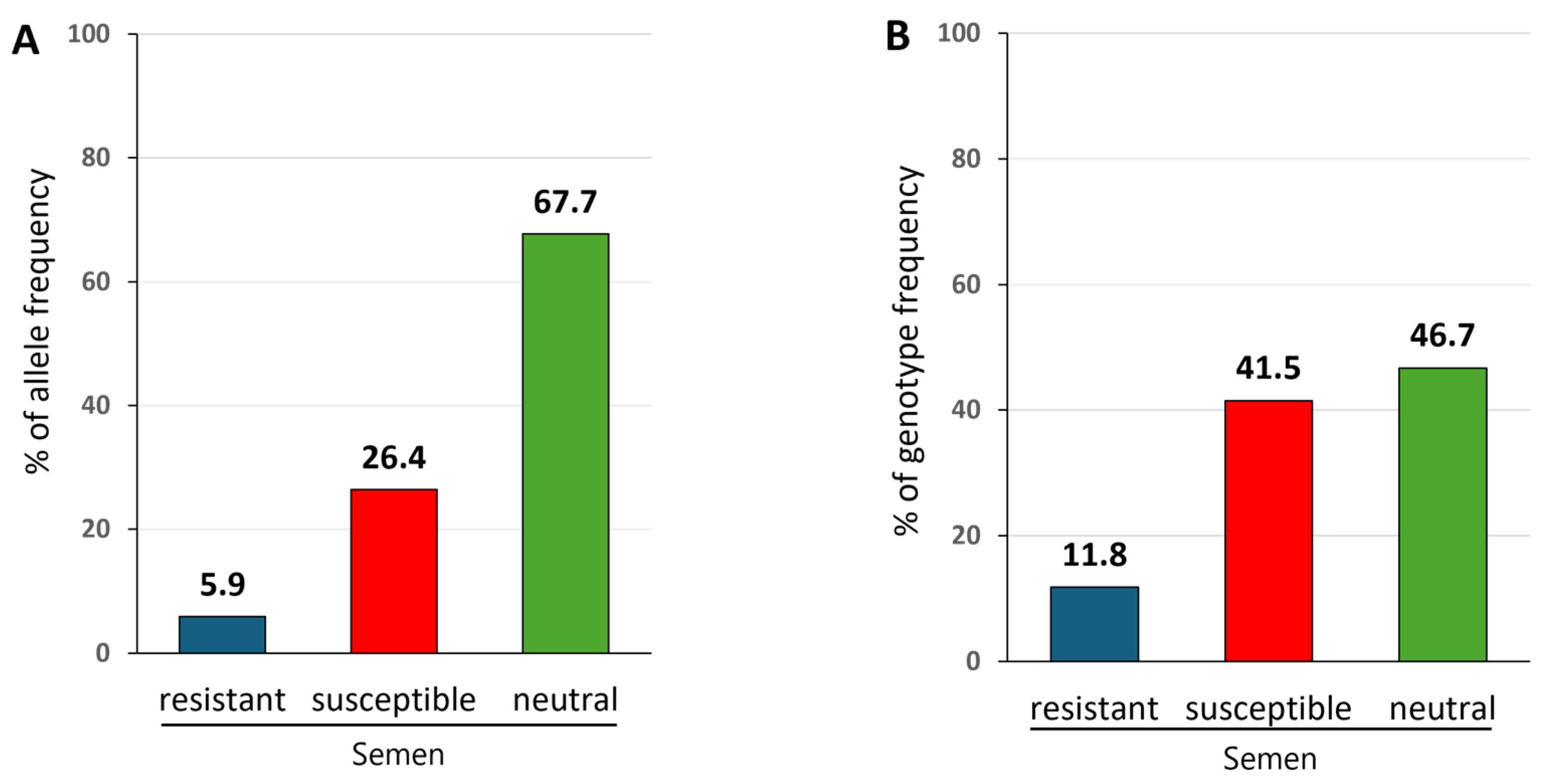
Figure 1. Allele and genotype frequencies of BoLA-DRB3 in widely used commercial frozen semen from Japanese Black cattle. Genomic DNAs were obtained from 178 widely used commercial frozen semen samples from Japanese Black sires in Japan, and BoLA-DRB3 alleles were typed using the PCR-SBT method: (A) Allele frequency. BoLA-DRB3*016:01 is a susceptibility-associated marker related to high PVL, BoLA-DRB3*009:02 and BoLA-DRB3*0:11:01 are resistance-associated markers related to low PVL, and all other alleles were neutral because they did not show a significant association with PVL. The X-axis shows allele classification, and the Y-axis shows the percentage of allele frequencies. (B) Genotype frequency. The 178 semen samples were divided into resistant, susceptible, and neutral semen groups based on the BoLA-DRB3 Genotype: Resistant semen carried at least one resistant allele, BoLA-DRB3*009:02 or *011:01, in their genome; susceptible semen carried at least one susceptible BoLA-DRB3*016:01 without a resistant allele in their genome; and neutral semen carried other alleles in their genome. The X-axis shows semen classification, and the Y-axis shows the percentage of genotype frequencies.
Pathogens 14 00837 g001
Figure 2. The average PVL values in the blood of the progeny derived from resistant, susceptible, and neutral semen were 6865, 14,758, and 11,241 copies per 105 cells, respectively. Three resistant semen samples carried at least one resistant allele, BoLA-DRB3*009:02 or *011:01, in their genome; 15 susceptible semen samples carried at least one susceptible allele, BoLA-DRB3*016:01, without a resistant allele in their genome; and 18 neutral semen samples carried other alleles. Blood samples were obtained from all 200 progeny, from which DNAs were extracted. The BoLA-DRB3 alleles were typed using the PCR-SBT method, and PVLs were measured using the CoCoMo-qPCR-2 method. Each semen group was subdivided based on the progeny genotype: resistant/resistant (R/R), resistant/susceptible (R/S), resistant/neutral (R/N), susceptible/neutral (S/N), neutral/neutral (N/N), and susceptible/susceptible (S/S) allele genotypes. The mean PVL was compared among the three groups, and the p-value was calculated using Student’s t-test after analysis of variance. Asterisks indicate significant differences (* p < 0.05). Only a single primary comparison was performed; thus, no correction for multiple comparisons was applied. All the statistical analyses were performed using R version 4.4.2 with RStudio (Posit Software, Boston, MA, USA).
Pathogens 14 00837 g002
Table 1. Distribution of BoLA-DRB3 alleles in 178 commercial frozen semen samples.
BoLA-DRB3 AlleleAllele FrequencySusceptibility of
Alleles b
n a%
*001:017(2.0)N
*002:0129(8.1)N
*005:0222(6.2)N
*005:0325(7.0)N
*005:082(0.6)N
*006:011(0.3)N
*007:0119(5.3)N
*008:016(1.7)N
*009:022(0.6)R
*010:0137(10.4)N
*011:0119(5.3)R
*012:0118(5.1)N
*013:0213(3.7)N
*014:01:018(2.2)N
*015:0147(13.2)N
*016:0194(26.4)S
*020:01:021(0.3)N
*027:034(1.1)N
*034:011(0.3)N
*040:021(0.3)N
a n = number of alleles. b S = BoLA-DRB3*016:01 is associated with high BLV PVL and is classified as a susceptible allele; R = BoLA-DRB3*009:02 and BoLA-DRB3*011:01 are associated with low PVL and are classified as resistant alleles; and N = all other alleles not showing significant association with PVL are neither susceptible nor resistant alleles, and are classified as neutral alleles.
Table 2. Frequency of 70 BoLA-DRB3 genotypes among 178 widely used commercial semen samples, number of selected semen samples used for progeny production, and number of progeny derived from the selected samples.
SemenProgeny from
Selected Semen
SemenProgeny from
Selected Semen
BoLA-DRB3
Genotype
FrequencySelected
Semen
nBoLA-DRB3
Genotype
FrequencySelected
Semen
n
n a%n%
Resistant *010:01/*013:0231.7 00
*011:01/*015:0142.2 00*002:01/*002:0121.1 00
*011:01/*002:0131.7 112*002:01/*005:0221.1 00
*011:01/*016:0131.7 00*002:01/*010:0121.1 00
*011:01/*013:0221.1 00*002:01/*012:0121.1 00
*009:02/*012:0110.6 00*005:03/*005:0321.1 114
*009:02/*016:0110.6 00*005:03/*010:0121.1 00
*011:01/*001:0110.6 116*007:01/*007:0121.1 00
*011:01/*005:0210.6 113*007:01/*015:0121.1 212
*011:01/*006:0110.6 00*008:01/*008:0121.1 00
*011:01/*007:0110.6 00*010:01/*010:0121.1 00
*011:01/*010:0110.6 00*012:01/*012:0121.1 11
*011:01/*012:0110.6 00*012:01/*015:0121.1 11
*011:01/*014:01:0110.6 00*013:02/*015:0121.1 11
Total2111.8 341*015:01/*027:0321.1 12
Susceptible *001:01/*005:0210.6 00
*016:01/*016:01169.0 751*001:01/*007:0110.6 00
*016:01/*010:01126.7 00*001:01/*010:0110.6 00
*016:01/*015:01116.2 11*001:01/*012:0110.6 00
*016:01/*005:0373.9 230*001:01/*015:0110.6 00
*016:01/*007:0173.9 24*002:01/*005:0310.6 14
*016:01/*002:0142.2 00*002:01/*013:0210.6 00
*016:01/*005:0242.2 25*002:01/*014:01:0110.6 00
*016:01/*013:0231.7 00*005:02/*005:0210.6 00
*016:01/*014:01:0131.7 00*005:02/*007:0110.6 17
*016:01/*008:0121.1 12*005:02/*014:01:0110.6 00
*016:01/*012:0121.1 00*005:02/*015:0110.6 00
*016:01/*001:0110.6 00*005:03/*005:0810.6 13
*016:01/*020:01:0210.6 00*005:03/*007:0110.6 00
*016:01/*034:0110.6 00*005:03/*012:0110.6 00
Total7441.6 1593*005:08/*007:0110.6 12
Neutral *007:01/*027:0310.6 00
*002:01/*015:0184.5 00*010:01/*014:01:0110.6 11
*010:01/*015:0163.4 11*010:01/*040:0210.6 00
*005:03/*015:0152.8 213*012:01/*013:0210.6 00
*005:02/*005:0342.2 23*012:01/*027:0310.6 00
*005:02/*010:0142.2 11*014:01:01/*015:0110.6 00
*005:02/*012:0131.7 00Total8346.6 1866
a n = number of semen samples or number of progeny blood samples.
Table 3. Distribution of BoLA-DRB3 genotypes in 178 commercial frozen semen samples from Japanese Black cattle.
Susceptibility
of Semen
BoLA-DRB3
Genotype
Frequency
n d(%)
Resistant a*009:02/*009:020(0.0)
*009:02/*016:011(0.6)
*009:02/neutral1(0.6)
*011:01/*011:010(0.0)
*011:01/*016:013(1.7)
*011:01/neutral16(8.9)
*009:02/*011:010(0.0)
Total21(11.8)
Susceptible b*016:01/*016:0116(8.9)
*016:01/neutral58(32.6)
Total74(41.5)
Neutral cneutral/neutral83(46.7)
Total83(46.7)
Total178(100.0)
a semen samples carrying at least one resistant allele (BoLA-DRB3*009:02 or BoLA-DRB3*011:01) were defined as resistant. b semen samples that carried at least one copy of the susceptible BoLA-DRB3*016:01 but did not carry a resistant allele were defined as susceptible. c semen samples that carried neither resistant nor susceptible alleles were classified as neutral. d n = number of semen samples.
Table 4. BoLA-DRB3 genotypes and BLV infection status in progeny derived from the selected semen.
Susceptibility of
Selected Semen
(Heads)
Progeny
GenotypeBLV-Infected RateAverage PVL ePVL Range
BLV-Positive n dTested n (%)
Resistant
(3)
R/R a1/2(50.0)124 124–124
R/S b3/5(60.0)9515 480–20,813
R/N c8/16(50.0)5663 97–26,305
S/N2/6(33.3)23,643 10,284–37,002
N/N5/12(41.7)1835 140–5384
Total19/41(46.3)6865
Susceptible
(15)
S/S3/13(23.1)33,725 8272–53,180
S/N11/55(20.0)15,745 1274–66,356
R/S7/9(77.8)9362 72–40,052
R/N1/1(100.0)3770 3770–3770
N/N4/15(26.7)10,007 3000–28,205
Total26/93(28.0)14,758
Neutral
(18)
S/N8/22(36.4)19,188 83–61,382
R/N0/2(0.0)ND f ND
N/N15/42(35.7)6179 26–43,941
Total23/66(34.8)11,241
Total 68/200(34.0)11,363
a R = resistant allele. b S = susceptible allele. c N = neutral allele. d n = number of progeny samples. e PVL = proviral load (Copies per 105 cells). f ND = not detected.
Disclaimer/Publisher’s Note: The statements, opinions and data contained in all publications are solely those of the individual author(s) and contributor(s) and not of MDPI and/or the editor(s). MDPI and/or the editor(s) disclaim responsibility for any injury to people or property resulting from any ideas, methods, instructions or products referred to in the content.