Article

Vector-Borne Agents in Species of Silky Anteater (Cyclopes Gray, 1821) from South America

by
Pedro Henrique Cotrin Rodrigues
1,
João Paulo Soares Alves
1,
Flávia Regina Miranda
2,
Cesar Rojano
3 and
Júlia Angélica Gonçalves Silveira
1,*
1
Laboratório de Protozoologia Veterinária, Departamento de Medicina Veterinária Preventiva, Escola de Veterinária, Universidade Federal de Minas Gerais, Belo Horizonte 31270-901, Minas Gerais, Brazil
2
Departamento de Ciência Animal, Universidade Estadual de Santa Cruz, Ilhéus 45662-900, Bahia, Brazil
3
Fundacion Cunaguaro, Yopal 850001, Casanare, Colombia
*
Author to whom correspondence should be addressed.
Pathogens 2025, 14(7), 718; https://doi.org/10.3390/pathogens14070718
Submission received: 12 June 2025 / Revised: 17 July 2025 / Accepted: 17 July 2025 / Published: 19 July 2025

Abstract

Cyclopes, the smallest of all known anteaters, has an insectivorous diet and is arboreal, rarely descending to the ground. There are scarce reports on diseases and pathogenic agents affecting this taxon. Hemopathogens are pathogenic agents that inhabit the blood of various vertebrate species. Protozoa such as Trypanosoma spp., Leishmania spp., Hepatozoon spp., and members of the order Piroplasmida, as well as hemoplasmas and Rickettsial bacteria of the genera Anaplasma and Ehrlichia, are among the most important in this group. The transmission of these pathogens generally occurs through arthropod vectors, which act as intermediate hosts. In addition, infections caused by hemopathogens can have adverse effects on host health, contributing to population declines in susceptible species. This study investigated infection by protozoa and hemotropic bacteria in blood samples from free-ranging silky anteaters from Brazil, Peru, and Colombia using molecular detection methods. Sixteen samples were obtained during expeditions conducted in these countries. DNA was extracted from blood samples, and PCR assays were performed to detect parasites from the order Piroplasmida, Hepatozoon spp., trypanosomatid agents including Leishmania spp., Trypanosoma evansi, T. cruzi, and T. vivax, as well as hemotropic bacteria of the genera Ehrlichia, Anaplasma, and Mycoplasma sp. Nucleotide sequencing was performed on positive samples. Of the total samples analyzed, 62.5% (10/16) tested positive for hemotropic Mycoplasma, 50% (8/16) for T. evansi, and 6.2% (1/16) for T. cruzi. There is a significant gap in knowledge regarding the diversity of hemopathogens affecting the genus Cyclopes, and future studies are needed to understand how these infections may impact the health of individuals.

1. Introduction

Silky anteaters are mammals belonging to the order Pilosa, family Cyclopedidae. Cyclopes didactylus Linnaeus, 1758, was historically considered the only species within the family Cyclopedidae. However, a taxonomic revision of the genus Cyclopes, using an integrative approach with morphological, morphometric, biogeographic, and molecular data, redefined its taxonomy. The authors demonstrated that four previously described species are considered valid: C. didactylus Linnaeus, 1758, C. ida Thomas, 1900, C. catellus Thomas, 1928, and C. dorsalis Gray, 1865. Additionally, three new species were described: C. rufus Miranda, 2017, C. thomasi Miranda, 2017, and C. xinguensis Miranda, 2017 [1].
Cyclopes spp. inhabit tropical forests at elevations above 1500 m across the American continent [2,3], with a distribution ranging from southern Mexico to South America [4,5]. This genus represents the smallest of all extant anteaters, with adults measuring, on average, between 15 and 35 cm in length [6,7] and rarely exceeding 400 g in body weight [5,8]. These species exhibit solitary, predominantly arboreal, and strictly insectivorous behavior, rarely descending to the ground [9]. Primarily nocturnal, they rest in the tree canopy during the day, typically not using the same tree for more than two consecutive days [6]. These ecological and behavioral characteristics contribute to this being the least studied species within the superorder Xenarthra [10].
The main threats identified for this taxon include agricultural expansion, deforestation, habitat fragmentation, and illegal capture and trade [11]. According to the IUCN Red List of Threatened Species, there is an urgent need for further research on the taxonomy, genetics, and basic ecology of this species to support its conservation [11]. Additionally, there are very few reports regarding diseases and pathogens affecting the genus Cyclopes [12].
Hemopathogens are pathogenic agents that inhabit the blood of various vertebrate species. Protozoa such as Trypanosoma spp. Gruby, 1843, Leishmania spp. Ross, 1903, Hepatozoon spp. Miller, 1908, and members of the order Piroplasmida Wenyon, 1926, as well as hemoplasmas Nowak, 1929, and Rickettsial bacteria of the genera Anaplasma Theiler, 1910, and Ehrlichia Moshkovski, 1945, are among the most significant in this group [13,14]. Transmission of these pathogens generally occurs through arthropod vectors that act as intermediate hosts, including triatomines, ticks, mosquitoes, and sandflies [15,16,17]. Infection by hemopathogens can negatively affect host health, potentially contributing to population declines in vulnerable species [18,19].
There is a significant knowledge gap regarding the diversity of hemopathogens affecting the genus Cyclopes, which hinders the assessment of their ecological and health impacts. Understanding the occurrence and distribution of these pathogens in understudied species is essential for the implementation of effective conservation and management strategies. In this context, the aim of this study was to investigate the presence of protozoan and hemotropic bacterial infections in blood samples from free-ranging Cyclopes spp. from Brazil, Peru, and Colombia using molecular detection methods.

2. Materials and Methods
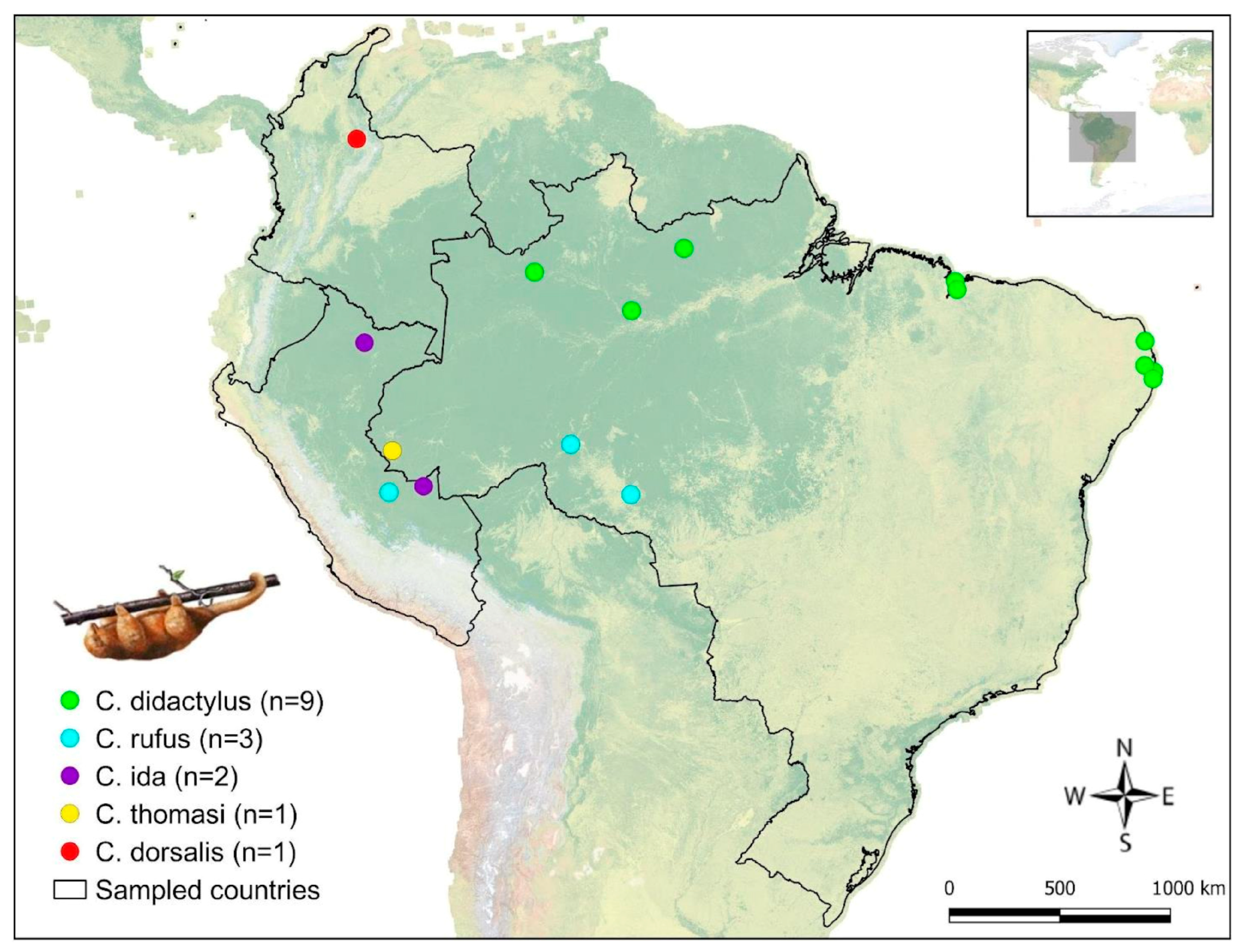
2.1. Sampling

Capture and sampling permits were granted by the Chico Mendes Institute for Biodiversity Conservation—ICMBio (SisBio permit numbers: 125811, 125813, and 133813).
Sampling was conducted between 2007 and 2016 during field expeditions in Brazil, Peru, and Colombia. Using active search methods, individuals were captured in their natural habitat during daylight hours and individually placed in cotton bags for administration of the anesthetic protocol. An intramuscular anesthesia protocol was applied, consisting of ketamine hydrochloride (8.0 mg/kg; Ketalar®, 50 mg/mL; Pfizer Laboratorie, São Paulo, Brazil) combined with midazolam (0.5 mg/kg; Dormonid®, 5 mg/5 mL; Roche Laboratorie, São Paulo, Brazil). The geographic location of each capture was recorded.
Immediately after anesthesia, approximately 2 mL of blood was collected via puncture of the cephalic, caudal, or femoral vein using 0.7 × 30 mm needles and a 5 mL syringe. The blood was stored in 3 mL EDTA tubes (BD Vacutainer®, São Paulo, Brazil) and maintained by refrigerating in a styrofoam container with ice throughout the field procedures, up to the time of laboratory analysis. Additionally, a thorough inspection of the animal’s body surface was performed, limited to macroscopic visual examination, in order to detect the presence of ectoparasites. During anesthesia, vital parameters were monitored, including heart rate and rhythm, respiratory rate, rectal temperature, and oxygen saturation (measured via pulse oximetry). In addition, complementary procedures were performed during immobilization, such as microchip implantation, tattooing, weighing, morphometric measurements, sex identification, age estimation (determined based on body mass, density of hair, and size), and biological sample collection. Individuals were kept under monitoring until full recovery from anesthesia (average recovery time of 40 min). Once fully recovered, the animals were released at the exact site of capture.
A total of 16 samples were collected. The identification of the samples and the geographical distribution of Cyclopes species included in this study are summarized in Table 1 and illustrated in Figure 1. Species identification was based on morphological and molecular methods, as described in a previous study [1].

2.2. Molecular Analysis

DNA was extracted from blood samples using the Wizard® Genomic DNA Purification Kit (Promega, Madison, WI, USA), following the manufacturer’s instructions. To verify the extraction quality, the integrity of the obtained DNA, and the presence of potential PCR inhibitors, the extracted samples were tested for the presence of the gene encoding glyceraldehyde-3-phosphate dehydrogenase (gapdh), a housekeeping gene in mammals [21].
Nested PCR and conventional PCR assays were performed to detect the presence of pathogens belonging to the order Piroplasmida, Hepatozoon spp., Ehrlichia spp., Anaplasma spp., hemotropic Mycoplasma, and trypanosomatids, including Leishmania spp., Trypanosoma evansi Steel, 1885, T. cruzi Chagas, 1909, and T. vivax Ziemann, 1905.
For all PCR assays, the reaction mixture for the first round contained 7.5 µL of GoTaq® Green Master Mix (Promega, Madison, WI, USA), 0.6 µL of a mixed primer solution (10 mM), and 5.4 µL of nuclease-free water. A volume of 1.5 µL of total DNA was added to the reaction mixture to achieve a final volume of 15 µL. For nested PCR assays, 1.5 µL of the amplified product from the first reaction was used as the DNA template for the second reaction. The sets of primers used for pathogen detection were applied with a touchdown PCR protocol, programmed as previously described. All primers, nucleotide sequences, target genes, and expected amplicon sizes used in this study are detailed in Table 2. In all reactions, ultrapure sterile water (Life Technologies®, Carlsbad, CA, USA) was used instead of template DNA as a negative control.
As positive controls, DNA extracted from different sources was used. For Ehrlichia spp., DNA was extracted from the whole blood of domestic dogs (Canis familiaris, Linnaeus, 1758) experimentally infected with Ehrlichia canis, Donatien and Lestoquard, 1935 (Jaboticabal strain) [40]. DNA from Anaplasma marginale Theiler, 1910 (UFMG1 strain), was obtained from the whole blood of a calf experimentally infected [41]. For the nested PCR targeting Piroplasmida/Hepatozoon spp., DNA was extracted from a calf experimentally infected with Babesia bovis Babes, 1888 (BbovMG strain), and B. bigemina, Smith and Kilborne, 1893 (BbigMG strain) [42]. For Anaplasma phagocytophilum Foggie, 1949, and other granulocytic agents from the family Anaplasmataceae, DNA from IDE8 tick cell cultures infected with A. phagocytophilum (isolated from a German dog) was kindly provided by Dr. Erich Zweygarth (Institut für Vergleichende Tropenmedizin und Parasitologie, Ludwig Maximilians Universität München). For Leishmania spp. testing, DNA from the reference strain L. infantum Nicolle, 1908 (MCAN/BR/2002/BH400), provided by the World Health Organization (WHO) and maintained in the cryobank of the Leishmania Biology Laboratory, was kindly provided by Prof. Maria Norma Melo (ICB/UFMG, Brazil). For Trypanosoma evansi, T. vivax, and hemotropic Mycoplasma, whole blood samples from naturally infected South American coatis (Nasua nasua Linnaeus, 1766), cattle (Bos taurus Linnaeus, 1758), and domestic cats (Felis catus Linnaeus, 1758), respectively, were used [43,44,45]. For T. cruzi, DNA was extracted from experimentally infected mice, kindly provided by the T. cruzi and Chagas Disease Biology Laboratory—ICB/UFMG.
All reactions were performed using the same thermal cycler—Applied Biosystems MiniAmp Thermal Cycler (Thermo Fisher Scientific, Waltham, MA, USA). PCR products were separated by electrophoresis on 1% agarose gels (40 min, 100 V), stained with GelRed™ (Biotium, Hayward, CA, USA), and visualized under ultraviolet light.

2.3. Genetic Sequencing

For sequencing, the PCR-positive products were purified using the QIAquick PCR Purification Kit (Qiagen Biotecnologia Brasil, São Paulo, Brazil) according to the manufacturer’s instructions. The purified amplicons were sequenced using the Sanger method [46] on an ABI3130 Genetic Analyzer (Applied Biosystems, Life Technologies, Carlsbad, CA, USA) with the BigDye® Direct Cycle Sequencing Kit v3.1 (Applied Biosystems) and the POP-7™ polymer as the separation matrix, employing the same primers used in the PCR reactions.
The forward and reverse sequences obtained were aligned, edited, and analyzed using MEGA (Molecular Evolutionary Genetics Analysis) version 11. The identity of each sequence was confirmed through alignment by homology with sequences available in GenBank using the BLASTn software [47].

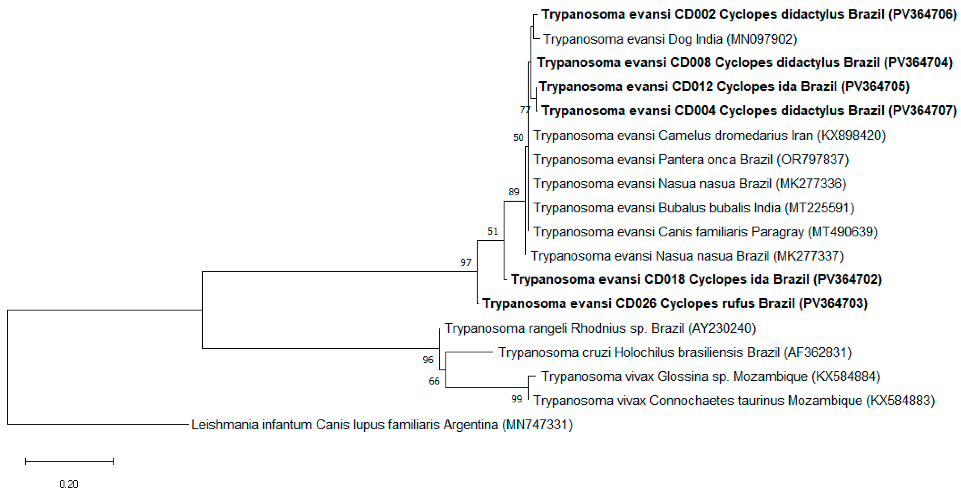
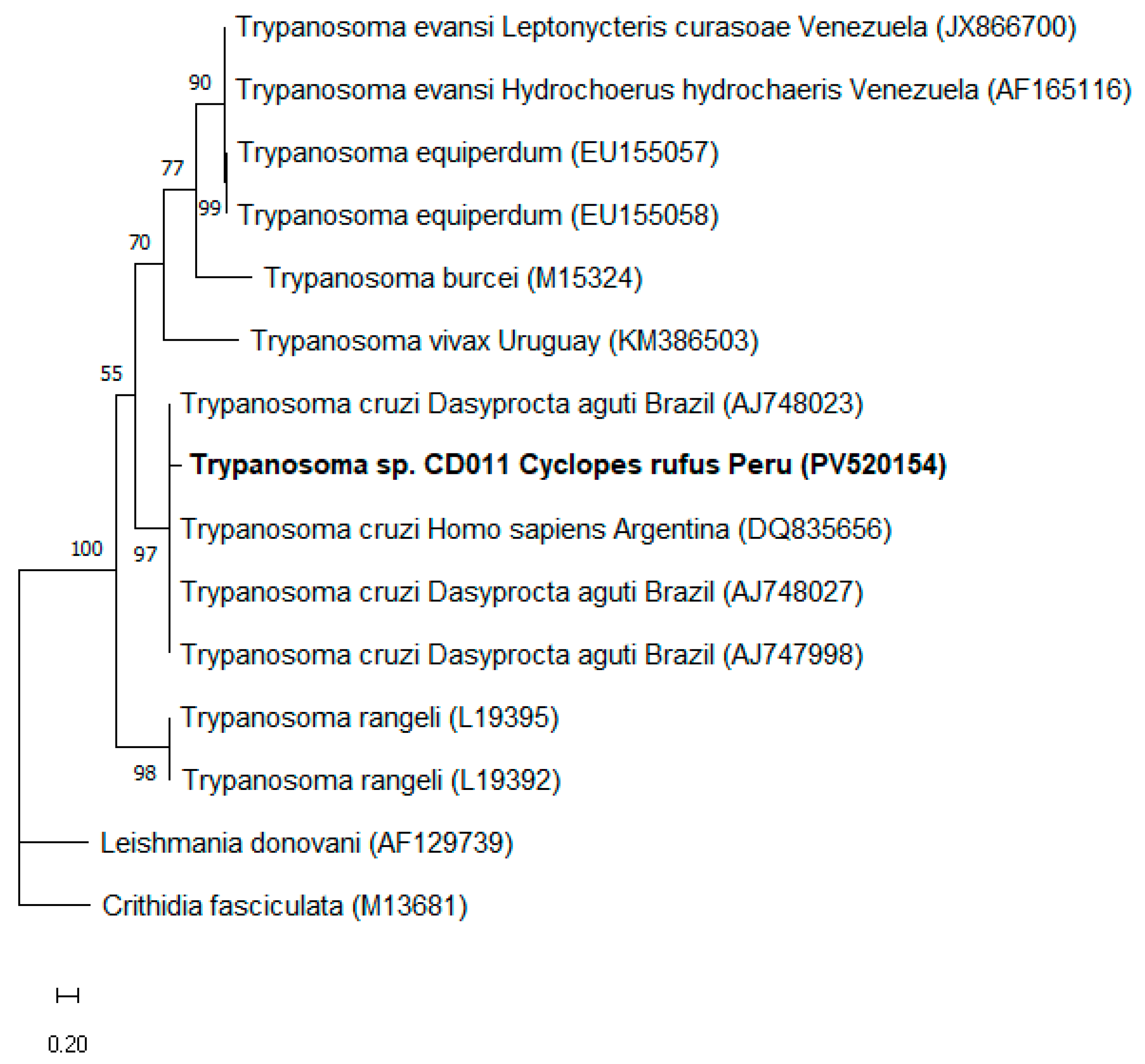
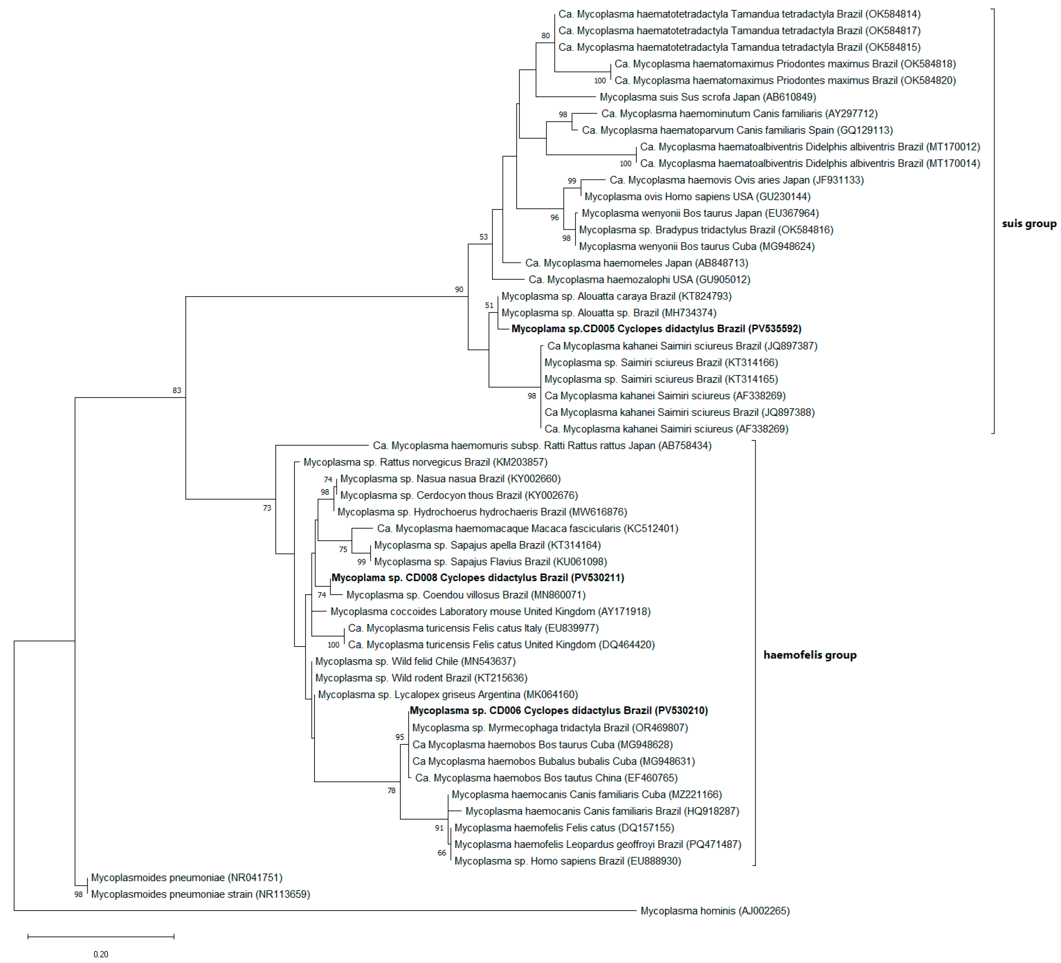
2.4. Phylogenetic Analysis

Phylogenetic trees were constructed using MEGA software, version 11. Multiple sequence alignments were performed with ClustalW, integrated into the platform, using sequences previously submitted to GenBank. The most appropriate evolutionary models were selected based on the lowest scores of the Bayesian Information Criterion (BIC) and the corrected Akaike Information Criterion (AICc). Representative sequences were included as an outgroup in the analysis. The phylogenetic tree was generated using the maximum likelihood (ML) method, based on the obtained alignments [48,49,50]. The robustness of the tree topology was tested through 1000 bootstrap replicates [51].

3. Results

All 16 DNA samples analyzed tested positive for the mammalian endogenous gene (gapdh). Subsequent molecular tests detected the presence of T. evansi (ITS-1 gene) in 50% (8/16) of the analyzed samples, including six samples from Brazil (four C. didactylus, one C. thomasi, and one C. rufus) and two from Peru (two C. ida). All eight samples were also positive for the amplification of the 18S rRNA gene of Kinetoplastida. Positive samples were subjected to genetic sequencing; however, only six ITS gene products yielded readable sequences (GenBank accession numbers: PV364702 to PV364707). BLASTn analyses showed percent identity ranging from 92% to 98.6% with T. evansi sequences from different hosts, including N. nasua (MK277343; MK277341) from Brazil, camel (Camelus spp. Linnaeus, 1758) (MH595480) from Iraq, and water buffalo (Bubalus bubalis Linnaeus, 1758) (MT225591) from India. Moreover, the maximum likelihood (ML) analysis based on the ITS-1 gene (219 bp), using the Jukes–Cantor (JC) model, clustered the T. evansi sequences detected in this study with sequences of the species sampled from C. familiaris, Camelus dromedarius, Panthera onca Linnaeus, 1758, N. nasua, and B. bubalis collected in India, Iran, Brazil, and Paraguay (Figure 2).
Additionally, 6.2% (1/16) of the individuals analyzed, represented by one specimen of C. rufus from Peru, tested positive for T. cruzi/T. rangeli (kDNA gene). Genetic sequencing of the amplified fragment (accession PV520154) revealed 87.6% identity with T. cruzi sequences previously isolated from agouti (Dasyprocta aguti Linnaeus, 1766) (AJ748023, AJ748027, AJ747998) and Homo sapiens Linnaeus, 1758 (DQ835656), from Brazil and Argentina, respectively. The sequencing reaction was performed in triplicate using both forward and reverse primers, and the resulting electropherograms were used to generate a consensus sequence, which consistently yielded similar identity values across all replicates. Pairwise alignment with the reference sequence AJ748023 showed 41 nucleotide differences over 329 base pairs (query cover 96%), supporting the calculated identity percentage. This same sample also tested positive for the 18S rRNA and kDNA genes of Kinetoplastida; however, the sequences obtained were not considered of sufficient quality for conclusive analyses. Phylogenetic inference performed using ML, based on kDNA gene sequences (307 bp) and the Tamura three-parameter (T92) substitution model, consistently clustered the Trypanosoma sp. sequence detected in this study within the same clade as T. cruzi sequences obtained from different hosts in Brazil and Argentina (Figure 3). Although the sequence clustered robustly with T. cruzi (bootstrap = 97%), it was conservatively labeled as Trypanosoma sp. in the phylogenetic tree due to the moderate identity value and relatively short fragment length. This clustering showed high statistical support, indicating a robust evolutionary relationship between the sequence obtained and previously described T. cruzi strains.
The screening PCR based on the 16S rRNA gene (600 and 193 bp) of hemoplasmas revealed a positivity rate of 62.5% (10/16), including seven individuals from Brazil (five C. didactylus, one C. rufus, and one C. thomasi) and three from Peru (one C. rufus and two C. ida). Additionally, 5 out of the 10 samples positive for the 16S rRNA gene were also positive for the 23S rRNA gene (800 bp) (4 C. didactylus from Brazil and 1 C. ida from Peru). PCR products with strong band intensity were selected for genetic sequencing; however, satisfactory results were obtained for only three 16S rRNA gene sequences (accessions PV530210, PV530211, PV535592). BLASTn analyses revealed high identity between one of the obtained sequences (CD005) and hemotropic Mycoplasma sp. previously detected in arboreal Neotropical primates of the genus Alouatta spp. Lacépède, 1799, in Brazil (KT824793, JQ897386, MH734376, MH734374), with percentage identity ranging from 97.7% to 98.5%. Furthermore, this sequence showed 95.4% to 96.1% identity with ‘Candidatus Mycoplasma kahanei’, previously described in Saimiri sciureus Linnaeus, 1758, another arboreal primate from Brazil (JQ897388) and the United States (AF338269). Another sequence (CD006) showed 99.4% to 99.8% identity with Mycoplasma sp. and ‘Ca. Mycoplasma haemobos’, previously detected in giant anteater (Myrmecophaga tridactyla Linnaeus, 1758) (OR469807) from Brazil and bovids (MG948628, MG948630, MG948631) sampled in Central America. The third and final readable sequence analyzed (CD008) showed 97.9% to 98.4% identity with Mycoplasma sp. isolated from Coendou villosus Coendou Lacépède, 1799, in Brazil (MN860071) and Mycoplasma lineages detected in Lycalopex griseus Gray, 1837 (MK064160), wild felids (MN543637), and wild rodents (KT215636). Phylogenetic inference performed using ML, based on the 16S rRNA gene and the Tamura three-parameter model with gamma distribution (T92+G), grouped the Mycoplasma spp. sequences detected in this study into two main clades, which, according to the authors of [52], can be divided into the haemofelis group (n = 2) and the suis group (n = 1), both supported by high bootstrap values. One sample from the suis group (CD005) clustered within the same clade as hemoplasmas previously detected in arboreal primates, with phylogenetic proximity to the ‘Ca. Mycoplasma kahanei’ clade. Of the two samples grouped in the haemofelis group, one (CD006) showed high similarity with Mycoplasma sequences isolated from M. tridactyla, both positioned within the same clade as ‘Ca. Mycoplasma haemobos’. Meanwhile, sample CD008 showed a closer phylogenetic relationship to Mycoplasma spp. detected in C. villosus, a Brazilian mammal that also has arboreal habits (Figure 4).
Among the 14 individuals positive for at least one of the pathogens investigated, 35.7% (5/14) presented co-infections involving two agents. Of these, 80% (4/5) corresponded to co-infection with hemotropic Mycoplasma and T. evansi, detected in two C. ida individuals from Peru, one C. thomasi, and one C. didactylus from Brazil. The remaining co-infection, representing 20% (1/5) of the cases, involved T. cruzi and hemotropic Mycoplasma in a C. rufus individual sampled in Peru. No co-infections involving more than two pathogens were identified. In the present study, no samples tested positive for pathogens of the order Piroplasmida, Hepatozoon sp., agents of the family Anaplasmataceae, or other trypanosomatids such as Leishmania spp. and T. vivax (Table 3). Additionally, no ectoparasites were observed on any of the body regions examined, based on macroscopic visual inspection.

4. Discussion

To the best of our knowledge, this is the first molecular detection of Trypanosoma evansi and hemotropic Mycoplasma in newly described species of the genus Cyclopes. These species are strictly arboreal and rarely descend to the ground, a behavior that can hinder their observation, capture, and the collection of blood samples. This fact makes the present study particularly relevant for advancing knowledge about the health status of this genus. Additionally, the taxonomic description of different Cyclopes species is relatively recent, and studies focused on these newly described species are still scarce.
There are very few studies on hemopathogens in silky anteaters. Between 1936 and 1938, Deane [53] examined 12 specimens of C. didactylus from the state of Pará, Brazil, using blood smears, tissue imprints, in vitro culture, and inoculation into other animals, but found no evidence of hemoflagellate infection. Later, Lainson et al. [54], using similar methods combined with xenodiagnosis, also failed to detect Trypanosomatidae in C. didactylus from the same region. However, when parasitemia is low, false-negative results may occur. The first isolation of a trypanosomatid identified as T. cruzi in C. didactylus was reported by Miles et al. [55] during a survey of wild mammals in the state of Pará. Since then, there have been no further records of studies aimed at detecting this or other pathogens in this genus.
Infections caused by Trypanosomatidae in xenarthrans have been reported in the state of Pará, Brazil, showing mixed infections with T. cruzi, T. rangeli Tejera, 1920, and L. infantum in the lesser anteater (Tamandua tetradactyla Linnaeus, 1758) [56]. Another study highlighted this species as an important reservoir for T. rangeli and an efficient vector of the parasite in the Brazilian Amazon region [57]. Additionally, T. cruzi was detected in two six-banded armadillos (Euphractus sexcinctus Linnaeus, 1758), one T. tetradactyla, and one nine-banded armadillo (Dasypus novemcinctus Linnaeus, 1758) in the Brazilian Pantanal. This was the first report of T. tetradactyla infected with T. cruzi in the Pantanal region, with positivity confirmed through hemoculture, showing high levels of parasitemia during the sampling period. The authors concluded that T. tetradactyla are important hosts for T. cruzi in the studied area [58]. However, the epidemiological relevance of Cyclopes spp. as hosts for this pathogen remains unclear.
Trypanosoma cruzi is a hemoflagellate responsible for Chagas disease, transmitted by triatomine bugs. Some authors have reported that the most favorable climatic conditions for the presence of triatomines include humid mesothermal climates, as higher temperatures promote a greater geographical dispersion of wild vectors [59,60]. These climatic conditions are present in the sampling areas, which may explain the detection of the pathogen in a blood sample from C. rufus collected in Peru. In that country, Chagas disease surveillance has been mandatory nationwide since 1999. Despite this, the distribution of cases remains irregular. Among the regions reporting Chagas disease in Peru, Ucayali—the origin of the positive silky anteater—accounts for 3.2% of the reported cases, reinforcing the circulation of the parasite in the region and, consequently, the possibility of infection in wild animals [61].
Trypanosoma evansi is a member of the Trypanosomatidae family that can parasitize a wide range of domestic and wild mammalian hosts and causes a trypanosomiasis known as ‘surra.’ In the Americas, the transmission of T. evansi occurs predominantly through mechanical means, via the blood of infected animals transmitted by hematophagous dipterans such as members of the Tabanidae family. Additionally, it can be transmitted vertically, iatrogenically, through the ingestion of infected prey, or via the action of hematophagous bats (Desmodus rotundus Geoffroy, 1810) [62,63,64]. It is a protozoan parasite found in both intravascular and extravascular fluids [65] and, in susceptible animals, can cause clinical manifestations. The infiltration and dissemination of T. evansi into the central nervous system have been reported, and neurological signs are common in horses [64,66]. Furthermore, animals subjected to stress, malnutrition, or pregnancy are more susceptible to the disease [66].
This study reports the presence of T. evansi parasitizing silky anteaters in tropical areas of Brazil and Peru, where this parasite was likely introduced during the 16th century with horses or mules brought by Spanish conquistadors [67,68]. Brazilian wild animals, such as deer, capybaras, and coatis, can become infected and may develop disease, including death, but may also act as reservoirs. However, the different habitats and behavioral patterns of wild and domestic hosts suggest that there are still unknown factors underlying the transmission cycles of T. evansi [36,63,69].
Little is known about the role of Xenarthra in the epidemiology of T. evansi infection. In a study conducted by Herrera et al. [63], which evaluated the infection by this agent in domestic and wild species in the Pantanal region of Brazil, PCR detection was reported in one (one out of eight) armadillo (Euphractus sp.). Santos et al. [58] investigated the role of different host species in the transmission cycles of Trypanosoma spp. in a central area of the Brazilian Pantanal. Among Xenarthra, they found PCR-positive results for T. evansi in two (2/29) six-banded armadillos (Euphractus sexcinctus).
Bacteria of the genus Mycoplasma, also known as hemoplasmas, can have significant implications for animal and human health [70,71,72]. These epierythrocytic microorganisms adhere to the surface of erythrocytes, and although generally considered to have low pathogenicity, they can cause clinical disease under conditions of immunosuppression or co-infection [18,73]. While vector-borne transmission by hematophagous arthropods is widely recognized, there is evidence supporting alternative transmission routes, such as transplacental transmission and exposure to the blood of infected animals during aggressive interactions [74,75,76,77]. Among Xenarthra, specific behaviors such as fights between anteaters (M. tridactyla) [78] and the shared use of burrows by armadillos [79] may facilitate both direct and vector-borne transmission of these hemopathogens, representing important ecological aspects to be considered in surveillance and conservation studies of these taxa. However, in the genus Cyclopes, given its strictly solitary nature, other transmission routes should be prioritized when evaluating health risks.
Recent studies have reported the occurrence of hemoplasmas in members of the order Xenarthra. De Oliveira et al. [80] reported the detection of these microorganisms in anteaters (M. tridactyla and T. tetradactyla), sloths (genera Bradypus Linnaeus, 1758, and Choloepus Illiger, 1811), and armadillos (Priodontes maximus Kerr, 1792, E. sexcinctus, and D. novemcinctus) sampled from different regions of Brazil. Notably, the study also described, for the first time, the presence of a 16S rRNA sequence with high identity (99.7%) to Mycoplasma wenyonii in pale-throated sloth (Bradypus tridactylus Linnaeus, 1758), in addition to the molecular characterization of two novel taxa, designated as ‘Ca. Mycoplasma haematotetradactyla’ and ‘Ca. Mycoplasma haematomaximus’.
Another study conducted by Sada et al. [19] also reported a considerably high positivity rate (52%) in armadillos (E. sexcinctus, P. maximus, and D. novemcinctus) and anteaters (M. tridactyla and T. tetradactyla) from the Southeast and Midwest regions of Brazil. The authors detected the presence of a hemoplasma phylogenetically related to ‘Ca. Mycoplasma haematomaximus’, previously described in P. maximus in the state of Mato Grosso do Sul. It is noteworthy that most of the animals sampled by Sada et al. [19] originated from Mato Grosso do Sul, a region characterized by high biodiversity and vector density.
These findings, together with the study by De Oliveira et al. [80], contribute significantly to advancing knowledge about the genetic diversity and distribution of hemoplasmas in free-ranging Xenarthra, highlighting the importance of these hosts in the ecology of these agents. However, the genus Cyclopes remains the least studied among Xenarthra, lacking investigations that assess the occurrence and diversity of hemoplasmas in these species and their implications for the health of this taxon.
The present study revealed a high positivity rate for hemoplasmas (62.5%) in individuals of the genus Cyclopes sampled in Brazil and Peru, representing the first report of the detection of this agent in this group. Phylogenetic analysis revealed a close relationship between two sequences obtained in this study (CD005 and CD008) and Mycoplasma spp. previously detected in C. villosus and neotropical arboreal primates of the genus Alouatta. This phylogenetic proximity suggests a possible sharing of hemoplasmas among species with similar ecological niches. Cyclopes, C. villosus, and Alouatta spp. all exhibit strictly arboreal habits and inhabit humid tropical forests of South America, often with spatial overlap in different vegetation strata. Such ecological proximity may facilitate exposure to common arthropod vectors. The genetic similarity between hemoplasmas associated with these hosts, combined with their arboreal lifestyle, raises the hypothesis that these microorganisms may circulate among sympatric species with convergent ecological behaviors through transmission routes that remain poorly understood.
The actual clinical impact of hemoplasma infections in these hosts is still uncertain, as bacterial co-infections may or may not compromise the health of individuals [18]. These results reinforce the need for longitudinal monitoring to assess the effects of infection over time.
Behavioral aspects of arboreal mammals may contribute to parasitism by arthropods of vectorial importance. Sympatric nocturnal arboreal primates from Madagascar (Avahi occidentalis Lorenz, 1898, and Lepilemur edwardsi Forsyth Major, 1894) were evaluated for parasite exposure risk, exploring how the ecology of sleeping sites influences infestation by ectoparasites and vector-borne hemopathogens. It was observed that L. edwardsi (uses tree hollows, exhibiting strong roosting site fidelity), but not A. occidentalis (sleeps in open branches and frequently changes roosting site), harbored nest-adapted hard and soft ticks, as well as mites. The authors concluded that sleeping in tree cavities promotes ectoparasite infestation but may offer protection against hemopathogens transmitted by mosquitoes [81].
Silky anteaters exhibit arboreal and nocturnal habits and frequently change their sheltering sites. These behavioral characteristics may make them more susceptible to parasitism by hematophagous dipterans than by ectoparasites that need to remain attached to the host to feed on blood. Dipterans of the family Tabanidae are predominantly active during the day or at twilight [15,63], meaning they are most active when Cyclopes spp. are at rest. In this context, the inactivity of silky anteaters during peak vector activity may increase their exposure to bites, as the absence of defensive behaviors can facilitate vector access. Therefore, it is plausible that tabanid flies are involved in the transmission of T. evansi and hemoplasmas to Cyclopes spp.
Previous studies on Xenarthra have reported the presence of ectoparasites, including ticks of the genera Amblyomma spp. Koch, 1844, and Rhipicephalus spp. Koch, 1844, as well as various flea species [82,83,84,85]. However, consolidated records regarding ectoparasites in species of the genus Cyclopes remain scarce. Miranda et al. [86] reported the parasitism of a C. didactylus individual by an engorged nymph of the tick Amblyomma humerale, a species endemic to South America whose adult stages are commonly associated with tortoises such as Chelonoidis denticulata Linnaeus, 1766, and C. carbonária Spix, 1824. This finding suggests that species of the genus Cyclopes may serve as accidental or transient hosts for immature stages of A. humerale. However, the absence of ectoparasite detection in the present study prevented the correlation between the findings and potential vectors, highlighting the need for further investigations to better understand the relationship between Cyclopes spp. and ectoparasites, as well as their potential role in the maintenance and dispersal of these arthropods in natural environments. The limitation of the inspection method, restricted to macroscopic visual examination, may have contributed to the absence of ectoparasite records, considering that immature forms or small-sized specimens might not be detected without the use of auxiliary tools. More sensitive approaches, such as the use of entomological combs and fine forceps, are recommended in future studies to maximize ectoparasite recovery, especially in species with dense fur.
This study investigated vector-borne hemopathogens using samples obtained from a biological bank previously collected for the genetic analysis of the animals [1]. However, the limited volume of the samples restricted the amplification of additional gene regions in the positive samples, as well as the repetition of sequencing reactions that yielded suboptimal results. Furthermore, the preservation status of the samples, which had been stored frozen, precluded the application of other direct parasitological methods, such as blood smears, Woo’s technique, buffy coat analysis, and pathogen culture and isolation. The absence of morphological examination is recognized as a limitation, as it hinders the confirmation of active infections and may lead to underdiagnosis in cases of mixed infections, which are common in wildlife. Additionally, molecular detection alone may reflect transient DNA presence from recent vector inoculation, rather than true host infection or susceptibility.
In conclusion, silky anteaters were found to be parasitized by T. evansi, T. cruzi, and hemotropic Mycoplasma. This study highlights the need for future research to evaluate the potential impacts of these infections on the health of Cyclopes spp. The results presented here may serve as a foundation for more comprehensive investigations and support wildlife health managers and conservation authorities in the development of management strategies and public policies aimed at the conservation of this species.

Author Contributions

P.H.C.R.: Conceptualization, Methodology, Validation, Investigation, Writing–Original Draft; J.P.S.A.: Conceptualization, Methodology, Validation, Investigation, Writing–Original Draft; F.R.M.: Conceptualization, Investigation, Methodology, Writing–Review and Editing; C.R.: Conceptualization, Investigation, Methodology, Writing–Review and Editing; J.A.G.S.: Conceptualization, Methodology, Validation, Investigation, Resources, Funding acquisition, Visualization, Supervision, Writing—Original Draft, Writing—review and editing. All authors have read and agreed to the published version of the manuscript.

Funding

This study was financed by scholarships from Coordenação de Aperfeiçoamento de Pessoal de Nível Superior (CAPES), financial support for the field, lab work and researcher grant from Conselho Nacional de Desenvolvimento Científico e Tecnológico (CNPq) (472802/2010-0), Fundação de Amparo à Pesquisa do Estado de Minas Gerais (FAPEMIG) (APQ-02095-16; APQ-00708-21; APQ-02531–24; APQ-06074-24) and Universidade Federal de Minas Gerais.

Institutional Review Board Statement

Capture and sampling permits were granted by the Chico Mendes Institute for Biodi-versity Conservation—ICMBio (SisBio permit numbers: 125811, Issued on: 13 December 2007; 125813, Issued on: 15 June 2010; and 133813, Issued on: 25 May 2014).

Informed Consent Statement

Not applicable.

Data Availability Statement

Data are contained within the article.

Acknowledgments

We are grateful to Monique Pool, Lizette Bermudez, Giamarco Rojas, and others for their assistance during the museum visits, as well as to the team of the Projeto Tamanduá. We also thank the Laboratory of Veterinary Protozoology (Protovet) at the Federal University of Minas Gerais for their technical and scientific support.

Conflicts of Interest

The authors declare that they have no financial, commercial, or any other conflicts of interest that could influence the results or the interpretation of the data presented in this study.

References

  1. Miranda, F.R.; Casali, D.M.; Perini, F.A.; Machado, F.A.; Santos, F.R. Taxonomic review of the genus Cyclopes Gray, 1821 (Xenarthra: Pilosa), with the revalidation and description of new species. Zool. J. Linn. Soc. 2018, 183, 687–721. [Google Scholar] [CrossRef]
  2. Machado, A.F.; Miranda, F.R. The potential distribution of Cyclopes didactylus, a silky anteater, reveals a likely unknown population and urgent need for forest conservation in Northeast Brazil. J. Trop. Ecol. 2022, 38, 454–461. [Google Scholar] [CrossRef]
  3. McNab, B.K. Energetics, population biology, and distribution of Xenarthrans, living and extinct. In The Evolution and Ecology of Armadillos, Sloths, and Vermilinguas; Montgomery, G.G., Ed.; Smithsonian Institution Press: Washington, DC, USA; London, OH, USA, 1985; pp. 219–232. [Google Scholar]
  4. Wetzel, R.M. Systematics, distribution, ecology, and conservation of South American edentates. In The Pymatuning Symposia in Ecology, Volume 6: Mammalian Biology in South America; Mares, M.A., Genoways, H.H., Eds.; University of Pittsburgh: Pittsburgh, PA, USA, 1982; pp. 345–375. [Google Scholar]
  5. Paglia, A.P.; Fonseca, G.A.B.; Rylands, A.B.; Herrmann, G.; Aguiar, L.M.S.; Chiarello, A.G.; Leite, Y.L.; Costa, L.P.; Siciliano, S.; Kierulff, M.C. Lista Anotada dos Mamíferos do Brasil, 2nd ed.; Occasional Papers in Conservation Biology 6; Conservation International: Arlington, TX, USA, 2012; pp. 1–76. [Google Scholar]
  6. Nowak, R.M. Walker’s Mammals of the World, 6th ed.; The Johns Hopkins University Press: Baltimore, MD, USA; London, OH, USA, 1999; Volume 1. [Google Scholar]
  7. Miranda, F.; Superina, M. New distribution records of the silky anteater Cyclopes didactylus (Mammalia, Pilosa, Cyclopedidae) in coastal northeastern Brazil. Mastozool. Neotrop. 2010, 17, 381–384. [Google Scholar]
  8. Eisenberg, J.F.; Redford, K.H. Mammals of the Neotropics: The Central Neotropics. Ecuador, Peru, Bolivia, Brazil; The University of Chicago Press: Chicago, CA, USA, 1999; Volume 3. [Google Scholar]
  9. Fonseca, G.A.B.; Herrmann, G.; Leite, Y.L.R.; Mittermeier, R.A.; Rylands, A.B.; Patton, J.L. Lista Anotada dos Mamíferos do Brasil; Occasional Papers in Conservation Biology 4; Conservation International: Arlington, TX, USA, 1996; pp. 1–38. [Google Scholar]
  10. Montgomery, G.G. Impacts of vermilinguas (Cyclopes, Tamandua: Xenarthra = Edentata) on arboreal ant populations. In The Evolution and Ecology of Armadillos, Sloths, and Vermilinguas; Montgomery, G.G., Ed.; Smithsonian Institution Press: Washington, DC, USA; London, OH, USA, 1985; pp. 351–363. [Google Scholar]
  11. Miranda, F.; Meritt, D.A.; Tirira, D.G.; Arteaga, M. Cyclopes didactylus. The IUCN Red List of Threatened Species. 2014. Available online: https://www.iucnredlist.org/species/6019/47440020 (accessed on 27 March 2025).
  12. Miranda, F.R.; Chiarello, A.G.; Röhe, F.; Miranda, G.H.B.; Vaz, S.M.; Avaliação do Risco de Extinção de Cyclopes didactylus (Linnaeus, 1758). Processo de Avaliação do Risco de Extinção dos Xenarthros Brasileiros, ICMBio. 2015. Available online: https://www.researchgate.net/profile/Alessandra-Bertassoni/publication/323457425_Avaliacao_do_Risco_de_Extincao_de_Tamandua_tetradactyla_Linnaeus_1758_no_Brasil_Evaluation_of_the_extinction_risk_of_the_Southern_Anteater_Tamandua_tetradactyla_Linnaeus_1758_in_Brazil/links/5a970536aca27214056b3456/Avaliacao-do-Risco-de-Extincao-de-Tamandua-tetradactyla-Linnaeus-1758-no-Brasil-Evaluation-of-the-extinction-risk-of-the-Southern-Anteater-Tamandua-tetradactyla-Linnaeus-1758-in-Brazil.pdf (accessed on 18 July 2025).
  13. Castillo, A.P.; Colácio, N.; Rodrigues, P.H.C.; Miranda, J.V.O.; Lima, P.C.S.; Motta, R.O.C.; Tinoco, H.P.; Coelho, C.M.; da Silveira, J.A.G. Parasitic Protozoa and Other Vector-Borne Pathogens in Captive Mammals from Brazil. J. Zool. Bot. Gard. 2024, 5, 754–773. [Google Scholar] [CrossRef]
  14. Ceylan, O.; Ma, Z.; Ceylan, C.; Ider, M.; Evci, A.; Mavinehir, A.; Xuan, X.; Sevinc, F. Feline vector-borne haemopathogens in Türkiye: The first molecular detection of Mycoplasma wenyonii and ongoing Babesia ovis DNA presence in unspecific hosts. BMC Vet. Res. 2024, 20, 365. [Google Scholar] [CrossRef] [PubMed]
  15. Baldacchino, F.; Desquesnes, M.; Mihok, S.; Foil, L.D.; Duvallet, G.; Jittapalapong, S. Tabanids: Neglected subjects of research, but important vectors of disease agents! Infect. Genet. Evol. 2024, 28, 596–615. [Google Scholar] [CrossRef] [PubMed]
  16. Muita, J.W.; Bargul, J.L.; Makwatta, J.O.; Ngatia, E.M.; Tawich, S.K.; Masiga, D.K.; Getahun, M.N. Stomoxys flies (Diptera, Muscidae) are competent vectors of Trypanosoma evansi, Trypanosoma vivax, and other livestock hemopathogens. PLoS Pathog. 2025, 21, e1012570. [Google Scholar] [CrossRef] [PubMed]
  17. Maia, C. Sand fly-borne diseases in Europe: Epidemiological overview and potential triggers for their emergence and re-emergence. J. Comp. Pathol. 2024, 209, 6–12. [Google Scholar] [CrossRef] [PubMed]
  18. Calchi, A.C.; Yogui, D.R.; Alves, M.H.; Desbiez, A.L.J.; Kluyber, D.; Vultão, J.G.; Arantes, P.V.C.; de Santi, M.; Werther, K.; Teixeira, M.M.G.; et al. Molecular detection of piroplasmids in mammals from the Superorder Xenarthra in Brazil. Parasitol. Res. 2023, 122, 3169–3180. [Google Scholar] [CrossRef] [PubMed]
  19. Sada, J.M.; Kluyber, D.; Lee, D.A.B.; Calchi, A.C.; Alves, M.H.; Machado, D.M.R.; Werther, K.; Machado, R.Z.; Desbiez, A.L.J.; André, M.R. Molecular detection and characterization of Anaplasmataceae agents, Bartonella spp. and hemoplasmas in armadillos and anteaters from Brazil. Acta Trop. 2024, 260, 107477. [Google Scholar] [CrossRef] [PubMed]
  20. QGIS Development Team. QGIS Geographic Information System; (Version 3.32.0); Open Source Geospatial Foundation Project: Beaverton, OR, USA, 2023; Available online: https://qgis.org (accessed on 27 March 2025).
  21. Birkenheuer, A.J.; Levy, M.G.; Breitschwerdt, E.B. Development and evaluation of a seminested PCR for detection and differentiation of Babesia gibsoni (Asian Genotype) and B. canis DNA in canine blood samples. J. Clin. Microbiol. 2003, 41, 4172–4177. [Google Scholar] [CrossRef] [PubMed]
  22. Zahler, M.; Rinder, H.; Schein, E.; Gothe, R. Detection of a new pathogenic Babesia microti-like species in dogs. Vet. Parasitol. 2000, 89, 241–248. [Google Scholar] [CrossRef] [PubMed]
  23. Silveira, J.A.; Rabelo, E.M.; Ribeiro, M.F. Detection of Theileria and Babesia in brown brocket deer (Mazama gouazoubira) and marsh deer (Blastocerus dichotomus) in the State of Minas Gerais, Brazil. Vet. Parasitol. 2011, 177, 61–66. [Google Scholar] [CrossRef] [PubMed]
  24. Soares, J.F.; Girotto, A.; Brandão, P.E.; Da Silva, A.S.; França, R.T.; Lopes, S.T.; Labruna, M.B. Detection and molecular characterization of a canine piroplasm from Brazil. Vet. Parasitol. 2011, 180, 203–208. [Google Scholar] [CrossRef] [PubMed]
  25. Kawahara, M.; Tajima, T.; Torii, H.; Yabutani, M.; Ishii, J.; Harasawa, M.; Isogai, E.; Rikihisa, Y. Ehrlichia chaffeensis infection of sika deer, Japan. Emerg. Infect. Dis. 2009, 15, 1991–1993. [Google Scholar] [CrossRef] [PubMed]
  26. Massung, R.F.; Slater, K.; Owens, J.H.; Nicholson, W.L.; Mather, T.N.; Solberg, V.B.; Olson, J.G. Nested PCR assay for detection of granulocytic ehrlichiae. J. Clin. Microbiol. 1998, 36, 1090–1095. [Google Scholar] [CrossRef] [PubMed]
  27. de la Fuente, J.; Ayoubi, P.; Blouin, E.F.; Almazán, C.; Naranjo, V.; Kocan, K.M. Gene expression profiling of human promyelocytic cells in response to infection with Anaplasma phagocytophilum. Cell Microbiol. 2005, 7, 549–559. [Google Scholar] [CrossRef] [PubMed]
  28. Bown, K.J.; Lambin, X.; Ogden, N.H.; Petrovec, M.; Shaw, S.E.; Woldehiwet, Z.; Birtles, R.J. High-resolution genetic fingerprinting of European strains of Anaplasma phagocytophilum by use of multilocus variable-number tandem-repeat analysis. J. Clin. Microbiol. 2007, 45, 1771–1776. [Google Scholar] [CrossRef] [PubMed]
  29. Criado-Fornelio, A.; Martinez-Marcos, A.; Buling-Saraña, A.; Barba-Carretero, J.C. Presence of Mycoplasma haemofelis, Mycoplasma haemominutum and piroplasmids in cats from southern Europe: A molecular study. Vet. Microbiol. 2003, 93, 307–317. [Google Scholar] [CrossRef] [PubMed]
  30. Kewish, K.E.; Appleyard, G.D.; Myers, S.L.; Kidney, B.A.; Jackson, M.L. Mycoplasma haemofelis and Mycoplasma haemominutum detection by polymerase chain reaction in cats from Saskatchewan and Alberta. Can. Vet. J. 2004, 45, 749–752. [Google Scholar] [PubMed]
  31. Mongruel, A.C.B.; Spanhol, V.C.; Valente, J.D.M.; Porto, P.P.; Ogawa, L.; Otomura, F.H.; Marquez, E.S.; André, M.R.; Vieira, T.S.W.J.; Vieira, R.F.C. Survey of vector-borne and nematode parasites involved in the etiology of anemic syndrome in sheep from Southern Brazil. Rev. Bras. Parasitol. Vet. 2020, 29, e007320. [Google Scholar] [CrossRef] [PubMed]
  32. de la Fuente, J.; Ruiz-Fons, F.; Naranjo, V.; Torina, A.; Rodríguez, O.; Gortázar, C. Evidence of Anaplasma infections in European roe deer (Capreolus capreolus) from southern Spain. Res. Vet. Sci. 2008, 84, 382–386. [Google Scholar] [CrossRef] [PubMed]
  33. Silveira, J.A.; Rabelo, E.M.; Ribeiro, M.F. Molecular detection of tick-borne pathogens of the family Anaplasmataceae in Brazilian brown brocket deer (Mazama gouazoubira) and marsh deer (Blastocerus dichotomus). Transbound. Emerg. Dis. 2012, 59, 353–360. [Google Scholar] [CrossRef] [PubMed]
  34. Noyes, H.A.; Stevens, J.R.; Teixeira, M.; Phelan, J.; Holz, P. A nested PCR for the ssrRNA gene detects Trypanosoma binneyi in the platypus and Trypanosoma sp. in wombats and kangaroos in Australia. Int. J. Parasitol. 1999, 29, 331–339. [Google Scholar] [CrossRef] [PubMed]
  35. Gilber, S.R.; Alban, S.M.; Gobor, L.; Bescrovaine, J.O.; Myiazaki, M.I.; Thomaz-Soccol, V. Comparison of conventional serology and PCR methods for the routine diagnosis of Trypanosoma cruzi infection. Rev. Soc. Bras. Med. Trop. 2013, 46, 310–315. [Google Scholar] [CrossRef] [PubMed]
  36. Silveira, J.A.; Rabelo, E.M.; Lacerda, A.C.; Borges, P.A.; Tomás, W.M.; Pellegrin, A.O.; Tomich, R.G.; Ribeiro, M.F. Molecular detection and identification of hemoparasites in pampas deer (Ozotoceros bezoarticus Linnaeus, 1758) from the Pantanal, Brazil. Ticks Tick Borne Dis. 2013, 4, 341–345. [Google Scholar] [CrossRef] [PubMed]
  37. Cortez, A.P.; Rodrigues, A.C.; Garcia, H.A.; Neves, L.; Batista, J.S.; Bengaly, Z.; Paiva, F.; Teixeira, M.M. Cathepsin L-like genes of Trypanosoma vivax from Africa and South America—Characterization, relationships and diagnostic implications. Mol. Cell. Probes 2009, 23, 44–51. [Google Scholar] [CrossRef] [PubMed]
  38. Vallejo, G.A.; Guhl, F.; Chiari, E.; Macedo, A.M. Species specific detection of Trypanosoma cruzi and Trypanosoma rangeli in vector and mammalian hosts by polymerase chain reaction amplification of kinetoplast minicircle DNA. Acta Trop. 1999, 72, 203–212. [Google Scholar] [CrossRef] [PubMed]
  39. El Tai, N.O.; Osman, O.F.; El Fari, M.; Presber, W.; Schönian, G. Genetic heterogeneity of ribosomal internal transcribed spacer in clinical samples of Leishmania donovani spotted on filter paper as revealed by single-strand conformation polymorphisms and sequencing. Trans. R. Soc. Trop. Med. Hyg. 2000, 94, 575–579. [Google Scholar] [CrossRef] [PubMed]
  40. Moreira, S.M. Estudo Retrospectivo (1998–2001) da Erliquiose Canina em Belo Horizonte: Avaliação Clínica e Laboratorial de Infecções Experimentais. Master’s Thesis, Universidade Federal de Minas Gerais, Belo Horizonte, Minas Gerais, Brazil, 2001. Available online: http://hdl.handle.net/1843/BUOS-8C8DX2 (accessed on 22 February 2025).
  41. Bastos, C.V.; Passos, L.M.; Facury-Filho, E.J.; Rabelo, E.M.; de la Fuente, J.; Ribeiro, M.F. Protection in the absence of exclusion between two Brazilian isolates of Anaplasma marginale in experimentally infected calves. Vet. J. 2010, 186, 374–378. [Google Scholar] [CrossRef] [PubMed]
  42. Costa, V.M.; Ribeiro, M.F.; Duarte, A.L.; Mangueira, J.M.; Pessoa, A.F.; Azevedo, S.S.; Barros, A.T.; Riet-Correa, F.; Labruna, M.B. Seroprevalence and risk factors for cattle anaplasmosis, babesiosis, and trypanosomiasis in a Brazilian semiarid region. Rev. Bras. Parasitol. Vet. 2013, 22, 207–213. [Google Scholar] [CrossRef] [PubMed]
  43. Castillo, A.P.; Miranda, J.V.O.; Fonseca, P.L.C.; Silva, S.O.; Lopes, R.E.N.; Spanhol, V.C.; Moreira, R.G.; Nicolino, R.R.; Queiroz, D.C.; de Araújo e Santos, L.C.G.; et al. Evidence of SARS-CoV-2 infection and co-infections in stray cats in Brazil. Acta Trop. 2024, 249, 107056. [Google Scholar] [CrossRef] [PubMed]
  44. Estevam, L.G.T.M.; Fonseca, A.A., Jr.; Silvestre, B.T.; Hemetrio, N.S.; Almeida, L.R.; Oliveira, M.M.; Silva, S.M.; Ribeiro, M.F.B.; Silveira, J.A.G. Seven years of evaluation of ectoparasites and vector-borne pathogens among ring-tailed coatis in an urban park in southeastern Brazil. Vet. Parasitol. Reg. Stud. Rep. 2020, 21, 100442. [Google Scholar] [CrossRef] [PubMed]
  45. Pereira, H.D.; Simões, S.V.D.; Souza, F.A.L.; Silveira, J.A.G.; Ribeiro, M.F.B.; Cadioli, F.A.; Sampaio, P.H. Aspectos clínicos, epidemiológicos e diagnóstico da infecção por Trypanosoma vivax em rebanho bovino no estado do Maranhão. Pesqui. Vet. Bras. 2018, 38, 896–901. [Google Scholar] [CrossRef]
  46. Sanger, F.; Nicklen, S.; Coulson, A.R. DNA sequencing with chain-terminating inhibitors. Proc. Natl. Acad. Sci. USA 1977, 74, 5463–5467. [Google Scholar] [CrossRef] [PubMed]
  47. Altschul, S.F.; Gish, W.; Miller, W.; Myers, E.W.; Lipman, D.J. Basic local alignment search tool. J. Mol. Biol. 1990, 215, 403–410. [Google Scholar] [CrossRef] [PubMed]
  48. Kimura, M. A simple method for estimating evolutionary rates of base substitutions through comparative studies of nucleotide sequences. J. Mol. Evol. 1980, 16, 111–120. [Google Scholar] [CrossRef] [PubMed]
  49. Saitou, N.; Nei, M. The neighbor-joining method: A new method for reconstructing phylogenetic trees. Mol. Biol. Evol. 1987, 4, 406–425. [Google Scholar] [CrossRef] [PubMed]
  50. Tamura, K.; Stecher, G.; Kumar, S. MEGA11: Molecular Evolutionary Genetics Analysis Version 11. Mol. Biol. Evol. 2021, 38, 3022–3027. [Google Scholar] [CrossRef] [PubMed]
  51. Felsenstein, J. Confidence limits on phylogenies: An approach using the bootstrap. Evolution 1985, 39, 783–791. [Google Scholar] [CrossRef] [PubMed]
  52. Neimark, H.; Johansson, K.E.; Rikihisa, Y.; Tully, J.G. Proposal to transfer some members of the genera Haemobartonella and Eperythrozoon to the genus Mycoplasma with descriptions of ‘Candidatus Mycoplasma haemofelis’, ‘Candidatus Mycoplasma haemomuris’, ‘Candidatus Mycoplasma haemosuis’ and ‘Candidatus Mycoplasma wenyonii’. Int. J. Syst. Evol. Microbiol. 2001, 51, 891–899. [Google Scholar] [CrossRef] [PubMed]
  53. Deane, L.M. Tripanosomideos de mamíferos da Região Amazônica. I. Alguns flagelados encontrados no sangue de mamíferos silvestres do Estado do Pará. Rev. Inst. Med. Trop. São Paulo 1961, 3, 15–28. [Google Scholar]
  54. Lainson, R.; Shaw, J.J.; Fraiha, H.; Miles, M.A.; Draper, C.C. Chagas’s Disease in the Amazon Basin: I. Trypanosoma cruzi infections in silvatic mammals, triatomine bugs and man in the State of Pará, North Brazil. Trans. R. Soc. Trop. Med. Hyg. 1979, 73, 193–204. [Google Scholar] [CrossRef] [PubMed]
  55. Miles, M.A.; Povoa, M.M.; de Souza, A.A.; Lainson, R.; Shaw, J.J.; Ketteridge, D.S. Chagas’s disease in the Amazon Basin: II. The distribution of Trypanosoma cruzi zymodemes 1 and 3 in Pará State, North Brazil. Trans. R. Soc. Trop. Med. Hyg. 1981, 75, 667–674. [Google Scholar] [CrossRef] [PubMed]
  56. De Araújo, V.A.; Boité, M.C.; Cupolillo, E.; Jansen, A.M.; Roque, A.L. Mixed infection in the anteater Tamandua tetradactyla (Mammalia: Pilosa) from Pará State, Brazil: Trypanosoma cruzi, T. rangeli and Leishmania infantum. Parasitology 2013, 140, 455–460. [Google Scholar] [CrossRef] [PubMed]
  57. Dias, F.B.; Quartier, M.; Romaña, C.A.; Diotaiuti, L.; Harry, M. Tamandua tetradactyla Linnaeus, 1758 (Myrmecophagidae) and Rhodnius robustus Larrousse, 1927 (Triatominae) infection focus by Trypanosoma rangeli Tejera, 1920 (Trypanosomatidae) in Attalea phalerata Mart. ex Spreng (Arecaceae) palm tree in the Brazilian Amazon. Infect. Genet. Evol. 2010, 10, 1278–1281. [Google Scholar] [CrossRef] [PubMed]
  58. Santos, F.M.; Barreto, W.T.G.; de Macedo, G.C.; Barros, J.H.D.S.; Xavier, S.C.D.C.; Garcia, C.M.; Mourão, G.; de Oliveira, J.; Rimoldi, A.R.; Porfírio, G.E.O.; et al. The reservoir system for Trypanosoma (Kinetoplastida, Trypanosomatidae) species in large neotropical wetland. Acta Trop. 2019, 199, 105098. [Google Scholar] [CrossRef] [PubMed]
  59. Carcavallo, R.U. Climatic factors related to Chagas disease transmission. Mem. Inst. Oswaldo Cruz 1999, 94 (Suppl. 1), 367–369. [Google Scholar] [CrossRef] [PubMed]
  60. Ferro, E.; Silva, A.M.; Sobral-Souza, T.; Vancine, M.H.; Muylaert, R.L.; de Abreu, A.P.; Pelloso, S.M.; de Barros Carvalho, M.D.; de Andrade, L.; Ribeiro, M.C.; et al. Spatial prediction of risk areas for vector transmission of Trypanosoma cruzi in the State of Paraná, southern Brazil. PLoS Negl. Trop. Dis. 2018, 12, e0006907. [Google Scholar] [CrossRef]
  61. Dirección General de Epidemiología. Análisis de la Situación de Salud del Perú; Ministério de Salud del Perú: Lima, Peru, 2010. Available online: https://www.dge.gob.pe/publicaciones/pub_asis/asis25.pdf (accessed on 3 June 2025).
  62. Campigotto, G.; Volpato, A.; Galli, G.M.; Glombowsky, P.; Baldissera, M.D.; Miletti, L.C.; Jaguezeski, A.M.; Stefani, L.M.; Da Silva, A.S. Vertical transmission of Trypanosoma evansi in experimentally infected rats. Exp. Parasitol. 2017, 174, 42–44. [Google Scholar] [CrossRef] [PubMed]
  63. Herrera, H.M.; Dávila, A.M.; Norek, A.; Abreu, U.G.; Souza, S.S.; D’Andrea, P.S.; Jansen, A.M. Enzootiology of Trypanosoma evansi in Pantanal, Brazil. Vet. Parasitol. 2004, 125, 263–275. [Google Scholar] [CrossRef] [PubMed]
  64. Hoare, C.A. The Trypanosomes of Mammals. A Zoological Monograph; Blackwell Scientific Publications: Oxford, UK, 1972. [Google Scholar]
  65. O’Donoghue, P. Haemoprotozoa: Making biological sense of molecular phylogenies. Int. J. Parasitol. Parasites Wildl. 2017, 6, 241–256. [Google Scholar] [CrossRef] [PubMed]
  66. OIE. Trypanosoma evansi Infection (Surra): Technical Disease Cards. OIE Terrestrial Manual. 2018, pp. 1–4. Available online: https://www.oie.int/fileadmin/Home/eng/Animal_Health_in_the_World/docs/pdf/Disease_cards/TRYPANO_EVANSI.pdf (accessed on 5 May 2025).
  67. Aregawi, W.G.; Agga, G.E.; Abdi, R.D.; Büscher, P. Systematic review and meta-analysis on the global distribution, host range, and prevalence of Trypanosoma evansi. Parasites Vectors 2019, 12, 67. [Google Scholar] [CrossRef] [PubMed]
  68. Luckins, A.G. Trypanosoma evansi in Asia. Parasitol. Today 1988, 4, 137–142. [Google Scholar] [CrossRef] [PubMed]
  69. Olifiers, N.; Jansen, A.M.; Herrera, H.M.; Bianchi, R.C.; D’Andrea, P.S.; Mourão, G.M.; Gompper, M.E. Co-Infection and Wild Animal Health: Effects of Trypanosomatids and Gastrointestinal Parasites on Coatis of the Brazilian Pantanal. PLoS ONE 2015, 10, e0143997. [Google Scholar] [CrossRef] [PubMed]
  70. Maggi, R.G.; Compton, S.M.; Trull, C.L.; Mascarelli, P.E.; Mozayeni, B.R.; Breitschwerdt, E.B. Infection with hemotropic Mycoplasma species in patients with or without extensive arthropod or animal contact. J. Clin. Microbiol. 2013, 51, 3237–3241. [Google Scholar] [CrossRef] [PubMed]
  71. Maggi, R.G.; Mascarelli, P.E.; Havenga, L.N.; Naidoo, V.; Breitschwerdt, E.B. Co-infection with Anaplasma platys, Bartonella henselae and Candidatus Mycoplasma haematoparvum in a veterinarian. Parasites Vectors 2013, 6, 103. [Google Scholar] [CrossRef] [PubMed]
  72. Hornok, S.; Micsutka, A.; Meli, M.L.; Lutz, H.; Hofmann-Lehmann, R. Molecular investigation of transplacental and vector-borne transmission of bovine haemoplasmas. Vet. Microbiol. 2011, 152, 411–414. [Google Scholar] [CrossRef] [PubMed]
  73. Tasker, S. Hemotropic Mycoplasma. Vet. Clin. N. Am. Small Anim. Pract. 2022, 52, 1319–1340. [Google Scholar] [CrossRef] [PubMed]
  74. Millán, J.; Martín-Maldonado, B.; Rodríguez-Pastor, R.; Martínez-Padilla, J.; Esperón, F. High diversity, novel genotypes, and vertical transmission of hemotropic Mycoplasma in micromammals. Comp. Immunol. Microbiol. Infect. Dis. 2024, 107, 102151. [Google Scholar] [CrossRef] [PubMed]
  75. Neimark, H.; Peters, W.; Robinson, B.L.; Stewart, L.B. Phylogenetic analysis and description of Eperythrozoon coccoides, proposal to transfer to the genus Mycoplasma as Mycoplasma coccoides comb. nov. and Request for an Opinion. Int. J. Syst. Evol. Microbiol. 2005, 55, 1385–1391. [Google Scholar] [CrossRef] [PubMed]
  76. Andrade, L.S.; de Souza, R.S.; Carvalho de Araujo, A.; Silva, S.O.; Melo, M.N.; Melo, F.G.; Ribeiro, G.H.S.; de Sousa, F.G.; Bastos, C.V.; Moreira, T.F.; et al. Hemopathogens in naturally infected bovine fetuses in Brazil. Ticks Tick-Borne Dis. 2024, 15, 102351. [Google Scholar] [CrossRef] [PubMed]
  77. Cohen, C.; Shemesh, M.; Garrido, M.; Messika, I.; Einav, M.; Khokhlova, I.; Tasker, S.; Hawlena, H. Haemoplasmas in wild rodents: Routes of transmission and infection dynamics. Mol. Ecol. 2018, 27, 3714–3726. [Google Scholar] [CrossRef] [PubMed]
  78. Kreutz, K.; Fischer, F.; Linsenmair, K.E. Observations of intraspecific aggression in giant anteaters (Myrmecophaga tridactyla). Edentata 2009, 10, 6–7. [Google Scholar] [CrossRef]
  79. Kluyber, D.; Desbiez, A.L.J.; Attias, N.; Massocato, G.F.; Gennari, S.M.; Soares, H.S.; Bagagli, E.; Bosco, S.M.G.; Garcés, H.G.; Ferreira, J.D.S.; et al. Zoonotic parasites infecting free-living armadillos from Brazil. Transbound. Emerg. Dis. 2021, 68, 1639–1651. [Google Scholar] [CrossRef] [PubMed]
  80. de Oliveira, L.B.; Calchi, A.C.; Vultão, J.G.; Yogui, D.R.; Kluyber, D.; Alves, M.H.; Desbiez, A.L.J.; de Santi, M.; Soares, A.G.; Soares, J.F.; et al. Molecular investigation of haemotropic Mycoplasmas and Coxiella burnetii in free-living Xenarthra mammals from Brazil, with evidence of new haemoplasma species. Transbound. Emerg. Dis. 2022, 69, e1877–e1891. [Google Scholar] [CrossRef] [PubMed]
  81. Hokan, M.; Strube, C.; Radespiel, U.; Zimmermann, E. Sleeping site ecology, but not sex, affect ecto- and hemoparasite risk, in sympatric, arboreal primates (Avahi occidentalis and Lepilemur edwardsi). Front. Zool. 2017, 14, 44. [Google Scholar] [CrossRef] [PubMed]
  82. Ezquiaga, M.C.; Gallo, J.A.; D’Agostino, R.L.; Udrizar Sauthier, D.E.; Abba, A.M.; Sanchez, J. Fleas and ticks in armadillos from Argentinean Patagonia: Diversity, abundance and distribution. Acta Trop. 2021, 219, 105911. [Google Scholar] [CrossRef] [PubMed]
  83. Miranda, F.R.; Teixeira, R.H.F.; Gazêta, G.S.; Serra-Freire, N.M.; Amorim, M. Presence of Amblyomma cajennense in wild giant armadillos (Priodontes maximus) of the Pantanal Matogrossense, Brazil. Edentata 2010, 11, 73–77. [Google Scholar] [CrossRef]
  84. Oliveira, G.M.B.; Martins, T.F.; Pereira, L.C.M.; Nicola, P.A.; Horta, M.C. Ocorrência de carrapatos em Tamandua tetradactyla (Linnaeus, 1758) na Caatinga, Nordeste do Brasil. Arq. Bras. Med. Vet. Zootec. 2017, 69, 865–869. [Google Scholar] [CrossRef]
  85. Soares, H.S.; Barbieri, A.R.; Martins, T.F.; Minervino, A.H.; de Lima, J.T.; Marcili, A.; Gennari, S.M.; Labruna, M.B. Ticks and rickettsial infection in the wildlife of two regions of the Brazilian Amazon. Exp. Appl. Acarol. 2015, 65, 125–140. [Google Scholar] [CrossRef] [PubMed]
  86. Labruna, M.B.; Camargo, L.M.; Terrassini, F.A.; Schumaker, T.T.; Camargo, E.P. Notes on parasitism by Amblyomma humerale (Acari: Ixodidae) in the state of Rondônia, western Amazon, Brazil. J. Med. Entomol. 2002, 39, 814–817. [Google Scholar] [CrossRef] [PubMed]
Figure 1. Map showing a partial representation of South America indicating the locations where animals were sampled. Circles represent the different sampled species according to their capture sites. Black lines represent the borders of the sampled countries (Brazil, Peru, and Colombia). Illustration of C. didactylus by Eisenberg and Redford [8]. The map was generated using QGIS (v. 3.32.0, 2023) with WGS-84 as the geographic reference system [20].
Pathogens 14 00718 g001
Figure 2. Phylogenetic tree based on a 219 bp alignment of the ITS-1 gene of Trypanosoma evansi, including 18 nucleotide sequences, constructed using the maximum likelihood (ML) method and the Jukes–Cantor (JC) evolutionary model. Numbers at the tree branches indicate bootstrap values based on 1000 replicates; values below 40 are not shown. The scale bar represents evolutionary distance. Sequences detected in the present study are highlighted in bold, with their GenBank accession numbers shown in parentheses. Leishmania infantum was used as the outgroup.
Pathogens 14 00718 g002
Figure 3. Phylogenetic tree based on a 300 bp alignment of the kDNA gene of Trypanosoma sp., including 16 nucleotide sequences, constructed using the maximum likelihood (ML) method and the Tamura 3-parameter (T92) evolutionary model. Numbers at the tree branches indicate bootstrap values based on 1000 replicates. The scale bar represents evolutionary distance. The sequence detected in the present study is highlighted in bold, with its GenBank accession number shown in parentheses. Leishmania donovani and Crithidia fasciculata were used as outgroups.
Pathogens 14 00718 g003
Figure 4. Phylogenetic tree based on a 584 bp alignment of the 16S rRNA gene of Mycoplasma spp., including 56 nucleotide sequences, constructed using the maximum likelihood (ML) method and the Tamura 3-parameter model with gamma distribution (T93+G). Numbers at the tree branches indicate bootstrap values based on 1000 replicates; values below 50 are not shown. The scale bar represents evolutionary distance. The sequences detected in the present study are highlighted in bold, with their GenBank accession numbers shown in parentheses. Mycoplasma hominis was used as the outgroup.
Pathogens 14 00718 g004
Table 1. Identification of the samples and geographical distribution of Cyclopes species included in this study.
Sample
Identification
SpeciesAgeLocation: Country/State/County
CD002C. didactylusAdultBrazil/MA/São Luís
CD004C. didactylusAdultBrazil/PE/Igarassu
CD005C. didactylusAdultBrazil/PE/Timbaúba
CD006C. didactylusAdultBrazil/PE/Jaboatão dos Guararapes
CD008C. didactylusAdultBrazil/PA/Oriximiná
CD010C. didactylusAdultBrazil/RN/Goianinha
CD011C. rufusAdultPeru/Ucayali/Atalaya
CD012C. idaAdultPeru/Ucayali/Purús
CD015C. didactylusAdultBrazil/AM/Santa Isabel do Rio Negro
CD018C. idaAdultPeru/Loreto/Maynas
CD022C. didactylusAdultBrazil/MA/Rosário
CD026C. rufusAdultBrazil/RO/Espigão do Oeste
CD030C. thomasiAdultBrazil/AC/Porto Walter
CD032C. didactylusAdultBrazil/AM/Manaus
CD034C. dorsalisAdultColombia/Santander/Girón
UFMG 6015C. rufusAdultBrazil/RO/Porto Velho
Table 2. Description of primer sequences, target identification, and expected PCR amplicon sizes for hemopathogen detection.
AgentsPrimerSequences (5’-3’)TargetFragment (bp)Reference
Piroplasmida/
Hepatozoon spp.
screening
1st
reaction
RIB-19CGGGATCCAACCTGGTTGATCCTGC18S rRNA1700[22]
RIB-20CCGAATTCCTTGTTACGACTTCTC
2nd
reaction
BAB-Rum FACCTCACCAGGTCCAGACAG18S rRNA430[23]
BAB-Rum RGTACAAAGGGCAGGGACGTA
Piroplasmida
screening
HSP70F2GGATCAACAAYGGMAAGAAChsp70720[24]
HSP70R2GBAGGTTGTTGTCCTTVGTCAT
Ehrlichia spp.
screening
1st
reaction
N516SCH 1 FACGGACAATTGCTTATAGCCTT16S rRNA1195[25]
N516SCH 1 RACAACTTTTATGGATTAGCTAAAT
2nd
reaction
N516SCH 2 FGGGCACGTAGGTGGACTAG16S rRNA443[25]
N516SCH 2 RCCTGTTAGGAGGGATACGAC
Anaplasma spp.
screening
1st
reaction
GE3aCACATGCAAGTCGAACGGATTATTC16S rRNA932[26]
GE10RTTCCGTTAAGAAGGATCTAATCTCC
2nd
reaction
GE9FAACGGATTATTCTTTATAGCTTGCT16SrRNA546[26]
GE2GGCAGTATTAAAAGCAGCTCCAGG
Anaplasma phagocytophilum
characterization
1st
reaction
MSP4AP5ATGAATTACAGAGAATTGCTTGTAGGmsp4849[27]
MSP4AP3TTAATTGAAAGCAAATCTTGCTCCTATG
2nd
reaction
msp4FCTATTGGYGGNGCYAGAGTmsp4450[28]
msp4RGTTCATCGAAAATTCCGTGGTA
Hemotropic Mycoplasma
screening/characterization
HBT FATACGGCCCATATCCCTACG16S rRNA600[29]
HBT RTCGCTCCACCACTTGTTCA
Hemotropic Mycoplasma
screening
HemFACGAAAGTCTGATGGAGCAATA16S rRNA170–193[30]
HemRACGCCCAATAAATCCGRATAAT
Hemotropic Mycoplasma
characterization
23S_HAEMO_FTGA GGG AAA GAG CCC AGA C23S rRNA800[31]
23S_HAEMO_RGGA CAG AAT TTA CCT GAC AAG G
Anaplasma marginale
screening/characterization
1st
reaction
MSP45GGGAGCTCCTATGAATTACAGAGAATTGTTTACmsp4872[32]
MSP43CCGGATCCTTAGCTGAACAGGAATCTTGC
2nd
reaction
AnapFCGCCAGCAAACTTTTCCAAAmsp4294[33]
AnapRATATGGGGACACAGGCAAAT
Kinetoplastida
screening
1st
reaction
SSU450FTGGGATAACAAAGGAGCA18S rRNA928–1984[34]
SSU450RCTGAGACTGTAACCTCAAAGC
2nd
reaction
TRY927FCAGAAACGAAACACGGGAG18S rRNA927[34]
TRY927RCCTACTGGGCAGCTTGGA
Kinetoplastida
screening/characterization
TCZ1CGAGCTCTTGCCCACACGGGTGCTkDNA180[35]
TCZ2CCTCCAAGCAGC GGATAGTTCAGG
Trypanosoma evansi
screening/characterization
1st
reaction
Te1FGCACAGTATGCAACCAAAAAITS280[36]
Te1RGTGGTCAACAGGGAGAAAAT
2nd
reaction
Te2FCATGTATGTGTTTCTATATGITS219[36]
Te1RGTGGTCAACAGGGAGAAAAT
Trypanosoma vivax
screening/characterization
DTO154ACAGAATTCCAGGGCCAATGCGGCTCGTGCTGGCatepsin L500[37]
DTO155TTAAAGCTTCCACGAGTTCTTGATGATCCAGTA
Trypanosoma cruzi/
Trypanosoma rangeli
screening/characterization
S35AAATAATGTACGGGKGAGATGCATGAkDNA330[38]
S36GGTTCGATTGGGGTTGGTGTAATATA
Leishmania spp.
screening/characterization
LITSRCTGGATCATTTT CCGATGITS1300–350[39]
L5.8STGATACCACTTA TCGCACTT
gapdhGAPDH FCCTTCATTGACCTCAACTACATgapdh400[21]
GAPDH RCCAAAGTTGTCATGGATGACC
Table 3. Hemopathogen infections detected in 16 silky anteaters (Cyclopes spp.) from Brazil, Peru, and Colombia.
Sample IdentificationCountryAgent
T. evansiT. cruziT. vivaxLeishmania spp.Mycoplasma spp.Piroplasmida/Hepatozoon spp.Ehrlichia spp.Anaplasma spp.
CD002
C. didactylus
BrazilPositive-------
CD004
C. didactylus
BrazilPositive-------
CD005
C. didactylus
Brazil----Positive---
CD006
C. didactylus
Brazil----Positive---
CD008
C. didactylus
BrazilPositive---Positive---
CD010
C. didactylus
Brazil----Positive---
CD011
C. rufus
Peru-Positive--Positive---
CD012
C. ida
PeruPositive---Positive---
CD015
C. didactylus
Brazil--------
CD018
C. ida
PeruPositive---Positive---
CD022
C. didactylus
Brazil----Positive---
CD026
C. rufus
BrazilPositive-------
CD030
C. thomasi
BrazilPositive---Positive---
CD032
C. didactylus
BrazilPositive-------
CD034
C. dorsalis
Colombia--------
UFMG 6015
C. rufus
Brazil----Positive---
Negative (-).
Disclaimer/Publisher’s Note: The statements, opinions and data contained in all publications are solely those of the individual author(s) and contributor(s) and not of MDPI and/or the editor(s). MDPI and/or the editor(s) disclaim responsibility for any injury to people or property resulting from any ideas, methods, instructions or products referred to in the content.

