Article

Mytilus galloprovincialis as a Natural Reservoir of Vibrio harveyi: Insights from GFP-Tagged Strain Tracking

1
Immunology, Microbiology and Parasitology Department, Faculty of Science and Technology, University of the Basque Country (UPV/EHU), 48940 Leioa, Biscay, Spain
2
Research Centre for Experimental Marine Biology and Biotechnology (Plentzia Marine Station, PiE-UPV/EHU), 48620 Plentzia, Biscay, Spain
3
Zoology and Animal Cell Biology Department, Faculty of Science and Technology, University of the Basque Country (UPV/EHU), 48940 Leioa, Biscay, Spain
*
Author to whom correspondence should be addressed.
Pathogens 2025, 14(7), 687; https://doi.org/10.3390/pathogens14070687
Submission received: 10 June 2025 / Revised: 9 July 2025 / Accepted: 10 July 2025 / Published: 13 July 2025

Abstract

Vibrios are widespread in marine environments, and their persistence is often linked to natural reservoirs such as filter-feeding bivalves. This study investigated the capacity of the Mediterranean mussel, Mytilus galloprovincialis, to act as a reservoir of Vibrio harveyi using a GFP-tagged strain in controlled experiments. Mussels (shell length 4–6 cm) were exposed to V. harveyi gfp in estuarine and seawater at 12 °C and 20 °C over six days. Bacterial accumulation in gills, digestive gland, and gonads, as well as in feces and pseudofeces, was quantified, and the immune response following microbial challenge was assessed by histopathological analysis. Mussels actively removed V. harveyi from the water, but not completely. Vibrios were rapidly accumulated in organs, with the highest densities in the digestive gland (up to 107–108 CFU g−1), and substantial bacterial loads detected in biodeposits (1.55–3.77 × 10⁷ CFU g−1). Salinity had a greater effect than temperature on bacterial accumulation, with consistently higher counts in seawater assays. Concurrently with bacterial accumulation, mussels activated their immune system, as evidenced by the detection of granulocytomas and hemocytic infiltrations. Overall, these results demonstrate that M. galloprovincialis accumulates V. harveyi in tissues and biodeposits, serving as a natural reservoir for this bacterium.

1. Introduction

Vibrio species are ubiquitous in marine and estuarine environments, thriving in warm waters between 15 °C and 30 °C, with salinity tolerances that vary by species [1]. Their abundance fluctuates due to multiple factors, including the availability of natural reservoirs where they persist at elevated densities, such as sediments, plankton, and shellfish [2,3]. Indeed, both pathogenic and non-pathogenic Vibrio species have been detected in mussels and other bivalves [3,4].
Vibrio harveyi is a halophilic member of the Vibrionaceae family that adapts readily to environmental changes [5,6]. It is capable of infecting a wide range of fish and invertebrates worldwide [7], which establishes it as a major bacterial pathogen in marine aquaculture. This bacterium has been associated with mass mortalities in bivalves and is recognized as a pathogen to several crustacean larvae [8], notably causing luminous vibriosis in shrimp, where infected animals exhibit bioluminescence. In fish, V. harveyi can induce extensive lesions, including eye damage, gastroenteritis, muscle necrosis, skin ulcers, tail rot, and vasculitis [7].
The Mediterranean mussel Mytilus galloprovincialis is a bivalve mollusk characterized by filter-feeding, a process by which it accumulates suspended particles, including microorganisms, within its internal organs and tissues. In the filtration process, the water enters the pallial cavity through the inhalant siphon and passes through the gills, which trap the particles and direct them to the labial palps, where are sorted before entering the mouth. The discarded particles are expelled as pseudofeces, a mixture of mucus-coated particles that are ejected periodically without being digested [9]. Those particles that reach the mouth pass through the esophagus to the stomach, which is connected to the digestive gland. After digestion, the wastes are egested as feces via the exhalant siphon [10]. The fate of filtered bacteria depends on resistance to immune response and the enzymes present in the digestive system of the bivalve; thus, lysozyme-resistant bacteria are rejected without degradation [11] with feces and pseudofeces. Therefore, bivalves themselves and their biodeposits can act as reservoirs of pathogenic bacteria and may play a role in bacterial transmission in marine food chains [12].
Although bacteria are a normal component of molluscan microbiota and are essential for the health and survival of the host [13], under certain environmental conditions, they can cause pathological effects and impair the immune system [14]. Upon microbial challenge, mussels respond by triggering an inflammatory response, which involves the activation of hemocytes and granulocytes in hemolymph and some soft tissues like the digestive gland [15]. Mollusks possess only innate immunity, and the hemocytes, which share many biological functions with vertebrate macrophages, are the immune effector cells in mollusk inflammation [16]. These immune cells generate cell-mediated immunity by means of phagocytosis and the synthesis and release of lysosomal enzymes, antimicrobial peptides, and reactive oxygen species (ROS) [15,17]. The evaluation of the inflammatory response, together with quantitative histopathological analysis, can provide valuable insights into disease impacts on mussels [16,18].
The present study aims to evaluate the capacity of Mytilus galloprovincialis, under different conditions, to act as a reservoir of V. harveyi through the accumulation of this microorganism in its tissues and residual materials. In addition, the impact of V. harveyi on the digestive tubule profile and on the immune response of M. galloprovincialis was examined.

2. Materials and Methods

2.1. Vibrio harveyi Strain and Inoculum Preparation

In this study, to address the challenge of distinguishing inoculated Vibrio from native mussel microbiota, we used V. harveyi gfp, a strain of V. harveyi ATCC 14126T modified to express the GFP (Green Fluorescent Protein) [6], enabling the specific detection and localization of the inoculated bacteria within mussel organs. The strain was stored in Microbank™ cryovials (Pro-Lab Diagnostics, Merseyde, UK) at −80 °C until use. For inoculum preparation, V. harveyi gfp was cultured aerobically in Marine Broth (PanReac AppliChem, Barcelona, Spain) supplemented with kanamycin (100 μg mL−1) at 26 °C with shaking (90 rpm) overnight to stationary phase. Cultures were harvested by centrifugation (4000× g, 4 °C, 15 min), washed twice with sterile saline solution (1.94% NaCl, w/v), and subsequently suspended in the same solution to a final density of approximately 2 × 10⁹ cells mL−1.

2.2. Water and Mussels

Water samples were collected from the Bay of Plentzia (43°24′54″ N 2°57’05″ W) and the Butroe river (43°22′18.06″ N, 2°54′52.01″ W), which discharges into the Plentzia estuary (Biscay Bay, northern Spain). The Plentzia estuary is a relatively shallow mesotidal system (tidal variation ~2.5 m) that constitutes the tidal section of the 7.9 km long Butroe river. It features a small leisure port near the tidal inlet, as well as the adjacent beaches of Plentzia and Gorliz. Pollution inputs are minimal; the local wastewater treatment plant (WWTP), which serves approximately 10,000 inhabitants, discharges its effluent outside the estuary through a submarine pipe extending about 1 km offshore at a depth of ~18 m [19]. The annual study carried out by the local government in terms of determining the health status of the estuary indicated that, overall, it is in good condition regarding physicochemical and biological parameters [20]. The collected samples were sequentially filtered through nitrocellulose membrane filters of 8, 0.8, 0.45, and 0.22 μm pore diameter (Millipore®, Merck Life Science S.U.L., Madrid, Spain). To simulate estuarine conditions, artificial estuarine water was prepared by combining two parts seawater and one part river water. Both the artificial estuarine water and seawater were then sterilized by autoclaving at 121 °C for 15 min. Salinity was measured using a RES-28 ATC refractometer (Tekcoplus Ltd., Kwun Tong, Hong Kong). The resulting salinity values were 23.27 (±0.17)‰ for the artificial estuarine water and 34.9 (±0.26)‰ for the seawater.
Prior to each experiment, approximately 70 mussels (Mytilus galloprovincialis) with shell lengths between 4 and 6 cm were collected at low tide in the Plentzia estuary, (43°24′33.6″ N 2°56′51.5″ W), between May and July. Individuals were gently rinsed with water from the sampling site to remove any external debris and then subjected to a depuration and acclimation process for 5 days in an open flow system before the assay. During this period, the organisms were maintained at a constant temperature of 18 °C in an aerated tank with a continuous supply of filtered natural seawater. In total, 240 mussels were used to carry out the experiments of this study.

2.3. Experimental Design

Artificial estuarine water and seawater were inoculated with V. harveyi gfp at a final density of 107 cells mL−1 and transferred to sterile wide-mouth glass bottles (600 mL of inoculated water sample per bottle). For each experimental condition, 10 bottles were used, each containing six mussels that were randomly selected. To stimulate mussel filtration activity, 90 µL of algal suspension (Isochrysis galbana) at a final concentration of 104 cells mL−1 was added. The bottles were kept without agitation but with continuous aeration throughout the trial. The experiments were conducted at 12 °C and 20 °C, representing average coastal water temperatures in the Basque Country (northern Spain) during cold and warm seasons, respectively [21]. Prior to the experiments, mussels were acclimatized for one hour at the corresponding temperature (12 °C or 20 °C), and three control mussels were collected and dissected.
Periodically (at 0, 2, 5, 10, and 20 min, and at 1, 24, and 144 h), mussels and water samples were collected. Mussels were then dissected to obtain the different organs (gills, digestive glands, and gonads) for subsequent microbiological and histological analyses (three mussels for each determination). Before and after dissection, the mussels were rinsed with sterile artificial estuarine water or seawater to remove unattached bacteria from the organs. Each organ was weighed, transferred to a tube containing 1 mL of sterile saline solution, manually crushed, and vigorously shaken prior to microbial analysis.
At the conclusion of the experiments, feces and pseudofeces were collected by filtering the surrounding water through 0.45 μm pore-diameter nitrocellulose filters (Millipore®). After weighing, the filters were transferred to vials containing 10 mL of sterile saline solution and vigorously shaken for 3 min.

2.4. Microbiological Determinations

Culturable V. harveyi gfp (colony-forming units, CFU g−1 or mL−1) present in organ extracts, biodeposits, or surrounding water were determined by plating on Marine Agar supplemented with kanamycin (100 μg mL−1). Plates were incubated in the dark at 26 °C for 24 h. Colonies emitting green fluorescence were counted under UV-A light using a UVGL-58 lamp.
The direct determination of V. harveyi gfp (Total Direct Count, TDC mL−1) in the water was carried out by epifluorescence microscopy. Formaldehyde-fixed samples (2% final concentration) were filtered through 0.22 μm pore-diameter black polycarbonate filters (IsoporeTM, Merck Life Science S.U.L., Madrid, Spain) and examined in a Nikon Eclipse E-400 epifluorescence microscope (Nikon Instruments Inc., Melville, NY, USA) equipped with a B-2A filter block (EX450-490 excitation filter, DM505 dichroic mirror, and BA520 barrier filter). Green fluorescent bacteria were counted in at least 20 randomly selected fields per sample.

2.5. Histological Determinations

Organs were dissected from specimens maintained in seawater at 20 °C and fixed in seawater-buffered 4% formaldehyde for 24 h. Following fixation, samples were dehydrated in an ethanol bath series, embedded in paraffin using a Leica ASP3005 tissue processor (Leica Microsystems AG, Wetzlar, Germany), and sectioned at 5 μm thickness with a Leica RM2125RTS microtome (Leica Microsystems AG) for histopathological analysis.
Some sections were stained with Hematoxylin–Eosin [22] using a Leica Autostainer XL staining station (Leica Microsystems AG) and mounted with DPX mounting medium (Merck Life Science S.U.L., Madrid, Spain) using the Leica CV5030 automatic mounter (Leica Microsystems AG). Microscopic slide observations were performed under an Olympus BX-61 light microscope (OlympusTM, Olympus Iberia S.A.U.. L’Hospitalet de Llobregat, Spain). Histological sections were observed under the microscope at 40×–400× magnification and alterations were annotated. Most common lesions appeared as a diffuse hemocytic infiltration of the vesicular connective tissue or as discrete focal accumulations. In some particular cases, there was more severe inflammation (granulocytomes) within the connective tissues, replacing significant proportions of the vesicular connective tissue and digestive diverticula. The hemocytic reaction was semi-quantitatively assessed according to the scale described by Villalba et al. [23], ranging from 0 (absence of hemocytic infiltrations and granulocytomas) to 3 (presence of hemocytic infiltrations and granulocytomas).
Additionally, at least 100 digestive tubule profiles per animal were examined and classified according to their morphology: adsorbing, holding, or atrophic phase [18,24].
The remaining sections were deparaffinized, hydrated, and analyzed for the presence of fluorescent bacteria using a Nikon Eclipse Ni epifluorescence microscope (Nikon Instruments) equipped with a filter block (D395/40× excitation filter, 425DCLP dichroic mirror, and D510/40m barrier filter).
For each animal, five images of the digestive gland were captured at 200× magnification using a camera-equipped Nikon DS-Ri microscope (Nikon Instruments). All images were acquired under identical conditions of light intensity, magnification, and exposure times using the Nis Elements F software (Nikon Instruments). Subsequently, color images were converted to grayscale and segmented to determine the gray intensity of the digestive epithelia. Intensity values were converted to percentages, with 100 representing absolute white and 0 representing absolute black. These analyses were performed using the FIJI/ImageJ program (National Institutes of Health).

2.6. Data Processing

Colony-forming units (CFU g−1 or mL−1) and total direct counts (TDC mL−1) were transformed to their decimal logarithms. The arithmetic mean and the standard deviation were calculated from three replicates for each time. For histopathological studies, the normal and symmetrical distribution of the data was checked. Statistical differences between groups were evaluated using one-way ANOVA followed by Tukey’s test. A probability level of p < 0.05 was considered statistically significant.

3. Results

3.1. Accumulation and Distribution of V. harveyi in Mussel Organs

The number of V. harveyi gfp bacteria in different organs varied over time and was influenced by incubation conditions (Figure 1). Vibrios rapidly accumulated inside mussels, with high densities (>105 CFU g−1) detected in all organs within the first 2–5 min. During the initial 20 min, differences were observed depending on water salinity. Mussels kept in seawater concentrated Vibrio cells preferentially in the digestive gland (107–108 CFU g−1), while those in estuarine water showed similar or higher bacterial counts in the gills compared to the digestive gland (106–107 CFU g−1). The gonads consistently showed significantly lower bacterial counts across all conditions.
Maximum Vibrio densities in mussels were detected between 20 and 60 min, with the digestive gland always showing the highest peak, around 108 CFU g−1. In this organ, bacterial numbers remained high until at least 24 h, except in mussels kept in artificial estuarine water at 12 °C, where the decline started earlier, beginning after the 1st hour (Figure 1A). In the gills and gonads, bacterial counts began to decrease between 20 and 60 min. After 6 days of exposure in estuarine water, 1.2 × 10³, 5.5 × 10², and 2.7 × 10³ CFU g−1 were enumerated in gills, gonads, and digestive glands for mussels kept at 12 °C, and 5.6 × 10³, 1.4 × 10³, and 5.8 × 10³ CFU g−1 at 20 °C. For specimens kept in seawater, these values were 5.2 × 10³, 4.7 × 10³, and 1.5 × 10⁴ CFU g−1 at 12 °C and 4.5 × 10⁴, 7.2 × 10³, and 1 × 10⁵ CFU g−1 at 20 °C. Overall, salinity had a greater influence than temperature on bacterial accumulation.

3.2. Bacterial Removal and Accumulation in Biodeposits

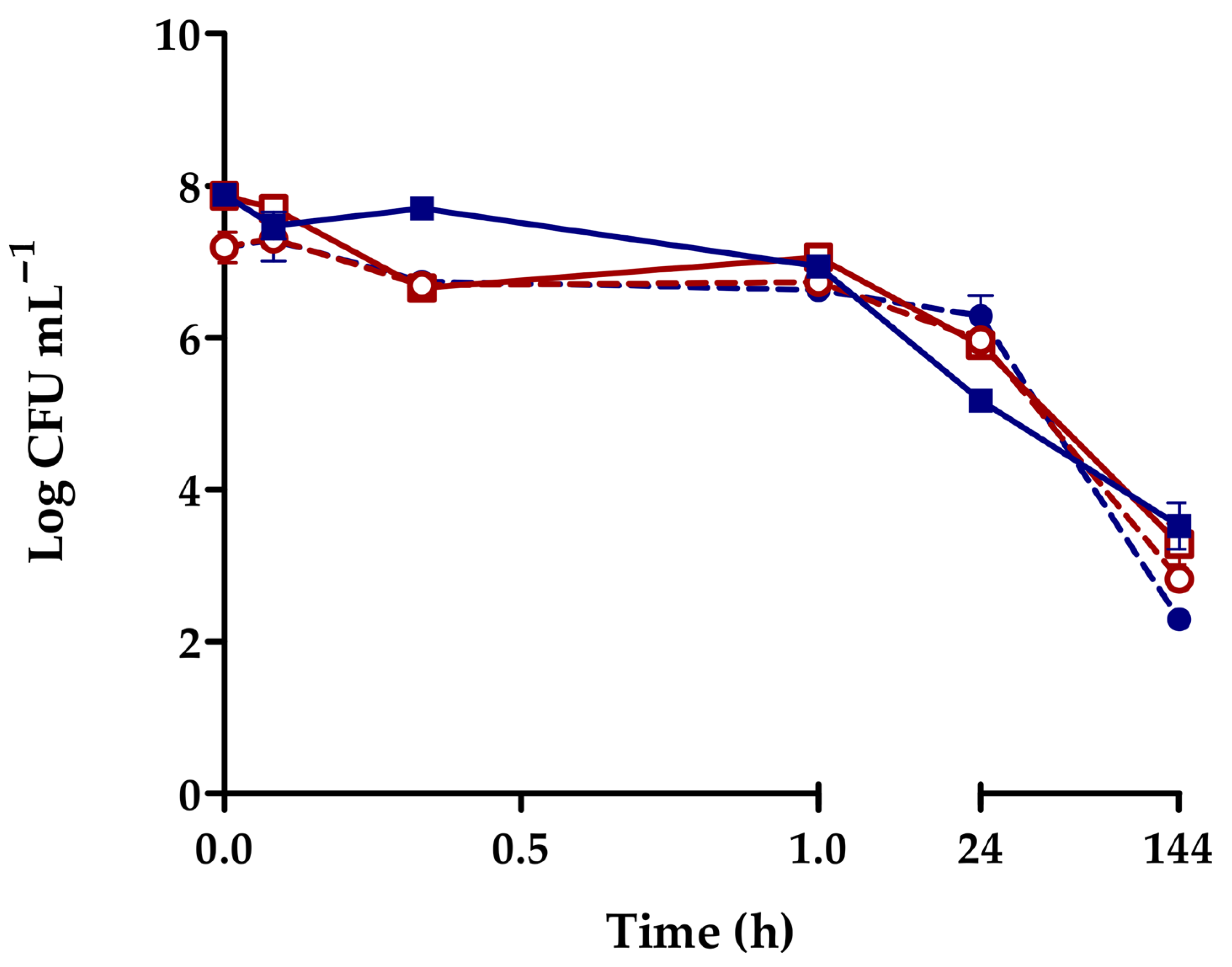
Mussels actively removed V. harveyi from both estuarine water and seawater at 12 °C and 20 °C, as evidenced by the changes in bacterial density in the surrounding water, which remained quite stable during the first hour of exposure, gradually declining thereafter. However, a complete elimination of the vibrios was not achieved, with levels remaining above 102–103 cells mL−1 after 6 days (Figure 2). The results were consistent for both culturable and total cells. In addition, feces and pseudofeces excreted by M. galloprovincialis were collected and analyzed at the end of the experiments. Similar V. harveyi gfp densities were recovered from these biodeposits, ranging from 1.55 × 10⁷ to 3.77 × 10⁷ CFU g−1 across all experimental conditions.

3.3. Histopathology and Immune Response of Mussels Exposed to V. harveyi

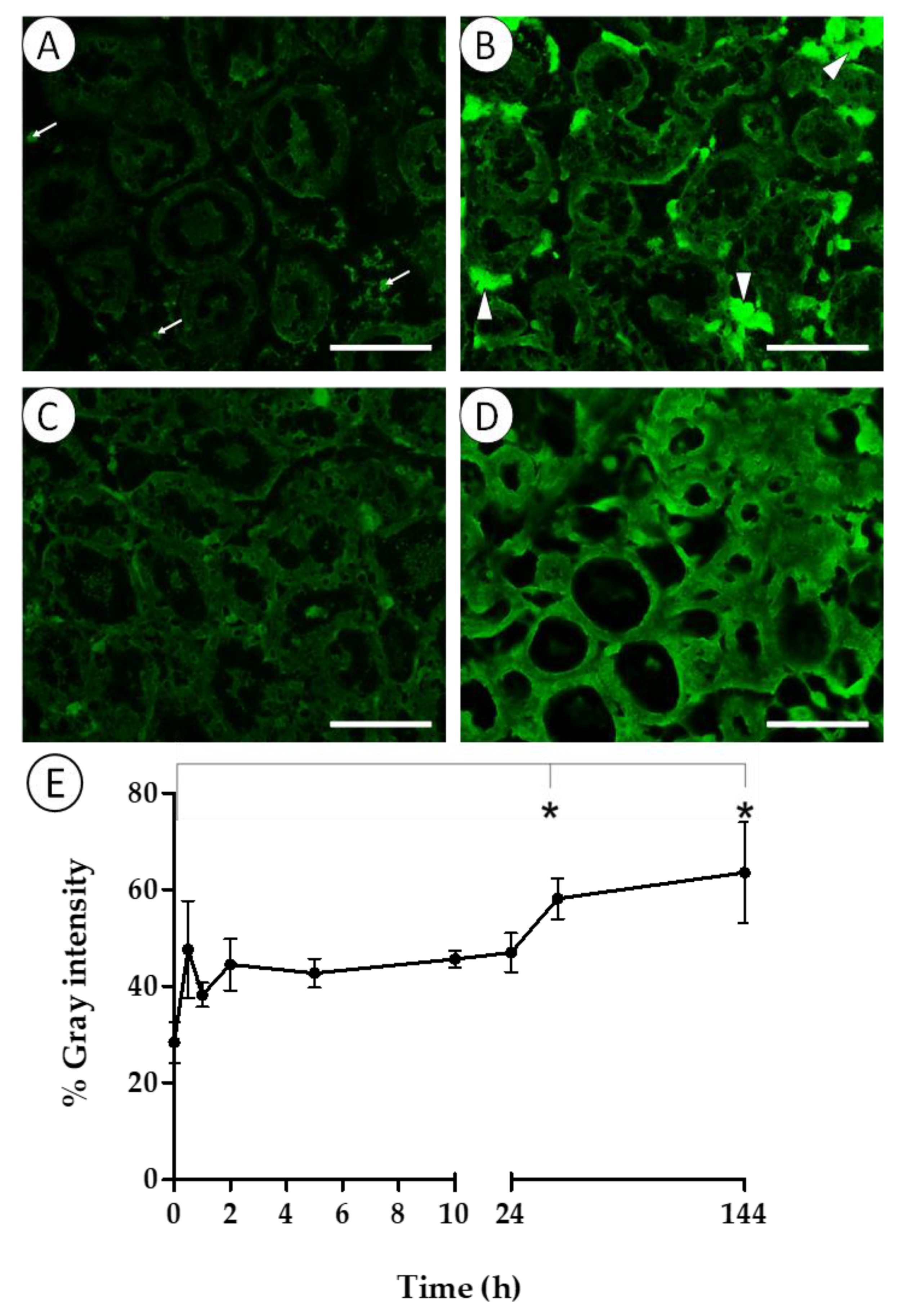
Epifluorescence microscopy revealed GFP-related fluorescence in the digestive epithelium and in cells such as hemocytes and adipogranular cells of specimens maintained in seawater at 20 °C (Figure 3A–D). The adipogranular cells showed extremely high fluorescent signals (Figure 3D); therefore, subsequent measurements focused on the epithelium of digestive diverticula. Fluorescence intensity increased after 30 min of exposure to V. harveyi gfp and continued to rise over time, with the highest signal intensity in the digestive epithelium after 144 h (Figure 3E). Significant differences (one-way ANOVA; F8,15 = 3.664; p = 0.013) in signal intensity were detected between 48 h and 144 h of exposure compared to the initial time point.
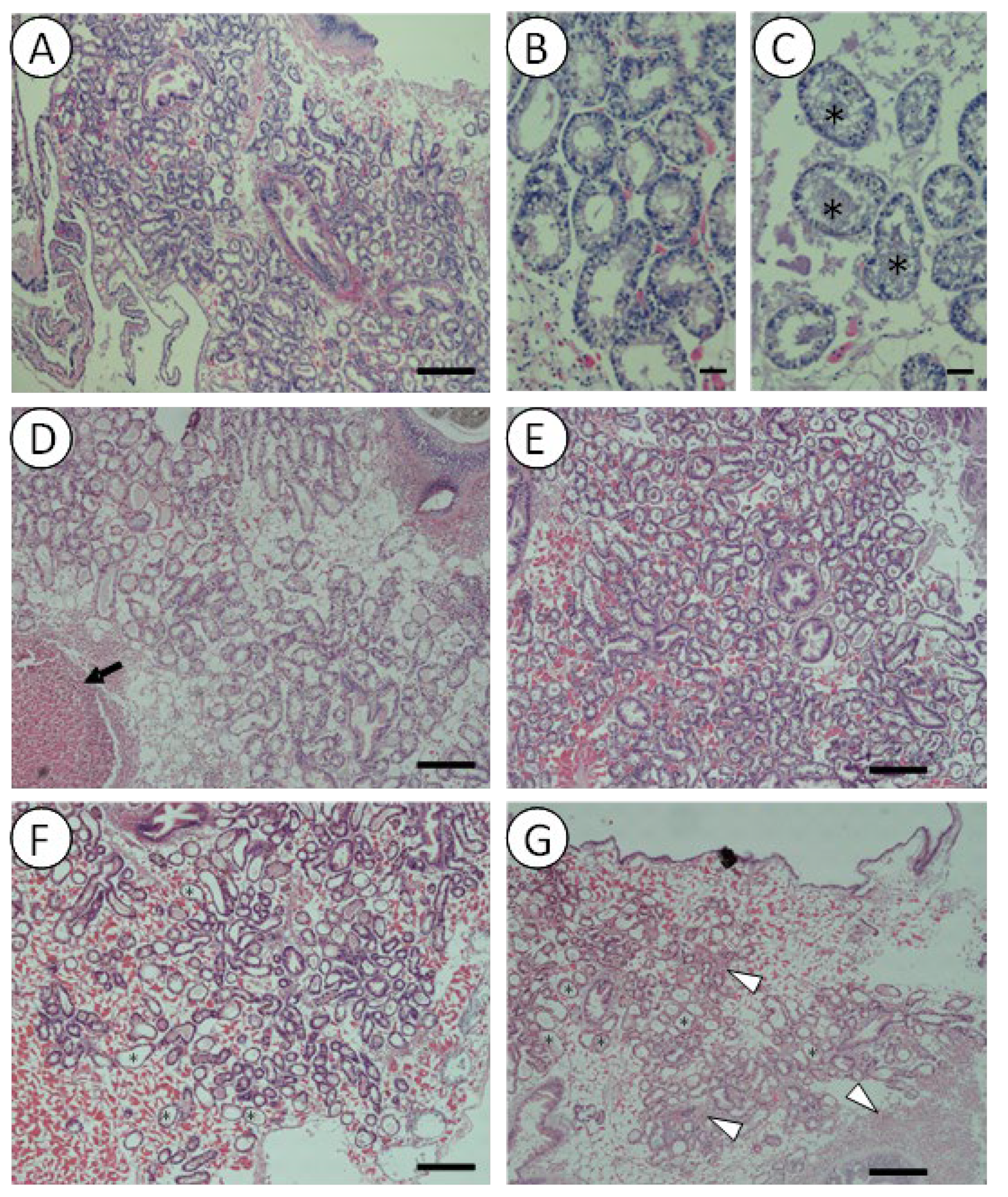
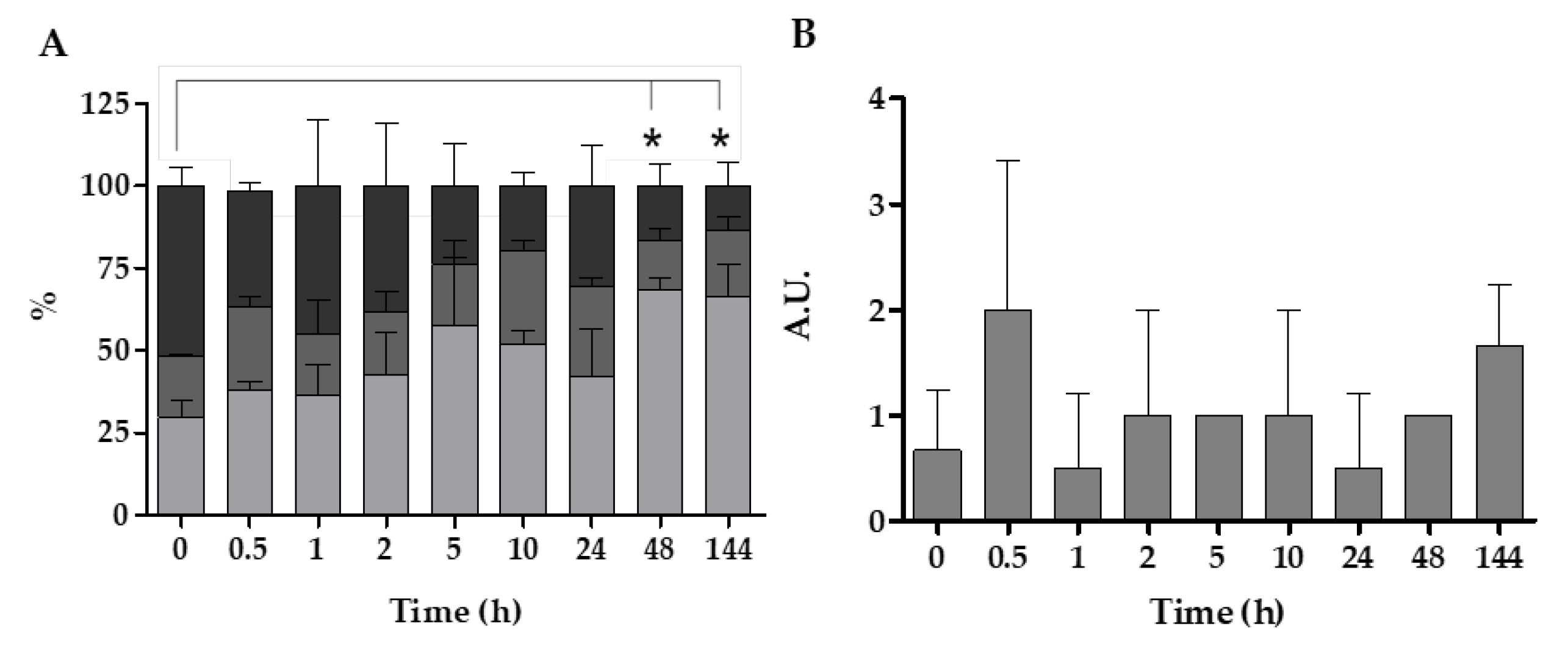
Histopathological analysis showed higher levels of altered health status in the digestive glands of mussels with longer exposure times to V. harveyi gfp. In general, after longer exposure times to V. harveyi, mussels presented higher interstitial connective tissue with a higher density percentage of atrophic digestive tubules or with thinner epithelium (Figure 4). Accordingly, they also presented a lower number of tubules in the resting or holding phase and an increased number of tubules in the atrophic phase, while tubules in the absorptive phase remained more or less constant throughout the experiment (Figure 5A). At this point, significant differences (one-way ANOVA; F8,15 = 3.802; p = 0.012) were also observed between 48 h and 144 h and time 0 of exposure.
Other immune system alterations, such as the presence of granulocytomas or hemocytic infiltrations, were also observed (Figure 4). The results indicate a rapid increase in the reaction of the immune system in the first 30 min of experimentation, with a slight decrease in the following hours, and a final increase after 144 h was also observed. However, the mentioned changes were not statistically significant, and immune system reactions remained mostly stable over time (Figure 5B).

4. Discussion

Marine bivalves are known to accumulate and concentrate different microorganisms in their tissues, and at times, they act as vectors for disease transmission [25,26,27]. Using a fluorescently labeled strain, we demonstrated in this study that the accumulation of vibrios is a rapid process, with the highest bacterial numbers detected in mussel organs within the first hour of exposure. This finding aligns with previous studies reporting peak accumulation times of 1 and 2 h for diverse bacterial species, including vibrios, in mussels [28,29] and other filter-feeding organisms such as clams [30].
Nevertheless, the existing literature provides limited insight regarding what occurs during exposure times shorter than one hour. Consequently, in the present study, we monitored the samples starting from the initial five minutes, a timeframe during which a substantial population of GFP-expressing vibrios was already detected within the mussel organs (Figure 1). Notwithstanding the inherent variability of mussels, the rapid appearance of vibrios in the organs is consistent with the high filtration rate of these mollusks. Indeed, assuming a filtration rate of 7.5 L h−1 [31], mussels are capable of filtering the entire volume of water contained in the experimental vessels in less than one minute.
The digestive gland was identified as the organ with the highest vibrio densities detected over time. This finding is not unexpected, given its critical function in food accumulation and both intracellular and extracellular digestion [32,33]. Although vibrios were predominantly concentrated in the digestive gland, comparable densities were also observed in the gills during the first few minutes, which reflects the importance of this organ in particle retention during filtration [9]. Similar accumulation patterns have been described in other bivalves; thus, Wang et al. [34] found the highest densities of V. parahaemolyticus in the digestive gland of oysters, followed by the gills.
The detection of V. harveyi in the gonads was lower, likely due to the contact of these organs with the pallial fluid rather than accumulation resulting from active ingestion. In natural conditions, pallial fluid contains higher bacterial densities than seawater due to its bioaccumulative capacity [35]. However, the gonads are covered by the gills and are therefore less exposed to pallial fluid.
After the initial accumulation of bacteria in mussel organs, their density began to decrease, mirroring the decline observed in the surrounding water. This reduction may result from bacterial digestion, elimination by the immune response of the mussel, or release through feces and pseudofeces. The first two processes could explain the increase in fluorescence detected in the digestive epithelium as the internal vibrio density decreased, since the lysis or digestion of vibrios would release GFP and thus increase fluorescence (Figure 3). Regardless, given the high filtration rate of mussels, the continuous presence of vibrios in the water suggests that only a fraction of the filtered microorganisms was effectively eliminated, while the remainder was returned to the medium, mainly as culturable bacteria, either free or as part of feces and pseudofeces. Accordingly, Williams et al. [36] demonstrated that part of the bacteria entering bivalves are eliminated through feces. When feces and pseudofeces are expelled, unlike the sessile organisms that generate them, they will be able to disperse via water currents, accumulate in superficial sediments, or be ingested by other organisms such as amphipods [37]. Thus, feces and pseudofeces containing pathogen microorganisms previously filtered by mussels may enter the food chain and contribute to disease dissemination [12,38].
Concurrently with bacterial accumulation in their organs, mussels responded by activating their immune system, as evidenced by the presence of alterations such as granulocytomas and hemocytic infiltrations (Figure 4 and Figure 5). This inflammatory response has been previously described in mussels exposed to different pathogens, toxins, or pollutants [18,39,40]. This process involves the recruitment of immune cells to the host tissues, which can itself cause tissue damage [41] and, consequently, may contribute to an increased percentage of atrophic digestive tubules. Furthermore, histological alterations in the digestive tubules may also result from microbial challenge, primarily affecting the digestive glands and leading to the thinning of the digestive tubule epithelium and changes in its profile and functionality [33], or even from starvation experienced by mussels during the assays.
Abiotic factors such as temperature and salinity play a role in the bioaccumulation of microorganisms in bivalves [42]. Fluctuations in salinity can influence growth, filtration rates, oxygen consumption, and immune function [43]; however, the halotolerance range remains unclear for most species [44]. In this study, salinity affected the differential retention of vibrios in the gills during the initial minutes of exposure, with lower retention observed in seawater compared to estuarine water (Figure 1). This fact may be associated with the role of Mytilus gills in modulating osmoregulation [45].
Previous studies have demonstrated that temperature significantly impacts the bioaccumulation of bacteria in M. edulis [42]. However, the literature on the effect of temperature on filtration rates has yielded conflicting conclusions. Thus, Chae et al. [46] observed higher effective depuration at slightly cold temperatures (15 °C), while Boroda et al. [47] suggested that, under cold stress, filter-feeding organisms tend to reduce their filtration rates by closing their shells as a physiological adjustment mechanism. Notably, the present study did not reveal significant differences in this regard, likely because the exposure temperatures used (12 °C and 20 °C) fall within the optimal temperature range for M. galloprovincialis persistence (10 °C and 32 °C) [47].

5. Conclusions

This study reveals that fluorescently labeled strains are a valuable tool for tracking bacterial dynamics in bivalves, enabling the precise quantification of Vibrio harveyi accumulation in Mytilus galloprovincialis organs. Our findings reveal the rapid bioaccumulation of vibrios, particularly in the digestive gland, in the first minutes of exposure across different environmental conditions. Although mussels responded to microbial challenge, evidenced by the presence of granulocytomas, hemocytic infiltrations, and increased fluorescence indicating bacterial digestion, V. harveyi persisted in tissues over time, establishing mussels as natural reservoirs. Moreover, the detection of viable vibrios in mussel biodeposits highlights their potential role in secondary transmission through feces and pseudofeces, emphasizing the dual ecological function of bivalves as both biofilters and vectors for aquatic pathogen dissemination.

Author Contributions

Conceptualization, I.A. and M.O.; formal analysis, A.A., I.A., B.Z. and M.O.; funding acquisition, I.A., B.Z. and M.O.; investigation, I.A.; methodology, A.A., F.O.U., M.G.-R., I.A.-V. and B.Z.; supervision, I.A. and M.O.; writing—original draft, I.A. and M.O.; writing—review and editing, A.A., I.A., B.Z. and M.O. All authors have read and agreed to the published version of the manuscript.

Funding

This research was funded by the Basque Government grant PRE_2021_1_0199 and research project MIMAS IT1657-22, as well as by the TED2021-132109BC21 award from the Ministry of Science and Innovation of the Spanish Government and the BlueAdapt grant 101057764 from the European Union.

Institutional Review Board Statement

The study was approved by the Ethics Committee on Research with Biological Agents and/or GMOs (CEIAB-UPV/EHU) (protocol code: M30_2021_164; 22 May 2021).

Informed Consent Statement

Not applicable.

Data Availability Statement

The data underlying this article will be shared on reasonable request to the corresponding author.

Conflicts of Interest

The authors declare no conflicts of interest.

References

  1. Thompson, F.L.; Iida, T.; Swings, J. Biodiversity of vibrios. Microbiol. Mol. Biol. Rev. 2004, 68, 403–431. [Google Scholar] [CrossRef] [PubMed]
  2. Erken, M.; Lutz, C.; McDougald, D. Interactions of Vibrio spp. with zooplankton. Microbiol. Spectr. 2014, 3. [Google Scholar]
  3. Romero, A.; Costa, M.M.; Forn-Cuni, G.; Balseiro, P.; Chamorro, R.; Dios, S.; Figueras, A.; Novoa, B. Occurrence, seasonality and infectivity of Vibrio strains in natural populations of mussels Mytilus galloprovincialis. Dis. Aquat. Org. 2014, 108, 149–163. [Google Scholar] [CrossRef] [PubMed]
  4. Albuixech-Martí, S.; Culloty, S.C.; Lynch, S.A. Co-occurrence of pathogen assemblages in a keystone species the common cockle Cerastoderma edule on the Irish coast. Parasitology 2021, 148, 1665–1679. [Google Scholar] [CrossRef]
  5. DeAngelis, C.M.; Saul-McBeth, J.; Matson, J.S. Vibrio responses to extracytoplasmic stress. Environ. Microbiol. Rep. 2018, 10, 511–521. [Google Scholar] [CrossRef]
  6. Orruño, M.; Parada, C.; Ogayar, E.; Kaberdin, V.R.; Arana, I. Effects of abiotic and biotic factors on Vibrio harveyi ATCC 14126T survival dynamics in seawater microcosms. Aquat. Microb. Ecol. 2019, 83, 109–118. [Google Scholar] [CrossRef]
  7. Zhang, X.-H.; He, X.; Austin, B. Vibrio harveyi: A serious pathogen of fish and invertebrates in mariculture. Mar. Life Sci. Tech. 2020, 2, 231–245. [Google Scholar] [CrossRef]
  8. Mohamad, N.; Amal, M.N.A.; Yasin, I.S.M.; Saad, M.Z.; Nasruddin, N.S.; Al-saari, N.; Sawabe, T. Vibriosis in cultured marine fishes: A review. Aquaculture 2019, 512, 734289. [Google Scholar] [CrossRef]
  9. Berg, D.J.; Fisher, S.W.; Landrum, P.F. Clearance and processing of algal particles by zebra mussels (Dreissena polymorpha). J. Great Lakes Res. 1996, 22, 779–788. [Google Scholar] [CrossRef]
  10. Lobo-da-Cunha, A. Structure and function of the digestive system in molluscs. Cell Tissue Res. 2019, 377, 475–503. [Google Scholar] [CrossRef]
  11. Birkbeck, T.H.; McHenery, J.G. Degradation of bacteria by Mytilus edulis. Mar. Biol. 1982, 72, 7–15. [Google Scholar] [CrossRef]
  12. Pietrak, M.R.; Molloy, S.D.; Bouchard, D.A.; Singer, J.T.; Bricknell, I. Potential role of Mytilus edulis in modulating the infectious pressure of Vibrio anguillarum 02β on an integrated multi-trophic aquaculture farm. Aquaculture 2012, 326–329, 36–39. [Google Scholar] [CrossRef]
  13. Masanja, F.; Yang, K.; Xu, Y.; He, G.; Liu, X.; Xu, X.; Jiang, X.; Luo, X.; Mkuye, R.; Deng, Y.; et al. Bivalves and microbes: A mini-review of their relationship and potential implications for human health in a rapidly warming ocean. Front. Mar. Sci. 2023, 10, 1182438. [Google Scholar] [CrossRef]
  14. Matozzo, V.; Ercolini, C.; Serracca, L.; Battistini, R.; Rossini, I.; Granato, G.; Quaglieri, E.; Perolo, A.; Finos, L.; Arcangeli, G.; et al. Assessing the health status of farmed mussels (Mytilus galloprovincialis) through histological, microbiological and biomarker analyses. J. Invertebr. Pathol. 2018, 153, 165–179. [Google Scholar] [CrossRef]
  15. Canesi, L.; Pruzzo, C. Specificity of innate immunity in bivalves: A lesson from bacteria. In Lessons in Immunity; Ballarin, L., Cammarata, M., Eds.; Elsevier: Amsterdam, The Netherlands, 2016; Chapter 6; pp. 79–91. [Google Scholar]
  16. Ottaviani, E. Immunocyte: The invertebrate counterpart of the vertebrate macrophage. Invertebr. Surviv. J. 2011, 8, 1–4. [Google Scholar]
  17. Vieira, G.C.; da Silva, P.M.; Barracco, M.A.; Hering, A.F.; Albuquerque, M.C.P.; Coelho, J.D.R.; Schmidt, É.C.; Bouzon, Z.; Rosa, R.D.; Perazzolo, L.M. Morphological and functional characterization of the hemocytes from the pearl oyster Pteria hirundo and their immune responses against Vibrio infections. Fish Shellfish. Immunol. 2017, 70, 750–758. [Google Scholar] [CrossRef]
  18. Carella, F.; Sardo, A.; Mangoni, O.; Di Cioccio, D.; Urciuolo, G.; De Vico, G.; Zingone, A. Quantitative histopathology of the Mediterranean mussel (Mytilus galloprovincialis L.) exposed to the harmful dinoflagellate Ostreopsis cf. ovata. J. Invertebr. Pathol. 2015, 127, 130–140. [Google Scholar] [CrossRef]
  19. Mijangos, L.; Ziarrusta, H.; Ros, O.; Kortazar, L.; Fernández, L.A.; Olivares, M.; Zuloaga, O.; Prieto, A.; Etxebarria, N. Occurrence of emerging pollutants in estuaries of the Basque Country: Analysis of sources and distribution, and assessment of the environmental risk. Water Res. 2018, 147, 152–163. [Google Scholar] [CrossRef]
  20. URA (Uraren Euskal Agentzia/Agencia Vasca del Agua). Available online: https://www.uragentzia.euskadi.eus/contenidos/documentacion/red_costa_2022/es_def/adjuntos/RSEETyC_2022_MEMORIA.pdf (accessed on 1 April 2024).
  21. Baña, Z.; Abad, N.; Uranga, A.; Azúa, I.; Artolozaga, I.; Unanue, M.; Iriberri, J.; Arrieta, J.M.; Ayo, B. Recurrent seasonal changes in bacterial growth efficiency, metabolism and community composition in coastal waters. Environ. Microbiol. 2020, 22, 369–380. [Google Scholar] [CrossRef]
  22. Bancroft, J.D.; Layton, C. The hematoxylins and eosin. In Bancroft’s Theory and Practice of Histological Techniques, 8th ed.; Suvarna, S.K., Layton, C., Bancroft, J.D., Eds.; Elsevier Publishing: Amsterdam, The Netherlands, 2019; Chapter 10; pp. 126–138. [Google Scholar]
  23. Villalba, A.; Mourelle, S.G.; López, M.C.; Carballal, M.J.; Azevedo, C. Marteiliasis affecting cultured mussels Mytilus galloprovincialis of Galicia (NW Spain). I. Etiology, phases of the infection, and temporal and spatial variability in prevalence. Dis. Aquat. Organ. 1993, 16, 61–72. [Google Scholar] [CrossRef]
  24. Langton, R.W. Synchrony in the digestive diverticula of Mytillus edulis L. J. Mar. Biol. Assoc. UK 1975, 55, 221–229. [Google Scholar] [CrossRef]
  25. Amoroso, M.G.; Langellotti, A.L.; Russo, V.; Martello, A.; Monin, M.; Di Bartolo, I.; Ianiro, G.; Di Concilio, D.; Galiero, G.; Fusco, G. Accumulation and depuration kinetics of rotavirus in mussels experimentally contaminated. Food Environ. Virol. 2019, 12, 48–57. [Google Scholar] [CrossRef] [PubMed]
  26. Mosteo, R.; Goñi, P.; Miguel, N.; Abadías, J.; Valero, P.; Ormad, M.P. Bioaccumulation of pathogenic bacteria and amoeba by zebra mussels and their presence in watercourses. Environ. Sci. Pollut. Res. Int. 2016, 23, 1833–1840. [Google Scholar] [CrossRef] [PubMed]
  27. Shapiro, K.; Silver, M.; Byrne, B.A.; Berardi, T.; Aguilar, B.; Melli, A.; Smith, W.A. Fecal indicator bacteria and zoonotic pathogens in marine snow and California mussels (Mytilus californianus). FEMS Microbiol. Ecol. 2018, 94, fiy172. [Google Scholar] [CrossRef]
  28. Marino, A.; Lombardo, L.; Fiorentino, C.; Orlandella, B.; Monticelli, L.; Nostro, A.; Alonzo, V. Uptake of Escherichia coli, Vibrio cholerae non-O1 and Enterococcus durans by, and depuration of mussels (Mytilus galloprovincialis). Int. J. Food Microbiol. 2005, 99, 281–286. [Google Scholar] [CrossRef]
  29. Herrfurth, D.; Oeleker, K.; Pund, R.-P.; Strauch, E.; Scheartz, K.; Leer, J.; Gölz, G.; Alter, T.; Huehn, S. Uptake and localization of Vibrio cholerae, Vibrio parahaemolyticus and Vibrio vulnificus in blue mussels (Mytilus edulis) of the Baltic Sea. J. Shellfish Res. 2013, 32, 855–859. [Google Scholar]
  30. Lopez-Joven, C.; de Blas, I.; Ruiz-Zarzuela, I.; Furones, M.D.; Roque, A. Experimental uptake and retention of pathogenic and nonpathogenic Vibrio parahaemolyticus in two species of clams: Ruditapes decussatus and Ruditapes philippinarum. J. Appl. Microbiol. 2011, 111, 197–208. [Google Scholar] [CrossRef]
  31. Figueras, A.; Moreira, R.; Sendra, M.; Novoa, B. Genomics and immunity of the Mediterranean mussel Mytilus galloprovincialis in a changing environment. Fish Shellfish Immunol. 2019, 90, 440–445. [Google Scholar] [CrossRef]
  32. Canesi, L.; Barmo, C.; Fabbri, R.; Ciacci, C.; Vergani, L.; Roch, P.; Gallo, G. Effects of Vibrio challenge on digestive gland biomarkers and antioxidant gene expression in Mytilus galloprovincialis. Comp. Biochem. Physiol. C Toxicol. Pharmacol. 2010, 152, 399–406. [Google Scholar] [CrossRef]
  33. Cuevas, N.; Zorita, I.; Costa, P.M.; Franco, J.; Larreta, J. Development of histopathological indices in the digestive gland and gonad of mussels: Integration with contamination levels and effects of confounding factors. Aquat. Toxicol. 2015, 162, 152–164. [Google Scholar] [CrossRef]
  34. Wang, D.; Yu, S.; Chen, W.; Zhang, D.; Shi, X. Enumeration of Vibrio parahaemolyticus in oyster tissues following artificial contamination and depuration. Lett. Appl. Microbiol. 2010, 51, 104–108. [Google Scholar] [CrossRef] [PubMed]
  35. Saco, A.; Rey-Campos, M.; Novoa, B.; Figueras, A. Transcriptomic response of mussel gills after a Vibrio splendidus infection demonstrates their role in the immune response. Front. Immunol. 2020, 11, 615580. [Google Scholar] [CrossRef] [PubMed]
  36. Williams, H.R.; Macey, B.M.; Burnett, L.E.; Burnett, K.G. Differential localization and bacteriostasis of Vibrio campbellii among tissues of the Eastern oyster, Crassostrea virginica. Dev. Comp. Immunol. 2009, 33, 592–600. [Google Scholar] [CrossRef]
  37. Kuehr, S.; Diehle, N.; Kaegi, R.; Schlechtriem, C. Ingestion of bivalve droppings by benthic invertebrates may lead to the transfer of nanomaterials in the aquatic food chain. Environ. Sci. Eur. 2021, 33, 35. [Google Scholar] [CrossRef]
  38. Sweat, L.H.; Busch, S.J.; Craig, C.A.; Dark, E.; Sailor-Tynes, T.; Wayles, J.; Sacks, P.E.; Walters, L.J. Oyster reefs are reservoirs for potential pathogens in a highly disturbed subtropical estuary. Environments 2023, 10, 205. [Google Scholar] [CrossRef]
  39. Ciacci, C.; Manti, A.; Canonico, B.; Campana, R.; Camisassi, G.; Baffone, W.; Canesi, L. Responses of Mytilus galloprovincialis hemocytes to environmental strains of Vibrio parahaemolyticus, Vibrio alginolyticus, Vibrio vulnificus. Fish Shellfish Immunol. 2017, 65, 80–87. [Google Scholar] [CrossRef]
  40. Gorbi, S.; Avio, G.C.; Benedetti, M.; Totti, C.; Accoroni, S.; Pichierri, S.; Bacchiocchi, S.; Orletti, R.; Graziosi, T.; Regoli, F. Effects of harmful dinoflagellate Ostreopsis cf. ovata exposure on immunological, histological and oxidative responses of mussels Mytilus galloprovincialis. Fish Shellfish Immunol. 2013, 35, 941–950. [Google Scholar] [CrossRef]
  41. De Vico, G.; Carella, F. Morphological features of the inflammatory response in molluscs. Res. Vet. Sci. 2012, 93, 1109–1115. [Google Scholar] [CrossRef]
  42. Olalemi, A.; Baker-Austin, C.; Ebdon, J.; Taylor, H. Bioaccumulation and persistence of faecal bacterial and viral indicators in Mytilus edulis and Crassostrea gigas. Int. J. Hyg. Environ. Health 2016, 219, 592–598. [Google Scholar] [CrossRef]
  43. Pourmozaffar, S.; Tamadoni Jahromi, S.; Rameshi, H.; Sadeghi, A.; Bagheri, T.; Behzadi, S.; Gozari, M.; Zahedi, M.R.; Abrari Lazarjani, S. The role of salinity in physiological responses of bivalves. Rev. Aquac. 2020, 12, 1548–1566. [Google Scholar] [CrossRef]
  44. Andreyeva, A.; Gostyukhina, O.; Gavruseva, T.; Sigacheva, T.; Tkachuk, A.; Podolskaya, M.; Chelebieva, E.; Kladchenko, E. Mediterranean mussels (Mytilus galloprovincialis) under salinity stress: Effects on antioxidant capacity and gill structure. J. Exp. Zool. A Ecol. Integr. Physiol. 2025, 343, 184–196. [Google Scholar] [CrossRef] [PubMed]
  45. Lockwood, B.L.; Somero, G.N. Transcriptomic responses to salinity stress in invasive and native blue mussels (genus Mytilus). Mol. Ecol. 2011, 20, 517–529. [Google Scholar] [CrossRef] [PubMed]
  46. Chae, M.J.; Cheney, D.; Su, Y.-C. Temperature effects on the depuration of Vibrio parahaemolyticus and Vibrio vulnificus from the American Oyster (Crassostrea virginica). J. Food Sci. 2009, 74, M62-6. [Google Scholar] [CrossRef] [PubMed]
  47. Boroda, A.V.; Kipryushina, Y.O.; Odintsova, N.A. The effects of cold stress on Mytilus species in the natural environment. Cell Stress Chaperones 2020, 25, 821–832. [Google Scholar] [CrossRef]
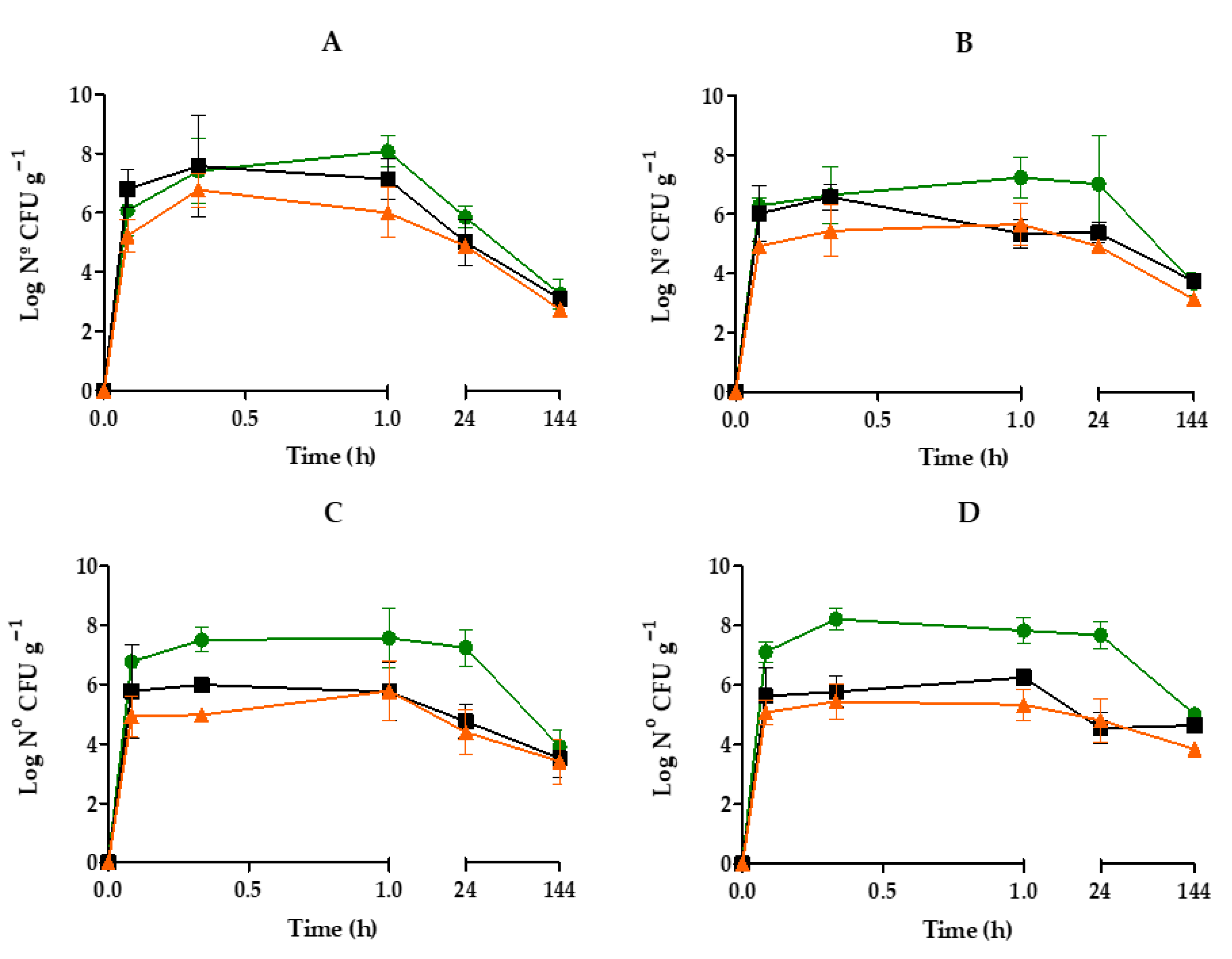
Figure 1. Dynamics of V. harveyi gfp in the different organs of M. galloprovincialis maintained in artificial estuarine water (A,B) and seawater (C,D) at 12 °C (A,C) or 20 °C (B,D). Gills (■), digestive gland (), and gonads (). The results show the mean values of three replicates (±SD).
Pathogens 14 00687 g001
Figure 2. Variation of culturable V. harveyi gfp in surrounding water in presence of M. galloprovincialis. Microcosms with artificial estuarine water (,) or seawater (,) maintained at 12 °C (closed symbols) and 20 °C (open symbols).
Pathogens 14 00687 g002
Figure 3. Fluorescence observed in the digestive glands of mussels maintained in seawater at 20 °C at time 0 (A), 2 h (B), 24 h (C), and 144 h (D) of exposure to V. harveyi gfp. White triangles indicate adipogranular cells and white arrows indicate hemocytes. Scale bar = 100 μm. Mean (±SD) fluorescence intensity converted to grayscale over the experimental time (E). Asterisks indicate significant differences (p < 0.05).
Pathogens 14 00687 g003
Figure 4. Micrographs of mussel digestive glands stained with Hematoxylin–Eosin at t0 (A,B) after 30 min (A,D), 2 h (E,B), 48 h (C,F), and 144 h (G,D) exposure to V. harveyi gfp in seawater at 20 °C. The white triangles indicate the hemocytic infiltrations; the black arrow indicates a granulocytoma and the asterisks, atrophic or disintegrating profiles. Scale bar = 100 μm (A,DG) and 25 µm (B,C).
Pathogens 14 00687 g004
Figure 5. Percentage of quantification (mean ± SD) of the different phases of the digestive tubules (A): █ holding, adsorbing, atrophic phases. Semi-quantification (A.U., arbitrary units) of the immune response (B) of mussels maintained in seawater at 20 °C and exposed to V. harveyi gfp. Asterisks indicate significant differences (p < 0.05).
Pathogens 14 00687 g005
Disclaimer/Publisher’s Note: The statements, opinions and data contained in all publications are solely those of the individual author(s) and contributor(s) and not of MDPI and/or the editor(s). MDPI and/or the editor(s) disclaim responsibility for any injury to people or property resulting from any ideas, methods, instructions or products referred to in the content.