Article

Copper-Induced Stimulation of Ectophosphatase Activity of Candida albicans

by
Anita Leocadio Freitas-Mesquita
1,2,
Fabiano Ferreira Esteves
1,2 and
José Roberto Meyer-Fernandes
1,2,*
1
Leopoldo de Meis Institute of Medical Biochemistry, Center of Health Science, Federal University of Rio de Janeiro, Rio de Janeiro 21941-590, RJ, Brazil
2
National Institute of Science and Technology in Structural Biology and Bioimaging, Rio de Janeiro 21941-590, RJ, Brazil
*
Author to whom correspondence should be addressed.
Pathogens 2025, 14(7), 667; https://doi.org/10.3390/pathogens14070667
Submission received: 30 May 2025 / Revised: 30 June 2025 / Accepted: 4 July 2025 / Published: 8 July 2025

Abstract

Candida albicans is an opportunistic fungal pathogen that can cause superficial and life-threatening infections, particularly in immunocompromised individuals. Its ability to adhere to host cells is critical for colonization and infection. In this context, investigating ectophosphatases is particularly relevant, as these enzymes have been associated with fungal adhesion to host cells. This study aimed to investigate the nature of copper-induced stimulation of ectophosphatase activity in C. albicans. Ectophosphatase activity was measured using p-nitrophenyl phosphate as substrate. Micromolar concentrations of CuCl2 markedly stimulated ectophosphatase activity, and its response to reducing agents and metal chelators suggested that this modulation does not involve redox reactions. The significant differences between the biochemical properties of basal (Cu2+-independent) and Cu2+-dependent ectophosphatase activities suggest the presence of at least two distinct ectophosphatases in C. albicans. Cu2+-independent ectophosphatase activity presented an acidic profile and was insensitive to Mg2+, whereas Cu2+-dependent ectophosphatase activity exhibited an alkaline profile and was also stimulated by Mg2+. Both activities were negatively modulated by classical phosphatase inhibitors, but Cu2+-dependent ectophosphatase had lower sensitivity compared to the basal activity. These findings highlight the role of copper as a modulator of C. albicans ectophosphatase activity and suggest potential implications for fungal adaptation during infection.

1. Introduction

Candida albicans was recently classified as a critical fungal pathogen in the World Health Organization (WHO)’s global priority list. This fungus is usually a harmless commensal but has the potential to become invasive, infectious, and even fatal by targeting numerous organ systems and acting as an opportunistic pathogen, especially in vulnerable, critically ill, and immunocompromised individuals [1,2,3]. C. albicans can cause two major types of infections in humans: superficial infections, such as oral or vaginal candidiasis, and life-threatening systemic infections [4]. Oral candidiasis has emerged as an early clinical clue for diagnosing acquired immunodeficiency syndrome (AIDS) and an indicator of its severity [5].
To establish infection, these opportunistic pathogens must evade the immune system, survive and replicate in the host environment, and spread to new tissues [6]. The expression of proteins involved in adhesion and invasion contributes significantly to the pathogenic potential of C. albicans. Adhesion is influenced by multiple factors, including the composition of cell wall proteins and the physical and chemical characteristics of the cell surface [7]. In this context, investigating ectophosphatases is particularly relevant, as these enzymes have been related to the adhesion of fungi to host cells [6,8,9,10,11,12].
Ectophosphatases are external surface membrane proteins widely distributed among various microorganisms, including numerous fungal species [13]. Several biochemical characterization studies have shown that ectophosphatase activities are typically modulated by divalent cations [13]. Notably, in Candida parapsilosis, a strong stimulation of ectophosphatase activity was observed in the presence of increasing millimolar concentrations of CuCl2 [10].
Copper is essential in biological systems due to its ability to transit between cuprous (Cu+) and cupric (Cu2+) oxidation, facilitating many important electron transfer reactions. Therefore, it acts as a prosthetic group for several enzymes involved in oxidation–reduction reactions, such as superoxide dismutase (SOD) [2,14]. On the other hand, copper can also promote the formation of potentially harmful reactive oxygen species via redox cycling between Cu2+/Cu+ states in the presence of dioxygen and physiological reducing agents [15]. To maintain copper homeostasis and prevent toxicity, C. albicans has developed complex regulatory mechanisms, including copper transporters and metal-binding proteins such as metallothioneins that sequester excess copper to avoid cellular damage [16].
In the present study, we found that micromolar concentrations of CuCl2 markedly stimulated the ectophosphatase activity of C. albicans. Considering the potential involvement of ectophosphatases in fungal infection and the importance of copper in various cellular processes, we aimed to investigate the nature of this stimulation. Additionally, we evaluated the differential biochemical responses between the basal and Cu2+-dependent ectophosphatase activities. Given the distinct biochemical profiles observed, we hypothesized that there are at least two distinct ectophosphatases in C. albicans.

2. Materials and Methods

2.1. Microorganisms and Growth Conditions

The experiments of this study were performed using the CAI-4 strain of Candida albicans (genotype Δura3::imm434/Δura3::imm434), an auxotrophic mutant for uridine obtained from the wild-type strain SC5314 [17]. This strain was used because its URA3-genetic construction facilitates molecular manipulations, which may be useful for future studies. C. albicans cells were cultivated at room temperature, under constant agitation, in Modified Sabouraud Medium (SAB-M; 2% glucose, 1% peptone, 0.5% yeast extract) supplemented with 50 µg/mL uridine. After 48 h of growth, the yeasts were washed three times with PBS (150 mM NaCl, 10 mM NaH2PO4, and 10 mM Na2HPO4, pH 7.2) and collected by centrifugation (1500× g, 10 min). Cell growth was estimated by counting the yeasts in a Neubauer chamber.

2.2. Materials

All reagents were obtained from E. Merck (D-6100 Darmstadt, Germany) or Sigma-Aldrich Chemical Co. (St. Louis, MO, USA). Distilled water was deionized using a Milli-Q resin system (Millipore Corp., Bedford, MA, USA) and was used for the preparation of all solutions.

2.3. Ectophosphatase Activity Measurements

The ectophosphatase activity of C. albicans was determined using p-nitrophenyl phosphate (p-NPP) as a substrate, measuring the rate of p-nitrophenol (p-NP) production. Intact cells (1 × 10⁸) were incubated for 1 h at room temperature in 0.5 mL of a reaction mixture containing, unless otherwise specified, 116 mM NaCl, 4 mM KCl, 5.4 mM D-glucose, 20 mM HEPES buffer (pH 7.4), and 5.0 mM p-NPP. Reactions were initiated by adding living cells or the substrate and stopet by adding 1.0 mL of 1 M NaOH. Absorbance was measured spectrophotometrically at 425 nm, using p-NP as a standard [18]. Ectophosphatase activity was calculated by subtracting nonspecific p-NPP hydrolysis measured in the absence of cells. Cell viability was assessed before and after incubations using the Trypan blue method [19]. Viability was not affected by the experimental conditions used in this study.

2.4. Statistical Analysis
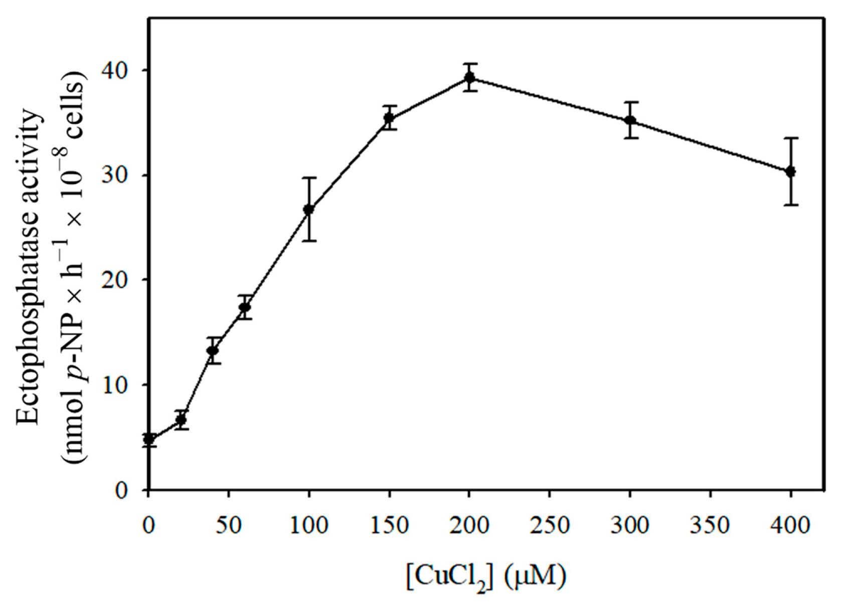
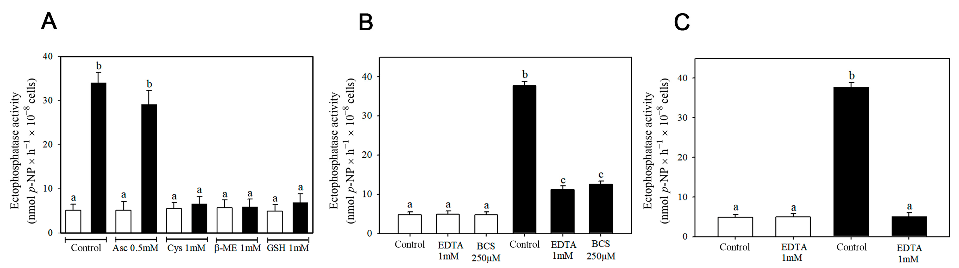
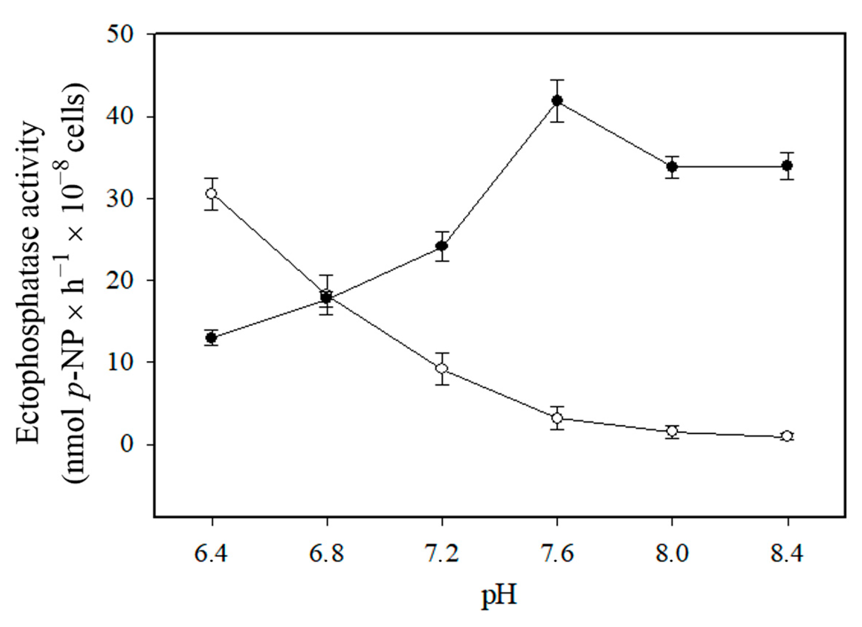
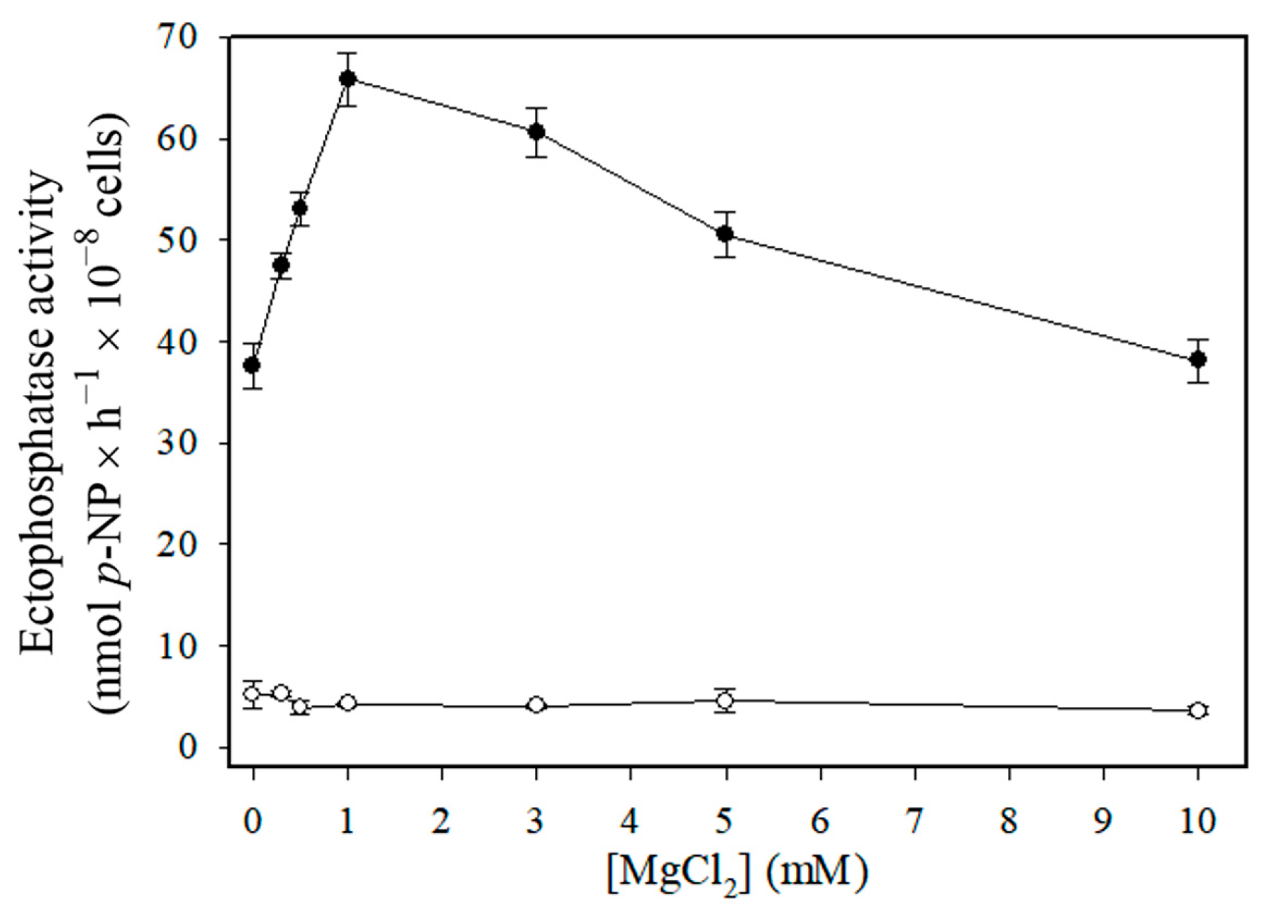
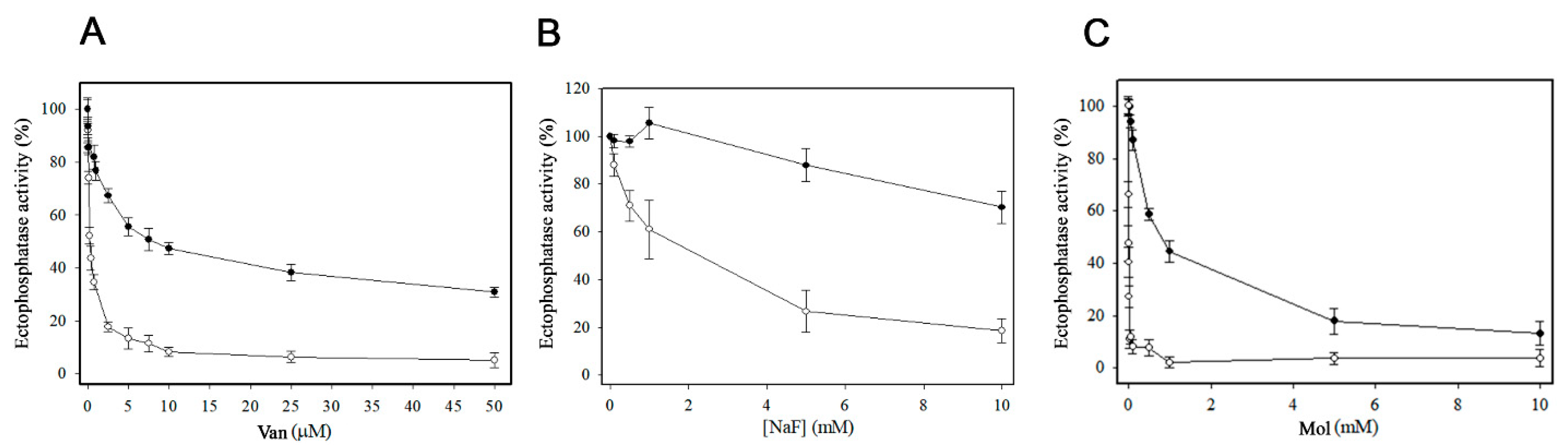
All experiments were performed in triplicate, and similar results were obtained from at least three independent cell suspensions. Data were analyzed statistically using one-way ANOVA followed by Tukey’s test, with GraphPad Prism software (GraphPad Software Inc., Version 8.0.1, San Diego, CA, USA). Results were considered statistically significant at p < 0.05, which was indicated by different lowercase letters above the bars.

3. Results

The ectophosphatase activity of Candida albicans increased in response to micromolar concentrations of CuCl2, reaching a maximum value (39.28 ± 1.32 nmols p-NP × 10−8 cells × h−1) at 200 µM CuCl2 (Figure 1). Therefore, all subsequent experiments were performed in the absence or presence of 200 µM CuCl2. The ectophosphatase activity measured after 30 min of pre-incubation with the metal was not significantly different from that without pre-incubation, indicating no dependence on pre-incubation.
Further assays were conducted to investigate the nature of this stimulation. To determine whether reducing agents could prevent copper-induced stimulation, ectophosphatase activity was measured in the presence of ascorbate (Asc), cysteine (Cys), β-mercaptoethanol (β-ME), and glutathione (GSH). Except for ascorbate, all other reducing agents tested prevented stimulation when added to the medium prior to the metal. In contrast, basal ectophosphatase activity was not affected by their presence (Figure 2A). To assess the reversibility of the copper-induced stimulation of ectophosphatase activity, we added the divalent ion chelator ethylenediaminetetraacetic acid (EDTA) or the copper-specific chelator bathocuproinedisulfonic acid disodium salt (BCS) to the reaction medium (Figure 2B). Although ectophosphatase activities remained significantly higher than in cells not exposed to copper, they were substantially lower than the ectophosphatase activity measured in the continuous presence of 200 µM CuCl2 due to the absence of chelators. It is possible that the chelators did not completely remove copper ions previously bound to the enzyme, thus accounting for this statistically significant difference. We also tested the effect of pre-incubating the cells with EDTA (Figure 2C), which resulted in a similar response to that observed with the reducing agents (Figure 2A), namely the reversal of the stimulation induced by CuCl2. It is important to note that basal ectophosphatase activity was not affected by chelators, whether they were added during the course of the reaction (Figure 2B) or pre-incubated with the cells (Figure 2C). These results suggest that copper stimulation does not involve the irreversible modification of C. albicans ectophosphatase.
To explore possible differences in biochemical properties, subsequent experiments compared the basal activity—referred to as Cu2+-independent ectophosphatase activity—with the Cu2+-dependent ectophosphatase activity of C. albicans. The Cu2+-dependent ectophosphatase activity was calculated by subtracting the Cu2+-independent activity from the activity measured in the presence of 200 µM CuCl2.
As shown in Figure 3, Cu2+-independent ectophosphatase activity increased under acidic pH conditions, whereas Cu2+-dependent ectophosphatase activity exhibited an alkaline profile. Unlike Cu2+-independent ectophosphatase activity, Cu2+-dependent ectophosphatase activity was stimulated by MgCl2, with maximal stimulation observed at 1 mM MgCl2 (Figure 4). Regarding the response to classical phosphatase inhibitors, we evaluated the effects of sodium orthovanadate (Van), sodium fluoride (NaF), and ammonium molybdate (Mol) on both Cu2+-dependent and Cu2+-independent ectophosphatase activities. All these compounds were able to inhibit both activities; however, Cu2+-dependent ectophosphatase activity was less sensitive to inhibition (Figure 5).

4. Discussion

Candida albicans is an opportunistic pathogen capable of causing a wide range of superficial and systemic infections, particularly under immunosuppressive conditions, such as those observed in HIV-positive patients [1,2,3]. In the present study, we identified a marked stimulation of ectophosphatase activity in the presence of micromolar concentrations of CuCl2 (Figure 1). Copper modulation has been previously described in phosphatases of other organisms. Beyond the previously mentioned study with C. parapsilosis [10], stimulatory effects of copper have been observed on the acid phosphatases of the protists Trypanosoma brucei [20] and Euglena gracilis [21], the yeast Yarrowia lipolytica [22], and the green alga Pseudokirchneriella subcapitata [23]. In Ophiocephalus punctatus (a freshwater fish), in vivo exposure to copper resulted in the activation of renal alkaline phosphatase [24].
Previous analyses of C. albicans isolates from the oral cavities of HIV-positive (HIV+) and HIV-negative (HIV) children revealed a strong association between ectophosphatase activity and HIV infection [6]. Interestingly, several studies have reported increased serum copper levels in HIV+ individuals, with differences ranging from approximately 1 to 10 µM [25,26,27,28]. Based on our findings, we hypothesize that the elevated ectophosphatase activity observed in HIV+ patients may be related to increased copper availability, potentially leading to enhanced enzymatic stimulation. Considering that serum copper levels in healthy individuals range from approximately 20 to 40 µM [25,26,27,28], the increase observed in HIV+ individuals is relatively subtle and may not directly justify a stimulation of C. albicans ectophosphatase activity. Nonetheless, it is important to emphasize that the periplasmic space constitutes a microenvironment rich in negatively charged components, which may favor the in situ accumulation of Cu2+ and thereby contribute to the increase in ectophosphatase activity. However, further studies will be necessary to better understand this possible association.
Regarding the nature of copper modulation, we found that pre-incubating intact cells with reducing agents—except ascorbate—completely prevented the stimulation of ectophosphatase activity induced by CuCl2 (Figure 2A). Therefore, this stimulation could be associated with conformational changes resulting from the oxidation of specific amino acid residues, due to the pro-oxidative nature of Cu2+ ions. In this scenario, the stimulatory effect would be expected to persist even after the removal of the metal. However, the addition of chelators to cells pre-incubated with the metal (Figure 2B) led to a significant reduction in ectophosphatase activity stimulation, suggesting that this effect is reversible. Furthermore, the pre-incubation of the cells with EDTA yielded similar results to those obtained with the reducing agents, preventing the stimulation of ectophosphatase activity by CuCl2 (Figure 2C). It is worth noting that reducing agents such as GSH and DTT can also act as metal chelators. The high nuclear charge of the Cu2+ ion favors the formation of a cupric–thiolate complex. GSH, for example, can coordinate and form a Cu2+-GSH complex in different conformations [29,30,31]. Considering that copper stimulation is a reversible effect and that only the reducing agents containing sulfhydryl groups were able to prevent the stimulation, we suggest that the action of Cu2+ on C. albicans ectophosphatase does not appear to involve a redox mechanism. However, since only ascorbate was tested as a non-thiol reducing agent, further studies using additional compounds are needed to fully confirm this hypothesis.
Since transition metals can induce oxidative stress through the production of hydroxyl radicals via the Fenton reaction [15,32], the stimulation of ectophosphatase activity could be associated with the presence of reactive oxygen species (ROS). If this hypothesis were correct, the generation of ROS due to the simultaneous presence of ascorbate and Cu2+ in the medium would be expected to increase the stimulation induced by the metal, as previously reported [33]. However, the level of stimulation promoted by the metal remained unchanged regardless of the presence or absence of ascorbate, allowing us to rule out this possibility (Figure 2A).
Therefore, the stimulation of ectophosphatase activity is likely dependent on the interaction between ectophosphatases and the Cu2+ ion. Considering that the primary ligands of Cu2+ ions in proteins are the imidazole ring of histidine residues and the thiol group of cysteine or methionine residues [34], it is plausible that such interactions induce conformational changes in ectophosphatases that enhance catalysis.
We observed significant differences between the biochemical properties of basal (Cu2+-independent) and Cu2+-dependent ectophosphatase activities. Since the experiments were conducted using intact cells, two possible explanations arise: (1) both activities are associated with the same enzyme, or (2) there are at least two distinct ectophosphatases in C. albicans. Considering the first hypothesis, the basal activity may reflect enzyme forms located in microenvironments less accessible to the metal ion or associated with cell wall components that limit their interaction with copper. However, given the distinct biochemical profiles observed, we consider it more plausible that two different enzymes are involved. Further studies using molecular biology and genetic manipulation techniques will be necessary to fully elucidate this question.
Cu2+-dependent ectophosphatase activity has a slightly alkaline optimum pH (7.6), while Cu2+-independent activity is much more effective at acidic pH (Figure 3). The existence of two enzymatic forms with different pH optima suggests a functional adaptation of C. albicans to different host niches, where pH can vary significantly. Cu2+-dependent activity also exhibited a significant and dose-dependent stimulation in the presence of MgCl2, while Cu2+-independent ectophosphatase activity was insensitive to this metal (Figure 4). This finding suggests a two-level regulatory mechanism, in which copper acts initially by promoting a conformational or functional change in the enzyme, then enabling the modulating action of magnesium. The modulation promoted by synergistic interaction between metal ions has been previously reported in phosphatases and other enzymatic activities [35,36].
Although both basal and Cu2+-independent and Cu2+-dependent ectophosphatase activities were inhibited by vanadate, sodium fluoride, and molybdate, Cu2+-dependent ectophosphatase activity presented lower sensitivity to all the inhibitors tested (Figure 5). This may reflect a conformational modulation promoted by copper binding, altering the active site microenvironment or the accessibility of inhibitors. In other enzyme systems, changes in cofactors have been attributed to changes in the main by-products or substrates [35]. The lower sensitivity observed may represent a functional advantage, allowing the enzyme to maintain its catalytic activity even in the presence of inhibitory compounds in the host extracellular environment.
In several studies [6,8,9,10,11,12], the irreversible profile of enzyme inhibition produced by vanadate has enabled the comparison of the ability of fungal cells to attach to host cells when ectophosphatase activity was fully functional versus when surface enzyme activity was inhibited by pretreatment with vanadate. The results suggest that ectophosphatase activity contributes to the adhesion process, potentially playing a key role in establishing infection [6,8,9,10,11,12]. In C. albicans, the pretreatment of yeast cells with vanadate significantly reduced their attachment to epithelial cells [6].
One of the most efficient host responses to fungal infections is a process termed nutritional immunity, a defense strategy that limits pathogen access to essential micronutrients or exposes them to toxic metal concentrations [37]. A widely recognized concept in innate immunity is host-imposed copper toxicity, in which elevated copper concentrations act as an effective biocide against microbial pathogens [38]. In this context, C. albicans also expresses cytosolic metallothioneins and a P1-type ATPase responsible for extruding Cu2+ ions across the plasma membrane, allowing fungal cells to survive even when exposed to high copper concentrations [16]. On the other hand, although serum copper levels increase, copper can become limiting in the kidney, the major site of C. albicans infection [39]. To overcome this condition, C. albicans readily adapts by increasing copper uptake and by exchanging metal cofactors for antioxidant SODs [2]. At the membrane level, copper exhibits high-affinity binding to phosphatidylserine residues, which can result in membrane damage. Accordingly, the plasma membrane architecture of C. albicans appears to be structured to protect the cell surface from copper-induced attack [14].
Taken together, our findings demonstrate that copper stimulates ectophosphatase activity in C. albicans, adding to our understanding of how this enzyme may function under copper fluctuations. Since previous studies have reported that ectophosphatases are involved in host–pathogen interactions, the copper-induced stimulation observed here could contribute to these processes. Further studies are needed to clarify this potential role.

Author Contributions

Conceptualization, F.F.E. and J.R.M.-F.; methodology, A.L.F.-M., F.F.E. and J.R.M.-F.; software, A.L.F.-M. and F.F.E.; validation, A.L.F.-M., F.F.E. and J.R.M.-F.; formal analysis, A.L.F.-M., F.F.E. and J.R.M.-F.; investigation, A.L.F.-M., F.F.E. and J.R.M.-F.; resources, J.R.M.-F.; data curation, A.L.F.-M., F.F.E. and J.R.M.-F.; writing—original draft preparation, A.L.F.-M.; writing—review and editing, F.F.E. and J.R.M.-F.; visualization, A.L.F.-M., F.F.E. and J.R.M.-F.; supervision, J.R.M.-F.; project administration, J.R.M.-F.; funding acquisition, J.R.M.-F. All authors have read and agreed to the published version of the manuscript.

Funding

This research was funded by the Brazilian agencies Conselho Nacional de Desenvolvimento Científico e Tecnológico (CNPq), grant number 310670/2021-7; Coordenação de Aperfeiçoamento de Pessoal de Nível Superior (CAPES), grant number 0012017; and Fundação Carlos Chagas Filho de Amparo à Pesquisa do Estado do Rio de Janeiro (FAPERJ), grant number E-26/201.136/2021.

Institutional Review Board Statement

Ethical review and approval were waived for this study since we did not use animal experimentation.

Informed Consent Statement

Patient consent was waived since we do not work with patients in this study.

Data Availability Statement

The data that support the findings of this study are available in Section 2.

Acknowledgments

We would like to thank Rosangela Rosa de Araújo for their excellent technical assistance.

Conflicts of Interest

The authors declare no conflicts of interest.

Abbreviations

The following abbreviations are used in this manuscript:
p-NPPp-nitrophenyl phosphate
AscAscorbate
CysCysteine
β-MEβ-mercaptoethanol
GSHGlutathione
EDTAEthylenediaminetetraacetic acid
BCSBathocuproinedisulfonic acid disodium salt
VanSodium orthovanadate
NaFSodium fluoride
MolAmmonium molybdate
ROSReactive oxygen species

References

  1. Kriegl, L.; Egger, M.; Boyer, J.; Hoenigl, M.; Krause, R. New treatment options for critically important WHO fungal priority pathogens. Clin. Microbiol. Infect. 2024, in press. [Google Scholar] [CrossRef]
  2. Li, C.X.; Gleason, J.E.; Zhang, S.X.; Bruno, V.M.; Cormack, B.P.; Culotta, V.C. Candida albicans adapts to host copper during infection by swapping metal cofactors for superoxide dismutase. Proc. Natl. Acad. Sci. USA 2015, 112, E5336–E5342. [Google Scholar] [CrossRef]
  3. Miceli, M.H.; Díaz, J.A.; Lee, S.A. Emerging opportunistic yeast infections. Lancet Infect. Dis. 2011, 11, 142–151. [Google Scholar] [CrossRef]
  4. Mayer, F.L.; Wilson, D.; Hube, B. Candida albicans pathogenicity mechanisms. Virulence 2013, 4, 119–128. [Google Scholar] [CrossRef]
  5. Keyvanfar, A.; Najafiarab, H.; Talebian, N.; Falah Tafti, M.; Adeli, G.; Ghasemi, Z.; Tehrani, S. Drug-resistant oral candidiasis in patients with HIV infection: A systematic review and meta-analysis. BMC Infect. Dis. 2024, 24, 546. [Google Scholar] [CrossRef]
  6. Portela, M.B.; Kneipp, L.F.; Ribeiro de Souza, I.P.; Holandino, C.; Alviano, C.S.; Meyer-Fernandes, J.R.; de Araújo Soares, R.M. Ectophosphatase activity in Candida albicans influences fungal adhesion: Study between HIV-positive and HIV-negative isolates. Oral Dis. 2010, 16, 431–437. [Google Scholar] [CrossRef]
  7. Talapko, J.; Juzbašić, M.; Matijević, T.; Pustijanac, E.; Bekić, S.; Kotris, I.; Škrlec, I. Candida albicans—The virulence factors and clinical manifestations of infection. J. Fungi 2021, 7, 79. [Google Scholar] [CrossRef]
  8. Collopy-Junior, I.; Esteves, F.F.; Nimrichter, L.; Rodrigues, M.L.; Alviano, C.S.; Meyer-Fernandes, J.R. An ectophosphatase activity in Cryptococcus neoformans. FEMS Yeast Res. 2006, 6, 1010–1017. [Google Scholar] [CrossRef]
  9. Cosentino-Gomes, D.; Rocco-Machado, N.; Santi, L.; Broetto, L.; Vainstein, M.H.; Meyer-Fernandes, J.R.; Schrank, A.; Beys-da-Silva, W.O. Inhibition of ectophosphatase activity in conidia reduces adhesion and virulence of Metarhizium anisopliae on the host insect Dysdercus peruvianus. Curr. Microbiol. 2013, 66, 467–474. [Google Scholar] [CrossRef]
  10. Kiffer-Moreira, T.; Pinheiro, A.A.S.; Alviano, W.S.; Barbosa, F.M.; Souto-Padrón, T.; Nimrichter, L.; Rodrigues, M.L.; Alviano, C.S.; Meyer-Fernandes, J.R. An ectophosphatase activity in Candida parapsilosis influences the interaction of fungi with epithelial cells. FEMS Yeast Res. 2007, 7, 621–628. [Google Scholar] [CrossRef]
  11. Kneipp, L.F.; Magalhães, A.S.; Abi-Chacra, E.A.; Souza, L.O.P.; Alviano, C.S.; Santos, A.L.S.; Meyer-Fernandes, J.R. Surface phosphatase in Rhinocladiella aquaspersa: Biochemical properties and its involvement with adhesion. Med. Mycol. 2012, 50, 570–578. [Google Scholar] [CrossRef]
  12. Kneipp, L.F.; Rodrigues, M.L.; Holandino, C.; Esteves, F.F.; Souto-Padrón, T.; Alviano, C.S.; Travassos, L.R.; Meyer-Fernandes, J.R. Ectophosphatase activity in conidial forms of Fonsecaea pedrosoi is modulated by exogenous phosphate and influences fungal adhesion to mammalian cells. Microbiology 2004, 150, 3355–3362. [Google Scholar] [CrossRef]
  13. Freitas-Mesquita, A.L.; Meyer-Fernandes, J.R. Biochemical properties and possible roles of ectophosphatase activities in fungi. Int. J. Mol. Sci. 2014, 15, 2289–2304. [Google Scholar] [CrossRef]
  14. Douglas, L.M.; Konopka, J.B. Plasma membrane architecture protects Candida albicans from killing by copper. PLoS Genet. 2019, 15, e1007911. [Google Scholar] [CrossRef]
  15. Falcone, E.; Stellato, F.; Vileno, B.; Bouraguba, M.; Lebrun, V.; Ilbert, M.; Morante, S.; Faller, P. Revisiting the pro-oxidant activity of copper: Interplay of ascorbate, cysteine, and glutathione. Metallomics 2023, 15, mfad040. [Google Scholar] [CrossRef]
  16. Weissman, Z.; Berdicevsky, I.; Cavari, B.Z.; Kornitzer, D. The high copper tolerance of Candida albicans is mediated by a P-type ATPase. Proc. Natl. Acad. Sci. USA 2000, 97, 3520–3525. [Google Scholar] [CrossRef]
  17. Fonzi, W.A.; Irwin, M.Y. Isogenic strain construction and gene mapping in Candida albicans. Genetics 1993, 134, 717–728. [Google Scholar] [CrossRef]
  18. Fernandes, E.C.; Meyer-Fernandes, J.R.; Silva-Neto, M.A.C.; Vercesi, A.E. Trypanosoma brucei: Ecto-phosphatase activity on the surface of intact procyclic forms. Z. Naturforsch. C 1997, 52, 351–355. [Google Scholar] [CrossRef]
  19. Patterson, M.K., Jr. Measurement of growth and viability of cells in culture. Methods Enzymol. 1979, 58, 141–152. [Google Scholar]
  20. Fernandes, E.C.; Granjeiro, J.M.; Aoyama, H.; Fonseca, F.V.; Meyer-Fernandes, J.R.; Vercesi, A.E. A metallo phosphatase activity present on the surface of Trypanosoma brucei procyclic forms. Vet. Parasitol. 2003, 118, 19–28. [Google Scholar] [CrossRef]
  21. Freitas-Mesquita, A.L.; Carvalho-Kelly, L.F.; Majerowicz, T.S.S.; Meyer-Fernandes, J.R. Euglena gracilis: Biochemical properties of a membrane bound ecto-phosphatase activity modulated by fluoroaluminate complexes and different trophic conditions. Eur. J. Protistol. 2023, 91, 126010. [Google Scholar] [CrossRef] [PubMed]
  22. Ito, H.; Inouhe, M.; Tohoyama, H.; Joho, M. Effect of copper on acid phosphatase activity in yeast Yarrowia lipolytica. Z. Naturforsch. C 2007, 62, 70–76. [Google Scholar] [CrossRef]
  23. Jonsson, C.M.; Aoyama, H. Effect of copper on the activation of the acid phosphatase from the green algae Pseudokirchneriella subcapitata. Biometals 2010, 23, 93–98. [Google Scholar] [CrossRef]
  24. Srivastava, D.K.; Pandey, K.C. Effect of copper on tissue acid and alkaline phosphatases in the green snakehead, Ophiocephalus punctatus (Bloch). Toxicol. Lett. 1982, 11, 237–241. [Google Scholar] [CrossRef] [PubMed]
  25. Graham, N.M.H.; Sorensen, D.; Odaka, N.; Brookmeyer, R.; Chan, D.; Willett, W.C.; Morris, J.S.; Saah, A.J. Relationship of serum copper and zinc levels to HIV-1 seropositivity and progression to AIDS. J. Acquir. Immune Defic. Syndr. 1991, 4, 976–980. [Google Scholar]
  26. Kassu, A.; Yabutani, T.; Mulu, A.; Tessema, B.; Ota, F. Serum zinc, copper, selenium, calcium, and magnesium levels in pregnant and non-pregnant women in Gondar, Northwest Ethiopia. Biol. Trace Elem. Res. 2008, 122, 97–106. [Google Scholar] [CrossRef] [PubMed]
  27. Moreno, T.; Artacho, R.; Navarro, M.; Pérez, A.; Ruiz-López, M.D. Serum copper concentration in HIV-infection patients and relationships with other biochemical indices. Sci. Total Environ. 1998, 217, 21–26. [Google Scholar] [CrossRef]
  28. Onwuli, D.; Waribo-Anthony, H.; Anyalebechi, O.; Ugochukwu, C. CD4+ T-Cell Count, Serum Zinc, Copper and Selenium Levels in HIV Sero-Positive Subjects on ART and ART Naïve Subjects in Port Harcourt, Nigeria. J. Biosci. Med. 2022, 10, 105–116. [Google Scholar]
  29. Ding, X.; Hua, Y.; Chen, Y.; Zhang, C.; Kong, X. Heavy metal complexation of thiol-containing peptides from soy glycinin hydrolysates. Int. J. Mol. Sci. 2015, 16, 8040–8058. [Google Scholar] [CrossRef]
  30. Krężel, A.; Lesniak, W.; Jezowska-Bojczuk, M.; Mlynarz, P.; Brasuñ, J.; Kozlowski, H.; Bal, W. Coordination of heavy metals by dithiothreitol, a commonly used thiol group protectant. J. Inorg. Biochem. 2001, 84, 77–88. [Google Scholar] [CrossRef]
  31. Ufnalska, I.; Drew, S.C.; Zhukov, I.; Szutkowski, K.; Wawrzyniak, U.E.; Wróblewski, W.; Frączyk, T.; Bal, W. Intermediate Cu(II)-thiolate species in the reduction of Cu(II)GHK by glutathione: A handy chelate for biological Cu(II) reduction. Inorg. Chem. 2021, 60, 18048–18057. [Google Scholar] [CrossRef] [PubMed]
  32. Shen, J.; Griffiths, P.T.; Campbell, S.J.; Utinger, B.; Kalberer, M.; Paulson, S.E. Ascorbate oxidation by iron, copper and reactive oxygen species: Review, model development, and derivation of key rate constants. Sci. Rep. 2021, 11, 7417. [Google Scholar] [CrossRef]
  33. Paletta-Silva, R.; Vieira-Bernardo, R.; Cosentino-Gomes, D.; Meyer-Fernandes, J.R. Leishmania amazonensis: Inhibition of 3’-nucleotidase activity by Cu2⁺ ions. Exp. Parasitol. 2012, 131, 63–68. [Google Scholar] [CrossRef] [PubMed]
  34. Rubino, J.T.; Franz, K.J. Coordination chemistry of copper proteins: How nature handles a toxic cargo for essential function. J. Inorg. Biochem. 2012, 107, 129–143. [Google Scholar] [CrossRef] [PubMed]
  35. Hung, H.-C.; Chang, G.-G. Differentiation of the slow-binding mechanism for magnesium ion activation and zinc ion inhibition of human placental alkaline phosphatase. Protein Sci. 2001, 10, 131–140. [Google Scholar] [CrossRef]
  36. Nam, K.; Arattu Thodika, A.R.; Tischlik, S.; Phoeurk, C.; Nagy, T.M.; Schierholz, L.; Ådén, J.; Rogne, P.; Drescher, M.; Sauer-Eriksson, A.E.; et al. Magnesium-induced structural reorganization in the active site of adenylate kinase. Sci. Adv. 2024, 10, eado5504. [Google Scholar] [CrossRef]
  37. Mackie, J.; Szabo, E.K.; Urgast, D.S.; Ballou, E.R.; Childers, D.S.; MacCallum, D.M.; Feldmann, J.; Brown, A.J.P. Host-imposed copper poisoning impacts fungal micronutrient acquisition during systemic Candida albicans infections. PLoS ONE 2016, 11, e0158683. [Google Scholar] [CrossRef]
  38. Li, C.; Li, Y.; Ding, C. The role of copper homeostasis at the host-pathogen axis: From bacteria to fungi. Int. J. Mol. Sci. 2019, 20, 175. [Google Scholar] [CrossRef]
  39. Jawale, C.V.; Biswas, P.S. Local antifungal immunity in the kidney in disseminated candidiasis. Curr. Opin. Microbiol. 2021, 62, 1–7. [Google Scholar] [CrossRef]
Figure 1. Influence of increasing CuCl2 concentrations on ectophosphatase activity of Candida albicans. Ectophosphatase activity was determined in the presence of increasing CuCl2 concentrations (0–400 µM). Values are expressed as the mean ± standard error from at least three independent experiments performed in triplicate.
Pathogens 14 00667 g001
Figure 2. Effect of reducing agents and metal chelators on the CuCl2-induced stimulation in ectophosphatase activity of Candida albicans. Fungal cells were pre-incubated with the reducing agents—ascorbate (Asc), cysteine (Cys), reduced glutathione (GSH), and β-mercaptoethanol (β-ME)—for 30 min prior to the start of the reaction. In the control group, no reducing agents were added. Ectophosphatase activity was measured in the absence (white bars) or presence of 200 µM CuCl2 (black bars), which was added 15 min after the addition of reducing agents (A). C. albicans cells were pre-incubated with buffer in the absence (white bars) or presence of 200 µM CuCl2 (black bars) for 30 min prior to the start of the reaction. After 15 min of pre-incubation, the chelating agents EDTA (1 mM) or BCS (250 µM) were added. Following an additional 15 min, ectophosphatase activity was measured in the absence (controls) or presence of the respective chelating agents (B). Fungal cells were pre-incubated with EDTA (1 mM) for 30 min prior to the start of the reaction. Control conditions were pre-incubated in the absence of the chelator. Ectophosphatase activity was then measured in the absence (white bars) or presence of 200 µM CuCl2 (black bars), which was added 15 min after the addition of EDTA (C). Values are expressed as the mean ± standard error from at least three independent experiments performed in triplicate. Different lowercase letters indicate statistically significant differences (p < 0.05).
Pathogens 14 00667 g002
Figure 3. Effect of pH on Cu2+-independent and Cu2+-dependent ectophosphatase activities of Candida albicans. Ectophosphatase activity was measured at different pH values in the absence or presence of 200 µM CuCl2. Cu2+-dependent ectophosphatase activity (black circles) was calculated by subtracting the Cu2+-independent activity (white circles) from the activity measured in the presence of 200 µM CuCl2. Values are expressed as the mean ± standard error from at least three independent experiments performed in triplicate.
Pathogens 14 00667 g003
Figure 4. Influence of increasing concentrations of MgCl2 on Cu2+-independent and Cu2+-dependent ectophosphatase activities of Candida albicans. Ectophosphatase activity was measured in the presence of increasing MgCl2 concentrations (1–10 mM), in the absence or presence of 200 µM CuCl2. Cu2+-dependent ectophosphatase activity (black circles) was calculated by subtracting the Cu2+-independent activity (white circles) from the activity measured in the presence of 200 µM CuCl2. Values are expressed as the mean ± standard error from at least three independent experiments performed in triplicate.
Pathogens 14 00667 g004
Figure 5. Influence of classical phosphatase inhibitors on Cu2+-independent and Cu2+-dependent ectophosphatase activities of Candida albicans. Ectophosphatase activity was measured in the presence of increasing concentrations of classical phosphatase inhibitors—sodium orthovanadate (Van; 1–50 µM, (A)), sodium fluoride (NaF; 1–10 mM, (B)), and ammonium molybdate (Mol; 1–10 mM, (C))—in the absence or presence of 200 µM CuCl2. Cu2+-dependent ectophosphatase activity (black circles) was calculated by subtracting the Cu2+-independent activity (white circles) from the activity measured in the presence of 200 µM CuCl2. Values are expressed as the mean ± standard error from at least three independent experiments performed in triplicate.
Pathogens 14 00667 g005
Disclaimer/Publisher’s Note: The statements, opinions and data contained in all publications are solely those of the individual author(s) and contributor(s) and not of MDPI and/or the editor(s). MDPI and/or the editor(s) disclaim responsibility for any injury to people or property resulting from any ideas, methods, instructions or products referred to in the content.

