Article

Genetic Identification of Brazilian Mammalian Hosts of Trypanosoma cruzi: Improving Blood Meal Source Discrimination in Vector-Borne Transmission

by
Quezia Moura Oliveira
1,2,†,
Thaíla Santos Pessanha
1,† and
Alena Mayo Iñiguez
1,*
1
Laboratório de Parasitologia Integrativa e Paleoparasitologia—LPIP, Instituto Oswaldo Cruz, Fiocruz, Avenida Brasil 4365, Rio de Janeiro 21040-360, RJ, Brazil
2
Programa de Pós-Graduação em Biologia Parasitária, Instituto Oswaldo Cruz, Fiocruz, Avenida Brasil 4365, Rio de Janeiro 21040-360, RJ, Brazil
*
Author to whom correspondence should be addressed.
These authors contributed equally to this work.
Pathogens 2025, 14(6), 579; https://doi.org/10.3390/pathogens14060579
Submission received: 1 April 2025 / Revised: 2 June 2025 / Accepted: 5 June 2025 / Published: 10 June 2025

Abstract

:
The detection of food sources of blood-sucking vectors is essential for a better understanding of the hosts, reservoirs, and other fauna that participate in the transmission web of hemoparasites. The molecular identification of triatomine blood meal sources (BMSs) has been shown to be highly sensitive and taxonomically specific when compared to the immunological method. The application of molecular cloning makes it possible to identify multiple BMS species and/or different individuals/haplotypes of the same vertebrate species in a single triatomine specimen. In Brazil, the molecular detection of BMSs is incipient, with insufficient genetic information on the species of animals involved in the transmission of Trypanosoma cruzi. In this work, we evaluated the sensitivity and specificity of a molecular approach using molecular cloning for the detection of multiple Brazilian mammalian species. The DNA was extracted from blood clots of 13 species of canids, bats, xenarthral, marsupials, and rodents. Serial proportions were used to formulate mixtures combining taxonomically close (belonging to the same family or order) and taxonomically distant (different families) species. The results showed that GenBank lacks reference sequences for some native species tested, such as the sylvatic rodent, Necromys lasiurus, and the wild canid, Lycalopex gymnocercus, for cytb and 12S rDNA, and the rodent Oecomys cleberi for 12S rDNA. The study also demonstrated that it is possible to detect multiple different species, even for those that are taxonomically close. This approach was proven to be efficient for the detection of species in equal and even in disparate unequal proportions, which could represent complementary information about the diversity of potential hosts of T. cruzi. The detection of multiple BMS species in mixed samples provides a more comprehensive and accurate landscape of T. cruzi transmission in nature.

Graphical Abstract

1. Introduction

American trypanosomiasis (AT), an infection caused by the protozoan Trypanosoma cruzi Chagas, 1909 (Kinetoplastida, Trypanosomatidae), is primarily a wild enzootic that is widely distributed in the mammalian fauna of Brazilian biomes [1]. In humans, this infection can cause Chagas disease (CD), which was discovered in 1909 by the Brazilian physician Carlos Chagas and is still considered today a neglected disease that can result in a debilitating and often fatal chronic condition [2]. It is estimated that about eight million people are infected with CD in Latin America, and more than seventy million are at risk of contracting the infection [3]. In Brazil, the prevalence of CD varies from 1.0 to 2.4%, equivalent to 1.9 to 4.6 million people infected with T. cruzi [4].
Trypanosoma cruzi can potentially infect all mammalian species, which play significant roles in the transmission cycle [5]. In Brazil, although more than 700 mammalian species have been described based on morphological data, genetic information is only available for a limited number [6]. This limitation hinders genetic studies for species identification, as many of them lack curated reference data. Certain mammals are frequently infected by T. cruzi, have been recognized as reservoirs of the parasite [5], and serve as food sources for the triatomine vector.
Brazilian marsupial opossums (Didelphimorphia) are classic reservoirs of T. cruzi and other trypanosomatids in the wild [7]. Trypanosoma cruzi was isolated and detected by PCR in the species Didelphis albiventris (Lund, 1840) and Monodelphis domestica (Wagner, 1842) found in the Caatinga region [8,9]. Infections in opossums are well-documented, and their parasite–host interactions have been extensively studied [7,8,10]. Bats (Chiroptera) have a long evolutionary history with trypanosomatids, and it has been postulated that they are the ancestral hosts responsible for introducing trypanosomatid species into the New World [11]. Trypanosoma cruzi infection has been reported in more than 20 species across all Brazilian biomes [8,12,13,14,15]. Rodents (Rodentia) are important hosts of T. cruzi, and some species represent the main protagonists in the transmission cycle in Northeast Brazil [16]. Synanthropic and wild rodents have been found to be infected in all Brazilian biomes of the country [8,9,17]. Wild and domestic canids (Carnivora) are commonly found infected, despite domestic dogs maintaining low parasitemia in Brazil [18]. Armadillos (Cingulata) are classic and ancient hosts, and their role as a wild reservoir was first defined in the discovery of CD by Dr. Carlos Chagas [2]. All these orders of mammals have been indicated as blood meal sources (BMSs) for T. cruzi vectors [16,19,20,21,22,23].
Siqueira (1960), using the immunological technique called precipitin, was the first researcher to demonstrate that it is possible to determine the BMSs of triatomines [24]. The precipitin test and Enzyme-Linked Immunosorbent Assay (ELISA) are based on an antigen–antibody reaction using sera from known mammals, birds, and reptiles, resulting in the formation of a precipitate when combined [24]. However, it is important to note that both tests have taxonomic limitations, providing results only up to the order or family level [25,26]. This leads to low sensitivity and specificity, failing to reflect the actual diversity of BMSs in hematophagous vectors. On the other hand, the molecular identification of BMSs proved to be highly sensitive and taxonomically specific compared to immunological methods. In addition, molecular cloning allows the identification of multiple BMS species and/or different individuals/haplotypes of the same species in a single triatomine specimen [6,7,8].
Studies of the BMSs of hematophagous insects have been applied in epidemiological surveys of infectious diseases in order to better understand the ecology of vectors and hosts, reservoirs, and other non-host environmental fauna that participate in the transmission of hemoparasites.
Recent molecular studies have improved our understanding of host diversity in the T. cruzi transmission network [16,22,27,28,29]. Using this approach, species of mammals, birds, and reptiles have been suggested to play different roles in the transmission networks of T. cruzi in populations of the Triatoma brasiliensis complex from the Brazilian semiarid region, State of Rio Grande do Norte [16,30]. Almeida and collaborators (2016) [28] suggest that rodents are the main BMSs, with distinct roles, such as Kerodon rupestris (Wied-Neuwied, 1820) in nature and Galea spixii (Wagler, 1831), potentially connecting T. cruzi networks in the wild and peridomestic ecotopes. It is known that different profiles of BMSs are consistent with local wild and domestic fauna. Furthermore, variations in blood meal preferences in some vector species, even in sympatry, were suggested [28].
Despite recent studies on the BMSs of triatomines, the deficiency of genetic information available in GenBank for wild mammals involved in T. cruzi transmission has been reported [28,31]. This absence of genetic data was observed in the study by Almeida and collaborators (2016) [28], in which the identification of some BMSs was taxonomically limited to the genera Proceratophrys and Mabuya. The difficulty in the taxonomic identification of species was also observed by Valença-Barbosa and collaborators (2022), showing the family Mustelidae as BMSs [31]. The cut-off for detecting BMSs at the species level was only considered when sequences presented a coverage higher than 97% and genetic identity above 97% and in the 75–97% range for genus/family identification [28].
Considering that BMS studies provide information on a wide diversity of species based on PCR and Sanger sequencing methodologies, the discrimination of multiple BMSs is exclusively obtained by methods such as molecular cloning or next-generation sequencing. From this, the following questions arise: (1) Is there bias in identifying the BMSs with higher proportions? (2) Are the techniques based on clone sequencing efficient at revealing secondary sources with equal or minor proportions? (3) Is the fauna of endemic mammals in Brazil recognized as hosts of T. cruzi represented in GenBank? The current work seeks to answer basic methodological questions to enrich the genetic identification of BMSs in the country.
In the current study, we evaluated the sensitivity and specificity of DNA barcoding and molecular cloning for detecting multiple Brazilian mammalian species that are close and distant taxonomically in individual samples. We hypothesized that molecular cloning is effective at discriminating mammal species, even at equal proportions or with a very low secondary representation in the sample.

2. Materials and Methods

2.1. Blood Clot Samples to Evaluate the Specificity and Sensitivity of the BMS Molecular Approach

To evaluate the specificity and sensitivity of the BMS molecular targets, blood clots from 14 species of mammals involved in the transmission of T. cruzi DNA were selected (Table 1). The dataset includes species from five orders—Didelphis albiventris, Didelphis aurita (Wied-Neuwied, 1826), Monodelphis domestica, Marmosa demerarae (O. Thomas, 1905) (Didelphimorphia), Canis lupus familiaris (Linnaeus, 1758), Cerdocyon thous (Linnaeus, 1766), Lycalopex gymnocercus (G. Fischer, 1814) (Carnivora), Oecomys cleberi (Locks, 1981), Necromys lasiurus (Lund, 1841), Rattus rattus (Linnaeus, 1758 (Rodentia), Desmondus rotundus (E. Geoffroy, 1810), Sturnira lilium (É. Geoffroy, 1810), Carollia perspicillata (Linnaeus, 1758) (Chiroptera), and Dasypus novemcinctus (Linnaeus, 1758) (Cingulata)—from 3 Brazilian biomes, including the Atlantic Forest, Pampa, and Cerrado. Mammal identification and blood clot collection procedures were conducted by veterinary professionals as part of previously performed fieldwork [32]. Blood clot collection was authorized by the Brazilian Institute of the Environment and Renewable Natural Resources (IBAMA) license no. 19037–1 and no. 10070–2.

2.2. DNA Extraction from Blood Clots

The flow of the methodological procedures is described in Figure 1. Blood clots were collected and stored in ethanol. DNA was extracted based on the ammonium acetate precipitation protocol [14]. The absolute ethanol was removed, and then the sample was centrifuged at 17,900× g for 10 min in buffer (38 mM NaCl, 10 mM EDTA, 5 mM Tris-Cl) to discard any residual ethanol. The supernatant was removed, and the pellet was resuspended in 200 μL of Digsol buffer (120 mM NaCl, 20 mM EDTA, 50 mM Tris-Cl, 1% SDS) and 20 μL of proteinase K at 20 mg/mL. The reaction was incubated in a thermo-shaker at 55 °C for 3 h. After the incubation, 400 μL of 4 M ammonium acetate was added. The DNA was resuspended in 25 μL of buffer (10 mM Tris-HCl pH 7.4; 1 mM EDTA, pH 8.0) and stored at −20 °C until molecular characterization.

2.3. Molecular Identification of Mammalian Species and T. cruzi Infection

The detection of mammalian species was based on the DNA barcoding approach with the application of the mitochondrial DNA targets cytb (358 bp) and 12S rDNA (215 bp) [33,34]. The PCR method was applied, with a final volume of 25 µL, including High Fidelity Buffer [600 mM Tris-SO4 (pH 8.9), 180 mM (NH4) 2SO4], 2.5 mM MgSO4, 2 mM each dNTP, 2.0 U of Platinum® Taq DNA Polymerase High Fidelity (Invitrogen, Paisley, Scotland), and 40 ng of DNA. To check if these blood clots were molecular positive for T. cruzi infection and their DTUs, PCR assays were performed based on the 18S rDNA and cytb molecular targets (200 pb) [35,36]. Positive (PCRs using Mus musculus DNA) [37] and negative (PCRs without DNA) PCR controls were always included.
PCR products were subjected to agarose (2%) gel electrophoresis, stained with GelRed (Biotium, Inc., Fremont, CA, USA), and visualized under UV light. Amplicons were purified using the Gel Band Purification Kit (GE Healthcare, Chicago, IL, USA), according to the manufacturer’s instructions. DNA direct sequencing was conducted using the Big Dye Terminator v 3.1 Cycle Sequencing Kit (Applied Biosystems, Foster City, CA, USA) and the ABI 3730 sequencer (Applied Biosystems, Waltham, MA, USA) at the RPT01A/FIOCRUZ sequencing facility. Sequences were deposited in GenBank (accession numbers: PV023314-PV023326).
Molecular cloning was performed using the pGEM®-T Easy Vector Systems protocol (Promega, Madison, WI, USA). Cloning products were sequenced as described above (Figure 1). All sequences obtained were submitted for editing, alignment, and visualization using Lasergene SeqManTM v.7.0 (DNASTAR, Madison, WI, USA) and Bioedit v.7.0.5 (Department of Microbiology, North Carolina State University, Raleigh, NC, USA) [38,39]. BLAS TNsearches were performed at NCBI (http://blast.ncbi.nlm.nih.gov/Blast.cgi (accessed on 31 May 2025)) to identify mammalian species using the cut-off values of 97% query coverage and 98% genetic identity. Phylogenetic trees were constructed using the MEGA v.11 software package, utilizing the Neighbor-Joining (NJ) and Maximum Likelihood (ML) methods. Statistical support for the branches was generated by 500 bootstrap replicates.

3. Results

3.1. Evaluation of the Specificity of Mammalian Species Identification Using BMS Molecular Targets and T. cruzi Infection

All blood clots were negative for T. cruzi infection and positive for the two BMS molecular targets cytb and 12S rDNA (Figure S1). Regarding the molecular identification of the mammalian species, it was observed that there are no sequences available in GenBank for the wild canid field fox Lycalopex gymnocercus or the wild mouse Oecomys cleberi for the two molecular targets cytb and 12S rDNA (Table 2). In addition, Necromys lasiurus (Pixuna) did not have 12S rDNA sequences available in GenBank (Table 2). The 12S rDNA and cytb markers demonstrated specificity in species discrimination in all the orders except for didelphids (Table 2). Marsupials of the genus Didelphis showed high intra-genus genetic proximity, showed no 12S rDNA genetic distance between D. aurita and D. marsupialis, and 1.84% genetic distance between D. aurita and D. albiventris, the species selected for this study (see below). Therefore, we decided not to perform Didelphis intra-genus mixing.
The 12S rDNA D. aurita sequence revealed a maximum genetic identity of 99.08% with the only two D. aurita sequences available in the database and, importantly, with the D. marsupialis sequence (NC057518). In addition, the D. aurita sequence obtained showed high genetic identity with D. albiventris at 98.16%. For the cytb marker, the D. aurita sequence obtained displayed a maximum genetic identity of 99.40% for the available D. aurita sequences. There are fourteen cytb sequences in the database for this species. Didelphis aurita showed 98.50% genetic identity with D. marsupialis and 96.40% with D. albiventris. Species that are not distributed in Brazil but showed a high genetic similarity with D. aurita in this study were D. pernigra (NC057519) at 97% and D. virginiana (NC001610) at 95.09%.
The 12S rDNA sequences generated from D. albiventris in this study revealed a maximum genetic identity of 99.36% with D. albiventris from GenBank (Table 2). There are only two sequences deposited in the database for this marker. Didelphis albiventris also showed high genetic identity with three other species of the same genus: D. imperfecta (NC057517) at 98.73% and both D. marsupialis and D. aurita at 98.09%. For the cytb target, the D. albiventris sequence had 99.85% maximum identity with the available D. albiventris data. Didelphis albiventris also exhibited a high genetic identity with three other species of the same genus: D. perniga with 96.26% genetic identity and D. marsupialis and D. aurita both with 95% genetic identity.
The Marmosa demerarae 12S rDNA sequence obtained presented a genetic identity of 99.05% with M. demerarae. It also showed 97.14% identity with M. murina (KX381772). For the cytb target, the M. demerarae sequence had a higher genetic identity of 98.80% with M. demerarae. Other species of the same genus were also identified: M. murina with 97.90%, M. limae (MN978639) with 97.31%, and M. constantiae (MK496216) with 96.47%. The 12S rDNA sequences generated by the Monodelphis domestica pointed to a maximum genetic identity of 97.36% with the available M. domestica sequences. For the cytb target, the M. domestica sequence had a higher genetic identity of 100% with M. domestica (Table 2).
The 12S rDNA sequence of the Lycalopex gymnocercus canid species showed a maximum genetic identity of 98.63% with Lycalopex sechurae and with other canid genera: 97.26% genetic identity with Speothos venaticus (NC053974), 96.80% with Chrysocyon brachyurus (KJ508409), and 95.89% with Canis lupus familiaris. The cytb sequence of L. gymnocercus also displayed a maximum genetic identity of 99.06% with L. sechurae, and with canids Speothos venaticus at 97.64%, and C. l. familiaris at 95.17%. One wild canid sample selected from a cloth bank as Cerdoncyon thous was identified as L. sechura based on 12S rDNA (Table 2). Therefore, a misidentification of the blood clot, probably with L. gymnocercus, was presumed. The sample was omitted from the cytb analysis and molecular cloning procedure. The C. l. familiaris sequence of the 12S rDNA target demonstrated the highest genetic identity of 100% with at least one thousand sequences of the species.
The Necromys lasiurus rodent sequence of the 12S rDNA marker showed a maximum genetic identity of 97.70% with Akodon montensis. There are no sequences available in the database for the Necromys genus. Neotoma floridana (AF294340) species, from a different genus, was also identified with 95.79% genetic identity. For the N. lasiurus cytb target, the maximum genetic identity was 98.57% with the species Bolomys lasiurus and 95.14% with B. temchuki (AY273913) and Oligoryzomys fornesi (EU192158). The 12S rDNA Rattus rattus sequence presented 100% genetic identity with R. rattus sequences. The species Sundamys infraluteus (NC036728) was also identified with 98.17% genetic identity. The cytb sequence had the highest genetic identity of 99% with the first one hundred R. rattus sequences deposited in the database. The Oecomys cleberi rodent 12S rDNA sequence obtained a maximum genetic identity of 98.92% with O. bicolor. There are no sequences deposited for this marker in the database for O. cleberi. Other species were also identified: Oecomys auyantepui (KX381574) with 97.85% genetic identity and Hapalomys delacouri (MZ159976), Euryoryzomys macconnelli (KX381426), and Acomys cahirinus (JN571145) all with 96.76% genetic identity. For the cytb marker, O. cebleri had the highest genetic identity of 99.16% with O. cleberi and 98.88% with O. bicolor.
The 12S rDNA sequence of the chiropteran D. rotundus pointed to a maximum genetic identity of 97% with D. rotundus and 90.60% with another bat species, Choeronycteris mexicana. For the cytb target, a sequence obtained from D. rotundus revealed the highest genetic identity of 99% with the available D. rotundus sequences, and the second highest identification with Diaemus youngii (FJ155475), with 89.05% genetic identity. The 12S rDNA sequence of the chiropteran Sturnira lilium showed the maximum identity of 100% with S. lilium, 99.17% with S. magna (AY395845), and 98.60% with S. tildae (NC022427). For cytb, the S. lilium sequence presented 100% genetic identity with S. lilium and 99.3% with S. tildae (NC022427). The 12S rDNA sequence of Carollia perspicillata showed maximum identity with C. perspicillata, 99.07% with C. bravicauda (NC066073), 98.14% with C. costanea (NC065677), and 96.74% with Micronycteris nicefori (AY395830). The C. perspicillata cytb sequence had 100% genetic identity with C. perspicillata for the first one hundred sequences available for this genetic marker.
The 12S rDNA sequence of the xenarthral Dasypus novemcinctus had the highest genetic identity of 100% with D. yepesi; 99.53% with the three species D. novemcinctus, D. sabanicola (NC028568), and D. bellus (MW963147); and 99.07% with the three species D. septemcinctus (KT818546), D. hybridus (NC028565), and D. guianensis (PP916004). There are only nine sequences available in GenBank for this marker. For the cytb target, the sequence of D. novemcinctus had a maximum identity of 99% with D. novemcinctus, 98.73% with D. yesepi (NC028570), and 92.72% with D. pilosus (NC028567).
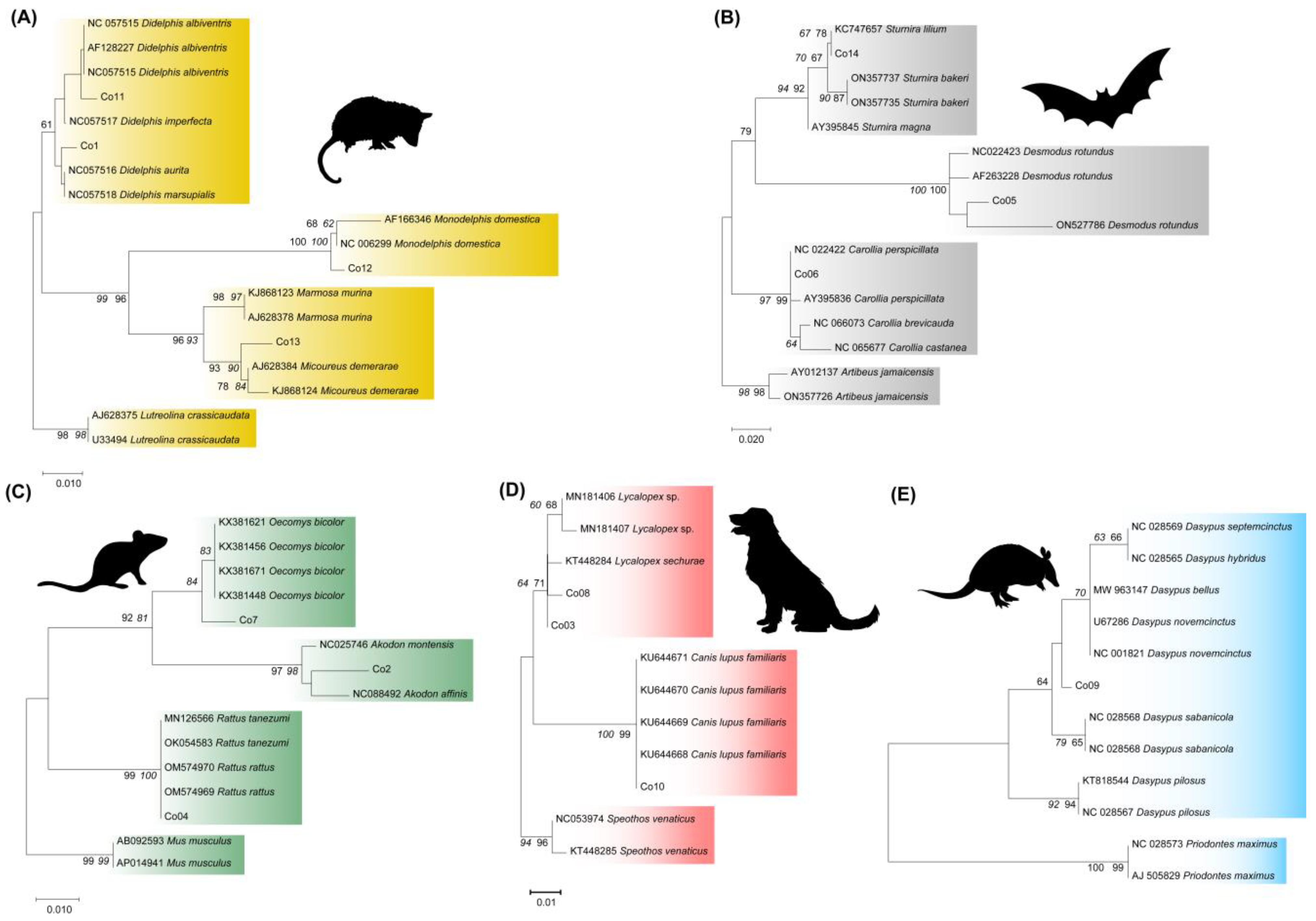
NJ and ML phylogenetic trees for each order were constructed using the sequences obtained from the 14 species and their best-scoring sequences in GenBank. Both reconstructions showed the same topology. The ML phylogeny that includes the Didelphimorphia species from the present study showed three genus-specific clusters: Didelphis spp., Monodelphis spp., and Marmosa spp. (Figure 2A). The sequences obtained from Monodelphis domestica (CO12) and Marmosa demerarea (CO13) were grouped into each genus cluster and were closely related to their corresponding GenBank species reference with high bootstrap values (Figure 2A). However, the Didelphis spp. cluster was moderately supported, and the sequences from D. aurita (CO01) and Didelphis albiventris (CO11) were poorly defined (Figure 2A), confirming our option of not conducting serial mixtures between these species.
The Chiropteran phylogenetic tree demonstrated that the D. rotundus (CO05), C. perspicillata (CO06), and S. lilium (CO14) sequences were located in each corresponding genus clade and were closely related to the GenBank species reference with high bootstrap values (Figure 2B).
The Rodentia phylogenetic reconstructions showed R. rattus (CO04) grouping in a species cluster (Figure 2C). However, Oecomys cleberi (CO07) was located in an O. bicolor species cluster, and Necromys lasiurus (CO02) grouped into an Akodon spp. clade (Figure 2C), reflecting the absence of these native species in the GenBank dataset, which limits accurate species-level mammalian identification and could consequently produce an incomplete and mistaken interpretation of the scenario of host–parasite transmission.
The ML 12S rDNA tree from canids (Figure 2D) showed two main well-defined clusters, one from Canis lupus familiaris, which includes the CO10 sequence, and a second one from the Lycalopex genus (Figure 2C). With the absence of a GenBank deposit of L. gymnocercus (CO08), the species sample (CO8) grouped into the Lycalopex genus clustered with the L. sechura sequence. The canid cloth sample named Cerdoncyon thous (CO3) clustered with L. sechura. Therefore, the misidentification of the blood clot sample with L. gymnocercus was confirmed. Finally, the Armadillo 12S rDNA phylogenetic analysis revealed the Dasypus novemcinctus (CO09) sequence near a cluster that included D novemcinctus reference sequences (Figure 2E).

3.2. Sensitivity Assessment of Detecting Multiple Blood Clot DNAs

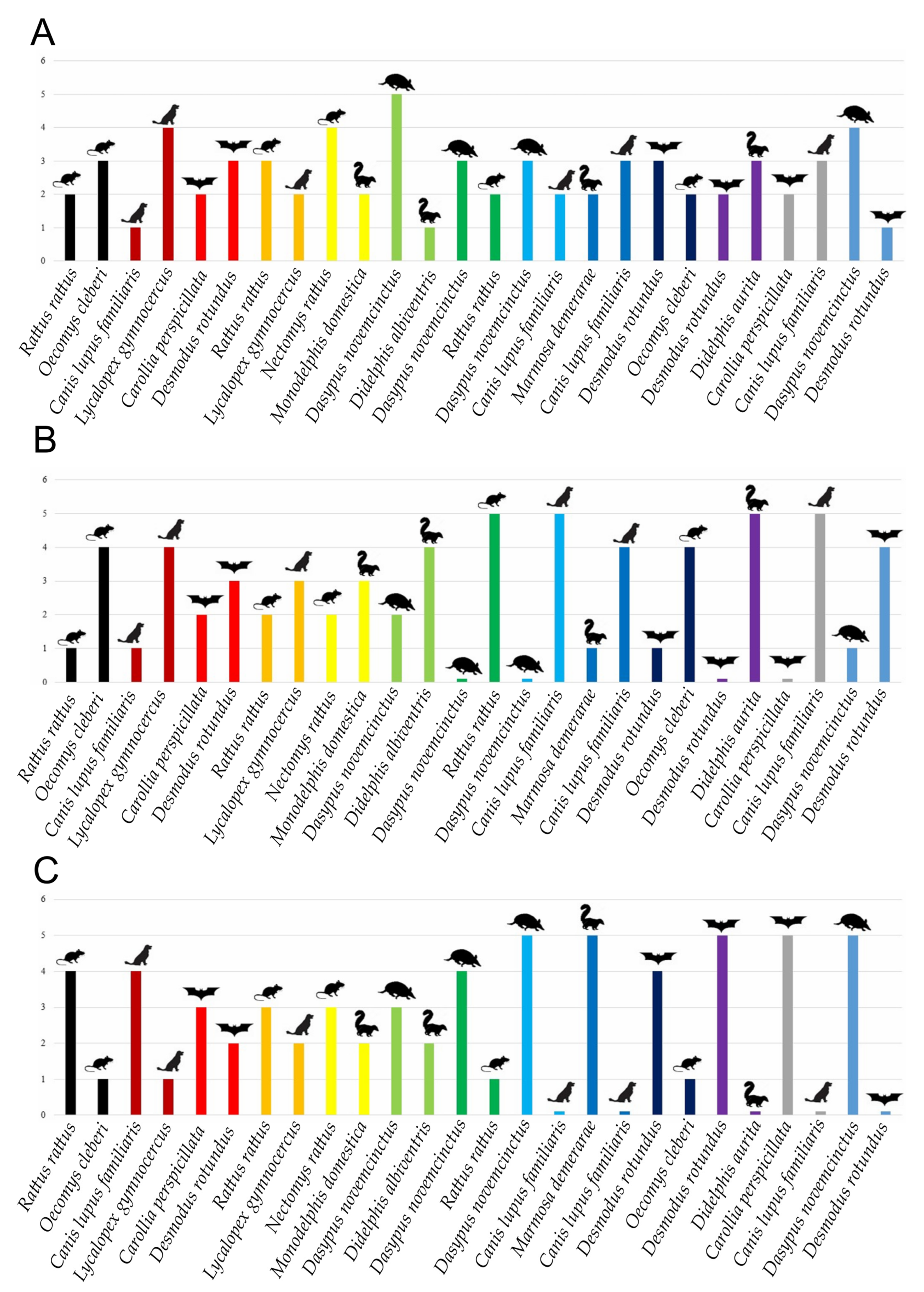
Clone sequencing throughout molecular cloning demonstrated the detection of all 13 species used in the 13 multiple mixtures in equal proportions (5/5), including those that are close and those that are unrelated taxonomically (Figure 3). It was possible to detect the species in a lower concentration in 17/26 multiple mixtures of mammalian species in disparate proportions (Figure 3B,C). In the proportions of 1/9 and 9/1, the species in the lower proportion were detected in 9/13 mixtures and 8/13 mixtures, respectively. All 13 species with a higher proportion were detected in the mixed samples, as expected. A total of 9/26 species with lower proportions in multiple mixtures were not detected (Figure 3B,C).

4. Discussion

4.1. Specificity of Molecular Species Identification

Identifying triatomine BMSs increases our understanding of the hosts involved in the transmission cycle of T. cruzi in nature. However, this analysis is usually performed using the precipitin technique, which has low sensitivity and specificity and does not permit an observation of the feeding eclecticism of these insect vectors. Moreover, knowledge of potential BMSs is essential, as this technique detects specific antibodies in the blood that respond to the animal protein origin.
On the other hand, with the application of molecular biology techniques, such as PCR and Sanger sequencing using DNA barcoding, it is possible to identify the BMSs at the species level. However, this method relies on a robust and complete dataset of the fauna from the area of the T. cruzi transmission network studied. There is a lack of available sequences for species in GenBank, particularly for mammals native to Brazil [16,22,28].
In the current work, we first evaluated the DNA barcoding approach in discriminating species of mammals involved in the transmission of T. cruzi in Brazil. It was observed that the BMS molecular markers 12S rDNA and cytb demonstrated specificity in the discrimination of the species from the orders selected, except for didelphids. Opossum species Didelphis aurita and D. albiventris showed a close genetic distance (less than 1%). This genetic identification has already been observed previously by Steiner et al. (2015), who identified the phylogenetic relationships of 19 species of Didelphis using two nuclear markers, the non-coding transthyretin intron 1 (TTR) and the inter-photoreceptor coding retinoid-binding protein exon 1 (IRBP), and the same two mitochondrial genes that were used in the present study, cytb and 12S rDNA [40].
For the marsupial species M. demerae, the subgenus Micoureus has already been clarified in the literature by Voss and Jansa (2009), even though the authors mentioned the possibility of keeping Micoureus as a genus due to the monophyly of the group [41]. It was possible to observe an inter-genus genetic distance between M. demerarae and M. domestica, and between the Monodelphis and Didelphis genera [41]. The high genetic distance among these genera has already been reported by Agrizzi and collaborators (2005), who constructed a phylogenetic tree using cytb gene sequences [42].
Regarding the canid species from the present study, the genetic distance considering the reference sequences of GenBank does not show genetic proximity among them. This was previously shown by Fuentes-González and Muñoz-Durán (2012), who studied the phylogenetic relationships of 35 canid species [43]. Canis lupus familiaris and C. lupus have a low genetic distance for both molecular markers used in this study. Studies using mitochondrial DNA markers demonstrated genetic proximity between these species, and it was suggested that C. lupus is the only ancestor of C. l. familiaris [44,45]. Even though C. lupus and C. l. familiaris are genetically close species, the question of whether they are two distant species has been discussed [45]. The molecular markers showed specificity in discriminating the canid species. The blood clot labeled as C. thous was identified as L. gymnocercus through the sequence analysis. Thus, this species was excluded from the mixture analysis in the present work due to the incorrect name of the blood clot sample.
It is known that a lack of reference sequences deposited in GenBank can lead to the incorrect identification of species. In BMS studies, this lack can cause the erroneous association of species that contribute to the T. cruzi life cycle. In the present work, L. gymnocercus GenBank deposits for both markers were absent, generating a maximum identity of the Brazilian field fox with L. sechurea, which is a species that does not occur in Brazil. This canid species inhabits the coastal deserts of northern Peru and southern Ecuador [44]. According to the Taxonomic Catalog of the Fauna of Brazil (Catálogo Taxonômico da Fauna do Brasil—http://fauna.jbrj.gov.br/fauna/listaBrasil (accessed on 31 May 2025), two Lycalopex species occurred in the country, L. gymnocercus and L. vetulus (Lund, 1842), both absent from GenBank. Therefore, we strongly recommend targeted sequencing efforts for absent and underrepresented species in the GenBank genetic database, some of which have been repeatedly highlighted in the present study.
The same situation was observed for the lack of an O. cleberi 12S rDNA reference sequence in the database, which generated genetic identification with other species of the genus, such as O. bicolor, O. auyantepui, and other rodent genera like Hapalomys delacouri, E. macconnelli, and A. cahirinus. Oecomys auyantepui is only found in the Brazilian Amazon biome [46], O. catherinae occurs in the Brazilian Atlantic Forest and Cerrado biomes [47], and O. bicolor occurs in the Pantanal, Cerrado, and Amazon biomes [48]. According to Musser and Carleton (2005), the genus Oecomys presents an unclear taxonomy [49]. This was confirmed by Suárez-Villota and collaborators (2017), who studied phylogenetic relationships and the delineation of Oecomys species using multiple cytogenetic, morphological, and molecular (mtDNA) analyses. The authors concluded that species of the genus Oecomys form a species complex and highlighted the importance of combining cytogenetics, morphology, and geographic information with molecular analyses in the delimitation of species [42]. The taxonomic revision of this group was also suggested by Rocha and collaborators (2015) [50]. Euryoryzomys macconelli occurs in the Brazilian Amazon biome, and the genetic distance from the Oecomys genus has already been demonstrated [51].
The 12S rDNA Necromys lasiurus sequence obtained in the current study was identified by BLAST/NCBI as Akodon montensis. No sequences of the Necromys genus are deposited for this target in GenBank. Necromys lasiurus and A. montensis belong to the same subfamily Sigmodontinae [52,53], which could be a reason why A. montensis appears as a BLAST/NCBI result. Using the cytb target, a sequence of Bolomys lasiurus presented 99% genetic identity with the N. lasiurus from this study. According to the Integrated Taxonomic Information System, B. lasiurus is currently an invalid species, a synonymous species of N. lasiurus. In this sense, we highlight the importance of taxonomically updating the GenBank deposits to avoid incorrect nomenclature and missing synonymy information. This failure could raise unrealistic ecological premises about the number and role of species serving as BMS, and whether the blood-feeding behavior of vectors is broad or restricted, with critical implications for understanding vector–host relationships.
It is important to mention that the three rodent species studied in this work do not present genetic proximity, as can be seen in the rodent phylogenetic study carried out by Vilela and collaborators (2014) [54].
The three chiropteran species selected for this work, C. perspicillata, D. rotundus, and S. lilium, showed high genetic distances among them, as can be seen in the work by Baker and Hoofer (2003), who performed a phylogenetic analysis of different genera of bats using mtDNA [55].
The bat species Sturnira lilium and S. tildae showed genetic distances of 1.4% and 0.7% for 12Sr DNA and cytb, respectively. The genus Sturnira is characterized as a group that contains complex phylogenetic relationships [56,57]. Due to their genetic similarity, S. tildae and S. lilum were suggested to be congeneric species [57] but not sister species [58].
In the present study, C. perspicillata and C. brevicauda showed high genetic proximity, but this can be explained because C. perspicillata and C. brevicauda are sister species and closely related to C. castanea [59]. The difficulty in identifying these species based only on morphological characters was reported [55].
The Xenarthra species D. novemcinctus showed high genetic proximity with D. yepesi for the 12S rDNA marker and with D. novemcinctus for the cytb marker. The high genetic proximity of these species was shown by a systematic analysis using the cytb marker [60]. The species D. yepesi, which showed the highest genetic identity with D. novemcinctus, is not considered a valid species by the Integrated Taxonomic Information System (ITIS). We found evidence of a synonymy issue with D. mazzai (Yepes, 1933), a valid species that occurs in northern Argentina, but has no genetic sequences deposited in GenBank for any gene [61]. These genes are widely used for the identification of BMSs, and their absence directly affects the accurate identification of the species acting as a BMS, thereby impairing the understanding of transmission dynamics.
It is important to mention that birds and reptiles, considered refractory to T. cruzi infection, also served as vector BMSs. However, the current evidence shows that these animals can be infected by T. cruzi, and, specifically, reptiles were proposed as silent hosts of the parasite [62,63]. This type of research should be stimulated to provide a larger and more diverse dataset of the Brazilian fauna, not only for BMS studies but also for other enzootic diseases, biodiversity studies, biogeography, taxonomy, and molecular systematics.

4.2. Sensitivity in Detecting Multiple DNA Samples from Blood Clots

DNA barcoding studies for the identification of triatomine BMSs are restricted to the detection of a single BMS, which limits the spectrum of potential species that participate in the parasite transmission network or in the maintenance of vector colonies. The application of this approach and subsequent molecular cloning allows the discrimination of multiple BMS species and/or different individuals of the same species by a haplotype analysis [64,65].
In the present study, the molecular cloning analysis demonstrated superior detection of the species with higher proportions, as expected. However, it was possible to detect species with lower concentrations in the different mixtures performed. For mixtures of the same proportion, both species were always detected. Relevant species present in smaller proportions were detected in 54% of the mixtures. From these results, it is possible to state that gene cloning using the 12S rDNA target is effective at detecting multiple BMSs and individuals of the same taxon, even in extremely unequal proportions. The specificity of the 12S rDNA target in the detection of DNA from vertebrate animals has previously been commented [64], and it has even been mentioned that this target is superior for identifying degraded samples compared to the cytb target. The sensitivity of the cloning technique for detecting multiple BMSs has already been mentioned in previous works [64,65]. However, this technique was only applied once previously in triatomine BMS studies in Brazil [36]. The next-generation sequencing method is also efficient at retrieving multiple BMSs in a single vector [66]. However, in comparison, molecular cloning is low-cost, relatively short, and less time-consuming, and does not require very specialized operators to perform the technique and to process the results.

4.3. The Importance of the Specificity and Sensitivity in Recovered Mammalian DNA for a Better Understanding of the Life Cycle of T. cruzi

The role of marsupials in maintaining the life cycle of T. cruzi is already well supported because these mammals share habitats with the triatomine vectors and feed on these insects and on reservoir species, such as small rodents, which can potentially be infected [10]. Legey and collaborators (1999) highlighted that some didelphids are important reservoirs of T. cruzi due to their ability to control the infection and not present clinical changes [67]. Some marsupial species, such as D. aurita and D. marsupialis, circulate in different ecotopes and can assist in the dispersion of the agent in sylvatic, domestic, and peridomestic environments [10,67]. In addition to moving between different ecotopes, marsupials can use all parts of the forest, being in contact with the different transmission cycles of T. cruzi [5]. The omnivorous diet of marsupials favors oral transmission by the predation of infected triatomines or other small mammals. Another marsupial species that was also identified as the main reservoir of T. cruzi in an outbreak of Chagas disease in Brazil was M. domestica [68]. Marmosa demerarae was found to be infected by the parasite [69]. The xenarthral species D. novemcinctus was identified as the second most important reservoir of T. cruzi after D. marsupialis [70].
In the domestic environment, C. l. familiaris, the domestic dog, is also reported as a major reservoir host of T. cruzi and as a risk factor for human infection [71,72,73]. Dogs may live inside houses and wander through wild ecotopes where they can acquire T. cruzi infection from sylvatic triatomines and/or infected prey. These mammals that circulate between domestic and wild ecotopes play very important roles in connecting the transmission network of the parasite [74]. In general, canids can maintain the T. cruzi infection throughout their lives [75].
Wild canids are part of a poorly studied taxon in terms of their possible roles in the transmission cycle of T. cruzi in nature [76]. These mammals are known to explore large areas and different habitats and can be considered parasite bioaccumulators, especially those that can be transmitted orally, such as T. cruzi [76]. This characteristic of wild canids to explore different ecotopes is an extremely critical aspect of the dispersion of the parasite [77].
Rodents are considered the main prey animals that circulate in peridomestic ecotopes [68]. These small mammals connect sylvatic, peridomestic, and domestic ecotopes, transporting the parasite to human dwellings, where they are transmitted to humans and domestic animals. For these reasons, these hosts are important in the study of T. cruzi transmission [78].
Almeida and coauthors (2024) [21] proposed that for the transmission cycle of the parasite to occur and lead to CD, five interactions are necessary: parasite, vectors, hosts, environments, and human action. The study highlights the role of humans in producing artificial domestic and peridomestic environments, such as the accumulation of wood piles, which favor the colonization of vectors and small mammals [21].
Rattus rattus, a synanthropic animal, has been suggested as a T. cruzi reservoir that connects different ecotopes [79]. The authors also mentioned that the proximity of these species to the domestic environment favors the contact of the parasite with the human population. The wild species of rodent, O. cleberi, was previously reported to be infected by T. cruzi with a high diversity of trypanosomatid species [80]. On the other hand, N. lasiurus is one of the most abundant species in the Brazilian Cerrado and is considered a significant host of T. cruzi in the biome [8].
For bats, D. rotundus and C. perspicillata were recently identified as reservoirs of T. cruzi in the Atlantic Forest biome [13]. This was not the first report in which bats were considered reservoirs of T. cruzi. In Colombia, one of the main food sources detected in triatomines infected by T. cruzi was bats [81]; in Ecuador, chiropter was identified as being responsible for the connection between the wild and household ecotopes [82]; in Argentina, D. rotundus was found to be infected with T. cruzi [83]; and in Venezuela, bats were identified as dispersers of the parasite [84]. The application of the NGS technique reinforced the roles of bats as hosts/reservoirs and as bioaccumulators of T. cruzi [13].

5. Conclusions

The present study aimed to respond to some basic questions regarding the BMS molecular methodology. The first question concerned the possibility of a bias in identifying BMSs with higher proportions. This was answered since we showed that species with very low proportions of 1/9 and 9/1 were detected in 17/26 of mixed samples, although species with higher proportions were identified in all 26 mixtures.
The second question concerned whether cloning sequencing is efficient at revealing secondary sources or equal proportions of BMSs. Our results demonstrated the detection of two mammalian species in equal proportions in all 26 mixed samples, and, as mentioned above, the secondary or low representative species were also recovered in 17/26 mixed samples. We are aware that the low concentration detection in mixed samples was partial, with a number of false-negative results (9/26). Future efforts to enhance assay sensitivity are necessary to address the false-negative results.
Finally, we questioned if the fauna of endemic mammals in Brazil recognized as hosts of T. cruzi are represented in GenBank. Despite the limited number of mammalian species evaluated, 3/13 species did not have GenBank sequences available for the two BMS molecular markers applied.
Thus, the study provides more detailed information on multiple BMS detection protocols that could reflect a succession of vector contact events, the diversity of mammalian hosts, and the complexity of the parasite transmission web.
Therefore, the data confirmed our hypothesis that molecular cloning is effective at distinguishing mammalian species, even with equal proportions or with a very low secondary representation in the sample. The integration of these methodologies in the detection of multiple BMSs could provide more detailed information that could reflect a series of vector contact events, the diversity of mammalian hosts, and the complexity of the parasite transmission network. Future research should further amplify the analysis of more Brazilian mammalian species and explore these dynamics in the wild to obtain effective information about the ecology of T. cruzi transmission in nature.

Supplementary Materials

The following supporting information can be downloaded at: https://www.mdpi.com/article/10.3390/pathogens14060579/s1, Figure S1. Amplification of mammalian DNA in blood clots and electrophoresis on an agarose gel. (A) Amplification of the molecular target 12S rDNA. Lane 1—Promega DNA Ladder 100 bp, Lane 2—CO01 = Didelphis aurita, Lane 3—CO02 = Akodon montensis, Lane 4—CO03 = Lycalopex schurae, Lane 5—CO04 = Rattus rattus, Lane 6—CO05 = Desmodus rotundus, Lane 7—CO06 = Carollia perspicillata, Lane 8—CO07 = Oecomys bicolor, Lane 9—CO08 = Lycalopex schurae, Lane 10—CO09 = Dasypus novemcinctus, Lane 11—CO10 = Canis lupus familiaris, Lane 12—CO11 = Didelphis albiventris, Lane 13—CO12 = Monodelphis domestica, Lane 14—CO13 = Micoureus demerarea, Lane 15—CO14 = Sturnira lilium, Lane 16—negative control. (B) Amplification of the molecular target cytb. Lane 1—Promega DNA Ladder 100 bp, Lane 2—CO01 = Didelphis aurita, Lane 3—CO02 = Akodon montensis, Lane 4—CO03 = Lycalopex schurae, Lane 5—CO04 = Rattus rattus, Lane 6—CO05 = Desmodus rotundus, Lane 7—CO06 = Carollia perspicillata, Lane 8—CO07 = Oecomys bicolor, Lane 9—CO08 = Lycalopex schurae, Lane 10—CO09 = Dasypus novemcinctus, Lane 11—CO10 = Canis lupus familiaris, Lane 12—CO11 = Didelphis albiventris, Lane 13—CO12 = Monodelphis domestica, Lane 14—CO13 = Micoureus demerarea, Lane 15—CO14 = Sturnira lilium, Lane 16—negative control.

Author Contributions

Conceptualization, A.M.I.; methodology, T.S.P. and Q.M.O.; software, Q.M.O.; validation, A.M.I.; investigation, T.S.P., Q.M.O. and A.M.I.; resources, A.M.I.; data curation, A.M.I.; writing—original draft preparation, T.S.P. and Q.M.O.; writing—review and editing, T.S.P., Q.M.O. and A.M.I.; supervision, A.M.I.; funding acquisition, A.M.I. All authors have read and agreed to the published version of the manuscript.

Funding

This research was funded by Fundação de Amparo a Pesquisa do Rio de Janeiro (http://www.faperj.br/) (A.M.I., Grant Numbers E-26/210.131/2023 and E-26/201.069/2021; Q.M.O., E-26/200.067/2021, 260339); Conselho Nacional de Desenvolvimento Científico e Tecnológico (http://www.cnpq.br/) (A.M.I., 315634/2021-9); and Coordenação de Aperfeiçoamento de Pessoal de Nível Superior (https://www.gov.br/capes/pt-br) (T.S.H., 88882.332462/2019-01; Q.M.O., 88887.644154/2021-00).

Institutional Review Board Statement

Not applicable.

Informed Consent Statement

Not applicable.

Data Availability Statement

All sequences generated in this study have been submitted to the GenBank database under the accession numbers: PV023314-PV023326.

Acknowledgments

We thank the PDTIS/FIOCRUZ genomic sequencing platform, Laboratório de Biologia de Tripanossomatideos IOC/FIOCRUZ, for donating the blood clots for this study, and the Laboratório de Parasitologia Integrativa e Paleoparasitologia for their valuable technical assistance during the study.

Conflicts of Interest

The authors declare no conflicts of interest.

References

  1. Jansen, A.M.; Xavier, S.C.D.C.; Roque, A.L.R. Landmarks of the Knowledge and Trypanosoma cruzi Biology in the Wild Environment. Front. Cell. Infect. Microbiol. 2020, 6, 10. [Google Scholar] [CrossRef]
  2. Chagas, C. Descoberta do Tripanozoma cruzi e verificação da Tripanozomiase Americana: Retrospecto histórico. Mem. Inst. Oswaldo Cruz 1922, 15, 67–76. [Google Scholar] [CrossRef]
  3. WHO. Chagas Disease (American Trypanosomiasis). Available online: https://www.who.int/health-topics/chagas-disease (accessed on 23 July 2024).
  4. Ministério da Saúde. Situação Epidemiológica. Available online: https://www.gov.br/saude/pt-br/assuntos/saude-de-a-a-z/d/doenca-de-chagas/situacao-epidemiologica (accessed on 24 July 2024).
  5. Jansen, A.M.; Xavier, S.C.C.; Roque, A.L.R. The multiple and complex and changeable scenarios of the Trypanosoma cruzi transmission cycle in the sylvatic environment. Acta Trop. 2015, 151, 1–15. [Google Scholar] [CrossRef]
  6. Abreu, E.F.; Casali, D.; Costa-Araújo, R.; Garbino, G.S.T.; Libardi, G.S.; Loretto, D.; Loss, A.C.; Marmontel, M.; Moras, L.M.; Nascimento, M.C.; et al. Lista de Mamíferos do Brasil. Zenodo. 2022. Available online: https://zenodo.org/record/7469767 (accessed on 10 January 2025).
  7. Deane, M.P.; Lenzi, H.L.; Jansen, A. Trypanosoma cruzi: Vertebrate and invertebrate cycles in the same mammal host, the opossum Didelphis marsupialis. Mem. Inst. Oswaldo Cruz 1984, 79, 513–515. [Google Scholar] [CrossRef]
  8. Jansen, A.M.; Xavier, S.C.d.C.; Roque, A.L.R. Trypanosoma cruzi transmission in the wild and its most important reservoir hosts in Brazil. Parasites Vectors 2018, 11, 502. [Google Scholar] [CrossRef]
  9. Dario, M.A.; Furtado, C.; Lisboa, C.V.; De Oliveira, F.; Santos, F.M.; D’Andrea, P.S.; Roque, A.L.R.; Das Chagas Xavier, S.C.; Jansen, A.M. Trypanosomatid Richness Among Rats, Opossums, and Dogs in the Caatinga Biome, Northeast Brazil, a Former Endemic Area of Chagas Disease. Front. Cell. Infect. Microbiol. 2022, 12, 851903. [Google Scholar] [CrossRef]
  10. Carreira, J.C.A.; Jansen, A.M.; De Nazareth Meirelles, M.; Costa ESilva, F.; Lenzi, H.L. Trypanosoma cruzi in the Scent Glands of Didelphis marsupialis: The Kinetics of Colonization. Exp. Parasitol. 2001, 97, 129–140. [Google Scholar] [CrossRef]
  11. Hamilton, P.B.; Cruickshank, C.; Stevens, J.R.; Teixeira, M.M.G.; Mathews, F. Parasites reveal movement of bats between the New and Old Worlds. Mol. Phylogenetics Evol. 2012, 63, 521–526. [Google Scholar] [CrossRef]
  12. Alves, F.M.; Rangel, D.A.; Vilar, E.M.; Pavan, M.G.; Moratelli, R.; Roque, A.L.R.; Jansen, A.M. Trypanosoma spp. Neobats: Insights about those poorly known trypanosomatids. Int. J. Parasitol. Parasites Wildl. 2021, 16, 145–152. [Google Scholar] [CrossRef]
  13. Dario, M.A.; Moratelli, R.; Schwabl, P.; Jansen, A.M.; Llewellyn, M.S. Small subunit ribosomal metabarcoding reveals extraordinary trypanosomatid diversity in Brazilian bats. PLoS Negl. Trop. Dis. 2017, 11, e0005790. [Google Scholar] [CrossRef]
  14. Torres, J.M.; De Oliveira, C.E.; Santos, F.M.; Sano, N.Y.; Martinez, É.V.; Alves, F.M.; Tavares, L.E.R.; Roque, A.L.R.; Jansen, A.M.; Herrera, H.M. Trypanosomatid diversity in a bat community of an urban area in Campo Grande, Mato Grosso do Sul, Brazil. Infect. Genet. Evol. 2024, 118, 105563. [Google Scholar] [CrossRef] [PubMed]
  15. Austen, J.M.; Barbosa, A.D. Diversity and Epidemiology of Bat Trypanosomes: A One Health Perspective. Pathogens 2021, 10, 1148. [Google Scholar] [CrossRef] [PubMed]
  16. Lilioso, M.; Reigada, C.; Pires-Silva, D.; Fontes Fvon, H.M.; Limeira, C.; Monsalve-Lara, J.; Folly-Ramos, E.; Harry, M.; Costa, J.; Almeida, C.E. Dynamics of food sources, ecotypic distribution and Trypanosoma cruzi infection in Triatoma brasiliensis from the northeast of Brazil. PLoS Negl. Trop. Dis. 2020, 14, e0008735. [Google Scholar] [CrossRef]
  17. Santos, F.M.; Sano, N.Y.; Liberal, S.C.; Dario, M.A.; Nantes, W.A.G.; Alves, F.M.; da Silva, A.R.; De Oliveira, C.E.; Roque, A.L.R.; Herrera, H.M.; et al. Kinetoplastid Species Maintained by a Small Mammal Community in the Pantanal Biome. Pathogens 2022, 11, 1205. [Google Scholar] [CrossRef]
  18. Jansen, A.M.; Roque, A.L.R.; Xavier, S.C.C. Trypanosoma cruzi enzootic cycle. In American Trypanosomiasis Chagas Disease; Elsevier: Amsterdam, The Netherlands, 2017; pp. 265–282. Available online: https://linkinghub.elsevier.com/retrieve/pii/B9780128010297000125 (accessed on 30 January 2025).
  19. Urbano, P.; Hernández, C.; Ballesteros, N.; Vega, L.; Alvarado, M.; Velásquez-Ortiz, N.; Martínez, D.; Barragán, K.; Ramírez, A.; Páez-Triana, L.; et al. Exploring dietary differences among developmental stages of triatomines infected with Trypanosoma cruzi in different habitats. Int. J. Parasitol. 2024, 54, 559–568. [Google Scholar] [CrossRef]
  20. Urbano, P.; Hernández, C.; Velásquez-Ortiz, N.; Ballesteros, N.; Páez-Triana, L.; Vega, L.; Urrea, V.; Ramírez, A.; Muñoz, M.; Ibarra-Cerdeña, C.N.; et al. Transmission ecology of Trypanosoma cruzi by Rhodnius prolixus (Reduviidae: Triatominae) infesting palm-tree species in the Colombian Orinoco, indicates risks to human populations. PLoS Negl. Trop. Dis. 2024, 18, e0011981. [Google Scholar] [CrossRef]
  21. Almeida, C.E.; Máximo, M.M.; Pires-Silva, D.; Takiya, D.M.; Valença-Barbosa, C.; Viana, M.C.; Reigada, C.; Iñiguez, A.M.; Harry, M.; Folly-Ramos, E. From molecules to ecosystems: Insights into a network of interactions for a Chagas disease outbreak using Triatoma brasiliensis as natural samplers. Acta Trop. 2024, 251, 107107. [Google Scholar] [CrossRef]
  22. Da Silva Leal, A.R.; Da Silva Ferreira, A.L.; De Araujo-Pereira, T.; Torres De Sousa, R.L.; Furtado Campos, J.H.; Tavares Dos Reis, R.; Finamore-Araujo, P.; Diotaiuti, L.; Ferreira, F.C.; Mendonça, V.J.; et al. Eco-epidemiological aspects and risk factors associated with human Chagas disease in rural areas of the state of Piauí, Brazil. BMC Infect. Dis. 2024, 24, 1335. [Google Scholar] [CrossRef]
  23. Daflon-Teixeira, N.F.; Coutinho, C.; Gomes, T.F.; Toma, H.K.; Duarte, R.; Bóia, M.N.; Carvalho-Costa, F.A.; Almeida, C.E.; Lima, M.M. Multiple Approaches to Address Potential Risk Factors of Chagas Disease Transmission in Northeastern Brazil. Am. J. Trop. Med. Hyg. 2019, 100, 296–302. [Google Scholar] [CrossRef]
  24. De Siqueira, A.F. Estudos Sôbre a Reação Identificação de Sangue da Precipitina Aplicada à Ingerido por Triatomíneos. Rev. Inst. Med. Trop. São Paulo 1960, 2, 41–53. [Google Scholar]
  25. Silva, M.B.A.; de Menezes, K.R.; de Farias, M.C.G.; Andrade, M.S.; Victor, C.C.A.; Lorosa, E.S.; Jurberg, J. Description of the feeding preferences of triatominae in the Chagas disease surveillance study for the State of Pernambuco, Brazil (Hemiptera: Reduviidae). Rev. Soc. Bras. Med. Trop. 2017, 50, 543–546. [Google Scholar] [CrossRef] [PubMed]
  26. Alencar, J.; De Mello, C.F.; Gil-Santana, H.R.; de Leão Giupponi, A.P.; Araújo, A.N.; Lorosa, E.S.; Guimarães, A.E.; dos Santos Silva, J. Feeding Patterns of Mosquitoes (Diptera: Culicidae) in the Atlantic Forest, Rio de Janeiro, Brazil. J. Med. Èntomol. 2015, 52, 783–788. [Google Scholar] [CrossRef] [PubMed]
  27. Verly, T.; Costa, S.; Lima, N.; Mallet, J.; Odêncio, F.; Pereira, M.; Moreira, C.J.D.C.; Britto, C.; Pavan, M.G. Vector competence and feeding-excretion behavior of Triatoma rubrovaria (Blanchard, 1843) (Hemiptera: Reduviidae) infected with Trypanosoma cruzi TcVI. PLoS Negl. Trop. Dis. 2020, 14, e0008712. [Google Scholar] [CrossRef]
  28. Almeida, C.E.; Faucher, L.; Lavina, M.; Costa, J.; Harry, M. Molecular Individual-Based Approach on Triatoma brasiliensis: Inferences on Triatomine Foci, Trypanosoma cruzi Natural Infection Prevalence, Parasite Diversity and Feeding Sources. PLoS Negl. Trop. Dis. 2016, 10, e0004447. Available online: https://repositorio.unesp.br/handle/11449/172623 (accessed on 6 April 2019). [CrossRef]
  29. Valença-Barbosa, C.; Fernandes, F.A.; Santos, H.L.C.; Sarquis, O.; Harry, M.; Almeida, C.E.; Lima, M.M. Molecular Identification of Food Sources in Triatomines in the Brazilian Northeast: Roles of Goats and Rodents in Chagas Disease Epidemiology. Am. J. Trop. Med. Hyg. 2015, 93, 994–997. [Google Scholar] [CrossRef]
  30. Lilioso, M.; Pires-Silva, D.; Fontes, F.V.H.M.; Oliveira, J.; da Rosa, J.A.; Vilela, R.V.; Folly-Ramos, E.; Almeida, C.E. Triatoma petrocchiae (Hemiptera, Reduviidae, Triatominae): A Chagas disease vector of T. brasiliensis species complex associated to reptiles. Infect. Genet. Evol. 2020, 82, 104307. [Google Scholar] [CrossRef]
  31. Valença-Barbosa, C.; Finamore-Araujo, P.; Moreira, O.C.; Alvarez, M.V.N.; Borges-Veloso, A.; Barbosa, S.E.; Diotaiuti, L.; de Souza, R.d.C.M. High Parasitic Loads Quantified in Sylvatic Triatoma melanica, a Chagas Disease Vector. Pathogens 2022, 11, 1498. [Google Scholar] [CrossRef]
  32. Rodrigues, M.S.; Lima, L.; Xavier, S.C.D.C.; Herrera, H.M.; Rocha, F.L.; Roque, A.L.R.; Teixeira, M.M.G.; Jansen, A.M. Uncovering Trypanosoma spp. diversity of wild mammals by the use of DNA from blood clots. Int. J. Parasitol. Parasites Wildl. 2019, 8, 171–181. [Google Scholar] [CrossRef]
  33. Kocher, T.D.; Thomas, W.K.; Meyer, A.; Edwards, S.V.; Paabo, S.; Villablanca, F.X.; Wilson, A.C. Dynamics of mitochondrial DNA evolution in animals: Amplification and sequencing with conserved primers. Proc. Natl. Acad. Sci. USA 1989, 86, 6196–6200. [Google Scholar] [CrossRef]
  34. Kitano, T.; Umetsu, K.; Tian, W.; Osawa, M. Two universal primer sets for species identification among vertebrates. Int. J. Leg. Med. 2006, 121, 423–427. [Google Scholar] [CrossRef]
  35. Borghesan, T.C.; Ferreira, R.C.; Takata, C.S.; Campaner, M.; Borda, C.C.; Paiva, F.; Milder, R.V.; Teixeira, M.M.; Camargo, E.P. Molecular Phylogenetic Redefinition of Herpetomonas (Kinetoplastea, Trypanosomatidae), a Genus of Insect Parasites Associated with Flies. Protist 2013, 164, 129–152. [Google Scholar] [CrossRef] [PubMed]
  36. Pessanha, T.S.; Herrera, H.M.; Jansen, A.M.; Iñiguez, A.M. “Mi Casa, Tu Casa”: The coati nest as a hub of Trypanosoma cruzi transmission in the southern Pantanal biome revealed by molecular blood meal source identification in triatomines. Parasites Vectors 2023, 16, 26. [Google Scholar] [CrossRef] [PubMed]
  37. Pessanha, T.S.; Pires, M.; Iñiguez, A.M. Molecular Detection of Blood Meal Source up to Three Months since the Last Meal: Experimental Starvation Resistance in Triatomines. Acta Trop. 2022, 232, 106507. [Google Scholar] [CrossRef]
  38. Edgar, R.C. MUSCLE: A multiple sequence alignment method with reduced time and space complexity. BMC Bioinform. 2004, 5, 113. [Google Scholar] [CrossRef]
  39. Nicholas, K.B.; Nicholas, H.B. GeneDoc: A Tool for Editing and Annotating Multiple Sequence Alignments. 1997. Available online: https://api.semanticscholar.org/ (accessed on 31 May 2025).
  40. Steiner, C.; Tilak, M.-K.; Douzery, E.J.; Catzeflis, F.M. New DNA data from a transthyretin nuclear intron suggest an Oligocene to Miocene diversification of living South America opossums (Marsupialia: Didelphidae). Mol. Phylogenetics Evol. 2005, 35, 363–379. [Google Scholar] [CrossRef]
  41. Voss, R.S.; Jansa, S.A. Phylogenetic Relationships and Classification of Didelphid Marsupials, an Extant Radiation of New World Metatherian Mammals. Bull. Am. Mus. Nat. Hist. 2009, 322, 1–177. [Google Scholar] [CrossRef]
  42. Agrizzi, J. Molecular Diagnosis of Atlantic Forest Mammals Using Mitochondrial DNA Sequences: Didelphid Marsupials. TOZJ 2012, 5, 2–9. [Google Scholar] [CrossRef]
  43. Fuentes-González, J.A.; Muñoz-Durán, J. Filogenia de los cánidos actuales (Carnivora: Canidae) mediante análisis de congruencia de caracteres bajo parsimonia. Actual. Biol. 2017, 34, 85–102. [Google Scholar] [CrossRef]
  44. Savolainen, P.; Zhang, Y.-P.; Luo, J.; Lundeberg, J.; Leitner, T. Genetic Evidence for an East Asian Origin of Domestic Dogs. Science 2002, 298, 1610–1613. [Google Scholar] [CrossRef]
  45. Vilà, C.; Savolainen, P.; Maldonado, J.E.; Amorim, I.R.; Rice, J.E.; Honeycutt, R.L.; Crandall, K.A.; Lundeberg, J.; Wayne, R.K. Multiple and Ancient Origins of the Domestic Dog. Science 1997, 276, 1687–1689. [Google Scholar] [CrossRef]
  46. Gentry, A. Mammal Species of the World, 3rd ed. In A Taxonomic and Geographic Reference; Don, E.W., DeeAnn, M.R., Eds.; Johns Hopkins University Press: Baltimore, ML, USA, 2005; Volume 2, 2142p, ISBN 0-8018-8221-4. A nomenclatural review. Bull. Zool. Nomencl. 2006, 63, 215–219. [Google Scholar]
  47. Asfora, P.H.; Palma, A.R.T.; Astúa, D.; Geise, L. Distribution of Oecomys catherinae Thomas, 1909 (Rodentia: Cricetidae) in northeastern Brazil with karyotypical and morphometrical notes. Biota Neotrop. 2011, 11, 415–424. [Google Scholar] [CrossRef]
  48. Mendes-Oliveira, A.C. Pequenos Mamíferos Não-Voadores da Amazônia Brasileira; Sociedade Brasileira de Mastozoologia: Rio de Janeiro, RJ, Brazil, 2014; Volume 1, 333p, ISBN 8563705024. [Google Scholar]
  49. Suárez-Villota, E.Y.; Carmignotto, A.P.; Brandão, M.V.; Percequillo, A.R.; Silva, M.J.D.J. Systematics of the genus Oecomys (Sigmodontinae: Oryzomyini): Molecular phylogenetic, cytogenetic and morphological approaches reveal cryptic species. Zool. J. Linn. Soc. 2017, 184, 182–210. [Google Scholar] [CrossRef]
  50. Rocha, R.G.; Justino, J.; Leite, Y.L.R.; Costa, L.P. DNA from owl pellet bones uncovers hidden biodiversity. Syst. Biodivers. 2015, 13, 403–412. [Google Scholar] [CrossRef]
  51. Kocher, A.; de Thoisy, B.; Catzeflis, F.; Huguin, M.; Valière, S.; Zinger, L.; Bañuls, A.; Murienne, J. Evaluation of short mitochondrial metabarcodes for the identification of Amazonian mammals. Methods Ecol. Evol. 2017, 8, 1276–1283. [Google Scholar] [CrossRef]
  52. Müller, L.; Gonçalves, G.L.; Cordeiro-Estrela, P.; Marinho, J.R.; Althoff, S.L.; Testoni, A.F. DNA Barcoding of Sigmodontine Rodents: Identifying Wildlife Reservoirs of Zoonoses. PLoS ONE 2013, 8, e80282. [Google Scholar] [CrossRef]
  53. FUNASA; Ministério da Saude. Manual de Controle de Roedores. In Assessoria de Comunicação e Educação em Saude; FUNASA: Tokyo, Japan, 2002. [Google Scholar]
  54. Vilela, J.F.; Mello, B.; Voloch, C.M.; Schrago, C.G. Sigmodontine rodents diversified in South America prior to the complete rise of the Panamanian Isthmus. J. Zool. Syst. Evol. Res. 2013, 52, 249–256. [Google Scholar] [CrossRef]
  55. Baker, G.C.; Smith, J.J.; Cowan, D.A. Review and re-analysis of domain-specific 16S primers. J. Microbiol. Methods 2003, 55, 541–555. [Google Scholar] [CrossRef]
  56. Pacheco, V.; Patterson, B.D. Phylogenetic Relationships of the New World Bat Genus Sturnira (Chiroptera: Phyllostomidae). Bull. Am. Mus. Nat. Hist. 1991, 206, 101–121. [Google Scholar]
  57. Dinelli, L.L. Três Espécies Crípticas em Sturnira lilium (Chiroptera: Phyllostomidae): Evidências Baseadas em Genes Mitocondriais. Master’s Thesis, Universidade Federal do Espírito Santo, Centro de Ciências Humanas e Naturais, Vitória, Brazil, March 2014. Available online: http://repositorio.ufes.br/handle/10/3844 (accessed on 31 May 2025).
  58. Villalobos, F.; Valerio, A.A. The phylogenetic relationships of the bat genus Sturnira Gray, 1842 (Chiroptera: Phyllostomidae). Mamm. Biol. 2002, 67, 268–275. [Google Scholar] [CrossRef]
  59. Lemos, T.H.; Da Cunha Tavares, V.; Moras, L.M. Character variation and taxonomy of short-tailed fruit bats from Carollia in Brazil. Zoologia 2020, 37, e34587. [Google Scholar] [CrossRef]
  60. De Machado, J.C.A. Biogeografia Cladística e Sistemática Molecular de Tatus do Gênero Dasypus e Uma Abordagem Filogeográfica da Espécie D. novemcinctus (Mammalia, Cingulata, Dasypodidae). Master’s Thesis, Universidade do Estado do Rio de Janeiro, Rio de Janeiro, Brazil, 2017. Available online: http://www.bdtd.uerj.br/handle/1/5874 (accessed on 31 May 2025).
  61. Feijó, A.; Cordeiro-Estrela, P. The Correct Name of the Endemic Dasypus (Cingulata: Dasypodidae) from North western Argentina. Zootaxa 2014, 3887, 88–94. [Google Scholar] [CrossRef] [PubMed]
  62. Martínez-Hernández, F.; Oria-Martínez, B.; Rendón-Franco, E.; Villalobos, G.; Muñoz-García, C.I. Trypanosoma cruzi, beyond the dogma of non-infection in birds. Infect. Genet. Evol. 2022, 99, 105239. [Google Scholar] [CrossRef] [PubMed]
  63. Botto-Mahan, C.; Correa, J.P.; Araya-Donoso, R.; Farías, F.; San Juan, E.; Quiroga, N.; Campos-Soto, R.; Reyes-Olivares, C.; González-Acuña, D. Lizards as Silent Hosts of Trypanosoma cruzi. Emerg. Infect. Dis. 2022, 28, 1250–1253. Available online: https://wwwnc.cdc.gov/eid/article/28/6/22-0079_article.htm (accessed on 1 August 2023). [CrossRef]
  64. Stevens, L.; Dorn, P.L.; Hobson, J.; de la Rua, N.M.; Lucero, D.E.; Klotz, J.H.; Schmidt, J.O.; Klotz, S.A. Vector Blood Meals and Chagas Disease Transmission Potential, United States. Emerg. Infect. Dis. 2012, 18, 646–649. [Google Scholar] [CrossRef]
  65. Stevens, L.; Monroy, M.C.; Rodas, A.G.; Dorn, P.L. Hunting, Swimming, and Worshiping: Human Cultural Practices Illuminate the Blood Meal Sources of Cave Dwelling Chagas Vectors (Triatoma dimidiata) in Guatemala and Belize. PLoS Negl. Trop. Dis. 2014, 8, e3047. [Google Scholar] [CrossRef]
  66. Arias-Giraldo, L.M.; Muñoz, M.; Hernández, C.; Herrera, G.; Velásquez-Ortiz, N.; Cantillo-Barraza, O.; Urbano, P.; Cuervo, A.; Ramírez, J.D. Identification of blood-feeding sources in Panstrongylus, Psammolestes, Rhodnius and Triatoma using amplicon-based next-generation sequencing. Parasites Vectors 2020, 13, 434. [Google Scholar] [CrossRef]
  67. Legey, A.P.; Pinho, A.P.; Xavier, S.C.; Marchevsky, R.; Carreira, J.C.; Leon, L.L.; Jansen, A.M. Trypanosoma cruzi in marsupial didelphids (Philander frenata and Didelphis marsupialis): Differences in the humoral immune response in natural and experimental infections. Rev. Soc. Bras. Med. Trop. 2003, 36, 241–248. [Google Scholar] [CrossRef]
  68. Roque, A.L.R.; Xavier, S.C.C.; da Rocha, M.G.; Duarte, A.C.M.; D’andrea, P.S.; Jansen, A.M. Trypanosoma cruzi Transmission Cycle Among Wild and Domestic Mammals in Three Areas of Orally Transmitted Chagas Disease Outbreaks. Am. J. Trop. Med. Hyg. 2008, 79, 742–749. [Google Scholar] [CrossRef]
  69. Herrera, L.; D’Andrea, P.S.; Xavier, S.C.C.; Mangia, R.H.; Fernandes, O.; Jansen, A.M. Trypanosoma cruzi infection in wild mammals of the National Park ‘Serra da Capivara’ and its surroundings (Piauí, Brazil), an area endemic for Chagas disease. Trans. R. Soc. Trop. Med. Hyg. 2005, 99, 379–388. [Google Scholar] [CrossRef]
  70. Raccurt, C.P. Trypanosoma cruzi in French Guinea: Review of accumulated data since 1940. Med. Trop. 1996, 56, 79–87. [Google Scholar]
  71. Beard, C.B.; Pye, G.; Steurer, F.J.; Rodriguez, R.; Campman, R.; Peterson, A.T.; Ramsey, J.; Wirtz, R.A.; Robinson, L.E. Chagas Disease in a Domestic Transmission Cycle in Southern Texas, USA. Emerg. Infect. Dis. 2003, 9, 103–105. [Google Scholar] [CrossRef] [PubMed]
  72. Cardinal, M.V.; Lauricella, M.A.; Ceballos, L.A.; Lanati, L.; Marcet, P.L.; Levin, M.J.; Kitron, U.; Gürtler, R.E.; Schijman, A.G. Molecular epidemiology of domestic and sylvatic Trypanosoma cruzi infection in rural northwestern Argentina. Int. J. Parasitol. 2008, 38, 1533–1543. [Google Scholar] [CrossRef] [PubMed]
  73. Morales, M.E.; Cimino, R.; Mackern-Oberti, J.P.; Martín, C.M.-S.; Cattan, P.E.; Superina, M. Eco-epidemiological Survey of Trypanosoma cruzi in Dogs from Mendoza, Argentina. Ecohealth 2025, 22, 79–90. [Google Scholar] [CrossRef]
  74. Ramírez, J.D.; Turriago, B.; Tapia-Calle, G.; Guhl, F. Understanding the role of dogs (Canis lupus familiaris) in the transmission dynamics of Trypanosoma cruzi genotypes in Colombia. Veter. Parasitol. 2013, 196, 216–219. [Google Scholar] [CrossRef]
  75. Rocha, F.L.; Roque, A.L.R.; De Lima, J.S.; Cheida, C.C.; Lemos, F.G.; de Azevedo, F.C.; Arrais, R.C.; Bilac, D.; Herrera, H.M.; Mourão, G.; et al. Trypanosoma cruzi Infection in Neotropical Wild Carnivores (Mammalia: Carnivora): At the Top of the T. cruzi Transmission Chain. PLoS ONE 2013, 8, e67463. [Google Scholar] [CrossRef]
  76. Sillero-Zubiri, C. Family Canidae (dogs). In Handbook Mammals of the World; Lynx Ediciones: Barcelona, Spain, 2009; pp. 352–447. [Google Scholar]
  77. Butler, J.; du Toit, J.; Bingham, J. Free-ranging domestic dogs (Canis familiaris) as predators and prey in rural Zimbabwe: Threats of competition and disease to large wild carnivores. Biol. Conserv. 2004, 115, 369–378. [Google Scholar] [CrossRef]
  78. Pinto, C.M.; Ocaña-Mayorga, S.; Lascano, M.S.; Grijalva, M.J. Infection by trypanosomes in marsupials and rodents associated with human dwellings in Ecuador. J. Parasitol. 2006, 92, 1251–1255. [Google Scholar] [CrossRef]
  79. Cab-Romero, M.; da Costa, A.P.; Costa, J.O.J.; de Oliveira Manhães, I.B.; da Silva, R.E.; Ruiz-Piña, H.A.; Reyes-Novelo, E.A.; Escobedo-Ortegona, J.; Tonhosolo, R.; Marcili, A. Synanthropic mammals in transmission cycle of Trypanosoma cruzi in Yucatán, Mexico. Braz. J. Glob. Health 2020, 1, 21–28. [Google Scholar] [CrossRef]
  80. Brandão, E.M.V.; Xavier, S.C.C.; Carvalhaes, J.G.; D’Andrea, P.S.; Lemos, F.G.; Azevedo, F.C.; Cássia-Pires, R.; Jansen, A.M.; Roque, A.L.R. Trypanosomatids in Small Mammals of an Agroecosystem in Central Brazil: Another Piece in the Puzzle of Parasite Transmission in an Anthropogenic Landscape. Pathogens 2019, 8, 190. [Google Scholar] [CrossRef]
  81. Hernández, C.; Salazar, C.; Brochero, H.; Teherán, A.; Buitrago, L.S.; Vera, M.; Soto, H.; Florez-Rivadeneira, Z.; Ardila, S.; Parra-Henao, G.; et al. Untangling the transmission dynamics of primary and secondary vectors of Trypanosoma cruzi in Colombia: Parasite infection, feeding sources and discrete typing units. Parasites Vectors 2016, 9, 620. [Google Scholar] [CrossRef] [PubMed]
  82. Pinto, C.M.; Ocaña-Mayorga, S.; Tapia, E.E.; Lobos, S.E.; Zurita, A.P.; Aguirre-Villacís, F.; MacDonald, A.; Villacís, A.G.; Lima, L.; Teixeira, M.M.G.; et al. Bats, Trypanosomes, and Triatomines in Ecuador: New Insights into the Diversity, Transmission, and Origins of Trypanosoma cruzi and Chagas Disease. PLoS ONE 2015, 10, e0139999. [Google Scholar] [CrossRef] [PubMed]
  83. Argibay, H.D.; Orozco, M.M.; Cardinal, M.V.; Rinas, M.A.; Arnaiz, M.; Segura, C.M.; Gürtler, R.E. First finding of Trypanosoma cruzi II in vampire bats from a district free of domestic vector-borne transmission in Northeastern Argentina. Parasitology 2016, 143, 1358–1368. [Google Scholar] [CrossRef] [PubMed]
  84. Añez, N.; Crisante, G.; Soriano, P.J. Trypanosoma cruzi congenital transmission in wild bats. Acta Trop. 2009, 109, 78–80. [Google Scholar] [CrossRef]
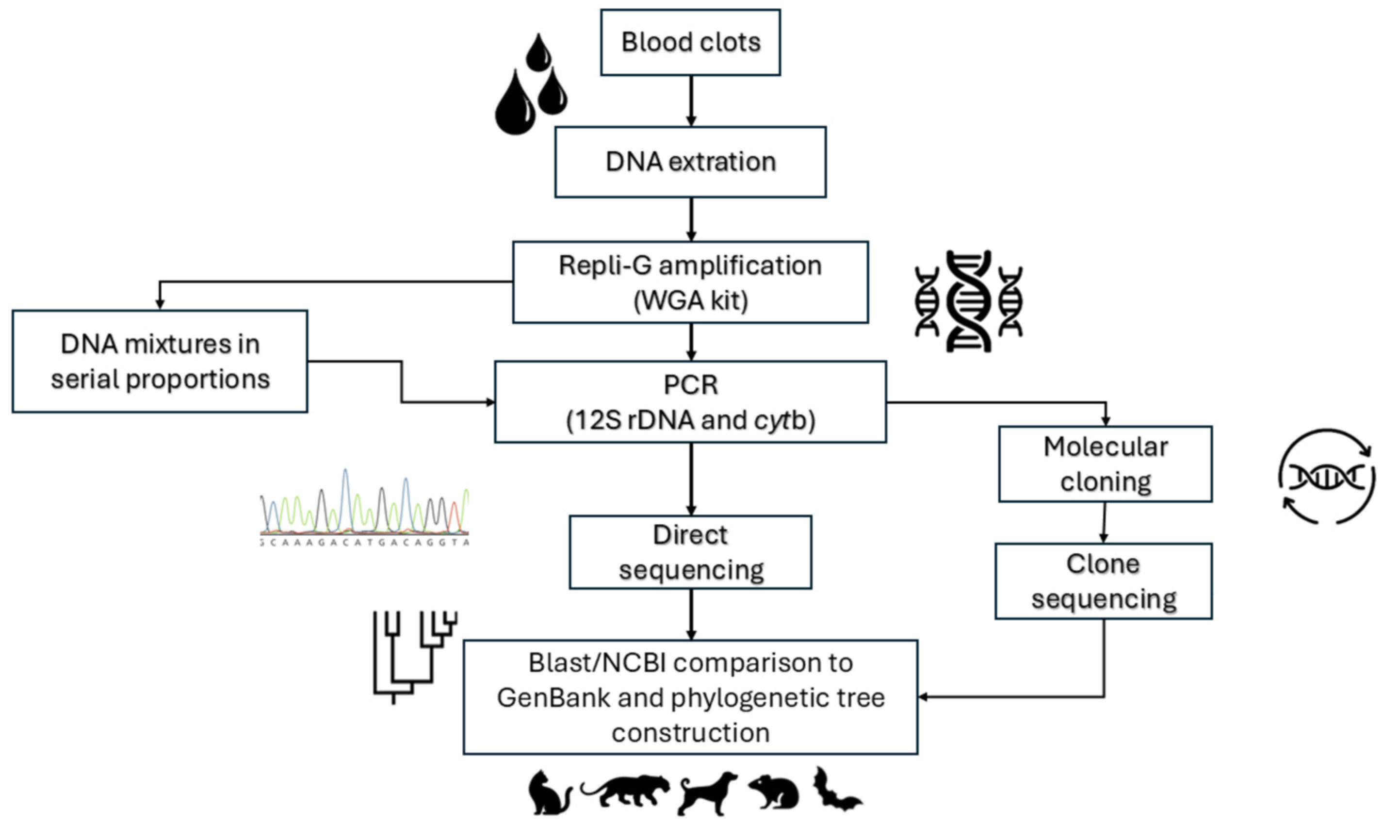
Figure 1. Diagram illustrating the flow of the methodological procedures applied in this study for the use of blood clots from different mammals through vertebrate BMS barcoding using both direct sequencing and clone sequencing approaches.
Pathogens 14 00579 g001
Figure 2. ML phylogenetic tree for the 12S rDNA target from the blood clots recovered from Brazilian mammals. (A) Opossums: CO01 = Didelphis aurita; CO11 = Didelphis albiventris, CO12 = Monodelphis domestica, CO13 = Micoureus demerarea. (B) Chiropterans: CO05 = Desmodus rotundus, CO06 = Carollia perspicillata, CO14 = Sturnira lilium (C) Canids: CO 03 and 08 = Lycalopex schurae, CO10 = Canis lupus familiaris. (D) Armadillo: CO09 = Dasypus novemcinctus. (E) Rodents: CO02 = Akodon montensis, CO04 = Rattus rattus, CO07 = Oecomys bicolor. The tree was constructed in MEGA v.11 software using ML and NJ methods and the Kimura 2-parameter model plus the gamma distribution with 500 bootstrap replicates. Reference sequences are indicated by species name and the GenBank numbers. The species used as outgroups were (A) the opossum Lutreolina crassicaudata (Desmarest, 1804); (B) chiropteran Artibeus jamaicensis Leach, 1821; (C) rodent Mus musculus (Linnaeus, 1758); (D) canids Speothos venaticus (Lund, 1842); and (E) armadillo Priodontes maximus (Kerr, 1792). Bootstrap values are shown on the internal nodes as ML = regular font and NJ = italic font.
Pathogens 14 00579 g002
Figure 3. Number of clones detected from 39 multiple mixtures of 13 mammalian species’ DNA. (A) Number of clones detected in the proportion of 5/5. (B) Number of clones detected in the proportion of 1/9 (C). Number of clones detected in the proportion of 9/1. Bars in the same color show the 13 mixtures performed. The animal symbols represent the orders, and the names of species included in each intrafamilial and interfamilial mixture are shown.
Pathogens 14 00579 g003
Table 1. Data regarding the samples collected from the Brazilian mammalian species for the present study.
Mammalian Species Common Name in BrazilCollection Site 1Brazilian Biome
Canis lupus familiarisDomestic dogMangaratiba-RJAtlantic Forest
Cerdocyon thousBush dogMamamguape-PBAtlantic Forest
Lycalopex gymnocercusField foxAlegrete-RSPampa
Oecomys cleberiTree mouseCumari-GOCerrado
Necromys lasiurusPixunaMamamguape-PBAtlantic Forest
Rattus rattusRatParaty-RJAtlantic Forest
Desmodus rotundusVampire batMamamguape-PBAtlantic Forest
Sturnira liliumFruit batMamamguape-PBAtlantic Forest
Carollia perspicillataShort-tail batMamamguape-PBAtlantic Forest
Didelphis albiventrisWhite-ear possumMamamguape-PBAtlantic Forest
Monodelphis domesticaShort-tailed cuicaMamamguape-PBAtlantic Forest
Marmosa (Micoureus) demeraraeCuicaMamamguape-PBAtlantic Forest
Didelphis auritaBlack-ear possumMamamguape-PBAtlantic Forest
Dasypus novemcinctusArmadilloMamamguape-PBAtlantic Forest
1 Brazilian States: RJ—Rio de Janeiro, PB—Paraíba, RS—Rio Grande do Sul, GO—Goiás.
Table 2. Results of the genetic analysis of mammalian species detection using the 12S rDNA and cytb rDNA BMS molecular markers.
Mammal SpeciesBlast/NCBI Genetic Identity ResultMaximum Genetic IdentityGenBank Accession NumberBlast/NCBI Genetic Identity ResultMaximum Genetic IdentityGenBank Accession Number
Molecular Target 12S rDNAMolecular Target cytb
Canis lupus familiarisCanis lupus familiaris100%KU291093Canis lupus familiaris99%DQ309764
Lycalopex gymnocercusLycalopex sechurae98.63%KT228284Lycalopex sechurae99.06%KT447686
Cerdocyon thousLycalopex sechurae98.00%KT448284NDNDND
Oecomys cleberiOecomys bicolor98.92%JF693852.1Oecomys cleberi98.88%KR190443
Necromys lasiurusAkodon montensis97.70%KF769456Bolomys lasiurus98.57%KR190443
Rattus rattusRattus rattus100%OM574970.1Rattus rattus99.00%KT232247
Desmodus rotundusDesmodus rotundus97.00%HG003310Desmodus rotundus99.00%HG003310
Sturnira liliumSturnira lilium100%KC747657Sturnira lilium100%KC747657
Carollia perspicillataCarollia perspicillata100%HG003309Carollia perspicillata100%FJ589715.1
Didelphis albiventrisDidelphis albiventris99.36%NC057515.1Didelphis albiventris99.85%AF089802
Monodelphis domesticaMonodelphis domestica99.53%AJ508398Monodelphis domestica100%KM071418
Marmosa (Micoureus) demeraraeMarmosa (Micoureus) demerarae99.05%AJ628384.1Marmosa (Micoureus) demerarae98.80%AJ487006
Didelphis auritaDidelphis aurita99.08%NC057518.1Didelphis aurita99.40%GU112886
Dasypus novemcinctusDasypus yesepi100%NC028570Dasypus novemcinctus99.00%KU253494
ND: The analysis was not performed.
Disclaimer/Publisher’s Note: The statements, opinions and data contained in all publications are solely those of the individual author(s) and contributor(s) and not of MDPI and/or the editor(s). MDPI and/or the editor(s) disclaim responsibility for any injury to people or property resulting from any ideas, methods, instructions or products referred to in the content.