Brief Report

Ex Vivo Drug Susceptibility of Plasmodium malariae Isolates to Antimalarial Drugs in Gabon

by
Yudi T. Pinilla
1,2,*,†,
Anton Hoffmann
1,2,†,
Maxim Viehweg
1,2,†,
Nathanaël Saison
1,2,
Stravensky Terence Boussougou Sambe
1,2,
Ange Gatien Doumba Ndalembouly
1,
Barclaye Ngossanga
1,
Florence Awamu
1,2,
Ayola Akim Adegnika
1,2,3 and
Steffen Borrmann
1,2,3
1
Centre de Recherches Médicales de Lambaréné, Lambaréné BP 242, Gabon
2
Institute for Tropical Medicine, University of Tübingen, 72074 Tübingen, Germany
3
German Center for Infection Research, 72074 Tübingen, Germany
*
Author to whom correspondence should be addressed.
These authors contributed equally to this work.
Pathogens 2025, 14(5), 453; https://doi.org/10.3390/pathogens14050453
Submission received: 16 March 2025 / Revised: 16 April 2025 / Accepted: 22 April 2025 / Published: 6 May 2025
(This article belongs to the Section Parasitic Pathogens)

Abstract

:
Plasmodium malariae is a neglected human malaria parasite despite its global distribution and propensity for persistent, sub-microscopic infections, which are associated with a mild but significant disease burden. Artemisinin-based therapies appear to be efficacious, but the susceptibility profiles of field isolates are largely unknown. We performed ex vivo assays with isolates collected from asymptomatic volunteers in Gabon. The mean concentrations required to inhibit 50% of growth (IC50) with chloroquine (n = 21), artesunate (n = 20), atovaquone (n = 21), and lumefantrine (n = 14) were 7.2 nM, 2.7 nM, 3.1 nM, and 7.4 nM, respectively. Our study provides novel data on the ex vivo susceptibility of P. malariae to several key antimalarials, including the first dataset for atovaquone.

1. Introduction

Plasmodium malariae is a neglected human malaria parasite, occurring often co-endemic with Plasmodium falciparum in regions such as sub-Saharan Africa, South America, Indonesia, Southeast Asia, and the Western Pacific [1]. Unlike P. falciparum, P. malariae has a longer, 72 h intraerythrocytic replication cycle, resulting in highly periodic fever peaks in non-immune patients, known as quartan malaria. The parasite is also known for its ability to cause chronic infections, which can persist and recur after years or even decades after the initial infection [2,3]. While P. malariae infections are generally far less pathogenic than those caused by P. falciparum, repeated and prolonged exposure can lead to anemia, contributing to the disease burden in affected populations [4].
Recent evidence suggests that P. malariae has become more prevalent in areas with low P. falciparum transmission following successful intervention efforts [5]. Sub-microscopic and thus, largely undiagnosed and untreated P. malariae infections constitute a significant challenge to malaria elimination efforts [6,7]. Whereas, microscopic-based surveys typically detect P. malariae infections in 1.3% to 5% of individuals [7,8], more sensitive molecular techniques, such as PCR or qPCR, reveal significantly higher prevalence rates of up to 30% and 10% in asymptomatic and symptomatic individuals, respectively [8,9,10]. Despite these findings, the actual burden of P. malariae remains poorly understood, largely due to the absence of routine diagnostic testing and the limited number of systematic epidemiological studies [1].
The World Health Organization (WHO) recommends artemisinin-based combination therapy (ACT) as the primary treatment for P. falciparum. However, there are currently no established treatment guidelines for P. malariae, and limited studies have assessed the efficacy of ACTs against this parasite [11,12,13]. In recent years, ACT resistance has emerged in Southeast Asia and a potential spread to other malaria-endemic regions jeopardizes global malaria control efforts [14]. In contrast to P. falciparum and P. vivax, evidence of chloroquine resistance in P. malariae remains limited [15]. However, more recently, treatment failures of artemether-lumefantrine for P. malariae malaria have been reported [16,17]. This highlights the need for further research to evaluate the in vivo and in vitro efficacy of these drugs. Due to the absence of continuous in vitro cultivation methods for P. malariae, ex vivo drug assays are currently the only option for gathering data on the drug susceptibility of this parasite. This study aims to evaluate the susceptibility of commonly used antimalarial drugs, including artesunate (AS), atovaquone (ATQ), lumefantrine (LUM), and chloroquine (CQ), against P. malariae.

2. Materials and Methods

2.1. Sample Collection

The study was conducted as a part of the CoMal project (DFG BO 2494/3-1), a cross-sectional prevalence study of non-falciparum malaria species in rural communities of the Moyen-Ogooué province of Gabon. Between January and April 2020, and again from April to August 2022, asymptomatic volunteers older than 1 year were screened for malaria infection by obtaining finger-prick blood samples. Thick blood smears were stained with 10% Giemsa solution for 45 min and examined microscopically by two experienced microscopists at 1000× magnification. Slide readings and calculations of the parasitemia were performed according to the Lambaréné method [18]. Only individuals with microscopic evidence of P. malariae infections (either as a mono-infection or mixed infection with P. falciparum or P. ovale), a hemoglobin concentration of ≥6 g/dL, and who had not taken antimalarial treatment within 3 months of sample collection were included. After obtaining written informed consent, blood samples were collected via venipuncture into sodium heparin vacutainers: 10 mL from participants aged ≥ 18 years and 5 mL from children > 5 years. Following sample collection, all patients were treated with artemether-lumefantrine (Coartem®), in accordance with the national treatment guidelines.

2.2. Ex Vivo Susceptibility Test

The ex vivo susceptibility of P. malariae isolates was assessed using a schizont maturation assay [19]. Fresh blood samples were centrifuged at 2000× g for 5 min at 37 °C, and the resulting red blood cell pellet was washed three times with incomplete culture medium (RPMI-1640 supplemented with 25 mM HEPES, 25 mM NaHCO3). Erythrocyte pellets were then resuspended in complete culture medium (RPMI-1640 supplemented with 25 mM HEPES, 25 mM NaHCO3, 20 mg/L hypoxanthine, 2.4 mM L-glutamine, 10 mg/L gentamycin, 5 g/L AlbuMAX II and 20% human AB serum), adjusted to a final hematocrit of 4%. The parasitized cells were distributed in 96-well drug plates, which were pre-dosed with 9 serial concentrations of CQ (2000 to 0.95 nM), AS (100 to 0.26 nM), ATQ (100 to 0.26 nM), and LUM 250 to 0.78 nM. CQ, AS, ATQ, and LUM were purchased from Sigma (Tokyo, Japan). Stock solutions of CQ (10 mM), AS (10 mM), ATQ (10 mM), were prepared in DMSO and LUM (10 mM) in RPMI medium. Each drug concentration was tested in triplicate.
Fifty microliters of the Blood Medium Mixture (BMM) was then added to each well. After the parasitized blood samples were added, the plates were incubated at 37 °C for up to 48 h in a multi-gas incubator (5% CO2, 5% O2, and 90% N2 with 80% humidity). Incubation was stopped when >50% of ring-stage parasites had differentiated to schizonts in the drug-free control wells. Schizont count per 100 parasites was determined by microscopy, with schizonts classified only when at least 4 merozoites per cell were discernable. The IC50 values were determined using nonlinear regression and a three-parameter dose–response curve. The stock drug solutions were validated using NF54 P. falciparum strain.

2.3. Real-Time qPCR Assay

An established qPCR assay was used to confirm P. malariae mono- or co-infected fresh isolates. A 500 µL sample of infected blood was mixed in a 1:2 ratio with DNA/RNA shield and RNA was extracted using the Microprep Plus Kit D7005 (Zymo Research, Irvine, CA, USA). The real-time qPCR was performed at the Institute for Tropical Medicine, Tübingen, Germany, following a previously described protocol [20]. Total RNA extraction was performed using the Quick-DNA/RNA™ Microprep Plus Kit (Zymo Research, Irvine, CA, USA) according to the manufacturer’s instructions. Purified RNA samples were immediately stored at −20 °C until use.
qPCR assays were conducted in parallel to detect P. malariae with the highest sensitivity, particularly in case of co-infections with P. falciparum or other Plasmodium species (LightCycler 480, Roche, Mannheim, Germany). The first assay identified P. malariae using primers that amplify a 100 bp fragment of the small-subunit rRNA gene (18 S), which is conserved across human malaria pathogens [21]. Each reaction required 1 µL RNA template, 5 µL SensiFAST™ One-Step Mix (Bioline, London, UK), 0.1 µL transkriptase, 0.2 µL 10 μM TaqMan probe, 0.4 µL of each 10 μM primer, and 2.9 µL water (10 µL total reaction volume).
The second assay differentiated between P. falciparum and other Plasmodium species, including P. malariae, P. vivax, and P. ovale, as described previously [21]. Primers and probes were designed using OligoArchitect online (Sigma-Aldrich, Tokyo, Japan), based on 18 S rRNA gene sequences from GenBank. Each reaction consisted of 1 µL RNA template, 5 µL SensiFAST™ One-Step Mix (Bioline, London, UK), 0.1 µTranscriptase, 0.4 µL of each 10 μM TaqMan probe, 0.8 µL of each 10 μM primer, and 1.5 µL water. After an initial transcription step of 20 min at 45 °C, followed by a hot-start step of 5 min at 95 °C, the cycling conditions were identical for both assays, allowing simultaneous runs in a single analysis step. The cycling conditions included 5 min at 95 °C, followed by 50 cycles of 93 °C for 15 s and 60 °C for 60 s. All samples were analyzed in triplicate, and each assay included a non-template control and a positive control in triplicates, respectively.

2.4. Statistical Analysis

Dose−response curves and 50% inhibitory concentrations (IC50) of the drugs of the clinical isolates were calculated by nonlinear regression analysis using GraphPad Prism (version 9) software (GraphPad Software Inc., San Diego, CA, USA). The type of distribution was evaluated using a Shapiro–Wilk or Kolmogorov–Smirnov test. Differences in drug susceptibility between drugs and P. falciparum and P. malariae were determined using a non-parametric Mann–Whitney test. A p-value < 0.05 was considered as significant. All statistical analyses were performed using GraphPad Prism (version 9) software (GraphPad Software Inc., San Diego, CA, USA).
The output of ultrasensitive qPCR reactions was examined by visual inspection and the cycle threshold (Ct) was calculated using LightCycler 480 Software (version 1.5.1.62) via the second derivative maximum method. A Ct value below the threshold of 40 indicated a positive sample.

3. Results

Between January and April 2020 and April and August 2022, a total of 1303 asymptomatic participants were screened for malaria. Of these, 42 (3.2%) participants were found to be infected with P. malariae by microscopy. Among the 42 infected participants, 24 (57%) had a P. malariae mono-infection, while 18 (43%) were co-infected with P. malariae and P. falciparum. Of the 42 P. malariae isolates collected, only 21 yielded ex vivo assay results. To confirm P. malariae mono-infection and co-infection with P. falciparum in the 21 isolates tested, qPCR was performed. The qPCR analyses showed that 9 out of the 21 isolates were P. malariae mono-infected (Table 1). The parasitemia of isolates ranged from 153 to 8620 parasites/µL, with a median assay duration of 36 h (range: 13 to 48 h). For the drug sensitivity analysis, only isolates with ≥50% ring-stage parasites at the start of the assay were included. Some drugs, such as AS and LUM, were not tested on certain isolates due to the insufficient blood volume collected.
IC50 values for the antimalarial drugs were determined for each isolate (Table 2 and Figure 1). Stock drug solutions were validated using the fully sensitive, laboratory-adapted P. falciparum NF54 strain, yielding consistently low nanomolar IC50 values (Table 2). The median ex vivo IC50 values for each drug were as follows: CQ (n = 21, IC50 = 7.2 nM, range: 3.8–26.7 nM), AS (n = 20, IC50 = 2.7 nM, range: 1.3–10.2 nM), ATQ (n = 21, IC50 = 3.1 nM, range: 1.5–11.3 nM), and LUM (n = 14, IC50 = 7.4 nM, range: 0.6–51.5 nM). All IC50s for chloroquine were below 35 nM, suggesting that P. malariae isolates from Gabon remain susceptible to this drug.

4. Discussion

Drug susceptibility profiles are well documented for P. falciparum and P. vivax, but there is limited knowledge regarding the activity of antimalarials against P. malariae isolates [22,23]. The high prevalence of sub-microscopic P. malariae infections in sub-Saharan African countries, along with its co-endemicity with P. falciparum, underscores the importance of understanding its drug susceptibility profile to inform effective treatment and elimination strategies. While in vitro and ex vivo cultivation of P. falciparum has facilitated extensive research and the development of effective drug treatments, the absence of similar cultivation systems for the asexual blood stages of P. malariae presents significant challenges to monitor existing antimalarial treatment strategies and validate novel antimalarial drug candidates [24]. In this study, we assessed the ex vivo susceptibility of P. malariae to four commonly used antimalarial drugs, chloroquine (CQ), artesunate (AS), atovaquone (ATQ), and lumefantrine (LUM), by testing freshly collected isolates.
The results showed generally low IC50 values for all tested drugs, with ATQ and AS displaying the highest potency, suggesting the continued susceptibility of P. malariae to these compounds. Our findings are consistent with those reported in earlier clinical studies on P. falciparum and P. malariae in Gabon [11,21,25]. Importantly, all of the CQ IC50 values were below the commonly referenced resistance thresholds [15], reinforcing the notion that P. malariae remains susceptible to CQ in Gabon. However, several limitations should be considered when interpreting our findings. Although 42 P. malariae infections were found in the study, the ex vivo susceptibility testing was successful in only 21 P. malariae isolates. This relatively small sample size limits the statistical power and generalizability of the results. Additionally, the study was conducted in a single region (Gabon), and the isolates cannot represent the genetic and phenotypic diversity of P. malariae found in other parts of sub-Saharan Africa or globally [26]. Larger, multi-country studies incorporating isolates from diverse transmission settings, including West, Central, and East Africa, as well as Southeast Asia and South America, are necessary to draw more robust data on the drug susceptibility of P. malariae.
A recent study from West Africa (Mali) has identified unusually high IC50 values for CQ, LUM and artemether against P. malariae isolates [22]. Although most clinical trials involving artemisinin derivatives or ACTs report sustained high efficacy [11,13], there have been a few instances of treatment failures in P. malariae infections [13,15,16]. The longer erythrocytic cycle of P. malariae may contribute to a natural refractoriness to 3-day ACT regimens, which may explain the persistence of infections, i.e., treatment failure, when using sensitive PCR during follow-up [16]. On the other hand, the survival of wild-type P. malariae parasites could potentially translate to lower selection pressure for mutants tolerant or resistant to partner drugs with short to medium elimination half-lives. More recently, a population-genomic analysis of global P. malariae isolates has identified mutations in PmDHFR that correspond to variants in PfDHFR conferring resistance to the slowly eliminated pyrimethamine [26], which we did not test in this study. The drug-resistant phenotype of these PmDHFR mutations were subsequently confirmed by ortholog replacement experiments in P. knowlesi [26].
Our study is the first to report the ex vivo sensitivity of P. malariae to ATQ. The results indicate that ATQ effectively inhibits P. malariae isolates, with IC50 values comparable to those reported for P. falciparum [27]. Atovaquone-proguanil is a highly effective non-ACT drug for the treatment of multidrug-resistant P. falciparum, and it is considered the drug of choice for both chemoprophylaxis in non-immune travelers to regions with reported multidrug resistance and the treatment of imported cases of acute uncomplicated P. falciparum infections in non-endemic countries. Given the frequent occurrence of P. malariae in Central and West Africa, often in mixed infections with P. falciparum, these findings underscore the complexity of malaria treatment in regions where species identification is difficult. Our findings support the continued use of combinations containing ATQ and LUM as effective oral regimens in case of diagnostic uncertainty or presumptive treatment of acute, uncomplicated malaria. This is particularly relevant in areas where mixed-species infections are prevalent or where limitations in diagnostic tool uncertainty limits species-specific treatment strategies.
Our results showed some variability in the IC50 values for LUM (ranging from 0.6 to 51.5 nM; median 7.2; interquartile range 3.0–10.9), mainly due to a single outlier (isolate #627). The observed variation may reflect phenotypic plasticity among the P. malariae isolates, potentially due to genetic variation. Nonetheless, technical and methodological factors could also have contributed to this variability. For instance, differences in the incubation duration in the ex vivo assay may have influenced schizont maturation and, consequently, the calculated IC50 values. Although only isolates with ≥50% ring-stage parasites were tested, some variability in parasite synchronization and maturation likely occurred, particularly in those samples with prolonged or shortened incubation times. While most assays were completed after 36 h, some were stopped earlier or later (e.g., isolate 1077 was incubated for 45 h, while isolate 1179 was only incubated for 13 h). The fact that 11 out of the 21 microscopically determined mono-infected P. malariae isolates were later shown by qPCR to be co-infected with P. falciparum could have also affected our results (such as in the case of isolate #627). In addition, and when compared to drugs like chloroquine (CQ) or artesunate (AS), suboptimal LUM solubility or inconsistencies in drug plate preparation could have affected the effective drug concentration, thereby impacting IC50 measurements. Without accompanying molecular data or repeat assays, it remains unclear whether this variability represents emerging tolerance or is simply an artifact of the assay conditions. Further investigation, including the genotyping of known drug resistance markers, is necessary to clarify this hypothesis. In addition, Ex Vivo response rates to lumefantrine in P. falciparum have also shown considerable variability [28,29].
In summary, our findings show that P. malariae isolates from Gabon remain susceptible to CQ, AS, ATQ, and LUM in an ex vivo assay. While these results are promising, a larger number of isolates, particularly from diverse geographic regions, are needed to provide a comprehensive understanding for guiding malaria control strategies. Diagnosing P. malariae infections remains challenging, particularly due to low parasite densities and the frequent occurrence of co-infections with other Plasmodium species. For prophylactic treatment, the WHO recommends drug combinations used in seasonal malaria chemoprevention in certain African settings; however, their effectiveness against P. malariae requires further study. Additionally, a better understanding of the impact of existing and emerging genetic variation on drug sensitivity of P. malariae is essential for developing effective, antimalarial-based elimination strategies.

Author Contributions

Conceptualization, Y.T.P., A.A.A. and S.B.; methodology, Y.T.P., A.H., M.V., N.S., S.T.B.S., A.G.D.N., B.N. and F.A.; formal analysis; Y.T.P., A.H., M.V., A.A.A. and S.B.; investigation, Y.T.P., A.H., M.V., A.A.A. and S.B.; resources, A.A.A. and S.B.; data curation, Y.T.P. and S.B.; writing—original draft preparation, Y.T.P., A.H., M.V. and S.B.; writing—review and editing, Y.T.P. and S.B.; project administration S.B. All authors have read and agreed to the published version of the manuscript.

Funding

This work was supported by German Research Foundation grants to S.B. (DFG BO 2494/3-1 and BO 2494/3-2).

Institutional Review Board Statement

This study was conducted in accordance with the Declaration of Helsinki, and the protocol was approved by the institutional review board of CERMEL (CEI-014/2018) and the ethics committee of the medical faculty and the university clinics of the University of Tübingen (010/2018BO2). Signed informed consent was obtained from all participants in this study. The study was part of the CoMal project (DFG BO 2494/3-1), a cross-sectional prevalence study of non-falciparum malaria species in rural communities of the Moyen-Ogooué province of Gabon.

Informed Consent Statement

Informed consent was obtained from all subjects involved in the study.

Data Availability Statement

The data presented in this research are available in the manuscript.

Acknowledgments

We thank the participants in Lambaréné, Gabon, for their willingness to participate in this study, and all expert microscopists of CERMEL.

Conflicts of Interest

The authors declare that they have no conflicts of interest.

Abbreviations

The following abbreviations are used in this manuscript:
PCRPolymerase Chain Reaction
WHOWorld Health Organization
ACTArtemisinin-based combination therapy
ASArtesunate
ATQAtovaquone
LUMLumefantrine
CQChloroquine
DNADeoxyribonucleic Acid
RNARibonucleic Acid
IC50Half-maximal inhibitory concentration
ATPGAtovaquone-proguanil
BMMBlood Medium Mixture

References

  1. Oriero, E.C.; Amenga-Etego, L.; Ishengoma, D.S.; Amambua-Ngwa, A. Plasmodium malariae, current knowledge and future research opportunities on a neglected malaria parasite species. Crit. Rev. Microbiol. 2021, 47, 44–56. [Google Scholar] [CrossRef] [PubMed]
  2. Sutherland, C.J. Persistent Parasitism: The Adaptive Biology of Malariae and Ovale Malaria. Trends Parasitol. 2016, 32, 808–819. [Google Scholar] [CrossRef]
  3. Collins, W.E.; Jeffery, G.M. Plasmodium malariae: Parasite and disease. Clin. Microbiol. Rev. 2007, 20, 579–592. [Google Scholar] [CrossRef]
  4. Langford, S.; Douglas, N.M.; Lampah, D.A.; Simpson, J.A.; Kenangalem, E.; Sugiarto, P.; Anstey, N.M.; Poespoprodjo, J.R.; Price, R.N. Plasmodium malariae Infection Associated with a High Burden of Anemia: A Hospital-Based Surveillance Study. PLoS Negl. Trop. Dis. 2015, 9, e0004195. [Google Scholar] [CrossRef]
  5. Yman, V.; Wandell, G.; Mutemi, D.D.; Miglar, A.; Asghar, M.; Hammar, U.; Karlsson, M.; Lind, I.; Nordfjell, C.; Rooth, I.; et al. Persistent transmission of Plasmodium malariae and Plasmodium ovale species in an area of declining Plasmodium falciparum transmission in eastern Tanzania. PLoS Negl. Trop. Dis. 2019, 13, e0007414. [Google Scholar] [CrossRef]
  6. Niño, C.H.; Cubides, J.R.; Camargo-Ayala, P.A.; Rodríguez-Celis, C.A.; Quiñones, T.; Cortés-Castillo, M.T.; Sánchez-Suárez, L.; Sánchez, R.; Patarroyo, M.E.; Patarroyo, M.A. Plasmodium malariae in the Colombian Amazon region: You don’t diagnose what you don’t suspect. Malar. J. 2016, 15, 576. [Google Scholar] [CrossRef]
  7. Mbama Ntabi, J.D.; Lissom, A.; Djontu, J.C.; Diafouka-Kietela, S.; Vouvoungui, C.; Boumpoutou, R.K.; Mayela, J.; Nguiffo-Nguete, D.; Nkemngo, F.N.; Ndo, C.; et al. Prevalence of non-Plasmodium falciparum species in southern districts of Brazzaville in The Republic of the Congo. Parasit Vectors 2022, 15, 209. [Google Scholar] [CrossRef]
  8. Woldearegai, T.G.; Lalremruata, A.; Nguyen, T.T.; Gmeiner, M.; Veletzky, L.; Tazemda-Kuitsouc, G.B.; Matsiegui, P.B.; Mordmüller, B.; Held, J. Characterization of Plasmodium infections among inhabitants of rural areas in Gabon. Sci. Rep. 2019, 9, 9784. [Google Scholar] [CrossRef]
  9. Camargo-Ayala, P.A.; Cubides, J.R.; Niño, C.H.; Camargo, M.; Rodríguez-Celis, C.A.; Quiñones, T.; Sánchez-Suárez, L.; Patarroyo, M.E.; Patarroyo, M.A. High Plasmodium malariae Prevalence in an Endemic Area of the Colombian Amazon Region. PLoS ONE 2016, 11, e0159968. [Google Scholar] [CrossRef]
  10. Abdulraheem, M.A.; Ernest, M.; Ugwuanyi, I.; Abkallo, H.M.; Nishikawa, S.; Adeleke, M.; Orimadegun, A.E.; Culleton, R. High prevalence of Plasmodium malariae and Plasmodium ovale in co-infections with Plasmodium falciparum in asymptomatic malaria parasite carriers in southwestern Nigeria. Int. J. Parasitol. 2022, 52, 23–33. [Google Scholar] [CrossRef]
  11. Borrmann, S.; Szlezák, N.; Binder, R.K.; Missinou, M.A.; Lell, B.; Kremsner, P.G. Evidence for the efficacy of artesunate in asymptomatic Plasmodium malariae infections. J. Antimicrob. Chemother. 2002, 50, 751–754. [Google Scholar] [CrossRef] [PubMed]
  12. Betson, M.; Clifford, S.; Stanton, M.; Kabatereine, N.B.; Stothard, J.R. Emergence of Nonfalciparum Plasmodium Infection Despite Regular Artemisinin Combination Therapy in an 18-Month Longitudinal Study of Ugandan Children and Their Mothers. J. Infect. Dis. 2018, 217, 1099–1109. [Google Scholar] [CrossRef] [PubMed]
  13. Tournoy, T.K.; Rosanas-Urgell, A.; Van Esbroeck, M.; Bottieau, E.; Huits, R. Plasmodium malariae after successful treatment of P. falciparum malaria with artemether-lumefantrine. Int. J. Infect. Dis. 2022, 119, 56–58. [Google Scholar] [CrossRef]
  14. Rosenthal, P.J.; Asua, V.; Bailey, J.A.; Conrad, M.D.; Ishengoma, D.S.; Kamya, M.R.; Rasmussen, C.; Tadesse, F.G.; Uwimana, A.; Fidock, D.A. The emergence of artemisinin partial resistance in Africa: How do we respond? Lancet Infect Dis. 2024, 24, e591–e600. [Google Scholar] [CrossRef]
  15. Maguire, J.D.; Sumawinata, I.W.; Masbar, S.; Laksana, B.; Prodjodipuro, P.; Susanti, I.; Sismadi, P.; Mahmud, N.; Bangs, M.J.; Baird, J.K. Chloroquine-resistant Plasmodium malariae in south Sumatra, Indonesia. Lancet 2002, 360, 58–60. [Google Scholar] [CrossRef]
  16. Smith, A.; Denholm, J.; Shortt, J.; Spelman, D. Plasmodium species co-infection as a cause of treatment failure. Travel Med. Infect. Dis. 2011, 9, 306–309. [Google Scholar]
  17. Dinko, B.; Oguike, M.C.; Larbi, J.A.; Bousema, T.; Sutherland, C.J. Persistent detection of Plasmodium falciparum, P. malariae, P. ovale curtisi and P. ovale wallikeri after ACT treatment of asymptomatic Ghanaian school-children. Int. J. Parasitol. Drugs Drug Resist. 2013, 3, 45–50. [Google Scholar] [CrossRef]
  18. Joanny, F.; Löhr, S.J.; Engleitner, T.; Lell, B.; Mordmüller, B. Limit of blank and limit of detection of Plasmodium falciparum thick blood smear microscopy in a routine setting in Central Africa. Malar. J. 2014, 14, 234. [Google Scholar]
  19. World Health Organization. In vitro Micro-Test (Mark III) for the Assessment of the Response of Plasmodium falciparum to Chloroquine, Mefloquine, Quinine, Amodiaquine, Sulfadoxine/Pyrimethamine and Artemisinin. WHO. CTD/MAL/97.20 Rev 2. Available online: https://iris.who.int/bitstream/handle/10665/67373/a76873.pdf?sequence=1&isAllowed=y (accessed on 16 February 2025).
  20. Pinilla, Y.T.; Boussougou-Sambe, S.T.; Gräßle, S.; Ngossanga, B.; Doumba-Ndalembouly, A.G.; Weierich, A.; Bingoulou, G.; Malinga, E.G.; Nguiffo-Nguete, D.; Ntoumi, F.; et al. Experimental Transmission of Plasmodium malariae to Anopheles gambiae. J. Infect. Dis. 2021, 223, 522–526. [Google Scholar] [CrossRef]
  21. Groger, M.; Veletzky, L.; Lalremruata, A.; Cattaneo, C.; Mischlinger, J.; Zoleko-Manego, R.; Endamne, L.; Klicpera, A.; Kim, J.; Nguyen, T. Prospective Clinical Trial Assessing Species-Specific Efficacy of Artemether-Lumefantrine for the Treatment of Plasmodium malariae, Plasmodium ovale, and Mixed Plasmodium Malaria in Gabon. Antimicrob. Agents Chemother. 2018, 62, 10–1128. [Google Scholar] [CrossRef]
  22. Dembele, L.; Aniweh, Y.; Diallo, N.; Sogore, F.; Sangare, C.P.O.; Haidara, A.S.; Traore, A.; Diakité, S.A.S.; Diakite, M.; Campo, B.; et al. Plasmodium malariae and Plasmodium falciparum comparative susceptibility to antimalarial drugs in Mali. J. Antimicrob. Chemother. 2021, 76, 2079–2087. [Google Scholar] [CrossRef] [PubMed]
  23. Siswantoro, H.; Russell, B.; Ratcliff, A.; Prasetyorini, B.; Chalfein, F.; Marfurt, J.; Kenangalem, E.; Wuwung, M.; Piera, K.A.; Ebsworth, E.P.; et al. In vivo and in vitro efficacy of chloroquine against Plasmodium malariae and P. ovale in Papua, Indonesia. Antimicrob. Agents Chemother. 2011, 55, 197–202. [Google Scholar] [CrossRef] [PubMed]
  24. van Schalkwyk, D.A.; Moon, R.W.; Duffey, M.; Leroy, D.; Sutherland, C.J. Ex Vivo susceptibility to new antimalarial agents differs among human-infecting Plasmodium species. Int. J. Parasitol. Drugs Drug Resist. 2021, 17, 5–11. [Google Scholar] [CrossRef] [PubMed]
  25. Mombo-Ngoma, G.; Kleine, C.; Basra, A.; Würbel, H.; Diop, D.A.; Capan, M.; Adegnika, A.A.; Kurth, F.; Mordmüller, B.; Joanny, F.; et al. Prospective evaluation of artemether-lumefantrine for the treatment of non-falciparum and mixed-species malaria in Gabon. Malar. J. 2012, 11, 120. [Google Scholar] [CrossRef]
  26. Ibrahim, A.; Mohring, F.; Manko, E.; van Schalkwyk, D.A.; Phelan, J.E.; Nolder, D.; Borrmann, S.; Adegnika, A.A.; Di Santi, S.M.; Alam, M.S.; et al. Genome sequencing of Plasmodium malariae identifies continental segregation and mutations associated with reduced pyrimethamine susceptibility. Nat Commun. 2024, 15, 10779. [Google Scholar] [CrossRef]
  27. Khositnithikul, R.; Tan-Ariya, P.; Mungthin, M. In vitro atovaquone/proguanil susceptibility and characterization of the cytochrome b gene of Plasmodium falciparum from different endemic regions of Thailand. Malar. J. 2008, 7, 23. [Google Scholar] [CrossRef]
  28. van Schalkwyk, D.A.; Pratt, S.; Nolder, D.; Stewart, L.B.; Liddy, H.; Muwanguzi-Karugaba, J.; Beshir, K.B.; Britten, D.; Victory, E.; Rogers, C.; et al. Treatment Failure in a UK Malaria Patient Harboring Genetically Variant Plasmodium falciparum From Uganda With Reduced In Vitro Susceptibility to Artemisinin and Lumefantrine. Clin. Infect. Dis. 2024, 78, 445–452. [Google Scholar] [CrossRef]
  29. Lingani, M.; Bonkian, L.N.; Yerbanga, I.; Kazienga, A.; Valéa, I.; Sorgho, H.; Ouédraogo, J.B.; Mens, P.F.; Schallig, H.D.F.H.; Ravinetto, R.; et al. In vivo/Ex Vivo efficacy of artemether-lumefantrine and artesunate-amodiaquine as first-line treatment for uncomplicated falciparum malaria in children: An open label randomized controlled trial in Burkina Faso. Malar. J. 2020, 19, 8. [Google Scholar] [CrossRef]
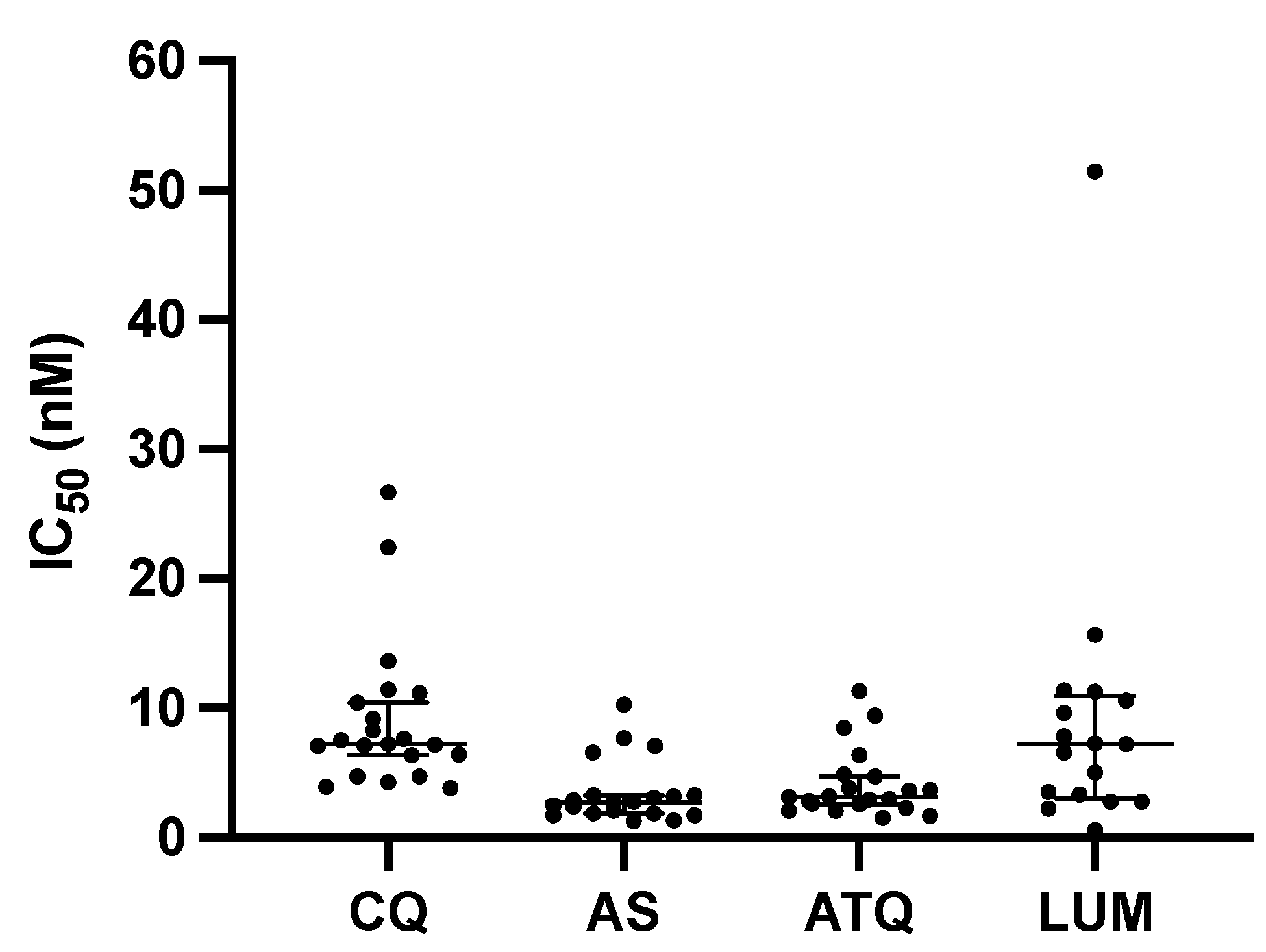
Figure 1. Distribution of IC50 values of P. malariae isolates for each drug (CQ, AS, ATQ, and LUM). CQ, chloroquine; AS, artesunate; ATQ, atovaquone; LUM, lumefantrine. Each data point for a particular drug shows mean IC50 values. Error bars indicate medians and 95% confidence intervals.
Pathogens 14 00453 g001
Table 1. Characteristics of the P. malariae isolates used for the susceptibility drug assay. ND: Not determined.
Isolate No.Parasite Count, No. Parasites/µL Plasmodium Species
Microscopy
Plasmodium Species
qPCR
12153P.mP.m
272610P.m-P.fP.m-P.f
1488620P.m-P.fP.m-P.f
1491013P.m-P.fP.m-P.f
167677P.mP.m
268831P.m-P.fP.m-P.f
269503P.m-P.fP.m-P.f
3251237P.mP.m
326773P.mP.m
3922960P.mP.m
589677P.mP.m-P.f
6272175P.mP.m-P.f
6781856P.m-P.fP.m-P.f
7422436P.mP.m
1037803P.m-P.fP.m-P.f
1063387P.mP.m
1077NDP.m-P.fND
1179462P.mP.m-P.f
1194406P.mP.m
1320715P.mP.m
1351358P.mP.m-P.f
Table 2. IC50 values (nM) for CQ, AS, ATQ, and LUM of all individual isolates as well as the NF54 control strain. NA: Not available. No IC50 data are available for these compounds.
Isolate No.CQ (nM)AS (nM)ATQ (nM)LUM (nM)Incubation Time (h)
127.141.7311.332.7636
273.932.093.602.2036
1489.207.686.376.6036
14922.452.903.813.5236
1676.397.103.697.2436
2687.206.554.887.8536
2693.832.363.179.6336
3257.512.762.5810.5936
3267.641.702.835.0536
3924.292.482.6511.3736
58926.673.098.4615.6836
6277.182.692.9551.4836
67810.4110.269.4511.2636
7427.10NA4.727.2836
10374.741.291.523.3236
10638.291.902.932.7736
107711.173.302.09NA45
11796.401.852.29NA13
119411.443.302.09NA24
13204.741.321.680.5936
135113.603.173.11NA48
NF545.813.034.443.5724
Disclaimer/Publisher’s Note: The statements, opinions and data contained in all publications are solely those of the individual author(s) and contributor(s) and not of MDPI and/or the editor(s). MDPI and/or the editor(s) disclaim responsibility for any injury to people or property resulting from any ideas, methods, instructions or products referred to in the content.