Article

Unravelling the Persistence of the Rare Serovar Salmonella Mikawasima in a Hospital Setting: A Whole-Genome Sequencing Study

1
Department of Microbiology, Croatian Institute of Public Health, 10000 Zagreb, Croatia
2
Food-and Water-Borne Infections Study Group (EFWISG), European Society of Clinical Microbiology and Infectious Disease (ESCMID), 4051 Basel, Switzerland
3
Department of Clinical Microbiology, University Hospital of Split, 21000 Split, Croatia
4
School of Medicine, University of Split, 21000 Split, Croatia
5
Department of Medicine, Icahn School of Medicine at Mount Sinai Morningside/West, 1000 Tenth Avenue, New York, NY 10019, USA
*
Author to whom correspondence should be addressed.
Pathogens 2025, 14(5), 408; https://doi.org/10.3390/pathogens14050408
Submission received: 17 March 2025 / Revised: 11 April 2025 / Accepted: 22 April 2025 / Published: 24 April 2025
(This article belongs to the Special Issue Emerging and Neglected Pathogens in the Balkans)

Abstract

:
Salmonella Mikawasima is a rare, mainly environmental serovar. In 2020, an outbreak was observed in neonatal and maternal wards of the University Hospital of Split and was established as an endemic until the end of 2024. Using whole-genome sequencing, this study aimed to analyse the phenotypic and genotypic characteristics of S. Mikawasima isolates and to elaborate whether the spread of the same clone occurred. Sequenced isolates were classified as ST2030, with the presence of aminoglycoside and extended spectrum beta-lactam resistance genes. Ten percent of the sequenced isolates exhibit multi-drug resistance. Identified virulence factors that include biofilm formation genes suggest the potential persistence of S. Mikawasima in the hospital environment, while spatial and temporal analysis reveal clonal expansion and possible horizontal transmission between different hospital wards. This study provides a deep understanding of the genomic composition of S. Mikawasima and emphasises the need for more stringent infection prevention measures, especially in vulnerable neonatal and postpartum settings, to mitigate the risk of healthcare-associated infections, and it should be followed by further microbiological and epidemiological investigations to identify the source of infection.

1. Introduction

Salmonella (S.) Mikawasima is a rare serovar belonging to S. enterica subspecies enterica. It is a part of the O:7 (C1) serogroup, showing antigenic profile formula O 6,7,14: y: e, n, z15 [1,2]. Its occurrence was firstly described in Turkey in 1967, when a previously unknown serovar was found in the digestive system of turtles. Since then, it has been found in Turkey in frog liver and in wild boars and birds in the north and south of Spain [3,4,5,6]. Apart from scarce reports of identification in animals and environmental and food samples, S. Mikawasima is also a rare cause of human salmonellosis outbreaks.
According to the European Food Safety Authority (EFSA) and European Centre for Disease Prevention and Control (ECDC) technical report and rapid outbreak assessment published in 2013, 671 human cases of S. Mikawasima were documented in EU/EEA countries and submitted to the European Surveillance System (TESSy) between 2007 and 2012 [7]. In the autumn of 2013, there was a major outbreak in the United Kingdom (U.K.), with 75 reported cases. It was likely a multistate outbreak with an unknown source. The case distribution exhibited significant seasonality across months, characterized by consistent peaks from September to November. It occurred mostly in adults, with at least 65% of cases documented in individuals over 25 years of age; there was no substantial variation between genders throughout the years, and there was a rather stable male-to-female ratio [7].
The next big cross-border outbreak was noted in 2019. The outbreak was first reported in the UK with 138 cases, Sweden with 31 cases, France with 18, Denmark with 2, and Ireland with 1 case. The epidemiologic data lack a complete travel history, and investigations pointed to the possible vehicle of infection distributed in different EU countries [8]. At the end of 2024, there was again a cross-border outbreak in Europe. Recent epidemiologic data from TESSy suggest travel-related infections, and also an infant community, but the details are not yet publicly available [9].
Reports on Salmonella species (spp.) as a causative agent of infections that are acquired during medical treatment in various healthcare facilities (healthcare-associated infections, HCAIs) are scarce [10,11,12,13].
The endemic occurrence of the S. Mikawasima in departments related to maternity and neonatal care at the University Hospital of Split (UHS) in the southern coast region of Croatia since April 2020 raises concerns [14], and it is necessary to employ all methods available to modern microbiological laboratories to gain high-resolution insights into the bacterial defence and virulence mechanisms so that, in synergy with clinical data, we can act on the source of infection and interrupt transmission pathways.
The aims of our study were to determine the phenotypic and genotypic characteristics of isolated strains of S. Mikawasima in UHS, to identify virulence and antimicrobial resistance (AMR) genes, to establish the relationship between the isolated strains, and to verify whether the isolates are part of the ongoing endemics or whether there were new entries and reinfections with different strains from the community. The specific research objectives included the application of whole-genome sequencing (WGS) and mutual comparison of genomes through core genome multi locus sequence typing (cgMLST). Because of the importance of understanding and dealing with the continued persistence of this rare serovar in neonatal hospital settings, this paper is a continuation of the study carried out by Novak at al., which showed that WGS can be a good tool for managing hospital infections [14].
We envisioned that managing this epidemic could benefit from our HERA2 project, which has, as one of its outcomes, scaling-up WGS capacities in outbreak detection, and that providing evidence of persistence can help in improving infection control measures and thus bring this epidemic to a close.

2. Materials and Methods

2.1. Study Design

This retrospective observational study was conducted at the Department of Clinical Microbiology, UHS, Croatia, from August 2023 to October 2024, and in parallel at the National Reference Laboratory for Salmonellae (NRLSalm), Division of Microbiology, Croatian Institute of Public Health (CIPH), Croatia.

2.2. Clinical Cases and Isolates

Bacterial strains were obtained from stool samples, rectal and throat swabs, and haemoculture collected during the study period from neonatal, paediatric, and maternal wards at UHS. The samples were then processed in the microbiological laboratory. Strains identified as S. Mikawasima were sent to NRLSalm for further genomic characterization. Only successfully sequenced strains that satisfied quality control criteria were included in this study.

2.3. Biochemical Characterization and Antimicrobial Susceptibility Testing

Initial samples were cultivated on Salmonella Shigella agar plates (Liofilchem, Roseto degli Abruzzi, Italy), and presumptive colonies were identified to species level using MALDI-TOF mass spectrometry (microflex® LRF; Bruker, Berlin, Germany). Strains identified as S. enterica, subsp. enterica, were serotyped according to White–Kaufmann–Le minor Scheme [1,2], using the slide agglutination technique with specific antisera (Sifin diagnostics gmbh, Berlin, Germany) for somatic O and flagellar H antigens. Antimicrobial susceptibility testing was conducted using the disk-diffusion method and Vitek 2 Compact system (bioMérieux, Lyon, France) with an AST-N379 card. Testing and interpretation were performed using European Committee on Antimicrobial Susceptibility Testing (EUCAST) breakpoints (v.13.1, and 14.0) [15]. The antimicrobial agents tested included ampicillin, ceftriaxone, ceftazidime, amoxicillin + clavulanic acid, chloramphenicol, trimethoprim-sulfamethoxazole, and pefloxacin as screening for ciprofloxacin. Extended-spectrum beta lactamase (ESBL) production in isolated strains was performed by double disk-diffusion test, using cefotaxime (CTX) and ceftazidime (CAZ) disks, with and without clavulanic acid (CLV), and Escherichia coli ATCC 25922 for the control.

2.4. Whole-Genome Sequencing and Bioinformatic Analysis

Upon identification, 38 isolated strains were successively referred to NRLSalm for further characterisation. DNA was extracted from pure bacterial cultures using the NucleoSpin Tissue kit (Macherey-Nagel, Dueren, Germany) following the manufacturer’s protocol, which included a 3 h proteinase K digestion at 56 °C. A WGS library with 2 × 150 bp reads was prepared using the Illumina DNA Prep kit and sequenced on an Illumina NextSeq 550 instrument (Illumina, San Diego, CA, USA). After sequencing, quality control was performed on acquired reads using FastQC v0.12.1, followed by read trimming with Fastp v0.23.4 [16,17]. De novo genome assembly was performed using the SPAdes genome assembler v3.10.1 with default parameters and the “careful” option [18]. The assembled genomes were then evaluated for quality and G+C content using Quast software v5.2.0 and Samtools v1.21 [19,20]. The analytical procedure adhered to acceptable quality control (QC) standards throughout the process. The samples that were not in line with quality parameter acceptance criteria (less than 4% deviation from the expected G+C content for species analysed, and total length of all contigs approximated to the known genome size of the target organism) were excluded from the study. All metrics related to the quality control and assembly are included as Supplementary Table S1.
The data generated in this study have been submitted to the European Nucleotide Archive (ENA).
Assembled genomes that passed quality requirements were identified against the RefSeq database with RefSeq masher v0.1.2 [21,22]. The reference genome used for Salmonella spp. analysis in our laboratory is GCF_000027025.1 [23]. Detailed serotype prediction was achieved using sistr_cmd v1.1.3 [24]. The multi-locus sequence types (MLSTs) were identified using mlst v2.23.0 [25]. High-resolution bacterial strain typing and evolutionary analysis by comparing genetic variations of conserved gene loci across genome (cgMLST), was performed using Ridom SeqSphere+ v4.0 [26]. This analysis was performed on our sequences but also on sequences obtained from the beginning of the epidemic and a set of publicly available S. Mikawasima sequences [14]. The distance matrices were visualised using the GrapeTree and iTOL online tools [27,28].
AMR mechanisms were determined using ResFinder v4.7.2 software that analyses de novo genome assemblies against ResFinder database: (2024-03-22) and PointFinder database: (2024-03-08) [29,30]. The analysis was conducted using a 90% identity threshold and 60% minimum length. Plasmids were searched using PlasmidFinder v2.1 with database version v2023-01-18 [31]. Mapping of resistance genes against plasmid sequences was performed using Mob-suite v3.1.9 and MobileElementfinder v1.0.3 with database v1.0.2 [32,33]. Abricate v1.0.1 was employed against VFDB database v20253-02-05 for identification of genes associated with virulence, and SPIFinder v2.0 was used to identify Salmonella spp. pathogenicity islands by running sequences against Pathogenic islands and resistance islands database (PAIDB) v2.0 [34,35,36,37].

3. Results

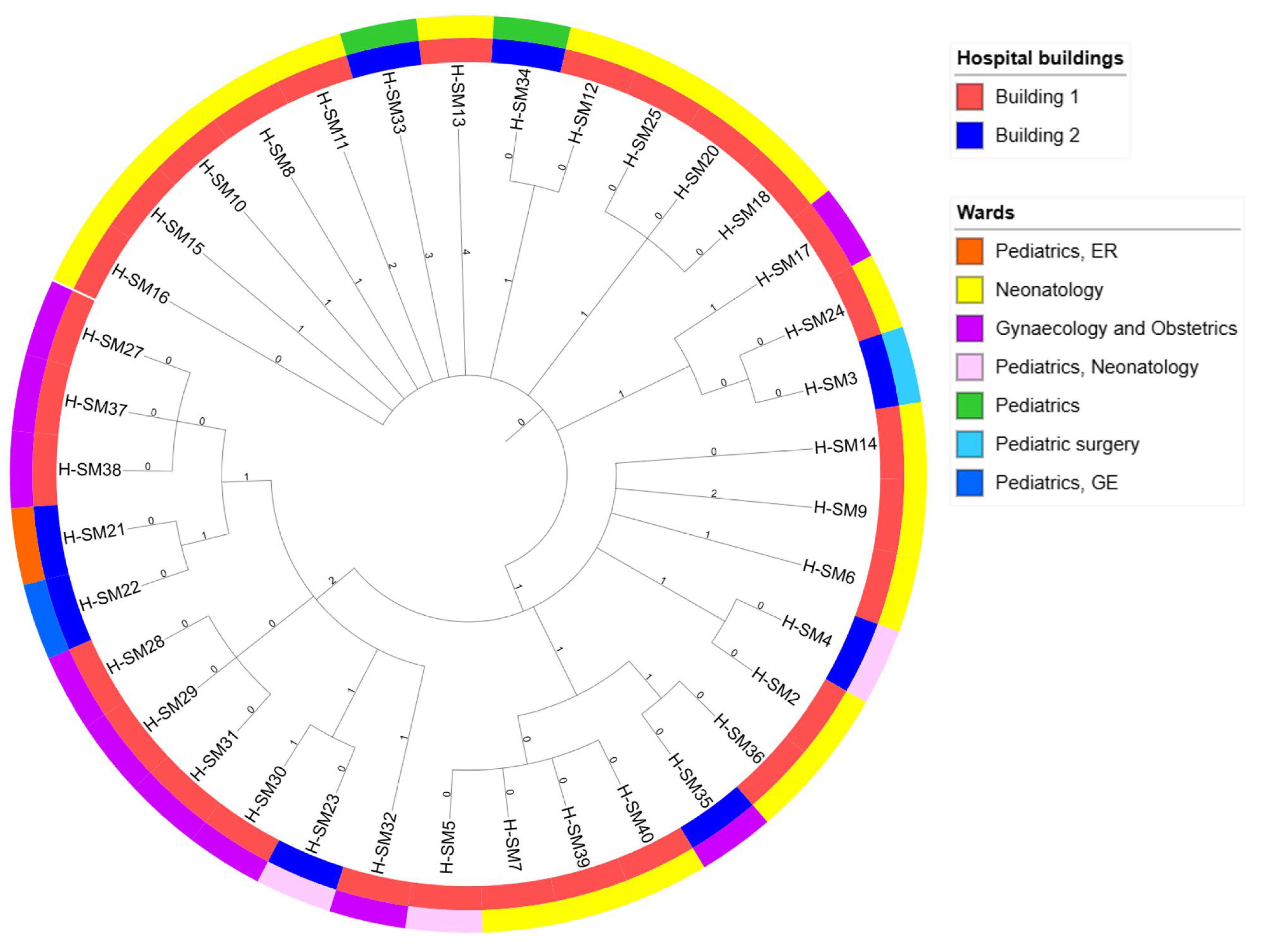
3.1. Isolates Metadata

Among 4979 tested stool samples and rectal swabs in UHC during the study period, 36 were positive for S. Mikawasima. Other serovars were sporadically isolated throughout the UHC, but none at the neonatal and maternal units, and they were not part of this study. One S. Mikawasima strain was isolated from a haemoculture, and one from a throat swab. Available information related to individual strains containing strain code, sampling dates, patient age, associated symptoms, hospital units, and antimicrobial resistance results are presented in Table 1.

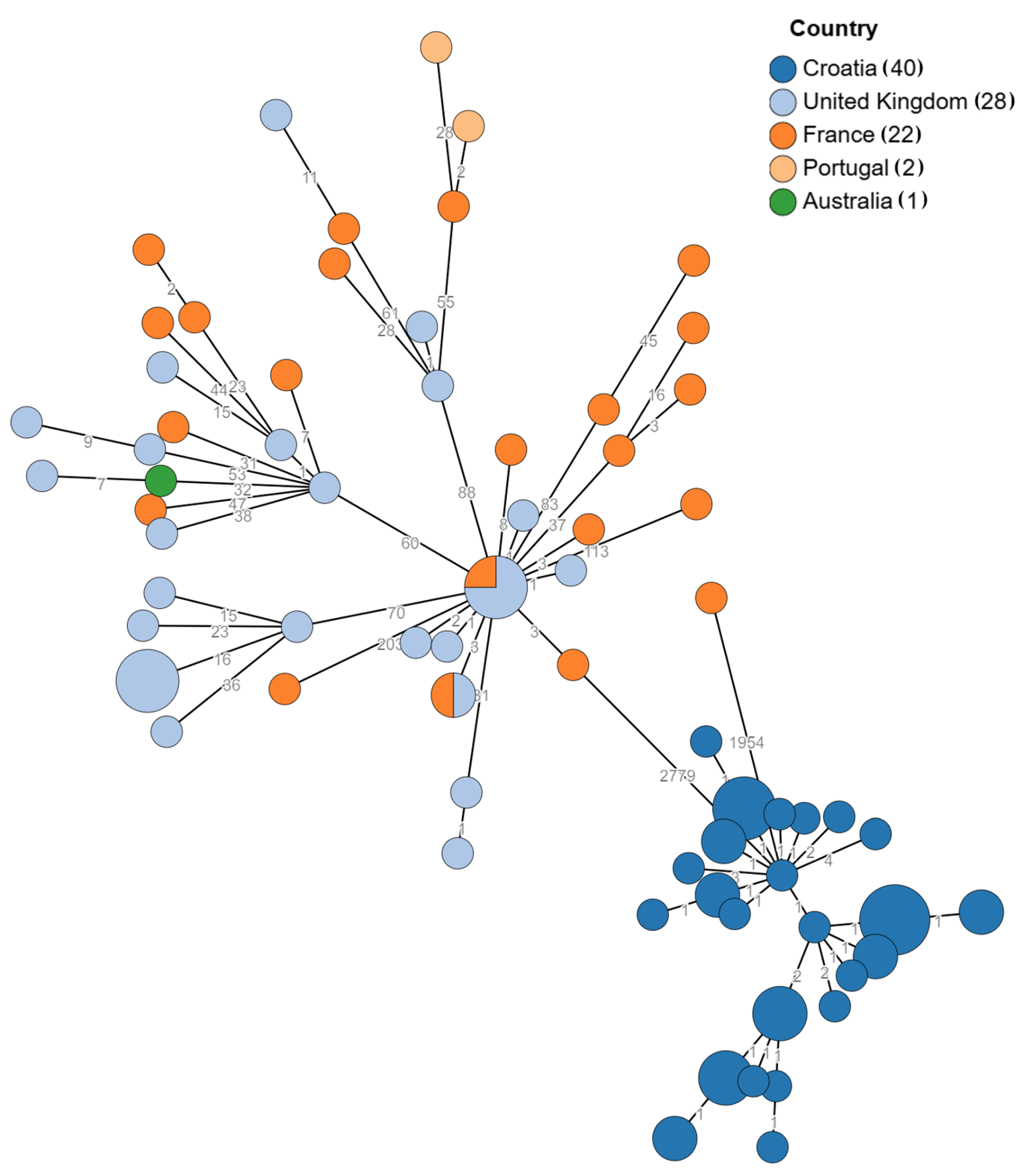
3.2. Genomic Relatedness

All isolates were identified as S. enterica, subsp. enterica, and serotype was predicted as S. Mikawasima, as were identified in UHS using the slide agglutination technique, showing a 7:y:e,n,z15 antigenic profile. The MLST scheme places all isolates in sequence type (ST) 2030. The distance matrix created with publicly available S. Mikawasima sequences from Enterobase and three sequences from the Novak at al. study shows that all isolates are a part of the same epidemiologic cluster, which is shown in Figure 1 [14,26].
To visualise relatedness of different S. Mikawasima strains, and to demonstrate their intrahospital location, we used the iTOL visualisation tool [28]. The phylogenetic tree is shown in Figure 2.
The distribution of samples, phenotypic ESBL patterns, and timelines according to occurrence in different hospital buildings and hospital units are shown in Table 2. It is visible from the table that majority of samples were isolated in the Neonatology Unit and closely related Gynaecology and Obstetrics Unit, placed in the same hospital building (Building 1). Sporadic but genetically closely related isolates were isolated from other buildings (Building 2) but from wards with a connection to the Neonatology Unit.

3.3. AMR Gene Profile and Phenotypic Correlation

All isolates harbour genes for aminoglycoside resistance, namely N-Acetyltransferases (aac): aac(6′)-Iaa, aac(6′)-Im and aph(2″)-Ib, (37/37, 100%).
Genes encoding beta-lactamase enzymes blaTEM-1B and blaSHV-2, and blaTEM-1B and blaSHV-2 together, are detected in 14/37 (37.8%), 6/37 (16.2%), and 17/37 (45.9%), respectively. Phenotypic expression of extended spectrum beta-lactamase was identified in 16 (43.3%) isolates.
Four out of thirty-seven (10.8%) isolates have the tet(D) gene that confers tetracycline resistance. These four isolates (H_SM3, H_SM22, H_SM33, and H_SM_34) are thus genotypically classified as multiple drug resistance (MDR) strains. All isolates carry the incompatibility group C plasmid (IncC) that can carry multiple resistance genes. These genes are often located within transposon integrons and other mobile genetic elements [38]. Mapping of resistance genes against the IncC plasmid sequence revealed that aac(6′)-Im and aph(2″)-Ib, blaTEM-1B are present on the IncC plasmid, while aac(6′)-Iaa is chromosomally encoded. The same is true for blaTEM-1B and tet(D) genes. In Table 3, the phenotypic antimicrobial susceptibility pattern and its correlation with the results of beta-lactamase profiling are shown. According to EUCAST guidelines, and based on clinical relevance, Salmonella spp. isolates are not phenotypically tested for susceptibility to aminoglycoside and tetracycline [15].

3.4. Virulence Genes as Biomarkers of Persistence

All isolates carry the following four Salmonella pathogenicity islands (SPI): SPI-1, SPI-2, SPI-3, and SPI-13.
Detected virulence genes are grouped in Table 4 into categories according to the VFDB database [39]. All the stated virulence factors are detected in all the tested strains. A complete list of detected virulence genes is presented in Supplementary Table S2.

4. Discussion

Data presented in this study show the complex behaviour of rarely detected Salmonella Mikawasima in a hospital environment. Distribution of samples between specific hospital units, the timeline of the isolates’ detection, and their correlation with the sequences deposited in GenBank from the Novak et al. study [14] suggest clonal expansion of ST 2030 in the same hospital settings from April 2020 until the end of 2024. Our research is a continuation of an earlier study due to the need for a deeper insight into the genomic characteristics of novel isolates throughout 2024 and in response to the dilemma whether this corresponds to the possible introduction of new clones from the community or further spread of the same clone within the hospital’s neonatology and paediatric settings.
The repeated detection of ESBL-positive ST 2030 in the same units over the studied period shows that S. Mikawasima became endemic in these specific units. Within all other hospital wards and units there were no S. Mikawasima isolates. Additionally, there were no other Salmonella spp. serovars isolated in the neonatal and paediatric settings in UHS.
Published reports on epidemic occurrences of Salmonella spp. in neonatal and paediatric hospital settings are scarce and include other different causative serovars. S. Tennessee caused epidemics in a neonatal intensive care unit in the U.S.A. and was connected to national epidemics linked to contaminated peanut butter. Multidrug-resistant S. Typhimurium caused prolonged two-year nosocomial epidemics in a paediatric ward in South Africa, and some authors hypothesised that the causative agent was introduced into the hospital setting from the community. The U.K.’s S. Enteritidis epidemic that affected mothers and new-borns was most likely initially spread by contaminated resuscitator equipment [11,12,13].
Detection of the same S. Mikawasima sequence type in other hospital units that include paediatric and gynaecology departments, even in two different buildings, suggests its ability to spread rapidly between different settings in a hospital environment. These other affected units handle postnatal care of neonates and mothers, indicating horizontal transfer between these wards, possibly through patient transfers, shared HCWs, or contaminated equipment.
Besides ST 2030 circulating in Europe, ST 1815 is an even more prominent S. Mikawasima sequence type that caused a cross-border outbreak in November 2019 [8]. At the end of 2024, there was again a cross-border outbreak of ST 1815 in Europe [9].
S. Mikawasima, as a relatively rare serovar, was not of particular interest in molecular studies of Salmonella spp. Apart from the Novak at al. paper [14], S. Mikawasima was described in the Joint EFSA and ECDC Rapid Outbreak Assessment after the 2013 increase in human infections in Europe [7]. They focused mainly on Pulse Field Gel Electrophoresis (PFGE) findings, as WGS was not widely available, and PFGE was still the gold standard in typing of Salmonella spp. [41]. An outbreak in Denmark, as a part of this 2013 increase, was analysed by WGS, but only for cluster detection [42]. Our study fulfils this knowledge gap and provides a deeper understanding of S. Mikawasima genomic composition and behaviour in a hospital environment.
The persistence of S. Mikawasima in hospital environments could be attributed to its formation and survival in biofilm outside of a human host. Our analysis revealed several adherence factors that are important for creation and survival of S. Mikawasima in a hospital environment. Curli fimbriae genes (csgA, csgB, csgC, csgE, csgF, csgG) are controlled by csgD and are critical in attachment to surfaces like stainless steel and plastics because of their role in the production of curli fimbriae and cellulose [43,44]. These fimbriae are more expressed when bacteria resides in a nutrient-depleted environment [45,46]. FimH and other type 1 fimbriae genes are important for adherence to different surfaces [47]. The Mig-14 gene, besides participating in polymyxin B resistance, promotes biofilm formation in harsh environments by regulating the expression of different virulence factors [40]. Although in this study biofilm formation assay was not performed, detection of these virulence genes indicates the genetic potential for environmental survival due to contamination and the potential for a permanent reservoir of S. Mikawasima in the hospital environment and/or medical equipment.
The presence of SPI-1 and SPI-2 points towards a type III secretion system (T3SS) that contributes to rapid intestinal colonisation, inflammation, and increased transmission in hospital environments [48,49]. Salmonella strains that do not carry SPI-1 and SPI-2 cannot induce inflammation [50]. There are studies that suggest that strains that carry SPI-1 display biofilm formation in media [51]. The function of SPI-3 is not so clear, but the data suggest that deletion of any of these three SPIs results in a reduction in adhesion capacity and possibly in biofilm formation [52]. SPI-13 contributes to intracellular survival of the bacteria [53].
The antimicrobial phenotypic beta-lactam resistance patterns show constant resistance to ampicillin and amoxicillin-clavulanic acid across almost all isolates (37/37, 100%). Exhibited phenotypic resistance levels to amoxicillin-clavulanate are slightly outside the 100% range (35/37, 94,6%). These phenotypic patterns of resistance are backed up by the detection of blaSHV-2 and blaTEM-1 genes. While in our strains, blaSHV-2 is chromosomally encoded, and blaTEM-1 is transferred across species by a broad host range of IncC plasmids that all our isolates carry.
In addition to resistance to aminopenicillins with or without beta-lactamase inhibitors, ESBL-positive strains express resistance to third generation cephalosporins with single strain resistance to ciprofloxacin (1/37, 0.93%). This specific case of phenotypic resistance does not have an underlying detected genetic basis of gene mutation confirmed through whole-genome sequencing. This warrants further investigation.
The presence of the gene tet(D) that confers resistance to tetracycline in four isolates, together with aminoglycoside resistance genes, designates them genotypically as MDR microorganisms, which diminishes treatment options, especially in vulnerable neonate patients for whom ampicillin in combination with aminoglycosides is a first-line empiric treatment [54]. The tet(D) gene is the least studied tet gene, encoding an efflux pump that expels tetracycline from bacterial cells in Salmonella isolates [55]. Its presence in four among a cluster of closely related strains remains a topic for further analysis, especially considering its chromosomal position. Different mobile genetic elements (insertion sequences, inverted repeats) detected throughout the genome might, in settings like the one described in this study, with the indication of continuous contamination and selective antimicrobial pressure, pose a high risk for persistence and spread of MDR strains, especially due to the presence of IncC plasmid. As cgMLST analysis considers only conserved regions of the genome, strains with no cgMLST allelic difference could have a different AMR profile.
Hospitals are a fundamental part of healthcare system, presenting a setting in which the most diversified and advanced treatment at the highest level is performed. This poses a risk for bacterial transmission and the emergence of healthcare-associated infections. Individuals at age extremes, including neonates and the elderly, are most susceptible to acquiring HCAIs. Prevalence rates of HCAIs and their bacterial causative agents in neonates vary across different hospital settings. As determined by ECDC point-prevalence survey of healthcare-associated infections in acute care hospitals conducted during 2011 and 2012 in 1149 hospitals across EU Member States, Iceland, Norway, and Croatia, prevalence of HCAIs in neonatology units was 3.5% (CI 95% 2.8–4.5%), paediatric surgery units 3.4% (CI 95% 2.3–4.9%), and general paediatric units 1.8% (CI 95% 1.4–2.4%) The prevalence rate of gastrointestinal infections acquired during hospitalization was 8.3% (CI 95% 6.4–10%). Although Enterobacterales were the most isolated microorganisms (15%) among detected pathogens causing all HCAIs, no Salmonella species was reported [56,57]. Another ECDC point-prevalence survey was conducted in the EU/EEA, including Croatia, during 2016/2017 [58], and according to the unpublished data from that study, the prevalence of HCAIs in Croatian paediatric and neonatal units was 5.2% and 1.8%, respectively. Among bacterial causative agents, no Salmonella species was detected.
Preventing the emergence of HCAIs, their epidemic or endemic occurrence, as well as interrupting the spread of already established ones, is only possible with a thorough understanding of the mechanisms of virulence and the antibiotic resistance that bacterial pathogens develop and utilize to successfully evade the effects of therapy and disinfection, thus maintaining themselves in the population and/or environment.
Our study indicates that, although S. Mikawasima is a rare serovar usually present in animals and contaminated environmental and food samples, it can emerge as a significant cause of hospital-acquired infections, especially in sensitive populations. It also can cause prolonged endemics in hospital settings. In Split-Dalmatia (SD) county, unlike other parts of Croatia, S. Mikawasima is the most frequently isolated serovar of Salmonella spp. [59,60]. Further genomic studies are needed to establish routes of transmission, the source of the infection, and to stop the spreading in hospital settings, as well as in the community.
The main drawback of this study is the lack of environmental samples. These samples were taken and processed in the local public health institute, and according to unofficial information, no source has been revealed. It is our hope that based on our research, which suggests the clonal spread within different and remote hospital settings, a more detailed investigation will be conducted, bearing in mind the behaviour of S. Mikawasima. It is evident that more effort is needed to implement rigorous measures for preventing HCAIs, particularly in neonatal care units where continuous antibiotic and disinfectant usage selects bacteria with a competitive advantage for survival in the hospital environment achieved by acquiring genes and mutations of antimicrobial resistance and different virulence factors, which we were able to show through means of analysis of WGS data.
Additionally, infection control and prevention measures should be conducted not only within a particular unit’s level but also at the hospital level, with the inclusion of all patient—HCW—equipment pathways. Comprehensive research across all sectors using the “One Health” approach is recommended to better understand and evaluate the risks linked to S. Mikawasima spreading.

Supplementary Materials

The following supporting information can be downloaded at: https://www.mdpi.com/article/10.3390/pathogens14050408/s1.

Author Contributions

Conceptualization, I.F. and A.N.; Data curation, I.T., D.J., L.K. and A.N.; Formal analysis, I.F., A.G.G., T.J., I.T., D.J. and H.J.; Funding acquisition, I.T.; Investigation, I.F., A.G.G., T.J., H.J. and L.K.; Methodology, A.G.G., T.J., M.T. and I.G.-B.; Software, D.J. and L.K.; Supervision, M.T., I.G.-B. and A.N.; Validation, I.F., T.J., I.T., M.T., I.G.-B. and A.N.; Visualization, D.J.; Writing—original draft, I.F. and A.G.G.; Writing—review and editing, I.F., A.G.G., T.J., I.T., M.T., I.G.-B., D.J., H.J. and A.N. All authors have read and agreed to the published version of the manuscript.

Funding

This research was conducted as a part of the up-scaling WGS capacities in outbreak detection under the HERA2 project, which received funding from the European Union’s Health and Digital Executive Agency (HaDEA) under Grant Agreement No. 101102440.

Institutional Review Board Statement

This study was conducted in accordance with the Declaration of Helsinki and approved by the Ethics Committee of University Hospital of Split (class: 520-03/25-01/35; reference number: 2181-147/01-06/LJ.Z.-25-02; date of approval: 27 February 2025).

Informed Consent Statement

Not applicable as this study involves retrospective analyses on bacterial isolates that were collected as a part of routine clinical care and are anonymised so they cannot be linked to individual patients. This is in line with the Protection of the Patient Rights Act and General Data Protection Regulation.

Data Availability Statement

The original data presented in the study are openly available from the European Nucleotide Archive at https://www.ebi.ac.uk/ena/browser/view/PRJEB86855 (accessed on 28 March 2025).

Conflicts of Interest

The authors declare no conflicts of interest.

References

  1. Issenhuth-Jeanjean, S.; Roggentin, P.; Mikoleit, M.; Guibourdenche, M.; de Pinna, E.; Nair, S.; Fields, P.I.; Weill, F.-X. Supplement 2008–2010 (No. 48) to the White–Kauffmann–Le Minor Scheme. Res. Microbiol. 2014, 165, 526–530. [Google Scholar] [CrossRef] [PubMed]
  2. Grimont, P.; Weill, F.-X. Antigenic Formulae of the Salmonella Serovars, 9th ed.; WHO Collaborating Centre for Reference and Research on Salmonella, Institute Pasteur: Paris, France, 2007; pp. 1–166. [Google Scholar]
  3. Gunel, E.; Polat Kilic, G.; Bulut, E.; Durul, B.; Acar, S.; Alpas, H.; Soyer, Y. Salmonella Surveillance on Fresh Produce in Retail in Turkey. Int. J. Food Microbiol. 2015, 199, 72–77. [Google Scholar] [CrossRef] [PubMed]
  4. Polo, F.; Figueras, M.J.; Inza, I.; Sala, J.; Fleisher, J.M.; Guarro, J. Prevalence of Salmonella Serotypes in Environmental Waters and Their Relationships with Indicator Organisms. Antonie Leeuwenhoek 1999, 75, 285–292. [Google Scholar] [CrossRef]
  5. Navarro-Gonzalez, N.; Mentaberre, G.; Porrero, C.M.; Serrano, E.; Mateos, A.; López-Martín, J.M.; Lavín, S.; Domínguez, L. Effect of Cattle on Salmonella Carriage, Diversity and Antimicrobial Resistance in Free-Ranging Wild Boar (Sus scrofa) in Northeastern Spain. PLoS ONE 2012, 7, e51614. [Google Scholar] [CrossRef]
  6. Jurado-Tarifa, E.; Torralbo, A.; Borge, C.; Cerdà-Cuéllar, M.; Ayats, T.; Carbonero, A.; García-Bocanegra, I. Genetic Diversity and Antimicrobial Resistance of Campylobacter and Salmonella Strains Isolated from Decoys and Raptors. Comp. Immunol. Microbiol. Infect. Dis. 2016, 48, 14–21. [Google Scholar] [CrossRef]
  7. European Food Safety Authority (EFSA). Unusual Increase of Salmonella Mikawasima Infections in Humans. EFSA Support. Publ. 2013, 10, 512E. [Google Scholar] [CrossRef]
  8. Communicable Disease Threats Report, 16-22 November 2024, Week 47. Available online: https://www.ecdc.europa.eu/en/publications-data/communicable-disease-threats-report-16-22-november-2024-week-47 (accessed on 21 April 2025).
  9. The European Surveillance System (TESSy). Available online: https://www.ecdc.europa.eu/en/publications-data/european-surveillance-system-tessy (accessed on 10 April 2025).
  10. Bobo, L.D.; Dubberke, E.R. Recognition and Prevention of Hospital-Associated Enteric Infections in the Intensive Care Unit. Crit. Care Med. 2010, 38, S324–S334. [Google Scholar] [CrossRef]
  11. Palmer, S.R.; Rowe, B. Investigation of Outbreaks of Salmonella in Hospitals. Br. Med. J. Clin. Res. Ed. 1983, 287, 891–893. [Google Scholar] [CrossRef]
  12. Boehmer, T.K.; Bamberg, W.M.; Ghosh, T.S.; Cronquist, A.; Fornof, M.E.; Cichon, M.K.; Gershman, K.; Vogt, R.L. Health Care-Associated Outbreak of Salmonella Tennessee in a Neonatal Intensive Care Unit. Am. J. Infect. Control 2009, 37, 49–55. [Google Scholar] [CrossRef]
  13. Smith, A.M.; Smouse, S.L.; Tau, N.P.; Bamford, C.; Moodley, V.M.; Jacobs, C.; McCarthy, K.M.; Lourens, A.; Keddy, K.H. GERMS-SA Surveillance Network Laboratory-Acquired Infections of Salmonella enterica Serotype Typhi in South Africa: Phenotypic and Genotypic Analysis of Isolates. BMC Infect. Dis. 2017, 17, 656. [Google Scholar] [CrossRef]
  14. Novak, A.; Dzelalija, M.; Goic-Barisic, I.; Kovacic, A.; Pirija, M.; Maravic, A.; Radic, M.; Marinovic, J.; Rubic, Z.; Carev, M.; et al. Phenotypic and Molecular Characterization of a Hospital Outbreak Clonal Lineage of Salmonella enterica Subspecies enterica Serovar Mikawasima Containing blaTEM-1B and blaSHV-2 That Emerged on a Neonatal Ward, During the COVID-19 Pandemic. Microb. Drug Resist. 2024, 30, 118–126. [Google Scholar] [CrossRef] [PubMed]
  15. Eucast: Previous Versions of Documents. Available online: https://www.eucast.org/ast_of_bacteria/previous_versions_of_documents (accessed on 31 January 2025).
  16. Babraham Bioinformatics—FastQC A Quality Control Tool for High Throughput Sequence Data. Available online: https://www.bioinformatics.babraham.ac.uk/projects/fastqc/ (accessed on 10 September 2024).
  17. Chen, S. Ultrafast One-Pass FASTQ Data Preprocessing, Quality Control, and Deduplication Using Fastp. iMeta 2023, 2, e107. [Google Scholar] [CrossRef] [PubMed]
  18. Prjibelski, A.; Antipov, D.; Meleshko, D.; Lapidus, A.; Korobeynikov, A. Using SPAdes De Novo Assembler. Curr. Protoc. Bioinforma. 2020, 70, e102. [Google Scholar] [CrossRef] [PubMed]
  19. Mikheenko, A.; Prjibelski, A.; Saveliev, V.; Antipov, D.; Gurevich, A. Versatile Genome Assembly Evaluation with QUAST-LG. Bioinforma. Oxf. Engl. 2018, 34, i142–i150. [Google Scholar] [CrossRef]
  20. Danecek, P.; Bonfield, J.K.; Liddle, J.; Marshall, J.; Ohan, V.; Pollard, M.O.; Whitwham, A.; Keane, T.; McCarthy, S.A.; Davies, R.M.; et al. Twelve Years of SAMtools and BCFtools. GigaScience 2021, 10, giab008. [Google Scholar] [CrossRef]
  21. O’Leary, N.A.; Wright, M.W.; Brister, J.R.; Ciufo, S.; Haddad, D.; McVeigh, R.; Rajput, B.; Robbertse, B.; Smith-White, B.; Ako-Adjei, D.; et al. Reference Sequence (RefSeq) Database at NCBI: Current Status, Taxonomic Expansion, and Functional Annotation. Nucleic Acids Res. 2016, 44, D733–D745. [Google Scholar] [CrossRef]
  22. Ondov, B.D.; Treangen, T.J.; Melsted, P.; Mallonee, A.B.; Bergman, N.H.; Koren, S.; Phillippy, A.M. Mash: Fast Genome and Metagenome Distance Estimation Using MinHash. Genome Biol. 2016, 17, 132. [Google Scholar] [CrossRef]
  23. Okoro, C.K.; Kingsley, R.A.; Connor, T.R.; Harris, S.R.; Parry, C.M.; Al-Mashhadani, M.N.; Kariuki, S.; Msefula, C.L.; Gordon, M.A.; de Pinna, E.; et al. Intracontinental Spread of Human Invasive Salmonella Typhimurium Pathovariants in Sub-Saharan Africa. Nat. Genet. 2012, 44, 1215–1221. [Google Scholar] [CrossRef]
  24. Yoshida, C.E.; Kruczkiewicz, P.; Laing, C.R.; Lingohr, E.J.; Gannon, V.P.J.; Nash, J.H.E.; Taboada, E.N. The Salmonella In Silico Typing Resource (SISTR): An Open Web-Accessible Tool for Rapidly Typing and Subtyping Draft Salmonella Genome Assemblies. PLoS ONE 2016, 11, e0147101. [Google Scholar] [CrossRef]
  25. Seemann, T. mlst Github. Available online: https://github.com/tseemann/mlst (accessed on 21 April 2025).
  26. Pallen, M.J. Updating Benchtop Sequencing Performance Comparison. Nat. Biotechnol. 2013, 31, 294–296. [Google Scholar] [CrossRef]
  27. Zhou, Z.; Alikhan, N.-F.; Sergeant, M.J.; Luhmann, N.; Vaz, C.; Francisco, A.P.; Carriço, J.A.; Achtman, M. GrapeTree: Visualization of Core Genomic Relationships among 100,000 Bacterial Pathogens. Genome Res. 2018, 28, 1395–1404. [Google Scholar] [CrossRef] [PubMed]
  28. Letunic, I.; Bork, P. Interactive Tree of Life (iTOL) v6: Recent Updates to the Phylogenetic Tree Display and Annotation Tool. Nucleic Acids Res. 2024, 52, W78–W82. [Google Scholar] [CrossRef] [PubMed]
  29. Bortolaia, V.; Kaas, R.S.; Ruppe, E.; Roberts, M.C.; Schwarz, S.; Cattoir, V.; Philippon, A.; Allesoe, R.L.; Rebelo, A.R.; Florensa, A.F.; et al. ResFinder 4.0 for Predictions of Phenotypes from Genotypes. J. Antimicrob. Chemother. 2020, 75, 3491–3500. [Google Scholar] [CrossRef] [PubMed]
  30. Camacho, C.; Coulouris, G.; Avagyan, V.; Ma, N.; Papadopoulos, J.; Bealer, K.; Madden, T.L. BLAST+: Architecture and Applications. BMC Bioinform. 2009, 10, 421. [Google Scholar] [CrossRef]
  31. Carattoli, A.; Zankari, E.; García-Fernández, A.; Voldby Larsen, M.; Lund, O.; Villa, L.; Møller Aarestrup, F.; Hasman, H. In Silico Detection and Typing of Plasmids Using PlasmidFinder and Plasmid Multilocus Sequence Typing. Antimicrob. Agents Chemother. 2014, 58, 3895–3903. [Google Scholar] [CrossRef]
  32. Robertson, J.; Nash, J.H.E. MOB-Suite: Software Tools for Clustering, Reconstruction and Typing of Plasmids from Draft Assemblies. Microb. Genom. 2018, 4, e000206. [Google Scholar] [CrossRef]
  33. Johansson, M.H.K.; Bortolaia, V.; Tansirichaiya, S.; Aarestrup, F.M.; Roberts, A.P.; Petersen, T.N. Detection of Mobile Genetic Elements Associated with Antibiotic Resistance in Salmonella enterica Using a Newly Developed Web Tool: MobileElementFinder. J. Antimicrob. Chemother. 2021, 76, 101–109. [Google Scholar] [CrossRef]
  34. Seemann, T. Abricate, Github. Available online: https://github.com/tseemann/abricate (accessed on 21 April 2025).
  35. Chen, L.; Zheng, D.; Liu, B.; Yang, J.; Jin, Q. VFDB 2016: Hierarchical and Refined Dataset for Big Data Analysis—10 Years On. Nucleic Acids Res. 2016, 44, D694–D697. [Google Scholar] [CrossRef]
  36. Roer, L.; Hendriksen, R.S.; Leekitcharoenphon, P.; Lukjancenko, O.; Kaas, R.S.; Hasman, H.; Aarestrup, F.M. Is the Evolution of Salmonella enterica Subsp. enterica Linked to Restriction-Modification Systems? mSystems 2016, 1, e00009-16. [Google Scholar] [CrossRef]
  37. Yoon, S.H.; Park, Y.-K.; Kim, J.F. PAIDB v2.0: Exploration and Analysis of Pathogenicity and Resistance Islands. Nucleic Acids Res. 2015, 43, D624–D630. [Google Scholar] [CrossRef]
  38. Fang, L.; Chen, R.; Li, C.; Sun, J.; Liu, R.; Shen, Y.; Guo, X. The Association between the Genetic Structures of Commonly Incompatible Plasmids in Gram-Negative Bacteria, Their Distribution and the Resistance Genes. Front. Cell. Infect. Microbiol. 2024, 14, 1472876. [Google Scholar] [CrossRef] [PubMed]
  39. VFDB—Pathogenesis of Salmonella. Available online: https://www.mgc.ac.cn/cgi-bin/VFs/genus.cgi?Genus=Salmonella (accessed on 21 April 2025).
  40. Sheng, X.; Wang, W.; Chen, L.; Zhang, H.; Zhang, Y.; Xu, S.; Xu, H.; Huang, X. Mig-14 May Contribute to Salmonella enterica Serovar Typhi Resistance to Polymyxin B by Decreasing the Permeability of the Outer-Membrane and Promoting the Formation of Biofilm. Int. J. Med. Microbiol. 2019, 309, 143–150. [Google Scholar] [CrossRef] [PubMed]
  41. Jacobs-Reitsma, W.F.; Maas, H.M.E. Eighteenth EURL-Salmonella Interlaboratory Comparison Study (2013) on Typing of Salmonella spp.; Rijksinstituut voor Volksgezondheid en Milieu RIVM: Utrecht, The Netherlands, 2016. [Google Scholar]
  42. Knegt, L.V.D.; Hald, T.; Boysen, L.; Pedersen, K.; Sørensen, A.I.V.; Korsgaard, H.; Perge, A.; Rosenquist, H. Annual Report on Zoonoses in Denmark 2013; DTU Food, National Food Institute: Lyngby, Denmark, 2014. [Google Scholar]
  43. Chia, T.W.R.; Goulter, R.M.; McMeekin, T.; Dykes, G.A.; Fegan, N. Attachment of Different Salmonella Serovars to Materials Commonly Used in a Poultry Processing Plant. Food Microbiol. 2009, 26, 853–859. [Google Scholar] [CrossRef]
  44. Harrell, J.E.; Hahn, M.M.; D’Souza, S.J.; Vasicek, E.M.; Sandala, J.L.; Gunn, J.S.; McLachlan, J.B. Salmonella Biofilm Formation, Chronic Infection, and Immunity Within the Intestine and Hepatobiliary Tract. Front. Cell. Infect. Microbiol. 2021, 10, 624622. [Google Scholar] [CrossRef] [PubMed]
  45. Laviniki, V.; Simoni, C.; Carloto, A.F.; Lopes, G.V. The Biofilm-Forming Ability of Salmonella Enterica Subsp. enterica Isolated from Swine-Feed Mills. Ciênc. Rural 2023, 54, e20230146. [Google Scholar] [CrossRef]
  46. Gaviria-Cantin, T.; Vargas, A.F.; Mouali, Y.E.; Jiménez, C.J.; Cimdins-Ahne, A.; Madrid, C.; Römling, U.; Balsalobre, C. Gre Factors Are Required for Biofilm Formation in Salmonella enterica Serovar Typhimurium by Targeting Transcription of the csgD Gene. Microorganisms 2022, 10, 1921. [Google Scholar] [CrossRef]
  47. Blumer, C.; Kleefeld, A.; Lehnen, D.; Heintz, M.; Dobrindt, U.; Nagy, G.; Michaelis, K.; Emödy, L.; Polen, T.; Rachel, R.; et al. Regulation of Type 1 Fimbriae Synthesis and Biofilm Formation by the Transcriptional Regulator LrhA of Escherichia coli. Microbiology 2005, 151, 3287–3298. [Google Scholar] [CrossRef]
  48. Lou, L.; Zhang, P.; Piao, R.; Wang, Y. Salmonella Pathogenicity Island 1 (SPI-1) and Its Complex Regulatory Network. Front. Cell. Infect. Microbiol. 2019, 9, 270. [Google Scholar] [CrossRef]
  49. Ramatla, T.; Khasapane, N.G.; Mlangeni, L.N.; Mokgokong, P.; Ramaili, T.; Ndou, R.; Nkhebenyane, J.S.; Lekota, K.; Thekisoe, O. Detection of Salmonella Pathogenicity Islands and Antimicrobial-Resistant Genes in Salmonella enterica Serovars Enteritidis and Typhimurium Isolated from Broiler Chickens. Antibiotics 2024, 13, 458. [Google Scholar] [CrossRef]
  50. Haneda, T.; Ishii, Y.; Shimizu, H.; Ohshima, K.; Iida, N.; Danbara, H.; Okada, N. Salmonella Type III Effector SpvC, a Phosphothreonine Lyase, Contributes to Reduction in Inflammatory Response during Intestinal Phase of Infection. Cell. Microbiol. 2012, 14, 485–499. [Google Scholar] [CrossRef]
  51. Jennings, M.E.; Quick, L.N.; Ubol, N.; Shrom, S.; Dollahon, N.; Wilson, J.W. Characterization of Salmonella Type III Secretion Hyper-Activity Which Results in Biofilm-Like Cell Aggregation. PLoS ONE 2012, 7, e33080. [Google Scholar] [CrossRef] [PubMed]
  52. Liu, G.; Lü, X.; Tian, Q.; Zhang, W.; Yi, F.; Zhang, Y.; Yu, S. Deletion of Salmonella Pathogenicity Islands SPI-1, 2 and 3 Induces Substantial Morphological and Metabolic Alternation and Protective Immune Potential. J. Integr. Agric. 2025, 24, 272–289. [Google Scholar] [CrossRef]
  53. Elder, J.R.; Paul, N.C.; Burin, R.; Guard, J.; Shah, D.H. Genomic Organization and Role of SPI-13 in Nutritional Fitness of Salmonella. Int. J. Med. Microbiol. 2018, 308, 1043–1052. [Google Scholar] [CrossRef]
  54. Neonatal Infection: Antibiotics for Prevention and Treatment; National Institute for Health and Care Excellence: Guidelines; National Institute for Health and Care Excellence (NICE): London, UK, 2024; ISBN 978-1-4731-5820-7.
  55. Pavelquesi, S.L.S.; de Oliveira Ferreira, A.C.A.; Rodrigues, A.R.M.; de Souza Silva, C.M.; Orsi, D.C.; da Silva, I.C.R. Presence of Tetracycline and Sulfonamide Resistance Genes in Salmonella spp.: Literature Review. Antibiotics 2021, 10, 1314. [Google Scholar] [CrossRef]
  56. Zingg, W.; Hopkins, S.; Gayet-Ageron, A.; Holmes, A.; Sharland, M.; Suetens, C.; ECDC PPS Study Group. Health-Care-Associated Infections in Neonates, Children, and Adolescents: An Analysis of Paediatric Data from the European Centre for Disease Prevention and Control Point-Prevalence Survey. Lancet Infect. Dis. 2017, 17, 381–389. [Google Scholar] [CrossRef]
  57. European Centre for Disease Prevention and Control. Point Prevalence Survey of Healthcare-Associated Infections and Antimicrobial Use in European Acute Care Hospitals: 2011–2012; Publications Office, LU: Luxembourg, 2013. [Google Scholar]
  58. European Centre for Disease Prevention and Control. Point Prevalence Survey of Healthcare-Associated Infections and Antimicrobial Use in European Acute Care Hospitals: 2016–2017; Publications Office, LU: Luxembourg, 2023. [Google Scholar]
  59. Seppälä, A.V. Epidemiological and Microbiological Characteristics of Salmonella spp. Infections in Outpatients in Split-Dalmatia County in 2022: A Cross-Sectional Study. Master’s Thesis, School of Medicine, University of Split, Split, Croatia, 2024. [Google Scholar]
  60. Barač Juretić, K.; Carev, M. Najčešći Bakterijski i Virusni Uzročnici Gastrointestinalnih Infekcija u Splitskodalmatinskoj Županiji u Razdoblju 2021–2023. Javno Zdr. 2024, 1, 30–34. Available online: https://nzjz-split.hr/casopis-javno-zdravstvo/ (accessed on 21 April 2025).
Figure 1. GrapeTree representation of cgMLST allelic distances between our strains and publicly available sequences. Branch lengths do not reflect the degree of sequence divergence because of the log scale representation chosen to show genetically similar sequences. Labels on branches represent number of different loci, and the size of the node represents the number of sequences without allelic differences.
Pathogens 14 00408 g001
Figure 2. Circular phylogenetic tree illustrating the genetic relationships among isolates. Each node corresponds to a specific isolate. Branch lengths do not reflect the degree of sequence divergence. Labels on branches represent number of different loci, and the legend depicts different hospital wards.
Pathogens 14 00408 g002
Table 1. Detailed metadata related to individual strains.
Strain CodeSampling DateAge/MonthsSampleSymptomsUnitAMR Profile *ESBL
H_SM2August 2023<1stooldiarrhoeaNeonatologyAM, AMC, CRO, CAZ, CTXpositive
H_SM3October 202313stooldiarrhoeaClinic for paediatric surgery, Department for abdominal surgeryAM, AMCnegative
H_SM4November 2023<1stooldiarrhoeaPaediatrics, neonatologyAM, AMC, CTX, CAZ, CIPpositive
H_SM5November 20231stooldiarrhoeaPaediatrics, neonatologyAM, AMC, CRO, CAZ, CTXpositive
H_SM6November 2023<1stooldiarrhoeaNeonatologyAM, AMC, CRO, CAZ, CTXpositive
H_SM7November 2023<1stooldiarrhoeaNeonatologyAM, AMC, CTX, CAZpositive
H_SM8January 2024<1rectal swabdiarrhoeaNeonatologyAM, AMC, CTXpositive
H_SM9January 2024<1rectal swabdiarrhoeaNeonatologyMissing datapositive
H_SM10January 2024<1rectal swabdiarrhoeaNeonatologyAM, AMC, CTXpositive
H_SM11January 20242rectal swabdiarrhoeaNeonatologyAM, AMC, CTXpositive
H_SM12January 20241throat swabasymptomatic colonisationNeonatologyAM, AMCpositive
H_SM13January 2024<1rectal swabdiarrhoeaNeonatologyAM, AMC, CTXpositive
H_SM14January 2024<1rectal swabdiarrhoeaNeonatologyAM, AMCnegative
H_SM15January 20241rectal swabdiarrhoeaNeonatologyAM, AMC, CAZpositive
H_SM16February 20243rectal swabdiarrhoeaNeonatologyAMpositive
H_SM17February 2024<1stooldiarrhoeaGynaecology and ObstetricsAM, AMC, CTXpositive
H_SM18December 20232rectal swabdiarrhoeaNeonatologyAM, AMCpositive
H_SM20February 2024<1rectal swabdiarrhoeaNeonatologyAM, AMC, CROpositive
H_SM21March 20241stooldiarrhoeaPaediatrics, ERAM, AMCnegative
H_SM22March 20241stooldiarrhoeaPaediatrics, GEAM, AMCnegative
H_SM23March 20243stooldiarrhoeaPaediatrics, neonatologyAM, AMCpositive
H_SM24July 2024<1haemoculturefeverNeonatologyAM, CAZpositive
H_SM25July 2024<1stooldiarrhoeaNeonatologyAM, CTX, CAZpositive
H_SM27July 2024<1rectal swabdiarrhoeaGynaecology and ObstetricsAM, AMCnegative
H_SM28August 2024<1stooldiarrhoeaGynaecology and ObstetricsAM, AMCnegative
H_SM29August 2024<1stooldiarrhoeaGynaecology and ObstetricsAM, AMCnegative
H_SM30August 2024<1rectal swabdiarrhoeaGynaecology and ObstetricsAM, AMCnegative
H_SM31August 2024<1stooldiarrhoeaGynaecology and ObstetricsAM, AMCnegative
H_SM32August 2024<1stooldiarrhoeaGynaecology and ObstetricsAM, AMCnegative
H_SM33July 20241stooldiarrhoeaPaediatricsAM, CTXpositive
H_SM34July 2024<1stooldiarrhoeaPaediatricsAM, CAZpositive
H_SM35September 2024<1rectal swabdiarrhoeaGynaecology and ObstetricsAM, AMCnegative
H_SM36September 20243rectal swabdiarrhoeaNeonatologyAM, AMC, CTX, CAZpositive
H_SM37September 2024<1rectal swabdiarrhoeaGynaecology and ObstetricsAM, AMCnegative
H_SM38September 2024<1stooldiarrhoeaGynaecology and ObstetricsAM, AMCnegative
H_SM39September 2024<1rectal swabdiarrhoeaNeonatologyAM, AMC, CTX, CAZpositive
H_SM40October 2024<1stooldiarrhoeaNeonatologyAM, AMCnegative
* AM—amikacin, AMC—amoxicillin + clavulanic acid, CAZ—ceftazidime, CTX—Cefotaxime, CRO—ceftriaxone.
Table 2. Timeline and distribution of isolates according to occurrence in two different buildings in the hospital setting.
August 2023October 2023November 2023December 2023January 2024February 2024March 2024July 2024August 2024September 2024October 2024
Building 2
Paediatric, neonatology unit 2 1
Paediatric, GE unit 1
Paediatric, ER 1
Clinic for paediatric surgery, Department for abdominal surgery 1
Paediatric 2
Building 1
Neonatology Unit1 217
1
2 2 21
Gynaecology and Obstetrics Unit 1 153
ESBL-positive strains; ESBL-negative strains.
Table 3. Correlation of AMR phenotype, ESBL production, and detected beta-lactamase genes.
ESBLAMR Phenotypic PatternblaTEM-1BblaSHV-2blaTEM-1B and blaSHV-2No. of Isolates
ESBL-negativeAmpicillin, amoxicillin-clavulanate131 14
ESBL-positiveAmpicillin 11
Ampicillin, amoxicillin-clavulanate1 23
Ampicillin, amoxicillin-clavulanate, cefotaxime 55
Ampicillin, amoxicillin-clavulanate, cefotaxime, ceftazidime 123
Ampicillin, amoxicillin-clavulanate, cefotaxime, ceftazidime, ciprofloxacin 11
Ampicillin, amoxicillin-clavulanate, ceftazidime 11
Ampicillin, amoxicillin-clavulanate, ceftriaxone 11
Ampicillin, amoxicillin-clavulanate, ceftazidime, cefotaxime, ceftriaxone 33
Ampicillin, cefotaxime 1 1
Ampicillin, cefotaxime, ceftazidime 1 1
Ampicillin, ceftazidime 2 2
Missing phenotypic data 11
Table 4. Categories of detected virulence genes.
Virulence Genes
AdherencecsgA, csgB, csgC, csgD, csgE, csgF, csgG, fimC, fimD, fimF, fimH, fimI, misL, sinH, steA, steB, steC,
Effector delivery systemsipA, sipB, sipC, sipD, invC, sopA, sopB, sopD, sopD2, sopE2, prgH, prgI, prgJ, prgK, invA, invB, invE, invF, invG, invH, invI, invJ, sicA, sicP, spaO, spaP, spaQ, spaR, spaS, ssaA, ssaB, ssaC, ssaD, ssaE, ssaF, ssaG, ssaH, ssaI, ssaJ, ssaK, ssaL, ssaM, ssaN, ssaO, ssaP, ssaQ, ssaR, ssaS, ssaT, ssaU, ssaV, sscA, sscB, orgA, orgB, orgC, sptP, sseJ, sseK2, sseL, pipB, pipB2, sifA, sifB, avrA, spiC/ssaB, sprP
ExotoxincdtB
Nutritional/Metabolic factormgtC, mgtB
Antimicrobial activity and biofilm formation [40]mig-14
Disclaimer/Publisher’s Note: The statements, opinions and data contained in all publications are solely those of the individual author(s) and contributor(s) and not of MDPI and/or the editor(s). MDPI and/or the editor(s) disclaim responsibility for any injury to people or property resulting from any ideas, methods, instructions or products referred to in the content.

