Article

Biocontrol Potential of Trichoderma spp. Against Phytophthora ramorum

by
Elisa Becker
1,*,
Nirusan Rajakulendran
2 and
Simon Francis Shamoun
1
1
Pacific Forestry Centre, Canadian Forest Service, Natural Resources Canada, 506 West Burnside Road, Victoria, BC V8Z 1M5, Canada
2
Independent Researcher, Toronto, ON M3H, Canada
*
Author to whom correspondence should be addressed.
Pathogens 2025, 14(2), 136; https://doi.org/10.3390/pathogens14020136
Submission received: 3 December 2024 / Revised: 20 January 2025 / Accepted: 21 January 2025 / Published: 2 February 2025

Abstract

:
Phytophthora ramorum, the cause of Sudden Oak Death and related diseases, threatens over 130 tree and shrub species. We evaluated the biocontrol potential of isolates from nine Trichoderma species against P. ramorum using growth-rate studies, dual-culture assays, and culture-filtrate assays. Results showed significant variation in Trichoderma growth rates and biocontrol potential. Some isolates exhibited rapid growth, effective overgrowth, and lethal effects against P. ramorum and produced potent antagonistic metabolites. Faster growth rates only partially correlated with biocontrol efficacy, indicating that factors beyond growth, such as metabolite production, play significant roles. Notably, isolates of T. koningii, T. viride, and the commercial product SoilGard™ (T. virens) showed promising efficacy. We calculated a combined biocontrol variable to rank isolates based on vigour and efficacy to aid in identifying promising candidates. Our findings support the use of Trichoderma spp. as biocontrol agents against P. ramorum and underscore the need for a multifaceted approach to selecting and optimizing isolates. Our evaluation demonstrated the importance of using different assays to assess specific mechanisms of action of biocontrol candidates. Future research should further explore these interactions to enhance the sustainable management of P. ramorum.

1. Introduction

Phytophthora ramorum, Werres, de Cock & Man in’t Veld [1], a globally significant invasive plant pathogen, causes substantial ecological and economic damage through diseases such as Sudden Oak Death in the U.S. and Sudden Larch Death and ramorum blight in Europe [2]. Its broad host range threatens forest biodiversity and the viability of the nursery industry. This oomycete affects over 130 species of trees and shrubs, including important nursery crops such as rhododendron, viburnum, and lilac, as well as forest species like maple and oak [3,4]. The pathogen produces sporangia on infected leaves and twigs, releasing motile zoospores that encyst and penetrate hosts under suitable conditions, initiating new infections [1]. In addition to causing foliar and stem diseases, P. ramorum survives in soil and can infect rhododendron roots, demonstrating its broad pathogenic capabilities [5,6].
The long-distance spread of P. ramorum has predominantly occurred through the transport of infected nursery stock. It has been isolated from diseased plants in nurseries across California, Oregon, Washington, and British Columbia (B.C.), with infected plants shipped to 32 U.S. states between 2003 and 2007 [3]. It has established in Florida and Mississippi, as evidenced by repeated detections in baiting surveys. In Europe, P. ramorum is considered an introduced pathogen found in nurseries and woodland gardens. This has led to quarantine regulations aimed at reducing further spread in North America and Europe. Despite detection in hundreds of nurseries and several wildland areas, its spread in natural environments in western North America remains limited to areas of California and southwestern Oregon. In B.C., P. ramorum is a quarantined pest posing a moderate risk to native forest ecosystems [7]. Since its detection in several B.C. nurseries in 2003, eradication actions have been mandated at all sites with infected plants [4,8]. While there are no cures for infected plants, several P. ramorum control measures are registered in the U.S. and Canada for preventive use on susceptible plants. These measures, alongside quarantine policies, focus on reducing inoculum levels and limiting human-facilitated pathogen spread. Minimizing the risk of infection is the primary defence against the spread of the pathogen to uninfected plants [9].
Trichoderma spp. are fast-growing, opportunistic fungi that function as avirulent plant symbionts. Their antagonistic abilities against plant pathogens have promoted their use as biological control agents [10,11,12,13]. They have shown potential as biocontrol agents against certain Phytophthora species [14]. For example, apple seedlings co-cultured with Phytophthora cactorum and Trichoderma spp. showed significant reductions in root damage and increased plant weight compared to those infected with P. cactorum alone [15].
Mycoparasitism, a process where one fungus parasitizes another, often involves the secretion of cell wall-degrading enzymes that hydrolyze the cell walls of target fungi or oomycetes [11]. Antibiosis, the production of inhibitory substances, is another major biocontrol mechanism employed by Trichoderma, which produces various antimicrobial compounds [16]. Other modes of action of Trichoderma strains include promoting plant growth and plant defence mechanisms and competing for nutrients and space [16,17,18,19]. To address the need for effective biocontrol agents against P. ramorum, this study investigates the potential of Trichoderma spp.
The objective of this study was to investigate the antagonistic properties of isolates from nine Trichoderma species, including three commercial biocontrol products, against P. ramorum. We conducted three in vitro biological assays to screen Trichoderma isolates as potential biological control candidates against P. ramorum.
  • Analysis of Temperature-Dependent Growth Rate: We began by performing a study of temperature-dependent growth rate on 51 Trichoderma isolates, hypothesizing that the growth rate of Trichoderma correlates with its biocontrol efficacy against P. ramorum. Trichoderma isolates that grow and colonize substrates rapidly could compete more effectively against P. ramorum.
  • Dual-Culture Assay for Direct Interaction: Next, we assessed the direct interaction, or mycoparasitism, of Trichoderma with P. ramorum. This assay used a dual-culture approach to measure the rates at which Trichoderma overgrew and killed an existing P. ramorum culture through direct interaction.
  • Antibiosis Microplate Assay for Antagonistic Effects: Additionally, we investigated the antibiosis of different Trichoderma isolates with respect to P. ramorum by testing sterile culture filtrates of Trichoderma isolates in a novel in vitro microplate assay to assess their antagonistic effects on P. ramorum germination and growth.
To conclude, we calculated a combined biocontrol variable to represent the general vigour and overall efficacy of the Trichoderma isolates.

2. Materials and Methods

2.1. Analysis of Temperature-Dependent Growth Rate

To measure the temperature-dependent linear growth rates of Trichoderma isolates (Table 1), a growth-rate study was performed at two temperatures: 10 °C and 20 °C. Agar plugs were taken from the leading edges of fresh cultures grown on potato dextrose agar (PDA) plates at 20 °C for 2 days. Growth-rate cultures were initiated on Petri plates (100 mm × 15 mm) containing 25 mL of PDA by placing an agar plug 5 mm from the edge of the plate. The plates were incubated at room temperature (~20 °C) for 4–8 h to allow the fungi to grow from the plug onto the agar surface. The plates were then transferred to incubators set at either 10 °C or 20 °C in darkness. Measurements of mycelial growth were taken along predetermined axes at 24, 48, 72, and 96 h after the acclimation period. Growth during the acclimation period was not included in the calculation of the overall growth rate. The linear growth rate was expressed in mm/day.

2.2. Dual Culture Assay for Direct Interaction

A dual-culture assay was used to evaluate direct interactions between the antagonist and pathogen, following the method of Goldfarb et al. [20]. To allow P. ramorum (NA1 lineage, accession 5073, isolated from Rhododendron) to establish before introduction of the faster-growing Trichoderma cultures, placements were timed accordingly (Figure 1). Each plate containing 15% V8 agar amended with 0.15% CaCO3 was bisected by a line drawn on the plate. An agar plug with an active culture of P. ramorum was placed on the center line, approximately 5 mm from the edge of the plate. The plate was incubated at 20 °C for 18 days or until the P. ramorum culture reached a diameter of 5.5 cm. To mark the farthest extent of P. ramorum growth, a line was drawn perpendicular to the center line. An agar plug of Trichoderma from the leading edge of a fresh 2-day-old PDA culture was placed on the center line, with its closest edge 5 mm from the marked extent of P. ramorum growth. After 4 days of dual culture growth, eight pairs of 6 mm plugs (one from each side of the center line), spanning a distance of 48 mm within the P. ramorum growth area, were collected. One plug from each pair was transferred to a plate containing 15% V8 agar amended with 0.15% CaCO3 to test for the presence of viable Trichoderma. The corresponding plug was transferred to a plate containing 15% V8 agar amended with 0.15% CaCO₃ plus 100 ppm benomyl (to inhibit Trichoderma growth) to test for the presence of viable P. ramorum. Re-isolation plates were monitored for the presence of P. ramorum or Trichoderma for up to 10 days (Figure 1).
The two parameters measured in this assay were the rate of overgrowth of the pathogen by the candidate Trichoderma culture and the rate of lethal effect. The rate of overgrowth was determined by measuring the linear distance on the dual culture plates over which Trichoderma overgrew the established P. ramorum culture. Growth of both P. ramorum and Trichoderma on their respective re-isolation plates indicated that Trichoderma had overgrown P. ramorum but had not killed it. The rate of lethal effect measured how effectively candidate cultures of Trichoderma spp. could kill P. ramorum. The absence of P. ramorum growth on the benomyl-amended re-isolation plates indicated it had been killed by the Trichoderma isolate (Figure 1).

2.3. Antibiosis Microplate Assay for Antagonistic Effect

2.3.1. Phytophthora Ramorum Sporangia Production

For sporangia production, fresh agar plugs of P. ramorum were transferred into 60 mm plates containing clarified 15% V8 broth amended with 1% CaCO3 and incubated for 3–5 days. After incubation, these liquid cultures were washed with sterile distilled water (sdH2O) to remove nutrient media, resuspended in sdH2O, and incubated for 3 days to induce sporangia production. To separate sporangia from mycelium, cultures were filtered through three layers of sterile cheesecloth. To stimulate zoospore release, the sporangia suspension was chilled at 4 °C for 1 h, then incubated at room temperature for 1–2 h, with zoospore release confirmed by microscopy. An aliquot of the zoospore suspension was transferred into a centrifuge tube and vortexed for 30 s to induce encystment. Encysted zoospores were counted using a hemocytometer, and appropriate dilutions were prepared in clarified 15% V8 broth amended with 0.15% CaCO3.

2.3.2. Production of Trichoderma Filtrates

Trichoderma isolates were cultured from agar plugs on PDA plates incubated at 20 °C in the dark for 2–3 days. Plates were subsequently placed under constant light until the cultures started to sporulate. Spore suspensions were prepared by flooding the plates with sdH2O and mixing with equal volumes of 20% glycerol, and the suspensions were then stored at –20 °C. On the day of inoculation, spores were counted using a hemocytometer and 100 mL of potato dextrose broth (PDB) was inoculated with Trichoderma spores to a final concentration of 10,000 spores/mL. Liquid cultures were incubated at room temperature with constant shaking at 130 rpm for 5 days. Mycelium was collected by filtering through three layers of cheesecloth and washed with 25 mL of sdH2O. Washed mycelium was aseptically transferred into 250 mL flasks containing 100 mL of sdH2O and incubated at room temperature with constant shaking at 130 rpm for 2 days. This culture filtrate was separated from mycelium by first pouring the liquid through three layers of cheesecloth, then filtering it through a 0.2 μm syringe filter to remove spores and mycelial fragments.

2.3.3. Antibiosis Microplate Assay

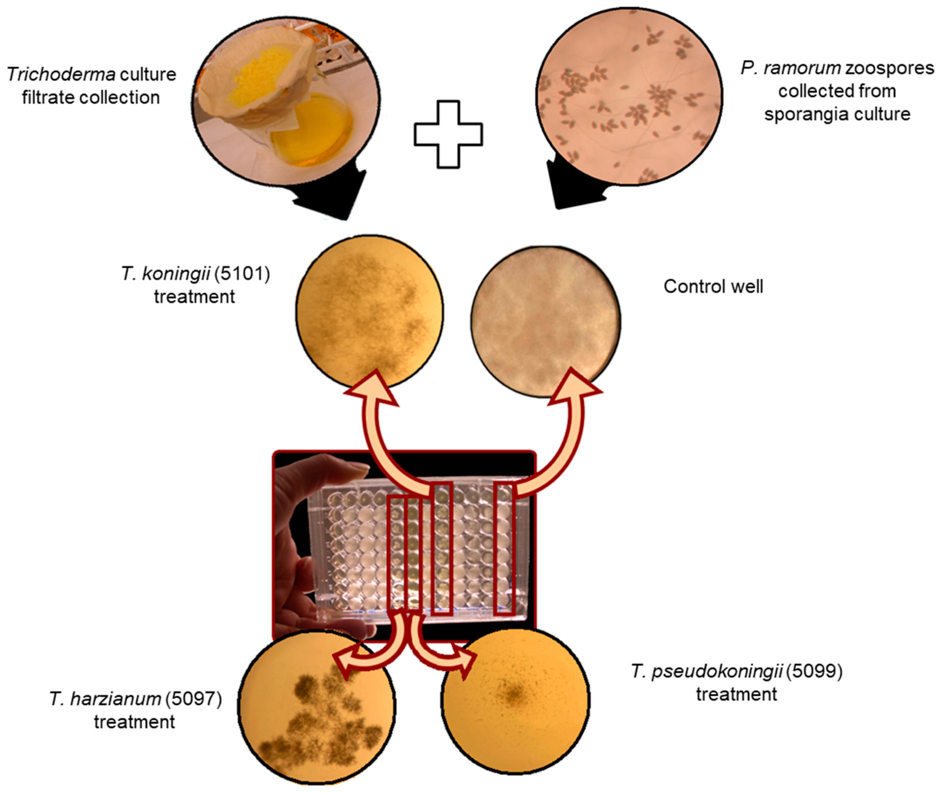
To test the activity of sterile filtrates, 100 μL of P. ramorum zoospore suspension (1000 zoospores/mL in 15% clarified V8) was mixed with 100 μL of Trichoderma culture filtrate in 96-well microplates (Figure 2). To generate the positive controls, 100 μL of sdH2O was substituted for Trichoderma culture filtrate. To generate the negative controls, 100 μL of 15% V8 broth was substituted for the P. ramorum zoospore suspension and mixed with 100 μL of Trichoderma filtrates. Absorbance at 650 nm was measured at 0, 24, 48, and 72 h. The percent inhibition was calculated using the following formula: % inhibition = [(Control − Treated)/Control] × 100.

2.4. Statistical Analyses

Statistical analyses were performed using R and RStudio with packages including tidyr, dplyr, psych, ggplot2, and RColorBrewer [21,22,23,24,25,26,27]. To create a combined biocontrol variable, values of each relevant variable were standardized (mean of 0 and standard deviation of 1) using the ’scale’ function, then combined into a single composite score by averaging the standardized variables.

3. Results

3.1. Analysis of Temperature-Dependent Growth Rate

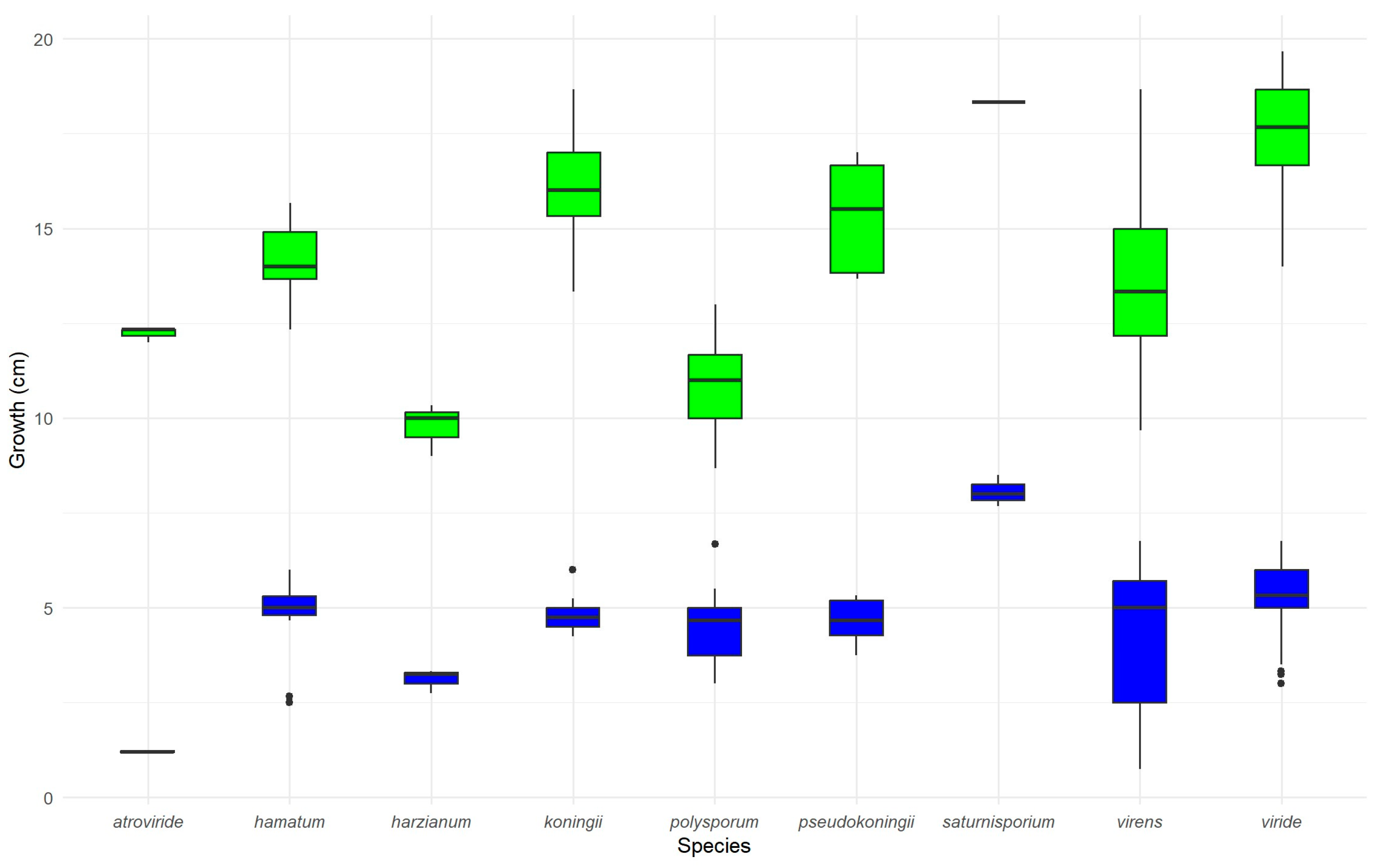
The mean radial growth of Trichoderma isolates at 10 °C after 3 days was 4.76 ± 1.25 cm (Figure 3). At 20 °C, the mean growth after 3 days was 14.72 ± 3.03 cm. The rate of linear growth ranged from 8.0 to 19.4 mm/day among 51 isolates (each with three replicates), with isolate 5035 (T. viride) growing the fastest.

3.2. Dual Culture Assay for Direct Interaction-Rate of Overgrowth

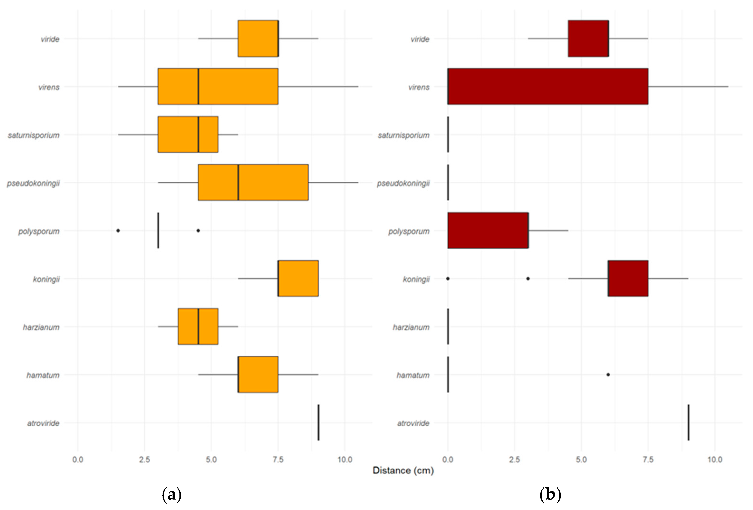
Overgrowth was assessed by the ability of Trichoderma cultures to overgrow an established P. ramorum culture (Figure 4). The mean overgrowth distance after 3 days was 5.95 ± 2.31 cm (Figure 4a). The commercial product SoilGard™ (T. virens) exhibited the highest overgrowth rate, at 10.5 mm/day.

3.3. Dual Culture Assay for Direct Interaction-Rate of Lethal Effect

The rate of lethal effect described the ability of Trichoderma isolates to mycoparasitize and kill P. ramorum through direct interactions (Figure 2). SoilGard (T. virens) demonstrated the highest rate of lethal effect at 10.5 mm/day, consistent with its high rate of overgrowth (Figure 4b). The ability of Trichoderma spp. to overgrow P. ramorum did not necessarily translate to a lethal effect. For example, T. hamatum had an average overgrowth rate of 6.0 mm/day; however, none of its isolates were able to mycoparasitize and kill P. ramorum. Similarly, T. polysporum (5003, 5018, 5025, 5026, 5034), T. virens (5095, 5096, 5098), T. koningii (5101), T. pseudokoningii (5093, 5094), and T. harzianum (5097) coexisted with P. ramorum without having a lethal effect on it. In addition, no Trichoderma isolate exhibited a lethal effect on P. ramorum from a distance. Trichoderma isolates had a lethal effect on P. ramorum only after overgrowing it.

3.4. Antibiosis Microplate Assay for Antagonistic Effect

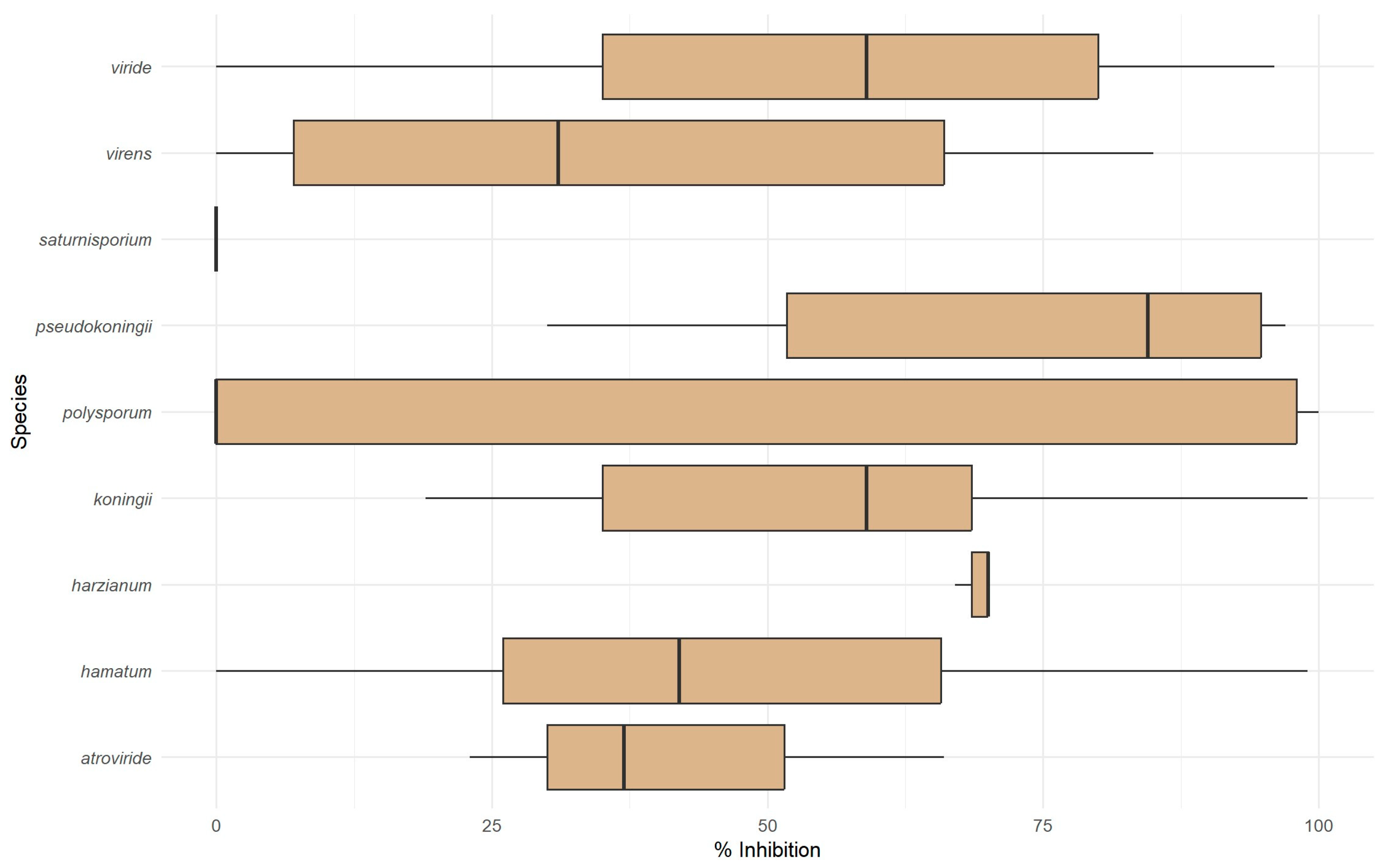
Culture filtrates collected under starvation conditions were tested for their inhibitory effects on P. ramorum (Figure 5). The average inhibition of P. ramorum growth by sterile culture filtrates of Trichoderma isolates was 49 ± 35%. The following isolates produced culture metabolites that inhibited P. ramorum growth by more than 75%: T. virens (SoilGard), T. hamatum (T382), T. koningii (5101), T. pseudokoningii (5099), T. viride (5008), and T. polysporum (5017, 5032, 5033, 5016, and 5001).

3.5. Correlation of Assay Variables

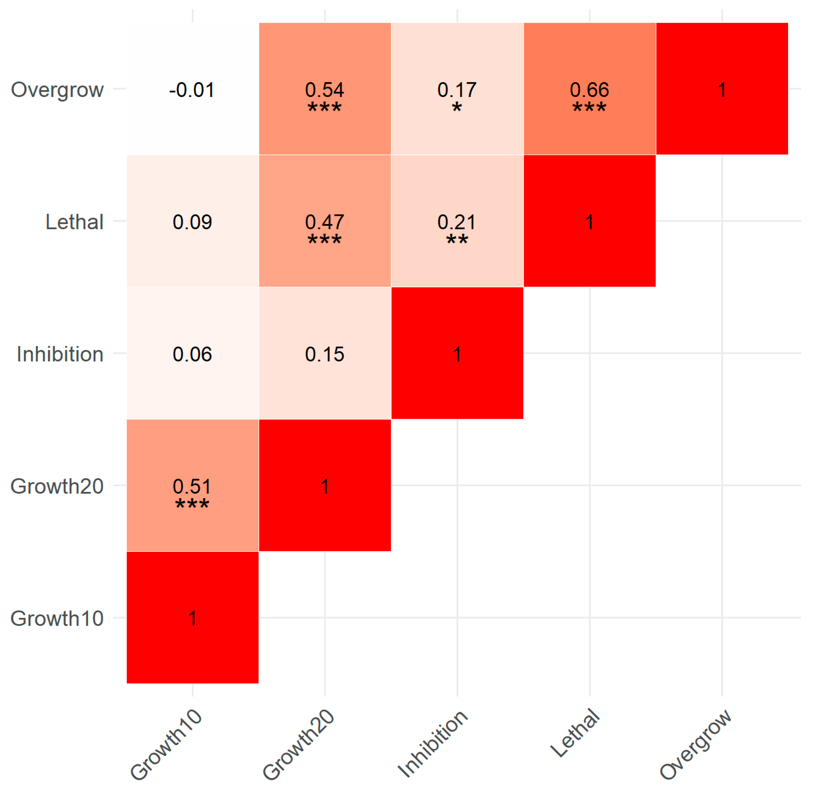
A strong positive correlation was observed between the growth of Trichoderma isolates at 10 °C and their growth at 20 °C (Figure 6; r = 0.506, p < 0.001). Likewise, a strong positive correlation (r = 0.544, p < 0.001) suggested that increased growth of Trichoderma isolates at 20 °C was associated with increased overgrowth of P. ramorum. The strongest positive correlation (r = 0.657, p < 0.001) indicated a significant relationship between the overgrowth and the lethal effect on P. ramorum exerted by Trichoderma. A moderate positive correlation (r = 0.214, p = 0.008) suggested that higher values of percent inhibition were associated with higher lethal effect values. A moderate positive correlation (r = 0.167, p = 0.039) indicated a relationship between percent inhibition and overgrowth of P. ramorum by Trichoderma.

3.6. Combined Biocontrol Variable

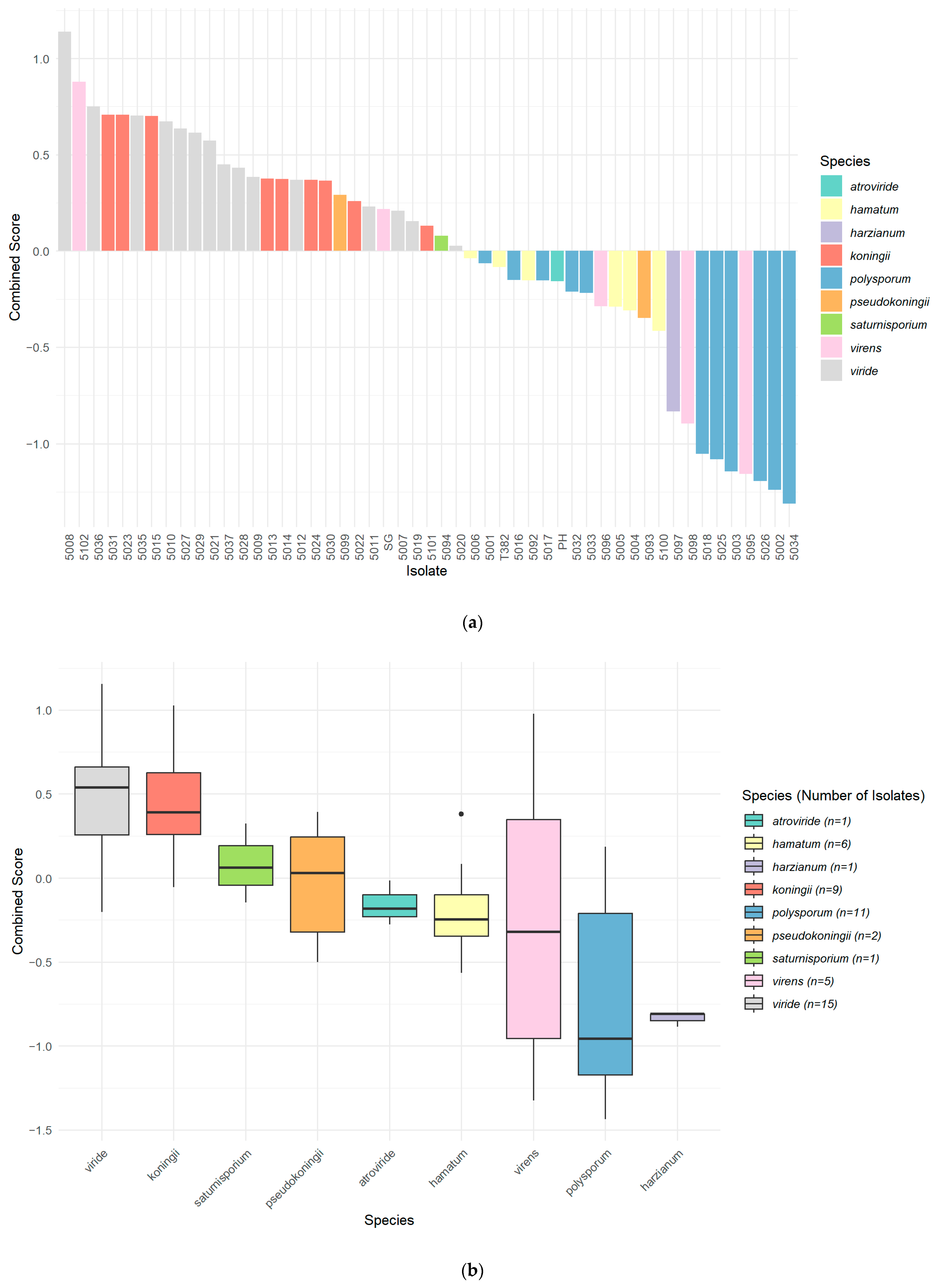
A combined variable representing the relative general vigour and biocontrol efficacy of the isolates was developed. This was done by standardizing and scaling each of the relevant variables (growth of Trichoderma at 10 °C, and at 20 °C, overgrowth of P. ramorum, lethal effect on P. ramorum, and percent inhibition of P. ramorum) and then combining them into a single composite score. The combined biocontrol variable was used to rank each isolate (Figure 7a) and species group (Figure 7b).

4. Discussion

Trichoderma spp. act as potent antagonists against plant pathogens through various direct and indirect modes of action, including mycoparasitism, antibiosis, lytic enzyme secretion, and competition for space and nutrients [10]. Through direct mechanisms such as mycoparasitism, the antagonist secretes lytic enzymes, antibiotics, and other toxic metabolites that synergistically antagonize the pathogen. Ideal biocontrol agents exhibit most of these characteristics, enabling effective control of target pathogens [19,28,29].
Given these diverse biocontrol mechanisms, our study assessed the effectiveness of various Trichoderma isolates against Phytophthora ramorum, a destructive pathogen causing significant nursery and forestry diseases. We focused on the isolates’ growth rates and specific mechanisms of action to identify promising candidates for biocontrol applications against P. ramorum. Specifically, we evaluated 51 isolates from nine Trichoderma species using criteria reflecting various desired biocontrol abilities or traits.
In our investigation, we hypothesized that the growth rate of Trichoderma isolates would be directly correlated with their biocontrol efficacy against P. ramorum. Our Trichoderma growth rate study indicated a wide variation among the 51 isolates screened, with growth rates at 20 °C ranging from 8.0 to 19.4 mm/day. Notably, 5035 (T. viride), the fastest-growing isolate, showed significant biocontrol potential, suggesting that rapid colonization through competition for space is an effective mechanism of action against P. ramorum. The correlation between growth rate and biocontrol efficacy was inconsistent, as several fast-growing isolates did not exhibit comparable biocontrol efficacy. While competition for space and nutrients is a classical mechanism of biological control [30], our results suggest that multiple mechanisms, including antibiosis and mycoparasitism, in addition to growth rate, contribute to the biocontrol capabilities of Trichoderma against P. ramorum.
Mycoparasitism involves direct interactions with the pathogen, such as coiling around the pathogen’s hyphae, penetration, and secretion of lytic enzymes [31,32,33]. These enzymes break down the cell walls of the pathogen, leading to its disintegration [33]. In our study, T. atroviride, T. koningii, and T. virens demonstrated effective mycoparasitism by overgrowing and directly killing P. ramorum in dual culture (Figure 1 and Figure 4a,b). This aligns with the results of studies showing that production of lytic enzymes by Trichoderma spp. contributes to their efficacy in suppressing root rot caused by Phytophthora capsici [18,33], suggesting that a similar mechanism acts against P. ramorum.
The dual-culture assays illuminated the complex relationship between Trichoderma growth rates and their biocontrol efficacy. While the commercial product SoilGard exhibited the highest rate of overgrowth and greatest lethal effect against P. ramorum, it was not statistically different from other isolates with varying growth rates. This underscores that while rapid growth aids in competition for space, it is not the sole determinant of biocontrol efficacy; other mechanisms like antibiosis are also crucial. Moreover, the observation that overgrowth did not always result in a lethal effect suggests that mechanisms beyond mycoparasitism, such as antibiosis, are involved in the biocontrol of P. ramorum.
The analysis of culture metabolites produced under starvation conditions provided further insights into the biocontrol mechanisms of Trichoderma isolates. Trichoderma spp. produce a variety of antibiotic-like substances and secondary metabolites that inhibit pathogen growth. These include compounds like Trichodermin and viridin [33] and volatile organic compounds (VOCs) with antimicrobial properties, contributing to antibiosis [10]. We found that several isolates, notably T. polysporum, produced antagonistic metabolites that significantly inhibited P. ramorum growth by more than 95%, emphasizing the role of antibiosis.
The results for T. polysporum were inconsistent between the two assays. While the microplate assay showed strong antibiosis (Figure 5), the dual-culture assay (Figure 4a,b) suggested limited mycoparasitic activity. This limited mycoparasitic activity may result from the slow linear growth rate of T. polysporum (Figure 3), suggesting that rapid growth is essential for effective mycoparasitism. Therefore, antibiosis through metabolite production was found to be a critical biocontrol mechanism of Trichoderma isolates against P. ramorum, especially for isolates with slower growth rates.
This study evaluated the biocontrol potential of various Trichoderma isolates against P. ramorum using several methodologies. Growth-rate studies at 20 °C served as a useful screening tool, generally correlating with indicators of biocontrol efficacy like overgrowth, lethal effect, and percentage inhibition. The dual-culture assays assessed direct antagonism by Trichoderma isolates through mycoparasitism. They provided insights into specific mechanisms of action, particularly the abilities to overgrow and to kill established cultures of P. ramorum. However, the dual-culture assays were complex and time-consuming, limiting their practicality for large-scale screening of mycoparasitic activity.
To efficiently assess the antibiosis mechanism, we developed a new microplate assay evaluating the inhibitory effects of Trichoderma culture filtrates on P. ramorum zoospore germination and growth. This assay evaluates antibiosis by testing the ability of sterile culture filtrates from Trichoderma to inhibit the pathogen. Using measurements of relative light absorbance to quantify P. ramorum zoospore germination and growth, this assay allowed us to efficiently screen multiple Trichoderma isolates for antibiosis. This efficient method facilitates easy replication, minimizes media usage, and provides rapid results, making it suitable for large-scale screening of biocontrol agents based on antibiosis.
The assays revealed different mechanisms of action. Selecting species and isolates for field studies requires considering the type of application. For instance, in a forest or nursery, a living Trichoderma isolate could be used as a foliar spray to prevent P. ramorum spread from leaf lesions, or as a soil treatment to minimize splash dispersal or root infection. The microplate assay we developed allows the selection of Trichoderma isolates with active metabolites against P. ramorum that could be used as a sterile product, eliminating persistence risk after application. These choices may be influenced by the specific P. ramorum hosts and the regulatory framework for biocontrol products. Future research should unravel these interactions and mechanisms to enhance the practical application of Trichoderma to manage P. ramorum infections [12,34]. Understanding the potential mechanisms of action of these isolates will enable us to further screen and tailor candidate agents to meet the specific requirements of diverse applications. Future studies should evaluate field and nursery applications to develop practical biocontrol strategies.

5. Conclusions

The predominantly positive correlations among variables suggest that a combined variable is useful for ranking isolates by biocontrol activity. This combined variable represented the isolates’ general vigour and biocontrol efficacy. Integrating these methods provided a comprehensive assessment of Trichoderma isolates’ biocontrol capabilities and mechanisms. The growth-rate study at 20 °C served as an efficient initial screening method. Although labour-intensive, the dual-culture assays provided detailed insights into direct antagonistic interactions. The new microplate assay enabled rapid, high-throughput evaluation of metabolite-mediated inhibition. Together, these approaches emphasize the importance of using diverse assays to select isolates with multiple mechanisms of action under varied conditions.
Our results partially support the hypothesis that Trichoderma isolates’ growth rates correlate with their biocontrol efficacy against P. ramorum. While rapid growth rates are advantageous and contribute to the competitive suppression of P. ramorum, our findings underscore that other factors, including antagonistic metabolites and possibly other mechanisms, play significant roles in the biocontrol process [28,35]. Our results align with those of previous studies demonstrating the importance of rapid colonization and metabolite production in effective biocontrol agents [10,36,37].
Advancements in developing and testing methods for applying promising Trichoderma isolates to plants or soil are continually progressing. These findings advance our understanding of biocontrol mechanisms against P. ramorum and have broader implications for sustainable disease management in forestry and agriculture [12,13,34,37].

Author Contributions

Conceptualization, E.B. and S.F.S.; methodology, E.B. and N.R.; software, E.B.; validation, E.B. and N.R.; formal analysis, E.B.; investigation, E.B.; resources, S.F.S.; data curation, E.B.; writing—original draft preparation, E.B.; writing—review and editing, E.B., S.F.S. and N.R.; visualization, E.B.; supervision, S.F.S.; project administration, S.F.S.; funding acquisition, S.F.S. All authors have read and agreed to the published version of the manuscript.

Funding

This research was funded by the NRCan CFS-Pest Risk Management Program, the CFIA, and NSERC support to SS. Additional funding was provided by NSERC (Visiting Fellowship to EB) and the YMCA (Student Internship to NR).

Institutional Review Board Statement

Not applicable.

Informed Consent Statement

Not applicable.

Data Availability Statement

The original data presented in the study are openly available in FigShare at https://doi.org/10.6084/m9.figshare.27961578.v1 (accessed on 20 January 2025).

Acknowledgments

The authors thank N. Grünwald for providing the P. ramorum isolate used in this study, and AmPac Biotech Corporation, Certis Biologicals, and Sylvan Bioproducts for supplying biocontrol agent samples. We also thank T. Osono and M. Elliott for collecting Trichoderma isolates. Special thanks to Robert Kowbel and Grace Sumampong for their excellent technical support.

Conflicts of Interest

The authors declare no conflicts of interest. The funders had no role in the design of the study; in the collection, analyses, or interpretation of data; in the writing of the manuscript; or in the decision to publish the results.

References

  1. Werres, S.; Marwitz, R.; Man In’t veld, W.A.; De Cock, A.W.A.M.; Bonants, P.J.M.; De Weerdt, M.; Themann, K.; Ilieva, E.; Baayen, R.P. Phytophthora ramorum sp. nov., a new pathogen on Rhododendron and Viburnum. Mycol. Res. 2001, 105, 1155–1165. [Google Scholar] [CrossRef]
  2. Parke, J.L.; Peterson, E.K. Sudden Oak Death, Sudden Larch Death, and Ramorum Blight. Plant Health Instr. 2019, 19. Available online: https://www.apsnet.org/edcenter/pdlessons/Pages/SuddenOakDeath.aspx (accessed on 20 January 2025).
  3. Grünwald, N.J.; Goss, E.M.; Press, C.M. Phytophthora ramorum: A Pathogen with a Remarkably Wide Host Range Causing Sudden Oak Death on Oaks and Ramorum Blight on Woody Ornamentals. Mol. Plant Pathol. 2008, 9, 729–740. [Google Scholar] [CrossRef]
  4. Shamoun, S.F.; Rioux, D.; Callan, B.E.; James, D.; Hamelin, R.; Bilodeau, G.J.; Elliott, M.; Lévesque, C.A.; Becker, E.; McKenney, D.; et al. An Overview of Canadian Research Activities on Diseases Caused by Phytophthora ramorum: Results, Progress, and Challenges. Plant Dis. 2018, 102, 1218–1233. [Google Scholar] [CrossRef] [PubMed]
  5. Rizzo, D.M. Phytophthora ramorum: Integrative Research and Management of an Emerging Pathogen in California and Oregon Forests. Annu. Rev. Phytopathol. 2005, 43, 309–350. [Google Scholar] [CrossRef] [PubMed]
  6. Shishkoff, N. Susceptibility of Some Common Container Weeds to Phytophthora ramorum. Plant Dis. 2004, 96, 1026–1032. [Google Scholar] [CrossRef] [PubMed]
  7. Canadian Food Inspection Agency. D-01-01: Phytosanitary Requirements to Prevent the Entry and Spread of Phytophthora ramorum (18th Revision); CFIA Directive D-01-01; Canadian Food Inspection Agency: Ottawa, ON, Canada, 13 June 2013. [Google Scholar]
  8. Canadian Food Inspection Agency. PI-010: Regulatory Response Protocol for Nurseries Confirmed with Phytophthora ramorum; CFIA—PI-010; Canadian Food Inspection Agency: Ottawa, ON, Canada, 29 April 2019. [Google Scholar]
  9. Grünwald, N.J.; Goss, E.M. Evolution and Population Genetics of Exotic and Re-emerging Pathogens: Novel Tools and Approaches. Annu. Rev. Phytopathol. 2011, 49, 249–267. [Google Scholar] [CrossRef]
  10. Saldana-Mendoza, S.A.; Pacios-Michelena, S.; Palacios-Ponce, A.; Chavez-Gonzalez, M.; Aguilar, C.N. Trichoderma as a Biological Control Agent: Mechanisms of Action, Benefits for Crops and Development of Formulation. World J. Microbiol. Biotechnol. 2023, 39, 269. [Google Scholar] [CrossRef] [PubMed]
  11. Vinale, F.; Sivasithamparam, K.; Ghisalberti, E.L.; Marra, R.; Woo, S.L.; Lorito, M. Trichoderma–Plant–Pathogen Interactions. Soil Biol. Biochem. 2008, 40, 1–10. [Google Scholar] [CrossRef]
  12. Elliott, M.; Shamoun, S.F.; Sumampong, G.; James, D.; Masri, S.; Varga, A. Evaluation of several commercial biocontrol products on European and North American populations of Phytophthora ramorum. Biocontrol Sci. Technol. 2009, 19, 709–721. [Google Scholar] [CrossRef]
  13. Shamoun, S.F.; Elliott, M. Phytophthora ramorum Werres, de Cock & Man in’t Veld, Sudden Oak Death/Encre des chênes rouges (Peronosporaceae). In Biological Control Programmes in Canada; Vankosky, M.A., Martel, V., Eds.; CABI: Wallingford, UK, 2024; pp. 593–601. [Google Scholar] [CrossRef]
  14. Kamoun, S.; Lamour, K. Oomycete Genetics and Genomics: Diversity, Interactions, and Research Tools. In Phytophthora: Genomics of Plant-Associated Fungi; Springer: Berlin/Heidelberg, Germany, 2002; pp. 197–213. [Google Scholar]
  15. Smith, V.L.; Wilcox, W.F.; Harman, G.E. Potential for Biological Control of Phytophthora Root and Crown Rots of Apple by Trichoderma and Gliocladium spp. Phytopathology 1990, 80, 880–885. [Google Scholar] [CrossRef]
  16. Howell, C.R. The Role of Antibiosis in Biocontrol. In Trichoderma and Gliocladium; Harman, G.E., Kubicek, C.P., Eds.; Taylor and Francis Ltd.: London, UK, 1998; Volume 2, pp. 173–183. [Google Scholar]
  17. Benítez, T.; Rincón, A.M.; Limón, M.C.; Codón, A.C. Biocontrol Mechanisms of Trichoderma Strains. Int. Microbiol. 2004, 7, 249–260. [Google Scholar]
  18. Gouit, S.; Chair, I.; Belabess, Z.; Legrifi, I.; Goura, K.; Tahiri, A.; Lazraq, A.; Lahlali, R. Harnessing Trichoderma spp.: A promising approach to control apple scab disease. Pathogens 2024, 13, 752. [Google Scholar] [CrossRef]
  19. Ramzan, U.; Abid, K.; Zafar, M.A.; Anwar, A.M.; Nadeem, M.; Tanveer, U.; Fatima, U. Trichoderma: Multitalented biocontrol agent. Int. J. Agric. Biosci. 2023, 12, 77–82. [Google Scholar] [CrossRef]
  20. Goldfarb, B.; Nelson, E.E.; Hansen, E.M. Trichoderma spp.: Growth Rates and Antagonism to Phellinus weirii in Vitro. Mycologia 1989, 81, 375–381. [Google Scholar] [CrossRef]
  21. R Core Team. R: A Language and Environment for Statistical Computing; Version 4.2.3; R Foundation for Statistical Computing: Vienna, Austria, 2024; Available online: https://www.R-project.org/ (accessed on 20 November 2024).
  22. RStudio Team. RStudio: Integrated Development Environment for R; Version 2023.03.0; RStudio, PBC: Boston, MA, USA, 2023; Available online: https://www.rstudio.com/ (accessed on 20 November 2024).
  23. Wickham, H.; Vaughan, D.; Girlich, M. tidyr: Tidy Messy Data. R Package Version 1.1.2. 2024. Available online: https://github.com/tidyverse/tidyr (accessed on 20 November 2024).
  24. Wickham, H.; François, R.; Henry, L.; Müller, K. dplyr: A Grammar of Data Manipulation, R Package Version 1.1.0; 2024. Available online: https://CRAN.R-project.org/package=dplyr (accessed on 20 November 2024).
  25. Revelle, W. psych: Procedures for Psychological, Psychometric, and Personality Research; R Package Version 2.3.0; Northwestern University: Evanston, IL, USA, 2024; Available online: https://CRAN.R-project.org/package=psych (accessed on 20 November 2024).
  26. Wickham, H. ggplot2: Elegant Graphics for Data Analysis; R Package Version 3.4.0; Springer-Verlag New York: New York, NY, USA, 2024; ISBN 978-3-319-24277-4. Available online: https://CRAN.R-project.org/package=ggplot2 (accessed on 20 November 2024).
  27. Neuwirth, E. RColorBrewer: ColorBrewer Palettes, R Package Version 1.1-3; 2024. Available online: https://CRAN.R-project.org/package=RColorBrewer (accessed on 20 November 2024).
  28. Contreras-Cornejo, H.A.; Macías-Rodríguez, L.; Cortés-Penagos, C.; López-Bucio, J. Trichoderma spp. as Inducers of Plant Growth and Systemic Resistance. Microb. Biotechnol. 2009, 2, 509–517. [Google Scholar] [CrossRef]
  29. Tariq, M.; Khan, A.; Asif, M.; Khan, F.; Ansari, T.; Shariq, M. Biological control: A sustainable and practical approach for plant disease management. Acta Agric. Scand. Sect. B—Soil Plant Sci. 2020, 70, 507–524. [Google Scholar] [CrossRef]
  30. Harman, G.E.; Howell, C.R.; Viterbo, A.; Chet, I.; Lorito, M. Trichoderma Species—Opportunistic, Avirulent Plant Symbionts. Nat. Rev. Microbiol. 2004, 2, 43–56. [Google Scholar] [CrossRef]
  31. Chet, I.; Inbar, J. Biological Control of Fungal Pathogens. Appl. Biochem. Biotechnol. 1994, 48, 37–43. [Google Scholar] [CrossRef]
  32. Pokhrel, A.; Adhikari, A.; Oli, D.; Paudel, B.; Pandit, S.; GC, B.; Tharu, B.R.R. Biocontrol Potential and Mode of Action of Trichoderma Against Fungal Plant Diseases. Acta Sci. Agric. 2022, 6, 10–21. [Google Scholar] [CrossRef]
  33. Diby, P.; Saju, K.A.; Jisha, P.J.; Sarma, Y.R.; Kumar, A.; Anandaraj, M. Mycolytic Enzymes Produced by Pseudomonas fluorescens and Trichoderma spp. Against Phytophthora capsici, the Foot Rot Pathogen of Black Pepper (Piper nigrum L.); Division of Crop Protection, Indian Institute of Spices Research, Marikkunnu P.O.: Calicut, India, 2005. [Google Scholar]
  34. Woo, S.L.; Ruocco, M.; Vinale, F.; Nigro, M.; Marra, R.; Lombardi, N.; Pascale, A.; Lanzuise, S.; Manganiello, G.; Lorito, M. Trichoderma-Based Products and Their Widespread Use in Agriculture. Open Mycol. J. 2014, 8, 71–126. [Google Scholar] [CrossRef]
  35. Rodrigues, A.O.; May De Mio, L.L.; Soccol, C.R. Trichoderma as a Powerful Fungal Disease Control Agent for a More Sustainable and Healthy Agriculture: Recent Studies and Molecular Insights. Planta 2023, 257, 31. [Google Scholar] [CrossRef]
  36. Hoitink, H.A.J.; Madden, L.V.; Dorrance, A.E. Systemic Resistance Induced by Trichoderma spp.: Interactions Between the Host, the Pathogen, the Biocontrol Agent, and Soil Organic Matter Quality. Phytopathology 2006, 96, 186–189. [Google Scholar] [CrossRef]
  37. Verma, M.; Brar, S.K.; Tyagi, R.D.; Surampalli, R.Y.; Valéro, J. Antagonistic Fungi, Trichoderma spp.: Panoply of Biological Control. Biochem. Eng. J. 2007, 37, 1–20. [Google Scholar] [CrossRef]
Figure 1. Trichoderma isolates in dual culture with 18-day-old P. ramorum cultures.
Pathogens 14 00136 g001
Figure 2. Microplate assay showing the effects of sterile Trichoderma culture extracts on P. ramorum germination and growth. The control wells contained only P. ramorum zoospores and showed uninhibited germination and growth, while the other wells also contained extracts of Trichoderma isolates and showed variable inhibition of P. ramorum growth relative to the controls. These examples show the least relative inhibition of P. ramorum in the top left (isolate 5101) and the most inhibition in the bottom right (isolate 5099).
Pathogens 14 00136 g002
Figure 3. Box plots showing the average radial growth of Trichoderma isolates, grouped by species, on PDA at two temperatures (10 °C in blue and 20 °C in green) after 3 days. The numbers of isolates per species were as follows: atroviride (1), hamatum (6), harzianum (1), koningii (9), polysporum (11), pseudokoningii (2), saturnisporium (1), virens (5), and viride (15). Each isolate had 3 replicates. Boxes represent the interquartile range (IQR), with medians indicated by horizontal lines. Whiskers extend to 1.5 times the IQR, and outliers are individual points.
Pathogens 14 00136 g003
Figure 4. Box plots showing the average overgrowth and lethal effects of Trichoderma species in dual culture with 18-day-old P. ramorum cultures, as measured in cm after 3 days. Panel (a) shows the average radial growth of Trichoderma spp. on the surface of media with established P. ramorum. Panel (b) displays the average distance over which Trichoderma species killed P. ramorum. Boxes represent the interquartile range (IQR), with medians indicated by horizontal lines. Whiskers extend to 1.5 times the IQR, and outliers are individual points.
Pathogens 14 00136 g004
Figure 5. Box plot illustrating the inhibition by Trichoderma culture filtrates of P. ramorum zoospore germination and growth. The x-axis shows percent inhibition of treated P. ramorum compared to the control (0–100%). Boxes represent the interquartile range (IQR), with medians indicated by horizontal lines. Whiskers extend to 1.5 times the IQR, and outliers are displayed as individual points.
Pathogens 14 00136 g005
Figure 6. Correlation matrix showing Spearman correlation coefficients between all growth and biocontrol activity variables (growth of Trichoderma at 10 °C and 20 °C, overgrowth of P. ramorum, lethal effect on P. ramorum, and percent inhibition of P. ramorum). Darker shades indicate stronger correlations. Significance levels: *** (p < 0.001), ** (p < 0.01), * (p < 0.05).
Pathogens 14 00136 g006
Figure 7. Mean combined scores of Trichoderma, as calculated from standardized variables related to biocontrol activity against P. ramorum (growth at 10 °C and 20 °C, overgrowth, lethal effect, and percent inhibition). In (a), scores of individual isolates are arranged from highest to lowest, with colors indicating species. Each bar represents the average score of an isolate based on three replicates. In (b), boxes show the distribution of scores for each species group, with the median as the midline, whiskers representing the interquartile range (IQR), and dots as outliers. Each isolate had 3 replicates. The number of isolates per species is shown in the legend.
Pathogens 14 00136 g007
Table 1. Trichoderma isolates screened in this study, with tree host, geographic location of collection, and name of collector or source company.
AccessionSpecies 1HostLocationCollected by/Source
PFC 5001 polysporum Black spruceComox, BCT. Osono
PFC 5002 polysporum Douglas firComox, BCT. Osono
PFC 5003 polysporum Black spruceComox, BCT. Osono
PFC 5004 hamatum Black spruceComox, BCT. Osono
PFC 5005 hamatum Black spruceComox, BCT. Osono
PFC 5006 hamatum Douglas firComox, BCT. Osono
PFC 5007 viride Black spruceComox, BCT. Osono
PFC 5008 viride Black spruceComox, BCT. Osono
PFC 5009 viride Black spruceComox, BCT. Osono
PFC 5010 viride Douglas firBerms, SKT. Osono
PFC 5011 viride Black spruceBerms, SKT. Osono
PFC 5012 viride Jack pineBerms, SKT. Osono
PFC 5013 koningii Douglas firGroundhog, ONT. Osono
PFC 5014 koningii Black spruceGroundhog, ONT. Osono
PFC 5015 koningii Douglas firGroundhog, ONT. Osono
PFC 5016 polysporum Douglas firGroundhog, ONT. Osono
PFC 5017 polysporum Douglas firGroundhog, ONT. Osono
PFC 5018 polysporum Black spruceGroundhog, ONT. Osono
PFC 5019 viride Black spruceGroundhog, ONT. Osono
PFC 5020 viride Douglas firGroundhog, ONT. Osono
PFC 5021 viride Black spruceGroundhog, ONT. Osono
PFC 5022 koningii Black spruceCPRS, QCT. Osono
PFC 5023 koningii Black spruceCPRS, QCT. Osono
PFC 5024 koningii Douglas firCPRS, QCT. Osono
PFC 5025 polysporum Douglas firCPRS, QCT. Osono
PFC 5026 polysporum Black spruceCPRS, QCT. Osono
PFC 5027 viride Black spruceCPRS, QCT. Osono
PFC 5028 viride Black spruceCPRS, QCT. Osono
PFC 5029 viride Black spruceCPRS, QCT. Osono
PFC 5030 koningii Douglas firNashwaak, NBT. Osono
PFC 5031 koningii Balsam firNashwaak, NBT. Osono
PFC 5032 polysporum Douglas firNashwaak, NBT. Osono
PFC 5033 polysporum Douglas firNashwaak, NBT. Osono
PFC 5034 polysporum Black spruceNashwaak, NBT. Osono
PFC 5035 viride Douglas firNashwaak, NBT. Osono
PFC 5036 viride Black spruceNashwaak, NBT. Osono
PFC 5037 viride Douglas firNashwaak, NBT. Osono
PFC 5092 hamatum Douglas firOregon, USAM. Elliott
PFC 5093 pseudokoningii Douglas firOregon, USAM. Elliott
PFC 5094 saturnisporum Douglas firOregon, USAM. Elliott
PFC 5095 virens Douglas firOregon, USAM. Elliott
PFC 5096 virens Douglas firOregon, USAM. Elliott
PFC 5097 harzianum Douglas firOregon, USAM. Elliott
PFC 5098 virens Douglas firOregon, USAM. Elliott
PFC 5099 pseudokoningii Douglas firOregon, USAM. Elliott
PFC 5100 hamatum Douglas firOregon, USAM. Elliott
PFC 5101 koningii Douglas firOregon, USAM. Elliott
PFC 5102 virens Douglas firOregon, USAM. Elliott
PlantHelper™ atroviride AmPac Biotech
T382 hamatum Sylvan Bioproducts
SoilGard™ virens Certis Biologicals
1 All are members of the Trichoderma genus.
Disclaimer/Publisher’s Note: The statements, opinions and data contained in all publications are solely those of the individual author(s) and contributor(s) and not of MDPI and/or the editor(s). MDPI and/or the editor(s) disclaim responsibility for any injury to people or property resulting from any ideas, methods, instructions or products referred to in the content.