Article

The First Study of Borrelia burgdorferi Sensu Lato Persistence in Small Mammals Captured in the Ixodes persulcatus Distribution Area in Western Siberia

1
Institute of Chemical Biology and Fundamental Medicine SB RAS, 630090 Novosibirsk, Russia
2
Omsk Research Institute of Natural Foci Infections, 644080 Omsk, Russia
*
Author to whom correspondence should be addressed.
Pathogens 2025, 14(12), 1200; https://doi.org/10.3390/pathogens14121200
Submission received: 10 October 2025 / Revised: 14 November 2025 / Accepted: 21 November 2025 / Published: 24 November 2025

Abstract

Borrelia burgdorferi sensu lato (s.l.) persistence in reservoir hosts is essential for the maintenance of the spirochaetes in the enzootic cycle. In this study, we investigated the persistence of Siberian B. burgdorferi s.l. strains in naturally infected voles and their transmission to Ixodes ticks. A long-term study conducted in 2013–2024 demonstrated the presence of Borrelia afzelii, Borrelia bavariensis, and, rarely, “Candidatus Borrelia sibirica” DNA in blood samples of small mammals. Among these, B. bavariensis exhibited the highest genetic diversity. All identified Borrelia species persisted in naturally infected Clethrionomys spp. voles throughout their lifespan (up to 50 weeks), providing the first evidence of long-term persistence of B. bavariensis and “Candidatus B. sibirica” in these hosts. Notably, the persistence of two Borrelia genospecies or several genovariants of a single genospecies within the same vole was common. Xenodiagnosis with laboratory-reared Ixodes spp. confirmed efficient transmission of all identified Borrelia genospecies to Ixodes persulcatus after 35–42 weeks of B. burgdorferi s.l. persistence. Moreover, B. bavariensis was transmitted to Ixodes pavlovskyi and I. persulcatus/I. pavlovskyi interspecies hybrids after at least 23 weeks of pathogen persistence. These findings demonstrate the reservoir competence of Clethrionomys spp. for B. afzelii, B. bavariensis, and “Candidatus B. sibirica”.

1. Introduction

Spirochetes of the Borrelia burgdorferi sensu lato (s.l.) species complex are widely distributed throughout the Northern Hemisphere. Among the 20 accepted and three proposed genospecies of this species complex, at least eight species are known to cause Lyme borreliosis (LB) in humans [1,2,3,4]. Of these, the most epidemiologically significant are Borrelia burgdorferi sensu stricto (s.s.), inhabiting mainly North America, as well as Borrelia afzelii, Borrelia bavariensis, and Borrelia garinii, which are widely distributed in Eurasia; these species are the most extensively studied [1,5,6].
The maintenance of B. burgdorferi s.l. in natural foci depends critically on its persistence in reservoir hosts, as these spirochetes are transmitted horizontally from infected vertebrate hosts to ticks but not transovarially. Small rodents and birds are the most common reservoir hosts. Birds are the main reservoirs for B. garinii, while small mammals are the main hosts for B. burgdorferi s.s., B. afzelii, and B. bavariensis [1,7,8,9,10,11]. The efficiency of this enzootic cycle relies on the ability of larval ticks to acquire the spirochetes from infected hosts and subsequently transmit them, as nymphs, to susceptible hosts. In contrast to immature stages, adult ticks play an auxiliary role as vectors because they feed primarily on large animals that are incompetent reservoir hosts [12,13]. In addition to systemic transmissions, B. burgdorferi s.l. can be effectively transmitted directly between ticks during simultaneous feeding on hosts in the absence of systemic infection (co-feeding transmission) [14].
The persistence of B. burgdorferi s.l. in small mammals and their transmission to ticks was studied in detail for US strains of B. burgdorferi s.s. and European strains of B. afzelii [15,16,17,18,19]. It has been shown that different strains within the same genospecies significantly vary in the duration of persistence in wild and laboratory mice and efficiency of systemic and co-feeding transmission [12,20]. Thus, some strains can persist in small mammals during their lifetime (up to 40 months) with high transmission efficiency, whereas other strains are rapidly cleared [21,22]. For some strains, the efficiency of systemic transmission decreased from the acute to chronic phase of infection [17]. Notably, co-feeding transmission was shown to be effective for rapidly cleared strains, while systemic transmission is more characteristic of strains with prolonged persistence [23].
Local populations of B. burgdorferi s.s. and B. afzelii usually contain multiple genetically diverse strains [15,24,25,26]. Up to ten genovariants of B. burgdorferi s.s. were found in a single I. scapularis tick [26] and up to six strains of B. afzelii were detected in a single individual vole [16], indicating that coinfection of several strains in the rodent hosts in nature is common. Some laboratory experiments demonstrated that competition between strains can reduce the host-to-tick transmission of both strains [27]. However, in other experiments, the asymmetry in competition can lead to the extinction of a less competitive strain [28].
In the Asian part of Russia, B. afzelii, B. bavariensis, and B. garinii are the most prevalent B. burgdorferi s.l. genospecies [6,29]. Borrelia afzelii and B. bavariensis are associated with Ixodes persulcatus ticks, whereas B. garinii is more frequently detected in the Ixodes pavlovskyi distribution area [29,30,31]. In addition, B. valasianna was detected in single I. persulcatus ticks [29,32]. Notably, a new proposed Borrelia genospecies with unknown pathogenicity, “Candidatus Borrelia sibirica”, was found in small mammals and ticks feeding on them in several I. persulcatus/I. trianguliceps/I. apronophorus sympatric areas of Western Siberia [33]. Since “Candidatus B. sibirica” has not been detected outside the I. apronophorus distribution area to date, it is assumed that I. apronophorus is the most likely vector for this candidate genospecies.
In the I. persulcatus distribution area in the Asian part of Russia, the prevalence of B. burgdorferi s.l. in small mammals has been examined in only a limited number of studies [33,34,35]. It was shown that the overall prevalence of B. burgdorferi s.l. in small mammals can exceed 50% in some years and that B. burgdorferi s.l. population was presented mainly by B. afzelii and B. bavariensis, with “Candidatus B. sibirica” detected only rarely.
European and Asian populations of B. bavariensis are genetically distinct [6]. To date, the reservoir competence of small mammals has been established for European, but not Asian, B. bavariensis genotypes [10]. Moreover, the long-term persistence of B. bavariensis in small mammals has not been studied for either European or Asian genotypes. Data on the persistence of “Candidatus B. sibirica” in small mammals is also lacking.
In this study, we investigated the role of small mammals as reservoir hosts for Borrelia genospecies circulating in the I. persulcatus distribution area of Western Siberia. The main objectives of the research were to determine the prevalence and genetic diversity of B. burgdorferi s.l. in small mammals, examine B. burgdorferi s.l. persistence in naturally infected voles, and evaluate the reservoir competence of Clethrionomys spp. voles for Borrelia genospecies circulating in the studied area using xenodiagnosis.

2. Materials and Methods

2.1. Sampling

All experiments with animals were conducted in compliance with the Animal Welfare Act at the Omsk Research Institute of Natural Foci Infections, according to the guidelines for experiments with laboratory animals (Supplement to the Order of the Russian Ministry of Health, no. 755, of 12 August 1977). This study was approved by the Bioethical Committee of the Omsk Research Institute of Natural Foci Infections (Protocol No.1, 20 March 2013; Protocol No. 4, 17 February 2016; Protocol No.1, 15 March 2024).
The study area, covering approximately 20 km2 (57°23′ N, 73°40′ E), was located in the Znamenskiy district of the Omsk province, Western Siberia, Russia, within the southern taiga subzone of the forest landscape zone (Figure 1). The landscape is characterized by continuous tracts of deciduous, mixed, and coniferous forests, predominantly pine. The terrain includes elevated areas and depressions featuring swampy birch and coniferous forests, as well as small transitional swamps.
Small mammals were captured during eleven sampling periods (2–4 weeks per period) from June to October in 2013–2018 and 2024 using live traps with standard bait—brown bread, saturated in unrefined sunflower oil, as previously described [36]. Between 50 and 150 traps were deployed in lines at 5-m intervals and inspected twice daily. A total of 150–300 traps were used per day, which corresponds to 1200–2500 trap-days per sampling period. Species identification was based on morphological characteristics, including body size, coat and tail coloration, and tail length [37,38]. A single blood sample was collected from each vole. Following initial sampling, animals were either labeled by ear tags or transported to the laboratory for further study. Blood samples (100 μL/animal) were taken from the lateral saphenous veins, collected into sterile tubes with 15 μL of 0.5 M EDTA, mixed with 200 μL lysis buffer (4 M guanidine thiocyanate, 0.1 M Tris−HCl pH 6.4, 0.045 M EDTA pH 8.0, and 1.3% Triton X-100), and stored at 4–8 °C until DNA extraction.
For subsequent examination of B. burgdorferi s.l. long-term persistence and their transmission to ticks, a number of sexually mature Clethrionomys spp. voles were transported to the laboratory. To minimize further pathogen exposure, the animals were manually cleared of all ectoparasites with fine tweezers prior to the study.

2.2. The Study of B. burgdorferi s.l. Persistence

In the laboratory, captured voles were kept individually in plastic cages with standard bedding under a natural light cycle throughout their lives. They received water and a balanced diet containing grains, vegetables, and legumes, supplemented with vegetable oil, ad libitum. Blood samples were taken from voles as described in Section 2.1 with intervals from 1 to 5 weeks and examined for the presence of B. burgdorferi s.l. DNA.

2.3. Laboratory Colonies of Ticks

The study of B. burgdorferi s.l. transmission was conducted by xenodiagnosis using laboratory colonies of ticks. The colony 5 of I. pavlovskyi and the colony 7/8–14 of I. persulcatus were obtained from ticks collected in Novosibirsk province in May 2013 and Omsk province in May 2014. Tick colonies were maintained for three and four generations, respectively, at 24–26 °C and ~100% humidity using standard methods [39]. To obtain interspecies hybrids, I. persulcatus females and I. pavlovskyi males from the second tick generations were crossed. To prevent conspecific mating, ticks were isolated individually upon reaching the engorged nymph stage. To verify the species identity of the tick colonies, a subset of larval offspring were genetically characterized for the mitochondrial cytochrome c oxidase subunit 1 (cox1) gene and nuclear multi-copy internal transcribed spacer (ITS2), as described previously (Table 1) [29,40]. The resulting larvae were stored at 4–8 °C until transmission experiments.

2.4. The Study of B. burgdorferi s.l. Transmission

Larvae from laboratory colonies of ticks were fed on Clethrionomys spp. voles until repletion. Up to 30 larvae were fed simultaneously on one animal. The engorged larvae were placed in containers and maintained at a temperature of 24–26 °C and ~100% humidity until molting. After molting, most nymphs were maintained under the same conditions for two weeks, then frozen and stored at −80 °C until DNA extraction.
In addition, several I. pavlovskyi nymphs were maintained at 24–26 °C and ~100% humidity for 110–140 days, after which they were fed on two-week-old white mice until repletion and maintained at 24–26 °C for three months until molting or the onset of morphogenetic diapauses. The ticks were then frozen and stored at −80 °C until DNA extraction.

2.5. DNA Extraction

To prevent cross-contamination, DNA extraction, PCR assay, and electrophoresis were conducted in separate rooms. Molted ticks were washed in bi-distilled water, 70% ethanol, and bi-distilled water and homogenized with a MagNA Lyser system (Roche Diagnostics, Basel, Switzerland). Total DNA was extracted from blood samples and homogenized ticks using the Proba NK kit (DNA-Technology, Moscow, Russia) according to the manufacturer’s protocol.

2.6. Detection and Genetic Characterization of B. burgdorferi s.l.

Detection of B. burgdorferi s.l. was carried out using nested PCR with primer sets flanking the 5S-23S rRNA intergenic spacer (IGS) of B. burgdorferi s.l. (Table 1). “Candidatus B. sibirica” was detected by nested reactions with species-specific primers by the IGS region (Table 1). The fragments of clpA and p83/100 genes were amplified using nested PCR with primers specified in Table 1. All PCR reactions were performed in 20 μL of the reaction mixture containing 1× PCR buffer, 200 μM of each dNTP, 2U of Taq DNA polymerase (Biolabmix, Novosibirsk, Russia), 0.5 μM of primers, and 2 μL of DNA for primary reactions or 2 μL of the primary PCR products for nested reactions. PCR protocol includes initial denaturation at 94 °C for 3 min followed by 35 cycles of denaturation at 94 °C for 0.5 min, annealing at temperatures specified in Table 1 for 0.5 min, and elongation at 72 °C for 1 min; final elongation was conducted at 72 °C for 5 min. Sterile bi-distilled water was used as a negative control. DNA of B. afzelii (str. Tom1303) and B. garinii (str. Tom3005) was used as a positive control. To distinguish B. afzelii from B. bavariensis and identify cases of mixed infections, the lengths of obtained PCR fragments of the p83/100 gene were compared; the lengths of B. afzelii and B. bavariensis fragments were 336 bp and 426 bp, respectively [29]. Genetic characterization for a number of positive specimens from molted ticks and blood samples was conducted by sequencing the clpA and p83/100 gene fragments in both directions.

2.7. Phylogenetic Analysis

The obtained PCR fragments were purified in 0.6% SeaKem® GTG-agarose (Lonza, Haifa, Israel). Sanger sequencing was conducted using BigDye Terminator V. 3.1 Cycling Sequencing Kit (Applied Biosystems, Carlsbad, CA, USA). Sanger reaction products were analyzed using an ABI 3500 Genetic Analyzer (Applied Biosystems, Carlsbad, CA, USA). The determined clpA and p83/100 gene sequences were compared with those available on the NCBI website using the BLASTN (https://blast.ncbi.nlm.nih.gov/Blast.cgi), accessed on 23 September 2025. In addition, the determined clpA gene sequences were analyzed using the PubMLST website (https://pubmlst.org/organisms/borrelia-spp), accessed on 23 September 2025. Phylogenetic trees were constructed using the Maximum likelihood (ML) method based on the Tamura-Nei model in MEGA 7.0 with 1000 bootstrap replicates [42].

2.8. Statistical Analysis

Statistical analysis was performed to compare the prevalence of B. burgdorferi s.l. genospecies in different hosts. Minimum infection rate (MIR) was calculated as the ratio of the number of positive pools to the total number of examined species. The 95% confidence intervals (CI) were computed using an Excel spreadsheet (http://www.pedro.org.au/english/downloads/confidence-interval-calculator/, accessed on 23 September 2025). Differences in the prevalence of infectious agents in different hosts were computed using the Pearson χ2 goodness-of-fit test (http://www.socscistatistics.com/tests/chisquare/, accessed on 23 September 2025). p < 0.05 was regarded as significant.

2.9. GenBank Accession Numbers

Nucleotide sequences determined in the study are available in the GenBank database under accession numbers: PX455120-PX455198.

3. Results

3.1. B. burgdorferi s.l. Prevalence in Small Mammals

A total of 737 small mammals, including 356 Clethrionomys rutilus, 210 Clethrionomys glareolus, 93 Clethrionomys rufocanus, 70 Microtus agrestis, 2 Microtus oeconomus, 4 Apodemus agrarius, and 2 Sorex araneus, were captured at a single site in Omsk province during 11 sampling periods between 2013 and 2018, and again in 2024 (Figure 1, Table 2). Blood samples were collected immediately after trapping and screened for the presence of B. burgdorferi s.l. DNA.
Borrelia burgdorferi s.l. DNA was found in 83/737 (11.3%) of blood samples; in different periods, the pathogen prevalence varied from 4.2% to 40.6% (Table 2). Analysis by host species revealed infection in 51 of 356 (14.3%) Cl. rutilus, 19 of 210 (9.0%) Cl. glareolus, 10 of 93 (10.8%) Cl. rufocanus, and 1 of 70 (1.4%) Mi. agrestis. Among the less common species, B. burgdorferi s.l. was identified in one Mi. oeconomus and one S. araneus, but was not detected in Ap. agrarius (Table S1). The prevalence of B. burgdorferi s.l. in each of the Clethrionomys species was significantly higher than in Mi. agrestis (p < 0.05); however, there were no significant differences in Borrelia prevalence among the different Clethrionomys species.
The genospecies of the identified spirochetes were successfully determined for 77 B. burgdorferi s.l. samples using species-specific PCR and/or sequencing. DNA of B. afzelii, B. bavariensis, and “Candidatus B. sibirica” were found in 33, 35, and 1 sample, respectively. Additionally, eight samples contained mixed DNA of both B. afzelii and B. bavariensis (Table 2).

3.2. Persistence of B. burgdorferi s.l. in Naturally Infected Voles

To study the long-term persistence of B. burgdorferi s.l. in naturally infected voles, the mature Clethrionomys spp. voles captured in July and September 2015 were periodically examined for the infection with B. burgdorferi s.l. The study group consisted of 47 voles (36 Cl. rutilus, 6 Cl. rufocanus, and 5 Cl. glareolus) that survived for at least seven weeks post-capture. The group included a similar proportion of males (55.3%) and females (44.7%) (Table 3). Blood samples were collected at 1- to 5-week intervals throughout the animals’ lives, with a maximum monitoring period of 50 weeks.
A total of approximately 740 blood samples were collected from the 47 voles, representing 8 to 25 samples per individual. All samples were screened for B. burgdorferi s.l. DNA. Infection was detected in 22 of the 47 voles (46.8%), comprising 17 Cl. rutilus, 3 Cl. glareolus, and 2 Cl. rufocanus (Table 3). The prevalence of infection was similar between males (12/26, 46.2%) and females (10/21, 47.6%).
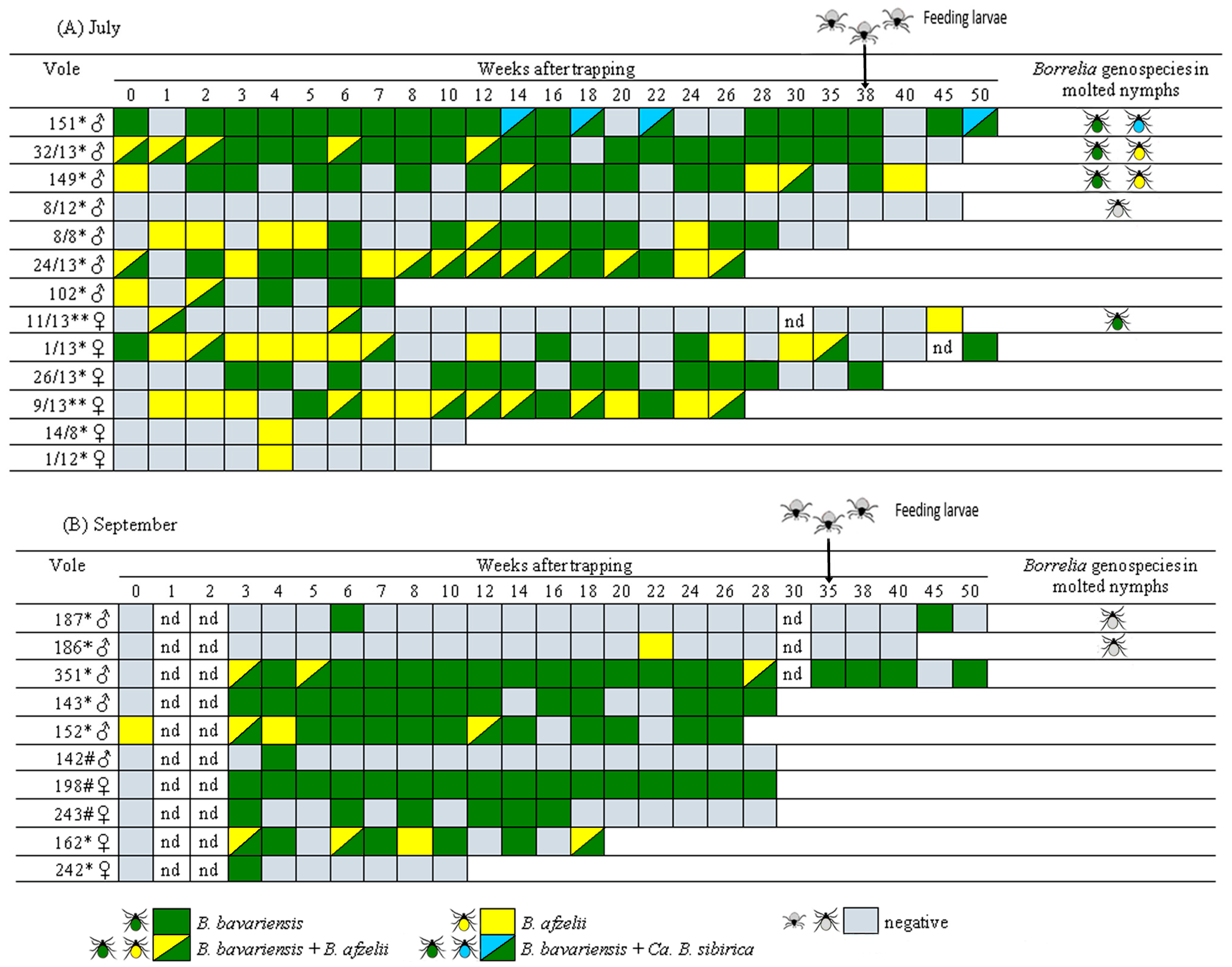
Among the PCR-positive voles, the proportion of positive samples per individual varied widely, from 5.0% to 94.4%. This distribution was polarized: a majority of infected voles (14 of 22; 63.6%) exhibited a high proportion of positive samples (>50%), while a substantial subset (7 of 22; 31.8%) showed a very low proportion (<15%) (Table 3). In individual voles, positive samples appeared randomly distributed over time, with no clear trend toward pathogen clearance (Figure 2). Notably, B. burgdorferi s.l. DNA was still detectable 50 weeks after initial capture in three of the five surviving voles.
Genospecies of B. burgdorferi s.l were determined for all 224 PCR-positive samples. Borrelia bavariensis was the most prevalent, identified in 146 samples, followed by B. afzelii detected in 34 samples. Mixed infections were also common, with 36 samples co-infected with B. afzelii and B. bavariensis, and four with “Candidatus B. sibirica” and B. bavariensis (Table 3). When including mixed infections, the overall prevalence of B. bavariensis was significantly higher than that of B. afzelii in blood samples from voles captured in both July and September (p < 0. 001). Furthermore, the prevalence of B. bavariensis was significantly higher in blood samples from voles captured in September (70/82, 85.4%) than in those from July (80/142, 56.3%; χ2 = 19.8, df = 1, p < 0.001). Conversely, infections with B. afzelii alone, as well as mixed B. afzelii/B. bavariensis infections, were significantly more frequent in the July group (Table 3). Similarly, the proportion of voles infected exclusively with B. bavariensis was higher in September, although this difference was not statistically significant due to the small sample size in the groups compared (Table 3, Figure 2).

3.3. Genetic Diversity of B. burgdorferi s.l. in Voles

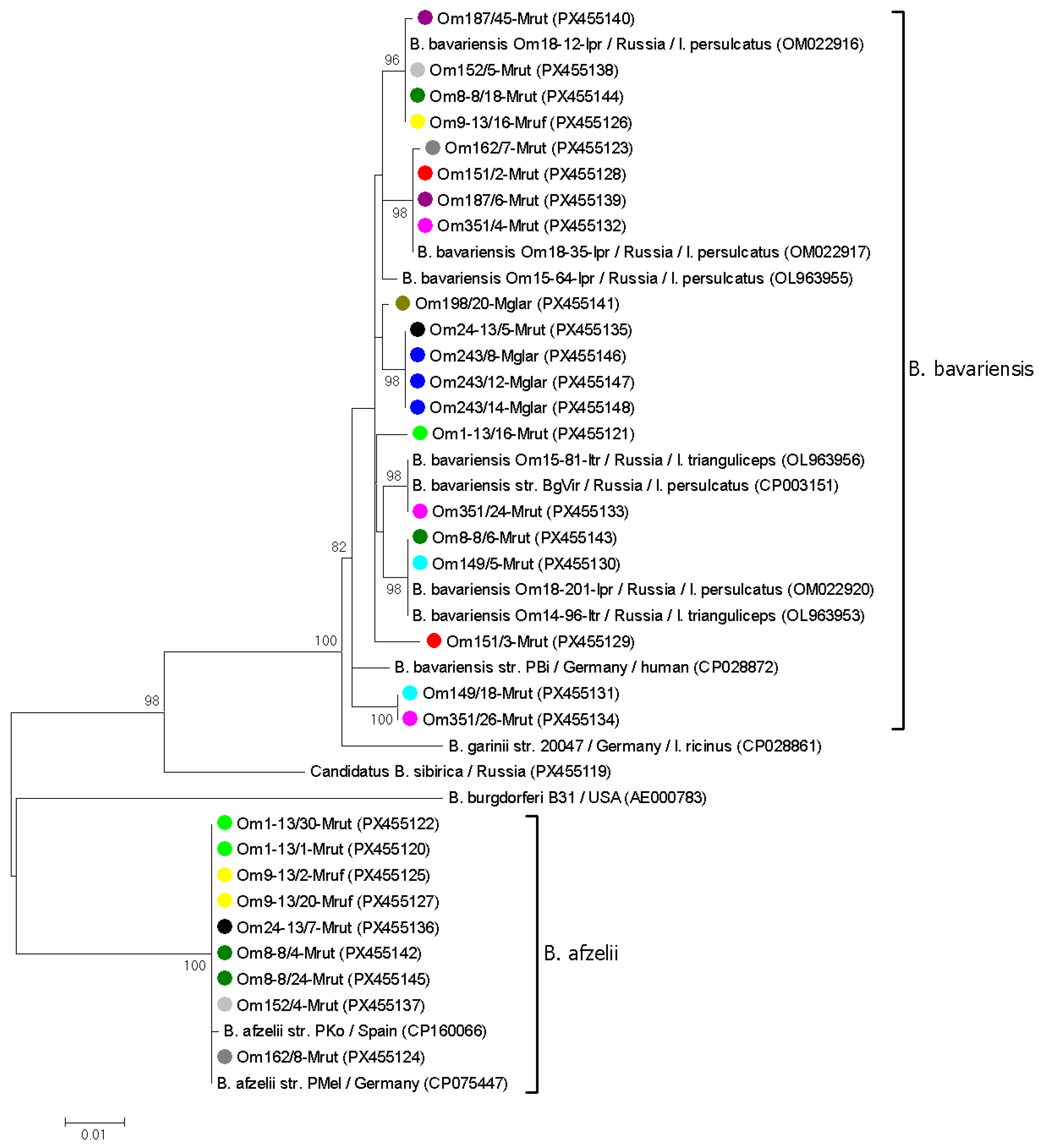
Randomly selected B. afzelii and B. bavariensis samples from 13 voles were genotyped by sequencing clpA gene fragments. Only 2 from 20 B. afzelii sequences contained a single polymorphic site; the remaining sequences matched the clpA allele 36 in the PubMLST database. In contrast, the clpA fragments of B. bavariensis were more variable. In total, 18 of the 38 B. bavariensis sequences contained from 1 to 6 polymorphic sites, indicating the simultaneous persistence of multiple genovariants. Among the remaining sequences, eleven distinct B. bavariensis genovariants were identified. Eight of these genovariants corresponded to known sequences in the PubMLST database (clpA alleles 60, 69, 72, 144, 197, and 207) and GenBank database (e.g., OM022917, PX097757). In addition, sequences from samples Om162/7-Mrut, Om187/45-Mrut, and Om198/20-Mglar were novel, differing by 1–3 substitutions from the closest sequences from I. persulcatus in Omsk province (OM022917, OM022916, and OL963955) (Figure 3).
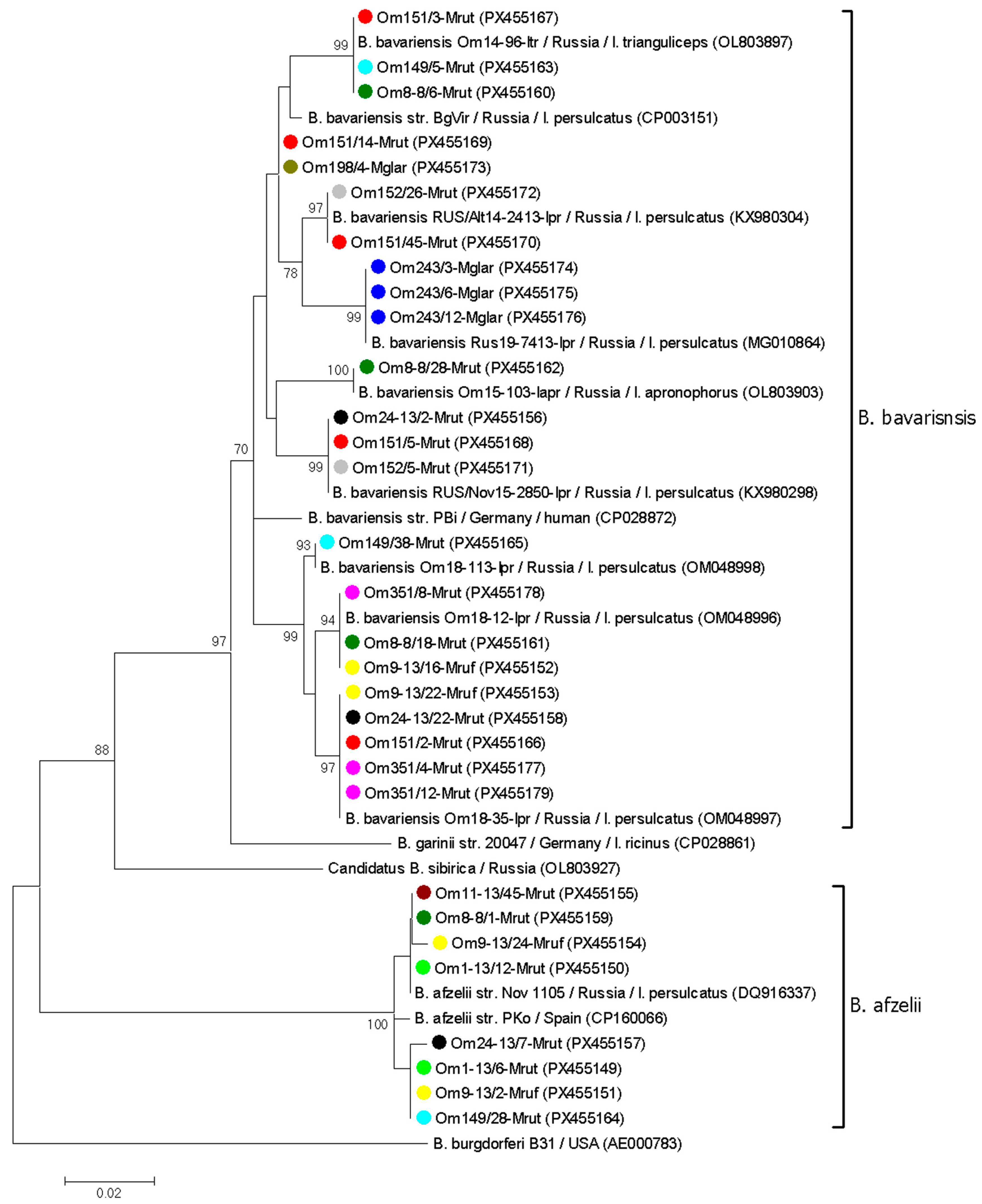
Similarly, a number of B. afzelii and B. bavariensis samples from twelve voles were genetically characterized based on p83/100 gene fragments. Four sequence variants, differing by 1–4 nucleotide substitutions, were identified among eight B. afzelii sequences. Two variants corresponded to common B. afzelii genovariants previously determined in I. persulcatus from Western Siberia (OM022930, DQ916337). The sequences from two other samples (Om9-13/24-Mruf, Om24-13/7-Mrut) differed from the known B. afzelii sequences by a single nucleotide substitution (Figure 4).
Consistent with the clpA genotyping results, B. bavariensis exhibited greater diversity in the p83/100 gene. In total, 22 of 45 B. bavariensis p83/100 gene sequences contained 1–14 polymorphic sites per sequence. The remaining 23 B. bavariensis sequences corresponded to nine genovariants, eight of which matched sequences previously reported in Ixodes spp. from Western Siberia (OM048997, OL803903, KX980298, OM048996, PX117462, KX980304, OM048998, and OL803897). Sequences from two samples (Om151/14-Mrut and Om198/4-Mglar) differed from their closest sequence (CP003151) by one substitution each (Figure 4).
Only sequences without mixed infections were included in the phylogenetic analysis of the clpA and p83/100 gene fragments. In the resulting trees, the obtained B. afzelii and B. bavariensis sequences, along with corresponding reference sequences, formed distinct, well-supported clades (Figure 3 and Figure 4). In most cases, B. burgdorferi s.l. sequences from the same vole host represented different genospecies or genovariants within a genospecies. For example, two different B. afzelii variants and two B. bavariensis genovariants based on the p83/100 gene were identified in samples from vole 9/13 (Figure 4). An exception was vole 243, for which all tested PCR-positive blood samples contained the same B. bavariensis genovariant for both the clpA and p83/100 genes (Figure 3 and Figure 4).

3.4. Transmission of B. burgdorferi s.l. by Ixodes spp.

The transmission of B. burgdorferi s.l. by Ixodes spp. ticks was assessed using xenodiagnosis with larvae from laboratory colonies of I. persulcatus, I. pavlovskyi, and their first-generation (F1) hybrids. A subset of larvae from each colony was genotyped based on the ITS2 and cox1 gene to confirm species identification.
To investigate the transmission of B. burgdorferi s.l. by I. persulcatus, laboratory-reared larvae were fed on seven voles at 35–42 weeks post-capture. All these voles were used in a prior persistence study (Section 3.2); they exhibited varying proportions of PCR-positive samples and were infected with different combinations of Borrelia genospecies: (i) B. bavariensis and “Candidatus B. sibirica” (vole #151), (ii) B. afzelii and B. bavariensis (voles #149 and #32/13), (iii) B. afzelii (vole #186), (iv) B. bavariensis (vole #187). Vole #8–12, which had no PCR-positive blood samples, served as a negative control (Figure 2, Table 4).
Engorged larvae that successfully molted to nymphs (116 of 240 fed larvae) were tested for the presence of B. burgdorferi s.l. DNA. The resulting nymphs were examined individually (5 ticks) or in 21 pools of 4–7 ticks each. DNA of B. burgdorferi s.l. was detected in nymphs that had fed, as larvae, on four voles. The minimum infection rate (MIR) for B. burgdorferi s.l. was 18.8–22.2% for ticks that fed on voles #151, #149, and #32/13 (which had 74–88% PCR-positive blood samples) and 10.0% for ticks that fed on vole #11/13 (which had 13% positive blood samples). No transmission was detected for ticks that fed on voles with 5–9% positive blood samples or on the uninfected control vole (Figure 2, Table 4).
The Borrelia genospecies detected in molted nymphs matched those found in the blood samples of the voles on which they had fed as larvae (Figure 2, Table 4). Among molted nymphs, B. bavariensis was the most frequently detected, with an MIR of 10.0–22.2% across ticks from different voles. This prevalence was higher than that of B. afzelii (MIR: 7.1–11.1%) and “Candidatus B. sibirica” (MIR: 7.1%). However, the difference in genospecies prevalence among ticks that fed on the same individual vole was not statistically significant (p > 0.1).
The transmission of B. burgdorferi s.l. to I. pavlovskyi and I. persulcatus/I. pavlovskyi hybrids (F1) was investigated using two Cl. rutilus voles (#6 and #24), captured in October 2017. Both voles tested positive for B. bavariensis DNA in all three analyzed blood samples; however, a detailed study of B. bavariensis persistence in these voles was not conducted. At 23 weeks post-capture, separate groups of 30 I. pavlovskyi and 30 hybrid larvae were fed on individual voles. Of these, 15 I. pavlovskyi and 14 hybrid larvae successfully molted to nymphs. A subset of these nymphs was individually screened for B. burgdorferi s.l. DNA, revealing B. bavariensis in three of six (50.0%) I. pavlovskyi and five of seven (71.4%) hybrid nymphs (Table 4).
For subsequent transmission experiments, a group of the molted I. pavlovskyi nymphs was used. At 110–140 days post-larval feeding, these nymphs were fed until repletion on two-week-old naïve laboratory mice and then maintained at room temperature for three months. During this period, four engorged nymphs molted successfully into adults (two males and two females), while three nymphs failed to molt and entered diapause. Borrelia bavariensis DNA was detected in one resulting female (Om24-Ipavl-F) and one engorged nymph (Om24-Ipavl-N-eng); the remaining ticks were PCR-negative.
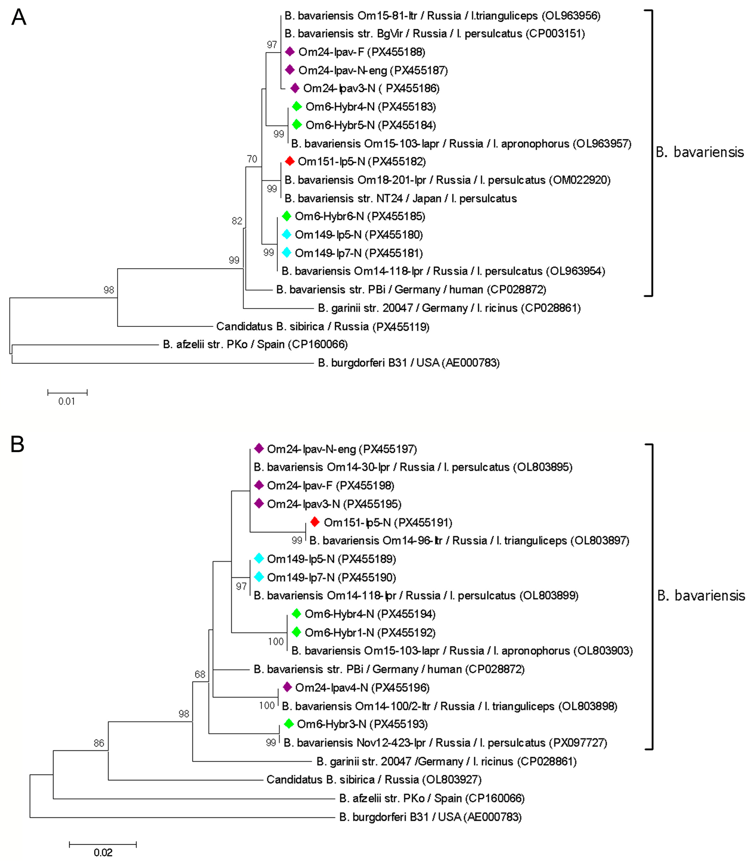
All B. bavariensis samples detected in individual molted ticks were genotyped based on clpA and p83/100 gene fragments. Among twelve clpA gene sequences obtained, three contained polymorphic sites. The remaining nine sequences corresponded to PubMLST alleles 59, 60, 72, and 168, previously identified in Ixodes spp. from the Asian part of Russia. Similarly, three of the thirteen p83/100 gene sequences contained polymorphic sites, while the other ten sequences matched six known variants (OL803899, OL803898, OL803897, OL803895, OL803903, MG010864) from Ixodes spp. in Western Siberia. Phylogenetic analysis of both gene fragments demonstrated that in most cases, ticks fed as larvae on the same vole were infected with an identical B. bavariensis variant. However, several ticks harbored distinct B. bavariensis genovariants that differed from those found in other ticks from the same host (Figure 5).

4. Discussion

Lyme borreliosis is the most common tick-borne disease in the Asian part of Russia [43]. Nevertheless, the enzootic cycles of Siberian B. burgdorferi s.l. strains, particularly their persistence in vertebrate hosts, remain poorly characterized. To address this knowledge gap, we conducted a comprehensive study of B. burgdorferi s.l. in small mammals at a site in the Omsk province of Western Siberia. The sampling area is characterized by high abundances of I. persulcatus and I. trianguliceps and a low abundance of I. apronophorus. Our investigation combined a long-term study of B. burgdorferi s.l. prevalence in small mammals captured between 2013 and 2024, analysis of spirochete persistence in naturally infected voles, and evaluation of Borrelia transmission from voles to xenodiagnostic ticks.
In all sampling periods, two LB agents, B. afzelii and B. bavariensis, were detected in small mammals at similar overall prevalence (Table 2). Notably, prevalence varied significantly between sampling periods (from 4.2% to 40.6%), likely due to fluctuations in the abundance and species composition of ticks and small mammals across seasons and years, as previously demonstrated [36]. Similarly, substantial variation in B. burgdorferi s.l. prevalence in small mammals (1.9–54.5% in different years) has been observed in the Middle Urals within the I. persulcatus distribution area [34]. Notably, the true prevalence of LB agents in voles is likely higher than observed, as our study showed that persisting spirochetes were not detected in all blood samples from infected voles (Figure 2).
In addition to common LB agents, the recently described “Candidatus B. sibirica” was detected in a common shrew in 2024. This species had previously been reported in Siberia only in 2015–2017 [33]. Its detection seven years later indicates the presence of a stable enzootic population of “Candidatus B. sibirica” in the Omsk province.
This study is the first to investigate the persistence of B. burgdorferi s.l. strains from the I. persulcatus distribution area in their mammalian hosts. We demonstrated that three genospecies—B. afzelii, B. bavariensis, and “Candidatus B. sibirica”—persisted in voles for the hosts’ entire lifespan, up to 45–50 weeks. The persistence patterns of B. afzelii and B. bavariensis were similar; both agents were distributed randomly among positive samples, with no observed clearance of either genospecies or replacement of one genospecies by the other (Figure 2). The higher prevalence of B. bavariensis in voles captured in September is likely driven by the seasonal dominance of I. trianguliceps, a tick species with a suspected association with this genospecies [33,44]. This finding provides the first evidence of long-term persistence for Asian B. bavariensis genotypes in small mammals. The observed prolonged persistence of B. afzelii is consistent with data from the I. ricinus distribution area [17,22,27]. Furthermore, the long-term persistence of “Candidatus B. sibirica” in a vole, combined with its initial detection in small mammals and the ticks feeding on them [33], indicates that this candidate species is also maintained in the enzootic cycle with small mammals.
However, prolonged Borrelia persistence observed in voles kept in the laboratory may differ from persistence dynamics in natural conditions. It has been suggested that animals under stress can exhibit higher pathogen infection rates due to immunosuppression and reactivation of latent infections [1,45].
In most voles, the simultaneous circulation of several B. burgdorferi s.l. isolates, including different genospecies and genovariants within a single species, was observed. These results are not surprising, as the study involved only mature voles that had likely experienced repeated tick exposure prior to capture. In the sampling area, Ixodes spp. are known to harbor both B. afzelii and B. bavariensis [33]. The high genetic diversity of Asian genotypes of B. bavariensis, both in this area and in other parts of the I. persulcatus distribution range, has been previously reported [6,33,46]. Consistent with findings from ticks, higher genetic variability of B. bavariensis compared to B. afzelii was found among the isolates persisting in voles, and the identified B. bavariensis variants correspond to those previously found in ticks (Figure 3 and Figure 4).
The persistence of B. burgdorferi s.l. in individual voles was dynamic, characterized by alternating detection of B. afzelii and B. bavariensis, either individually or simultaneously. This alternation could result from competition between Borrelia species or genovariants, as shown for B. burgdorferi s.s. strains [28] or from stochastic variations in spirochete abundance among different genovariants, as demonstrated for multiple clones of a single B. burgdorferi s.s. strain persisting in mice [19].
Another possible explanation involves the transformation of spirochetes into dormant forms [47,48], which can revert to active forms, potentially leading to the observed alternation of genovariants. Furthermore, B. burgdorferi s.l. can colonize various internal organs in rodents, including the hearts, spleens, joints, and bladder [49,50], from which spirochetes may be periodically released into the bloodstream. This dynamic of persistent infection may also lead to the alternation of Borrelia genotypes in peripheral blood. These proposed mechanisms of Borrelia persistence could explain our other observation that a substantial subset of PCR-positive voles had only single positive blood samples, whereas in most infected voles the proportion of positive blood samples exceeded 50% (Table 3).
The method of xenodiagnoses, which includes the feeding of laboratory-reared larvae on animals and subsequent analysis of molted ticks for the presence of pathogens, is considered the strongest evidence of reservoir competence of tested animals [1]. To date, in Eurasia, reservoir competence of small mammals for B. burgdorferi s.l. has been conclusively demonstrated for rodents within the I. ricinus distribution area, but not for those in the I. persulcatus range. Specifically, Apodemus mice have been confirmed as competent reservoirs for B. afzelii and B. bavariensis (European genotype), and Cl. glareolus for B. burgdorferi s.s. and B. afzelii [1,10].
In this study, we first showed via xenodiagnosis that all circulating Siberian genospecies of B. burgdorferi s.l. can be transmitted to laboratory-reared I. persulcatus larvae and survive through at least one molting cycle. Notably, all Borrelia species were successfully transmitted after persisting in voles for at least 38 weeks, confirming the long-term persistence of viable spirochetes. The successful transmission of B. bavariensis, B. afzelii, and “Candidatus B. sibirica” was shown for ticks that fed on four, two, and one voles, respectively (Table 4, Figure 2). Unexpectedly, B. bavariensis was transmitted by ticks that fed on a vole, with only two blood samples positive for this agent, indicating that even transient spirochetemia can yield viable, infectious pathogens (Figure 2). The obtained results provide the first reliable evidence of reservoir competence of Cl. rutilus for B. bavariensis (Asian genotypes), B. afzelii, and “Candidatus B. sibirica”, and of Cl. rufocanus for B. bavariensis (Asian genotypes).
Borrelia bavariensis transmission was demonstrated not only for I. persulcatus, but also for I. pavlovskyi and interspecies hybrids. Moreover, in one case, B. bavariensis was maintained by I. pavlovskyi through two consecutive transtadial transmissions—from larva to nymph and from nymph to female. The reliability of these laboratory results was proved by the genetic characterization of all molted nymphs, which confirmed tick species identity and Borrelia genospecies. Nevertheless, the obtained data contradict the results of previous field studies from Russian Siberia and the Far East, which reported a low prevalence (1–2%) of B. bavariensis in I. pavlovskyi adults [29,31,51]. Thus, despite the laboratory-confirmed association between B. bavariensis and I. pavlovskyi, this association appears to be rare in nature. Similarly, the observed transmission of “Candidatus B. sibirica” to I. persulcatus is unlikely to be of biological significance, as this candidate species has previously been detected only within the range of I. apronophorus [33]. The observed discrepancy between laboratory and field results may be explained by ecological competition, either among B. burgdorferi s.l. genospecies or between tick species, which primarily determines pathogen associations in nature.
Notably, the genetic diversity of B. bavariensis in molted ticks was lower than in voles (Figure 3, Figure 4 and Figure 5). This may be due to the population “bottlenecks” during spirochete acquisition and tick molting, which could eliminate certain B. burgdorferi s.l. genovariants [19,52].
However, the findings regarding the tick-pathogen relationship are preliminary. A limitation of this study is that the effective transmission of B. bavariensis to I. pavlovskyi and hybrids, and of “Candidatus B. sibirica” to I. persulcatus, was demonstrated using larvae that fed on a single vole. Furthermore, the analysis of B. burgdorferi s.l. transmission in pooled nymphs does not allow an accurate assessment of transmission efficiency. Further experiments using laboratory strains of different Borrelia genospecies are required to validate the obtained results and compare transmission efficiency across tick species and bacterial genospecies.
In conclusion, B. afzelii, B. bavariensis, and, in single cases, “Candidatus B. sibirica” can persist in the blood of wild voles during their life. A simultaneous persistence of several B. burgdorferi s.l. genospesies or genovariants was found within the same species in most of the infected voles. It was first demonstrated by xenodiagnosis that all genospecies can be effectively transmitted to I. persulcatus; in addition, B. bavariensis can be transmitted to I. pavlovskyi and I. persulcatus/I. pavlovskyi interspecies hybrids.

Supplementary Materials

The following supporting information can be downloaded at: https://www.mdpi.com/article/10.3390/pathogens14121200/s1, Table S1: Borrelia burgdorferi s.l. prevalence in the blood of small mammals by seasons.

Author Contributions

Conceptualization, V.R.; Methodology, V.R. and V.Y.; Formal Analysis, V.Y. and V.F.; Investigation, V.R., V.Y., Y.I., Y.S., V.F., A.K., G.R., and T.E.; Resources, V.Y., G.R.; Writing—Original Draft Preparation, V.R.; Writing—Review and Editing, N.T.; Supervision, N.T.; Funding Acquisition, V.R. All authors have read and agreed to the published version of the manuscript.

Funding

This research was funded by the Russian Science Foundation, research project No. 24-24-00390.

Institutional Review Board Statement

All experiments with animals were conducted in compliance with the Animal Welfare Act at the Omsk Research Institute of Natural Foci Infections, according to the guidelines for experiments with laboratory animals (Supplement to the Order of the Russian Ministry of Health, no. 755, of 12 August 1977). This animal study was approved by the Bioethical Committee of the Omsk Research Institute of Natural Foci Infections (Protocol No.1, 20 March 2013; Protocol No. 4, 17 February 2016; Protocol No.1, 15 March 2024).

Informed Consent Statement

Not applicable.

Data Availability Statement

The data are contained within the article.

Acknowledgments

The authors are grateful to Marat Makenov, Nadezhda Berezovskaya, and Nadezhda Palgova for their invaluable assistance with field research and animal care. During the preparation of this manuscript, the authors used DeepSeek (https://www.deepseek.com) for language editing and proofreading. After using this tool, the authors reviewed and edited the content as needed and take full responsibility for the publication’s content.

Conflicts of Interest

The authors declare no conflicts of interest.

References

  1. Wolcott, K.A.; Margos, G.; Fingerle, V.; Becker, N.S. Host association of Borrelia burgdorferi sensu lato: A review. Ticks Tick Borne Dis. 2021, 12, 101766. [Google Scholar] [CrossRef]
  2. Rudenko, N.; Golovchenko, M.; Grubhoffer, L.; Oliver, J.H., Jr. Updates on Borrelia burgdorferi sensu lato complex with respect to public health. Ticks Tick Borne Dis. 2011, 2, 123–128. [Google Scholar] [CrossRef]
  3. Rudenko, N.; Golovchenko, M.; Vancova, M.; Clark, K.; Grubhoffer, L.; Oliver, J.H., Jr. Isolation of live Borrelia burgdorferi sensu lato spirochaetes from patients with undefined disorders and symptoms not typical for Lyme borreliosis. Clin. Microbiol. Infect. 2016, 22, 267.e9–15. [Google Scholar] [CrossRef] [PubMed]
  4. Pritt, B.S.; Mead, P.S.; Johnson, D.; Neitzel, D.F.; Respicio-Kingry, L.B.; Davis, J.P.; Schiffman, E.; Sloan, L.M.; Schriefer, M.E.; Replogle, A.J.; et al. Identification of a novel pathogenic Borrelia species causing Lyme borreliosis with unusually high spirochaetaemia: A descriptive study. Lancet Infect. Dis. 2016, 16, 556–564. [Google Scholar] [CrossRef] [PubMed]
  5. Eisen, L. Vector competence studies with hard ticks and Borrelia burgdorferi sensu lato spirochetes: A review. Ticks Tick Borne Dis. 2020, 11, 101359. [Google Scholar] [CrossRef]
  6. Rollins, R.E.; Sato, K.; Nakao, M.; Tawfeeq, M.T.; Herrera-Mesías, F.; Pereira, R.J.; Kovalev, S.; Margos, G.; Fingerle, V.; Kawabata, H.; et al. Out of Asia? Expansion of Eurasian Lyme borreliosis causing genospecies display unique evolutionary trajectories. Mol. Ecol. 2023, 32, 786–799. [Google Scholar] [CrossRef]
  7. Margos, G.; Vollmer, S.A.; Cornet, M.; Garnier, M.; Fingerle, V.; Wilske, B.; Bormane, A.; Vitorino, L.; Collares-Pereira, M.; Drancourt, M.; et al. A new Borrelia species defined by multilocus sequence analysis of housekeeping genes. Appl. Environ. Microbiol. 2009, 75, 5410–5416. [Google Scholar] [CrossRef]
  8. Rizzoli, A.; Hauffe, H.; Carpi, G.; Vourc, H.G.; Neteler, M.; Rosa, R. Lyme borreliosis in Europe. Euro. Surveill. 2011, 16, 19906. [Google Scholar] [CrossRef] [PubMed]
  9. Trevisan, G.; Cinco, M.; Trevisini, S.; di Meo, N.; Chersi, K.; Ruscio, M.; Forgione, P.; Bonin, S. Borreliae Part 1: Borrelia Lyme Group and Echidna-Reptile Group. Biology 2021, 10, 1036. [Google Scholar] [CrossRef]
  10. Huegli, D.; Hu, C.M.; Humair, P.F.; Wilske, B.; Gern, L. Apodemus species mice are reservoir hosts of Borrelia garinii OspA serotype 4 in Switzerland. J. Clin. Microbiol. 2002, 40, 4735–4737. [Google Scholar] [CrossRef]
  11. Takano, A.; Nakao, M.; Masuzawa, T.; Takada, N.; Yano, Y.; Ishiguro, F.; Fujita, H.; Ito, T.; Ma, X.; Oikawa, Y.; et al. Multilocus sequence typing implicates rodents as the main reservoir host of human-pathogenic Borrelia garinii in Japan. J. Clin. Microbiol. 2011, 49, 2035–2039. [Google Scholar] [CrossRef] [PubMed]
  12. Kurtenbach, K.; Hanincová, K.; Tsao, J.I.; Margos, G.; Fish, D.; Ogden, N.H. Fundamental processes in the evolutionary ecology of Lyme borreliosis. Nat. Rev. Microbiol. 2006, 4, 660–669. [Google Scholar] [CrossRef] [PubMed]
  13. Raileanu, C.; Silaghi, C.; Fingerle, V.; Margos, G.; Thiel, C.; Pfister, K.; Overzier, E. Borrelia burgdorferi Sensu Lato in Questing and Engorged Ticks from Different Habitat Types in Southern Germany. Microorganisms 2021, 9, 1266. [Google Scholar] [CrossRef] [PubMed]
  14. Voordouw, M.J. Co-feeding transmission in Lyme disease pathogens. Parasitology 2015, 142, 290–302. [Google Scholar] [CrossRef]
  15. Baum, E.; Hue, F.; Barbour, A.G. Experimental infections of the reservoir species Peromyscus leucopus with diverse strains of Borrelia burgdorferi, a Lyme disease agent. mBio 2012, 3, e00434-12. [Google Scholar] [CrossRef]
  16. Andersson, M.; Scherman, K.; Råberg, L. Multiple-strain infections of Borrelia afzelii: A role for within-host interactions in the maintenance of antigenic diversity? Am. Nat. 2013, 181, 545–554. [Google Scholar] [CrossRef]
  17. Jacquet, M.; Margos, G.; Fingerle, V.; Voordouw, M.J. Comparison of the Lifetime Host-to-Tick Transmission Between Two Strains of the Lyme disease pathogen Borrelia afzelii. Parasites Vectors 2016, 9, 645. [Google Scholar] [CrossRef]
  18. Hodzic, E.; Feng, S.; Holden, K.; Freet, K.J.; Barthold, S.W. Persistence of Borrelia burgdorferi following antibiotic treatment in mice. Antimicrob. Agents Chemother. 2008, 52, 1728–1736. [Google Scholar] [CrossRef]
  19. Rego, R.O.; Bestor, A.; Štefka, J.; Rosa, P.A. Population bottlenecks during the infectious cycle of the Lyme disease spirochete Borrelia burgdorferi. PLoS ONE 2014, 9, e101009. [Google Scholar] [CrossRef]
  20. Derdáková, M.; Dudičák, V.; Brei, B.; Brownstein, J.S.; Schwartz, I.; Fish, D. Interaction and transmission of two Borrelia burgdorferi sensu stricto strains in a tick-rodent maintenance system. Appl. Environ. Microbiol. 2004, 70, 6783–6788. [Google Scholar] [CrossRef]
  21. Hanincová, K.; Ogden, N.H.; Diuk-Wasser, M.; Pappas, C.J.; Iyer, R.; Fish, D.; Schwartz, I.; Kurtenbach, K. Fitness variation of Borrelia burgdorferi sensu stricto strains in mice. Appl. Environ. Microbiol. 2008, 74, 153–157. [Google Scholar] [CrossRef] [PubMed]
  22. Gern, L.; Siegenthaler, M.; Hu, C.M.; Leuba-Garcia, S.; Humair, P.F.; Moret, J. Borrelia burgdorferi in rodents (Apodemus flavicollis and A. sylvaticus): Duration and enhancement of infectivity for Ixodes ricinus ticks. Eur. J. Epidemiol. 1994, 10, 75–80. [Google Scholar] [PubMed]
  23. States, S.L.; Huang, C.I.; Davis, S.; Tufts, D.M.; Diuk-Wasser, M.A. Co-feeding transmission facilitates strain coexistence in Borrelia burgdorferi, the Lyme disease agent. Epidemics 2017, 19, 33–42. [Google Scholar] [CrossRef] [PubMed]
  24. Wang, I.N.; Dykhuizen, D.E.; Qiu, W.; Dunn, J.J.; Bosler, E.M.; Luft, B.J. Genetic diversity of ospC in a local population of Borrelia burgdorferi sensu stricto. Genetics 1999, 151, 15–30. [Google Scholar] [CrossRef]
  25. Perez, D.; Kneubühler, Y.; Rais, O.; Jouda, F.; Gern, L. Borrelia afzelii ospC Genotype diversity in Ixodes ricinus questing ticks and ticks from rodents in two Lyme borreliosis endemic areas: Contribution of co-feeding ticks. Ticks Tick-Borne Dis. 2011, 2, 137–142. [Google Scholar] [CrossRef]
  26. Qiu, W.G.; Dykhuizen, D.E.; Acosta, M.S.; Luft, B.J. Geographic uniformity of the Lyme disease spirochete (Borrelia burgdorferi) and its shared history with tick vector (Ixodes scapularis) in the Northeastern United States. Genetics 2002, 160, 833–849. [Google Scholar] [CrossRef]
  27. Genne, D.; Rossel, M.; Sarr, A.; Battilotti, F.; Rais, O.; Rego, R.O.M.; Voordouw, M.J. Competition between strains of Borrelia afzelii in the host tissues and consequences for transmission to ticks. ISME J. 2021, 15, 2390–2400. [Google Scholar] [CrossRef]
  28. Rynkiewicz, E.C.; Brown, J.; Tufts, D.M.; Huang, C.I.; Kampen, H.; Bent, S.J.; Fish, D.; Diuk-Wasser, M.A. Closely-related Borrelia burgdorferi (sensu stricto) strains exhibit similar fitness in single infections and asymmetric competition in multiple infections. Parasites Vectors 2017, 10, 64. [Google Scholar] [CrossRef]
  29. Rar, V.; Livanova, N.; Tkachev, S.; Kaverina, G.; Tikunov, A.; Sabitova, Y.; Igolkina, Y.; Panov, V.; Livanov, S.; Fomenko, N.; et al. Detection and genetic characterization of a wide range of infectious agents in Ixodes pavlovskyi ticks in Western Siberia, Russia. Parasit. Vectors 2017, 10, 258. [Google Scholar] [CrossRef]
  30. Rar, V.; Chicherina, G.; Igolkina, Y.; Fedorets, V.; Epikhina, T.; Tikunova, N. Spectrum of Ixodidae Ticks Attacking Humans in Novosibirsk Province, Russian Siberia, and Their Association with Tick-Borne Bacterial Agents. Pathogens 2025, 14, 315. [Google Scholar] [CrossRef]
  31. Mukhacheva, T.A.; Kovalev, S.Y. Borrelia spirochetes in Russia: Genospecies differentiation by real-time PCR. Ticks Tick Borne Dis. 2014, 5, 722–726. [Google Scholar] [CrossRef]
  32. Kurilshikov, A.M.; Fomenko, N.V.; Stronin, O.V.; Tikunov, A.Y.; Kabilov, M.R.; Tupikin, A.E.; Tikunova, N.V. Complete Genome Sequencing of Borrelia valaisiana and Borrelia afzelii Isolated from Ixodes persulcatus Ticks in Western Siberia. Genome Announc. 2014, 2, e01315-14. [Google Scholar] [CrossRef] [PubMed]
  33. Sabitova, Y.; Rar, V.; Tikunov, A.; Yakimenko, V.; Korallo-Vinarskaya, N.; Livanova, N.; Tikunova, N. Detection and genetic characterization of a putative novel Borrelia genospecies in Ixodes apronophorus/Ixodes persulcatus/Ixodes trianguliceps sympatric areas in Western Siberia. Ticks Tick Borne Dis. 2023, 14, 102075. [Google Scholar] [CrossRef]
  34. Kovalevskii, Y.V.; Korenberg, E.I.; Gorelova, N.B. Long-term dynamics of the epizootic process in natural foci of ixodid tick borreliosis in mountain taiga forests of the Middle Ural. Parazitologiia 2004, 38, 105–121. (In Russian) [Google Scholar] [PubMed]
  35. Rar, V.A.; Epikhina, T.I.; Tikunova, N.V.; Bondarenko, E.I.; Ivanov, M.K.; Iakimenko, V.V.; Mal’kova, M.G.; Tantsev, A.K. DNA detection of pathogens transmitted by Ixodid ticks in blood of small mammals inhabiting the forest biotopes in Middle Irtysh Area (Omsk Region, West Siberia). Parazitologiia 2014, 48, 37–53. (In Russian) [Google Scholar]
  36. Rar, V.; Yakimenko, V.; Makenov, M.; Tikunov, A.; Epikhina, T.; Tancev, A.; Bobrova, O.; Tikunova, N. High prevalence of Babesia microti ‘Munich’ type in small mammals from an Ixodes persulcatus/Ixodes trianguliceps sympatric area in the Omsk region, Russia. Parasitol. Res. 2016, 115, 3619–3629. [Google Scholar] [CrossRef] [PubMed]
  37. Gromov, I.M.; Polyakov, I.Y. Fauna of the USSR. Mammals, Ser. 3: Voles (Microtinae); Nauka: Leningrad, Russia, 1977; 502p. (In Russian) [Google Scholar]
  38. Gureev, A.A. Fauna of the USSR. Mammals, Ser. 4: Insectivores; Nauka: Leningrad, Russia, 1979; 501p. (In Russian) [Google Scholar]
  39. Yakimenko, V.V.; Malkova, M.G.; Shpynov, S.N. Ixodid Ticks of the Western Siberia; Omsk Scientific Vestnik: Omsk, Russia, 2013; p. 276. (In Russian) [Google Scholar]
  40. Rar, V.; Livanova, N.; Sabitova, Y.; Igolkina, Y.; Tkachev, S.; Tikunov, A.; Babkin, I.; Golovljova, I.; Panov, V.; Tikunova, N. Ixodes persulcatus/pavlovskyi natural hybrids in Siberia: Occurrence in sympatric areas and infection by a wide range of tick-transmitted agents. Ticks Tick Borne Dis. 2019, 10, 101254. [Google Scholar] [CrossRef]
  41. Margos, G.; Gatewood, A.G.; Aanensen, D.M.; Hanincová, K.; Terekhova, D.; Vollmer, S.A.; Cornet, M.; Piesman, J.; Donaghy, M.; Bormane, A.; et al. MLST of housekeeping genes captures geographic population structure and suggests a European origin of Borrelia burgdorferi. Proc. Natl. Acad. Sci. USA 2008, 105, 8730–8735. [Google Scholar] [CrossRef]
  42. Kumar, S.; Stecher, G.; Tamura, K. MEGA7: Molecular Evolutionary Genetics Analysis Version 7.0 for Bigger Datasets. Mol. Biol. Evol. 2016, 33, 1870–1874. [Google Scholar] [CrossRef]
  43. Korenberg, E.I.; Pomelova, V.G.; Osin, N.S. Infections with Natural Focality Transmitted by Ixodid Ticks; Ginzburg, A., Zlobin, V., Eds.; Kommentariy: Moscow, Russia, 2013; 463p. (In Russian) [Google Scholar]
  44. Korenberg, E.I.; Kovalevskii, Y.V.; Gorelova, N.B.; Nefedova, V.V. Comparative analysis of the roles of Ixodes persulcatus and I. trianguliceps ticks in natural foci of ixodid tick-borne borrelioses in the Middle Urals, Russia. Ticks Tick Borne Dis. 2015, 6, 316–321. [Google Scholar] [CrossRef]
  45. Gylfe, A.; Bergström, S.; Lundström, J.; Olsen, B. Reactivation of Borrelia infection in birds. Nature 2000, 403, 724–725. [Google Scholar] [CrossRef]
  46. Scholz, H.C.; Margos, G.; Derschum, H.; Speck, S.; Tserennorov, D.; Erdenebat, N.; Undraa, B.; Enkhtuja, M.; Battsetseg, J.; Otgonchimeg, C.; et al. High prevalence of genetically diverse Borrelia bavariensis-like strains in Ixodes persulcatus from Selenge Aimag, Mongolia. Ticks Tick Borne Dis. 2013, 4, 89–92. [Google Scholar] [CrossRef]
  47. Meriläinen, L.; Herranen, A.; Schwarzbach, A.; Gilbert, L. Morphological and biochemical features of Borrelia burgdorferi pleomorphic forms. Microbiology 2015, 161, 516–527. [Google Scholar] [CrossRef] [PubMed]
  48. Brorson, O.; Brorson, S.H. Transformation of cystic forms of Borrelia burgdorferi to normal, mobile spirochetes. Infection 1997, 25, 240–246. [Google Scholar] [CrossRef] [PubMed]
  49. Goodman, J.L.; Jurkovich, P.; Kodner, C.; Johnson, R.C. Persistent cardiac and urinary tract infections with Borrelia burgdorferi in experimentally infected Syrian hamsters. J. Clin. Microbiol. 1991, 29, 894–896. [Google Scholar] [CrossRef]
  50. Sonnesyn, S.W.; Manivel, J.C.; Johnson, R.C.; Goodman, J.L. A guinea pig model for Lyme disease. Infect. Immun. 1993, 61, 4777–4784. [Google Scholar] [CrossRef] [PubMed]
  51. Nikitin, A.Y.; Sabitova, Y.V.; Rar, V.A.; Morozov, I.M.; Gordeiko, N.S.; Allenov, A.V.; Kaverina, G.B.; Babkin, I.V.; Tikunova, N.V.; Andaev, E.I. Role of Ixodes pavlovskyi (Acari, Ixodidae) in Borreliosis Epizootic Process at the Island Russky. Probl. Osobo Opasnykh Infektsii 2021, 1, 116–121. (In Russian) [Google Scholar] [CrossRef]
  52. Piesman, J.; Oliver, J.R.; Sinsky, R.J. Growth kinetics of the Lyme disease spirochete (Borrelia burgdorferi) in vector ticks (Ixodes dammini). Am. J. Trop. Med. Hyg. 1990, 42, 352–357. [Google Scholar] [CrossRef]
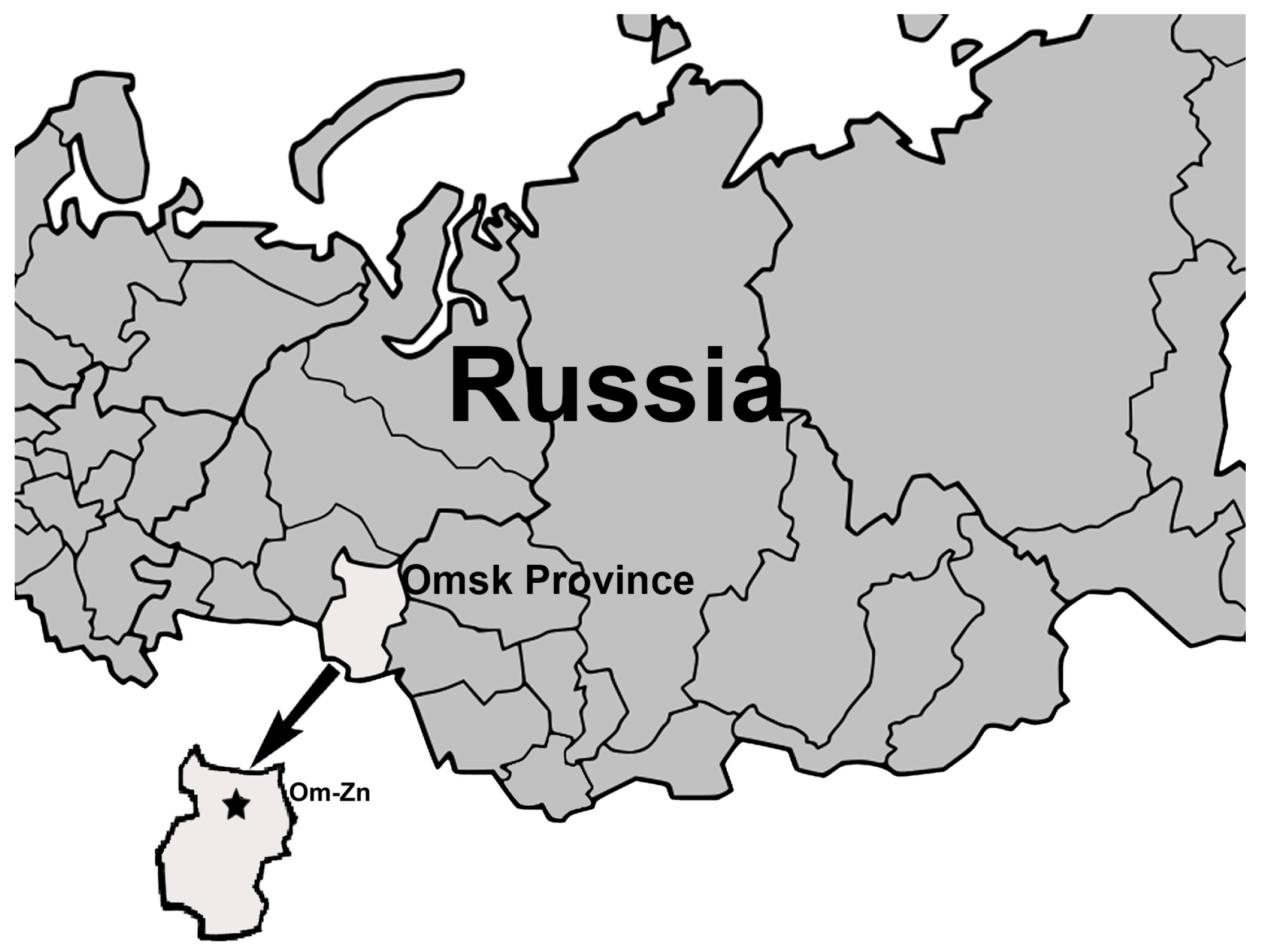
Figure 1. The map shows the location of a sampling site.
Pathogens 14 01200 g001
Figure 2. Persistence of B. burgdorferi s.l. in naturally infected voles and their transmission to Ixodes ticks. PCR-positive vole blood samples and molted nymphs are marked by color: B. bavariensis by green, B. afzelii by yellow, “Candidatus B. sibirica” by blue. PCR-negative blood samples, molted nymphs, and larvae are marked in grey. *—Clethrionomys rutilus; **—Clethrionomys rufocanus; #—Clethrionomys glareolus; nd—not determined.
Pathogens 14 01200 g002
Figure 3. The phylogenetic tree was constructed by the ML method based on nucleotide sequences of a 775 bp fragment of the clpA gene of Borrelia burgdorferi s.l. identified in blood samples of voles. Sample names include the vole identifier, time post-capture (in weeks), and host species. Different blood samples from the same individual vole are color-coded. The scale bar indicates an evolutionary distance of 0.01 nucleotide per position in the sequence. Significant bootstrapping values (>70%) are shown on the nodes. B. burgdorferi B31 was used as an outgroup.
Pathogens 14 01200 g003
Figure 4. The phylogenetic tree constructed by the ML method based on nucleotide sequences of 276–366 bp fragments of the p83/100 gene of Borrelia burgdorferi s.l. identified in blood samples of voles. Sample names include the vole identifier, time post-capture (in weeks), and host species. Different blood samples from the same individual vole are color-coded. The scale bar indicates an evolutionary distance of 0.02 nucleotide per position in the sequence. Significant bootstrapping values (>70%) are shown on the nodes. B. burgdorferi B31 was used as an outgroup.
Pathogens 14 01200 g004
Figure 5. Phylogenetic trees constructed by the ML method based on a 775 bp fragment of the clpA gene (A) and 366–369 bp fragments of the p83/100 gene (B) of Borrelia bavariensis identified in molted ticks. Sample names include the vole identifier, tick species, tick identifier, and tick stage. Samples derived from molted ticks that had fed as larvae on the same vole are color-coded. Significant bootstrapping values (>65%) are shown on the nodes. B. burgdorferi B31 was used as an outgroup.
Pathogens 14 01200 g005
Table 1. Primers used for identification and genotyping of Ixodes spp. and B. burgdorferi s.l.
LocusOrganismReactionPrimer NamePrimer Sequences 5′-3′T * (°C)References
ITS2IxodidaeconventionalF-ITS2cacactgagcacttactctttg57[29]
R1-ITS2actggatggctccagtattc
cox1I. persulcatusconventionalIxodes-Facctgatatagctttccctcg55[29]
Ipers-Rttgattcctgttggaacagc
I. pavlovskyiconventionalIxodes-Facctgatatagctttccctcg55[29]
Ipav-Rtaatccccgtggggacg
IGSB. burgdorferi s.l. Primary NC1cctgttatcattccgaacacag50[29]
NC2tactccattcggtaatcttggg
Nested NC3tactgcgagttcgcgggag50
NC4cctaggcattcaccatagac
Ca. B. sibirica”NestedBsibataaaacattctaaaaaaatgaaca50This study
NC4cctaggcattcaccatagac [29]
clpA B. burgdorferi s.l.Primary clpAF1237aaagatagatttcttccagac50[41]
clpAR2218gaatttcatctattaaaagctttc
Nested clpAF1255gacaaagcttttgatattttag50
clpAR2104caaaaaaaacatcaaattttctatctc
p83/100B. burgdorferi s.l. Primary F7ttcaaagggatactgttagagag50[29]
F10aagaaggcttatctaatggtgatg
Nested F5acctggtgatgtaagttctcc54
F12ctaacctcattgttgttagactt
* Annealing temperature.
Table 2. Borrelia burgdorferi s.l. prevalence in the blood of small mammals.
Sampling
Periods
No. of Tested MammalsNo (%/95% CI) of Samples
Containing DNA of B. burgdorferi s.l.
No of
Genotyped Samples
No (%/95% CI *) of Samples Containing DNA
BaBbavBsBa + Bbav
June 2013383 (7.9/2.7–20.8)3 2100
September 2013597 (11.9/5.9–22.5)7 2500
July 201412414 (11.3/6.9–18.1)125502
September 20141195 (4.2/1.8–9.5)53101
July 20157910 (12.7/7.0–21.8)105203
September 2015874 (4.6/1.8–11.2)41201
September 2016956 (6.3/2.9–13.1)21100
June 2017336 (18.2/8.6–34.4)62400
October 2017438 (18.6/9.7–32.6)82600
September 2018287 (25.0/12.7–43.4)74300
August 20243213 (40.6/25.5–57.7)136511
Total73783 (11.3/9.2–13.8)7733 (42.9/32.4–54.0)35 (45.5/34.8–56.5)1 (1.3/0.2–7.0)8 (10.4/5.4–19.2)
%*—of genotyped samples. Abbreviations: Ba—B. afzelii, Bbav—B. bavariensis, Bs—“Candidatus B. sibirica”.
Table 3. Persistence of B. burgdorferi s.l. in voles captured in July and September 2015.
Both Sampling
Periods
July 2015September 2015Statistical Difference Between Sampling Periods
All examined voles
   Total number472423
   No. of males (%/95% CI)26 (55.3/41.3–68.6)13 (54.2/35.1–72.1)13 (56.5/36.8–74.3)
   No. of females (%/95% CI)21 (44.7/31.4–58.8)11 (45.8/27.9–64.9)10 (43.5/25.6–63.2)
PCR-positive voles
   Total number221210
   No. of males (%/95% CI)12 (54.6/34.7–73.1)6 (50.0/25.4–74.6)6 (60.0/31.3–83.2)
   No. of females (%/95% CI)10 (45.4/26.9–65.3)6 (50.0/25.4–74.6)4 (40.0/16.8–68.7)
Number (%/95% CI) of voles with a portion of PCR-positive blood samples:
   50–100% 14 (63.6/43.0–80.3) 9 (75/46.8–91.1) 5 (50.0/23.7–76.3)
   15–50%1 (4.6/0.8–21.8)01 (10.0/1.8–40.4)
   0–15%7 (31.8/16.3–52.7)3 (25/8.9–53.2)4 (40.0/16.8–68.7)
Number (%/95% CI) of voles with a persistence of:
   B. bavariensis7 (31.8/16.3–52.7)1 (8.3/1.5–35.4)6 (60.0/31.3–83.2)
   B. afzelii3 (13.6/4.8–33.3)2 (16.7/4.7–44.8)1 (10.0/1.8–40.4)
   B. bavariensis + B. afzelii11 (50.0/30.7–69.3)8 (66.7/39.1–86.2)3 (30.0/10.8–60.3)
   B. bavariensis + Ca. B. sibirica1 (4.6/0.8–21.8)1 (8.3/1.5–35.4)0
PCR-positive samples
   Total number224142 82
Number (%/95% CI) of blood samples containing DNA:
   B. bavariensis150 (67.0/60.6–72.8)80 (56.3/48.1–64.2)70 (85.4/76.1–91.4)χ2 = 19.8, p < 0.001
   B. afzelii34 (15.2/11.1–20.5)30 (21.1/15.2–28.6)4 (4.9/1.9–11.9)χ2 = 10.7, p = 0.001
   B. bavariensis + B. afzelii36 (16.1/11.8–21.5)28 (19.7/14.0–27.0)8 (9.8/5.0–18.1)χ2 = 3.8, p = 0.05
   B. bavariensis + Ca. B. sibirica4 (1.8/0.7–4.5)4 (2.9/1.1–7.6)0
Table 4. Borrelia burgdorferi s.l. transmission by Ixodes spp. larvae.
VolesFeeding LarvaeMolted NymphsMIR */Prevalence #
(%/95% CI) of Bbsl, Ba, Bbav, and Bs in Molted Nymphs
IDBbsl Species in VolesTick SpeciesTime After Vole Capture (Weeks) IDNo of Ticks in a PoolPresence of Bbsl DNABbsl Species in Molted Ticks
151Bbav,Ip38151-Ip15+Bbav, BsBbsl: 21.4/7.6–47.6 *
BsIp38151-Ip37+BbavBbav: 21.4/7.6–47.6 *
Ip38151-Ip41---Bs: 7.1/1.3–31.5 *
Ip38151-Ip51+Bbav
149BbavIp38149-Ip15+Bbav, BaBbsl: 22.2/10.6–40.8 *
BaIp38149-Ip25+Bbav, BaBbav: 22.2/10.6–40.8 *
Ip38149-Ip37+Bbav, BaBa: 11.1/3.9–28.1 *
Ip38149-Ip47+Bbav
Ip38149-Ip51+Bbav
Ip38149-Ip61--
Ip38149-Ip71+Bbav
32-13BbavIp3832-13-Ip17+BbavBbsl: 18.8/8.9–35.3 *
BaIp3832-13-Ip27+BbavBbav: 18.8/8.9–35.3 *
Ip3832-13-Ip35+BbavBa: 6.3/1.7–20.2 *
Ip3832-13-Ip45+Bbav
Ip4232-13-Ip54+Bbav, Ba
Ip4232-13-Ip64+Bbav, Ba
11-13BbavIp4211-13-Ip15--Bbsl: 10.0/1.8–40.4 *
BaIp4211-13-Ip25+BbavBbav: 10.0/1.8–40.4 *
186BaIp35186-Ip15--NA
Ip35186-Ip25--
Ip35186-Ip35--
187BbavIp35187-Ip14--NA
Ip35187-Ip24--
8-12-Ip428-12-Ip15--NA
Ip428-12-Ip15--
24BbavIpav2324-Ipav11--Bbsl: 50.0/18.8–81.2 #
Ipav2324-Ipav21+BbavBbav: 50.0/18.8–81.2 #
Ipav2324-Ipav31+Bbav
Ipav2324-Ipav41+Bbav
Ipav2324-Ipav51--
Ipav2324-Ipav61--
6BbavHybr236-Hybr11+BbavBbsl: 71.4/35.9–91.8 #
BbavHybr236-Hybr21--Bbav: 71.4/35.9–91.8 #
BbavHybr236-Hybr31+Bbav
BbavHybr236-Hybr41+Bbav
BbavHybr236-Hybr51+Bbav
BbavHybr236-Hybr61+Bbav
BbavHybr236-Hybr71--
Nymphs were analyzed 14 days after molting. MIR *—was calculated for ticks in pools; Prevalence #—was calculated for individual ticks. Abbreviations: Bbsl—B. burgdorferi s.l., Ba—B. afzelii, Bbav—B. bavariensis, Bs—“Candidatus B. sibirica”, Ip—I. persulcatus, Ipav—I. pavlovskyi, Hybr—I. persulcatus/I. pavlovskyi hybrids.
Disclaimer/Publisher’s Note: The statements, opinions and data contained in all publications are solely those of the individual author(s) and contributor(s) and not of MDPI and/or the editor(s). MDPI and/or the editor(s) disclaim responsibility for any injury to people or property resulting from any ideas, methods, instructions or products referred to in the content.