Article

Bovine Clinical E. coli Mastitis in Italian Dairy Herds Is Not Associated with a Specific Pathotype

1
Department of Biomedical, Surgical and Dental Sciences-One Health Unit, School of Medicine, University of Milan, Via Pascal 36, 20133 Milan, Italy
2
Department of Clinical and Community Sciences, School of Medicine, University of Milan, Via Celoria 22, 20133 Milan, Italy
3
Associazione Regionale Allevatori della Lombardia, Via Kennedy 30, 26013 Crema, Italy
*
Author to whom correspondence should be addressed.
Pathogens 2025, 14(11), 1181; https://doi.org/10.3390/pathogens14111181
Submission received: 27 October 2025 / Revised: 7 November 2025 / Accepted: 13 November 2025 / Published: 18 November 2025
(This article belongs to the Section Bacterial Pathogens)

Abstract

Background: Escherichia coli is a cause of severe clinical bovine mastitis; however, it is not yet fully understood what makes mastitis-associated bacteria different from commensal strains at the genetic level. The goal of this study was to compare the genomic features, sequence types, virulence, and antibiotic resistance profiles of E. coli isolated from healthy cows and cows with clinical mastitis in Northern Italy. Methods: Between 2023 and 2024, 46 E. coli isolates, 23 from healthy animals and 23 from mastitis cases were recovered. Standard phenotypic approaches and Oxford Nanopore sequencing were used to investigate the genomic landscape of the strains. Results: Phylogroups A and B1 were the most common in both groups. MLST showed several types, with ST10 (19.6%), ST58 (13.0%), and ST69 (8.7%) being the most common. There was no lineage that was uniquely able to describe the isolates as Mammary Pathogenic Escherichia coli (MPEC); indeed, the ST distribution and phylogeny were the same in both groups. A total of 47.8% of isolates had antimicrobial resistance determinants, with β-lactamases (21.7%) and tetA (15.2%) being the most common. No significant differences in resistance rates were observed between mastitis and healthy isolates. Pangenome investigation found a large pool of accessory genes, but no genomic signature that distinguished mastitis from commensal isolates across the MPEC. Conclusions: Bovine E. coli isolated from milk of both healthy and mastitic cows share sequence types, resistance rates, and accessory genome content, supporting the absence of a unique MPEC pathotype and highlighting the ecological versatility of these bacteria.

1. Introduction

Bovine mastitis remains a significant health issue in the dairy industry, resulting in considerable economic losses and impacting animal health and milk quality. Escherichia coli (E. coli) is one of the primary pathogens implicated in acute mastitis [1].
E. coli can be classified into six major phylogenetic groups: A, B1, B2, D, F, and E [2]. Pathogenic E. coli strains are usually categorized into several pathotypes depending on their virulence characteristics, disease-causing ability and localization of the infection (e.g., Enterotoxigenic E. coli (ETEC), mainly found in human diseases in developing countries; Enteropathogenic E. coli (EPEC), linked to diarrhea in infants; and Shiga toxin-producing E. coli (STEC), responsible for causing severe foodborne disease) [2]. E. coli involved in mastitis differs from the classical pathogenic groups. Indeed, the pathogenesis of E. coli mastitis is based on the release of LPS from the bacteria, and not to the presence of specific genes [3,4]. Recent papers suggest a different hypothesis that involves the presence of specific genetic characteristics beyond the development of E. coli mastitis implying the possibility to classify them as Mammary Pathogenic E. coli (MPEC) [5,6,7]. However, the specific characteristics of MPEC have not yet been described despite extensive research over the last few years [8]. Strains associated with this suggested pathotype seem to exhibit increased proliferation in milk and are more resistant to phagocytosis. However, they lack the most common virulence factors that are usually linked to pathogenicity in other types of infections [3,9]. On the other hand, this description does not adequately explain numerous features of MPEC. For instance, mastitis isolates are substantially less genetically varied than bovine commensal isolates [8], and not all E. coli may induce clinical mastitis in experimental models of the disease [10]. As a result, the complete etiology of mastitis caused by E. coli is not fully understood.
To contribute in reducing the knowledge gap on this topic, we performed a comparative genomic analysis of 46 E. coli strains isolated from quarter milk samples of Italian dairy cows, healthy or with clinical mastitis, to ascertain the genomic differences in terms of chromosome dimension, specific virulence factors and antibiotic resistance genes, as well as determination of sequence type (ST) to corroborate scientific knowledge about MPEC.

2. Materials and Methods

2.1. Bacterial Isolation and Identification

Bovine E. coli isolates were obtained from quarter milk samples taken in dairy herds of Lombardy region and isolated in the laboratory of the Associazione Regionale Allevatori della Lombardia (ARAL, Crema, Italy) between 2023 and 2024. Twenty-three E. coli from healthy cows and 23 E. coli associated with mastitis were included in this study. Quarter milk samples (QMS) were collected aseptically from bovine udders, following the National Mastitis Council (NMC) guidelines to minimize contamination. Samples were immediately refrigerated at 4 °C and processed within 24 h of collection in ARAL laboratories. The isolates from healthy cows were selected from routine post-calving QMS showing the presence of a pure culture of suspected E. coli. Clinical isolates were collected from QMS sent to the ARAL lab for diagnosis of mild/severe mastitis. Only one isolate/herd for each category were collected; therefore, the isolates came from 38 different herds. For isolation, 10 µL of each milk sample was streaked onto MacConkey agar plates and incubated aerobically at 37 °C for 18–24 h. Lactose-fermenting, typically pink and medium to large colonies, were subcultured onto eosin methylene blue (EMB, Microbiol, Sardinia, Italy) agar to confirm the presence of E. coli. Colonies exhibiting a characteristic metallic green sheen on EMB were considered presumptive E. coli. These colonies were further subjected to Gram staining, revealing Gram-negative rods, and confirmed by Vitek™ system (Biomerieux, Lion, France). After isolation and identification, bacteria were streaked onto blood agar plates and then delivered to the laboratory of the One Health unit of the Department of Biomedical, Surgical and Dental Sciences in Milan, where a single colony from each sample was resuspended in Tryptic Soy Broth (TSB, Microbiol, Sardinia, Italy) and incubated overnight at 37 °C. The next day, 50 µL of bacterial cultures were spread on Tryptic Soy Agar (TSA) and incubated at 37 °C for 24 h.

2.2. DNA Extraction, and Nanopore Sequencing Library Preparation

Isolated colonies on TSA were resuspended straight in 750 µL of the lysis solution from ZYmoBIOMICS™ DNA Miniprep Kit (Zymo Research, Irvine, CA, USA), and DNA was extracted following manufacturer’s instructions. DNA quality and quantity was assessed using NanoReady Touch series Micro Volume (UV-Vis) spectrophotometer (Aurogene, Rome, Italy), making sure that the A260/A280 and A260/A230 ratios fell between 1.8 and 2, respectively.
The Rapid Sequencing DNA V14-barcoding kit (SQK-RBK114.96; Oxford Nanopore Technologies, ONT, Oxford, UK) was used to barcode each sample, which included 200 ng of DNA. Twelve barcoded samples were loaded in MinION FLO-MIN114 v10.4.1 flow cell (ONT, Oxford, UK) and sequenced in a MinION Mk1C (ONT, Oxford, UK) for maximum 72 h.

2.3. Bioinformatic Genome Sequencing Analysis

The same workflow analysis described elsewhere was used [11]. Briefly, .pod5 files were basecalled (--dna_r10.4.1_e8.2_400bps_hac@v5.0.0), adapters trimmed, and demultiplexed (--config configuration.cfg --barcode_kits SQK-RBK114.96 --trim_barcodes; min_score threshold default 60) with Dorado (v0.8.2) on GPU A100 HPC. Summary statistics were obtained with NanoPlot (v1.44.0) (--verbose --tsv_stats --N50 --fastq). Reference guided filtration with a 1 kbp threshold was achieved using FiltLong (v0.2.1), blasting mastitis-associated (MA) Escherichia coli against a reference strain AJQW01000001.1 (referred to as ‘Ref_xx.fasta’) (--assembly Ref_xx.fasta --trim --min_lenght 1000 --keep_percent 90). Healthy-derived genomes were de novo assembled using Flye (v2.8.1-b1676) (--nano-corr --genome-size 5 m --asm-coverage 50 --trestle). Assembled contigs were polished with Medaka (v2.0.1) (medaka_consensus --t 12 --m dna_r10.4.1_e8.2_400bps_hac@v4.1.0). Genomic completeness and contamination were derived using CheckM2 (v.1.0.2) (check2m predict --threads 12 --x fna). The NCBI Prokaryotic Genome Annotation Pipeline (PGAP) was used to annotate genomes and find out the total numbers of coding sequences, rRNAs, and tRNAs. Multilocus sequence types (MLSTs) were determined by uploading the genomes to the Center for Genomic Epidemiology (https://www.genomicepidemiology.org/; accessed on 28 October 2024), using the online tool for MLST prediction. Finally, pan-genomes were visualized using both an online tool (IPGA) and a command-line pipeline (ANVI’O).

2.4. Bioinformatic Analysis of Antimicrobial-Resistance Genes and Virulence Factors

To determine whether antibiotic-resistance genes (ARGs) were present, the Comprehensive Antibiotic Resistance Database (CARD), the National Centre for Biotechnology Information (NCBI), ResFinder, Plasmidfinder, and the Virulence Factor, were all consulted for genomes analysis using Abricate (v1.0.1) (abricate/path/to/fna/*.fna --db card, vfdb, resfinder, ncbi, plasmidfinder –minid 95 –csv > /path/to/output/.csv). A threshold identity of ≥95% was established for each sample in the study.

2.5. Statistical Analysis

In this study customized bioinformatic pipelines were used to analyse genomic data. All categorical data were analyzed using χ2 Test. Quantitative data were analyzed using a one-way ANOVA test and Dunnett’s multiple comparisons test in GraphPad Prism 10; the quantitative data were presented as the mean and standard deviation (SD).

3. Results

3.1. General Features of the Genomes

After genome assembly, the summary statistics of the sequencing data from a Na-nopore Mk1C include a mean coverage of 184.6× with a N50 of 3 731 408,13 bp, a genome size of 4,962,502.283 bp, and a GC% content of 51. The whole-genome assemblies were deposited at GenBank repository under the bioproject PRJNA1242576. Table 1 summarizes the main statistics of the polished and assembled genomes.
This study analyzed 46 E. coli isolates from Bos taurus milk in Italy, consisting of 23 isolates from healthy animals (H group) and 23 from animals with clinical mastitis (MA group), and characterize genomic features in relation to bovine health status. The assembly level exhibited variability, with the number of contigs per assembly ranging from a single unit (noted in five isolates, specifically H_EC26, H_EC43, H_EC77, MA_EC101, and MA_EC102) to a maximum of 45 (H_EC55). The median fragment count was 4, with a mean of 6.8 fragments (SD = 8). No significant changes in fragment distribution were seen between the H and MA groups (H median: 3, MA median: 4), suggesting same assembly capability. Complete, circular assemblies (“Y” in the table) were obtained in 11 genomes (24%), distributed across both groups (6 H, 5 MA). The assembled genome sizes were conformed to species expectations, ranging from 4.26 Mbp (MA_EC96) to 5.41 Mbp (MA_EC93), with averages (±SD) of 4.90 ± 0.19 Mbp for H isolates and 4.99 ± 0.21 Mbp for MA isolates. Mastitis-associated isolates exhibited many high-mass outliers (≥5.3 Mbp genome size: MA_EC89, MA_EC93), indicative of augmented accessory gene richness, despite minor overall group differences (mean difference: +0.09 Mbp favouring MA). GC content exhibited notable stability across all isolates, averaging 51% (mean: 51.02%, SD: 0.19%), with H_EC55 being slightly elevated at 52%. Gene annotation estimated a total coding sequence (CDS) count ranging from 4257 (MA_EC96) to 5192 (MA_EC89). The average CDS count for healthy isolates was 4659 (SD: 142), whereas the MA group showed an average of 4684 CDS (SD = 175), indicating a minor, non-significant increase in the MA group. The sequencing coverage across was predominantly high (mean: 174×, SD: 85×; median: 167×), peaking at 394× in H_EC86 and 396× in MA_EC96, hence guaranteeing base-call precision and dependable identification of genetic variations. Genome completeness, evaluated by CheckM2, consistently reached 100% across all assemblies, providing robust evidence for the absence of substantial assembly gaps or omitted essential genes. The levels of suspected contaminant DNA were consistently low (mean: 0.26%, SD: 0.27%), varying from undetectable (H_EC81: 0.00%) to a peak of 1.29% (H_EC55); just three isolates (all from the H group) surpassed 1% contamination, indicating minimal impact from environmental or laboratory artefacts. Neither completeness nor contamination scores shown variation among health groups.
A whole-genome based phylogenetic tree (Figure 1) was built with the online tool Integrated Prokaryotes Genome and pan-genome Analysis service IPGA (v.1.09, https://nmdc.cn/ipga/) (accessed on 10 April 2025).
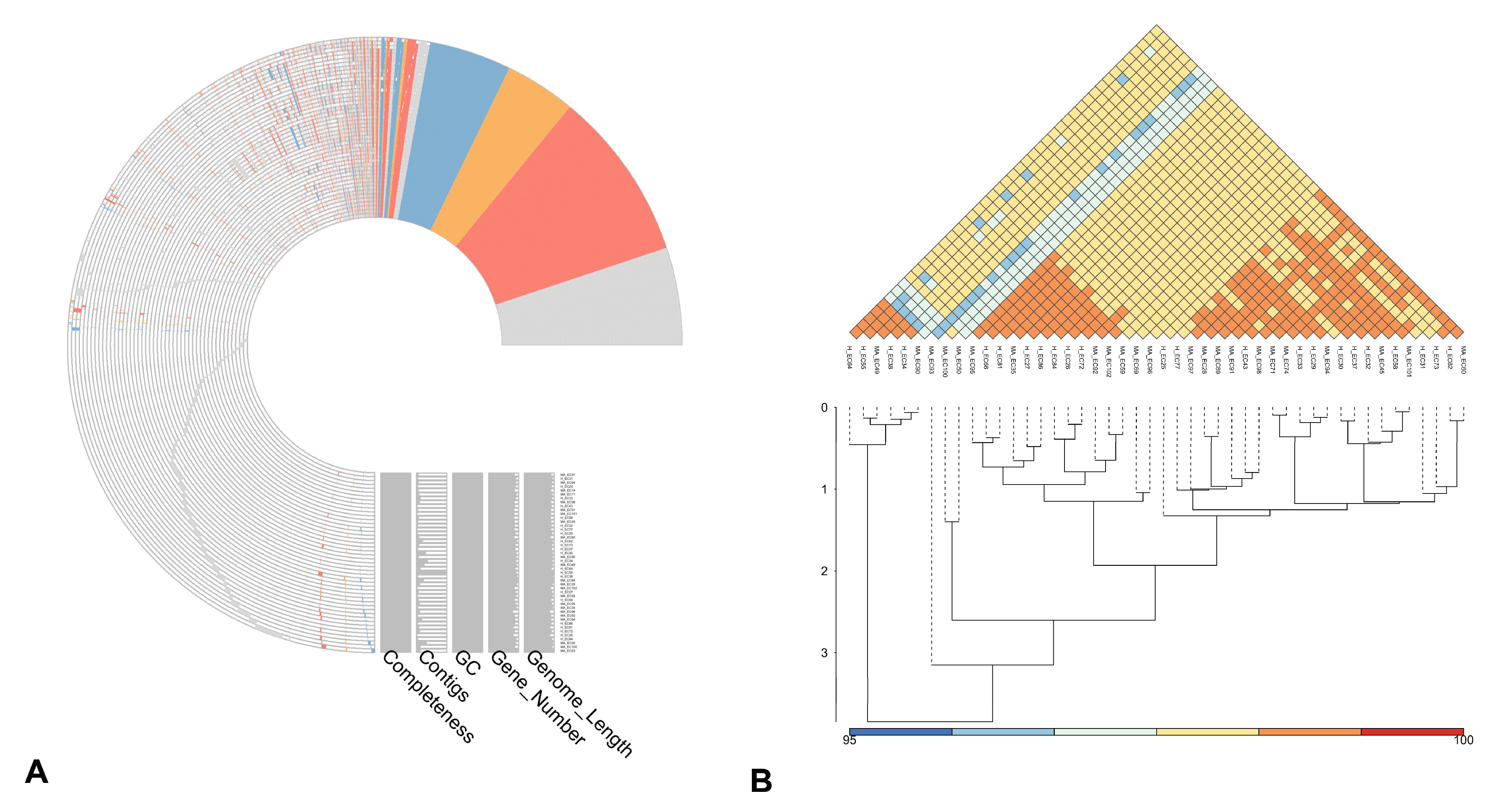
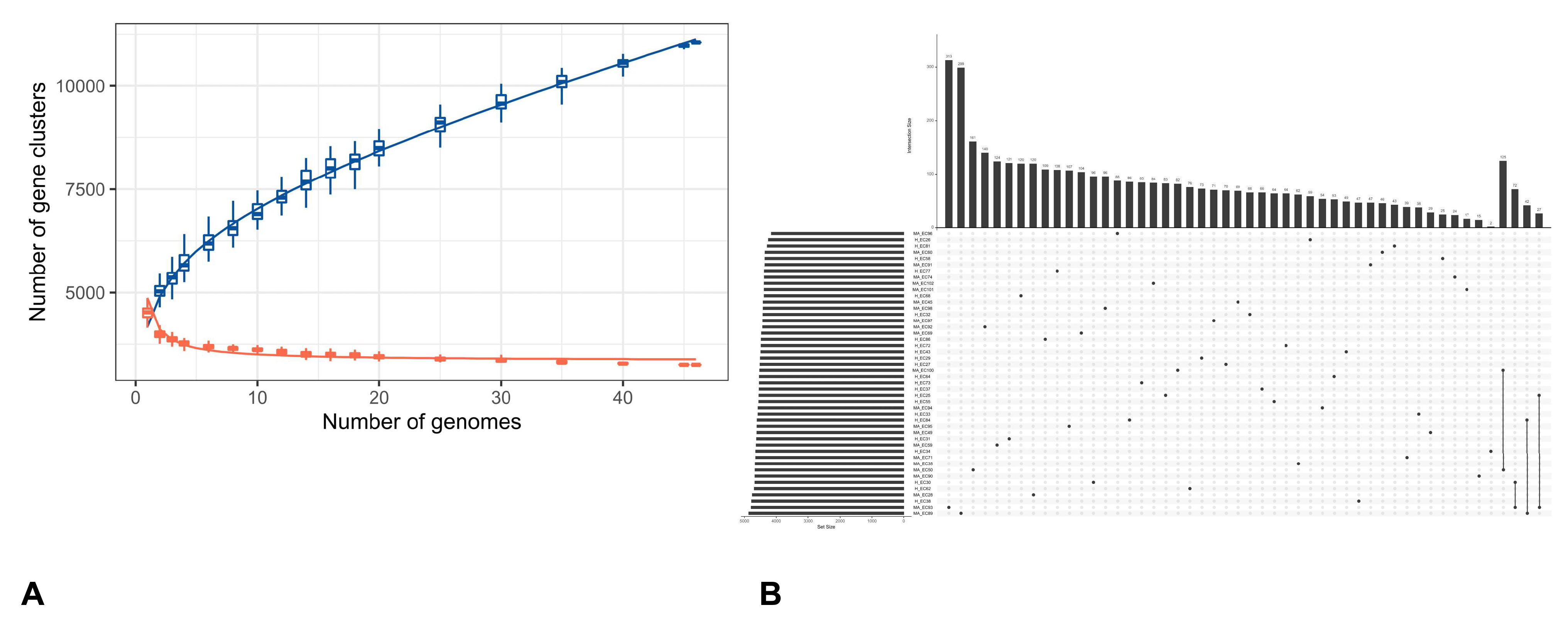
Figure 2A reports the pan-genome profile of all E. coli strains derived from Clusters of Orthologous Genes (COG) annotation, where the colored components represent the shared core cluster genes by isolates under analysis. More in dept, red indicates genes involved in metabolism (2659), orange represents genes related to information processing and storage (1168), and blue shows genes involved in cellular processes and signaling (1544). Finally, portions colored in grey and left blank pin out poorly characterized (570) or not annotated (5494) genes, respectively. Moreover, the average nucleotide identity (ANI) analysis (Figure 2B) among all strains revealed a few strains with the lowest identity of 96%, while the most show an identity of 98%. On a total of 46 analyzed genomes, 11,046 pan-gene clusters were identified (Figure 3A), with a range from 2 to 313 of the distinct gene clusters in each genome (Figure 3B).
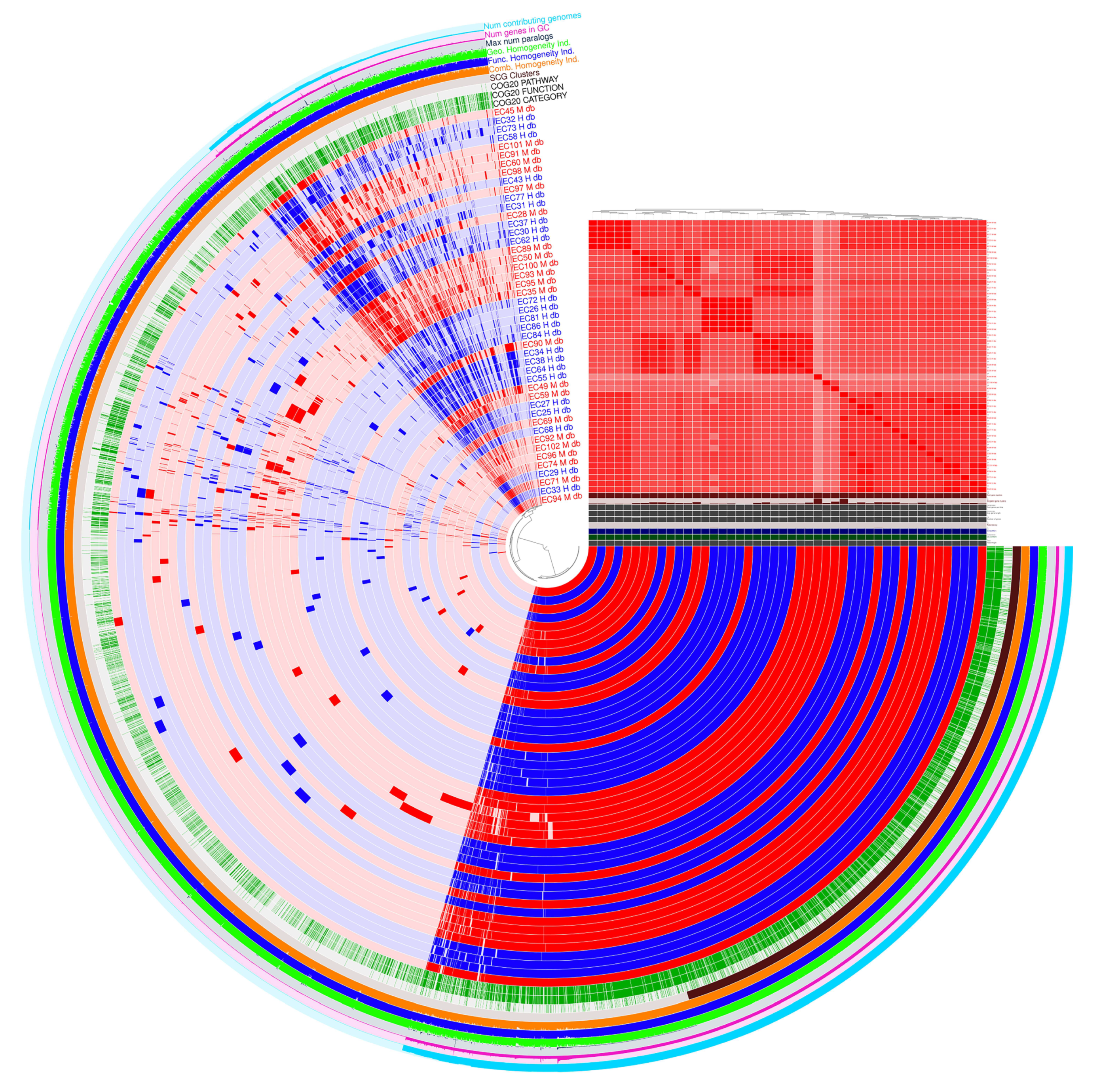
The pangenome representation derived from ANVI’O (Figure 4), describes more in-depth how our genomes are clustered, reporting how many genes are present compared to the proportion of genomes that include them. The pangenome is composed of different categories: (a) 2882 core genes (99% ≤ strains ≤ 100%), (b) 455 soft-core genes (95% ≤ strains < 99%), (c) 2213 shell genes (15% ≤strains < 95%), and (d) 10,326 cloud genes (0% ≤ strains < 15%).

3.2. Pangenome Visualization with PPANGGOLIN Pipeline

The rarefaction curve (Supplementary Figure S1) measures the pangenome diversity across all examined genomes, categorizing gene content into pangenome, soft accessory, soft core, exact accessory, and exact core. The pangenome expands from 5312 to 10,000 gene families across 46 genomes, with no saturation seen in rarefaction analysis according to Heaps’ law. The soft accessory and soft core gene families increase with more genomes, but the soft core stabilizes earlier, marking near-universal genes. The strict core remains constant at about 3196 gene families shared by all strains, likely representing essential functions. Individual genomes contain roughly 4500–4800 genes and 4320–4850 families. MA_EC28 has 4873 genes in 4712 families, including 3572 soft core and 3822 persistent families, while cloud genes range widely from 134 to 778 per genome.
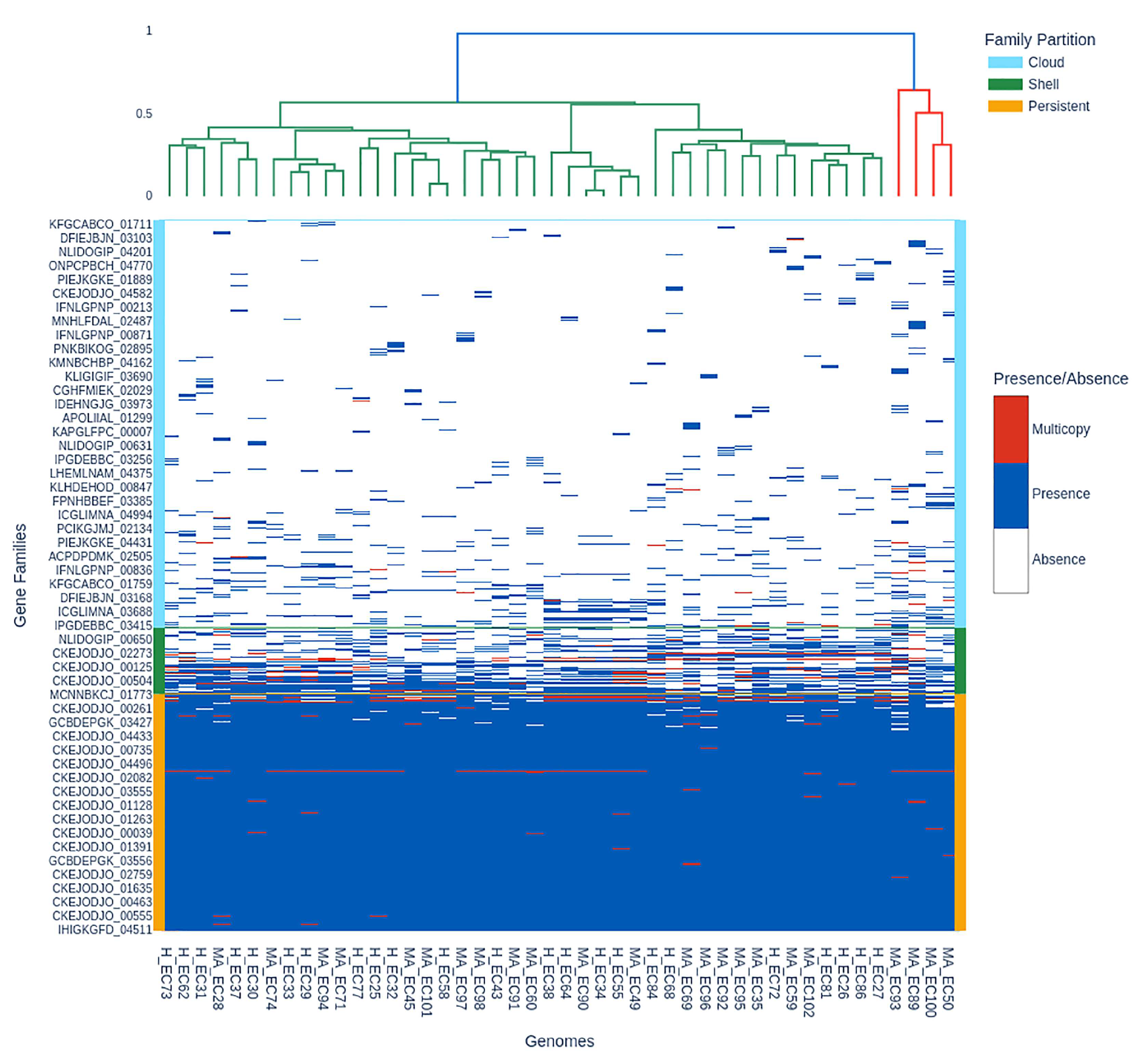
The presence–absence matrix (Figure 5) lists each gene family (rows) across all included genomes (columns), with colors indicating presence (blue), multicopy (red), and absence (white) and family partition identity (cloud, shell, persistent) on the right and in the dendrogram.
In the lowest half of Figure 5, the blue block represents the persistent genome (median per genome: ~3739–3849 gene families). This matches the rarefaction plateau for persistent and exact core genomes (3196 gene families). The shell and cloud genomes have presence and absence polymorphism, shown by blue and white in the upper matrix. The cloud partition of MA_EC93 has 778 gene families, while H_EC84 has 380. Red indicates multicopy families, including up to 221 families (MA_EC89) in pathogenic isolates, indicating transposons, duplications, or plasmid dynamics. The dendrogram shows that genomes cluster by gene content, matching partition identity.
E. coli pangenome evolution is shown by the U-shape gene family frequency distribution (Supplementary Figure S2).

3.3. Antibiotic Resistance

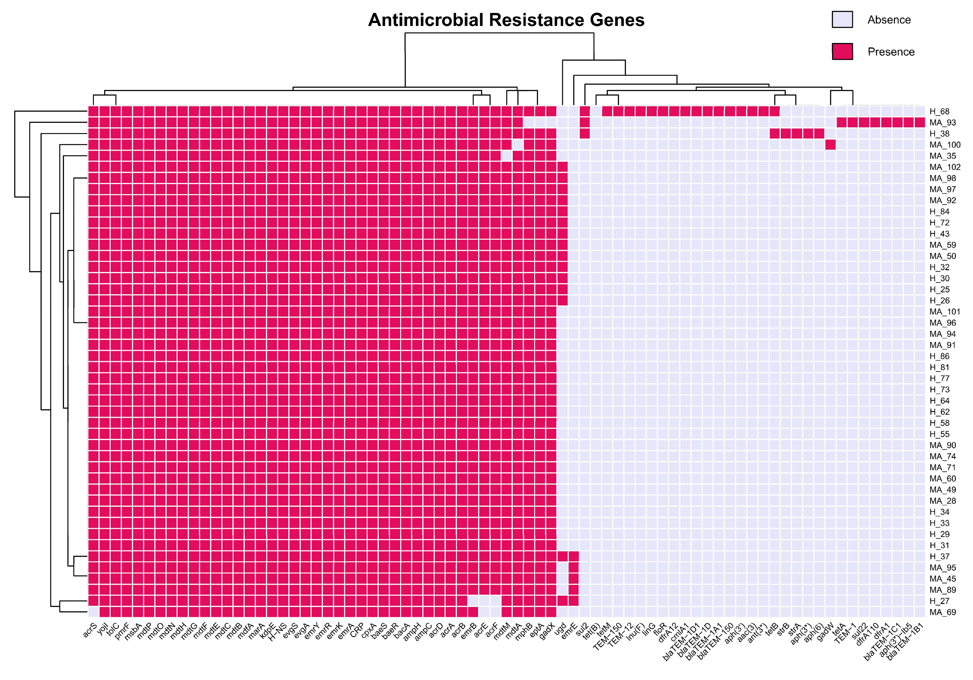
In Figure 6 we detected a 100% prevalence (46/46) of efflux pump-related ARGs for multidrug-resistant Gram-negative bacteria as well as resistance to beta-lactam antibiotics (ampC, ampH). In addition, bacA, a resistance gene that alters or deactivates the bacitracin and pmrF, a colistin-resistance gene that acts on changes at the level of the lipopolysaccharide of the exterior wall of Gram-negative bacteria, are found in all samples. There are no significant statistical differences between healthy and mastitis-associate groups (χ2 Test). Moreover, it is showable that most shared resistance genes are present in all our samples, even though some bacteria present specificity, but not necessarily to create clusters.
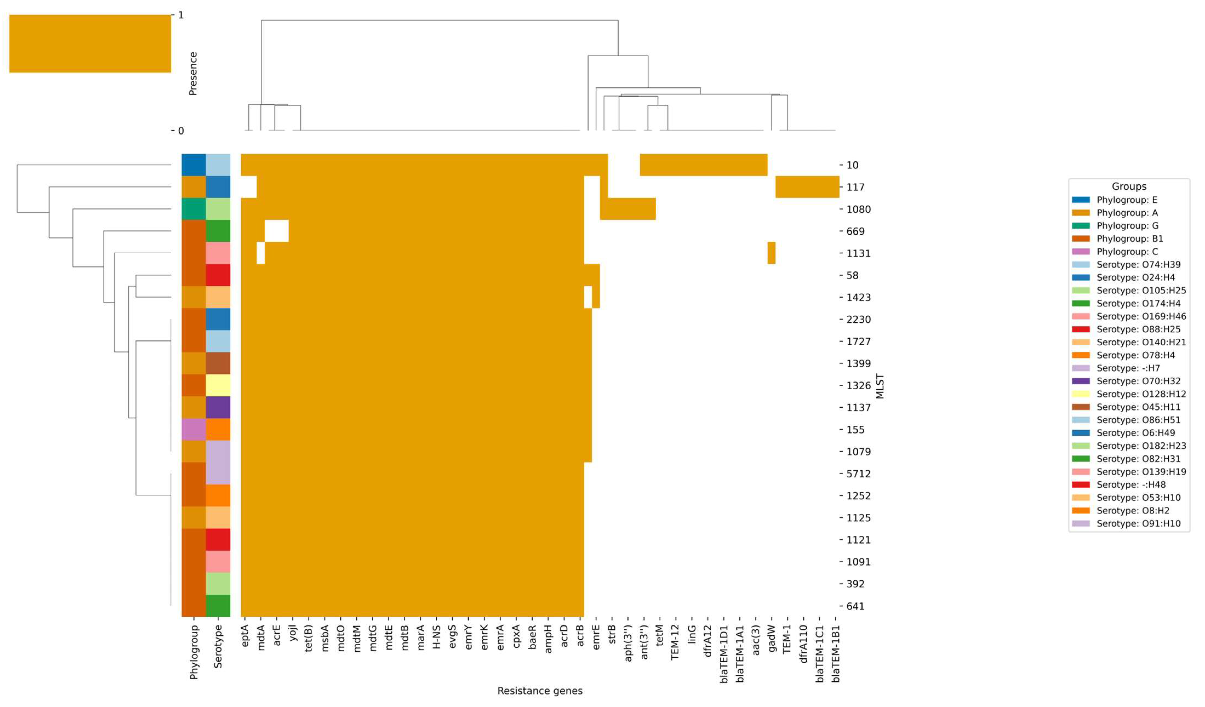
The second heatmap (Figure 7) illustrates the presence (ocher) of different resistance genes (mainly those encoding multidrug efflux pumps and their regulators, including mdtA, mdtB, mdtC, mdtE, mdtF, mdtG, mdtM, mdtO, acrA, acrB, acrD, acrE, acrF, and marA), across all isolates, irrespective of their origin, phylogroup, serotype, or MLST. The presence of these genes indicates that they are the core components of the E. coli genome, likely providing baseline resistance to a wide range of antibiotics, and are therefore evolutionarily conserved due to their essential role in bacterial survival across various environments. The heatmap illustrates a disparity in the distribution of acquired-resistance genes, including tet(B), aph(3’), blaTEM-1, dfrA1, and aac(3), which are predominantly present in the mastitis associated strains. This suggests that the selective pressure from antibiotic treatment in mastitis likely facilitates the acquisition of these genes within specific clonal lineages. This observation is corroborated by the clustering patterns: isolates from mastitis cases, particularly those in phylogroups A and B1 and MLSTs such as ST1080 and ST117, tend to cluster together and are more prone to possess acquired-resistance genes. Conversely, isolates from healthy cows, including those with MLSTs ST1125, ST58, and ST392 and serotypes like O139:H19 and O8:H2, typically lack these genes and cluster separately, indicating their diminished exposure to antibiotics and reduced selective pressure for resistance acquisition. The serotype distribution underscores this trend, with mastitis-associated serotypes like O105:H25 and O78:H4 more commonly linked to acquired-resistance genes, whereas serotypes found in healthy cows, such as O139:H19 and O8:H2, are predominantly lacking these genes.
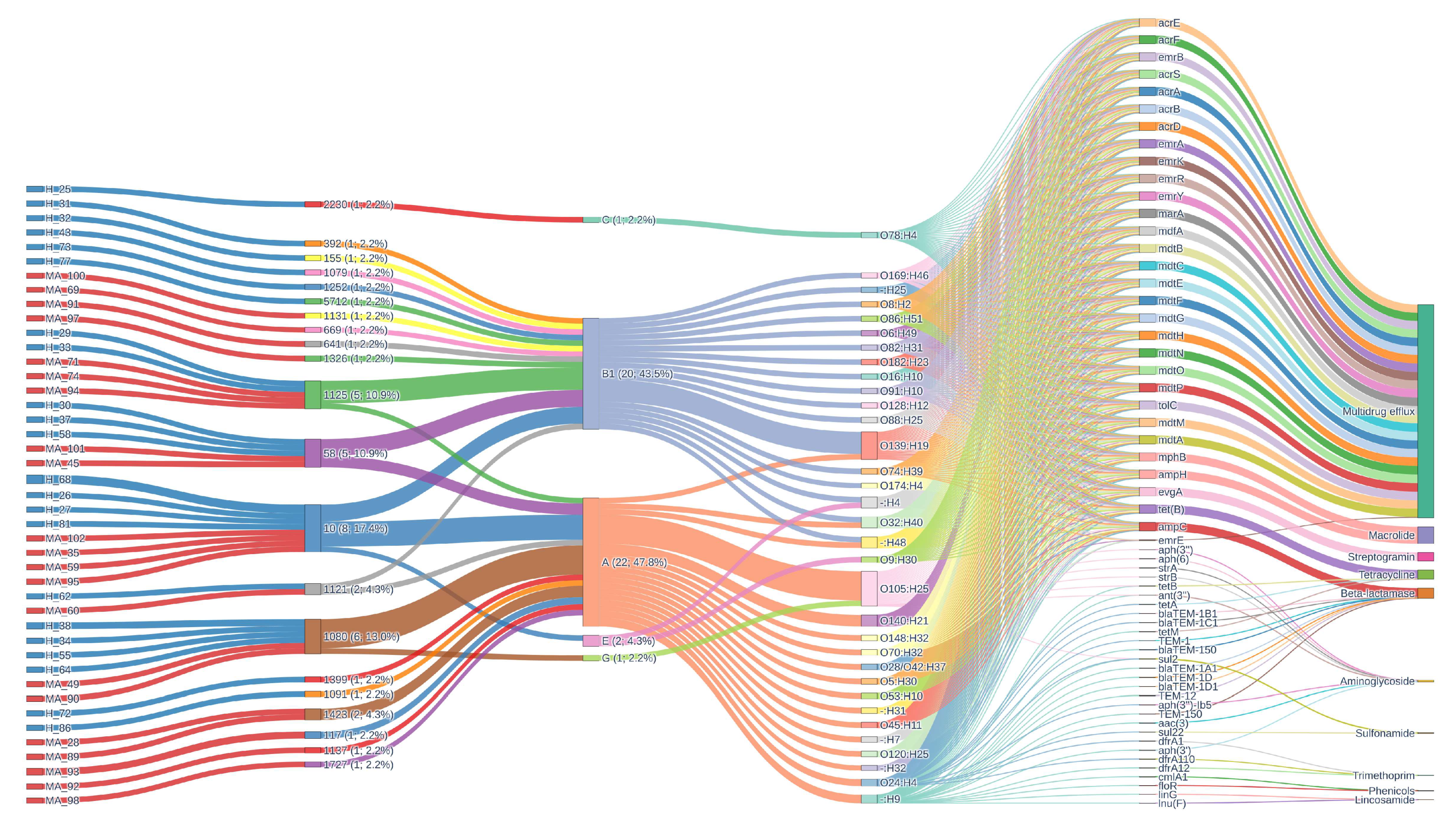
The alluvial plot (Figure 8) provides a comprehensive analysis of resistance gene architecture, which reveals a highly intricate and dynamic landscape of antimicrobial resistance. This landscape is distinguished by a diversity of sequence types (STs), phylogroups, serotypes, and resistance mechanisms. ST1080 (17.4%), ST1080 (13.0%), ST58 (10.9%), ST1125 (10.9%), ST1121 (4.3%), and ST1423 (4.3%) are the most prevalent STs. A broad distribution of additional rare STs, each constituting 2.2% or less of the population, is also present. This diversity is reflected in the phylogroup composition, which is primarily composed of group A (47.8%) and B1 (43.5%), with minor contributions from E, C, and G. The complex genetic structure of the population is further emphasized by serotype analysis, which reveals the heterogeneity of the sampled isolates. O105:H25 (13.0%), O139:H19 (10.9%), O140:H21 (4.3%), and a variety of other serotypes, including non-typeable variants, all contribute to this structure.
The resistance gene landscape is primarily characterized by multidrug efflux systems, which are universally present in all isolates (100%). This is indicative of a strong selective pressure for broad-spectrum resistance and the potential impact of both clinical and environmental antimicrobial exposure. High frequencies of beta-lactamase, macrolide, streptogramin, and tetracycline resistance determinants, each of which is nearly ubiquitous within the dataset, complement the prevalence of multidrug efflux mechanisms. This indicates a significant burden of resistance, as the mean number of resistance mechanisms per sample is 5.2 and an average of 30.6 resistance genes per isolate, emphasizing the collection’s extensive genetic capacity for antimicrobial resistance. In contrast, sulfonamide, phenicol, trimethoprim, and lincosamide resistance mechanisms are sporadically distributed, suggesting either recent acquisition, limited selective pressure, or potential fitness costs associated with these mechanisms. Aminoglycoside resistance is less common, occurring in only 2.2–4.3% of isolates.
The genetic structure within the population is resolved through the comprehensive mapping of ST–phylogroup–serotype combinations. It is important to note that the co-occurrence of specific clonal backgrounds and serotypes with distinct resistance profiles is exemplified by combinations such as ST1080/A/O105:H25 (10.9%), ST1125/B1/O139:H19 (8.7%), and other prominent lineages. The flow of genetic information from STs through phylogroups and serotypes to specific resistance determinants is elegantly illustrated in the alluvial diagrams, which depict the architecture of resistance genes. This emphasizes the modularity and plasticity of the bacterial genome by illustrating the intricate relationship between functional gene content and clonal origin. Frequent horizontal gene transfer events and recombination are evident, resulting in a mosaic architecture of resistance determinants that can swiftly adapt to changing selective pressures. The figure also aids in the identification of potential locations for gene exchange and the emergence of multidrug-resistant clones, which have substantial implications for the management of antimicrobial resistance and public health. Beta-lactamase, macrolide, tetracycline, streptogramin, and multidrug efflux resistance are consistently present at 100% prevalence in both groups. However, phenicol and lincosamide resistance mechanisms are absent in MA_ but present at low frequency in H_.

3.4. Virulence Factors

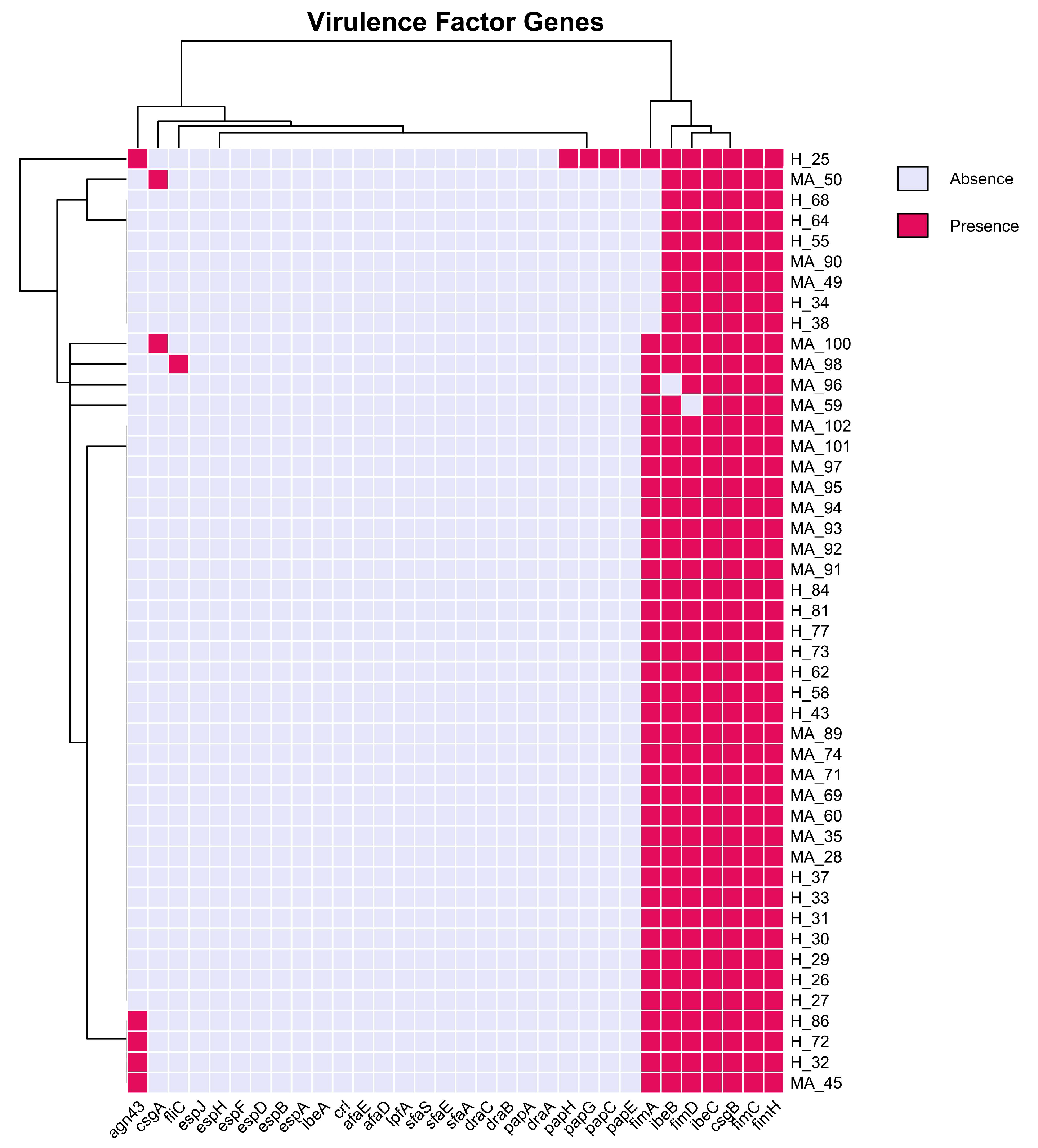
According to the literature [12,13,14], epithelial cell adhesion and invasion are critical factors for the onset and duration of mastitis in the mammary gland, but these features are not considered critical in the case of E. coli mastitis, being related mainly to LPS release. The results of our investigation on virulence adhesion factors (VFs) (Figure 9), such as the presence of fimbriae, flagella, adhesins, and type III secretion system (T3SS) confirm this information. Indeed, we could not identify a unique set of VFs that mastitis-associated E. coli strains carried, which could potentially account for the more pathogenic phenotype in comparison to healthy donors. Besides, it seems that no difference is present within strains, underlining the ubiquitous presence of type 1 fimbria for adhesion to epithelial cells (fimACDH), surface fibers for biofilm formation and adhesion enhancement (csgB), and VFs that facilitate invasion into the epithelial cells (ibeB, ibeC).
The virulence gene distribution (Figure 10) is a landscape that is both highly conserved and not, with each isolate containing a diverse array of virulence determinants that collectively offer substantial pathogenic potential. The isolates considered possess an extensive genetic capacity for virulence, as evidenced by the mean number of virulence mechanisms per sample.
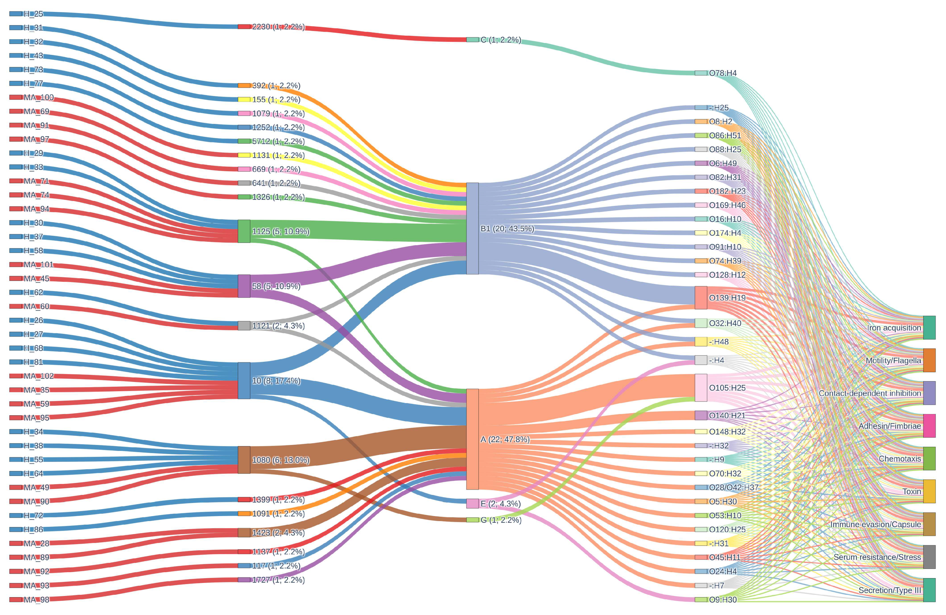
Genes associated with iron acquisition, motility/flagella, contact-dependent inhibition, adhesin/fimbriae, chemotaxis, toxin production, immune evasion/capsule formation, serum resistance/stress response, and type III secretion systems are included in this comprehensive repertoire. The conserved baseline of pathogenic potential that may be essential for survival, colonization, and infection in diverse host environments is suggested by the universal presence of these fundamental virulence mechanisms across all isolates, regardless of ST, phylogroup, or serotype. In addition to disclosing unique combinations and potential lineage-specific adaptations, the alluvial diagram for virulence genes vividly illustrates the convergence of diverse STs and serotypes onto a shared set of virulence mechanisms.
Across both the H_ and MA_ groups, the distribution of virulence mechanisms is remarkably consistent, with all main mechanisms present at a 100% prevalence.
Mapping ST–phylogroup–serotype combinations shows how the genetic structure of virulence varies across the population. ST1080/A/O105:H25 (10.9%) and ST1125/B1/O139:H19 (8.7%) are two examples of pairings that show that certain clonal backgrounds and serotypes can be found together with different virulence profiles. This method makes it possible to find epidemiologically relevant clones that might be connected to higher virulence, transmission, or the possibility of an outbreak.
The idea of a conserved core of virulence determinants is supported by the fact that different groups have several virulence mechanisms. All isolates have iron acquisition, motility/flagella, contact-dependent inhibition, adhesin/fimbriae, chemotaxis, toxin generation, immune evasion/capsule formation, serum resistance/stress response, and type III secretion systems.

4. Discussion

4.1. Phylogenetic Diversity and Core Genome Structure

To better understand the evolutionary context of the isolates, we first examined their phylogenetic distribution and core genome composition. Our comparative genomic study was done on a dataset of 46 Escherichia coli (E. coli) isolates from bovine milk in Italy. The dataset included an equal number of healthy (H_) and mastitis-affected animals (MA_). The key question our study tried to answer was whether mastitis-associated E. coli (MPEC) is a pathotype with unique genetic or virulence traits. This question is still very much up for dispute in the field, and our results can provide further data for this discussion. Our whole-genome sequencing and analysis showed that the technical quality was quite high. The genomes we got were almost complete and well-assembled, and the assembly statistics were the same for both health status groups. Most of the isolates clustered into phylogroups A and B1, which are the same two main groups that have been found in the literature for both commensal bovine and mastitis-associated strains. This supports the idea that there is a lot of phylogenetic diversity in these strains [8,15,16]. No unique clustering of MPEC based on phylogroup, MLST, or serotype was observed, echoing the consensus that MPEC are genetically heterogeneous and do not segregate into a monophyletic pathotype [8,17]. These results support previous findings that bovine E. coli populations are genetically diverse and that mastitis-associated isolates cannot be defined as a distinct evolutionary lineage.

4.2. Pan-Genome Composition and Accessory Gene Variability

Building on the phylogenetic results, we next explored the pan-genome composition to assess genomic plasticity and accessory gene variability. Our pan-genome analysis found 11,046 gene clusters, 2882 of which were core genes; this was in line with recent large-scale genome studies that showed how flexible E. coli’s genome is in cows [15]. Both healthy and mastitis-associated isolates had a lot of different accessory genes. These included cloud and shell genes that would have adaptive roles in the mammary niche. However, there was no collection of genes that were present in all MPEC and not in healthy cohorts. Many reports say that there is no conserved “mastitis virulence gene set”, and several comparative genomics studies have come to the same conclusion [16,18,19]. The virulence gene content of commensal and pathogenic isolates is very similar, and the known virulence determinants that are common in other ExPEC pathotypes (like Uropathogenic E. coli [UPEC] and Neonatal Meningitis-causing E. coli [NMEC]) are not often found or are not useful for distinguishing bovine mastitis [15,16,20,21]. Our accessory genome analysis, as well as recent publications, found enrichment in some strains for iron acquisition operons (fec, iro, iuc), adhesins, or capsule genes, but these features were distributed among both healthy and mastitis isolates [8,21]. This means considerable variation within the accessory genome, contributing to the persistent rise in the rarefaction curve and illustrating adaptation to diverse environments, acquisition of mobile genetic elements, and differing virulence factors.

4.3. Antimicrobial Resistance and Virulence Genes Patterns in Mastitis Pathogenicity

To investigate whether distinct antimicrobial resistance genes and virulence factors could explain mastitis pathogenicity, we examined gene patterns in comparison with existing ExPEC pathotypes.
One interesting result we found in our dataset, which is in line with what other researchers have found recently, is that all the bacteria carried antimicrobial resistance genes (AMRGs) and β-lactam resistance determinants (ampC, ampH). All the isolates had genes like bacA (resistance to bacitracin) and pmrF (resistance to polymyxin). This shows that milk-related E. coli has a lot of baseline antimicrobial resistance, but the number and variety of accessory resistance genes were not very different between healthy and MA animals. This is a pattern that has been seen in other research that looked at the resistance gene composition in bovine E. coli populations [20,21]. This pattern indicates that specific serotype-MLST-phylogroup combinations are more susceptible to acquiring and propagating resistance genes, maybe due to their ecological environment, genetic composition, and exposure to clinical interventions. The hierarchical clustering of genes and isolates on the heatmap (Figure 9) visually substantiates these associations, with core resistance genes forming a compact cluster present in all strains, while acquired-resistance genes exhibit smaller, more dispersed clusters linked to specific lineages and clinical contexts. Significantly, several healthy cow isolates group with mastitis-associated isolates, indicating either recent acquisition of resistance genes or possible misclassification, underscoring the dynamic nature of resistance gene spread. The biological findings are substantial: although intrinsic resistance mechanisms are universally present and likely crucial for fundamental bacterial physiology and survival, the intermittent yet epidemiologically significant occurrence of acquired-resistance genes in mastitis-associated isolates highlights the influence of antibiotic usage on the resistome of bovine E. coli populations. The elevated prevalence of acquired-resistance genes in mastitis cases has significant implications for animal health and antibiotic stewardship, indicating that existing treatment practices may unintentionally favor and disseminate resistant clones. The prevalence data in the dataset suggests that certain resistance mechanisms (multidrug efflux, beta-lactamase, macrolide, and tetracycline resistance) are universally or nearly universally distributed across both H_ and MA_ groups. This suggests that strong selective pressures and/or efficient horizontal gene transfer events are facilitating the dissemination of these determinants. Conversely, certain groups exhibit the absence or rarity of mechanisms such as phenicol and lincosamide resistance, which may be indicative of ecological or evolutionary constraints that restrict their dissemination. This pattern may indicate that environmental or host-associated factors significantly influence the resistance gene repertoire of bacterial populations, as it may be indicative of variations in selective pressures, antimicrobial use, or ecological context between the distinct groups.
Researchers are still very interested in finding specific genetic traits or phenotypes that are linked to some strains of E. coli being able to induce clinical mastitis. Some early investigations and reviews suggested that a new form of E. coli called “mammary pathogenic E. coli” (MPEC) might be emerging [6]. However, more recent and thorough genomic studies have not been able to consistently find unique virulence or fitness genes that are only found in mastitis isolates [8,15,21]. Our results strongly support this view; MA_ strains had only small increases in accessory genome size and gene count. This is consistent with the idea that sporadic acquisition of extra genetic material (e.g., plasmids, prophages, capsule islands) may change how pathogenic a strain is but does not define a pathotype.

4.4. Ecological and Epidemiological Insights

To interpret the genetic findings within a broader ecological and epidemiological perspective, we examined how environmental and host-related factors may shape the observed genomic patterns. Recent studies that looked at larger genome datasets have looked for accessory genes, single nucleotide polymorphisms, or gene expression profiles that could help tell MPEC apart. Researchers have suggested several genes that are interesting, such as the ferric dicitrate receptor operon (fec), genes that code for adhesins (yad fimbriae), or genes that are involved in making exopolysaccharides or capsules (e.g., wbpl, capD). These genes are mostly linked to disease isolates or the severity of clinical presentation [22]. In our study, we detected iron acquisition and adherence factors in several isolates, often more prevalent in MPEC, but these same genes also occurred occasionally in H_ strains: no single virulence determinant could predict phenotype. This emphasizes the bacterial genome’s adaptability and its ability to acquire virulence determinants through both vertical inheritance and horizontal acquisition. This implies that the selective pressures that influence the content of virulence genes are consistent across various ecological or host-associated contexts, or that the core set of virulence genes is necessary for fitness in a wide variety of environments. The presence of supplementary or accessory virulence determinants may alter pathogenesis in a strain-specific manner, resulting in variations in clinical outcomes, host range, or environmental persistence. A recent genome-wide study found that mutations and allelic variants of adhesins, biofilm genes (pga operon), and secretion system components were linked to either mild or severe mastitis in complicated ways [15,16]. However, these were affected by both phylogenetic background and accessory genome content, not by clear-cut pathotype-specific sequence markers [15]. Moreover, core genome phylogeny frequently exhibited a stronger correlation with variations in gene content than with disease association, reinforcing the notion that evolutionarily divergent lineages of E. coli may employ different genetic repertoires and tactics to prosper in the mammary gland [15,16]. Consequently, the emergence of a “pan-pathotype” with definitive markers seems improbable; instead, a mosaic and polygenic model of mastitis pathogenicity has gained substantial validation [15,16]. Our results align closely with genome-wide studies conducted by Leimbach et al. (2017) [8,15,16], Olson et al. (2024) [8,15,16], Kempf et al. (2016) [8,15,16], and others, which consistently did not identify MPEC as genetically separate from commensal bovine E. coli, highlighting the functional redundancy and adaptability of this species. The observed variability in surface antigens (O/H serotypes), sequence types, and accessory genes among mastitis isolates reinforces the conclusion that pathogenicity in the mammary gland arises not from the acquisition of a universal mastitis gene set, supporting the current knowledge that a non-genetic factor (LPS release) is the trigger for acute clinical mastitis. However, this does not exclude that a combination of core fitness genes, the capacity to evade host immune responses (e.g., serum resistance and biofilm formation), and the random acquisition of accessory modules may augment survival and pro-inflammatory potential [16,21,23].

4.5. Limitations and Future Perspectives

Although this study provides valuable genomic insights into the complexity of MPEC, we recognise that several limitations and open questions still remain. The scenario is significantly different from human ExPEC, EHEC, or EPEC pathotypes, where virulence gene signatures or toxins (e.g., stx, eae, pap) are distinctly associated with clinical syndromes. Our investigation, along with others, revealed that typical extraintestinal pathogenic E. coli (ExPEC) genes were frequently absent or shared between both groups, and no definitive “mastitis marker” was identified [24]. Traits such as the group III polysaccharide capsule observed in experimental hypervirulent MPEC models (e.g., M12 strain), while capable of providing increased fitness in infection models, are infrequently found in natural populations and do not constitute a population-wide mastitis-specific arsenal. Adding unusual and non-typeable variants to the population increases its genomic and functional diversity even more. These variants may act as reservoirs for new virulence genes or as steps in the evolution of new harmful lineages. As summarized in the alluvial plot of virulence genes, the mosaic structure of virulence determinants, reflect frequent recombination and horizontal gene transfer events. This shows how adaptable and modular the bacterial genome is. This plasticity makes it easier for organisms to quickly adjust to changes in their environment or host. It also allows hypervirulent or multidrug-resistant clones to appear, which have better fitness and disease-causing potential. The fact that these processes are found in so many places suggests that they are necessary for survival and success in a wide range of settings. Additional or accessory virulence genes can give organisms specialized capabilities that help them adapt to certain hosts or niches and improve the overall fitness and evolutionary success of the population.
Functional studies indicate that specific genes may influence the degree of mastitis (mild versus severe), and clusters related to adherence and exopolysaccharide synthesis have variable correlations with illness outcomes; nevertheless, these factors are neither exclusive nor essential for the hypothesized MPEC phenotype [15]. We considered isolates only from mild and severe mastitis, but not from acute mastitis, due to the need for very rapid treatment in this latter case. This may be considered a limitation since certain resistance and virulence genes are often associated with more severe clinical presentations.
Moreover, our data corroborate a robust alternative hypothesis suggesting that most of E. coli related to clinical mastitis are opportunistic commensals of the bovine gut or environment, adept at colonizing various niches. Therefore, other risk factors such as unproper milking practices and hygiene, physiological or pathological stress, in conjunction with stochastic host susceptibility, may lead to disease [8,16]. The absence of pathotype-defining features in MPEC is corroborated by evidence indicating that, apart from the predominance in phylogroups A and B1 (which also prevail in the H_ strains), neither horizontal gene transfer nor concentrated virulence gene distinguishes mastitis isolates, as supposed from existing literature [20,24].

5. Conclusions

In conclusion, our comparative genomic investigation of 46 E. coli isolates from healthy and mastitis dairy cows in Italy demonstrates significant genetic variety and flexibility within this crucial bovine-associated bacterial species. Despite methodologically sound study design and sampling, we identified no consistent genomic or phylogenetic markers capable of consistently differentiating mastitis-associated E. coli from those obtained from healthy animals. The pan-genome analysis uncovered a vast accessory genome and several cloud genes, alongside a restricted collection of universally shared core genes, further emphasizing the flexibility and mosaic nature of the E. coli population in the bovine mammary environment. Our study contributes to the consensus that the genetic basis of E. coli mastitis is intricate and multifaceted, with no singular genomic marker or virulence profile sufficiently accounting for disease association and underscores the dynamic evolutionary context E. coli encounters at the animal–environment interface in dairy systems.

Supplementary Materials

The following supporting information can be downloaded at: https://www.mdpi.com/article/10.3390/pathogens14111181/s1, Figure S1: Rarefaction curve showing gene family accumulation with increasing numbers of H and MA genomes, highlighting an open pangenome and around 3196 gene families; Figure S2: U-shape frequency plot of gene family distribution.

Author Contributions

Conceptualization, A.Z. and G.M.; methodology, G.L. and G.M.; software, A.S. and V.M.S.; formal analysis, L.C. and F.Z.; investigation, L.C., F.Z., and G.M.; resources, A.Z. and L.B.; data curation, A.S. and V.M.S.; writing—original draft preparation, P.A.M. and G.L.; writing—review and editing, A.Z., P.A.M., and L.B.; visualization, G.L.; funding acquisition, A.Z. All authors have read and agreed to the published version of the manuscript.

Funding

This research received no external funding.

Institutional Review Board Statement

The study falls under Art. 2 (Excluded cases from the discipline) of Legislative Decree 26/2014 ‘Implementation of Directive 2010/63/EU on the protection of animals used for scientific purposes’): to veterinary clinical practices for non-experimental purposes*, the definition of which is according to the Note of 24 July 2017 of the Italian Ministry of Health—‘By veterinary clinical practice for non-experimental purposes is meant therapeutic, curative or diagnostic activity carried out with the informed consent of the owner of the animal affected by an ongoing pathology, natural and i.e., not experimentally provoked or induced’. No authorisation is therefore required from Animal Welfare Body.

Informed Consent Statement

Not applicable.

Data Availability Statement

The whole-genome assemblies were deposited at GenBank repository under the bioproject PRJNA1242576 (https://www.ncbi.nlm.nih.gov/bioproject/PRJNA1242576, accessed on 16 September 2025).

Acknowledgments

Computational resources provided by INDACO Platform (https://www.indaco.unimi.it/), which is a project of High-Performance Computing at the University of Milan https://www.unimi.it/it.

Conflicts of Interest

The authors declare no conflicts of interest.

References

  1. Riemann, T.; Bergmann, A. Epidemiology, pathogenesis, treatment and prevention of bovine acute Escherichia coli mastitis, a literature review. DTW Dtsch. Tierarztl. Wochenschr. 2000, 107, 444–454. [Google Scholar]
  2. Donnenberg, M. (Ed.) Escherichia coli Pathotypes and Principles of Pathogenesis; Elsevier: Amsterdam, The Netherlands, 2013; ISBN 978-0-12-397048-0. [Google Scholar]
  3. Buitenhuis, B.; Røntved, C.M.; Edwards, S.M.; Ingvartsen, K.L.; Sørensen, P. In Depth Analysis of Genes and Pathways of the Mammary Gland Involved in the Pathogenesis of Bovine Escherichia coli- Mastitis. BMC Genom. 2011, 12, 130. [Google Scholar] [CrossRef]
  4. Burvenich, C.; Van Merris, V.; Mehrzad, J.; Diez-Fraile, A.; Duchateau, L. Severity of E. coli Mastitis Is Mainly Determined by Cow Factors. Vet. Res. 2003, 34, 521–564. [Google Scholar] [CrossRef] [PubMed]
  5. Shpigel, N.Y.; Elazar, S.; Rosenshine, I. Mammary Pathogenic Escherichia coli. Curr. Opin. Microbiol. 2008, 11, 60–65. [Google Scholar] [CrossRef] [PubMed]
  6. Orsi, H.; Guimarães, F.F.; Leite, D.S.; Guerra, S.T.; Joaquim, S.F.; Pantoja, J.C.F.; Hernandes, R.T.; Lucheis, S.B.; Ribeiro, M.G.; Langoni, H.; et al. Characterization of Mammary Pathogenic Escherichia coli Reveals the Diversity of Escherichia coli Isolates Associated with Bovine Clinical Mastitis in Brazil. J. Dairy Sci. 2023, 106, 1403–1413. [Google Scholar] [CrossRef]
  7. Campos, F.C.; Castilho, I.G.; Rossi, B.F.; Bonsaglia, É.C.R.; Dantas, S.T.A.; Dias, R.C.B.; Fernandes Júnior, A.; Hernandes, R.T.; Camargo, C.H.; Ribeiro, M.G.; et al. Genetic and Antimicrobial Resistance Profiles of Mammary Pathogenic E. coli (MPEC) Isolates from Bovine Clinical Mastitis. Pathogens 2022, 11, 1435. [Google Scholar] [CrossRef]
  8. Leimbach, A.; Poehlein, A.; Vollmers, J.; Görlich, D.; Daniel, R.; Dobrindt, U. No Evidence for a Bovine Mastitis Escherichia coli Pathotype. BMC Genom. 2017, 18, 359. [Google Scholar] [CrossRef]
  9. Dogan, B.; Rishniw, M.; Bruant, G.; Harel, J.; Schukken, Y.H.; Simpson, K.W. Phylogroup and lpfA Influence Epithelial Invasion by Mastitis Associated Escherichia coli. Vet. Microbiol. 2012, 159, 163–170. [Google Scholar] [CrossRef] [PubMed]
  10. Blum, S.E.; Heller, E.D.; Jacoby, S.; Krifucks, O.; Leitner, G. Comparison of the Immune Responses Associated with Experimental Bovine Mastitis Caused by Different Strains of Escherichia coli. J. Dairy Res. 2017, 84, 190–197. [Google Scholar] [CrossRef]
  11. Badis, A.; Heleili, N.; Merradi, M.; Ayachi, A.; Martino, P.A.; Meroni, G.; Soggiu, A. Outbreak of Carbapenem-Resistant High-Risk Clone ST244 of Pseudomonas Aeruginosa in Dogs and Cats in Algeria. Antibiotics 2025, 14, 230. [Google Scholar] [CrossRef]
  12. Passey, S.; Bradley, A.; Mellor, H. Escherichia coli Isolated from Bovine Mastitis Invade Mammary Cells by a Modified Endocytic Pathway. Vet. Microbiol. 2008, 130, 151–164. [Google Scholar] [CrossRef] [PubMed]
  13. Dogan, B.; Klaessig, S.; Rishniw, M.; Almeida, R.A.; Oliver, S.P.; Simpson, K.; Schukken, Y.H. Adherent and Invasive Escherichia coli Are Associated with Persistent Bovine Mastitis. Vet. Microbiol. 2006, 116, 270–282. [Google Scholar] [CrossRef]
  14. Almeida, R.A.; Dogan, B.; Klaessing, S.; Schukken, Y.H.; Oliver, S.P. Intracellular Fate of Strains of Escherichia coli Isolated from Dairy Cows with Acute or Chronic Mastitis. Vet. Res. Commun. 2011, 35, 89–101. [Google Scholar] [CrossRef] [PubMed]
  15. Olson, M.A.; Cullimore, C.; Hutchison, W.D.; Grimsrud, A.; Nobrega, D.; De Buck, J.; Barkema, H.W.; Wilson, E.; Pickett, B.E.; Erickson, D.L. Genes Associated with Fitness and Disease Severity in the Pan-Genome of Mastitis-Associated Escherichia coli. Front. Microbiol. 2024, 15, 1452007. [Google Scholar] [CrossRef]
  16. Kempf, F.; Slugocki, C.; Blum, S.E.; Leitner, G.; Germon, P. Genomic Comparative Study of Bovine Mastitis Escherichia coli. PLoS ONE 2016, 11, e0147954. [Google Scholar] [CrossRef]
  17. Cruz-Soto, A.S.; Toro-Castillo, V.; Munguía-Magdaleno, C.O.; Torres-Flores, J.E.; Flores-Pantoja, L.E.; Loeza-Lara, P.D.; Jiménez-Mejía, R. Relación Genética, Formación de Biopelículas, Movilidad y Virulencia de Escherichia coli Aislada de Mastitis Bovina. Rev. Mex. Cienc. Pecu. 2020, 11, 167–182. [Google Scholar] [CrossRef]
  18. Olson, M.A.; Siebach, T.W.; Griffitts, J.S.; Wilson, E.; Erickson, D.L. Genome-Wide Identification of Fitness Factors in Mastitis-Associated Escherichia coli. Appl. Environ. Microbiol. 2018, 84, e02190-17. [Google Scholar] [CrossRef]
  19. Germon, P.; Foucras, G.; Smith, D.G.E.; Rainard, P. Invited Review: Mastitis Escherichia coli Strains—Mastitis-Associated or Mammo-Pathogenic? J. Dairy Sci. 2025, 108, 4485–4507. [Google Scholar] [CrossRef]
  20. Nüesch-Inderbinen, M.; Käppeli, N.; Morach, M.; Eicher, C.; Corti, S.; Stephan, R. Molecular Types, Virulence Profiles and Antimicrobial Resistance of Escherichia coli Causing Bovine Mastitis. Vet. Rec. Open 2019, 6, e000369. [Google Scholar] [CrossRef]
  21. Goulart, D.B.; Mellata, M. Escherichia coli Mastitis in Dairy Cattle: Etiology, Diagnosis, and Treatment Challenges. Front. Microbiol. 2022, 13, 928346. [Google Scholar] [CrossRef] [PubMed]
  22. Yoon, S.; Lee, Y.J. Molecular Characteristics of Escherichia coli from Bulk Tank Milk in Korea. J. Vet. Sci. 2022, 23, e9. [Google Scholar] [CrossRef] [PubMed]
  23. Fernandes, J.B.C.; Zanardo, L.G.; Galvão, N.N.; Carvalho, I.A.; Nero, L.A.; Moreira, M.A.S. Escherichia coli from Clinical Mastitis: Serotypes and Virulence Factors. J. Vet. Diagn. Investig. 2011, 23, 1146–1152. [Google Scholar] [CrossRef] [PubMed]
  24. Zuo, J.; Lv, Z.; Lian, L.; Wu, Z.; Fu, S.; Zhang, H.; Wu, J.; Pan, Z.; Yu, Y.; Chen, W.; et al. Difference Analysis on Virulence Genes, Biofilms and Antimicrobial Susceptibility of Escherichia coli from Clinical and Subclinical Bovine Mastitis. Vet. Sci. 2025, 12, 132. [Google Scholar] [CrossRef] [PubMed]
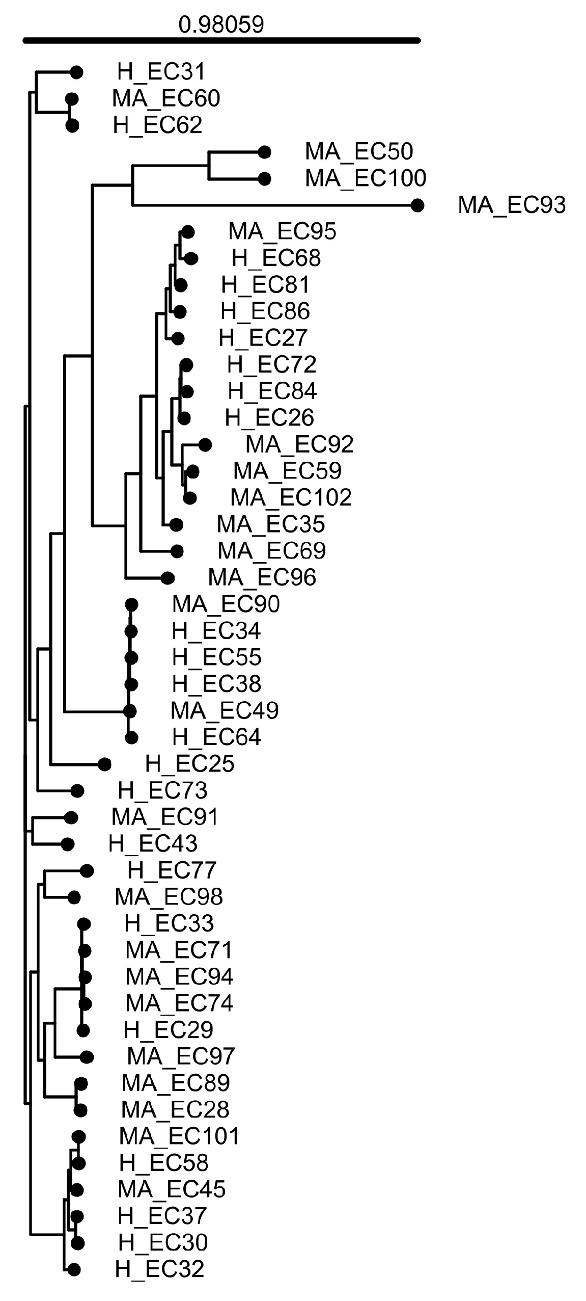
Figure 1. IPGA construction of a whole-genome-based phylogenetic tree. The tree illustrates the evolutionary relationships among E. coli strains in this study. The branch lengths reflect genomic differences, while the clustering suggests genetic similarity and possible shared origins. The major branching point, or the evolutionary split from which all strains in the phylogenetic tree originate, has significant statistical support (confidence value, 0.98059). H, healthy; MA, mastitis-associated.
Pathogens 14 01181 g001
Figure 2. Pan-genome analysis of E. coli isolates. (A) Pan-genome profile based on gene clusters. (B) Heatmap and hierarchical clustering based on pairwise average nucleotide identity (ANI). Colors indicate the degree of average nucleotide identity between each pair, with red corresponding to higher ANI values (more genomic similarity). The hierarchical clustering beneath shows how genomes are grouped together in hierarchical clusters.
Pathogens 14 01181 g002
Figure 3. Pan-genome analysis of E. coli isolates (Continued): (A) Number of pan-gene clusters (blue) and sequenced genomes (orange) among isolates. This type of graph helps to gain information on the genetic variability and evolution of the species under study based on the blue curve shape according to the number of genomes analyzed. More specifically, we can define whether the pan-genome is ‘open’—as in our case—when the number of clusters keeps increasing with the insertion of new sequenced genomes, or ‘closed’, when the growth of the blue line stabilizes. (B) Upset plot of comparisons among unique clusters of genes.
Pathogens 14 01181 g003
Figure 4. Pangenome visualization of E. coli strains visualized by ANVI’ O. Blue and red genomes represent strains derived from healthy donors and associated with mastitis, respectively. Gene cluster presence or absence determines how the samples are grouped in the central dendrogram. The samples are shown in the phylogenetic tree in the order determined by the ANI % of identity. A red square in the evolutionary tree represents each sample cluster, signifying ANI percentage identity values more than 99%.
Pathogens 14 01181 g004
Figure 5. Presence–absence matrix and hierarchical clustering of gene families across H and MA E. coli genomes. Persistent (core) families dominate the lower block, variable accessory families partition the upper section, and shell/cloud diversity informs strain clustering.
Pathogens 14 01181 g005
Figure 6. Heatmap of antimicrobial resistance genes in E. coli. Fuchsia, Presence; Liliac, Absence.
Pathogens 14 01181 g006
Figure 7. Heatmap and hierarchical clustering of resistance gene profiles in 46 E. coli isolates analyzed.
Pathogens 14 01181 g007
Figure 8. Alluvial diagram showing the distribution of resistance genes and their associations with phylogroups, sequence types, and resistance phenotypes.
Pathogens 14 01181 g008
Figure 9. Heatmap of virulence factors in E. coli. Fuchsia, Presence; Liliac, Absence.
Pathogens 14 01181 g009
Figure 10. Alluvial diagram showing the distribution of virulence genes and their associations with phylogroups, sequence types, and resistance phenotypes.
Pathogens 14 01181 g010
Table 1. Assembly statistics and genomic characteristics of 46 Escherichia coli isolates from Bos taurus milk, collected in Italy (2023–2024). For each isolate (23 from healthy: H_; 23 from mastitis-associated: MA_ animals), summary metrics are provided, including year of isolation, assembly level, assembly structure (Y: circular; N: linear), fragment number, genome size, GC content, CDS count, sequencing.
Isolate IDBioSampleGenomeYr of
Isolation
Assembly
Level
CircularityNo. of
Fragments
Genome
Size (Mbp)
GC (%)CDSCOV a
(x)
Com b
(%)
Cont c
(%)
H_EC25SAMN47596209JBPKIM0000000002023ContigY24.985146942221000.43
H_EC26SAMN47596210GCA_051912635.12023ContigY14.665143191491000.31
H_EC27SAMN47596211JBPKIL0000000002023ContigY35514734821000.5
H_EC29SAMN47596212JBPKIK0000000002023ContigN24.945147051991000.77
H_EC30SAMN47596213JBPKIJ0000000002023ContigN145.11514829791000.18
H_EC31SAMN47596214JBPKII0000000002023ContigN25.075147621691000.24
H_EC32SAMN47596215JBPKIH0000000002023ContigN24.895145231341000.17
H_EC33SAMN47596216JBPKIG0000000002023ContigN65.025147511131000.13
H_EC34SAMN47596217JBPKIF0000000002023ContigN175.06514797921000.17
H_EC37SAMN47596218JBPKIE0000000002023ContigY24.975146631161000.14
H_EC38SAMN47596219JBPKID0000000002023ContigY25.175149761761000.18
MA_EC28SAMN47596220JBPKIC0000000002023ContigN55.095148891721000.26
MA_EC35SAMN47596221JBPKIB0000000002023ContigY25.135148371191000.26
MA_EC45SAMN47596222JBPKIA0000000002023ContigN24.835145191671000.37
MA_EC49SAMN47596223JBPKHZ0000000002023ContigN125.15149151191000.21
MA_EC50SAMN47596224JBPKHY0000000002023ContigN155.165147971141001.24
MA_EC59SAMN47596225JBPKHX0000000002023ContigY25.075148182261000.12
MA_EC60SAMN47596226JBPKHW0000000002023ContigN44.735144671631000.08
MA_EC69SAMN47596227JBPKHV0000000002023ContigN84.855146212751000.13
MA_EC71SAMN47596228JBPKHU0000000002024ContigN45.055148231261000.34
MA_EC74SAMN47596229JBPKHT0000000002024ContigN34.835145351941000.16
MA_EC89SAMN47596230JBPKHS0000000002024ScaffoldN115.385151922011000.5
MA_EC90SAMN47596231JBPKHR0000000002024ContigN35.095148372741000.18
H_EC43SAMN47596232GCA_051912625.12023ContigY14.925146003181000.15
H_EC55SAMN47596233JBPKHQ0000000002023ContigN455.23524852531001.29
H_EC58SAMN47596234JBPKHP0000000002023ContigN34.775144381141000.09
H_EC62SAMN47596235JBPKHO0000000002023ContigN95.15149231441000.2
H_EC64SAMN47596236JBPKHN0000000002023ContigN64.925146991971000.16
H_EC68SAMN47596237JBPKHM0000000002023ContigY34.845145311301000.03
H_EC72SAMN47596238JBPKHL0000000002024ContigN54.915145762431000.21
H_EC73SAMN47596239JBPKHK0000000002024ContigN45.025147181451000.25
H_EC77SAMN47596240GCA_051912615.12024ScaffoldN14.835144752331000.1
H_EC81SAMN47596241JBPKHJ0000000002024ContigN44.695143661331000
H_EC84SAMN47596242JBPKHI0000000002024ContigN25.02514821851000.18
H_EC86SAMN47596243JBPKHH0000000002024ContigY24.885146323941000.21
MA_EC91SAMN47596244JBPKHG0000000002024ContigN24.785144401981000.04
MA_EC92SAMN47596245JBPKHF0000000002024ContigN24.835145622711000.12
MA_EC93SAMN47596246JBPKHE0000000002024ContigY65.415151212971000.48
MA_EC94SAMN47596247JBPKHD0000000002024ContigN35.015147801531000.38
MA_EC95SAMN47596248JBPKHC0000000002024ContigN54.995147402841000.12
MA_EC96SAMN47596249JBPKHB0000000002024ContigN54.65142573961000.11
MA_EC97SAMN47596250JBPKHA0000000002024ContigY34.855145001691000.05
MA_EC98SAMN47596251JBPKGZ0000000002024ContigN54.835144801711000.07
MA_EC100SAMN47596252JBPKGY0000000002024ContigN65.055145921671000.18
MA_EC101SAMN47596253GCA_051912605.12024ContigN14.795144582781000.08
MA_EC102SAMN47596254GCA_051912595.1 2024ContigN14.795145042371000.22
Abbreviations: a coverage (COV), b completeness (Com), and c contamination (Cont).
Disclaimer/Publisher’s Note: The statements, opinions and data contained in all publications are solely those of the individual author(s) and contributor(s) and not of MDPI and/or the editor(s). MDPI and/or the editor(s) disclaim responsibility for any injury to people or property resulting from any ideas, methods, instructions or products referred to in the content.