Abstract
Background: Pseudomonas aeruginosa infection is a major driver of morbidity and mortality in cystic fibrosis (CF), yet disease severity varies widely among people with CF (pwCF). This clinical heterogeneity suggests the involvement of host genetic modifiers beyond CFTR. We previously identified sphingosine 1-phosphate receptor 1 (S1PR1) as a candidate gene associated with susceptibility to P. aeruginosa. Here, we investigated its role in modulating airway epithelial responses to infection. Methods: Using CRISPR/Cas9, we generated S1PR1-knockout bronchial epithelial cells with (IB3-1) and without (C38) CFTR mutations. We assessed cell viability, cytotoxicity, and interleukin-8 secretion following exposure to P. aeruginosa exoproducts. S1PR1 protein expression was evaluated in lung tissue from pwCF and non-CF individuals using immunohistochemistry. Results: S1PR1-mutant cells produced truncated, non-functional peptides. In CFTR-mutant cells, S1PR1 loss reduced viability, increased cytotoxicity, and significantly enhanced interleukin-8 production in response to P. aeruginosa exoproducts. These effects were not observed in CFTR-competent cells. Notably, S1PR1 protein levels were markedly lower in lung tissue from pwCF compared to non-CF individuals. Conclusions: S1PR1 deficiency exacerbates epithelial damage and inflammatory responses to P. aeruginosa in CF models. These findings highlight S1PR1 as a potential contributor to infection severity and a promising target for therapeutic strategies in pwCF.
1. Introduction
The identification of modifier genes contributing to individual risk factors for lung pathology is both a clinical necessity and a forthcoming challenge in achieving effective treatment for cystic fibrosis (CF). CF presents particular challenges due to the complex relationship between genotype (CFTR function) and phenotype (clinical outcome) []. The disease phenotype primarily manifests as lung and exocrine pancreatic insufficiency, liver fibrosis, and male infertility. The absence of functional CFTR protein in the airways leads to airway surface dehydration, impaired mucus clearance, increased susceptibility to infections, and inflammation []. Since the early descriptions of CF, Pseudomonas aeruginosa has been recognized as the predominant respiratory pathogen in this disease, with pulmonary exacerbations remaining a major cause of morbidity and mortality []. With the advent of new modulator therapies, the landscape of CF has evolved significantly, improving clinical outcomes and extending life expectancy, yet P. aeruginosa continues to be a significant respiratory pathogen [].
The clinical outcome of P. aeruginosa airway infections varies significantly among people with CF (pwCF) with the same CFTR mutation, ranging from mild to severe life-threatening conditions []. This clearly indicates that the CFTR genotype alone cannot account for the diversity of the lung phenotype []. Before extensive genetic data became available, the prevailing concept attributed the heterogeneity of the clinical lung phenotype primarily to environmental influences, including the risk of acquiring different types of pathogens with varying pathogenic potential. However, the role of host genetics was somewhat underestimated as a contributing factor to this complexity. In recent years, thanks to human genome sequencing, there has been a growing interest in understanding genetic variations, commonly referred to as “modifier genes,” which can influence the CF phenotype [,].
To complement human genetic studies, we employed animal models to identify potential disease modifiers. We previously demonstrated that the genetically diverse Collaborative Cross mouse model population exhibits a wide range of heritable responses to respiratory P. aeruginosa infections [,]. This mouse population serves as a valuable tool for identifying genetic disease modifiers, encompassing multiple genetic loci, or polymorphic variations that affect morbidity and mortality in P. aeruginosa pneumonia. Based on significant changes in survival times, a quantitative trait locus (QTL) consisting of 31 protein-coding genes was mapped. Within this locus, sphingosine-1-phosphate receptor 1 (S1PR1) was identified based on genome-wide significance and disease gene prioritization. S1PR1 encodes a G-protein-coupled receptor (S1PR1) that binds the bioactive signaling molecule sphingosine-1-phosphate (S1P) and plays a role in various physiological processes, including inflammation mediated by viral and bacterial infections [,]. S1PR1 is expressed in a variety of cell types, such as endothelial and epithelial cells, T cells, natural killer T cells, innate lymphoid cells, and dendritic cells [,,]. In humans, 15 nonsynonymous S1PR1 SNPs have been identified by the U.S. National Heart, Lung, and Blood Institute Exome Sequencing Project []. Although specific pathogenic mutations in the S1PR1 gene have not been reported in pwCF, several lines of evidence point to its potential role as a modifier gene influencing the pulmonary phenotype. In particular, functional S1PR1 variants have been implicated in chronic respiratory disorders, including asthma [], idiopathic pulmonary fibrosis [], and acute lung injury [], supporting the idea that genetic variants of S1PR1 may contribute to disease heterogeneity and inflammatory dysregulation in the lungs. Functional validation of the S1PR1 through pharmacological targeting in C57BL/6NCrl mice confirmed its relevance in the pathophysiology of P. aeruginosa infections []. Furthermore, the bioactive signaling molecule S1P inhibits CFTR activity via adenosine monophosphate-activated kinase (AMPK) [], establishing a link to CF.
Since there appears to be a relationship between S1PR1 and CFTR, we explored the specific impact of S1PR1 mutations on epithelial cytotoxicity and inflammation within the CF context. Through the creation of a novel cellular model, we demonstrated that S1PR1 mutation, in combination with CFTR dysfunction, enhances cytotoxicity both under basal conditions and upon stimulation with P. aeruginosa exoproducts. This alteration also amplifies the inflammatory response triggered by P. aeruginosa exoproducts. Furthermore, our investigation of lung tissues from pwCF revealed a conspicuous absence or significant reduction in S1PR1 protein signals compared to non-CF individuals. These findings underscore the potential role of S1PR1 as a potential modifier in CF and emphasize the need for further exploration into its implications.
2. Materials and Methods
2.1. Ethics Statement
Research on the bacterial isolate from the individual with CF has been approved by the responsible physician at the CF center at Hannover Medical School, Germany. The person gave informed consent before the sample collection. Approval for the storing of biological materials was obtained by the Hannover Medical School, Germany. The use of human bronchi, obtained from pwCF who underwent lung transplant, was approved by the Ethical Committee of the Gaslini Institute (Genova, Italy), in accordance with the guidelines of the Italian Ministry of Health. Each person provided written informed consent to the study using a form that was also approved by the Ethical Committee (approval #13).
2.2. Bacterial Strain and pwCF
P. aeruginosa clinical isolate AA2 was obtained from sputa or throat swabs from a person homozygous for F508del CFTR mutation attending the Medizinische Hochschule of Hannover, Germany, at the onset of chronic colonization. Genotypic and phenotypic data of P. aeruginosa strains were published previously []. P. aeruginosa was cultured in trypticase soy broth (TSB) at 37 °C.
2.3. Generation and Sequence of S1PR1 Mutant Cells by CRISPR/Cas9
IB3-1 cells derived from the bronchial epithelium of a person with CF (F508del/W1282X genotype), and their CFTR-corrected counterpart, C38 cells, obtained by transfection with wt CFTR [], were used. Cells were co-transduced with two lentiviral vectors encoding single guide RNAs (sgRNAs) at a high multiplicity of infection (MOI) in the presence of polybrene to enhance transduction. This vector also carries puromycin resistance for clone selection (Cas9.FLAG.P2A.PuroR transgene; Addgene Plasmid #52963) []. Subsequently, transduced cells were selected by the administration of puromycin in 6-well culture plates. Subsequently, single transduced cells were sorted using flow cytometry into separate wells of a 96-well plate. Genomic DNA from single clone cells was extracted using a DNeasy Blood & Tissue Kit (Qiagen, Milano, Italy). Deletions in the S1PR1 gene were confirmed through amplicon sequencing. Gene amplicons were obtained through PCR (Supplementary Table S1). Sequencing libraries were prepared following the TruSeq DNA Nano protocol according to the manufacturer instructions, and sequencing was carried out on an Illumina Novaseq 6000 platform (2 × 100 nt). The quality of the raw reads was assessed using FastQC v0.12.1, and adapter and quality trimming was performed using Trimmomatic v0.39 (Illuminaclip:adapter_file.fa:2:30:20 leading:3 trailing:3 slidingwindow:4:20 minlen:50). Reads were then aligned to the Homo sapiens S1PR1 gene sequence (RefSeq accession: NG_016181.1) using Minimap2 v2.28-r1209 using the -x splice preset. Peptide generation predictions were carried out using the Translate tool available on the Expasy platform (link: https://web.expasy.org/translate/, accessed on 5 April 2024).
2.4. Bacterial Exoproducts Preparation
Three overnight (O/N) liquid growths of the P. aeruginosa AA2 strain (deriving from individual colonies grown on tryptic soy agar plates) were refreshed in tryptic soy broth (TSB) at an optical density at 600 nm (OD600) of 0.1 and grown at 37 °C 200 rpm. After 4 h, the bacterial growth media were collected and centrifuged, and the pellets were resuspended in TSB, the OD600 measured, and the bacterial growths were refreshed at 0.1 OD600 in TSB and grown at 37 °C at 200 rpm. After 6 h, the bacterial growth media were merged and centrifuged. The supernatant (SN) was collected, filtered with 0.22 µm filters, aliquoted, and stored at −80 °C.
2.5. Cell Stimulation
IB3-1 and C38 cells (either SP1R1-mutated or not), grown as previously described [], were plated at a density of 4 × 10⁴ cells/cm2 in duplicate in wells of 6-well plates (for the ELISA) or in quadruplicate in 96-well plates (for the Alamar assay). The day after, AA2 extracellular supernatant was used to stimulate the cells (at 10% in the cell medium, a concentration selected based on previous dose–response analyses performed in our laboratory, in 1 mL volume in the 6-well plates and 300 µL in the 96-well plates). As a control, filtered TSB was used at the same percentage. In the 96-well plates, the Alamar Blue was added to each well at a final concentration of 2%.
2.6. Cytotoxicity and ELISA
After 24 h from the stimulation, the cell medium was centrifuged and the SN used to quantify interleukin-8 (IL-8) release by ELISA assay following manufacturer indications (R&D Systems Duoset ELISA Development System). For the cytotoxicity assay [], at specific time-points (20 min, and 2, 4, 6, and 24 h), 50 µL of medium were transferred to the wells of black 96-well plates, and fluorescence was read at the Victor X5 microplate reader (Perkin Elmer) (emission: 595/560). The fluorescence values obtained, subtracted of the blank, were normalized on those of naïve cells at 20 min and expressed as percentage.
2.7. Immunostaining Against S1PR1
Localization of S1PR1 was conducted via indirect immunofluorescence. After fixation with 4% paraformaldehyde (PFA), cells were incubated with an anti-human S1PR1 monoclonal antibody (EDG-1/S1P1/S1PR1 (A-6)|SCBT—Santa Cruz Biotechnology, Heidelberg, Germany) used at 1:200 in PBS with bovine serum albumin (BSA) 4%. Secondary antibody (1:500) was a Texas Red-labeled goat anti-mouse. Daco Fluorescent Mounting Medium was used to cover the slides. Immunofluorescence images were recorded with an EM-CCD Hamamatsu C9100 camera (Hamamatsu Photonics, Hamamatsu City, Japan) mounted on an UltraVIEW Spinning Disk Confocal Microscope (Perkin Elmer, Waltham, MA, USA). Sections of human lungs were deparaffinized and stained with the anti-human S1PR1 monoclonal antibody (1:100). Secondary antibody (1:50) was a biotinylated rabbit anti-mouse. Pierce™ Substrate Kit (Thermo Fisher, Monza, Italy) was used for revelation, followed by hematoxylin and eosin staining. Slides were visualized with Axioplan2 (Zeiss, Jena, Germany) with AxioCam provided with the CCD MRc5 (Zeiss).
2.8. Statistics
Statistical analyses were performed with GraphPad Prism version 9 (GraphPad Software, Inc., San Diego, CA, USA), using a one-way ANOVA test with the Šidák correction for multiple comparisons to compare IL-8 levels and a two-way ANOVA test with the Bonferroni correction for multiple comparisons to compare cell viability.
3. Results
3.1. Generation of Mutant Cell Lines for S1PR1 Through CRISPR/Cas9 Genetic Editing
Our goal was to generate a genetically defined human cell line that would enable us to investigate the role of S1PR1 in the presence or absence of a CFTR mutation. Human bronchial immortalized cell lines IB3-1, which is deficient for CFTR (F508del/W1282X) and C38, which has a corrected CFTR, were engineered to introduce a mutation in the S1PR1 gene using the CRISPR/Cas9 technique. Three S1PR1 sgRNAs (Table 1) were designed using a bioinformatics tool that predicts the most specific and efficient guides (link: http://chopchop.cbu.uib.no/, accessed on 19 November 2017). These three sgRNAs target the only annotated coding exon of S1PR1 at different positions Specifically, W3 sgRNA targets the initial portion of the S1PR1 gene, W6 targets the central portion, and W9 targets the final portion.
Table 1.
The 20-nucleotide protospacer sequences of the guides (sgRNA) designed to target the second exon of the human S1PR1 gene.
Subsequently, the three sgRNAs were cloned into a lentiviral vector that encodes both the guides and Cas9, along with the gene for puromycin resistance for clone selection [] (Figure 1). The lentiviral vector was chosen to enable the co-expression of Cas9 and the guide of interest.
Figure 1.
Schematic representation of the lentiviral vector encoding the Cas9 nuclease, sgRNA, and puromycin resistance, used for the transduction of the human bronchial immortalized cell lines C38 and IB3-1.
The IB3-1 and C38 cell lines were co-transduced with two lentiviral vectors encoding sgRNAs W3 and W9, while guide W6 was kept as a reserve. Subsequently, transduced cells were selected by the administration of puromycin. Using flow cytometry, individual cells were isolated, and clones were selected and grown individually for expansion to create homogeneous cell populations of IB3-1 S1PR1mut/mut and C38 S1PR1mut/mut lines. Following the same procedure, control cells were obtained, including IB3-1 S1PR1wt/wt and C38 S1PR1wt/wt cells. These cells were transduced with an empty control viral vector, lacking sgRNA. As a result, these cells express S1PR1, but since they were treated under the same conditions as the mutant clones, they serve as the best positive control.
To sequence the alleles of candidate clones, we initially amplified the S1PR1 target locus through PCR and subsequently performed next generation sequencing. The sequencing revealed that our clones displayed various deletions. Specifically, for the C38 S1PR1mut/mut cell line, we selected Clone 23, which exhibited a 550 bp deletion after codon 13 in one allele and a 480 bp deletion after codon 12 in the other allele (Supplementary Alignments S1A and B). In the IB3-1 S1PR1mut/mut cell line, clone 8 displayed a 550 bp deletion after codon 13 in one S1PR1 gene allele and a 17 bp deletion, followed by another 455 bp deletion, after codon 13 in the other allele (Supplementary Alignments S1C and D). Bioinformatic analysis predicting peptide generation found that clone 23 (C38 S1PR1mut/mut) is expected to generate 6 and 5 peptides from each allelic sequence, whereas clone 8 (IB3-1 S1PR1mut/mut) could generate 5 and 11 peptides (Supplementary Alignments S1). When we investigated the presence of transmembrane domains in the generated peptides to assess possible functionality, we found substantial or complete deletion of these domains. The presence of frameshift mutations, premature stop codons, and the absence of functional domains in our clones determine the generation of small peptides that do not lead to functional receptors.
3.2. Immunolocalization of S1PR1 and Its Mutant Form in the Presence or Absence of CFTR
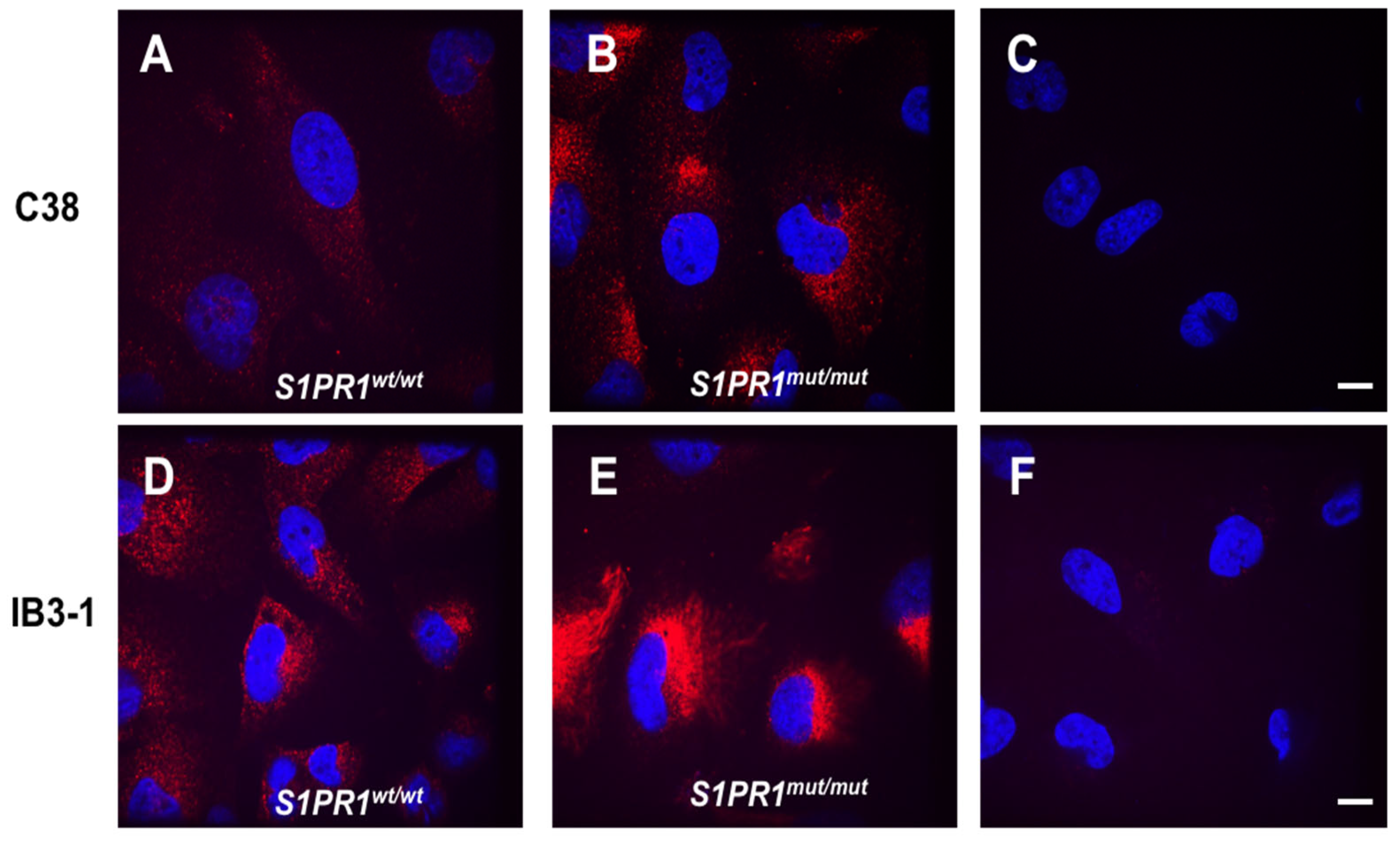
The engineered cell lines generated in this work serve as models for assessing the role of the S1PR1 protein in the pathophysiology. Clone 8 (IB3-1 S1PR1mut/mut) and clone 23 (C38 S1PR1mut/mut) were chosen as representatives of all isolated clones. The immunolocalization of S1PR1 in our cell lines reveals different patterns (Figure 2). In C38 S1PR1wt/wt cells, confocal microscopy shows that S1PR1 appears to be uniformly expressed throughout the cell (Figure 2A). The mutation in S1PR1 of C38 S1PR1mut/mut cells results in a shortened protein (159 aa deletion) and other small peptides being generated by each clone allele that are still recognized by S1PR1 antibody (Supplementary Alignments S1A and B). These peptides are primarily localized to the perinuclear cytoplasmic region (Figure 2B) since they are not correctly processed, as their localization significantly differs between wt/wt and S1PR1 mutant cells.
Figure 2.
Localization of S1PR1 and its mutant form in C38 and IB3-1 cell lines. Representative images display immunofluorescence staining with anti-S1PR1 antibody (in red) in C38 S1PR1wt/wt (A), C38 S1PR1mut/mut (B), IB3-1 S1PR1wt/wt (D), and IB3-1 S1PR1mut/mut (E). The nucleus was stained with 4,6-Diamidino-2-phenylindole dihydrochloride (DAPI) (in blue) (A–F). Different localizations were analyzed using confocal microscopy (Perkin Elmer, Waltham, MA, USA). Negative control cells were stained by omitting the primary S1PR1 antibody (C,F). Scale bar: 10 µm.
When analyzing cell lines with mutations in CFTR, we observed that in IB3-1 S1PR1wt/wt cells, S1PR1 is diffuse throughout the cell and also accumulates in a perinuclear location (Figure 2D). The mutation of S1PR1 in IB3-1 generated several peptides of different lengths that mainly accumulate the protein predominantly in the perinuclear cytoplasmic region (Figure 2E). These findings imply that mutations in S1PR1 disrupt protein processing and trafficking, further contributing to cellular stress and pathology in addition to the effects of CFTR mutations.
3.3. Response of IB3-1 and C38 Cell Lines Carrying S1PR1 or Its Mutant to Stimulation with P. aeruginosa Exoproducts
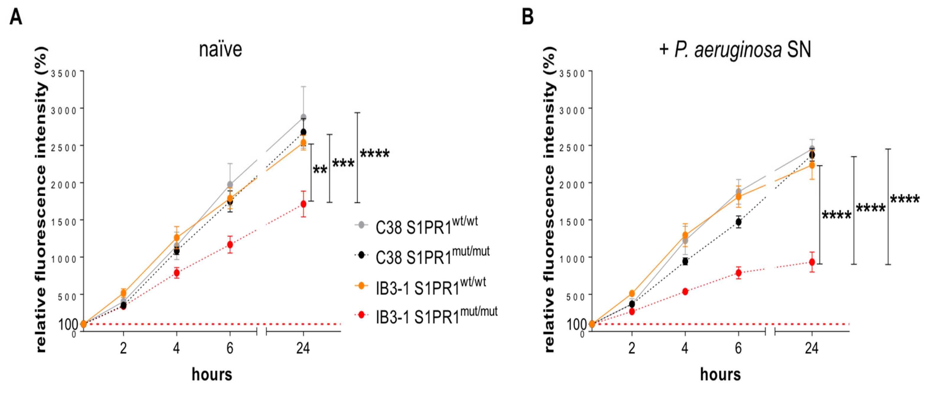
Next, we investigated the role of the S1PR1 protein in both baseline conditions and in the responses induced by P. aeruginosa infection. Specifically, Clone 8 (IB3-1 S1PR1mut/mut) and clone 23 (C38 S1PR1mut/mut) and their isogenic S1PR1wt/wt cell lines were exposed to the exoproducts of a clinical strain of P. aeruginosa known as AA2 []. Our aim was to assess both cell viability, cytotoxicity and cytokine secretion at baseline and following bacterial stimulation. As a control, we used stimulation with the bacterial culture medium, named tryptic soy broth (TSB). Cell viability and cytotoxicity was quantified by measuring metabolic activity with the Alamar Blue assay. First, metabolic activity was significantly lower in naïve IB3-1 S1PR1mut/mut cells compared to their isogenic S1PR1wt/wt counterpart (Figure 3A), consistent with increased baseline cytotoxicity in the CFTR-deficient background. A similar trend was observed in cells exposed to the TSB control (Supplementary Figure S1), indicating a role of S1PR1 in determining cell viability and cytotoxicity under basal conditions. When comparing the two genetic backgrounds, the effect of S1PR1 mutation was evident only in the CFTR-deficient IB3-1 cells, whereas no significant differences were observed between S1PR1mut/mut and S1PR1mut/mut C38 cells. This finding strenghtens the role of CFTR in determining the protective effects of S1PR1 against cellular cytotoxicity and in maintaining cell viability.
Figure 3.
Cell viability in IB3-1 and C38 cell lines, both S1PR1mut/mut and S1PR1wt/wt. A time-course analysis of cell viability (2, 4, 6 and 24 h) was performed using the Alamar Blue assay in naïve IB3-1 S1PR1mut/mut, C38 S1PR1mut/mut, IB3-1 S1PR1wt/wt and C38 S1PR1wt/wt (A). Cells have been stimulated with exoproducts from P. aeruginosa AA2 strain supernatant (SN) (B). Data, from four independent experiments, are expressed as means ± standard errors of the means (SEM). The red dashed lines indicate the 100%, corresponding to the fluorescence of naïve cells at 20 min. ** p < 0.01, *** p < 0.001, **** p < 0.0001, according with the two-way ANOVA test with the Bonferroni correction for multiple comparisons.
Stimulation with P. aeruginosa exoproducts resulted in a further decrease in cell viability in the IB3-1 S1PR1mut/mut cell line compared to naïve (Supplementary Figure S1). Under these conditions, the IB3-1 S1PR1mut/mut cell line exhibited a significantly increased difference compared to S1PR1wt/wt and other cell lines, including C38 S1PR1wt/wt and C38- S1PR1mut/mut (Figure 3B). No significant differences were observed in IB3-1 S1PR1wt/wt or in C38 cells, either S1PR1mut/mut or S1PR1wt/wt. These findings indicate that S1PR1 mutation increases baseline cytotoxicity in the CFTR-deficient genetic background, and that P. aeruginosa exoproducts further exacerbate this phenotype.
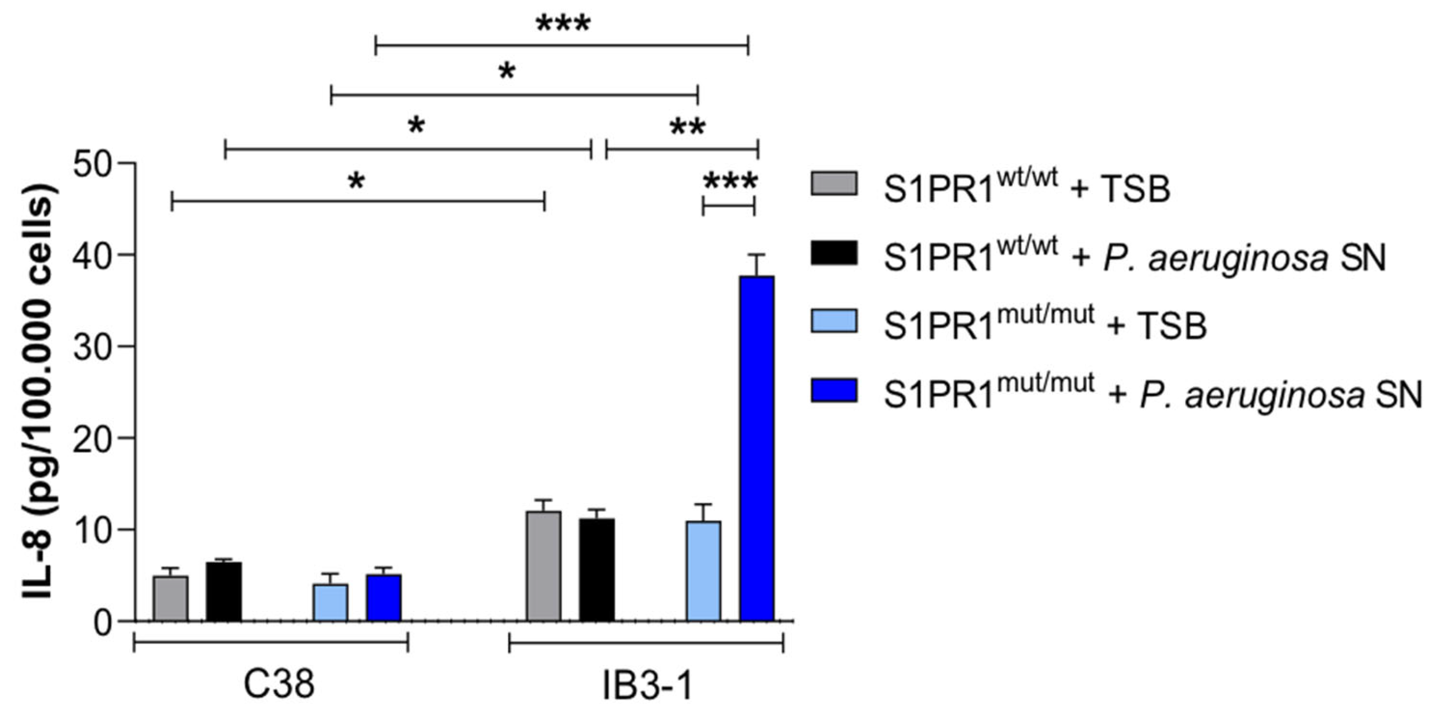
Next, we evaluated the role of S1PR1 in modulating cytokine release, with a particular focus on IL-8, a cytokine responsible for recruiting neutrophils during lung infections. To do this, we stimulated the cell lines by adding the exoproducts of the P. aeruginosa strain AA2 into the cell culture medium. As a control, we subjected the same cell lines to a similar treatment, but added only the bacterial culture medium. Following a 24-h incubation period, we observed distinct IL-8 cytokine secretion profiles using the ELISA assay. In the CFTR-deficient background, S1PR1 mutation was associated with higher IL-8 levels upon exoproduct stimulation in the IB3-1 S1PR1mut/mut cell line, while C38 cells remained unaffected (Figure 4).
Figure 4.
IL-8 release in IB3-1 and C38 cell lines, both S1PR1mut/mut and S1PR1wt/wt. IL-8 release was assessed by ELISA after stimulation with the exoproducts of the P. aeruginosa strain AA2 added to the cell culture medium. Data, from four independent experiments, are expressed as mean ± standard error of the mean (SEM). * p ≤ 0.05, ** p < 0.01, *** p < 0.001, according with the one-way ANOVA test with the Šidák correction for multiple comparisons.
3.4. Protein Expression of S1PR1 in Lung Tissues of pwCF and Non-CF Individuals
To translate our findings to humans, we verified the presence of S1PR1 in lung tissues obtained from pwCF in comparison to non-CF individuals. Specifically, for this study, we selected 5 pwCF, all of whom were homozygous for the F508del mutation in CFTR, with one exception where the subject was heterozygous. The F508del is a processing mutation that removes a single amino acid from the CFTR protein, leading to mistrafficking and incorrect folding. The R1162X is a nonsense mutation containing an early stop signal that causes the production of the CFTR protein to stop prematurely. These individuals represented severe clinical conditions in both cases. They were compared with non-CF individuals (Table 2), all of whom had undergone transplantation and were in severely compromised health conditions for non-CF pathologies.
Table 2.
List of pwCF and non-CF individuals, including CFTR genotype, analyzed in the study.
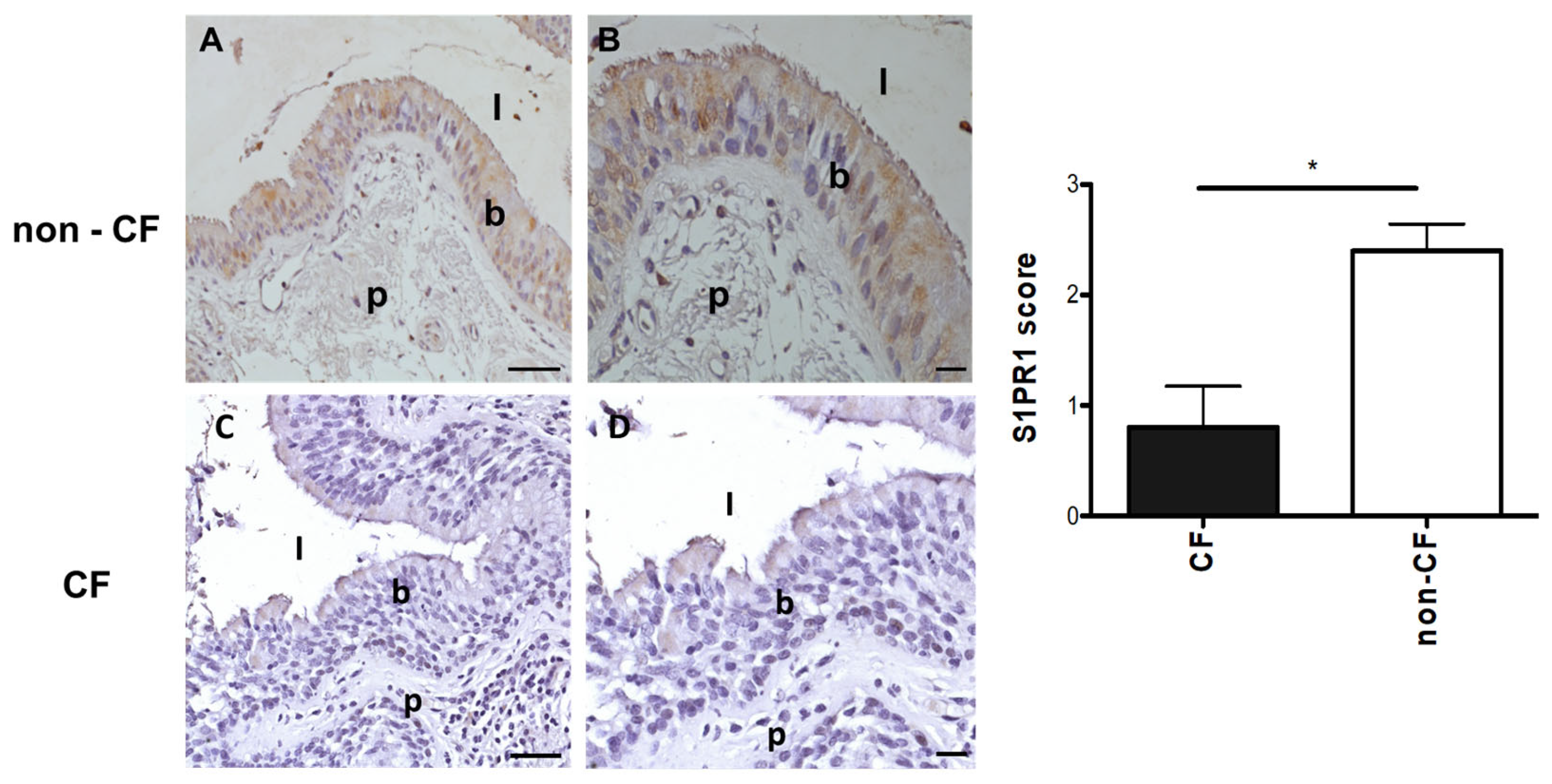
In the tissues, we evaluated the level and localization of the S1PR1 protein through immunohistochemistry. Our immunohistochemistry study successfully identified S1PR1 protein expression in the alveolar epithelium, lung vascular endothelium, and bronchial epithelial cells (Figure 5). However, we observed that signals for S1PR1 protein were either absent or significantly reduced in the lungs of CF individuals in comparison to non-CF ones.
Figure 5.
Staining for S1PR1 in human bronchial tissues. Immunohistochemical analysis of S1PR1 was conducted in human bronchial tissues obtained from non-CF individuals (n = 5) and pwCF (n = 5). Representative images show positive staining with the anti-S1PR1 antibody in non-CF airways, as seen in the individual BE67 ((A), 20×; (B), 40×), who has a wild-type CFTR. In contrast, the staining is negative in BE79, homozygous for F508del mutation ((C), 20×; (D), 40×). Data in the graph represent the score assigned to the presence of S1PR1 in the lung sections from CF (n = 5) and non-CF individuals (n = 5) and are expressed as means ± standard errors of the means (SEM). * p ≤ 0.05, two-tailed paired samples t-test. (A,C): scale bar: 50 um; (B,D): scale bar: 25 um. l: lumen; b: bronchial cells; p: parenchyma.
4. Discussion
CF is characterized by considerable phenotypic variability, even among individuals carrying the same CFTR mutations, and numerous studies have demonstrated that this heterogeneity is partly attributable to modifier genes. Genes such as TGFB1 (involved in inflammation and fibrosis regulation), IL8 (cytokine signaling), EDNRA (vascular tone and remodeling), EHF and APIP (oxidative stress response), and MUC5B and SLC9A3 (airway mucus and ion transport) have been associated with variations in lung function, infection susceptibility, and inflammation in pwCF [,,]. These genetic factors highlight the role of the host background in modulating epithelial stress responses and inflammatory signaling. Within this framework, S1PR1 emerges as a novel candidate modifier gene potentially influencing the pulmonary phenotype in CF. S1PR1 mediates several physiological processes and plays a central role in immune and vascular regulation. S1PR1 is ubiquitous and is expressed in nearly every cell line [], but it is especially abundant and extensively studied in immune cells and endothelial cells, while its role in epithelial cells remains unclear. S1PR1’s broad distribution underlies its pleiotropic effects across various organs [,,]. At the peripheral level, immune and vascular cells carry out multiple functions such as actin remodeling, chemotaxis, lymphocyte egress, vascular integrity, organogenesis, including angiogenesis, cell growth and proliferation, and immune response []. However, the role of S1PR1 in the context of infection and respiratory diseases, especially CF, has not been fully elucidated.
This study aimed to specifically investigate the role of S1PR1 in the context of CFTR deficiency and bacterial challenge. Thanks to gene editing based on CRISPR/Cas9 technology and the use of lentiviral vectors, we engineered the two human bronchial epithelial IB3-1 and C38 cell lines and successfully introduced mutations in S1PR1. The S1PR1 gene consists of two exons, with only exon 2 being protein-coding, thus making it the selected target for CRISPR/Cas9 guides. As a result, we generated a CFTR-mutant cell line with mutations in S1PR1 (IB3-1 S1PR1mut/mut) and a wild-type CFTR cell line also carrying S1PR1 mutations (C38 S1PR1mut/mut). This pairwise setup enabled us to evaluate the role of S1PR1 in the presence or absence of CFTR dysfunction. Matched controls were also generated via transduction with an empty lentiviral vector to obtain S1PR1wt/wt cells.
The immunofluorescence staining indicates that S1PR1 in C38 cells (wild-type CFTR) is broadly distributed throughout the cell. However, after mutation, S1PR1 is predominantly localized in a perinuclear position, suggesting the presence of a non-functional protein in compartments such as the Golgi apparatus or endoplasmic reticulum. In the case of defective S1PR1, it would likely undergo degradation and be internalized by lysosomes. Supporting this, Chavez et al. have observed that phosphorylation on tyrosine 143 of S1PR1 in endothelial cells leads to its internalization and inhibition of its binding with S1P []. Mutations in S1PR1 at tyrosine 143 internalize the protein, sequestering it in the cytoplasm, with results highly similar to the findings obtained in this work. Notably, in IB3-1 cells (CFTR-mutant), the localization of even wt S1PR1 protein shows partial perinuclear accumulation, and the mutation further intensifies mislocalization. Sequence analysis revealed that mutations of S1PR1 in IB3-1 produced several peptides of varying lengths, which primarily relocate and accumulate in the perinuclear cytoplasmic region. These results suggest that S1PR1 mutations disrupt protein processing and trafficking, leading to additional cellular stress and pathology beyond the effects of CFTR mutations. Our data, using the Alamar Blue assay, showed that S1PR1 mutation in the CFTR-deficient genetic background (IB3-1) reduces metabolic activity under basal conditions, thereby affecting cell viability and increasing cytotoxicity. These effects are further exacerbated upon exposure to P. aeruginosa exoproducts, highlighting the impact on epithelial resilience to infection.
It is known in the literature that the F508del mutation retains the CFTR protein in the endoplasmic reticulum in a glycosylated and immature form, which is rapidly degraded intracellularly. This prevents its localization in the plasma membrane [,]. These phenomena lead to intracellular stress, affecting the maturation of proteins and likely S1PR1 as well. Our study utilizes the IB3-1 human bronchial epithelial cell line derived from a person with CF, specifically deficient for CFTR (F508del/W1282X), along with its corrected counterpart, C38. Theoretically, both cell lines retain the mutated CFTR protein in the endoplasmic reticulum, leading to similar endoplasmatic stress. However, the presence of wt/wt CFTR in C38 cells, unlike in IB3-1 cells, may rescue other cellular stress factors such as elevated oxidative stress [] and abnormal lipid metabolism []. Indeed, it is well known that the inactivity of the channel, as well as bacterial infections, is also responsible for producing a redox imbalance in epithelial cells and alterations in the lipid composition of the plasma membrane. Notably, the sphingolipid-richlipid rafts are organizing platforms where specific proteins involved in the signaling mediated across the plasma membrane are recruited, including S1PR1 []. Consequently, it is plausible that CF cells may experience impaired S1PR1 stability and activity due to these intricate alterations in lipid composition and cellular processes. Future studies could provide a more detailed characterization of the mechanisms through which S1PR1 impacts the pathophysiology of the CF epithelium and host defenses during bacterial infections.
Previous studies have consistently demonstrated that the dysfunction of the S1PR1 receptor signaling system leads to heightened inflammation primarily as a consequence of increased endothelial barrier permeability. Recently, Hao et al. [] reported that S1PR1 is downregulated in the lungs of people with idiopathic pulmonary fibrosis and the endothelial-conditional S1PR1 knockout mouse model exhibited inflammation and fibrosis. The lack of S1PR1 in the vascular endothelium results in a ruptured endothelial barrier, increased inflammation and collagen deposition. Our study has further established a connection between S1PR1 and CFTR: in CFTR-mutant bronchial cells with mutated S1PR1, exposure to P. aeruginosa exoproducts triggered a pronounced IL-8 response, suggesting that S1PR1 influences the host’s inflammatory reactivity to pathogens in a CF-dependent manner. Undoubtedly, the presence or absence of S1PR1 is a variable implicated in inflammation and its associated events, such as the secretion of inflammatory cytokines like IL-8. Simultaneously, the inflammatory pathway of S1PR1 seems to be intricately linked to the mutation in CFTR.
Finally, immunohistochemistry on tissues from non-CF individuals revealed the expression of the S1PR1 protein in bronchial epithelial cells, alveolar pulmonary epithelium, and vascular endothelium, whereas lungs from pwCF exhibited a marked reduction or almost complete absence of signal. This observation is consistent with the fact that the CF tissues analyzed were obtained from explanted lungs of pwCF with advanced disease and severely impaired respiratory function. In such cases, the presence of mutations or regulatory alterations in S1PR1 may have led to markedly reduced protein expression, which in turn could have been associated with more severe clinical manifestations. It is plausible that, in pwCF with milder disease, S1PR1 expression might be comparatively higher, reflecting a less compromised regulatory balance. Future studies including tissues from pwCF with different degrees of disease severity will be instrumental in clarifying the relationship between S1PR1 expression, genetic variation, and CF progression.
Overall, these findings support the concept that S1PR1 acts as a modifier influencing the severity of CF lung disease and shaping the epithelial response to bacterial infection. A deeper understanding of this pathway could provide new perspectives for therapeutic modulation of inflammation and host–pathogen interactions in CF.
Supplementary Materials
The following supporting information can be downloaded at: https://www.mdpi.com/article/10.3390/pathogens14111146/s1, Table S1: PCR primers used for the generation of amplicons for amplicon sequencing; Supplementary Alignments S1; Figure S1: Cell viability after P. aeruginosa stimulation.
Author Contributions
Conceptualization, A.B.; methodology, C.C. (Cristina Cigana), C.C. (Claudia Caslini), A.M., B.A.-F., N.I.L. and L.V.; formal analysis, C.C. (Cristina Cigana) and L.V.; investigation, C.C. (Cristina Cigana), C.C. (Claudia Caslini), A.M., B.A.-F. and L.V.; resources, A.L. and A.B.; data curation, C.C. (Cristina Cigana), L.V. and A.B.; writing—original draft preparation, A.B.; writing—review and editing, C.C. (Cristina Cigana), A.M., L.V., A.L. and A.B.; funding acquisition, A.B. All authors have read and agreed to the published version of the manuscript.
Funding
This research was funded by the Italian CF Research Foundation (FFC number 9/2014 with the contribution of Delegazione FFC di Milano and FFC number 15/2016 with the contribution of Delegazione FFC della Valpolicella, Delegazione FFC di Manciano Grosseto e Famiglia Catalano, Gruppo di Sostegno FFC di Riola Sardo) to A.B.
Institutional Review Board Statement
The use of human bronchi, obtained from pwCF undergoing lung transplant, was approved by the Ethical Committee of the Gaslini Institute (Genova, Italy), in accordance with the guidelines of the Italian Ministry of Health. Each person provided written informed consent to the study using a form that was also approved by the Ethical Committee (approval #13, 24 September 2010).
Informed Consent Statement
Research on the bacterial isolate from the individual with CF has been approved by the responsible physician at the CF center at Hannover Medical School, Germany. The person with CF gave informed consent before the sample collection. Approval for storing of biological materials was obtained by the Hannover Medical School, Germany.
Data Availability Statement
Sequencing data are publicly available at ENA under the project number PRJEB76287 samples ERS20182475 (C38 clone 23) and ERS20182474 (IB3-1 clone 8).
Acknowledgments
We thank B. Tummler (Klinische Forschergruppe, Medizinische Hochschule, Hannover, Germany) for supplying the P. aeruginosa AA2 clinical strain. We thank L.L.V. Galietta (Gaslini Institute (Genova, Italy); currently at TIGEM, Pozzuoli, Napoli) for supplying the human bronchi, obtained from patients undergoing lung transplants.
Conflicts of Interest
The authors declare no conflicts of interest. The funders had no role in the design of the study; in the collection, analyses, or interpretation of data; in the writing of the manuscript; or in the decision to publish the results.
Abbreviations
The following abbreviations are used in this manuscript:
| BSA | bovine serum albumin |
| CF | cystic fibrosis |
| CFTR | cystic fibrosis transmembrane conductance regulator |
| DAPI | 4,6-Diamidino-2-phenylindole dihydrochloride |
| IL-8 | interleukin-8 |
| MOI | multiplicity of infection |
| OD | optical density |
| O/N | overnight |
| PBS | phosphate-buffered saline |
| PFA | paraformaldehyde |
| pwCF | people with CF |
| S1PR1 | sphingosine 1-phosphate receptor 1 |
| SEM | standard errors of the means |
| sgRNA | single guide RNA |
| SN | supernatant |
| TSB | tryptic soy broth |
References
- Shteinberg, M.; Haq, I.J.; Polineni, D.; Davies, J.C. Cystic fibrosis. Lancet 2021, 397, 2195–2211. [Google Scholar] [CrossRef] [PubMed]
- Lund-Palau, H.; Turnbull, A.R.; Bush, A.; Bardin, E.; Cameron, L.; Soren, O.; Wierre-Gore, N.; Alton, E.W.; Bundy, J.G.; Connett, G.; et al. Pseudomonas aeruginosa infection in cystic fibrosis: Pathophysiological mechanisms and therapeutic approaches. Expert Rev. Respir. Med. 2016, 10, 685–697. [Google Scholar] [CrossRef]
- Gibson, R.L.; Burns, J.L.; Ramsey, B.W. Pathophysiology and management of pulmonary infections in cystic fibrosis. Am. J. Respir. Crit. Care Med. 2003, 168, 918–951. [Google Scholar] [CrossRef] [PubMed]
- Allen, L.; Allen, L.; Carr, S.B.; Davies, G.; Downey, D.; Egan, M.; Forton, J.T.; Gray, R.; Haworth, C.; Horsley, A.; et al. Future therapies for cystic fibrosis. Nat. Commun. 2023, 14, 693. [Google Scholar] [CrossRef]
- Cutting, G. Cystic fibrosis genetics: From molecular understanding to clinical application. Nat. Rev. Genet. 2015, 16, 45–56. [Google Scholar] [CrossRef]
- Cutting, G.R. Modifier genes in Mendelian disorders: The example of cystic fibrosis. Ann. N. Y. Acad. Sci. 2010, 1214, 57–69. [Google Scholar] [CrossRef] [PubMed]
- Knowles, M.; Drumm, M. The influence of genetics on cystic fibrosis phenotypes. Cold Spring Harb. Perspect. Med. 2012, 2, a009548. [Google Scholar] [CrossRef]
- Lore, N.I.; Iraqi, F.A.; Bragonzi, A. Host genetic diversity influences the severity of Pseudomonas aeruginosa pneumonia in the Collaborative Cross mice. BMC Genet. 2015, 16, 106. [Google Scholar] [CrossRef]
- Lore, N.I.; Sipione, B.; He, G.; Strug, L.J.; Atamni, H.J.; Dorman, A.; Mott, R.; Iraqi, F.A.; Bragonzi, A. Collaborative Cross Mice Yield Genetic Modifiers for Pseudomonas aeruginosa Infection in Human Lung Disease. mBio 2020, 11. [Google Scholar] [CrossRef]
- Bryan, A.M.; Del Poeta, M. Sphingosine-1-phosphate receptors and innate immunity. Cell. Microbiol. 2018, 20, e12836. [Google Scholar] [CrossRef] [PubMed]
- Rosen, H.; Gonzalez-Cabrera, P.J.; Sanna, M.G.; Brown, S. Sphingosine 1-phosphate receptor signaling. Annu. Rev. Biochem. 2009, 78, 743–768. [Google Scholar] [CrossRef]
- O’Sullivan, C.; Dev, K.K. The structure and function of the S1P1 receptor. Trends Pharmacol. Sci. 2013, 34, 401–412. [Google Scholar] [CrossRef]
- Aoki, M.; Aoki, H.; Ramanathan, R.; Hait, N.C.; Takabe, K. Sphingosine-1-Phosphate Signaling in Immune Cells and Inflammation: Roles and Therapeutic Potential. Mediat. Inflamm. 2016, 2016, 8606878. [Google Scholar] [CrossRef]
- Allende, M.L.; Zhou, D.; Kalkofen, D.N.; Benhamed, S.; Tuymetova, G.; Borowski, C.; Bendelac, A.; Proia, R.L. S1P1 receptor expression regulates emergence of NKT cells in peripheral tissues. FASEB J. 2008, 22, 307–315. [Google Scholar] [CrossRef]
- Obinata, H.; Gutkind, S.; Stitham, J.; Okuno, T.; Yokomizo, T.; Hwa, J.; Hla, T. Individual variation of human S1P(1) coding sequence leads to heterogeneity in receptor function and drug interactions. J. Lipid Res. 2014, 55, 2665–2675. [Google Scholar] [CrossRef] [PubMed]
- Sun, X.; Ma, S.F.; Wade, M.S.; Flores, C.; Pino-Yanes, M.; Moitra, J.; Ober, C.; Kittles, R.; Husain, A.N.; Ford, J.G.; et al. Functional variants of the sphingosine-1-phosphate receptor 1 gene associate with asthma susceptibility. J. Allergy Clin. Immunol. 2010, 126, 241–249, 249 e241-243. [Google Scholar] [CrossRef] [PubMed]
- Hao, M.; Fu, R.; Tai, J.; Tian, Z.; Yuan, X.; Chen, Y.; Wang, M.; Jiang, H.; Ji, M.; Lai, F.; et al. S1PR1 serves as a viable drug target against pulmonary fibrosis by increasing the integrity of the endothelial barrier of the lung. Acta Pharm. Sin. B 2023, 13, 1110–1127. [Google Scholar] [CrossRef] [PubMed]
- Maceyka, M.; Spiegel, S. Sphingolipid metabolites in inflammatory disease. Nature 2014, 510, 58–67. [Google Scholar] [CrossRef]
- Malik, F.A.; Meissner, A.; Semenkov, I.; Molinski, S.; Pasyk, S.; Ahmadi, S.; Bui, H.H.; Bear, C.E.; Lidington, D.; Bolz, S.S. Sphingosine-1-Phosphate Is a Novel Regulator of Cystic Fibrosis Transmembrane Conductance Regulator (CFTR) Activity. PLoS ONE 2015, 10, e0130313. [Google Scholar] [CrossRef]
- Bragonzi, A.; Paroni, M.; Nonis, A.; Cramer, N.; Montanari, S.; Rejman, J.; Di Serio, C.; Doring, G.; Tummler, B. Pseudomonas aeruginosa microevolution during cystic fibrosis lung infection establishes clones with adapted virulence. Am. J. Respir. Crit. Care Med. 2009, 180, 138–145. [Google Scholar] [CrossRef]
- Zeitlin, P.; Lu, L.; Rhim, J.; Cutting, G.; Stetten, G.; Kieffer, K.A.; Craig, R.; Guggino, W.B. A cystic fibrosis bronchial epithelial cell line: Immortalization by adeno-12-SV40 infection. Am. J. Respir. Cell Mol. Biol. 1991, 4, 313–319. [Google Scholar] [CrossRef]
- Sanjana, N.E.; Shalem, O.; Zhang, F. Improved vectors and genome-wide libraries for CRISPR screening. Nat. Methods 2014, 11, 783–784. [Google Scholar] [CrossRef]
- Lorè, N.; Cigana, C.; De Fino, I.; Riva, C.; Juhas, M.; Schwager, S.; Eberl, L.; Bragonzi, A. Cystic Fibrosis-Niche Adaptation of Pseudomonas aeruginosa Reduces Virulence in Multiple Infection Hosts. PLoS ONE 2012, 7, e35648. [Google Scholar] [CrossRef] [PubMed]
- Longhin, E.M.; El Yamani, N.; Runden-Pran, E.; Dusinska, M. The alamar blue assay in the context of safety testing of nanomaterials. Front. Toxicol. 2022, 4, 981701. [Google Scholar] [CrossRef] [PubMed]
- Corvol, H.; Blackman, S.M.; Boelle, P.Y.; Gallins, P.J.; Pace, R.G.; Stonebraker, J.R.; Accurso, F.J.; Clement, A.; Collaco, J.M.; Dang, H.; et al. Genome-wide association meta-analysis identifies five modifier loci of lung disease severity in cystic fibrosis. Nat. Commun. 2015, 6, 8382. [Google Scholar] [CrossRef] [PubMed]
- Zhou, Y.H.; Gallins, P.J.; Pace, R.G.; Dang, H.; Aksit, M.A.; Blue, E.E.; Buckingham, K.J.; Collaco, J.M.; Faino, A.V.; Gordon, W.W.; et al. Genetic Modifiers of Cystic Fibrosis Lung Disease Severity: Whole-Genome Analysis of 7,840 Patients. Am. J. Respir Crit. Care Med. 2023, 207, 1324–1333. [Google Scholar] [CrossRef]
- Tsai, H.C.; Han, M.H. Sphingosine-1-Phosphate (S1P) and S1P Signaling Pathway: Therapeutic Targets in Autoimmunity and Inflammation. Drugs 2016, 76, 1067–1079. [Google Scholar] [CrossRef]
- Gaire, B.P.; Lee, C.H.; Sapkota, A.; Lee, S.Y.; Chun, J.; Cho, H.J.; Nam, T.G.; Choi, J.W. Identification of Sphingosine 1-Phosphate Receptor Subtype 1 (S1P(1)) as a Pathogenic Factor in Transient Focal Cerebral Ischemia. Mol. Neurobiol. 2018, 55, 2320–2332. [Google Scholar] [CrossRef]
- Pan, S.; Mi, Y.; Pally, C.; Beerli, C.; Chen, A.; Guerini, D.; Hinterding, K.; Nuesslein-Hildesheim, B.; Tuntland, T.; Lefebvre, S.; et al. A monoselective sphingosine-1-phosphate receptor-1 agonist prevents allograft rejection in a stringent rat heart transplantation model. Chem. Biol. 2006, 13, 1227–1234. [Google Scholar] [CrossRef]
- Bravo, G.A.; Cedeno, R.R.; Casadevall, M.P.; Ramio-Torrenta, L. Sphingosine-1-Phosphate (S1P) and S1P Signaling Pathway Modulators, from Current Insights to Future Perspectives. Cells 2022, 11, 2058. [Google Scholar] [CrossRef]
- Chavez, A.; Schmidt, T.T.; Yazbeck, P.; Rajput, C.; Desai, B.; Sukriti, S.; Giantsos-Adams, K.; Knezevic, N.; Malik, A.B.; Mehta, D. S1PR1 Tyr143 phosphorylation downregulates endothelial cell surface S1PR1 expression and responsiveness. J. Cell Sci. 2015, 128, 878–887. [Google Scholar] [CrossRef]
- Dalemans, W.; Barbry, P.; Champigny, G.; Jallat, S.; Dott, K.; Dreyer, D.; Crystal, R.G.; Pavirani, A.; Lecocq, J.P.; Lazdunski, M. Altered chloride ion channel kinetics associated with the delta F508 cystic fibrosis mutation. Nature 1991, 354, 526–528. [Google Scholar] [CrossRef] [PubMed]
- Li, C.; Ramjeesingh, M.; Reyes, E.; Jensen, T.; Chang, X.; Rommens, J.M.; Bear, C.E. The cystic fibrosis mutation (delta F508) does not influence the chloride channel activity of CFTR. Nat. Genet. 1993, 3, 311–316. [Google Scholar] [CrossRef] [PubMed]
- Galli, F.; Battistoni, A.; Gambari, R.; Pompella, A.; Bragonzi, A.; Pilolli, F.; Iuliano, L.; Piroddi, M.; Dechecchi, M.C.; Cabrini, G.; et al. Oxidative stress and antioxidant therapy in cystic fibrosis. Biochim. Biophys. Acta 2012, 1822, 690–713. [Google Scholar] [CrossRef] [PubMed]
- Dobi, D.; Loberto, N.; Bassi, R.; Pistocchi, A.; Lunghi, G.; Tamanini, A.; Aureli, M. Cross-talk between CFTR and sphingolipids in cystic fibrosis. FEBS Open Bio 2023, 13, 1601–1614. [Google Scholar] [CrossRef]
- Sonnino, S.; Prinetti, A.; Mauri, L.; Chigorno, V.; Tettamanti, G. Dynamic and structural properties of sphingolipids as driving forces for the formation of membrane domains. Chem. Rev. 2006, 106, 2111–2125. [Google Scholar] [CrossRef]
Disclaimer/Publisher’s Note: The statements, opinions and data contained in all publications are solely those of the individual author(s) and contributor(s) and not of MDPI and/or the editor(s). MDPI and/or the editor(s) disclaim responsibility for any injury to people or property resulting from any ideas, methods, instructions or products referred to in the content. |
© 2025 by the authors. Licensee MDPI, Basel, Switzerland. This article is an open access article distributed under the terms and conditions of the Creative Commons Attribution (CC BY) license (https://creativecommons.org/licenses/by/4.0/).