Article

Whole Genome Sequencing and Comparative Genomics of the Emerging Pathogen Burkholderia pseudomallei Isolated from Two Travel-Related Infections in Hungary

1
Department of National Biosafety Laboratory, National Center for Public Health and Pharmacy, H-1097 Budapest, Hungary
2
School of PhD Studies, Semmelweis University, H-1097 Budapest, Hungary
3
Department of Bacteriology, National Center for Public Health and Pharmacy, H-1097 Budapest, Hungary
4
Microbiology Laboratory, Aladar Petz University Teaching Hospital, H-1097 Győr, Hungary
5
Central Department of Anesthesiology and Intensive Care, Aladar Petz University Teaching Hospital, H-1097 Győr, Hungary
6
Department of Otorhinolaryngology, Head and Neck Surgery, North-Pest Central Hospital—Military Hospital, H-1097 Budapest, Hungary
7
Department of Clinical Microbiology, North-Pest Central Hospital—Military Hospital, H-1097 Budapest, Hungary
8
Department of Internal Medicine, North-Pest Central Hospital—Military Hospital, H-1097 Budapest, Hungary
9
Communicable Diseases and Immunization Unit, National Center for Public Health and Pharmacy, H-1097 Budapest, Hungary
10
Institute of Medical Microbiology, Faculty of Medicine, Semmelweis University, H-1097 Budapest, Hungary
*
Authors to whom correspondence should be addressed.
Pathogens 2025, 14(11), 1108; https://doi.org/10.3390/pathogens14111108
Submission received: 19 September 2025 / Revised: 20 October 2025 / Accepted: 23 October 2025 / Published: 31 October 2025
(This article belongs to the Special Issue Updates on Human Melioidosis)

Abstract

Background: Burkholderia pseudomallei, the causative agent of melioidosis, is a neglected tropical pathogen that has been increasingly encountered in Europe through travel-related infections. Clinical manifestations range from localized abscesses to life-threatening sepsis, posing diagnostic challenges in non-endemic regions. Methods: We report two travel-associated melioidosis cases confirmed in Hungary between 2008 and 2024. Whole-genome sequencing (WGS), multilocus sequence typing (MLST), and core-genome MLST (cgMLST) were performed for molecular characterization. In parallel, a systematic review of travel-related melioidosis cases reported in Europe (1980–2025) was conducted according to PRISMA 2020 guidelines. Data were retrieved from PubMed, Scopus, Google Scholar, and the PubMLST database. Results: In silico MLST identified two distinct sequence types (STs): a novel ST1643, and ST1051, previously reported in Asia and Australia. Both isolates clustered within the Asian clade, confirming an imported origin. Virulence profiling revealed major determinants, including the Yersinia-like fimbriae (YLF) cluster, fhaB3, and ITS type C. The ST1643 isolate carried the bimABm variant and multiple resistance genes (blaOXA-57, blaPenI, and amrAB efflux system), while ST1051 harbored blaOXA-59. The literature review identified 82 studies encompassing 195 European cases, most originating from Southeast Asia, with pneumonia, followed by septic form and abscess as the predominant presentation. We found only eight neuromelioidosis cases in Europe. Conclusions: This study represents the first report of neuromelioidosis in Hungary, and the first global description of ST1643. Combined genomic and epidemiological data highlight the need for improved clinical awareness, genomic surveillance, and diagnostic preparedness in non-endemic regions, as global travel and climate change expand the distribution of melioidosis.

1. Introduction

Melioidosis (Whitmore’s disease, soil fever) is an emerging infectious disease caused by the environmental bacterium Burkholderia pseudomallei, which was first isolated in Burma in 1912 [1,2,3]. It is classified as a risk group 3 (RG3) pathogen and has been designated as a Class B bioterrorism agent by the Centers for Disease Control and Prevention (CDC) [4]. In 2015, the global prevalence of melioidosis was estimated at approximately 165,000 cases, resulting 89,000 deaths [5,6,7]. B. pseudomallei is endemic between the latitudes of 20 degrees N and 20 degrees S in Southeast and South Asia, and northern Australia [2,7,8]. The incidence of affected fields has been observed to steadily expand, with 46 countries currently considered endemic. Furthermore, cases have been observed in an additional 33 countries by 2024 [2]. The highest prevalence is found in ‘hyperendemic zones’ in Southeast Asia and northern Australia, with the peak being during the rainy season [9]. Global transportation systems, particularly transport of contaminated products, may facilitate bacterial spread [10]. The Burkholderia genus is known for its extreme surviving ability and broad resistance to many disinfectants. It has been widely reported that B. pseudomallei can survive for up to two decades in distilled water or in different beverages [11,12,13,14,15]. Melioidosis is recognized as a zoonosis, as a wide range of animals are susceptible to the bacterium, including iguanas, non-human primates, dogs, crocodiles, goats, and sheep, although animals, as well as humans, are usually dead-end hosts [8,16,17,18]. The most common acquisition of human infections is via exposure to various contaminated environmental materials by inhalation, ingestion, or direct contact with abraded skin [9]. More than 99% of the fatal cases occur predominantly among agricultural workers in the endemic areas [16]. Person-to-person transmission, including sexual transmission or vertical transmission through breastfeeding, is rare but possible [19,20,21,22]. The typical incubation period varies from 1 to 21 days with 4 days [23,24]. The infection can potentially remain latent for up to 62 years, or reactivate after months or years, as the bacteria can evade the host’s immune system [25].
The infection can be localized or disseminated, and the most frequent type of melioidosis is pneumonia, but sepsis, multiple abscesses, and other forms such as wound infections, genitourinary infections, enteric infection, even neurological forms can also occur [21,26,27,28,29,30,31]. Differentiation from the clinical presentation of some malignant diseases, autoimmune diseases, tuberculosis, malaria, rickettsiosis, leptospirosis, plague, dengue, or staphylococcal bacteraemia may pose a considerable challenge [32]. Reinfections are not uncommon, as there is no cross-protection between different genotypes. Individual susceptibility to melioidosis is associated with several predisposing health conditions, including, but not limited to, diabetes mellitus, cirrhosis, chronic renal disease, cancer, and alcoholism [33,34]. The mortality rate of melioidosis usually varies from 14 to 50%, depending on the condition of the patient, and the lack of prompt and appropriate therapy and its fulminant septic form is still nearly 100% fatal [21,28,29,30,34,35,36].
B. pseudomallei possesses intrinsic resistance to several commonly used antibiotics, including penicillin, first- and second-generation cephalosporin, and macrolide. Consequently, intensive treatment of melioidosis typically relies on ceftazidime or meropenem. Although acquired resistance to these frontline agents remains relatively rare, it can arise through alterations in penicillin-binding proteins, increased β-lactamase production, or modifications to efflux pump activity. Globally, resistance rates vary, with higher prevalence reported for tetracyclines and ciprofloxacin, underscoring the importance of early detection and careful antibiotic selection to ensure effective therapy [37]. Currently, the first-choice therapy is initially the IV administration of ceftazidime or meropenem for 10–14 days, followed by trimethoprim/sulfamethoxazole per-os (or amoxicillin–clavulanate or doxycycline as alternatives) for 3–6 months [38]. The patient could recover without any residual symptoms, but fatal outcomes or relapses can occur despite the adequate therapy [39]. Currently, no effective vaccines are available [40].
Regarding microbiological diagnosis, cultivation of the organism is the gold standard, but it has a low sensitivity, and hence, a low negative predictive value. Consequently, molecular diagnostic methods, including PCR-based, CRISPR-Cas-based or whole genomic-based methods slowly become first line diagnostic methods; however, standardization is still ongoing [41,42].
B. pseudomallei possesses one of the largest bacterial genomes (~7.2 Mb), distributed across two chromosomes. It is heavily armed with virulence factors that enable it to survive in extreme environments and cause a wide range of clinical disease presentations [43]. The prognosis of the infection may relate to the presence or absence of the different virulence factors, including capsule, pili, flagella, lipopolysaccharide (LPS), quorum-sensing members, two types of Type 3 secretory systems (T3SS), Type 6 secretory systems (T6SS) and exotoxins [44,45]. Toxin A (ToxA) is a part of T3SS and plays a significant role in the ability to infect and damage host tissues. Moreover, ToxA can enter the host cell and interfere with key cellular processes like cell cycle regulation, actin cytoskeleton dynamics, and apoptosis, often leading to cell death and tissue damage [44,45]. A key virulence factor, the putative autotransporter Burkholderia intracellular motility A (BimA), enables B. pseudomallei and B. mallei to move via actin-based motility. This protein also aids bacterial spread and shields them from autophagy [45,46]. Understanding BimA and its role in B. pseudomallei infection is crucial, because it could help identify potential therapeutic targets. By inhibiting BimA, it might be possible to reduce the ability of bacteria to spread in the host, potentially reducing the severity of melioidosis [46].
B. pseudomallei belongs to the Type II O-polysaccharide and has three different LPS types: namely, A, B, and B2. The LPS gene can change the phenotype without changing the genotype [47]. LPS type A is commonly found in Asia, LPS type B in Australia and Asia, and LPS type B2 in Australia, as well [48]. The internal transcribed spacer (ITS) typing differentiates B. pseudomallei strains based on ITS region variation, providing useful information for epidemiological studies and tracking sources of infections. In the case of B. pseudomallei, ITS types are often classified into different categories based on sequence analysis. ITS type A is typically found in isolates from Southeast Asia, especially from Thailand and Malaysia, while ITS type B is often associated with isolates from Australia. ITS type C is frequently observed in isolates from India and other parts of Asia [45]. B. pseudomallei possesses three filamentous hemagglutinin genes (fhaB), of which the fhaB3 variant has been associated with positive blood cultures and is negatively correlated with localized skin infections, mainly without sepsis [44,45]. Yersinia-like fimbriae (YLF) and Burkholderia thailandensis-like flagellum and chemotaxis (BTFC) gene clusters are also known to have distinct geographic distributions. The YLF gene cluster has been associated more with clinical isolates than with environmental ones [39,44,45].
While cases among returning travelers are frequent worldwide, B. pseudomallei epidemiology in Europe remains poorly understood and likely underreported [35,49]. In Europe, melioidosis is currently not listed as a communicable disease threat, according to the Commission Implementing Decision (EU) 2018/945 of 22 June 2018. Climate change is expected to influence the spread of infectious diseases in Europe by creating environmental conditions that favor bacterial growth and establishment. Understanding B. pseudomallei, especially its emergence in Europe, highlights the need for increased surveillance and innovative strategies to manage its threats. The pathogenicity of B. pseudomallei, alongside its resilience against common antibiotics, calls for urgent attention, particularly given its historical implications as a potential biological threat, as highlighted by its notorious use during World War II [45,50]. Therefore, establishing robust preventative measures and fostering interdisciplinary research efforts will be crucial in mitigating the risk that B. pseudomallei poses to human and animal health in Europe and beyond. However, until now, autochthonic cases in connection with B. pseudomallei were not confirmed in Europe [51,52].

2. Materials and Methods

2.1. B. pseudomallei Isolates

In Hungary, between 2008 and 2024, the National Reference Laboratory for Highly Pathogenic Bacteria at the National Center for Public Health and Pharmacy tested different types of clinical specimens from 100 patients with travel anamnesis from well-known endemic areas. Moreover, 3 bacterial isolates were submitted to the laboratory for confirmation, from which 2 were confirmed as B. pseudomallei, and 1 was Pseudomonas aeruginosa. All procedures with living cultures were performed under BSL-3 conditions at the National Biosafety Laboratory, National Center for Public Health and Pharmacy (Table 1). The bacterial strain stocks were stored at −80 °C in cryoprotective media for further analysis. For whole genomic sequencing, the strains were grown on a Luria–Bertani (LB) agar plate for 24 h at 37 °C ambient air. The purity of the strains was checked by using Matrix-Assisted Laser Desorption/Ionization Time-of-Flight, MALDI-TOF MS (Bruker Daltonics Inc., Bremen, Germany) after a trifluoroacetic acid (TFA) inactivation method, as described previously [53].

2.2. Molecular Methods

DNA isolation was performed from the isolates by using the QIAamp DNA Mini Kit (Qiagen, Hilden, Germany) according to the manufacturer instructions. Multiple qPCRs were performed, targeted to the orf2 of the Type III Secretion System (T3SS) [54]. After a quality check by using a Qubit Flex instrument (Thermo Fisher Scientific, Grand Island, NY, USA), the DNA libraries were prepared by using an Illumina DNA Prep kit (Illumina, Inc., San Diego, CA, USA), following the manufacturer instructions without any modifications to the protocol. Pooled libraries were quantified by using the Qubit Flex instrument (Thermo Fisher Scientific, Grand Island, NY, USA). Whole genome sequencing was performed on the Illumina MiSeq platform (Illumina Inc., San Diego, CA, USA), using MiSeq reagent kit version 2, for 300 cycles (Illumina Inc., San Diego), generating 2 × 150 bp paired-end sequencing runs. For bioinformatics analysis, a custom-made pipeline was developed. Briefly, the raw reads were checked with FastQC version 0.12.0, and reads with a Phred score under 20 were discarded [55]. Quality-based filtering and trimming were performed using TRIMMOMATIC version 0.39, and de novo assembly was conducted using Spades version 3.15.3, with the careful option to perform mismatch correction [56,57]. Structural gene prediction and functional annotation was performed with the Bakta version 1.11.3 and RAST server [58,59]. The presence of potential CRISPR-Cas systems was checked using a CRISPRCasFinder version 2.0.3 [60]. Virulence-associated gene analysis and annotation was conducted by using a custom-made database, originally downloaded from the Virulence Factor Database (VFDB) downloaded on 30 September 2025, and the analyses were performed with the MyDBFinder 2.0 and Ridom SeqSphere+ version 8.0 tools (www.ridom.de, accessed on 30 September 2025) [36,61]. Assembled genomes were analyzed for the identification of resistance genes using the Comprehensive Antibiotic Resistance Database (CARD) and the ResFinder database. Only resistance genes exhibiting a coverage of >80% and an identity (proportion of exact nucleotide matches) of >75% were accepted [62]. Further typing of the two assembled genomes was performed using a Ridom SeqSphere+ [63] Additionally, 410 Burkholderia pseudomallei genomes from the National Center for Biotechnology Information (NCBI) were downloaded (Supplementary Data S1). K96243 was used as a reference genome. The public database for molecular typing (PubMLST) web software was used to determine the in silico MLST sequence types (http://pubmlst.org/bpseudomallei/, accessed on 30 September 2025). The relatedness of B. pseudomallei isolates, based on their MLST profiles, was manually verified using the MLST database (http://eburst.mlst.net/). Concatenated sequences for all sequence types (STs) included in this study were downloaded from the MLST website (http://pubmlst.org/bpseudomallei/, accessed on 30 September 2025). A minimum spanning tree, based on cgMLST, was constructed using RIDOM SeqSphere+ (Münster, Germany) and visualized by iTOL version 7.2 [64]. Furthermore, the PubMLST isolate database (n = 7510) was analyzed and compared with the two isolates under investigation, using GrapeTree version 1.52.0 [65]. Visualization and extraction of the two samples’ related information was performed by using GrapeTree.

2.3. Update on European Melioidosis Cases

The review followed the Preferred Reporting Items for Systematic Reviews and Meta-Analyses (PRISMA) guidelines for systematic literature reviews.

2.3.1. Search Strategy

A search was conducted for the literature review; the search covered 1980–2025 and the literature was obtained from PubMed, Web of Science, PubMLST, and a web-based direct search for five specific search criteria: “Burkholderia pseudomallei in Europe” “melioidosis cases in Europe” and “neuromelioidosis”, and “melioidosis travel related”. Additionally, melioidosis cases were extracted from the pubMLST database [66]. All articles with available online full texts that were published in peer-reviewed journals and country reports until 30 September 2025 were included, with no restrictions for gender, age group, region, or the language in which they were published. The English, German, and French languages were included, but we did not exclude results in other languages either. Citation searching was performed to look for additional articles. The articles were separately screened. Titles and abstracts based on the inclusion/exclusion criteria were checked. Full texts of the articles, or reports, were independently selected according to the eligibility criteria. All articles or reports described clinical data relating to human cases of melioidosis in Europe. Discrepancies in the decision to include the study and data extraction were resolved through consensus and by including a third reviewer.

2.3.2. Eligibility Criteria

Patients of all ages who were diagnosed with melioidosis were screened for eligibility.

2.3.3. Data Extraction and Analysis

Two reviewers extracted data independently, using a standardized form containing study characteristics (author, year of publication) and patient demographics. For each variable, only cases where the detail was explicitly reported as present or absent were included in the denominator, with the number reported as present serving as the numerator. Data on epidemiology (geographic area of presentation, comorbidities), clinical signs and symptoms (neurological findings, etc.), diagnosis (method of diagnosis and specimen type), and clinical outcomes (death, cure with or without residual neurological deficits) were collected. The data were recorded in a Microsoft Excel spreadsheet (Supplementary Data S2).
Nucleotide Sequence Accession Numbers
Sequences have been deposited at the Sequence Read Archive (SRA) under the accession of SUB12019197, BioSample: SAMN26814095 and SAMN26814096.

3. Results

3.1. Whole-Genomic Sequencing and Comparative Genomics Results

The main genomic characteristics of the two Hungarian isolates are summarized in Table 2. Genome sizes were 7.2 Mb (584_2008_OEK) and 7.13 Mb (831_2019_NNK), with GC contents of 67.9% and 68.1%, respectively. Both genomes contained ~7400 predicted coding sequences and 299–300 annotated virulence-associated genes. Differences were noted in the number of tRNA and rRNA genes, but both isolates harbored comparable numbers of non-coding RNAs and other genomic elements.

3.2. In Silico Multilocus Sequence Typing

In silico MLST assigned isolate 584_2008_OEK to a novel sequence type, ST1643, carrying a previously undescribed ndh allele. This new ST has been deposited in the pubMLST database. Isolate 831_2019_NNK belonged to the previously reported ST1051 (Table 3).
As of June 2025, the pubMLST database contained 7510 isolates, of which only 74 originated from Europe and 58 (78.4%) were linked to human disease, including the two isolates from Hungary. The origin of infection remains unknown for 33 cases (44.6%), while 24 cases (32.4%) were attributed to Asia and 7 (9.4%) to Africa, while 1 (1.4%) was attributed to the Caribbean Islands. Data regarding the molecular analysis of South Asian strains remain limited. ST1051 has seven entries in the database, reported from clinical isolates in Vietnam (n = 2) and India (n = 2), and environmental isolates in Australia (n = 3). The isolate 584_2008_OEK was a single ST (ST1643, ID5254). The isolate 831_2019_NNK was assigned to an existing sequence type, ST1051, which has seven records in the pubMLST database (Table 4).

3.3. cgMLST Analysis

Comparative cgMLST was performed with 410 global B. pseudomallei genomes obtained from NCBI. Both Hungarian isolates showed marked genetic divergence from each other (Figure 1). Isolates 584_2008_OEK and 831_2019_NNK were positioned in distinct clades, consistent with their different sequence types.
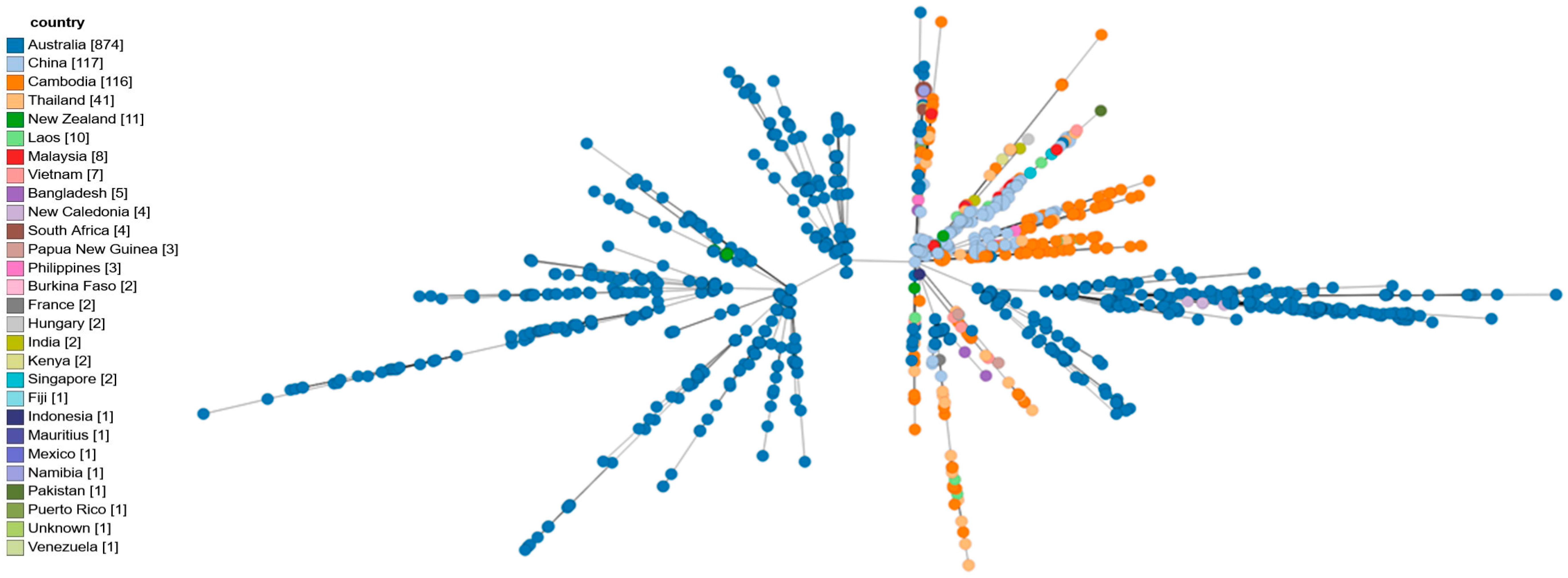
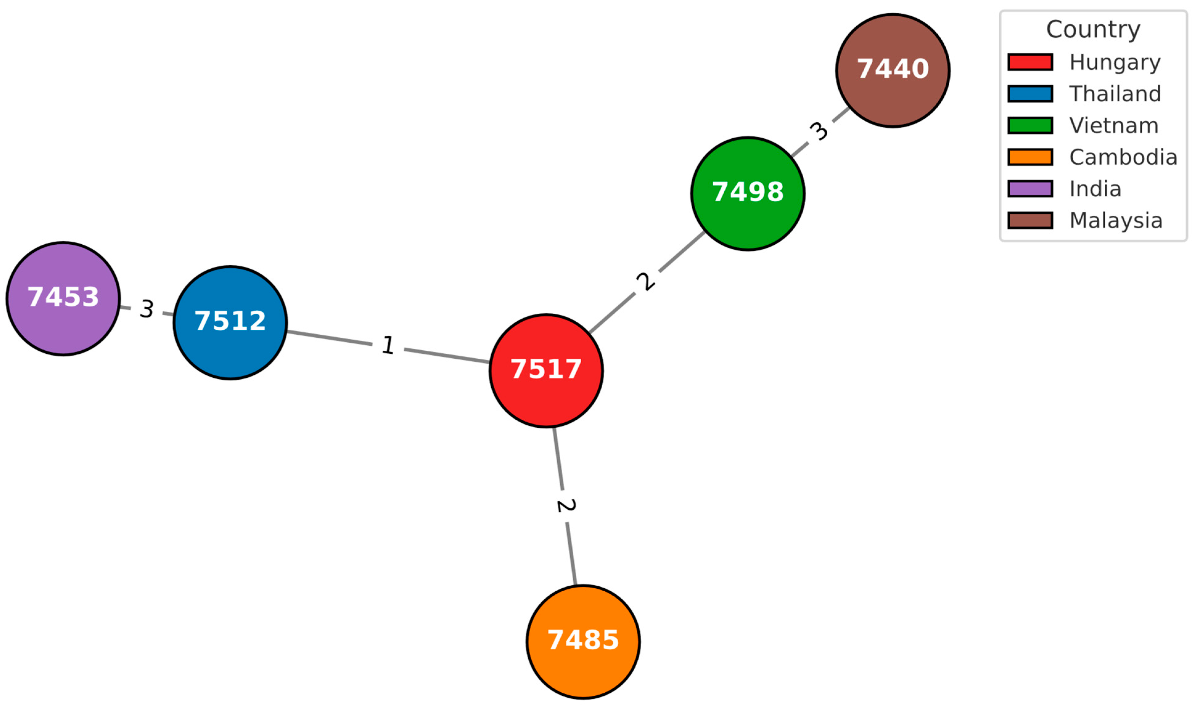
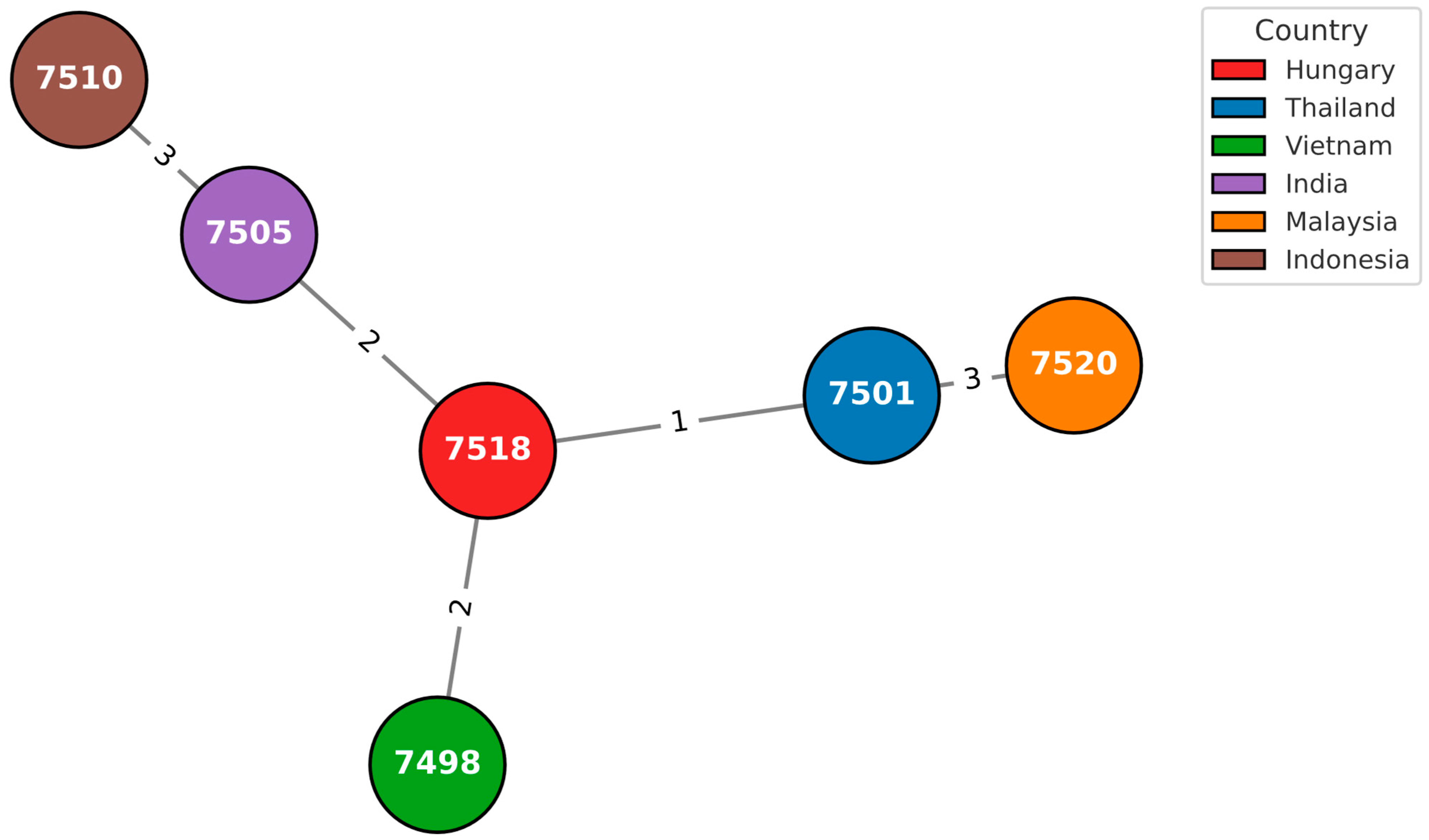
A total of 7510 isolates were analyzed and compared with the two isolates under investigation using the GrapeTree module on PubMLST. Visualization and extraction of sample-specific information were performed with GrapeTree, enabling detailed comparative analysis of the two isolates within the global dataset (Figure 2, Figure 3 and Figure 4).

3.4. Antimicrobial Resistance Genes

Isolate 584_2008_OEK carried blaOXA-57, blaPenI, and the efflux pump operon amrAB, along with the outer membrane protein omp38. In contrast, isolate 831_2019_NNK contained only blaOXA-59, a gene commonly present in B. pseudomallei.

3.5. Virulence-Associated Genes

Draft genomes were analyzed to predict the presence of relevant virulence-associated genes. Both strains were found to harbor the most relevant B. pseudomallei-associated known virulence factors (Supplementary Data S3).
According to the RAST server, the main virulence-associated subsystems were identified (Supplementary Data S4). Isolate 584_2008_OEK contained 299 virulence-associated genes, while the isolate 831_2019_NNK contained 300 virulence-associated genes. The strain 584_2008_OEK was classified as LPS serotype A, while strain 831_2019_NNK was LPS serotype B. Furthermore, our data revealed that the entire wcb operon (BPSL2786 and BPSL2810), which was crucial for LPS biosynthesis, was present in both isolates, without key attenuating mutations [48]. Both strains harbored the Yersinia-like fimbria (YLF) gene cluster, but were negative for the B. thailandensis-like flagellum and chemotaxis (BTFC) gene cluster [45]. We also identified the B. mallei-like actin polymerization gene (bimA), with the bimABm variant in strain 584_2008_OEK and the bimABp variant in strain 831_2019_NNK. Both strains also tested positive for fhaB3, and when exhibiting a 622 bp sequence length corresponding to ITS type C, we observed the presence of the toxA gene, which encodes for the toxin A protein [45]. The isolate 584_2008/OEK contained blaOXA-57 and blaPenI β-lactamases in addition to amrAB genes, encoding an efflux system [42]. The outer membrane protein Omp38 was also detected. In contrast, isolate 831_2019_NNK contained only the blaOXA-59 gene [43].

3.6. Literature Review on Neuromelioidosis Cases in Europe

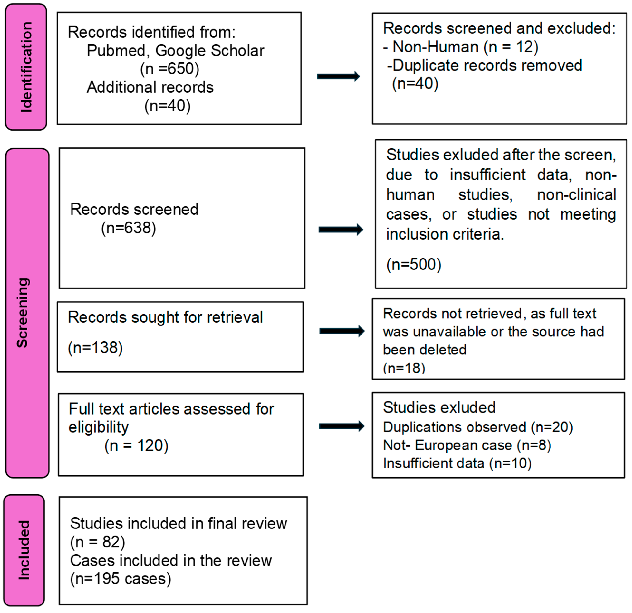
By September 2025, at least around 195 imported melioidosis cases had been documented across Europe between 1980 and 2025 (Figure 5). As of 2019, 184 cases were recorded. After 2019, seven more cases were recorded, until 2024 [8,35,67,68]. Additionally, we found four more cases in 2025 [8,35,69]. A total of 195 cases were recorded between these periods (Table 5) [35,70]. The majority of affected patients were male: (n = 146) 74% of the total cases; median age was 49%. Only one pediatric case was observed. Co-morbidities were poorly captured, according to the absence of surveillance mechanisms in Europe. Respiratory diseases, sepsis, and abscess formation were the most common presenting features. The most cases have been reported in the UK (n = 69), followed by The Netherlands (n = 39), France (n = 24), and Germany (n = 14). The most frequent travel anamnesis was Thailand with 73 cases.
Furthermore, we found only nine neurological cases in Europe, including meningitis, meningoencephalitis, and cerebral abscess; all patients were men between 26 and 58 years old (median age was 50 years), and Thailand was the most frequent travel destination (n = 4), followed by India (n = 1), Cambodia (n = 1), and Sri Lanka (n = 1). One patient visited several countries before the infection, and in one case, the travel anamnesis was not specified in the manuscript (Table 6).
The pulmonary-related melioidosis form was frequent, with a total of 46 cases, followed by the unspecified category and cutaneus/soft tissue infections. The literature discusses only three possible autochthonous occurrences in Europe: one represents Bologna, Italy, where B. pseudomallei was reported in tap water (6 out of 85 specimens) in 2000. However, the confirmation of B. pseudomallei by any specific laboratory methods has never been reported. Moreover, a soil culture was positive for B. pseudomallei in the ‘Jardin des Plantes’ in Paris; this was identified after melioidosis infections, which were connected to a panda imported from China. Only one potential human autochthonous case was reported in Germany, but there is insufficient evidence available at present, because the incubation period of melioidosis can be years, and the patient traveled a lot two years earlier, but detailed information is not available [29].

4. Discussion

This study aimed to provide an analysis of whole genome sequencing data from B. pseudomallei clinical isolates associated with travel-related infections in Hungary, and also to provide a short update on European cases.
Between 2008 and 2024, two B. pseudomallei strains were identified in Hungary from travel-related infections. The first case, in 2008, presented with meningoencephalitis. Neuromelioidosis is rare, with fewer than 100 cases reported worldwide, and it carries a ~25% mortality rate, even with optimal therapy [28,29,32]. Survivors often experience long-term neurological deficits [29,32]. In Europe, delayed recognition may worsen outcomes, as neuromelioidosis can mimic autoimmune neurological syndromes, including an acute disseminated encephalomyelitis (ADEM)-like syndrome, or a Guillain–Barré-like syndrome, or it can present as a spatial sparsii, tomiso. Recognizing the correct etiology is critical because treatments for these conditions are completely different [32]. B. pseudomallei therapy is based on prolonged antibiotic therapy, whereas ADEM and Guillain–Barré are immune-mediated, requiring immunosuppressive or immunomodulatory therapy. Misdiagnosis can be fatal, making prompt recognition and correct therapy crucial [28]. In contrast, a neck abscess is a common manifestation of the disease, and belongs to the one of the most frequent forms worldwide. In the literature, we found only eight neuromelioidosis cases in Europe and according to our knowledge, 584_OEK_2008 was the first sequenced meningoencephalitis case in Europe. The absence of pathognomonic features has been a real public health concern in recent years [2,9,16]. B. pseudomallei exemplifies the complexities of infectious diseases that transcend geographical boundaries. Delayed diagnosis of travel-related melioidosis in non-endemic regions frequently leads to increased morbidity and mortality. The global incidence of melioidosis has sharply increased, reflecting the growing severity of this worldwide health concern [2,9,16].
The high-level genetic diversity among different strains, particularly the different carrying genes involved in extreme adaptability to different environmental niches, makes it possible to estimate the survival ability in the temperate climate and the real burden of B. pseudomallei in Europe [18]. Currently, the environmental risk of B. pseudomallei in Hungary is considered low. Global suitability models show that the bacterium predominantly thrives in tropical and subtropical climates, whereas as currently, the climate in Hungary is temperate–continental, conditions generally did not favor its persistence in soil or water. However, climate change and extreme weather events, such as heavy rainfall or flooding, could increase local environmental suitability, particularly in warmer and wetter areas or regions with standing water, like irrigated fields or floodplains. Although the risk remains low, targeted monitoring is advisable among travelers. This includes raising clinical awareness in hospitals and the reference laboratory to ensure prompt identification of potential melioidosis cases, conducting environmental sampling using established international protocols, and maintaining epidemiological surveillance of both human and animal infections, including imported cases. Updating local risk models with regional climate and soil data can further help to identify areas that may become vulnerable in the future [8,16,51,75]. The literature discusses only three possible autochthonous occurrences in Europe; however, there is currently insufficient evidence to conclusively prove the presence of bacteria [29,76].
In B. pseudomallei, the currently used sequencing technologies and bioinformatics analysis pipelines often struggle to discriminate properly between different strains. This limitation arises from several factors, including MLST sequence type (ST) homoplasy, sequencing bias, and the lack of dedicated analysis pipelines for ‘chimeric’ or ‘genetically engineered’ microorganisms [41,77]. The in silico MLST analysis of the isolate from our neuromelioidosis case revealed a novel ndh (ndh 87) allele, which belonged to a new sequence type, ST 1643. ST 1051 showed significant genetic overlap with Australian strains, despite the presence of Yersinia-like fimbriae (YLF), a feature typically associated with Asian strains. This observation aligns with the concept of homoplasy, a common challenge in genomics-based studies of B. pseudomallei strains. Homoplasy occurs despite the different geographical origin and virulence features, which can determine the geographical location as well. Our results also highlight the problem that there are currently no reliable genotyping methods available for B. pseudomallei, which is obstructing the epidemiological investigations, as well as identification in the case of a potential bioterror event [41,77].
According to our results, we identified almost 300 virulence-associated genes in the genomes, including critical factors that are likely to play an important role in human virulence. Genomic analysis revealed the presence of the Yersinia-like fimbria (YLF) gene cluster in both strains. This cluster, frequently observed in Asian strains and known for its horizontal acquisition, is implicated in host–cell adhesion and biofilm formation, thereby enhancing pathogenicity. In contrast, neither strain possessed the B. thailandensis-like flagellum and chemotaxis (BTFC) gene cluster, a genetic element that is characteristic of Australian isolates. The BTFC cluster is recognized for its role in motility and chemotaxis, which contributes to bacterial adaptation across various environmental niches [44,47]. We also identified the B. mallei-like actin polymerization gene (bimA), with the Bm variant in strain 584_2008_OEK and the Bp variant in strain 831_2019_NNK. The BimA Bm variant is strongly associated with neurological disease, as indicated by several publications [27,28,29,30,60]. BimA plays a critical role in actin-based motility within host cells, facilitating bacterial invasion and dissemination. Furthermore, the BimA Bm is a potential target for CRISPR-Cas-based detection methods in neurologic melioidosis cases. Such cutting-edge methods are particularly important in non-endemic areas, as their limit of detection is significantly lower than the currently available diagnostic approaches [42]. Both strains exhibited an ITS with a 622 bp sequence length, corresponding to ITS type C: a characteristic commonly observed in Asian isolates [41]. Both strains tested positive for fhaB3. FhaB3 is implicated in the adhesion to host cells and tissue invasion, contributing to the strain’s ability to persist in human and animal hosts. This gene is frequently associated with increased virulence, particularly in strains that cause severe infections, such as pneumonia or sepsis. The presence of fhaB3 further suggests the potential for these strains to exhibit enhanced pathogenicity in clinical settings [44,45]. The B. pseudomallei capsule polysaccharide is a critical virulence factor that is essential for immune evasion. Our data revealed that the entire wcb operon (BPSL2786 and BPSL2810) was present in both isolates, with no key mutations identified that could potentially attenuate the strains [48]. The capsule plays a significant role in resisting phagocytosis by host immune cells and protecting against antimicrobial peptides. We observed the presence of the toxA gene, which encodes for the toxin A protein, a known virulence factor in B. pseudomallei. Toxin A is involved in the disruption of host cell processes, particularly by targeting the host cell’s cytoskeleton, which impairs immune responses and promotes bacterial survival. Another key virulence factor identified was the type III secretion system (T3SS), which is associated with the ability to inject bacterial effector proteins directly into host cells, manipulating host cell functions to favor bacterial replication and immune evasion [44,45]
The isolate 584_2008/OEK contained blaOXA-57 and blaPenI β-lactamases, along with amrAB genes, encoding an efflux system. This is clinically relevant, as these genes may contribute to aminoglycoside resistance, a key factor in treatment failure. The outer membrane protein Omp38 was also detected, which is associated with low permeability and resistance to multiple antibiotics [61]. Omp38 is involved in the uptake of essential nutrients and plays a role in the resistance of the bacteria to host immune responses. In contrast, isolate 831_2019_NNK contained only the blaOXA-59 gene, which naturally occurs in B. pseudomallei strains, and is linked to resistance to β-lactam antibiotics. The main characteristics are summarized in Table 7.
Approximately 40% of the reported cases presented with respiratory or pulmonary infections, making this the most common clinical manifestation observed [35,67,70]. This finding aligns with previous studies indicating that pneumonia is one of the predominant forms of melioidosis. Most cases originated from Southeast Asia, particularly Thailand, reflecting the endemic nature of the disease in this region. The mean patient age was around 50 years, and the majority were male, consistent with the demographic profile typically described in the literature. Only one patient in the dataset was younger than 18 years old, representing less than 1% of all reported cases with a known age. This finding confirms that melioidosis predominantly affects adults, with pediatric cases being exceptionally rare among imported infections. The rarity of childhood cases may reflect both lower exposure risk and underdiagnosis in this age group [22]. These results highlight the importance of recognizing respiratory involvement in imported melioidosis and emphasize the need to consider travel history when evaluating patients with pneumonia of unclear origin. Only nine cases in the dataset involved central nervous system (CNS) manifestations, accounting for approximately 2–5% of all reports. These included meningoencephalitis, cerebral melioidosis, intracranial melioidosis, and brain abscesses—representing some of the most severe and uncommon forms of melioidosis. Notably, one additional case initially classified as a respiratory disease also exhibited brain abscesses, suggesting that CNS involvement may be underrecognized when it coexists with pulmonary infection. Clinically, these findings highlight the need for a high index of suspicion in returning travelers presenting with fever, headache, focal neurological deficits, or atypical pneumonia.
Early diagnosis and prolonged, targeted antimicrobial therapy are crucial to prevent fatal outcomes in neurological melioidosis. In addition, MRI findings can play a key role in supporting the diagnosis of neurological melioidosis. The presence of the so-called “tunnel sign”, reflecting the appearance of elongated, tubular abscess tracts within the brain parenchyma, has been described as a characteristic radiologic feature [78]. Recognition of this imaging pattern may assist in differentiating melioidosis from other pyogenic or granulomatous CNS infections and should prompt microbiological confirmation and appropriate antimicrobial therapy.

5. Conclusions

Melioidosis, often referred to as one of the “great mimickers,” presents a significant challenge for laboratories worldwide, especially in non-endemic areas. This is due to the absence of a pathognomonic clinical syndrome and the ability of B. pseudomallei to manifest clinical symptoms that mimic other diseases such as cancer, autoimmune disorders, and tuberculosis [9,32].
There is an urgent need to update diagnostic protocols for tropical or travel-related infections. We strongly recommend that all patients returning from endemic areas who present with fever be tested for melioidosis, in addition to the well-known “classic” tropical pathogens such as malaria, dengue, and rickettsial infections [35]. One of our goals is to raise awareness of potential B. pseudomallei infections in clinicians in Hungary, particularly among patients returning from endemic regions [16].
Today, next-generation sequencing technologies have become the most powerful and accurate tools for identifying and characterizing various pathogens. Epidemiological investigations should be extended to include geographic attribution and the determination of the origin of different infections, including RG3 pathogens [38,41]. Whole genome sequencing, combined with a robust bioinformatics pipeline, can aid in early diagnosis (e.g., bimA, fhaB3, LPS, BTFC, and ITS identification) and provide insights into possible alternative treatments if necessary [44,45]. Furthermore, the ability of B. pseudomallei to form biofilms (which directly affects its reduced cultivation) and its prolonged survival in free-living organisms such as amoebas, along with its extreme adaptability, raises important biosecurity concerns. The potential for B. pseudomallei to adapt to temperate climates remains uncertain, due to the lack of comprehensive studies [5,13,43]. To address this phenomenon, further research is urgently needed.

Supplementary Materials

The following supporting information can be downloaded at: https://www.mdpi.com/article/10.3390/pathogens14111108/s1. Supplementary Data S1: Burkholderia pseudomallei isolates used in this study to perform cgMLST. Supplementary Data S2: Travel related melioidosis cases in Europe between 1980–2025. Supplementary Data S3: Presence of the main Burkholderia-related virulence genes according to VFDB. Supplementary Data S4: Presents the different types of virulence genes between the two isolates.

Author Contributions

J.H.: methodology, review, software, investigation, writing—original draft and review and editing, visualization. Á.T.: software, validation, methodology, writing—review and editing. S.T.: curation, review; B.P.: curation; Z.K.: review and editing; M.K.: microbiological investigation, B.G.: resources; E.B.: case management, microbiological investigation; L.R.: case management, microbiological investigation; Á.R.: case management, microbiological investigation; E.L.: case management, microbiological investigation; T.M.: resources; T.E.: data analysis; N.D.-M.: data analysis; Z.M.: data analysis, epidemiological investigation. All authors have read and agreed to the published version of the manuscript.

Funding

This research received no external funding.

Institutional Review Board Statement

Ethics Committee approval was not required, as the Hungarian legislation on handling personal health information (Law no. 1997. XLVII.) empowers the National Center for Public Health and Pharmacy to analyze data and take necessary measures in the interest of public health. Personal data have been handled in accordance with legal regulations and the Center’s data protection rules.

Informed Consent Statement

Our institution operates as a national reference laboratory. These investigations are carried out as part of mandatory public health surveillance and diagnostic duties, as defined by Hungarian law. Specifically, we act under the provisions of Government Decree No. 18/1998 (VI.3) of the Ministry of Welfare on the Epidemiological Requirements of Certain Public Health Laboratories (“18/1998. (VI.3.) NM rendelet az egyes közegészségügyi-járványügyi feladatok ellátására szolgáló laboratóriumok működéséről”). As a result, these procedures do not constitute a clinical or academic research study involving human participants. Therefore, no informed consent form is required or applicable in this context.

Data Availability Statement

Sequences have been deposited at the GenBank under the accession of SUB12019197, BioSample: SAMN26814095 and SAMN26814096.

Acknowledgments

We are thankful to Mária Takács and Tibor Pál for their constructive feedback on the manuscript. We would like to also thank Zsuzsanna Hegedűsné Richter and Filoména Király for their kind assistance.

Conflicts of Interest

The authors declare no conflicts of interest.

Abbreviations

The following abbreviations are used in this manuscript:
ABCATP-binding cassette transporters
AraCTranscriptional regulator protein
ATPAdenosine triphosphate
BapABacterial adhesion protein
BCCBurkholderia cepacia complex
BPCBurkholderia pseudomallei complex
BimABacterial invasion protein
BipB, BipC, BipDBip proteins are part of the type III secretion system (T3SS)
BopABacterial outer membrane protein involved in adhesion or invasion mechanisms
cgMLSTCore genome multilocus sequence typing
CRISPRsClustered regularly interspaced short palindromic repeats
CheA, CheD, CheW, CheYProteins involved in chemotaxis
DNADeoxyribonucleic acid
ITSInternal transcribed spacer
LPSLipopolysaccharide
MALDI-TOFMatrix-assisted laser desorption/ionization time of flight
MLSTMultilocus sequence typing
NADHNicotinamide adenine dinucleotide (NAD)
OMPOuter membrane proteins
PCRPolymerase chain reaction
qPCRQuantitative PCR
RNARibonucleic acid
SNPSingle nucleotide polymorphism
T3SSType III secretion system
T3SS1Type 3 secretion system- cluster 1
T6SS1/5Type 6 secretion system- cluster 1/cluster 5
VFVirulence factors
VFDBVirulence factor database
WGSWhole genome sequencing
YadABacterial adhesion protein

References

  1. White, N. Melioidosis. Lancet 2003, 361, 1715–1722. [Google Scholar] [CrossRef]
  2. Meumann, E.M.; Limmathurotsakul, D.; Dunachie, S.J.; Wiersinga, W.J.; Currie, B.J. Burkholderia pseudomallei and melioidosis. Nat. Rev. Microbiol. 2024, 22, 155–169. [Google Scholar] [CrossRef]
  3. Gassiep, I.; Armstrong, M.; Norton, R. Human Melioidosis. Clin. Microbiol. Rev. 2020, 33, e00006-19. [Google Scholar] [CrossRef] [PubMed]
  4. Centers for Disease Control and Prevention (U.S.); National Institutes of Health (U.S.). Biosafety in Microbiological and Biomedical Laboratories, 6th ed.; U.S. Department of Health and Human Services: Washington, DC, USA, 2020.
  5. Phillips, E.D.; Garcia, E.C. Garcia Burkholderia pseudomallei. Trends Microbiol. 2023, 32, 105–106. [Google Scholar] [CrossRef] [PubMed]
  6. Benoit, T.J.; Blaney, D.D.; Doker, T.J.; Gee, J.E.; Elrod, M.G.; Rolim, D.B.; Inglis, T.J.J.; Hoffmaster, A.R.; Bower, W.A.; Walke, H.T. A Review of Melioidosis Cases in the Americas. Am. J. Trop. Med. Hyg. 2015, 93, 1134–1139. [Google Scholar] [CrossRef] [PubMed]
  7. Birnie, E.; James, A.; Peters, F.; Olajumoke, M.; Traore, T.; Bertherat, E.; Trinh, T.T.; Naidoo, D.; Steinmetz, I.; Wiersinga, W.J.; et al. Melioidosis in Africa: Time to Raise Awareness and Build Capacity for Its Detection, Diagnosis, and Treatment. Am. J. Trop. Med. Hyg. 2022, 106, 394–397. [Google Scholar] [CrossRef]
  8. Norman, F.F.; Blair, B.M.; Chamorro-Tojeiro, S.; González-Sanz, M.; Chen, L.H. The Evolving Global Epidemiology of Human Melioidosis: A Narrative Review. Pathogens 2024, 13, 926. [Google Scholar] [CrossRef]
  9. Limmathurotsakul, D.; Peacock, S.J. Melioidosis: A clinical overview. Br. Med. Bull. 2011, 99, 125–139. [Google Scholar] [CrossRef]
  10. Gee, J.E.; Bower, W.A.; Kunkel, A.; Petras, J.; Gettings, J.; Bye, M.; Firestone, M.; Elrod, M.G.; Liu, L.; Blaney, D.D.; et al. Multistate Outbreak of Melioidosis Associated with Imported Aromatherapy Spray. N. Engl. J. Med. 2022, 386, 861–868. [Google Scholar] [CrossRef]
  11. Pumpuang, A.; Chantratita, N.; Wikraiphat, C.; Saiprom, N.; Day, N.P.; Peacock, S.J.; Wuthiekanun, V. Survival of Burkholderia pseudomallei in distilled water for 16 years. Trans. R. Soc. Trop. Med. Hyg. 2011, 105, 598–600. [Google Scholar] [CrossRef]
  12. Wuthiekanun, V.; Amornchai, P.; Langla, S.; White, N.J.; Day, N.P.J.; Limmathurotsakul, D. Survival of Burkholderia pseudomallei and Pathogenic Leptospira in Cola, Beer, Energy Drinks, and Sports Drinks. Am. J. Trop. Med. Hyg. 2020, 103, 249–252. [Google Scholar] [CrossRef]
  13. Shams, A.M.; Rose, L.J.; Hodges, L.; Arduino, M.J. Survival of Burkholderia pseudomallei on Environmental Surfaces. Appl. Environ. Microbiol. 2007, 73, 8001–8004. [Google Scholar] [CrossRef]
  14. O’Rourke, A.; Lee, M.D.; Nierman, W.C.; Everroad, R.C.; Dupont, C.L. Genomic and phenotypic characterization of Burkholderia isolates from the potable water system of the International Space Station. PLoS ONE 2020, 15, e0227152. [Google Scholar] [CrossRef] [PubMed]
  15. Sarli, D.A.; Sánchez, L.A.; Delgado, O.D. Burkholderia gladioli MB39 an Antarctic Strain as a Biocontrol Agent. Curr. Microbiol. 2021, 78, 2332–2344. [Google Scholar] [CrossRef] [PubMed]
  16. Savelkoel, J.; Dance, D.A.B.; Currie, B.J.; Limmathurotsakul, D.; Wiersinga, W.J. A call to action: Time to recognise melioidosis as a neglected tropical disease. Lancet Infect. Dis. 2022, 22, e176–e182. [Google Scholar] [CrossRef]
  17. Larsen, J.C.; Johnson, N.H. Pathogenesis of Burkholderia pseudomallei and Burkholderia mallei. Mil. Med. 2009, 174, 647–651. [Google Scholar] [CrossRef]
  18. Birnie, E.; Biemond, J.J.; Wiersinga, W.J. Drivers of melioidosis endemicity: Epidemiological transition, zoonosis, and climate change. Curr. Opin. Infect. Dis. 2022, 35, 196–204. [Google Scholar] [CrossRef]
  19. Abbink, F.C.; Orendi, J.M.; de Beaufort, A.J. Mother-to-Child Transmission of Burkholderia pseudomallei. N. Engl. J. Med. 2001, 344, 1171–1172. [Google Scholar] [CrossRef]
  20. Chang, C.Y.; Lau, N.L.J.; Currie, B.J.; Podin, Y. Disseminated melioidosis in early pregnancy—An unproven cause of foetal loss. BMC Infect. Dis. 2020, 20, 201. [Google Scholar] [CrossRef]
  21. Currie, B.J.; Ward, L.; Cheng, A.C. The Epidemiology and Clinical Spectrum of Melioidosis: 540 Cases from the 20 Year Darwin Prospective Study. PLOS Neglected Trop. Dis. 2010, 4, e900. [Google Scholar] [CrossRef] [PubMed]
  22. Jarrett, O.; Seng, S.; Fitzgerald, D.A. Paediatric melioidosis. Paediatr. Respir. Rev. 2023, 50, 31–37. [Google Scholar] [CrossRef]
  23. Nemhauser, J.; LaRocque, R.; Alvarado-Ramy, F.; Angelo, K.; Ericsson, C.; Gertz, A.; Kozarsky, P.; Ostroff, S.; Ryan, E.; Shlim, D.; et al. Copyright Page. In CDC Yellow Book; Oxford University Press: New York, NY, USA, 2024. [Google Scholar]
  24. Cheng, A.C.; Currie, B.J. Melioidosis: Epidemiology, Pathophysiology, and Management. Clin. Microbiol. Rev. 2005, 18, 383–416. [Google Scholar] [CrossRef]
  25. Ngauy, V.; Lemeshev, Y.; Sadkowski, L.; Crawford, G. Cutaneous Melioidosis in a Man Who Was Taken as a Prisoner of War by the Japanese during World War II. J. Clin. Microbiol. 2005, 43, 970–972. [Google Scholar] [CrossRef] [PubMed]
  26. Zueter, A.; Yean, C.Y.; Abumarzouq, M.; Rahman, Z.A.; Deris, Z.Z.; Harun, A. The epidemiology and clinical spectrum of me-lioidosis in a teaching hospital in a North-Eastern state of Malaysia: A fifteen-year review. BMC Infect. Dis. 2016, 16, 333. [Google Scholar] [CrossRef]
  27. Virk, H.S.; Mukhopadhyay, C.; Wiersinga, W.J. Melioidosis: A Neglected Cause of Community-Acquired Pneumonia. Semin. Respir. Crit. Care Med. 2020, 41, 496–508. [Google Scholar] [CrossRef] [PubMed]
  28. Deuble, M.; Aquilina, C.; Norton, R. Neurologic Melioidosis. Am. J. Trop. Med. Hyg. 2013, 89, 535–539. [Google Scholar] [CrossRef] [PubMed]
  29. Dimitriou, N.G.; Flüh, G.; Zange, S.; Aytulun, A.; Turowski, B.; Hartung, H.-P.; Meuth, S.G.; Gliem, M. Case report: First case of neuromelioidosis in Europe: CNS infection caused by Burkholderia pseudomallei. Front. Neurol. 2022, 13, 899396. [Google Scholar] [CrossRef]
  30. Andersen, E.W.; Mackay, M.T.; Ryan, M.M. Ryan Neurologic Melioidosis: Case Report of a Rare Cause of Acute Flaccid Paralysis. J. Pediatr. 2016, 170, 319–321. [Google Scholar] [CrossRef]
  31. Saravu, K.; Kadavigere, R.; Shastry, A.B.; Pai, R.; Mukhopadhyay, C. Neurologic melioidosis presented as encephalomyelitis and subdural collection in two male labourers in India. J. Infect. Dev. Ctries. 2015, 9, 1289–1293. [Google Scholar] [CrossRef]
  32. Karunanayake, P. Melioidosis: Clinical aspects. Clin. Med. 2022, 22, 6–8. [Google Scholar] [CrossRef]
  33. Guo, R.F.; Wong, F.L.; Perez, M.L. Splenic abscesses in a returning traveler. Infect. Dis. Rep. 2015, 7, 10–12. [Google Scholar] [CrossRef]
  34. Raj, S.; Sistla, S.; Sadanandan, D.M.; Kadhiravan, T.; Rameesh, B.M.S.; Amalnath, D. Clinical Profile and Predictors of Mortality among Patients with Melioidosis. J. Glob. Infect. Dis. 2023, 15, 72–78. [Google Scholar] [CrossRef]
  35. Le Tohic, S.; Montana, M.; Koch, L.; Curti, C.; Vanelle, P. A review of melioidosis cases imported into Europe. Eur. J. Clin. Microbiol. Infect. Dis. 2019, 38, 1395–1408. [Google Scholar] [CrossRef]
  36. Limmathurotsakul, D.; Wongratanacheewin, S.; Teerawattanasook, N.; Wongsuvan, G.; Chaisuksant, S.; Chetchotisakd, P.; Chaowagul, W.; Day, N.P.; Peacock, S.J. Increasing Incidence of Human Melioidosis in Northeast Thailand. Am. J. Trop. Med. Hyg. 2010, 82, 1113–1117. [Google Scholar] [CrossRef]
  37. Schweizer, H.P. Mechanisms of Antibiotic Resistance in Burkholderia pseudomallei: Implications for Treatment of Melioidosis. Futur. Microbiol. 2012, 7, 1389–1399. [Google Scholar] [CrossRef]
  38. Wiersinga, W.J.; Virk, H.S.; Torres, A.G.; Currie, B.J.; Peacock, S.J.; Dance, D.A.B.; Limmathurotsakul, D. Melioidosis. Nat. Rev. Dis. Primers 2018, 4, 17107. [Google Scholar] [CrossRef] [PubMed]
  39. Lipsitz, R.; Garges, S.; Aurigemma, R.; Baccam, P.; Blaney, D.D.; Cheng, A.C.; Currie, B.J.; Dance, D.; Gee, J.E.; Larsen, J.; et al. Workshop on Treatment of and Postexposure Prophylaxis for Burkholderia pseudomallei and B. mallei Infection. Emerg. Infect. Dis. 2010, 18, e2. [Google Scholar] [CrossRef] [PubMed]
  40. Galeas-Pena, M.; Morici, L.A. Vaccine Development Against Melioidosis. In Vaccines for Neglected Pathogens: Strategies, Achievements and Challenges; Springer International Publishing: Cham, Switzerland, 2023; pp. 329–344. [Google Scholar]
  41. Lichtenegger, S.; Trinh, T.T.; Assig, K.; Prior, K.; Harmsen, D.; Pesl, J.; Zauner, A.; Lipp, M.; Que, T.A.; Mutsam, B.; et al. Development and Validation of a Burkholderia pseudomallei Core Genome Multilocus Sequence Typing Scheme to Facilitate Molecular Surveillance. J. Clin. Microbiol. 2021, 59, e0009321. [Google Scholar] [CrossRef]
  42. Pakdeerat, S.; Boonklang, P.; Angchagun, K.; Chomkatekaew, C.; Apichaidejudom, N.; Dokket, Y.; Faosap, A.; Wongsuwan, G.; Wuthiekanun, V.; Aramrueung, P.; et al. Benchmarking CRISPR-BP34 for point-of-care melioidosis detection in low-income and middle-income countries: A molecular diagnostics study. Lancet Microbe 2024, 5, e379–e389. [Google Scholar] [CrossRef] [PubMed]
  43. Holden, M.T.G.; Titball, R.W.; Peacock, S.J.; Cerdeño-Tárraga, A.M.; Atkins, T.; Crossman, L.C.; Pitt, T.; Churcher, C.; Mungall, K.; Bentley, S.D.; et al. Genomic plasticity of the causative agent of melioidosis, Burkholderia pseudomallei. Proc. Natl. Acad. Sci. USA 2004, 101, 14240–14245. [Google Scholar] [CrossRef]
  44. Bzdyl, N.M.; Moran, C.L.; Bendo, J.; Sarkar-Tyson, M. Pathogenicity and virulence of Burkholderia pseudomallei. Virulence 2022, 13, 1945–1965. [Google Scholar] [CrossRef]
  45. Sarovich, D.S.; Price, E.P.; Webb, J.R.; Ward, L.M.; Voutsinos, M.Y.; Tuanyok, A.; Mayo, M.; Kaestli, M.; Currie, B.J. Variable virulence factors in Burkholderia pseudomallei (Melioidosis) associated with human disease. PLoS ONE 2014, 9, e91682. [Google Scholar] [CrossRef]
  46. Stevens, M.P.; Stevens, J.M.; Jeng, R.L.; Taylor, L.A.; Wood, M.W.; Hawes, P.; Monaghan, P.; Welch, M.D.; Galyov, E.E. Identification of a bacterial factor required for actin-based motility of Burkholderia pseudomallei. Mol. Microbiol. 2005, 56, 40–53. [Google Scholar] [CrossRef] [PubMed]
  47. Tuanyok, A.; Stone, J.K.; Mayo, M.; Kaestli, M.; Gruendike, J.; Georgia, S.; Warrington, S.; Mullins, T.; Allender, C.J.; Wagner, D.M.; et al. The Genetic and Molecular Basis of O-Antigenic Diversity in Burkholderia pseudomallei Lipopolysaccharide. PLOS Neglected Trop. Dis. 2012, 6, e1453. [Google Scholar] [CrossRef] [PubMed]
  48. Sarkar-Tyson, M.; Thwaite, J.E.; Harding, S.V.; Smither, S.J.; Oyston, P.C.F.; Atkins, T.P.; Titball, R.W. Polysaccharides and virulence of Burkholderia pseudomallei. J. Med. Microbiol. 2007, 56, 1005–1010. [Google Scholar] [CrossRef]
  49. Dance, D.A. Melioidosis: The tip of the iceberg? Clin. Microbiol. Rev. 1991, 4, 52–60. [Google Scholar] [CrossRef] [PubMed]
  50. Siddique, M.H.; Samad, M.A.; Memoon, A.; Naqvi, S.Z.H.; Rehman, F.U.; Kalim, F.; Ali, A.; Qureshi, M.A.; Khawar, W. Burkholderia (Mallei and Pseudomallei) Related Zoonosis Drastic Zoonotic and Biological Warfare Potential. Int. J. Agric. Biosci. 2023, 4, 82–99. [Google Scholar] [CrossRef]
  51. Chewapreecha, C.; Holden, M.T.G.; Vehkala, M.; Välimäki, N.; Yang, Z.; Harris, S.R.; Mather, A.E.; Tuanyok, A.; De Smet, B.; Le Hello, S.; et al. Global and regional dissemination and evolution of Burkholderia pseudomallei. Nat. Microbiol. 2017, 2, 16263. [Google Scholar] [CrossRef]
  52. Li, S.; Xu, L.; Jiao, Y.; Li, S.; Yang, Y.; Lan, F.; Chen, S.; Man, C.; Du, L.; Chen, Q.; et al. Risk Assessment of Global Animal Melioidosis Under Current and Future Climate Scenarios. Animals 2025, 15, 455. [Google Scholar] [CrossRef]
  53. Lasch, P.; Nattermann, H.; Erhard, M.; Stämmler, M.; Grunow, R.; Bannert, N.; Appel, B.; Naumann, D. MALDI-TOF Mass Spectrometry Compatible Inactivation Method for Highly Pathogenic Microbial Cells and Spores. Anal. Chem. 2008, 80, 2026–2034. [Google Scholar] [CrossRef]
  54. Novak, R.T.; Glass, M.B.; Gee, J.E.; Gal, D.; Mayo, M.J.; Currie, B.J.; Wilkins, P.P. Development and Evaluation of a Real-Time PCR Assay Targeting the Type III Secretion System of Burkholderia pseudomallei. J. Clin. Microbiol. 2006, 44, 85–90. [Google Scholar] [CrossRef] [PubMed]
  55. Leggett, R.M.; Ramirez-Gonzalez, R.H.; Clavijo, B.J.; Waite, D.; Davey, R.P. Sequencing quality assessment tools to enable da-ta-driven informatics for high throughput genomics. Front. Genet. 2013, 4, 288. [Google Scholar] [CrossRef]
  56. Bolger, A.M.; Lohse, M.; Usadel, B. Trimmomatic: A flexible trimmer for Illumina sequence data. Bioinformatics 2014, 30, 2114–2120. [Google Scholar] [CrossRef]
  57. Bankevich, A.; Nurk, S.; Antipov, D.; Gurevich, A.A.; Dvorkin, M.; Kulikov, A.S.; Lesin, V.M.; Nikolenko, S.I.; Pham, S.; Prjibelski, A.D.; et al. SPAdes: A new genome assembly algorithm and its applications to single-cell sequencing. J. Comput. Biol. 2012, 19, 455–477. [Google Scholar] [CrossRef] [PubMed]
  58. Schwengers, O.; Jelonek, L.; Dieckmann, M.A.; Beyvers, S.; Blom, J.; Goesmann, A. Bakta: Rapid and standardized annotation of bacterial genomes via alignment-free sequence identification. Microb. Genom. 2021, 7, 000685. [Google Scholar] [CrossRef]
  59. Aziz, R.K.; Bartels, D.; Best, A.A.; DeJongh, M.; Disz, T.; Edwards, R.A.; Formsma, K.; Gerdes, S.; Glass, E.M.; Kubal, M.; et al. The RAST server: Rapid annotations using subsystems technology. BMC Genom. 2008, 9, 75. [Google Scholar] [CrossRef]
  60. Couvin, D.; Bernheim, A.; Toffano-Nioche, C.; Touchon, M.; Michalik, J.; Néron, B.; Rocha, E.P.C.; Vergnaud, G.; Gautheret, D.; Pourcel, C. CRISPRCasFinder, an update of CRISRFinder, includes a portable version, enhanced performance and inte-grates search for Cas proteins. Nucleic Acids Res. 2018, 46, W246–W251. [Google Scholar] [CrossRef] [PubMed]
  61. Chen, L.; Zheng, D.; Liu, B.; Yang, J.; Jin, Q. VFDB 2016: Hierarchical and refined dataset for big data analysis—10 years on. Nucleic Acids Res. 2016, 44, D694–D697. [Google Scholar] [CrossRef]
  62. Florensa, A.F.; Kaas, R.S.; Clausen, P.T.L.C.; Aytan-Aktug, D.; Aarestrup, F.M. ResFinder—An open online resource for identification of antimicrobial resistance genes in next-generation sequencing data and prediction of phenotypes from geno-types. Microb. Genom. 2022, 8, 000748. [Google Scholar] [CrossRef]
  63. Jünemann, S.; Sedlazeck, F.J.; Prior, K.; Albersmeier, A.; John, U.; Kalinowski, J.; Mellmann, A.; Goesmann, A.; von Haeseler, A.; Stoye, J.; et al. Updating benchtop sequencing performance comparison. Nat. Biotechnol. 2013, 31, 294–296. [Google Scholar] [CrossRef]
  64. Letunic, I.; Bork, P. Interactive Tree Of Life (iTOL): An online tool for phylogenetic tree display and annotation. Bioinformatics 2007, 23, 127–128. [Google Scholar] [CrossRef] [PubMed]
  65. Zhou, Z.; Alikhan, N.-F.; Sergeant, M.J.; Luhmann, N.; Vaz, C.; Francisco, A.P.; Carriço, J.A.; Achtman, M. GrapeTree: Visualization of core genomic relationships among 100,000 bacterial pathogens. Genome Res. 2018, 28, 1395–1404. [Google Scholar] [CrossRef]
  66. Jolley, K.A.; Bray, J.E.; Maiden, M.C.J. Open-access bacterial population genomics: BIGSdb software, the PubMLST.org website and their applications. Wellcome Open Res. 2018, 3, 124. [Google Scholar] [CrossRef]
  67. Birnie, E.; Savelkoel, J.; Reubsaet, F.; Roelofs, J.J.; Soetekouw, R.; Kolkman, S.; Cremers, A.L.; Grobusch, M.P.; Notermans, D.W.; Wiersinga, W.J.; et al. Melioidosis in travelers: An analysis of Dutch melioidosis registry data 1985–2018. Travel Med. Infect. Dis. 2019, 32, 101461. [Google Scholar] [CrossRef] [PubMed]
  68. Saïdani, N.; Griffiths, K.; Million, M.; Gautret, P.; Dubourg, G.; Parola, P.; Brouqui, P.; Lagier, J.-C. Melioidosis as a travel-associated infection: Case report and review of the literature. Travel Med. Infect. Dis. 2015, 13, 367–381. [Google Scholar] [CrossRef]
  69. Hinchcliffe, A.; Hylton, C.; Brown, K.; Mortimer, K.; Gurusinghe, D. An unusually sore head: Melioidosis case report. Clin. Microbiol. Infect. 2025, 31, 1400–1401. [Google Scholar] [CrossRef]
  70. O’COnnor, C.; Kenna, D.; Walsh, A.; Zamarreño, D.V.; Dance, D. Imported melioidosis in the United Kingdom: Increasing in-cidence but continued under-reporting. Clin. Infect. Pract. 2020, 7–8, 100051. [Google Scholar] [CrossRef]
  71. Hesstvedt, L.; Reikvam, D.H.; Dunlop, O. Neurological melioidosis in Norway presenting with a cerebral abscess. IDCases 2015, 2, 16–18. [Google Scholar] [CrossRef]
  72. Lübbert, C.; Wendt, S.; Sander, C.; Beeskow, A.; Becker-Rux, D. Intracranial hypertension and vasculitic infarction in a patient with severe cerebral melioidosis. Lancet Infect. Dis. 2018, 18, 1160. [Google Scholar] [CrossRef]
  73. Corell, A.; Yilmaz, A.; Almotairi, F.S.; Farahmand, D. Intracranial Manifestation of Melioidosis: A Case Report and Long-Term Follow-Up. Cureus 2020, 12, e12367. [Google Scholar] [CrossRef] [PubMed]
  74. Demas, A.; Labbé, F.; Vandendriessche, A.; Langlois, V. Focal pachymeningitis in a returning traveler: Don’t forget melioidosis. IDCases 2023, 33, e01834. [Google Scholar] [CrossRef] [PubMed]
  75. Limmathurotsakul, D.; Golding, N.; Dance, D.A.B.; Messina, J.P.; Pigott, D.M.; Moyes, C.L.; Rolim, D.B.; Bertherat, E.; Day, N.P.J.; Peacock, S.J.; et al. Predicted global distribution of Burkholderia pseudomallei and burden of melioidosis. Nat. Microbiol. 2016, 1, 15008. [Google Scholar] [CrossRef]
  76. Mahidol Oxford Tropical Medicine Research Unit Raising Awareness of Melioidosis. Available online: https://www.melioidosis.info/infobox.aspx?pageID=101 (accessed on 9 September 2025).
  77. DiEuliis, D.; Imperiale, M.J.; Berger, K.M. Biosecurity Assessments for Emerging Transdisciplinary Biotechnologies: Revisiting Biodefense in an Age of Synthetic Biology. Appl. Biosaf. 2024, 29, 123–132. [Google Scholar] [CrossRef] [PubMed]
  78. Gupta, N.; Singh, S.; Kumar, T.P.; Malla, S.; Sethi, A.; Boodman, C.; Broucke, S.V.D.; Vlieghe, E.; Bottieau, E.; Grobusch, M.P.; et al. Tunnel sign on magnetic resonance imaging in neuromelioidosis: A systematic literature review. New Microbes New Infect. 2025, 68, 101639. [Google Scholar] [CrossRef]
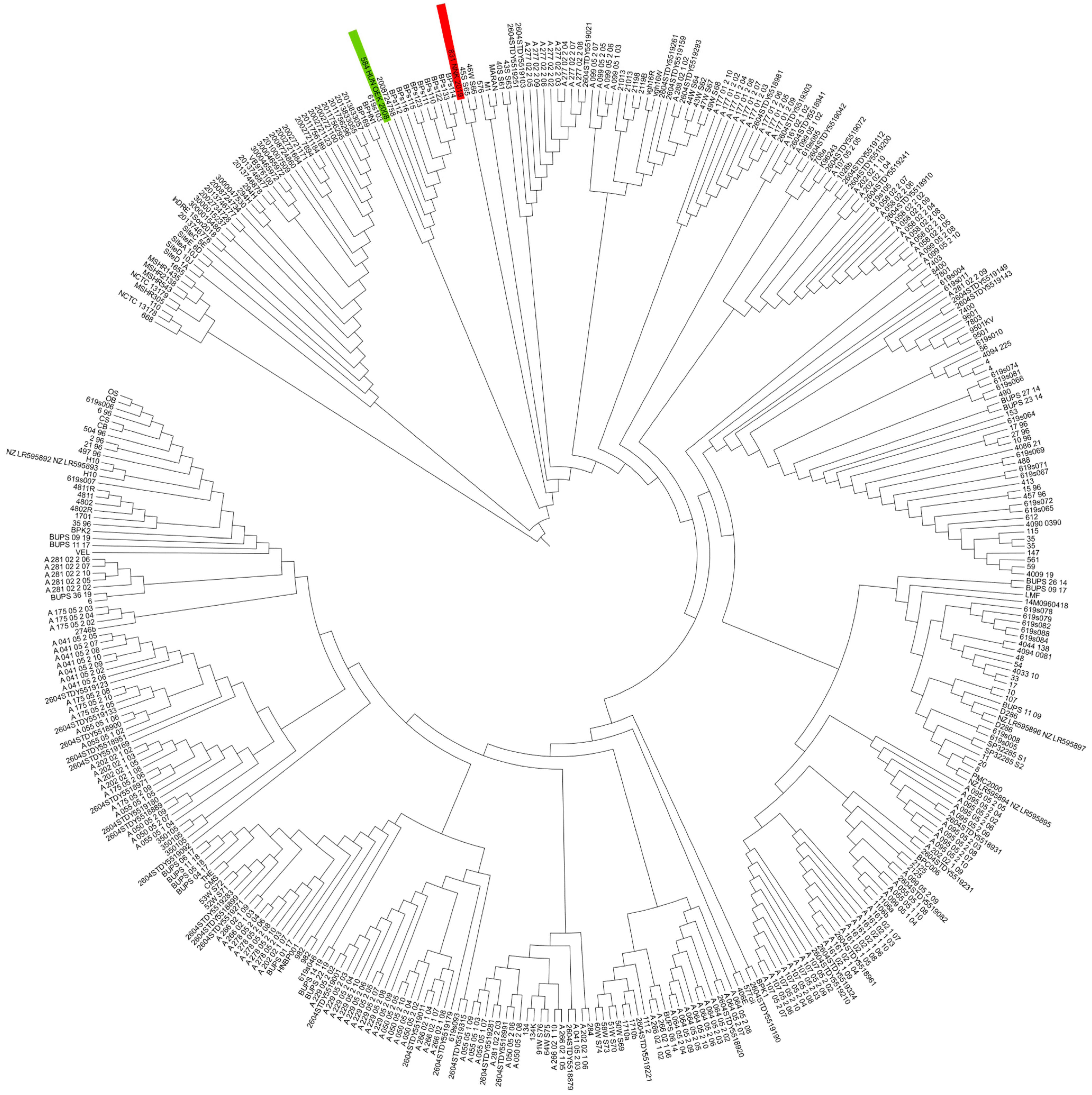
Figure 1. Comparison of Hungarian B. pseudomallei strains (n = 2) with global isolates (n = 410). The tree was built based on cgMLST. The NJ-tree shows clustering of global isolates like that of a previously constructed global SNP phylogeny, and provides high resolution for closely related isolates on a global level. The cgMLST-based neighbor-joining tree of 401 global B. pseudomallei isolates is constructed by using 4221 target genes. K96243 was used as the reference strain. The red line shows the clade where 831_NNK_2019 was included and the green line shows the clade where 584_OEK_2008 was included.
Pathogens 14 01108 g001
Figure 2. Allelic profiles of 7510 isolates from the PubMLST database. The visualization was performed using GrapeTree. Each node represents a distinct sequence type (ST) and is colored by geographic origin. Hungarian isolates clustered within the Asian lineage, showing close allelic relationships (1–3 loci difference) to sequence types from Southeast Asia, including Thailand, Vietnam, and India. The overall topology demonstrates a clear separation between Asian and Australian clades, while isolates from Africa and the Americas form distant clusters. These findings support the imported origin of Hungarian cases and reflect the global population structure of B. pseudomallei. Data source: PubMLST (accessed January 2025).
Pathogens 14 01108 g002
Figure 3. GrapeTree analysis of Burkholderia pseudomallei isolates, based on MLST data (PubMLST ID 7517). Each node represents a unique sequence type (ST), while colors correspond to the country of origin. Node ID 7517, representing the Hungarian isolate with the novel sequence type ST1643, is highlighted within the Asian cluster. The black connecting lines indicate the number of allelic differences between isolates, illustrating the genetic relationships among them. The position of ID 7517 shows a limited allelic overlap with neighboring isolates, suggesting an independent lineage within the Asian clade. Nearby nodes, such as ID 7512, ID 7498, and ID 7453, represent reference isolates from Southeast Asia, showing limited allelic overlap with 7517. Numbers displayed on the connecting lines indicate allelic distances, i.e., the number of MLST loci differing between isolates. The position of ID 7517 within the network shows minimal connectivity from larger allelic distances to neighboring nodes, suggesting that ST1643 forms an independent lineage within the Asian clade. Colors correspond to the geographic origin of isolates, as retrieved from the PubMLST database (accessed January 2025). Figure 3 demonstrates the phylogenetic position of the novel sequence type ST1643 (ID 7517). This strain shows limited allelic overlap with known isolates, suggesting an independent lineage within the Asian clade. Its unique ndh 87 allele confirms its status as a previously undescribed genotype, now deposited in the PubMLST database.
Pathogens 14 01108 g003
Figure 4. B. pseudomallei Grapetree screen on ID 7518, based on the MLST data. Each node represents a distinct isolate labeled with its PubMLST ID, and node colors correspond to the country of origin. The Hungarian isolate (ID 7518, ST1051) occupies the central position within the network and shares short allelic distances (1–3 loci) with Southeast Asian isolates, including Thailand (ID 7501), Vietnam (ID 7498), and India (ID 7505). The connecting lines denote the number of differing MLST loci, reflecting close genetic relatedness to strains circulating in Asia. This pattern supports the imported origin of the Hungarian isolate within the established Asian clade. Colors correspond to the countries listed in the legend. Data source: PubMLST (accessed January 2025).
Pathogens 14 01108 g004
Figure 5. PRISMA flow diagram for selections of melioidosis clinical cases in Europe between 1980 and 2025.
Pathogens 14 01108 g005
Table 1. Burkholderia pseudomallei strain collection in Hungary.
Sample IDTravel HistoryIncubation Period
(Calculated from Arrival Home)
Specimen TypeSymptomsTherapyAntibiotic TherapyPatient StatusCulture and Identification System
Case 1584_OEK_2008India21 daysBloodWeakness, fever, headache, facial neuralgia, sepsis ICU supportive care + antibiotic therapyEmpiric therapy: Rocephin, Herpesin; after the diagnosis, meropenem 3 × 1 g/day and Amikacin 1 g/day for 18 days; Recovered with long term neurological manifestationsBlood agar, chocolate agar followed by Biolog System
CNS infection; CSF protein 1.69; Cell count 343; glucose 2.8; CRP elevated;
Case 2831_NNK_2019Thailand14 daysAbscessRespiratory symptoms followed by neck pain, swollen lymph nodesUltrasound showed a neck abscess, which was surgically removed + antibiotic therapy Amoxicillin Clavulanate followed the standard dosageFully recoveredBlood agar, chocolate agar followed by MALDI-TOF MS
CNS = central nervous system; CSF = cerebrospinal fluid; ICU = intensive care unit; CRP = C-reactive protein. Normal ranges: CSF protein 0.15–0.45 g/L; CSF glucose 2.5–4.4 mmol/L (≈45–80 mg/dL; ≈60–70% of concurrent blood g glucose); CSF white cell count, 0–5 cells/µL; serum CRP 0–5 mg/L.
Table 2. Main genome characteristics of the Hungarian strains.
Isolate_1
(Signed 584_2008_OEK)
Isolate_2
(Signed 831_2019_NNK)
Genome size 7.2 Mb7.13 Mb
GC content 67.9%68.1%
CDS 58775857
Gene codes 73947460
Virulence-associated genes (annotated) 299300
tRNAs6083
rRNAs42
tmRNA11
ncRNA3032
ncRNA regions1919
sORF77
oriC22
oriV00
oriT00
Gap40
CRISPR-Cas systems00
Table 3. Multilocus sequence typing results of the strains.
Strain IDMLST
acegltBgmhDlepAlipAnarkndhSTIsolate IDGenomic
ID
584_2008_OEK112641887164352547517
831_2019_NNK11261121105165547518
Table 4. Burkholderia pseudomallei ST 1051 isolates from the PubMLST Database.
pubMLST IDIsolateAliasesCountryTravel HistoryYearSource
3230BCC23BPCC 24Australiana1999Environmental
3231BCC24BPCC 25Australiana1999Environmental
3232BCC32BPCC 33Australiana2000Environmental
5004Ma154 Indiano2016Human
5331H 04-2015 Vietnamno2015Human
5335QB 03 Vietnamno2015Human
5512Ma-35 Indiano2012Human
7518831_2019_NNK HungaryThailand2019Human
Table 5. Melioidosis cases in Europe between 1980 and 2025.
CountryYearsNumber of CasesPatient CharacteristicsSource Country/RegionClinical Presentation
United Kingdom1988–202569Male (n = 49), Female (n = 19); not specified (n = 1)Thailand (n = 18); India (n = 3); Nigeria (n = 2); Banghladesh (n = 5); Cambogia (n = 2); Caribbean (n = 2); Ghana (n = 2); Malaysia (n = 2); Nigeria (n = 2); Borneo (n = 1); Singapore (n = 1); Vietnam (n = 1); China (n = 1); Shanghai (n = 1); Palau (n = 1); Pakistan (n = 1); Multiple country (n = 6), not specified (n = 6); Asia (n = 22)Respiratory infection (n = 31), abscess (n = 13), sepsis (n = 17); genito-urinary infection (n = 6), gastrintestinal infection (n = 3), central nervous system infection (n = 2), cutaneus/soft tissue infection (n = 2);sore head (n = 1); not specified (n = 15)
The Netherlands1990–201839Male (n = 29), female (n = 10)Thailand (n = 21), Brazil (n = 3); Vietnam (n = 3), Indonesia (n = 4); Gambia (n = 1); Sri Lanka (n = 1); Nepal (n = 1); Myanmar (n = 1); Malaysia (n = 1); Australia (n = 3), Panama (n = 1); Cambodia (n = 1); Australia (n = 1); not specified (n = 5),Sepsis (n = 10), abscess (n = 10), respiratory infection (n = 20); genito-urinary infection (n = 6); central nervous system (n = 1), otitis externa (n = 1); mycotic aneurysm (n = 1)
Finland1995–20144Male (n = 3), female (n = 1)Thailand (n = 4) Cutaneous/soft tissues infection (n = 3), genito-urinary infection (n = 1)
Belgium2001–20124Male (n = 2), female (n = 2)Bangladesh (n = 1); Vietnam (n = 1); Thailand (n = 1); Madagascar (n = 1)Respiratory infection (n = 1), cutaneus/soft tissue infection (n = 1); genito-urinary infection (n = 1); lymphadenopathy (n = 1)
Denmark1982–20249Male (n = 6); Female (n = 1)Kenya (n = 1); Thailand (n = 4); Laos (n = 1); Vietnam (n = 1); Cambodia (n = 1); Southeast Asia (n = 1)Sepsis (n = 1); cutaneus/soft tissue infection (n = 1); genito-urinary infection (n = 1); abscess (n = 1); respiratory infection (n = 4)
Spain2009, 2011, 2023–20245Male (n = 4); Female (n = 1)Gambia (n = 1); Guinea Bissau (n = 1); Senegal (n = 1); Madagascar (n = 1); West-Africa (n = 1); Colombia (n = 1); Thailand (n = 1)Sepsis (n = 2); cutaneus/soft tissue infection (n = 1); abscess (n = 1); osteomyelitis (n = 1)
France1995–202421Male (n = 18), Female (n = 3)Africa (n = 1); Thailand (n = 6); Cambodia (n = 4); Cameron (n = 1); Madagascar (n = 1); Guadalupe (n = 1), Vietnam (n = 3); Indonesia (n = 1); Phillippines (n = 1); not specified (n = 1)Respiratory infection (n = 11); cervical lymphadenitis (n = 2); abdominal infection (n = 2); mycotic aneurysm (n = 3); saccular aneurysm (n = 1); central nervous system infection (n = 1); osteomyelytis (n = 1)
Germany1996–202414Male (n = 11); Female (n = 3)South East Asia (n = 1); China (n = 1); Taiwan (n = 7); Maldives (n = 1); Sri Lanka (n = 1); Cambodia (n = 1); Thailand (n = 2), Vietnam (n = 1); Indonesia (n = 1); Mexico (n = 1); Dominican Republic (n = 1); USA (n = 1); Costa Rica (n = 1); No significant travel history (n = 1)Respiratory infection (7); central nervous system infection (n = 1); abscess (n = 1); pericardial effusion (n = 1), urosepsis (n = 1); abdominal mycotic aortic aneurysm (n = 1); wound infection (n = 1)
SwedenNot specified5Male (n = 4); Female (n = 1)Thailand (n = 4); Malaysia (n = 1)Respiratory infection (n = 1); central nervous system infection (n = 1); otitis externa (n = 1); abscess (n = 2); cutaneus/soft tissue (n = 1)
Austria2014; 20202Male (n = 2)Thailand (n = 2)Sepsis (n = 1); lymphadenitis (n = 1)
Hungary2008–20192Male (n = 1); Female (n = 1)India, ThailandCentral nervous system infection (n = 1); abscess (n = 1)
Portugal 2011; 20253Male (n = 1); Female (n = 1); Not specified (n = 1)Thailand (n = 1), Brazil (n = 1), Not specified (n = 1)Erythema nodosum (n = 1); sepsis (n = 1); gluteal abscess (n = 1); not specified (n = 1)
Slovenia20071Male (n = 1)Asia (Brunei)Osteomyelitis of parietal bone
Italy1997, 2002, 2014,3Male (n = 2), female (n = 1)Thailand (n = 2); Singapure (n = 1)Respiratory infection (n = 3), multiple abscess (n = 1)
Norway2011, 20143Male (n = 3)Sri Lanka (n = 1); Thailand (n = 1); Cambodia (n = 1)Bacteraemia and splenic and prostatic abscesses (n = 2); neurological (n = 1)
Switzerland2008–20124Male (n = 4)Thailand (n = 3), Caribbean, Martinique Island (n = 1)Respiratory infection (n = 1); abscess (n = 1); systemic inflammatory response syndrome (n = 1); cutaneous/soft tissues (n = 1)
IcelandNot specified4Male (n = 4)Thailand (n = 3); Southeast Asia (n = 1); not specified (n = 1)Respiratory infection (n = 2); necrotizing granulomatous inflammation (n = 1); pleural abscess (n = 1); osteomyelitis (n = 1)
Table 6. Neuromelioidosis cases in Europe.
CountryYearTravel AnamnesisClinical PictureReference
The Netherlands1993Not specifiedMeningoencephalitis[67]
Hungary2008IndiaMeningoencephalitisin this study
Norway2014CambogiaCerebral abscess[71]
The Netherlands2015ThailandBrain abscesses[67]
Germany2018ThailandCerebral melioidosis[72]
Sweden2021ThailandIntracranial melioidosis[73]
Germany2022Unknown, several countries, possibility of autochthonous infectionNeuromelioidosis[29]
France2023ThailandPneumonia, Meningitis[74]
United Kingdom2024Sri LankaNeuromelioidosis[35,69]
Table 7. Main genomic and phenotypic characteristics of the Hungarian B. pseudomallei isolates.
FeatureIsolate 584_2008_OEKIsolate 831_2019_NNKImplications
Clinical manifestationNeuromelioidosis
(meningoencephalitis)
Neck abscessBm variant of bimA, associated with neurological disease
MLST/sequence type (ST)ST 1640 (novel)ST 1051ST 1051 shows overlap with Australian strains
bimA variantBmBpBm variant linked to neurological complications
YLF gene clusterPresentPresentEnhances adhesion and biofilm formation; common in Asian strains
BTFC clusterAbsentAbsentCharacteristic of Australian isolates; affects motility/chemotaxis
ITS typeType C (622 bp)Type C (622 bp)Commonly observed in Asian isolates
fhaB3PresentPresentAdhesion and tissue invasion; contributes to virulence
Capsule polysaccharide operon (wcb)Present, no key mutationsPresent, no key mutationsImportant for immune evasion
Toxin A (toxA)PresentPresentDisrupts host cell cytoskeleton
T3SSPresentPresentType III secretion system; facilitates host cell manipulation
β-lactamase genesblaOXA-57, blaPenI, amrABblaOXA-59Contributes to antibiotic resistance (aminoglycosides, β-lactams)
Geographical link/phylogenyFirst neuromelioidosis case in EuropeLinked to Asian strains via YLF clusterHighlights travel-related risk and global strain diversity
Disclaimer/Publisher’s Note: The statements, opinions and data contained in all publications are solely those of the individual author(s) and contributor(s) and not of MDPI and/or the editor(s). MDPI and/or the editor(s) disclaim responsibility for any injury to people or property resulting from any ideas, methods, instructions or products referred to in the content.