Article

Pathogen Detection and Resistome Analysis in Healthy Shelter Dogs Using Whole Metagenome Sequencing

1
School of Veterinary Medicine, Texas Tech University, Amarillo, TX 79106, USA
2
College of Veterinary Medicine, Lincoln Memorial University, Orange Park, FL 32073, USA
3
Center for Infectious, Zoonotic and Vector-Borne Diseases, Lincoln Memorial University, Harrogate, TN 37752, USA
4
Richard A. Gillespie College of Veterinary Medicine, Lincoln Memorial University, Harrogate, TN 37752, USA
*
Authors to whom correspondence should be addressed.
Pathogens 2025, 14(1), 33; https://doi.org/10.3390/pathogens14010033
Submission received: 29 October 2024 / Revised: 3 January 2025 / Accepted: 3 January 2025 / Published: 5 January 2025
(This article belongs to the Special Issue One Health: New Approaches, Research and Innovation to Zoonoses)

Abstract

:
According to the Humane Society, 25 to 40 percent of pet dogs in the United States are adopted from animal shelters. Shelter dogs can harbor bacterial, viral, fungal, and protozoal pathogens, posing risks to canine and human health. These bacterial pathogens may also carry antibiotic resistance genes (ARGs), serving as a reservoir for antimicrobial resistance (AMR) transmission. This study aimed to utilize whole metagenome sequencing (WMS) to screen for microbial pathogens and assess the resistome in healthy shelter dogs. Fecal samples from 58 healthy shelter dogs across 10 shelters in Kentucky, Tennessee, and Virginia were analyzed using WMS. Genomic DNA was extracted, and bioinformatics analyses were performed to identify pathogens and ARGs. The WMS detected 53 potentially zoonotic or known pathogens including thirty-eight bacterial species, two protozoa, five yeast species, one nematode, four molds, and three viruses. A total of 4560 ARGs signatures representing 182 unique genes across 14 antibiotic classes were detected. Tetracycline resistance genes were most abundant (49%), while β-lactam resistance genes showed the highest diversity with 75 unique ARGs. ARGs were predominantly detected in commensal bacteria; however, nearly half (18/38, 47.4%) of known bacterial pathogens detected in this study carried ARGs for resistance to one or more antibiotic classes. This study provides evidence that healthy shelter dogs carry a diverse range of zoonotic and antibiotic-resistant pathogens, posing a transmission risk through fecal shedding. These findings highlight the value of WMS for pathogen detection and AMR surveillance, informing therapeutic and prophylactic strategies to mitigate the transmission of pathogens among shelter dog populations and the risk associated with zoonoses.

1. Introduction

In the United States, 65 million households owned a pet dog in 2023 [1]. Interestingly, the US Humane Society report shows that over a quarter of these dogs were adopted from animal shelters [2]. The animal shelters typically admit dogs irrespective of age, breed, sex, and source or health status, with over 40% of their intake comprising stray or abandoned animals that often present with poor health [3]. The operational policies and procedures of animal shelters vary widely, with some shelters implementing strict quarantine and pre-transfer medical requirements, whereas most others have no such requirements. Additionally, due to the high traffic and the intermixing of animals from different sources and medical backgrounds, shelter dogs are frequently exposed to various infectious agents and often experience a variety of stressors that further increase their susceptibility to infectious agents.
Most published epidemiologic studies focused on targeted, pathogen-specific surveillance in the United States show that shelter dogs can harbor pathogens. For instance, a few studies have reported that shelter dogs can be carriers of zoonotic pathogens such as Campylobacter spp. (13% to 75% prevalence) or Salmonella spp. (1.9% to 8.3% prevalence) [4,5,6,7]. We previously reported the fecal prevalence of Campylobacter (13.6%, 8/59) and urinary shedding of Leptospira (13.1%, 26/198) among the shelter dog population in the tri-state area of western Virginia, eastern Tennessee, and southeastern Kentucky [8,9]. From nasal and perianal swab cultures collected at a dog shelter in Colorado, methicillin-resistant Staphylococcus pseudintermedius (MRSP) was detected in one out of two-hundred dogs (0.5%) and methicillin-sensitive S. pseudintermedius in six out of two-hundred dogs (3%) [10]. Gastrointestinal parasites such as Ancylostoma caninum (20.7% to 26.4%) and Trichuris vulpis (12% to 14.1%) were detected from fecal samples collected from 529 shelter dogs in Texas [11]. In Oklahoma, Trypanosoma cruzi infection in shelter dogs was detected by PCR [three of one-hundred and eighty-nine (1.6%)] and via sero-surveillance [twenty-six of one-hundred and ninety-seven (13.2%)] [12]. Moreover, several vector-borne infectious agents were identified in shelter dogs, including Anaplasma phagocytophilum, Anaplasma platys, Babesia sp. (Coco), Borrelia burdgoferi, Dirofilaria immitis, Ehrlichia canis, Ehrlichia chaffeensis, Ehrlichia ewingii, Rickettsia rickettsii, and Hepatozoon americanum [13]. Similarly, up to 33% of the dogs from multiple shelters at the western US–Mexico border were reported to be infected with tick-borne pathogens, including Rickettsia rickettsii, Echrlichia canis, and Anaplasma platys [14]. Recently, a large-scale study conducted from May 2014 to June 2020 reported that 1393 (23.5%) of the 5920 sheltered dogs housed in 98 different animal welfare organizations in the United States tested positive for canine distemper virus (CDV) [15]. These studies collectively show that healthy shelter dogs harbor a variety of zoonotic and canine pathogens, highlighting their potential as reservoirs for microbial transmission to humans (shelter workers and adopters), other animals, and among shelter dogs.
Most pathogen surveillance studies in shelter dogs have focused on detecting a limited number of pathogens using traditional culture, serological, or molecular techniques. However, these targeted pathogen detection approaches fail to capture the full range of microbial pathogens that shelter dogs may harbor which are significant to both animals including canine and public health. This limitation increases the risk of pathogen transmission from carrier dogs to other animals or humans. Given the high turnover and movement of shelter dogs between shelters and households, comprehensive pathogen screening is crucial for understanding the full pathogen load of individual dogs. Such screening can inform the design of targeted therapeutic and preventive strategies, reducing the risk of outbreaks in shelters. Additionally, it can help guide decisions regarding adoption suitability/pre-adoption interventions, particularly for families with high-risk individuals, such as young children (<5 years), older adults (>60 years), or those with underlying health conditions. In this study, we employed whole metagenome sequencing (WMS) as a comprehensive tool to detect a wide spectrum of canine and zoonotic pathogens, as well as the antimicrobial resistome, in fecal samples collected from shelter dogs.

2. Materials and Methods

2.1. Sampling

A total of 58 healthy dogs (35 male and 23 female) from ten shelters (coded as AB, BC, BR, CC, JO, KR, KW, LC, SL, and UC) in the Cumberland Gap Region of Kentucky, Tennessee, and Virginia were selected for this study. The age of the dogs ranged from <1 year (n = 17), 1 to 2 years (n = 23), and >2 years (n = 18). In the summer of 2019, freshly voided fecal samples (5–50 g) were collected from each dog in a sterile bag, immediately placed on ice, and shipped to the laboratory. The fecal samples were stored at −80 °C until further processing for DNA extraction. The genomic DNA was extracted from each fecal sample using the QIAamp DNA Stool Mini Kit (Qiagen, Germantown, MD, USA). Informed consent was obtained from the animal shelter directors for the collection of the freshly voided fecal samples. The collection of these samples was exempted by the Institutional Animal Care and Use Committee (IACUC) at the Lincoln Memorial University.

2.2. Whole Metagenome Sequencing (WMS)

The total genomic DNA was extracted from the individual fecal samples using QIAamp DNA Stool Mini Kit (Qiagen, Germantown, MD, USA) following the manufacturer’s instructions. The individual DNA samples were sequenced and analyzed using the ZymoBIOMICS shotgun sequencing pipeline (Zymo Research, Irvine, CA, USA) and ZymoBIOMICS bioinformatics analysis pipeline. Briefly, genomic DNA samples were profiled with shotgun metagenomic sequencing. Sequencing libraries were prepared with the Nextera® DNA Flex Library Prep Kit (Illumina, San Diego, CA, USA) with up to 100 ng DNA input following the manufacturer’s protocol using internal dual-index 8 bp barcodes with Nextera® adapters (Illumina, San Diego, CA, USA). The ZymoBIOMICS® Microbial Community DNA Standard (Zymo Research, Irvine, CA, USA) was used as a positive control for each targeted library preparation. Negative controls (i.e., blank library preparation control) were included to assess the level of bioburden carried by the wet lab process. All libraries were quantified with TapeStation® (Agilent Technologies, Santa Clara, CA, USA) and then pooled in equal abundance. The final pool was quantified using qPCR. The final library was sequenced (paired-end, 2 × 150 bp) on the Illumina NovaSeq® (Illumina, San Diego, CA, USA).

2.3. Data Analysis

The raw sequence reads from each sample were analyzed using the ZymoBIOMICS bioinformatics analysis pipeline. Briefly, raw sequence reads were trimmed to remove low-quality fractions and adapters with Trimmomatic-0.33 [16]: quality trimming was done by a sliding window with a 6 bp window size and a quality cutoff of 20 and reads with size lower than 70 bp were removed. The microbial composition was profiled with Centrifuge [17] using bacterial, viral, fungal, mouse, and human genome datasets. Strain-level abundance information was extracted from the Centrifuge outputs for downstream analysis including (1) alpha- and beta-diversity; (2) microbial composition with QIIME [18]; and (3) taxa abundance heatmaps with hierarchical clustering (based on Bray–Curtis dissimilarity). Antimicrobial resistance genes (ARGs) were identified with the DIAMOND sequence aligner [19]. The data from ARG and microbial composition profiling were extracted to an excel file. All the ARGs were then manually assigned to a specific class of antibiotics. The microbial community data were screened to identify individual pathogens and categorized into four major pathogen groups: (i) known canine pathogens, (ii) known/potentially zoonotic pathogens, (iii) opportunistic pathogens of canines or humans, and (iv) pathogens of other animals. The outcomes of the shotgun metagenomics were described using descriptive statistics to summarize the results of microbiome diversity, the prevalence of pathogens, and the resistomes (ARGs) in shelter dogs. The differences in microbial communities between sexes, age groups, and shelters were compared using the Shannon diversity index by one-way ANOVA with Tukey’s post hoc using NCSS 2023 version 23.0.1 (NCSS, Kaysville, UT, USA).

3. Results and Discussion

3.1. Fecal Microbiome Diversity and Composition in Shelter Dogs

From the WMS, a total of approximately 565 million raw reads were obtained, with an average of 9.75 million reads per sample (Supplementary Table S1). After the quality trimming and removal of host reads and unclassified reads, a total of 135 million reads were identified as microbial reads, with an average of 2.32 million reads per dog (range = 0.7 to 4.87 million reads from individual dogs).
Bacterial taxonomic units (BTUs) predominated in all samples (Supplementary Table S2), constituting 99.5% of the fecal microbiome, contributing to a greater phylogenetic diversity compared to eukaryotes (0.06%) and viruses (0.46%). These results corroborate with previous gut microbiome studies in dogs [20,21]. The sequenced reads were taxonomically assigned to 12 bacterial phyla [Acidobacteria (0.003% BTUs, n = one species), Actinobacteria (1.98% BTUs, n = 63 species), Bacteroidetes (70.63% BTUs, n = 110 species), Chlamydiae (0.06% BTUs, n = two species), Chloroflexi (0.001% BTUs, n = one species), Deferribacteres (0.01% BTUs, n = one species), Deinococcus-Thermus (0.08% BTUs, n = three species), Firmicutes (10.73% BTUs, n = 235 species), Fusobacteria (1.44% BTUs, n = seven species), Proteobacteria (14.93% BTUs, n = 86 species), Spirochaetes (0.14% BTUs, n = six species), and Verrucomicrobia (0.0004% BTUs, n = one species)]; six Eukaryote phyla [Apicomplexa (n = one species), Ascomycota (n = seven species), Basidiomycota (n = three species), Discosea (n = one species), Mucoromycota (n = one species), and Nematoda (n = one species)]; three pathogenic viral genera [canine minute virus, canine parvovirus, and an enterovirus]; and 10 bacteriophages [Escherichia, Shigella, Salmonella, Bacteroides, Enterococcus, Faecalibacterium, Lactobacillus, Parabacteroides, Stenotrophomonas, and Streptococcus phages and one unknown virus] (Figure 1). In concordance with previous studies [20,21], the predominant bacterial phyla in individual dogs included Bacteroidetes (68.7% OTUs, 95% confidence interval (73.3%, 64%)), Proteobacteria (15.7% OTUs, 95% confidence interval (19.35%, 12%)), and Firmicutes (11.6% OTUs, 95% confidence interval (13.7%, 9.6%)), these being the most abundant, followed by Actinobacteria (2.4% OTUs, 95% confidence interval (3.4%, 1.36%)) and Fusobacteria (1.3% OTUs, 95% confidence interval (2.3%, 0.29%)).
The Shannon diversity index (SDI) for the fecal microbiome of individual shelter dogs ranged from 0.73 (sample SL6) to 3.59 (sample JO3), with an average SDI of 2.48 (Supplementary Table S1, Figure 1). At the shelter level, SDI values ranged from 1.89 (shelter SL) to 3.02 (shelter KR). No statistically significant differences in SDI were observed between male and female dogs or between the three age groups of dogs.

3.2. Fecal Pathogen Prevalence in the Shelter Dog Population

WMS identified a total of 53 potential or known pathogen species (Figure 2). These included bacteria (n = 38 species, distributed across 18 genera), protozoa (n = two, Toxoplasma gondii and Balamuthia mandrillaris), yeast (n = five, Candida parapsilosis, Malassezia globose, Malassezia restricta, Malassezia sympodialis, and Malassezia spp.), nematode (n = one, Trichuris trichiura), mold (n = four, Alternaria alternata, Fusarium fracticaudum, Fusarium proliferatum, and Saksenaea oblongispora), and viruses (n = three, canine parvovirus 2, canine parvovirus 1, and an enterovirus). These pathogens were broadly classified based on their known or potential pathogenicity in dogs, humans, or other animal species, resulting in four categories: known canine pathogen, known or potential zoonotic pathogen, opportunistic pathogen for canines or humans, and other animal pathogen.

3.2.1. Known Canine Pathogens

Three known canine pathogens, Carnivore protoparvovirus 1, Carnivore bocaparvovirus 1, and Bordetella bronchiseptica, were identified by fecal metagenome sequencing in two shelters. Shelter KR harbored all three pathogens, while shelter KW carried Carnivore bocaparvovirus 1. Carnivore protoparvovirus 1 (also called canine parvovirus 2 or CPV-2) was detected in fecal samples from a total of three dogs (5.2%), including two (28.6%) from shelter KW (KW1 and KW3) and one from shelter KR (KR5). In shelter KR, a fecal sample from one dog (KR4) was positive for Carnivore bocaparvovirus 1 (also called canine minute virus or CPV-1) and another dog (KR7) was positive for Bordetella bronchiseptica. All the dogs except KR7 were less than two years of age, with KW1 and KW3 less than a year old. CPV-2 is known to cause severe gastroenteritis, especially in young puppies of less than 6 months of age, whereas CPV-1 is known to cause a range of pathologies, including enteritis, pneumonitis, and neonatal death, in dogs [22]. Parvoviral gastroenteritis is a highly contagious viral disease that is known to pose a significant health challenge in animal shelters, often leading to mortality and significant economic burden due to the high cost of treatment and euthanasia [23]. While the fecal shedding of CPV in a few healthy dogs in a shelter can pose a significant risk of transmission to other dogs, it is currently unknown if the CPV detected via metagenomics is a virulent strain or a vaccine strain. Similarly, Bordetella bronchiseptica, a causative agent of contagious kennel cough, was detected in one dog (KR7). Given that the usual habitat of Bordetella bronchiseptica is the upper respiratory tract of dogs, its detection in fecal samples is unexpected. However, this may occur through two potential mechanisms: dogs that are coughing may swallow respiratory secretions containing the bacteria, leading to its passage through the gastrointestinal tract. Additionally, dogs frequently engage in behaviors such as sniffing or licking the perianal area of other dogs, which could introduce the bacteria into the fecal material [24].

3.2.2. Known or Potentially Zoonotic Pathogens

A total of 12 species (10 bacteria, one nematode, and one protozoa) of known or potentially zoonotic pathogens were identified in this study (Figure 2). These pathogens included Campylobacter upsaliensis, Campylobacter jejuni, Salmonella enterica, Helicobacter canis, Clostridioides difficile, Clostridium perfringens, Clostridium botulinum, Brachyspira pilosicoli, Mycobacterium tuberculosis, Balamuthia mandrillaris, Toxoplasma gondii, and Trichuris trichiura. Given that the significance of each of these as animal infectious agents varies widely depending on several factors, the pathogenicity and zoonotic potential of these bacteria in animals and humans is discussed below.
Campylobacter upsaliensis was detected in fecal samples from 75.9% (44/58) of dogs, representing all ten shelters, whereas Campylobacter jejuni was detected in 20.7% (12/58) of dogs, representing six shelters. Published studies show a 50 to 90% prevalence of Campylobacter fecal shedding [4,5,6], with both C. jejuni and C. upsaliensis being frequently detected in the shelter dog population. C. jejuni is frequently implicated as a cause of canine campylobacteriosis [25], whereas C. upsaliensis is known to cause mild to moderate enteric disease in dogs [25,26]. Both C. jejuni and C. upsaliensis are also the most significant zoonotic pathogens in people, especially in children and the elderly [27,28], with an annual estimated incidence of 20 cases of Campylobacter-associated illness for every 100,000 people in the United States [29,30]. Evidence suggests that zoonotic transmission of C. jejuni occurs through direct contact with the feces of infected dogs [31,32,33]. In some cases, Campylobacter infection can be invasive in people, leading to severe complications including Guillain–Barré syndrome [34].
Salmonella enterica, an enteric zoonotic pathogen known to infect a wide variety of animals and humans, was detected in one (1.7%) of the dogs from a single (10%) shelter. One study reported fecal shedding of Salmonella in 4.9% (26/226) of shelter dogs in Texas [35]. A multi-laboratory survey conducted in the United States reported 2.5% (60/2422) of Salmonella prevalence in diarrheic and non-diarrheic dogs [36], suggesting that Salmonella shedding in healthy dogs is not uncommon. Although dogs can acquire Salmonella infection from a variety of sources, raw foods are considered major risk factors for the acquisition of infection and have caused major outbreaks involving both dogs and humans [37]. The fecal shedding of Salmonella in dogs poses a zoonotic risk of transmission to humans [38].
Non-Helicobacter pylori helicobacters (NHPH) such as Helicobacter canis are known to naturally colonize the gastrointestinal tract of dogs. In this study, Helicobacter canis was detected in 65.5% (38/58) of dogs from all ten shelters (Figure 2). Previously, H. canis prevalence in canine feces has been reported to range from 5.8% (23/390) in Chile and 21% (20/95) in Sweden to 100% (14/14) in Taiwan [39,40,41]. H. canis is occasionally reported from enterohepatic disease in dogs [42]. Moreover, close contact with dogs and cats was reported as the primary risk factor among immunocompromised human patients living with H. canis-infected dogs, suggesting the potential for zoonotic transmission of H canis from dogs to humans [43,44]. However, the pathogenicity of H. canis in dogs and its zoonotic potential is likely underrecognized, most likely due to its fastidious nature that poses challenges in culturing, likely leading to false-negative cultures.
Clostridioides difficile (previously known as Clostridium difficile) was detected in a total of ten (17.2%) dogs, representing nine shelters, Clostridium perfringens was detected in eleven (19%) dogs from eight shelters, and Clostridium botulinum was detected in six (10.3%) dogs, representing four shelters (Figure 2). While the virulence profiling of pathogens is beyond the scope of this study, none of the C. difficile positive fecal samples were positive for the presence of the binary toxin gene (cdtB) (data not shared). The prevalence of toxigenic strains of C. difficile in dogs has been reported to range from 17% in healthy adult dogs [45] to 90% in puppies [46,47] and from 19 to 33% in outpatients and hospitalized dogs that are treated with antibiotics [48,49,50,51]. Dogs that visit human healthcare facilities, interact with hospitalized children, or live with an owner treated with antibiotics show an increased risk of C. difficile fecal shedding [52]. Thus, the zoonotic transmission of C. difficile between dogs and people is likely; however, a significant knowledge gap remains in our understanding of the role of C. difficile in enteric disease in dogs, primarily due to the lack of a concrete correlation between C. difficile infection and enteric disease [53]. C. perfringens is also frequently detected in the feces of healthy dogs and dogs with acute hemorrhagic diarrhea syndrome. Cpa gene (encoding phospholipase C toxin)-positive C. perfringens that also carry other toxin genes are more likely to be associated with disease in dogs [9,54,55]. Although screening for all potential toxin-encoding genes for C. perfringens-positive fecal samples from shelter dogs was beyond the scope of this study, four (AB4, JO2, KR5, and KW2) out of eleven C. perfringens-positive fecal samples sequenced in this study were also positive for the Cpa gene. Nine out of eleven C. perfringens-positive fecal samples sequenced in this study were previously reported as Cpa gene-positive by PCR [9]. While the role of toxin-positive C. perfringens as a potential zoonotic pathogen remains poorly recognized, Clostridium botulinum, a known human pathogen, was detected in fecal samples from a total of six (10.3%) dogs from three shelters including LC (n = three), UC (n = one), SL (n = one), and KW (n = one). Intestinal colonization of C. botulism has been reported in naturally infected and experimentally inoculated dogs without clinical signs of the disease [56]. Moreover, C. botulism infection with clinical disease has been linked to eating poorly stored dry food [57] and rotten bovine [58], pigeon [59], or duck [60] carcasses. The likely sources of C. botulinum infection in shelter dogs tested in this study remain unknown; however, the fecal prevalence of C. botulinum in shelter dogs poses a risk of the persistent transmission of infection among dogs. Zoonotic transmission of C. botulinum from dogs to people has not been conclusively demonstrated; however, the risk cannot be ruled out because C. botulism is also known to be transmitted to humans by a variety of transmission routes [61].
Brachyspira pilosicoli, a causative agent of intestinal spirochetosis in various animal species including dogs, pigs, poultry, rodents, and humans [62,63], was detected in the fecal samples of three (5.2%) dogs from two (20%) shelters. Although B. pilosicoli is commonly detected in the feces of dogs and has been sporadically isolated from cases of canine diarrhea [62,64], its role as a potential pathogen in dogs is yet to be conclusively determined. Comparative analyses of B. pilosicoli isolates from several species, including dogs and humans, suggest B. pilosicoli has a clear zoonotic potential [63,65,66].
The fecal samples of two (3.4%) of dogs from two (20%) shelters examined in this study were positive for Mycobacterium tuberculosis. While humans are major reservoir hosts of M. tuberculosis, dogs often serve as sentinels of human infection within households and can develop both pulmonary and disseminated forms of tuberculosis [67,68]. M. tuberculosis infection in dogs following exposure to infected humans (reverse zoonoses) and transmission from dogs to humans has been reported in many parts of the world, with sporadic reports from the United States [69,70,71].
Besides bacterial pathogens, the fecal samples of four (6.9%) dogs from two (20%) shelters tested positive for Balamuthia mandrillaris, a free-living amoeba that causes encephalitis in competent and immunocompromised humans, horses, dogs, sheep, and nonhuman primates. Amoebas are found in soil and are likely transmitted through the inhalation of airborne cysts or by the direct contamination of skin wounds. A few cases of Balamuthia amoebic encephalitis in dogs have been reported in the United States [72,73]. However, its true incidence in dogs and zoonotic potential from dogs to humans remains poorly understood, likely because the clinical signs often mimic other forms of encephalitis and only a few laboratories perform appropriate diagnostics [74].
Toxoplasma gondii, an Apicomplexan protozoan that infects almost all warm-blooded animals including livestock, birds, cats, dogs, and humans, was detected in seven (12.1%) dogs from five shelters. Interestingly, dogs do not produce infective oocysts of T. gondii, but rather acquire infection via the ingestion of infectious oocysts from the environment contaminated with cat feces leading to seroconversion and, in some cases, clinical disease [75]. Most infected dogs can shed infectious oocysts in their feces and mechanically transport infection to humans and other animals via contaminated body surfaces, the mouth, and the feet. Thus, dogs not only serve as vehicles for the zoonotic transmission of infection to humans but also as sentinels for environmental contamination with infective oocysts [76].
The intestinal nematode Trichuris trichiura (whipworm), a causative agent of human trichuriasis, was detected in eight (13.8%) dogs from five (50%) shelters. Trichuriasis is a neglected tropical disease that affects >800 million people worldwide, including in the southern United States [77]. The primary hosts of T. trichiura are humans and non-human primates. Canine whipworm infections are generally considered to be T. vulpis. However, T. trichiura eggs have been occasionally detected in stool samples from dogs [78]. Although zoonotic infections with other Trichiura spp. such as T. suis (from pigs) and Trichiura vulpis (from dogs) have been reported in humans [79], potential zoonotic transmission of T. trichiura from shelter dogs to humans or between dogs remains unknown. The detection of T. trichiura in multiple dogs from multiple shelters in this study suggests a rather high prevalence which warrants further investigations on potential zoonotic risks. Finally, the signatures of Enterovirus C (human poliovirus 1 Mahoney) were detected in six (10.3%) dogs from four (40%) shelters. Interestingly, human enteroviruses, including polioviruses, have been isolated from dog feces previously [80,81].

3.2.3. Opportunistic Pathogens (Canine and Human)

Twenty-five opportunistic pathogen species, including seven genera of bacteria, three genera of mold, two genera of yeast, and an enterovirus, were identified in shelter dogs. Seven opportunistic pathogens, including Enterococcus faecium (46.6% of dogs and 100% of shelters), Enterococcus hirae (15.5% of dogs and 60% of shelters), Escherichia coli (51.7% of dogs and 90% of shelters), Staphylococcus aureus (8.6% of dogs and 40% of shelters), Streptococcus gallolyticus (51.7% of dogs and 80% of shelters), Malassezia restricta (8.6% of dogs and 50% of shelters), and an enterovirus (10.3% of dogs and 40% of shelters), were commonly detected. Other opportunistic pathogens, including Proteus mirabilis, Staphylococcus epidermidis, Streptococcus pasteurianus, Fusarium fracticaudum, Saksenaea oblongispora, and Malassezia globosa, were detected from two to three dogs from up to three shelters. Opportunistic pathogens, including Enterobacter cloacae, Enterobacter ludwigii, Enterococcus faecalis, Pasteurella multocida, Streptococcus canis, Streptococcus mitis, Streptococcus sinensis, Alternaria alternata, Fusarium proliferatum, Candida parapsilosis, Malassezia sp., and Malassezia sympodialis, were detected, representing one dog each from eight different shelters. Also noteworthy is that the opportunistic pathogens listed here are also known to be a component of the normal canine gut microflora; thus, the detection of these organisms in fecal samples can be expected and this finding does not necessarily signify as primary concern for infection. However, several of these organisms have been frequently reported as a cause of secondary opportunistic infection in both animals and humans. A range of underlying health conditions—such as cancer, wounds, urinary calculi, heart disease, and stressful situations—can predispose individuals to secondary infections caused by these microbes.

3.2.4. Other Animal Pathogens

Thirteen species belonging to seven genera of bacteria were characterized as other animal pathogens. Chlamydia abortus was detected in all the shelters, with 81% of dogs positive, in this study. Clostridium spiroforme was detected in 25.9% of dogs from 60% of the shelters investigated in the study.
Collectively, these findings underscore the diverse and complex fecal pathogen landscape within the shelter dog population, encompassing a wide range of pathogens of known and potential significance to both canine and human health. This study shows that WMS can be effectively employed to detect and monitor a battery of zoonotic and other infectious pathogens from fecal samples from shelter dogs. The WMS-based detection of obligate pathogen signatures in fecal samples from healthy dogs in this study likely suggests inapparent carrier infections. The detection of opportunistic pathogen signatures from healthy dogs must be inferred with caution unless supported by follow-up investigations, including clinical findings, culture-based results, or immune responses from individual animals or shelter populations [82,83].

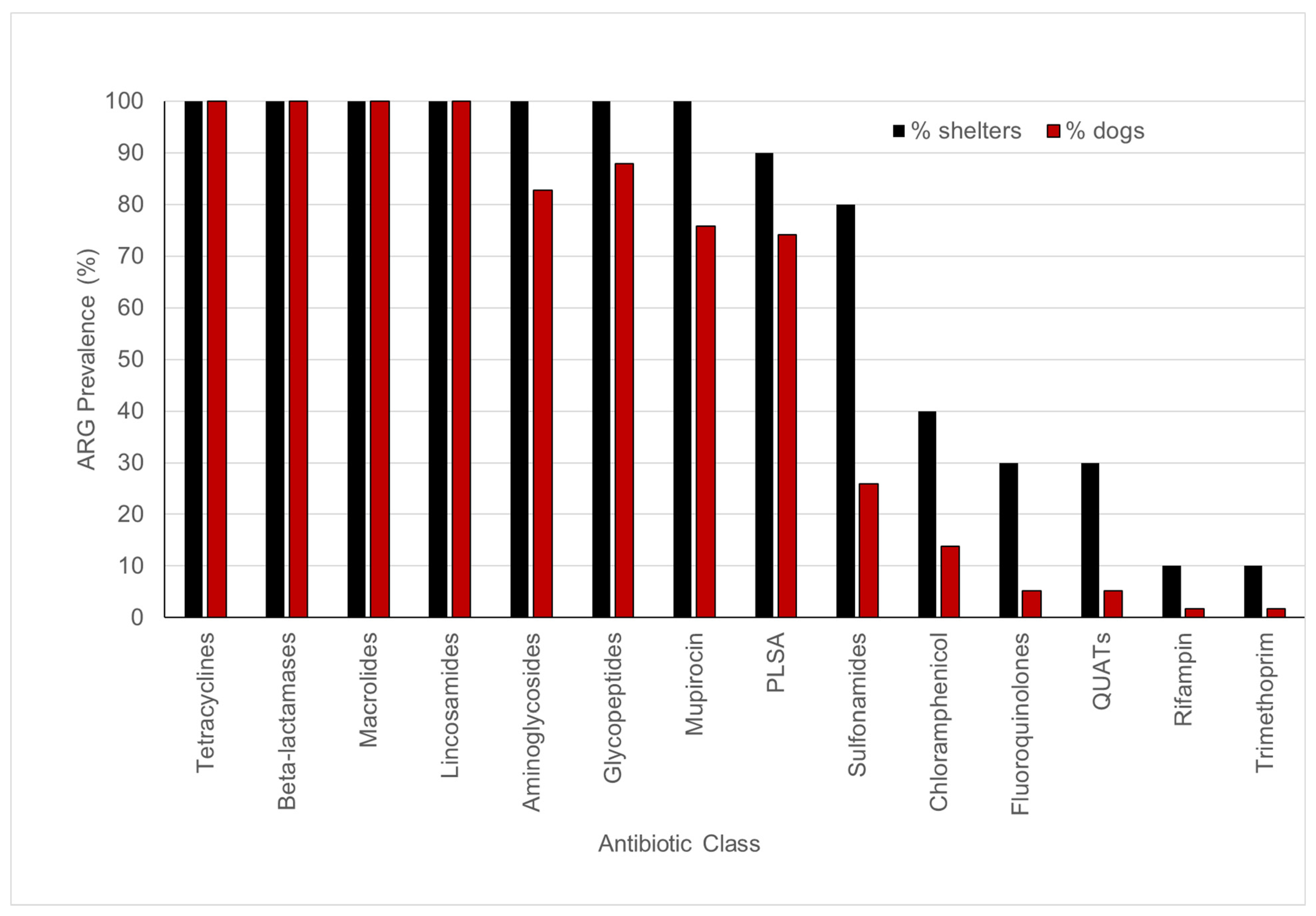
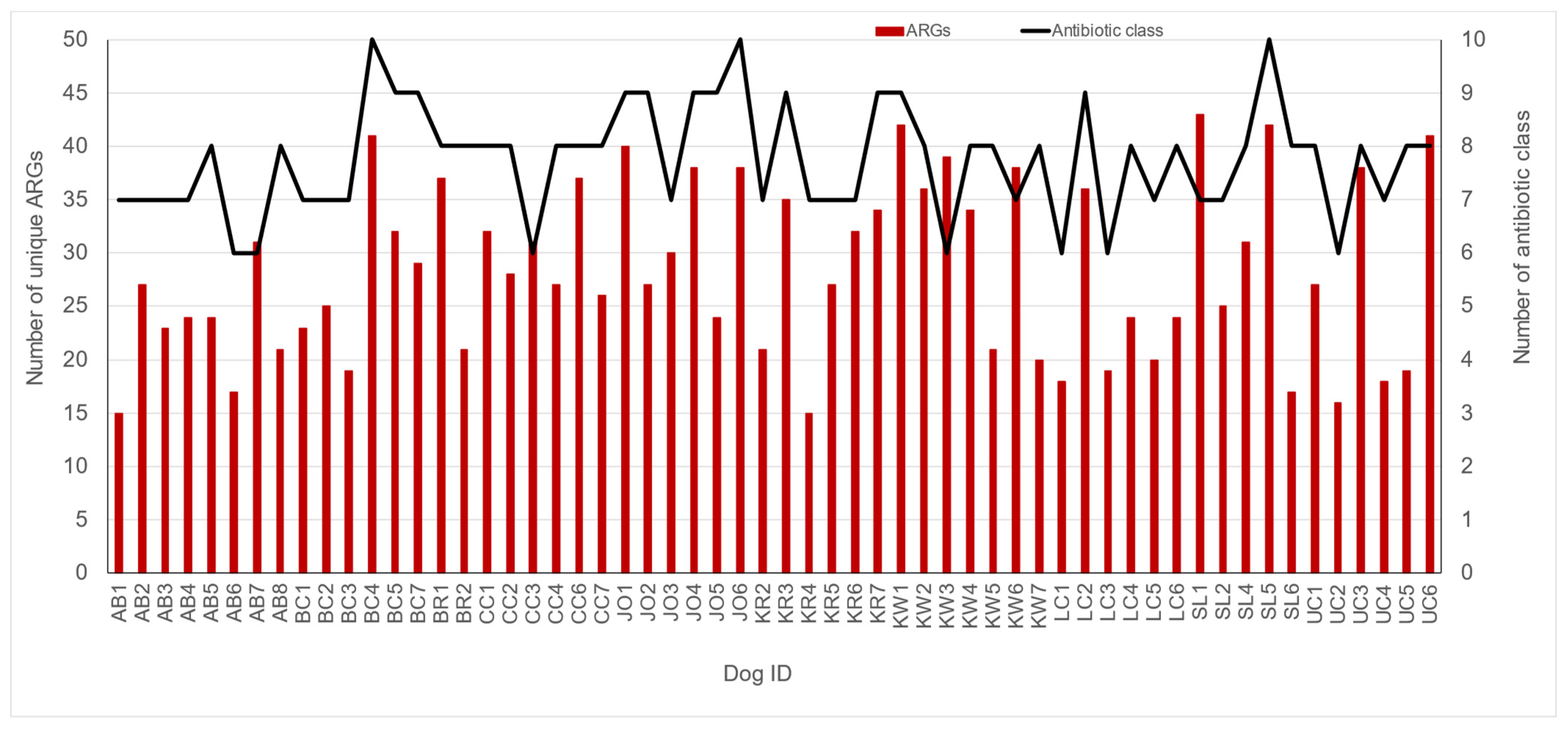
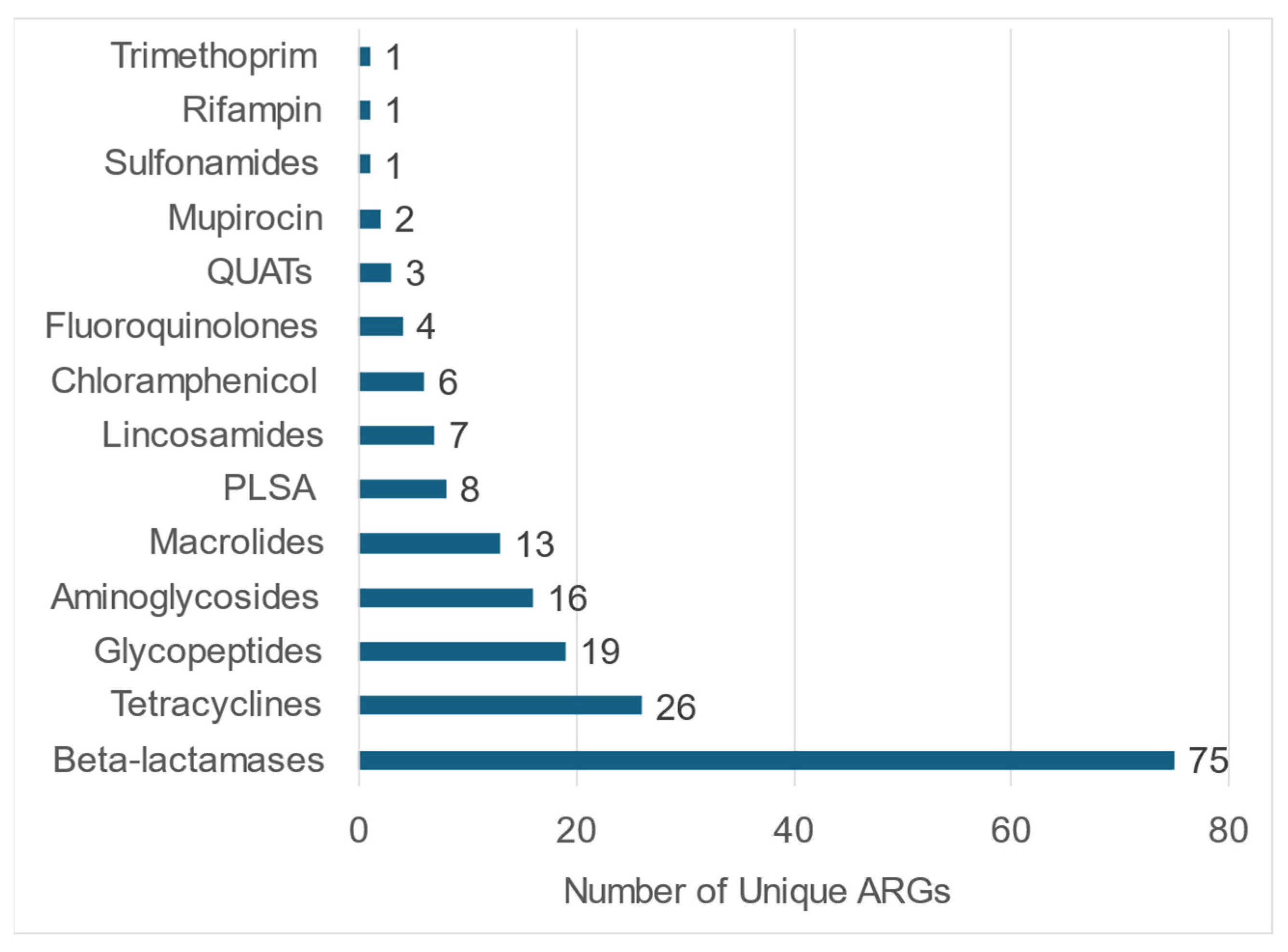
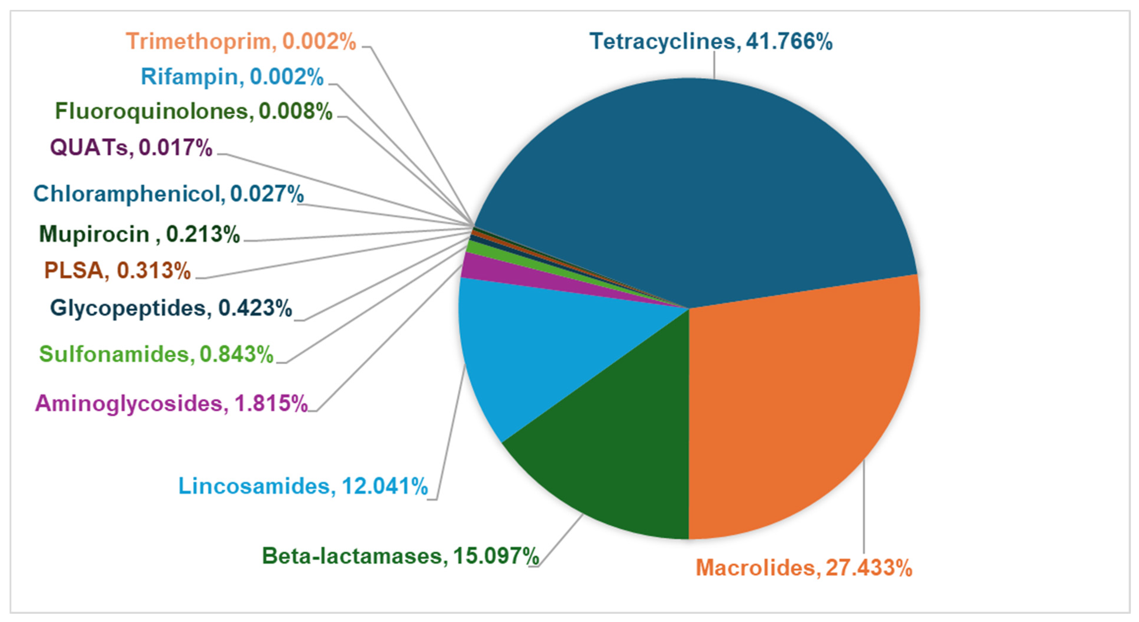
3.3. Fecal Resistome in Shelter Dog Population

The WMS of fecal samples from 58 shelter dogs yielded a total of 48,267 ARG-specific reads, resulting in the identification of 4560 ARG signatures spanning 182 unique ARGs encoding resistance to 14 different antibiotic classes, including disinfectants such as quaternary ammonium compounds (QUATs) (Supplementary Table S3). The abundance of ARG-specific reads in individual dogs ranged from 0.4% (UC3) to 4.2% (LC6) (Supplementary Table S3). ARGs encoding resistance to tetracycline, β-lactamase, macrolide, and lincosamide were detected in fecal samples from all (100%) dogs tested in this study (Figure 3). The fecal samples from the majority of dogs also tested positive for ARGs encoding resistance to pleuromutilin, lincosamide, and streptogramin A (PLSAs, 74.1%), mupirocin (75.9%), glycopeptides (87.9%), and aminoglycosides (82.8%). A few dogs tested positive for ARGs encoding resistance to sulfonamides (25.9%) and chloramphenicol (13.8%), whereas the ARGs encoding resistance to fluoroquinolones (5.2%), QUATs (5.2%), rifampicin (1.7%), and trimethoprim (1.7%) were rarely detected (Figure 3). On average, the fecal samples of shelter dogs tested in this study harbored 28.5 unique ARGs (range = 15 to 43 ARGs/dog) encoding resistance against six to ten antibiotic classes (Figure 4). Notably, the most diverse array of ARGs (75 unique ARGs) encoded resistance to the β-lactam class of antibiotics followed by the number of unique ARGs encoding resistance to tetracyclines, glycopeptides, aminoglycosides, and macrolides (Figure 5). Interestingly, the WMS sequenced the highest percentage (41.8%) of the total ARG reads encoding resistance to tetracyclines, followed by 27.4%, 15.1%, 12%, and 1.81% of the total reads encoding resistance to macrolides, beta-lactamases, lincosamides, and aminoglycosides, respectively, while <1% of the total ARG reads encoded resistance to the rest of the antibiotic classes (Figure 6). Collectively, these data show that each dog harbored a diverse array of ARGs, potentially serving as a reservoir of diversity of antimicrobial resistance.
The host gut with its residing bacterial community is considered an important reservoir of ARGs (resistome) where gut commensals and pathogens often share the ARGs encoded by mobile genetic elements [84]. The 4560 ARG signatures (representing 48,267 ARG-specific reads) identified in this study were associated with 224 unique bacterial species spanning 103 bacterial genera (Figure 7, Supplementary Table S4). Predominantly, 82.06% of the total 4560 ARG signatures were detected in non-pathogenic bacterial genera, with 32% of the total ARG signatures associated with Bacteroides, followed by 12.15% of the ARG signatures detected in unknown bacterial genera (Figure 7, Supplementary Table S4). In the context of pathogenic bacterial species, 47.4% (18 out of 38 bacterial pathogens) carried ARG signatures encoding resistance to at least one class of antibiotic (Supplementary Table S5). This accounted for 5.7% (261/4560) of the ARG signatures in the pathogenic bacterial genera.
While the detected ARGs were associated with almost half of the pathogen species, not all the dogs with pathogen signatures were positive for ARGs. ARGs were detected in Clostridium spiroforme (7% [1/15] of positive dogs), Clostridioides difficile (20% [2/10] of positive dogs), Clostridium botulinum (17% [one of six] of positive dogs), Enterobacter cloacae (100% [one of one] of positive dogs), Salmonella enterica (100% [one of one] of positive dogs), Streptococcus agalactiae (25% [one of four] of positive dogs), Streptococcus pasteurianus (50% [one of two] of positive dogs), Campylobacter jejuni (25% [3/12] of positive dogs), Clostridium perfringens (27% [3/11] of positive dogs), Enterococcus hirae (56% [five of nine] of positive dogs), Proteus mirabilis (100% [two of two] of positive dogs), Streptococcus gallolyticus (20% [6/30] of positive dogs), Streptococcus suis (75% [three of four] of positive dogs), Trichuris trichiura (38% [three of eight] of positive dogs), Staphylococcus epidermidis (67% [two of three] of positive dogs), Staphylococcus aureus (40% [two of five] of positive dogs), Enterococcus faecium (4% [1/27] of positive dogs), and E. coli (63% [19/30] of positive dogs) (Supplementary Table S5).
The fecal WMS resistance profiling in this study highlights the prevalence and diversity of the antibiotic resistance genes (ARGs) carried by shelter dogs. Research shows that pet dogs often share their antimicrobial resistance (AMR) profiles with their owners, indicating potential transmission of AMR within households [85,86,87]. The resistome detected in this study was predominantly associated with non-pathogenic gut microflora with less than 20% ARGs identified in various categories of pathogenic species. The role of shelter dogs in maintaining and disseminating antimicrobial resistance remains unclear. Clinical decision-making should carefully align with current antibiotic stewardship guidelines, which may require follow-up culture and phenotypic antimicrobial susceptibility testing [88]. Nevertheless, our findings underscore the importance of ongoing monitoring and surveillance to mitigate the potential health risks for both dogs and humans.

4. Conclusions

This study demonstrates that shelter dogs harbor several known and potential pathogens of importance to canines, humans, and other animal species. The presence of ARGs in fecal samples suggests a potential role in the persistence and spread of antimicrobial resistance, which could contribute to treatment failures. Both zoonotic pathogens and ARGs pose a significant public health risk to shelter workers and adopters. WMS offers a valuable, comprehensive screening tool to detect a battery of pathogens and ARG carriage in dynamic animal populations such as shelter dogs which can serve as sentinels of zoonotic and other infectious disease, enabling evidence-based strategies for preventing, treating, and controlling pathogen transmission.

Supplementary Materials

The following supporting information can be downloaded at: https://www.mdpi.com/article/10.3390/pathogens14010033/s1, Supplementary Table S1. Number of reads and Shannon diversity index from fecal samples of 58 shelter dogs. Supplementary Table S2. Taxonomic abundance in 58 shelter dogs. Supplementary Table S3. Abundance of ARGs detected in fecal samples from shelter dogs. Supplementary Table S4. ARG signatures and reads associated with microbial genera in the fecal metagenome from fifty-eight dogs from ten shelters. Supplementary Table S5. Prevalence of pathogens and antibiotic-resistant genes detected in pathogens from shelter dogs.

Author Contributions

A.V. and D.H.S. conceived the fecal microbiome study, K.C. implemented shelter recruitment and sample collection, S.S. performed the data analysis and prepared the manuscript draft, and S.S., D.H.S., A.V. and K.C. reviewed and finalized the manuscript draft. All authors have read and agreed to the published version of the manuscript.

Funding

This study was funded through an institutional research grant from Richard A. Gillespie College of Veterinary Medicine, Lincoln Memorial University to KC and AV (Grant#19KC001).

Institutional Review Board Statement

Not applicable.

Informed Consent Statement

This study was conducted on freely voided fecal samples collected under a protocol exempted by the Institutional Animal Care and Use Committee at Lincoln Memorial University. Informed consent was obtained from the animal shelter directors for the collection and use of these fecal samples in research.

Data Availability Statement

The data presented in this study are available within the article or supplementary materials. The raw sequence reads associated with this study are available in the NCBI Sequence Read Archive (SRA) under BioProject ID PRJNA1197100, with BioSample numbers ranging from SAMN45772830 to SAMN45772887.

Acknowledgments

We wish to thank Joey Morgan, Kaitlyn Amsler, and Marina Taylor for their technical assistance. This study was funded through an institutional research grant from the Richard A. Gillespie College of Veterinary Medicine, Lincoln Memorial University to KC and AV (Grant#19KC001). The funders had no role in the study design, sample collection, analysis and interpretation of the data, or manuscript writing.

Conflicts of Interest

The authors declare no conflicts of interest.

References

  1. Megna, M. Pet Ownership Statistics 2024. Available online: https://www.forbes.com/advisor/pet-insurance/pet-ownership-statistics/#sources_section (accessed on 7 October 2024).
  2. TheHumaneSociety. Pets by the Numbers. Available online: https://humanepro.org/page/pets-by-the-numbers (accessed on 7 October 2024).
  3. ShelterAnimalsCount. 2023 Annual Analysis Report. Available online: https://www.shelteranimalscount.org/wp-content/uploads/2024/01/Full-Year-2023-Report.pdf (accessed on 10 October 2024).
  4. Parsons, B.N.; Williams, N.J.; Pinchbeck, G.L.; Christley, R.M.; Hart, C.A.; Gaskell, R.M.; Dawson, S. Prevalence and shedding patterns of Campylobacter spp. in longitudinal studies of kennelled dogs. Vet. J. 2011, 190, 249–254. [Google Scholar] [CrossRef] [PubMed]
  5. Acke, E.; Whyte, P.; Jones, B.R.; McGill, K.; Collins, J.D.; Fanning, S. Prevalence of thermophilic Campylobacter species in cats and dogs in two animal shelters in Ireland. Vet. Rec. 2006, 158, 51–54. [Google Scholar] [CrossRef]
  6. Leahy, A.M.; Cummings, K.J.; Rodriguez-Rivera, L.D.; Hamer, S.A.; Lawhon, S.D. Faecal Campylobacter shedding among dogs in animal shelters across Texas. Zoonoses Public. Health 2017, 64, 623–627. [Google Scholar] [CrossRef] [PubMed]
  7. LaLonde-Paul, D.; Cummings, K.J.; Rodriguez-Rivera, L.D.; Wu, J.; Lawhon, S.D. Ciprofloxacin resistance among Campylobacter jejuni isolates obtained from shelter dogs in Texas. Zoonoses Public. Health 2019, 66, 337–342. [Google Scholar] [CrossRef]
  8. Spangler, D.; Kish, D.; Beigel, B.; Morgan, J.; Gruszynski, K.; Naikare, H.; Nahar, V.K.; Coarsey, M.D.; Verma, A. Leptospiral shedding and seropositivity in shelter dogs in the Cumberland Gap Region of Southeastern Appalachia. PLoS ONE 2020, 15, e0228038. [Google Scholar] [CrossRef]
  9. Verma, A.; Carney, K.; Taylor, M.; Amsler, K.; Morgan, J.; Gruszynski, K.; Erol, E.; Carter, C.; Locke, S.; Callipare, A.; et al. Occurrence of potentially zoonotic and cephalosporin resistant enteric bacteria among shelter dogs in the Central and South-Central Appalachia. BMC Vet. Res. 2021, 17, 313. [Google Scholar] [CrossRef]
  10. Gingrich, E.N.; Kurt, T.; Hyatt, D.R.; Lappin, M.R.; Ruch-Gallie, R. Prevalence of methicillin-resistant staphylococci in northern Colorado shelter animals. J. Vet. Diagn. Investig. 2011, 23, 947–950. [Google Scholar] [CrossRef] [PubMed]
  11. Rodriguez, J.Y.; Cummings, K.J.; Hodo, C.L.; Hamer, S.A. A repeated cross-sectional study of intestinal parasites in Texas shelter dogs using fecal flotation and saline sedimentation. Parasitol. Res. 2023, 122, 237–243. [Google Scholar] [CrossRef] [PubMed]
  12. Allen, K.E.; Lineberry, M.W. Trypanosoma cruzi and Other Vector-Borne Infections in Shelter Dogs in Two Counties of Oklahoma, United States. Vector Borne Zoonotic Dis. 2022, 22, 273–280. [Google Scholar] [CrossRef] [PubMed]
  13. Patterson, G.; Tanhauser, M.; Schmidt, P.; Spangler, D.; Faulkner, C.; Faulkner, V.; Kish, D.; Gruszynski, K.; Naikare, H.; Coarsey, M.D.; et al. Serosurvey of arthropod-borne diseases among shelter dogs in the Cumberland Gap Region of the United States. BMC Vet. Res. 2020, 16, 221. [Google Scholar] [CrossRef]
  14. Backus, L.; Foley, J.; Chung, C.; Virata, S.; Zazueta, O.E.; Lopez-Perez, A. Tick-borne pathogens detected in sheltered dogs during an epidemic of Rocky Mountain spotted fever, a One Health challenge. J. Am. Vet. Med. Assoc. 2022, 261, 375–383. [Google Scholar] [CrossRef] [PubMed]
  15. Allen, C.; Ellis, A.; Liang, R.; Lim, A.; Newbury, S. Prolonged persistence of canine distemper virus RNA, and virus isolation in naturally infected shelter dogs. PLoS ONE 2023, 18, e0280186. [Google Scholar] [CrossRef] [PubMed]
  16. Bolger, A.M.; Lohse, M.; Usadel, B. Trimmomatic: A flexible trimmer for Illumina sequence data. Bioinformatics 2014, 30, 2114–2120. [Google Scholar] [CrossRef] [PubMed]
  17. Kim, D.; Song, L.; Breitwieser, F.P.; Salzberg, S.L. Centrifuge: Rapid and sensitive classification of metagenomic sequences. Genome Res. 2016, 26, 1721–1729. [Google Scholar] [CrossRef]
  18. Caporaso, J.G.; Kuczynski, J.; Stombaugh, J.; Bittinger, K.; Bushman, F.D.; Costello, E.K.; Fierer, N.; Pena, A.G.; Goodrich, J.K.; Gordon, J.I.; et al. QIIME allows analysis of high-throughput community sequencing data. Nat. Methods 2010, 7, 335–336. [Google Scholar] [CrossRef]
  19. Buchfink, B.; Xie, C.; Huson, D.H. Fast and sensitive protein alignment using DIAMOND. Nat. Methods 2015, 12, 59–60. [Google Scholar] [CrossRef] [PubMed]
  20. Swanson, K.S.; Dowd, S.E.; Suchodolski, J.S.; Middelbos, I.S.; Vester, B.M.; Barry, K.A.; Nelson, K.E.; Torralba, M.; Henrissat, B.; Coutinho, P.M.; et al. Phylogenetic and gene-centric metagenomics of the canine intestinal microbiome reveals similarities with humans and mice. ISME J. 2011, 5, 639–649. [Google Scholar] [CrossRef]
  21. Suchodolski, J.S. Analysis of the gut microbiome in dogs and cats. Vet. Clin. Pathol. 2022, 50, 6–17. [Google Scholar] [CrossRef]
  22. Jager, M.C.; Tomlinson, J.E.; Lopez-Astacio, R.A.; Parrish, C.R.; Van De Walle, G.R. Small but mighty: Old and new parvoviruses of veterinary significance. Virol. J. 2021, 18, 210. [Google Scholar] [CrossRef]
  23. Horecka, K.; Porter, S.; Amirian, E.S.; Jefferson, E. A Decade of Treatment of Canine Parvovirus in an Animal Shelter: A Retrospective Study. Animals 2020, 10, 939. [Google Scholar] [CrossRef]
  24. Lavan, R.; Knesl, O. Prevalence of canine infectious respiratory pathogens in asymptomatic dogs presented at US animal shelters. J. Small Anim. Pract. 2015, 56, 572–576. [Google Scholar] [CrossRef] [PubMed]
  25. Acke, E. Campylobacteriosis in dogs and cats: A review. N. Z. Vet. J. 2018, 66, 221–228. [Google Scholar] [CrossRef]
  26. Procter, T.D.; Pearl, D.L.; Finley, R.L.; Leonard, E.K.; Janecko, N.; Reid-Smith, R.J.; Weese, J.S.; Peregrine, A.S.; Sargeant, J.M. A cross-sectional study examining Campylobacter and other zoonotic enteric pathogens in dogs that frequent dog parks in three cities in south-western Ontario and risk factors for shedding of Campylobacter spp. Zoonoses Public. Health 2014, 61, 208–218. [Google Scholar] [CrossRef] [PubMed]
  27. Same, R.G.; Tamma, P.D. Campylobacter Infections in Children. Pediatr. Rev. 2018, 39, 533–541. [Google Scholar] [CrossRef] [PubMed]
  28. Igwaran, A.; Okoh, A.I. Human campylobacteriosis: A public health concern of global importance. Heliyon 2019, 5, e02814. [Google Scholar] [CrossRef] [PubMed]
  29. Centers for Disease Control and Prevention (CDC). Campylobacter Infection (Campylobacteriosis). Available online: https://www.cdc.gov/campylobacter/about/index.html (accessed on 8 October 2024).
  30. Couturier, B.A.; Hale, D.C.; Couturier, M.R. Association of Campylobacter upsaliensis with Persistent Bloody Diarrhea. J. Clin. Microbiol. 2012, 50, 3792–3794. [Google Scholar] [CrossRef]
  31. Damborg, P.; Olsen, K.E.P.; Møller Nielsen, E.; Guardabassi, L. Occurrence of Campylobacter jejuni in Pets Living with Human Patients Infected with C. jejuni. J. Clin. Microbiol. 2004, 42, 1363–1364. [Google Scholar] [CrossRef] [PubMed]
  32. Gras, L.M.; Smid, J.H.; Wagenaar, J.A.; Koene, M.G.J.; Havelaar, A.H.; Friesema, I.H.M.; French, N.P.; Flemming, C.; Galson, J.D.; Graziani, C.; et al. Increased risk for Campylobacter jejuni and C. coli infection of pet origin in dog owners and evidence for genetic association between strains causing infection in humans and their pets. Epidemiol. Infect. 2013, 141, 2526–2535. [Google Scholar] [CrossRef]
  33. Steneroden, K.K.; Hill, A.E.; Salman, M.D. Zoonotic disease awareness in animal shelter workers and volunteers and the effect of training. Zoonoses Public. Health 2011, 58, 449–453. [Google Scholar] [CrossRef]
  34. Fitzgerald, C. Campylobacter. Clin. Lab. Med. 2015, 35, 289–298. [Google Scholar] [CrossRef] [PubMed]
  35. Leahy, A.M.; Cummings, K.J.; Rodriguez-Rivera, L.D.; Rankin, S.C.; Hamer, S.A. Evaluation of Faecal Salmonella Shedding Among Dogs at Seven Animal Shelters across Texas. Zoonoses Public. Health 2016, 63, 515–521. [Google Scholar] [CrossRef]
  36. Reimschuessel, R.; Grabenstein, M.; Guag, J.; Nemser, S.M.; Song, K.; Qiu, J.; Clothier, K.A.; Byrne, B.A.; Marks, S.L.; Cadmus, K.; et al. Multilaboratory Survey To Evaluate Salmonella Prevalence in Diarrheic and Nondiarrheic Dogs and Cats in the United States between 2012 and 2014. J. Clin. Microbiol. 2017, 55, 1350–1368. [Google Scholar] [CrossRef] [PubMed]
  37. Nichols, M.; Stapleton, G.S.; Rotstein, D.S.; Gollarza, L.; Adams, J.; Caidi, H.; Chen, J.; Hodges, A.; Glover, M.; Peloquin, S.; et al. Outbreak of multidrug-resistant Salmonella infections in people linked to pig ear pet treats, United States, 2015–2019: Results of a multistate investigation. Lancet Reg. Health-Am. 2024, 34, 100769. [Google Scholar] [CrossRef] [PubMed]
  38. Morse, E.V.; Duncan, M.A.; Estep, D.A.; Riggs, W.A.; Blackburn, B.O. Canine salmonellosis: A review and report of dog to child transmission of Salmonella enteritidis. Am. J. Public. Health 1976, 66, 82–83. [Google Scholar] [CrossRef] [PubMed]
  39. Ekman, E.; Fredriksson, M.; Trowald-Wigh, G. Helicobacter spp. in the saliva, stomach, duodenum and faeces of colony dogs. Vet. J. 2013, 195, 127–129. [Google Scholar] [CrossRef]
  40. Ashaolu, J.O.; Tsai, Y.J.; Liu, C.C.; Ji, D.D. Prevalence, diversity and public health implications of Helicobacter species in pet and stray dogs. One Health 2022, 15, 100430. [Google Scholar] [CrossRef] [PubMed]
  41. Ochoa, S.; Ojeda, J.; Martínez, O.A.; Vidal-Veuthey, B.; Collado, L. Exploring the role of healthy dogs as hosts of enterohepatic Helicobacter species using cultivation-dependent and -independent approaches. Zoonoses Public. Health 2021, 68, 344–352. [Google Scholar] [CrossRef] [PubMed]
  42. Fox, J.G.; Drolet, R.; Higgins, R.; Messier, S.; Yan, L.; Coleman, B.E.; Paster, B.J.; Dewhirst, F.E. Helicobacter canis isolated from a dog liver with multifocal necrotizing hepatitis. J. Clin. Microbiol. 1996, 34, 2479–2482. [Google Scholar] [CrossRef]
  43. Lardinois, B.; Belkhir, L.; Verroken, A. Helicobacter canis: A Review of Microbiological and Clinical Features. Front. Microbiol. 2022, 12, 814944. [Google Scholar] [CrossRef]
  44. van Wezel, E.M.; van der Beek, E.S.J.; Siebrecht, M.A.N.; Stel, A.J.; Wouthuyzen-Bakker, M.; Meessen, N.E.L. Pet-related bacterial zoonotic infections: Three cases of severe infections in the immunocompromised host. IDCases 2022, 30, e01623. [Google Scholar] [CrossRef] [PubMed]
  45. Stone, N.E.; Sidak-Loftis, L.C.; Sahl, J.W.; Vazquez, A.J.; Wiggins, K.B.; Gillece, J.D.; Hicks, N.D.; Schupp, J.M.; Busch, J.D.; Keim, P.; et al. More than 50% of Clostridium difficile Isolates from Pet Dogs in Flagstaff, USA, Carry Toxigenic Genotypes. PLoS ONE 2016, 11, e0164504. [Google Scholar] [CrossRef]
  46. Buogo, C.; Burnens, A.P.; Perrin, J.; Nicolet, J. Presence of Campylobacter spp., Clostridium difficile, C. perfringens and salmonellae in litters of puppies and in adult dogs in a shelter. Schweiz. Arch. Tierheilkd. 1995, 137, 165–171. [Google Scholar] [PubMed]
  47. Perrin, J.; Buogo, C.; Gallusser, A.; Burnens, A.P.; Nicolet, J. Intestinal carriage of Clostridium difficile in neonate dogs. Zentralbl Vet. B 1993, 40, 222–226. [Google Scholar] [CrossRef] [PubMed]
  48. Struble, A.L.; Tang, Y.J.; Kass, P.H.; Gumerlock, P.H.; Madewell, B.R.; Silva, J. Fecal Shedding of Clostridium Difficile in Dogs: A Period Prevalence Survey in a Veterinary Medical Teaching Hospital. J. Vet. Diagn. Investig. 1994, 6, 342–347. [Google Scholar] [CrossRef]
  49. Clooten, J.; Kruth, S.; Arroyo, L.; Weese, J.S. Prevalence and risk factors for Clostridium difficile colonization in dogs and cats hospitalized in an intensive care unit. Vet. Microbiol. 2008, 129, 209–214. [Google Scholar] [CrossRef] [PubMed]
  50. Hussain, I.; Sharma, R.K.; Borah, P.; Rajkhowa, S.; Hussain, I.; Barkalita, L.M.; Hasin, D.; Choudhury, M.; Rupnik, M.; Deka, N.K.; et al. Isolation and characterization of Clostridium difficile from pet dogs in Assam, India. Anaerobe 2015, 36, 9–13. [Google Scholar] [CrossRef] [PubMed]
  51. Usui, M.; Suzuki, K.; Oka, K.; Miyamoto, K.; Takahashi, M.; Inamatsu, T.; Kamiya, S.; Tamura, Y. Distribution and characterization of Clostridium difficile isolated from dogs in Japan. Anaerobe 2016, 37, 58–61. [Google Scholar] [CrossRef]
  52. Weese, J.S. Clostridium (Clostridioides) difficile in animals. J. Vet. Diagn. Investig. 2020, 32, 213–221. [Google Scholar] [CrossRef]
  53. Hernandez, B.G.; Vinithakumari, A.A.; Sponseller, B.; Tangudu, C.; Mooyottu, S. Prevalence, Colonization, Epidemiology, and Public Health Significance of Clostridioides difficile in Companion Animals. Front. Vet. Sci. 2020, 7, 512551. [Google Scholar] [CrossRef]
  54. Unterer, S.; Busch, K. Acute Hemorrhagic Diarrhea Syndrome in Dogs. Vet. Clin. N. Am. Small Anim. Pract. 2021, 51, 79–92. [Google Scholar] [CrossRef] [PubMed]
  55. Marks, S.L.; Rankin, S.C.; Byrne, B.A.; Weese, J.S. Enteropathogenic Bacteria in Dogs and Cats: Diagnosis, Epidemiology, Treatment, and Control. J. Vet. Intern. Med. 2011, 25, 1195–1208. [Google Scholar] [CrossRef] [PubMed]
  56. Barsanti, J.A. Botulism, Chapter 40, 4th ed.; Elsevier Saunders: Philadelphia, PA, USA, 2012. [Google Scholar]
  57. Lamoureux, A.; Pouzot-Nevoret, C.; Escriou, C. A case of type B botulism in a pregnant bitch. J. Small Anim. Pract. 2015, 56, 348–350. [Google Scholar] [CrossRef] [PubMed]
  58. Gonçalves, S.R.F.; Neves, A.K.d.R.; Silva, T.P.R.; Oliveira, A.A.d.F. Botulismo em cão sem raça definida-relato de caso. Med. Veterinária 2020, 13, 325–328. [Google Scholar] [CrossRef]
  59. Bruchim, Y.; Steinman, A.; Markovitz, M.; Baneth, G.; Elad, D.; Shpigel, N.Y. Toxicological, bacteriological and serological diagnosis of botulism in a dog. Vet. Rec. 2006, 158, 768–769. [Google Scholar] [CrossRef]
  60. Farrow, B.R.; Murrell, W.G.; Revington, M.L.; Stewart, B.J.; Zuber, R.M. Type C botulism in young dogs. Aust. Vet. J. 1983, 60, 374–377. [Google Scholar] [CrossRef] [PubMed]
  61. Lonati, D.; Schicchi, A.; Crevani, M.; Buscaglia, E.; Scaravaggi, G.; Maida, F.; Cirronis, M.; Petrolini, V.M.; Locatelli, C.A. Foodborne Botulism: Clinical Diagnosis and Medical Treatment. Toxins 2020, 12, 509. [Google Scholar] [CrossRef] [PubMed]
  62. Oxberry, S.L.; Hampson, D.J. Colonisation of pet shop puppies with Brachyspira pilosicoli. Vet. Microbiol. 2003, 93, 167–174. [Google Scholar] [CrossRef]
  63. Hampson, D.J. The Spirochete Brachyspira pilosicoli, Enteric Pathogen of Animals and Humans. Clin. Microbiol. Rev. 2018, 31. [Google Scholar] [CrossRef]
  64. Hidalgo, A.; Rubio, P.; Osorio, J.; Carvajal, A. Prevalence of Brachyspira pilosicoli and “Brachyspira canis” in dogs and their association with diarrhoea. Vet. Microbiol. 2010, 146, 356–360. [Google Scholar] [CrossRef]
  65. Duhamel, G.E.; Trott, D.J.; Muniappa, N.; Mathiesen, M.R.; Tarasiuk, K.; Lee, J.I.; Hampson, D.J. Canine Intestinal Spirochetes Consist of Serpulina pilosicoli and a Newly Identified Group Provisionally Designated “ Serpulina canis ” sp. nov. J. Clin. Microbiol. 1998, 36, 2264–2270. [Google Scholar] [CrossRef]
  66. Hampson, D.J.; Oxberry, S.L.; La, T. Potential for Zoonotic Transmission of Brachyspira pilosicoli. Emerg. Infect. Dis. 2006, 12, 869–870. [Google Scholar] [CrossRef]
  67. Barker, E.N.; O’Halloran, C.; Gunn-Moore, D.A. Review canine tuberculosis—An emerging concern. Vet. J. 2024, 305, 106111. [Google Scholar] [CrossRef] [PubMed]
  68. O’Halloran, C.; Barker, E.N.; Hope, J.C.; Gunn-Moore, D.A. Canine tuberculosis: A review of 18 new and 565 previously reported confirmed cases. Vet. J. 2024, 304, 106089. [Google Scholar] [CrossRef] [PubMed]
  69. Foster, E.S.; Scavelli, T.D.; Greenlee, P.G.; Gilbertson, S.R. Cutaneous lesion caused by Mycobacterium tuberculosis in a dog. J. Am. Vet. Med. Assoc. 1986, 188, 1188–1190. [Google Scholar] [PubMed]
  70. Hackendahl, N.C.; Mawby, D.I.; Bemis, D.A.; Beazley, S.L. Putative transmission of Mycobacterium tuberculosis infection from a human to a dog. J. Am. Vet. Med. Assoc. 2004, 225, 1573–1577. [Google Scholar] [CrossRef]
  71. Sykes, J.E.; Cannon, A.B.; Norris, A.J.; Byrne, B.A.; Affolter, T.; O’Malley, M.A.; Wisner, E.R. Mycobacterium tuberculosis complex infection in a dog. J. Vet. Intern. Med. 2007, 21, 1108–1112. [Google Scholar] [CrossRef] [PubMed]
  72. Foreman, O.; Sykes, J.; Ball, L.; Yang, N.; De Cock, H. Disseminated Infection with Balamuthia mandrillaris in a Dog. Vet. Pathol. 2004, 41, 506–510. [Google Scholar] [CrossRef]
  73. Chien, R.C.-C.; Telford, C.R.; Roy, S.; Ali, I.K.M.; Shieh, W.-J.; Confer, A.W. Canine amoebic meningoencephalitis due to Balamuthia mandrillaris. Vet. Parasitol. Reg. Stud. Rep. 2018, 13, 156–159. [Google Scholar] [CrossRef]
  74. Centers for Disease Control and Prevention (CDC). Balamuthia amebic encephalitis--California, 1999–2007. MMWR Morb. Mortal. Wkly. Rep. 2008, 57, 768–771. [Google Scholar]
  75. Dubey, J.P.; Murata, F.H.A.; Cerqueira-Cézar, C.K.; Kwok, O.C.H.; Yang, Y.; Su, C. Toxoplasma gondii infections in dogs: 2009–2020. Vet. Parasitol. 2020, 287, 109223. [Google Scholar] [CrossRef] [PubMed]
  76. Dini, F.M.; Stancampiano, L.; Poglayen, G.; Galuppi, R. Risk factors for Toxoplasma gondii infection in dogs: A serological survey. Acta Vet. Scand. 2024, 66, 14. [Google Scholar] [CrossRef] [PubMed]
  77. Centers for Disease Control and Prevention (CDC). Trichuriasis. Available online: https://www.cdc.gov/dpdx/trichuriasis/index.html#print (accessed on 10 October 2024).
  78. Areekul, P.; Putaporntip, C.; Pattanawong, U.; Sitthicharoenchai, P.; Jongwutiwes, S. Trichuris vulpis and T. trichiura infections among schoolchildren of a rural community in northwestern Thailand: The possible role of dogs in disease transmission. Asian Biomed. 2010, 4, 49–60. [Google Scholar] [CrossRef]
  79. Else, K.J.; Keiser, J.; Holland, C.V.; Grencis, R.K.; Sattelle, D.B.; Fujiwara, R.T.; Bueno, L.L.; Asaolu, S.O.; Sowemimo, O.A.; Cooper, P.J. Whipworm and roundworm infections. Nat. Rev. Dis. Primers 2020, 6, 44. [Google Scholar] [CrossRef] [PubMed]
  80. Waldman, E.A.; Moreira, R.C.; Saez, S.G.; Souza, D.F.C.; Carmona, R.D.C.C.; Takimoto, S.; Cortes, V.D.A. Human enterovirus infection in stray dogs. Some aspects of interest to public health. Rev. Inst. Med. Trop. São Paulo 1996, 38, 157–161. [Google Scholar] [CrossRef]
  81. Fieldhouse, J.K.; Wang, X.; Mallinson, K.A.; Tsao, R.W.; Gray, G.C. A systematic review of evidence that enteroviruses may be zoonotic. Emerg. Microbes Infect. 2018, 7, 164. [Google Scholar] [CrossRef] [PubMed]
  82. Miller, S.; Chiu, C. The Role of Metagenomics and Next-Generation Sequencing in Infectious Disease Diagnosis. Clin. Chem. 2021, 68, 115–124. [Google Scholar] [CrossRef]
  83. Simner, P.J.; Miller, S.; Carroll, K.C. Understanding the Promises and Hurdles of Metagenomic Next-Generation Sequencing as a Diagnostic Tool for Infectious Diseases. Clin. Infect. Dis. 2018, 66, 778–788. [Google Scholar] [CrossRef] [PubMed]
  84. Anthony, W.E.; Burnham, C.-A.D.; Dantas, G.; Kwon, J.H. The Gut Microbiome as a Reservoir for Antimicrobial Resistance. J. Infect. Dis. 2021, 223, S209–S213. [Google Scholar] [CrossRef] [PubMed]
  85. Røken, M.; Forfang, K.; Wasteson, Y.; Haaland, A.H.; Eiken, H.G.; Hagen, S.B.; Bjelland, A.M. Antimicrobial resistance—Do we share more than companionship with our dogs? J. Appl. Microbiol. 2022, 133, 1027–1039. [Google Scholar] [CrossRef]
  86. Stege, P.B.; Hordijk, J.; Sandholt, A.K.S.; Zomer, A.L.; Viveen, M.C.; Rogers, M.R.C.; Salomons, M.; Wagenaar, J.A.; Mughini-Gras, L.; Willems, R.J.L.; et al. Gut Colonization by ESBL-Producing Escherichia coli in Dogs Is Associated with a Distinct Microbiome and Resistome Composition. Microbiol. Spectr. 2023, 11, e00063-23. [Google Scholar] [CrossRef]
  87. Zhao, R.; Hao, J.; Yang, J.; Tong, C.; Xie, L.; Xiao, D.; Zeng, Z.; Xiong, W. The co-occurrence of antibiotic resistance genes between dogs and their owners in families. iMeta 2022, 1, e21. [Google Scholar] [CrossRef]
  88. Maddock, K.J.; Burbick, C.R.; Cole, S.D.; Daniels, J.B.; Lecuyer, T.E.; Li, X.-Z.; Loy, J.D.; Sanchez, S.; Stenger, B.L.S.; Diaz-Campos, D. A One Health perspective on the use of genotypic methods for antimicrobial resistance prediction. J. Am. Vet. Med. Assoc. 2024, 262, 303–312. [Google Scholar] [CrossRef] [PubMed]
Figure 1. Fecal microbiome of 58 shelter dogs by shelter (AB, BC, BR, CC, JO, KR, KW, LC, SL, and UC), sex, and age (years). Bar plots (top) show the relative abundance of operational taxonomic units (OTU) of different bacterial phyla, eukaryotic phyla, and viruses, and the box plots (bottom) show alpha diversity as measured by the Shannon diversity index (SDI).
Pathogens 14 00033 g001
Figure 2. Fecal pathogen prevalence in 58 healthy dogs from 10 different shelters. Bars show the relative prevalence of bacterial, protozoal, fungal, and viral pathogen signatures. A total of 53 pathogen signatures were detected in fecal samples.
Pathogens 14 00033 g002
Figure 3. Prevalence (%) of antimicrobial resistance genes (ARGs) encoding resistance against different classes of antibiotics in fecal samples from fifty-eight dogs from 10 shelters. PLSA, pleuromutilin, lincosamide, and streptogramin A; QUATs, quaternary ammonium compounds.
Pathogens 14 00033 g003
Figure 4. Antimicrobial resistance genes’ (ARGs) prevalence in fecal samples from fifty-eight dogs from ten shelters. Each bar represents the number of unique ARGs detected, whereas the line represents the number of antibiotic classes detected.
Pathogens 14 00033 g004
Figure 5. Number of unique antimicrobial resistance genes (ARGs) detected for each antibiotic class.
Pathogens 14 00033 g005
Figure 6. Abundance (%) of antimicrobial resistance gene (ARG) reads for each antibiotic class.
Pathogens 14 00033 g006
Figure 7. Abundance (%) of antimicrobial resistance gene (ARG) signatures associated with 103 bacterial genera in the fecal metagenome from fifty-eight dogs from ten shelters. Abundance of ARG signatures for genera ≤ 1% are shared in Supplementary Table S4.
Pathogens 14 00033 g007
Disclaimer/Publisher’s Note: The statements, opinions and data contained in all publications are solely those of the individual author(s) and contributor(s) and not of MDPI and/or the editor(s). MDPI and/or the editor(s) disclaim responsibility for any injury to people or property resulting from any ideas, methods, instructions or products referred to in the content.

