Article

Survival of Escherichia coli in Edible Land Snails: Implications for Heliciculture and Public Health

by
Mary Nkongho Tanyitiku
1,
Graeme Nicholas
1,
Jon J. Sullivan
2,
Igor C. Njombissie Petcheu
3 and
Stephen L. W. On
1,*
1
Department of Wine, Food and Molecular Biosciences, Lincoln University, Lincoln 7647, New Zealand
2
Department of Pest-Management and Conservation, Lincoln University, Lincoln 7647, New Zealand
3
Global Mapping and Environmental Monitoring, Yaounde P.O. Box 755, Cameroon
*
Author to whom correspondence should be addressed.
Pathogens 2024, 13(3), 204; https://doi.org/10.3390/pathogens13030204
Submission received: 10 January 2024 / Revised: 18 February 2024 / Accepted: 19 February 2024 / Published: 26 February 2024

Abstract

:
Background: Land snails are considered a delicacy in many countries in Europe and sub-Saharan Africa. However, the interaction of microbial pathogens with land snails may present a public health threat when handling and/or consuming snails. This study examines the survival of Escherichia coli in edible land snails in a model system. Methods: Well-studied Shigatoxigenic (STEC) and non-STEC strains were compared. Mature Helix spp. were experimentally fed with E. coli-inoculated oats for 48 h. The snail feces after inoculation were periodically sampled and cultured for a 30-day period and subjected to microbiological analyses. Results: The average rate of decline of the non-STEC strain CSH-62 in the feces of live snails was significantly (p < 0.05) faster than that of STEC ERL 06-2503. In addition, the viable population of E. coli ERL 06-2503 significantly (p < 0.05) persisted for a longer time in the intestine of land snails than E. coli CSH-62. Conclusion: The results showed that the viable population of the E. coli strains examined demonstrated first-order kinetics, and their survival (CFU/mL) appeared significantly (p < 0.05) dependent on the E. coli pathotype. In addition, the continuous enumeration of E. coli in snail faeces indicated that land snails could serve as a mode of transmission of microbial pathogens to susceptible hosts, including humans. Further research is recommended to better quantify the direct and indirect health risks of pathogen transmission by edible snails to humans.

1. Introduction

Humans consumed snails as early as 10.5 thousand years ago [1]. Land snails were prominent during the era of the Roman Empire, where it was a common aphrodisiac practice to eat snail meat in the courts of the emperor [1,2]. Today, edible land snails are defined by regulation (EC) No. 853/2004 as terrestrial gastropods of the Helix and Achatinidae families [3]. Although Helix spp. (Helix pomatia Linné, Helix aspersa Muller, Helix lucorum) remain a delicacy in many European cuisines, species of the Achatinidae family, specifically Achatina achatina, Achatina fulica, and Archachatina marginata, are popularly consumed in the tropical and savannah zones of Africa [3,4,5,6,7,8]. Its global consumption is currently at 450–500,000 tons per year. Snail meat consumption is in increasing demand due to its ready availability, high-quality protein, balance of omega-3 and omega-6 fatty acids, low fat, high calcium, magnesium, and iron contents [1,9,10,11].
Snail-consuming populations often source edible land snails from non-standard locations, including swamps, ditches, forests, gardens, footpaths, farmlands, and domestic waste areas [7,12,13,14,15]. It has been estimated that only 55,000 tons (13.8%) of the total snails consumed in the world each year are obtained from snail breeding farms [1]. France [11], for example, which is famous for its cuisine of Helix spp. (escargots), consumes 44,000 tons per year, yet only 1% are obtained through snail farming [16]. Consumption of snails obtained from unregulated sources may pose public health risks. Significant disease-causing pathogenic microorganisms including Escherichia coli, Campylobacter spp., Salmonella spp., Listeria spp., Yersinia spp., among others, have been detected in studies of edible land snails in both snail farms and free-living environments [4,5,8,13,15,17,18,19,20]. Notably, 57% of snails sampled in Cameroon were found to harbour Shiga toxin-producing E. coli (STEC) [19].
Shigatoxigenic E. coli is a leading cause of foodborne disease; at least 130 of the 151 serogroups of STEC have been reported to be associated with human illnesses around the world [21]. They are responsible for conditions ranging from mild enteritis to haemorrhagic colitis, which in some cases leads to complications including haemolytic uremic syndrome (HUS) and even death [21]. As a mode of transmission of E. coli to humans, some studies have reported the survival of E. coli in the faeces of cattle [21,22,23], sheep [22], pigs [22], Canada goose [24], rats, and pigeons [25]. To our knowledge, there are no existing data on the survival characteristics of microbial pathogens in edible land snails.
Considering foodborne disease risks that could arise in snail handling and/or consumption, the purpose of this current study was to evaluate, in controlled conditions, the colonisation potential and survival in feces, of E. coli in edible land snails. We also highlight the public health risks of E. coli in the faeces of edible land snails and its implications for heliciculture.

2. Materials and Methods

2.1. E. coli Isolates and Inoculum Pre-Preparation

Two E. coli strains were used in this study. E. coli ERL 06-2503 is a Shiga-toxigenic variant (serotype O157:H7, containing stx2, eaeA and ehxA genes) that was found to bind strongly to the bovine intestines in ex vivo experiments [26]. It was obtained from a stock culture collection of the Enteric Reference laboratory, a department of the Institute of Environmental Science and Research (ESR, Christchurch, New Zealand). Another strain, E. coli CSH-62, is a non-invasive, non-Shigatoxigenic laboratory-adapted strain that has often been used in transformation experiments [27,28].
Before the inoculation of experimental snails (see Section 2.2), each strain of E. coli was recovered from a 40% glycerol stock solution that had been stored at −80 °C. A loopful of each strain of E. coli was individually streaked on Tryptone Bile Glucuronic, TBX agar (Oxoid, Hampshire, UK) plates and cultured at 37 °C for 20 h. Serial dilutions of 10−1 to 10−6 of a single colony in cultured TBX plates were again subcultured in TBX. The presence of stx1 and stx2 genes for pathogenic E. coli ERL 06-2503 in the cultured plates were verified using a high-fidelity DNA polymerase (repliQa Hifi toughmix: Quantabio, MA, USA), as described in previous PCR protocols [19].

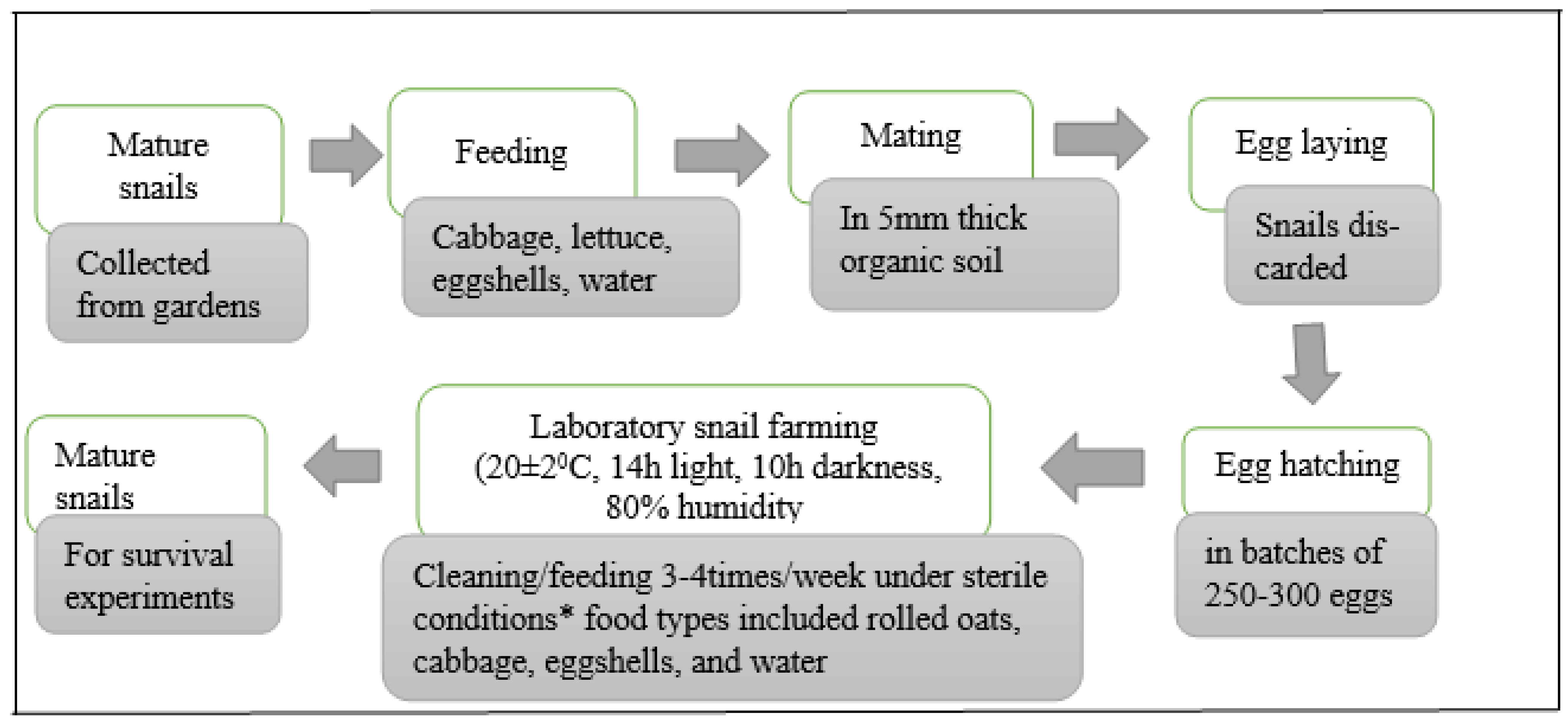
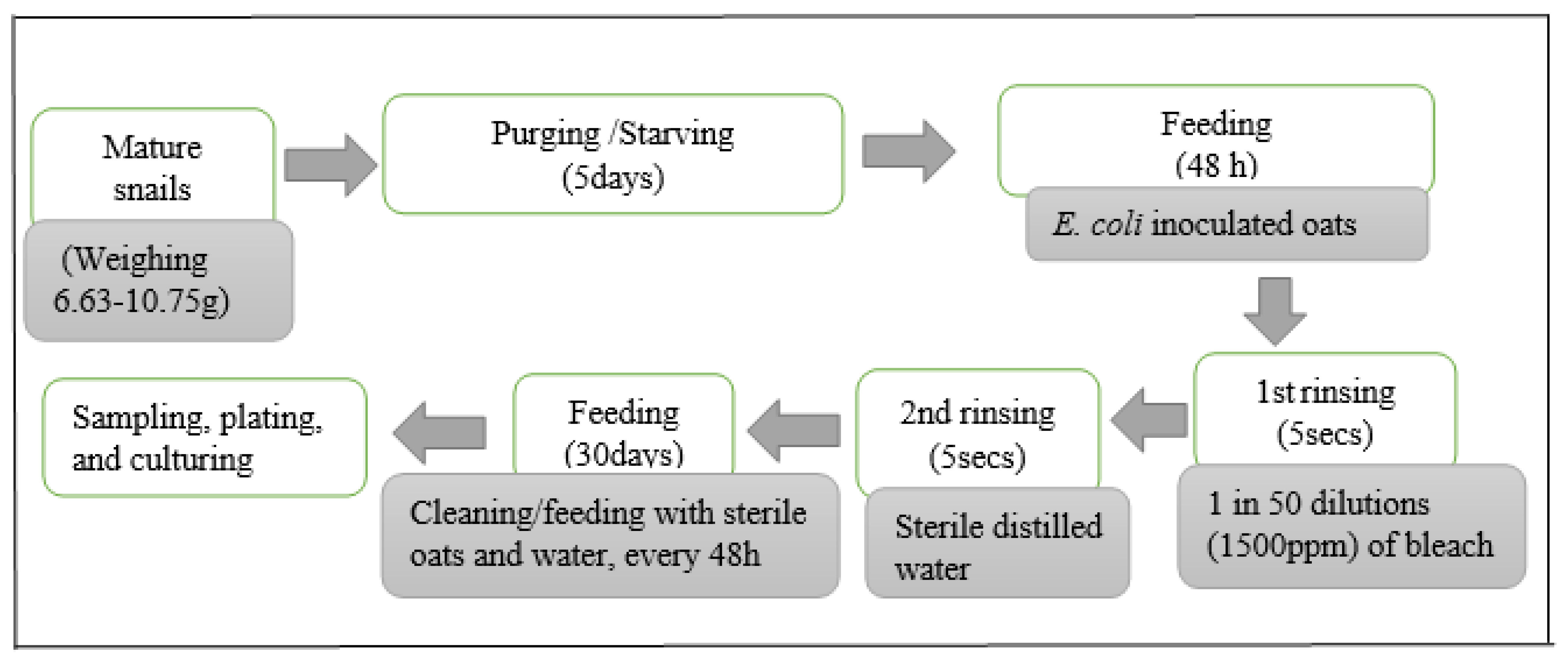
2.2. Preparation of Experimental Snails

The snails were prepared for inoculation experiments based on established methods for laboratory snail farming [29] and outdoor snail farming or heliciculture [6,30,31]. In brief, 50 mature land snails (Helix aspersa) of reproductive age were collected from vegetable farms in Christchurch, New Zealand, during the summer months of November to January 2019. Snail species were identified by their morphological characteristics; for example, the shells were brown with 4 to 5 whorls and measured 30 to 45 mm [1,30].
Snails were transported to Lincoln University, New Zealand, maintained in optimal growth conditions (20 ± 2 °C, 14 h light, 10 h darkness, 80% humidity), and fed with vegetables (cabbage, lettuce) and eggshells (a calcium carbonate supplement) until egg laying commenced [29,31]. Aseptically, egg batches were embedded into 5 mm thick pre-autoclaved organic soil (Garden box, Christchurch, New Zealand), kept moist in sterile distilled water, and contained in 2-L perforated Sistema containers (Sistema Plastics, Auckland, New Zealand) pre-treated with exposure to ultraviolet light for ≥2 h. Within 18–21 days, juvenile snails emerged from the organic soil and the snails were raised till maturity, that is, 18 months. A summary of the rearing procedure is presented in Figure 1.

2.3. Inoculation of Experimental Snails

All inoculation experiments were conducted in a biological safety hood. Bacteria suspensions (1 mL) were transferred into 15 mL Eppendorf tubes containing 1 g of moist rolled oats each. The rolled oats were rehydrated according to the manufacturer’s instructions (Harraways, Dunedin, New Zealand). The inoculant concentration in the rolled oats was such that when initially examined on TBX agar at 36 °C for 20 h, the bacterial colonies were approximately 106 CFU/mL, based on a 10−3 dilution of a suspension made to an optical density of 1.0 determined at 600 nm (SmartSpec, Bio-Rad, Hercules, CA, USA). A total of 40 tubes were prepared, thoroughly mixed, and poured into a sterile 2-L perforated Sistema container (Sistema Plastics, Auckland, New Zealand). 40 mature land snails, previously purged with rolled oats for 36 h and then starved for 48 h [29], were then introduced into the mixture of bacteria-rolled oats for 48 h. In principle, this implied that 1 g of inoculated oats corresponded to 1 land snail. Oats used throughout the study had been previously screened for the presence of E. coli (using methods described by [32], modified by the use of 5 g samples in the protocol) and tested negative for this organism. The procedure is summarised in Figure 2.
After 48 h of exposure and feeding on inoculated oats, the snails were rinsed for ca. 5 s with 15 mL of 5% sodium hypochlorite (Janola premium bleach, Pental Products Pty Ltd., Melbourne, Australia) and then 15 mL of sterile distilled water to achieve surface decontamination. Preliminary experiments with snails intentionally contaminated with E. coli demonstrated that this protocol was effective in snail surface decontamination when rinse water was cultured on Nutrient agar no. 2 (Oxoid, Hampshire, UK) at 37 °C for 18 h. Snails remained viable after this treatment.

2.4. E. coli Survival Experiments

After the brief rinsing with 5% sodium hypochlorite and distilled water, the 40 snails from each E. coli strain inoculation were used as the starting samples for the 30-day survival experiments (Figure 2). Snails were then fed with 40 g of sterile moist rolled oats, and periodically every 48 h, their faeces were collected at the point of cleaning and feeding with fresh rolled oats. The enumeration of E. coli in snail faeces was conducted based on Silva, Marta [32], with slight modifications. 5 g of pooled snail faeces from each inoculated group in one “Sistema” container were thoroughly vortexed in 45 mL of Phosphate buffered saline, PBS (10−1 dilution), and the suspension was allowed to stand for 30 mins. Subsequent dilutions (10−1 to 10−6) of the sample were prepared, and 1 mL each of three appropriate dilutions (10−1, 10−2, 10−4) were inoculated into Durham tubes containing 2 mL of E. coli (EC) broth. The samples were then incubated at 44.5 ± 0.2 °C/24 h in a shaker incubator (ThermoScientific, Auckland, New Zealand), rotating at 120 rpm.
Using a spread plate method, 50 µL of serial dilutions (10−1 to 10−6) of EC broth samples were plated on TBX agar. The plates were incubated at 37 °C for 20 h, and 30–250 bacteria colonies were visually counted using a 6X LED illuminated linen tester (Magnifiers, Christchurch, New Zealand). The surviving colonies of E. coli were confirmed as blue-green colonies in TBX agar plates [33]. Further E. coli confirmation tests for inconclusive colonies were performed using previously described PCR protocols [19]. The same procedure was used to screen for E. coli in rolled oats prior to feeding inoculated snails during the entire 30-day survival experiments. It should be noted that 30 days were determined as the maximum period for survival experiment that allowed for adequate faecal sample collection for microbiological analyses, since it was observed that snails barely survived beyond this period under the aforementioned post-inoculated laboratory feeding and growth conditions.

2.5. Statistical Analysis

All experiments were carried out in triplicate. The values of CFU/mL for each timepoint were expressed as the mean ± standard deviation of the three separate survival experiments, with the mean values used as the basis for statistical analysis. A commercial spreadsheet software (Microsoft 2010) was used to perform all calculations. R programming (version 4.0.3) was used in the plotting of the best-fit graphs and in performing statistical analyses, that is, Analysis of Variance (ANOVA) and student t-tests. The mean number of CFU/mL from each sampling time was calculated and represented as logarithmic transformed data; that is, each calculated CFU/mL was replaced with the transformed value log10(x), where x = CFU/mL. The normal distribution of each logarithmic transformed data set was graphically verified (with a confidence level of 95%). Linear regression analysis was used to determine the best relationship between the two E. coli strains. The D-values (the time required to reduce the E. coli population by 1log cycle) were also determined by taking the negative reciprocal of the slope from the linear regression equations [34].

3. Results

The number of CFU/mL for each strain decreased over the examination period (Supplementary Tables S1–S5). The average rate of decline of the laboratory-adapted E. coli strain CSH-62 in the faeces of live snails was significantly (p < 0.05) faster than the pathogenic (Shiga-toxigenic) E. coli ERL 06-2503. Furthermore, the viable population of the Shiga-toxigenic E. coli strain ERL 06-2503 persisted significantly (p < 0.05) for a longer time in the intestinal tract of land snails than that of the laboratory-adapted E. coli CSH-62. These trends are clearly seen in the graph using the “best fit” algorithm in the “R” software environment (Version R 4.2.0) (Figure 3).
Strain ERL 06-2503 showed a relatively rapid decrease to 6.78 log10 CFUmL−1 within the first two days but was constant for the next 14 days before a marked decrease at day 28; in contrast, E. coli strain CSH-62 decreased steadily throughout the experiment (Figure 3). For both strains, viable bacteria remained detectable up to the end of the sampling period, that is, approximately 165,000 CFU/mL for E. coli CSH-62 and 403,000 CFU/mL for E. coli ERL 06-2503. The mean decline in the viable population demonstrated first-order kinetics. With no clear evidence of shoulders or tails, this permitted the calculation of the decimal reduction times (D-values) from the linear parts of the survival curves [22,34].
E. coli CSH-62 displayed three timepoints at which a 1-log decrease in its viable population was evident (Table 1). In the first two days, a 0.95 log10 CFUmL−1 decrease in viable counts was observed. The calculated D value (time for a 1 log decrease in E. coli) for this initial time period (0 to 2 days) was 1.59 days. An additional decline of 1.28 log10 CFUmL−1 in viable bacteria occurred during the next 18 days. The calculated D-value for this second phase (days 2 to 18) was 14.62 days. Finally, the third decrease in the viable count of 1.07 log10 CFUmL−1 for the remaining 30 days of the study occurred between day 18 and day 26. Its D-value was 25.36 days.
However, corresponding analysis of the results for STEC strain ERL 06-2503 revealed only two timepoints at which a 1-log decrease was seen (Table 1). An initial decrease in viable counts of 1.11 log10 CFUmL−1 was observed on day 2 after inoculation. The calculated D-value for day 0 to 2 was 1.38 days. The second decrease of 1.03 log10 CFUmL−1 in viable count was observed on day 22. The calculated D-value (day 2 to 22) was 18.08 days. From day 22 to 30, we obtained a D-value of 28.96 days, of which a further 1.03 log10 CFUmL−1 of viable bacteria was observed on day 30 (the last day of sampling).

4. Discussion

Snail farmers all over the world are experimenting with different types of indoor and outdoor snail production systems. The challenges in adapting well-defined snail farming systems are due to the ability of snails to adversely respond to changes in climatic conditions [6,31]. In both farming systems, it is well known that land snails, such as African giant snails, are hosts to many parasites and bacterial pathogens [4,5,8,13,15,17,18,19,20,35]. Indeed, our previous work [19] detected at least one enteric pathogen in every snail sample examined, with STEC found in 57% of the samples examined. In this research, we studied E. coli interactions with land snails, Helix spp., to gain an understanding of the colonisation dynamics and infectious potential of this relationship. Our results show that E. coli can survive in edible land snails for at least a month and that pathogenic variants such as STEC may survive for longer.
Land snails are known to occupy various environments and are susceptible to E. coli contamination due to (1) their free-living nature, (2) their close contact with the soil and (3) their random feeding patterns [4,5,8,19]. Previous research has also detected different strains of pathogenic E. coli in the faeces of healthy animals that were raised in indoor and grassland systems [22,23]. For example, to establish the transmission pattern of E. coli O157: H7 between cattle groups, Scott and McGee [36] reported that a total of 8.7% (6/69) of cattle shed E. coli O157: H7 in the first month, with faecal shedding increasing to 52% (36/69) in the third month after infection. Kudva and Blanch [23] recovered E. coli O157: H7 in the range of <102 to <106 CFU/g in the manure of inoculated sheep, further demonstrating that E. coli could survive for 21 months in bovine faeces, despite fluctuating environmental conditions. Furthermore, Avery and Moore [22] demonstrated that E. coli from the faeces of indoor cattle, sheep and pigs can survive on grass for at least 5–6 months. Under such conditions, host animals are continually re-exposed to strains from the environment and possess body temperatures favourable to E. coli multiplication. Snails are poikilothermic and, in our experiments, exposed only once to the inoculant. Despite these conditions, both pathogenic and laboratory-adapted strains of E. coli were detected for 30 days. This suggests that the faeces of infected snails in the environment could act as a significant reservoir for the transmission of E. coli to humans and/or animals. Moreover, our study suggests that current helicicultural practices expose snail handlers, and subsequently consumers, to foodborne pathogens.
The presence of E. coli in the faeces of snails further revealed that snails as intermediate hosts and/or the consumption of raw or undercooked snail meat may not necessarily be the only route to foodborne illnesses associated with land snails. For example, snails are known to leave their slime and faeces on locally produced green leafy vegetables in gardens, which is an example of this research’s initial snail harvesting location. Humans could directly ingest these bacterial pathogens from the faeces of snails present in their daily gardening. For example, in 2021, E. coli O157: H7 foodborne outbreaks were reported in baby spinach, leafy greens, and several unknown food sources in the United States [37]. Mukherjee et al. [38] reported a sporadic case of E. coli O157 infection in a child who acquired E. coli O157: H7 from garden soil that had been previously fertilized with manure from cattle in Minnesota, USA. Through PCR and pulsed-field gel electrophoresis techniques, it was demonstrated that E. coli O157:H7 strains can survive on manure-amended soil for more than 2 months [38].
Using the non-invasive laboratory-adapted E. coli strain CSH-62 and Shiga-toxigenic pathogenic E. coli strain ERL 06-2503, this research has demonstrated that the outcome of E. coli interactions with land snails may be significantly (p < 0.05) strain dependent. The STEC strain used was specifically chosen for its ability to robustly colonize an ex vivo bovine intestinal model; more robustly, in fact, than other strains studied [26]. Strain variance was strongly evidenced in our study. Strain CSH-62 demonstrated an approximate 3 log10 CFUmL−1 decline in its viable population, while the pathogenic E. coli strain ERL 06-2503 demonstrated 2 log10 CFUmL−1 reduction in its viable population. Similar results have been reported [39], where invasive E. coli K1 had the ability to penetrate and remain viable in the free-living protozoan Acanthamoeba, while non-invasive E. coli K-12 was progressively killed.
Although it is unclear how both E. coli strains maintained a somewhat prolonged survival in land snails, their continued persistence in edible land snails represents significant risks to public health. Previous recommendations [31,40] indicate that the purging/starvation of land snails for 3–5 days prior to human consumption will accelerate the process of ridding the snail digestive system of soil, food stuffs and microorganisms. However, this is contrary to our findings, as both pathogenic and laboratory-adapted E. coli strains were actively viable beyond day 5 after inoculation and during the entire period whereby the sole nutrition source was soaked oats, a commonly used purging agent. Our results provide evidence that land snails may host both pathogenic and non-pathogenic E. coli for prolonged periods (30 days), which may be transmitted to susceptible hosts, including humans. More research is required to understand the fate of E. coli after this experimental research period, the survival mechanisms of the different E. coli serotypes in the guts of edible land snails, and the spectrum of survival behaviours from additional strains and pathovars of importance to human health.

5. Conclusions

In summary, this study revealed that upon ingestion and continuous shedding in the faeces of snails, E. coli could survive in edible land snails for more than a month. Although the results indicate that the viable populations of E. coli declined significantly within the 30-day experimental period, the ability of E. coli to survive throughout the study under controlled laboratory conditions precluding reinfection is of great significance for helicicultural practices and public health, especially in regions where the carriage of pathogenic Shigatoxigenic strains is so prevalent [19] and where snails are frequently consumed as a source of nutrition. We accept that our results, based on only two (albeit well characterised) strains, may be regarded as preliminary, an unfortunate casualty of issues concerning access to laboratories during the COVID-19 lockdown period in New Zealand. Nonetheless, we feel the data further illustrate differences between E. coli strains in their host colonisation ability, as identified before in a cattle gut model system [26]. We also hope that this inaugural study of E. coli colonisation and survival in edible snails can inform better helicicultural practices to reduce the risks of foodborne illness. Clearly, there are implications for existing practices where snails are sourced from native, uncontrolled conditions, and there may well be advantages in this respect in encouraging a more widespread practice of snail rearing under more hygienic and controlled conditions. The need for legislation to formalise such interventions is open to conjecture.

Supplementary Materials

The following supporting information can be downloaded at: https://www.mdpi.com/article/10.3390/pathogens13030204/s1, Table S1: Reformatted data; Table S2: Means SDs CSH62; Table S3: Means SDs STEC; Table S4: Raw data; Table S5: Corrected data.

Author Contributions

Conceptualization, S.L.W.O. and M.N.T.; methodology, S.L.W.O. and M.N.T.; formal analysis, S.L.W.O. and M.N.T.; investigation, M.N.T.; resources, S.L.W.O.; writing—original draft preparation, M.N.T.; writing—review and editing, S.L.W.O., M.N.T., G.N. and I.C.N.P.; visualization, M.N.T.; supervision, J.J.S., S.L.W.O., G.N. and I.C.N.P.; project administration, S.L.W.O.; funding acquisition, S.L.W.O. and M.N.T. All authors have read and agreed to the published version of the manuscript.

Funding

This work was supported by the New Zealand Aid programme for a doctoral scholarship to MNT.

Institutional Review Board Statement

Ethical review and approval were waived for this study since such requirements are not required in New Zealand for studies of molluscs.

Data Availability Statement

All applicable data are given in the Supplementary Tables S1–S5.

Acknowledgments

We thank Janine Koster and Liz McCormick for providing land snails for the preliminary stages of this research.

Conflicts of Interest

The authors declare no conflicts of interests.

References

  1. Paszkiewicz, W.; Kozyra, I.; Rzeżutka, A. A Refinement of an International Standard Method (ISO/TS 15216–2:2013) to Allow Extraction and Concentration of Human Enteric Viruses from Tissues of Edible Snail Species. Food Anal. Methods 2014, 8, 799–806. [Google Scholar] [CrossRef]
  2. Okafor, F.C. The Varied Roles of Snails (Gastropod Molluscs) in the Dynamics of Human Existence. Master’s Thesis, University of Lagos, Lagos, Nigeria, 2009; 153p. [Google Scholar]
  3. APHA. The Animal and Plant Health Agency. Import of Snail Meat for Human Consumption. Import Information Note (IIN) BAL/3. Available online: http://apha.defra.gov.uk/official-vets/Guidance/bip/iin/animal-prod-hum-cons.htm (accessed on 7 May 2022).
  4. Akpomie, O.O. Assessment of health implication associated with snails and snail farm soils in Warri and Sapele, Delta state, Nigeria. Niger. J. Sci. Environ. 2013, 12, 50–56. [Google Scholar]
  5. Barimah, M.N.Y.S. Microbiological Quality of Edible Land Snails from Selected Markets in Ghana. Master’s Thesis, Department of Microbiology, University of Ghana Medical School, Accra, Ghana, 2013. [Google Scholar]
  6. Cobbinah, J.R.; Vink, A.; Onwuka, B. Snail Farming: Production, Processing and Marketing; Agromisa: Wageningen, The Netherlands, 2008. [Google Scholar]
  7. Mohammed, S.; Ahmed, A.; Adjei, D. Opportunities for Increasing Peasant Farmers Income through Snail Production in Ghana. Sch. J. Agric. Vet. Sci. 2014, 1, 195–200. [Google Scholar]
  8. Nyoagbe, L.A.; Appiah, V.; Josephine, N.; Daniel, L.; Isaac, A. Evaluation of African giant snails (Achatina and Archachatina) obtained from markets (wild) and breeding farms. Afr. J. Food Sci. 2016, 10, 94–104. [Google Scholar] [CrossRef]
  9. Kocatepe, D.; Çelik, Y. Snail Consumption Habits and Health. Poster in Ecology 2017 International Symposium. 2017. 2p. Available online: https://www.researchgate.net/publication/321330613_Snail_Consumption_Habits_And_Health#fullTextFileContent (accessed on 18 June 2022).
  10. Pissia, M.A.; Matsakidou, A.; Kiosseoglou, V. Raw materials from snails for food preparation. Future Foods 2021, 3, 100034. [Google Scholar] [CrossRef]
  11. Tardugno, R.; Virga, A.; Nava, V.; Mannino, F.; Salvo, A.; Monaco, F.; Giorgianni, M.; Cicero, N. Toxic and Potentially Toxic Mineral Elements of Edible Gastropods Land Snails (Mediterranean Escargot). Toxics 2023, 11, 317. [Google Scholar] [CrossRef]
  12. Ngenwi, A.A.; Mafeni, J.M.; Etchu, K.A.; Oben, F.T. Characteristics of Snail Farmers and Constraints to Increased Production in West and Central Africa. Afr. J. Environ. Sci. Technol. 2010, 4, 274–278. [Google Scholar]
  13. Parlapani, F.F.; Neofitou, C.; Boziaris, I.S. Microbiological quality of raw and processed wild and cultured edible snails: Microbiological quality of edible snails. J. Sci. Food Agric. 2014, 94, 768–772. [Google Scholar] [CrossRef]
  14. Waldhorn, D.R. Snails Used for Human Consumption: The Case of Meat and Slime. Available online: https://rethinkpriorities.org/publications/snails-used-for-human-consumption (accessed on 1 June 2022).
  15. Tanyitiku, M.N.; Nicholas, G.; Sullivan, J.J.; Njombissie Petcheu, I.C.; On, S.L.W. Snail Meat Consumption in Buea, Cameroon: The Methodological Challenges in Exploring Its Public Health Risks. Int. J. Qual. Methods 2022, 21, 16094069221078132. [Google Scholar] [CrossRef]
  16. Kiebre-Toe, M.; Borges, E.; Maurin, F.; Richard, Y.; Kodjo, A. Etude de la flore bacterienne a ‘erobie’ a Gram n ‘egatif de l’escargot d’ ‘elevage’ (Helix aspersa). Rev. De Médecine Vétérinaire 2003, 154, 606–610. [Google Scholar]
  17. Adegoke, A.A.; Bukola, A.T.C.; Comfort, I.U.; Olainka, A.A.; Amos, K.O. Snails as meat source: Epidemiological and nutritional perspectives. J. Microbiol. Antimicrob. 2010, 2, 001–005. [Google Scholar]
  18. Nwuzo, A.C.; Iroha, I.R.; Moses, I.B.; Ugbo, E.N.; Agumah, N.B.; Orji, J.; Ogene, L. Isolation and Characterization of Bacterial Species Associated with Edible Snails (Achatina achatina) sold in Major Markets within Abakaliki Metropolis. Biolife 2016, 4, 494–497. [Google Scholar]
  19. Tanyitiku, M.N.; Nicholas, G.; Njombissie Petcheu, I.C.; Sullivan, J.J.; On, S.L. Public health risk of foodborne pathogens in edible African land snails, Cameroon. Emerg. Infect. Dis. 2022, 28, 1715. [Google Scholar] [CrossRef]
  20. Temelli, S.; Dokuzlu, C.; Sen, M.K.C. Determination of microbiological contamination sources during frozen snail meat processing stages. Food Control 2006, 17, 22–29. [Google Scholar] [CrossRef]
  21. Capps, K.M.; Ludwig, J.B.; Shridhar, P.B.; Shi, X.; Roberts, E.; DebRoy, C.; Cernicchiaro, N.; Phebus, R.K.; Bai, J.; Nagaraja, T.G. Identification, Shiga toxin subtypes and prevalence of minor serogroups of Shiga toxin-producing Escherichia coli in feedlot cattle feces. Sci. Rep. 2021, 11, 8601. [Google Scholar] [CrossRef] [PubMed]
  22. Avery, S.M.; Moore, A.; Hutchison, M.L. Fate of Escherichia coli originating from livestock faeces deposited directly onto pasture. Lett. Appl. Microbiol. 2004, 38, 355–359. [Google Scholar] [CrossRef]
  23. Kudva, I.T.; Blanch, K.; Hovde, C.J. Analysis of Escherichia coli O157:H7 survival in ovine or bovine manure and manure slurry. Appl. Environ. Microbiol. 1998, 64, 3166–3174. [Google Scholar] [CrossRef]
  24. Moriarty, E.M.; Weaver, L.; Sinton, L.W.; Gilpin, B. Survival of Escherichia coli, Enterococci and Campylobacter jejuni in Canada Goose Faeces on Pasture. Zoonoses Public Health 2012, 59, 490–497. [Google Scholar] [CrossRef]
  25. Cízek, A.; Literák, I.; Scheer, P. Survival of Escherichia coli O157 in faeces of experimentally infected rats and domestic pigeons. Lett. Appl. Microbiol. 2000, 31, 349–352. [Google Scholar] [CrossRef]
  26. Brandt, S.M.; Paulin, S.M. Quantifying colonization potential of enterohemorrhagic Escherichia coli O157 using bovine in vitro organ culture and immunofluorescent staining. Foodborne Pathog. Dis. 2012, 9, 1064–1070. [Google Scholar] [CrossRef]
  27. Nakane, K.; Kawamura, K.; Goto, K.; Arakawa, Y. Long-Term Colonization by bla(CTX-M)-Harboring Escherichia coli in Healthy Japanese People Engaged in Food Handling. Appl. Environ. Microbiol. 2016, 82, 1818–1827. [Google Scholar] [CrossRef]
  28. Thorne, G.M.; Farrar, W.E. Genetic Properties of R Factors Associated with Epidemic Strains of Shigella dysenteriae Type I from Central America and Salmonella typhi from Mexico. J. Infect. Dis. 1973, 128, 132–136. [Google Scholar] [CrossRef]
  29. Garcia, A.; Perea, J.; Mayoral, A.; Acero, R.; Martos, J.; Gomez, G.; Peña, F. Laboratory rearing conditions for improved growth of juvenile Helix aspersa Muller snails. Lab. Anim. 2006, 40, 309–316. [Google Scholar] [CrossRef]
  30. Thompson, R.; Cheney, S. Raising Snails; U.S. Department of Agriculture, National Agricultural Library: Beltsville, MD, USA, 1996.
  31. Murphy, B. Breeding and Growing Snails Commercially in Australia: A Report for the Rural Industries Research and Development Corporation. Available online: http://www.rirdc.gov.au/reports/NAP/00-188.htm (accessed on 18 June 2022).
  32. Silva, N.d.; Marta, T.H.; Valeria, J.C.A.; Neliane, S.F.A.; Margarete, O.M.; Renato, G.A.R. Microbiological Examination Methods of Food and Water: A Laboratory Manual, 2nd ed.; CRC Press/Balkema: Leiden, The Netherlands; Boca Raton, FL, USA, 2018. [Google Scholar]
  33. Vergine, P.; Salerno, C.; Barca, E.; Berardi, G.; Pollice, A. Identification of the faecal indicator Escherichia coli in wastewater through the -D-glucuronidase activity: Comparison between two enumeration methods, membrane filtration with TBX agar, and Colilert(R)-18. J. Water Health 2016, 15, 209–217. [Google Scholar] [CrossRef] [PubMed]
  34. Edelson-Mammel, S.G.; Buchanan, R.L. Thermal inactivation of Enterobacter sakazakii in rehydrated infant formula. J. Food Prot. 2004, 67, 60–63. [Google Scholar] [CrossRef] [PubMed]
  35. Meffowoet, C.P.; Kouam, K.M.; Kana, J.R.; Tchakounte, F.M. Infestation rate of African giant snails (Achatina fulica and Archachatina marginata) by parasites during the rainy season in three localities of Cameroon. J. Vet. Med. Res. 2020, 7, 1–8. [Google Scholar]
  36. Scott, L.; McGee, P.; Minihan, D.; Sheridan, J.J.; Earley, B.; Leonard, N. The characterisation of E. coli O157:H7 isolates from cattle faeces and feedlot environment using PFGE. Vet. Microbiol. 2006, 114, 331–336. [Google Scholar] [CrossRef]
  37. CDC. Center for Disease Control 2021 Outbreaks. Available online: https://www.cdc.gov/ecoli/outbreaks.html (accessed on 18 June 2022).
  38. Mukherjee, A.; Cho, S.; Scheftel, J.; Jawahir, S.; Smith, K.; Diez-Gonzalez, F. Soil survival of Escherichia coli O157:H7 acquired by a child from garden soil recently fertilized with cattle manure. J. Appl. Microbiol. 2006, 101, 429–436. [Google Scholar] [CrossRef]
  39. Alsam, S.; Jeong, S.; Sissons, J.; Dudley, R.; Kim, K.; Khan, N. Escherichia coli interactions with Acanthamoeba: A symbiosis with environmental and clinical implications. J. Med. Microbiol. 2006, 55, 689–694. [Google Scholar] [CrossRef]
  40. Cerezal Mezquita, P.; Madariaga, P.; Segovia, Y. A practical approach to preparation and meat preservation by refrigeration or freezing of the land snail (helix aspersa müller). J. Muscle Foods 2009, 20, 401–419. [Google Scholar] [CrossRef]
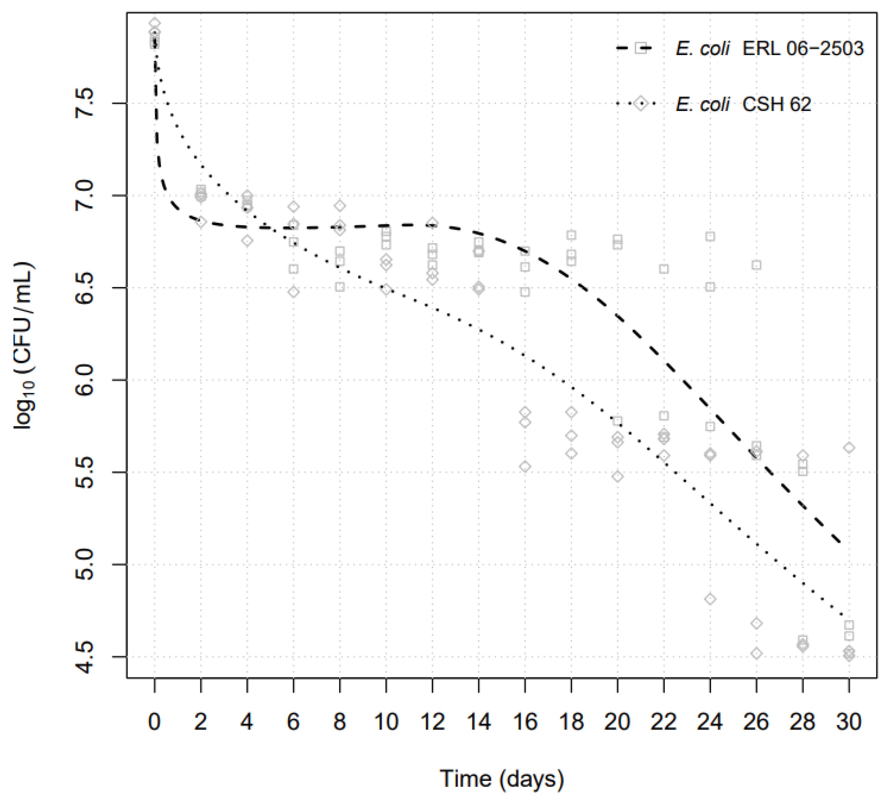
Figure 1. Preparation schedule of snails for bacterial inoculation. * Regular sterilization (121 °C/20 min) of all types of food, materials and equipment used.
Pathogens 13 00204 g001
Figure 2. Inoculation schedule of snails for E. coli survival experiments.
Pathogens 13 00204 g002
Figure 3. Survival in snails of E. coli strains STEC ERL 06-2503 and laboratory-adapted strain CSH 62 during the 30-day examination period, using the “best fit” algorithm in the “R” software environment.
Pathogens 13 00204 g003
Table 1. Calculated D-values of E. coli strains examined within the 30-day period at timepoints showing a ≥1-log decrease in colony forming units.
E. coli Isolates Day of ~1log Cyclelog10 CFUmL−1D-Value (Days)
E. coli CSH-6226.9521.59
185.67514.62
264.60425.36
E. coli ERL 06-250326.7951.38
225.76518.08
Disclaimer/Publisher’s Note: The statements, opinions and data contained in all publications are solely those of the individual author(s) and contributor(s) and not of MDPI and/or the editor(s). MDPI and/or the editor(s) disclaim responsibility for any injury to people or property resulting from any ideas, methods, instructions or products referred to in the content.

