Review

Diagnosis and Management of Infections in Patients with Mendelian Susceptibility to Mycobacterial Disease

by
Aparna Dalvi
1,
Umair Ahmed Bargir
1,
Gita Natraj
2,
Ira Shah
3 and
Manisha Madkaikar
1,*
1
Department of Pediatric Immunology, ICMR National Institute of Immunohaematology, Mumbai 400012, India
2
Seth GS Medical College and KEM Hospital, Mumbai 400012, India
3
Bai Jerbai Wadia Hospital for Children, Mumbai 400012, India
*
Author to whom correspondence should be addressed.
Pathogens 2024, 13(3), 203; https://doi.org/10.3390/pathogens13030203
Submission received: 20 December 2023 / Revised: 15 February 2024 / Accepted: 23 February 2024 / Published: 25 February 2024
(This article belongs to the Special Issue Infection in Inborn Errors of Immunity)

Abstract

:
The diagnosis and treatment of patients with mendelian susceptibility to mycobacterial disease (MSMD) pose consistent challenges due to the diverse infection spectrum observed in this population. Common clinical manifestations include Bacillus Calmette-Guérin vaccine (BCG) complications in countries where routine BCG vaccination is practiced, while in non-BCG-vaccinating countries, Non-Tuberculous Mycobacteria (NTM) is prevalent. In tuberculosis-endemic regions, Mycobacterium tuberculosis (MTB) has a high prevalence, along with other intracellular organisms. Isolating these organisms presents a significant challenge, and treatment is often initiated without confirming the specific species. This review primarily focuses on the methods and challenges associated with diagnosing and treating MSMD patients.

1. Introduction

Inborn errors of immunity (IEI) are a group of disorders that result in an impaired immune response [1]. IEIs are classified by the International Union of Immunology Societies (IUIS) depending on the clinical and immunological evaluation of the defects. Mendelian susceptibility to mycobacterial diseases (MSMD) is a rare IEI that is grouped under defects of intrinsic and innate immunity [2,3]. MSMD predisposes to clinical disease caused by weakly virulent mycobacteria such as the Bacillus Calmette-Guérin (BCG) vaccine, nontuberculous mycobacteria (NTM), or environmental mycobacteria (EM). Infections due to more virulent M. tuberculosis have also been reported in MSMD patients. These patients present early in childhood, whereas milder forms can have an onset in adulthood and even remain asymptomatic. Mycobacterial diseases range from localized to disseminated or persistent life-threatening infections. Patients with MSMD may also suffer from infections due to intramacrophagic bacteria, fungi, parasites, and viruses [4,5,6,7,8,9,10,11]. Mutations in 21 different genes (IFNGR1, IFNGR2, IFNG, IL12RB1, IL12RB2, IL23R, IL12B, ISG15, USP18, ZNFX1, TBX21, STAT1, TYK2, IRF8, CYBB, JAK1, RORC, NEMO, SPPL2A, MCTS1, and IRF1) with more than 35 different genetic etiologies of MSMD have been described [12,13,14,15,16,17,18]. These deficiencies exhibit either autosomal recessive, autosomal dominant, or X-linked patterns of inheritance. Confirming the underlying genetic defects in these patients provides a better therapeutic approach and genetic counseling for the affected families. In most suspected MSMD cases, patients are treated empirically without confirming a microbiological and molecular diagnosis. This review mainly focuses on approaches and challenges associated with diagnosing and managing MSMD patients.

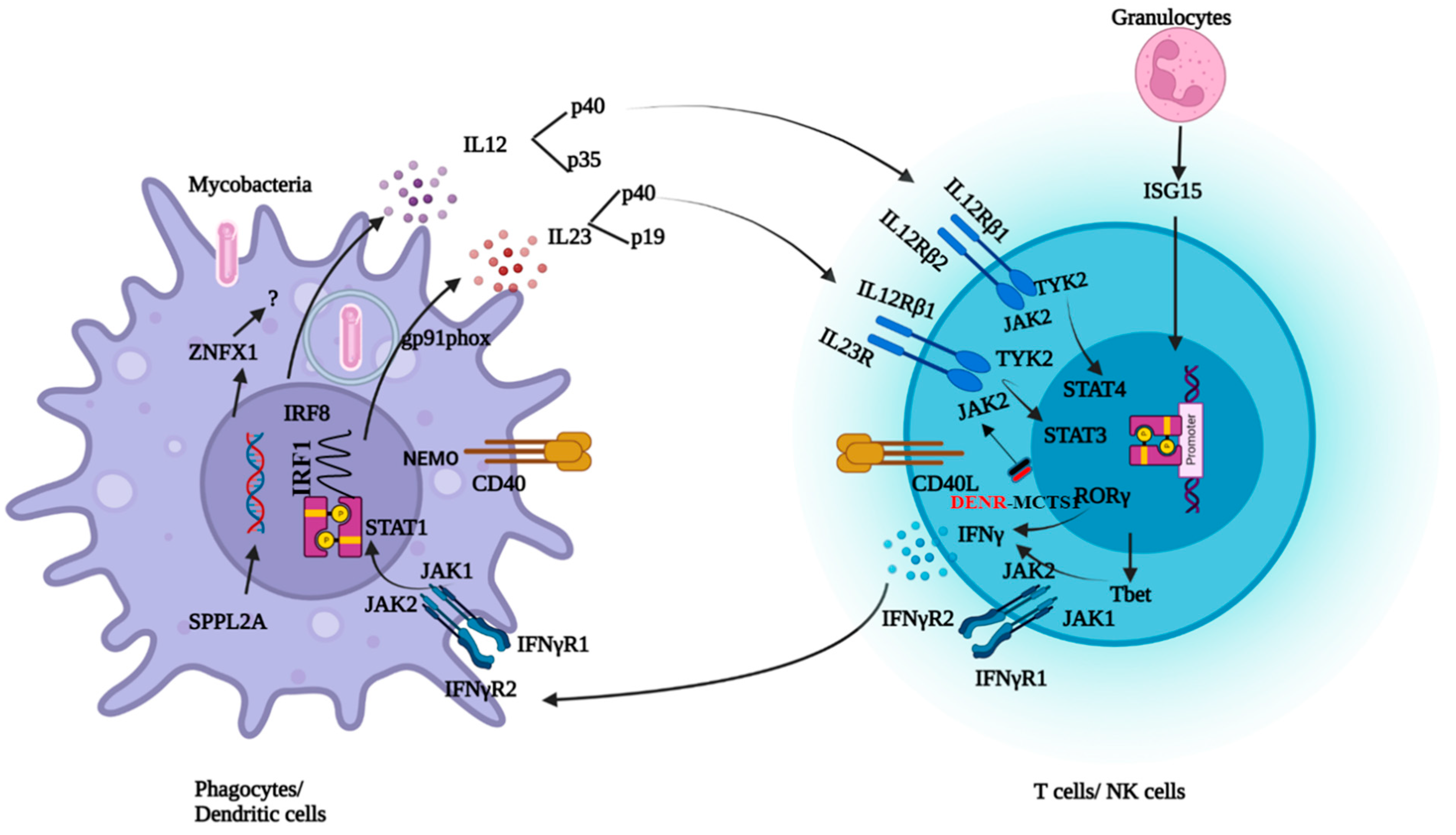
2. Pathophysiology and Clinical Manifestations of MSMD

Type I cytokines such as IFNγ, IL12, and IL23 play a crucial role in the control of intracellular microorganisms. Once the bacteria are internalized, macrophages recognize the bacterial pathogen-associated molecular patterns and become activated. This activation produces cytokines such as IL12, IL23, and tumor necrosis factor-α (TNF-α). TNF-α plays a role in the formation of granulomas. IL12 leads to the differentiation of T cells into Th1 subpopulations and induces IFNγ secretion. IFNγ further activates the macrophages to eliminate the bacteria. In 1996, the discovery of IFNγR1 deficiency was reported as the first genetic etiology of MSMD with BCG infection. Over the period of discoveries of new genes in MSMD, this condition has been termed an inborn error of IFNγ immunity [7]. To mount an effective immune response against mycobacterial infections, the integrity of the IL12/23/ISG15-IFN-γ circuit is necessary (Figure 1). Mutations in the 21 genes involved in this circuit lead to either an impaired response or the production of IFNγ [8]. Molecular defects in the IL12RB1 and IFNGR1 genes together constitute about 80% of MSMD cases [9].
IL12Rβ1 deficiency is the most common genetic cause of MSMD. IL12 and IL-23 share a common IL12Rβ1 chain encoded by IL12RB1. Therefore, IL12 and IL23 signaling are defective in patients with IL12Rβ1 deficiencies. The clinical phenotype ranges from early deaths in infancy to asymptomatic adulthood, suggesting an incomplete penetrance [19,20]. Patients are vulnerable to mycobacterial disease, Salmonella infections, and chronic mucocutaneous candidiasis (CMC). About 25% of the patients with IL12Rβ1 deficiency develop fungal infections, including CMC [8,9,10]. It is unclear why only some patients with MSMD show susceptibility to fungal infections. Patients with IL-12p40, IL-12Rβ1, or IL-23R deficiency exhibit normal levels of circulating leukocytes from various subsets, including cell types that typically express IL-23R, such as NK, iNKT, mucosal-associated invariant T (MAIT), Vδ2+ γδ T, TH1, TH17, and TH1* cells. However, the susceptibility of certain individuals with deficiencies in IL-12B, IL-12Rβ1, or IL-23R to chronic mucocutaneous candidiasis (CMC) indicates the crucial role of human IL-23 in defending against mucocutaneous Candida spp. infections mediated by IL-17A/F. Recent findings showed that IL-23 induces IL-17A exclusively in MAIT cells from healthy individuals, a response absent in IL-23R-deficient patients. The incomplete penetrance of CMC in these patients is not solely explained by individual variability in IL-17A/F production by leukocyte subsets. Instead, it may be due to the preservation of C. albicans-specific memory CD4+ T cell development in IL-23R-deficient patients and the fact that IL-17A induction is governed by IL-23 in only one lymphocyte subset (MAIT cells), at least in ex vivo conditions [21].
In IL12Rβ1 patients, other clinical manifestations, such as autoimmunity and inflammatory bowel disease (IBD), have been described. Impairment in IL12Rβ1 signaling led to deficiencies in the differentiation of Th17 cells, which are vital for maintaining intestinal homeostasis. Th17 cells contribute to this balance by producing key cytokines, including IL-6, TGF-β, IL-17, and IL-1β, particularly in mucosal damage scenarios. They also play a pivotal role in defending against extracellular pathogens and stimulating the release of antimicrobial peptides from intestinal epithelial cells. The observed reduction in the Th17 cell population in the patient strongly suggests its potential involvement in the development of inflammatory bowel disease in this individual, aligning with the clinical manifestations seen in IL-12Rb1 patients, such as autoimmunity and IBD [22,23,24].
The second most common cause of MSMD is IFNγR1 deficiency. Two forms of autosomal recessive complete IFNγR1 deficiencies are described, with the presence of a non-functional receptor or a receptor that does not express at the cell surface. These complete forms of IFNγR1 are severe forms of MSMD. Patients present early, before 3 years of age, and suffer from disseminated BCG and/or EM with poorly characterized granuloma lesions. Infections with M. tuberculosis, viruses, and bacteria have also been reported [25,26,27,28]. In partial IFNγR1 deficiencies, the receptor is expressed on the cell surface, but the response to IFNγ is impaired. These are less severe forms than a complete IFNγR1 deficiency. In AD with partial IFNγR1 deficiency, there is an accumulation of truncated receptors on the surface due to a mutation in the intracellular domain lacking a receptor recycling motif. Higher amounts of protein are detected on the surface. Multifocal osteomyelitis is commonly seen in these patients. Recently, it was reported that AD IFNγR1-deficient and AD STAT1-deficient cells exhibit resistance to the inhibitory effects of IFNγ on osteoclast differentiation and bone resorption. Additionally, the IFNγ induced inhibition of NFATC1 mRNA, a critical transcription factor involved in osteoclast differentiation, was compromised in both AD IFNγR1-deficient and AD STAT1-deficient cells. These findings suggest that the increased osteoclastogenesis is responsible for the occurrence of osteomyelitis in these patients. Osteomyelitis is relatively rare in patients with complete IFNγR1 and STAT1 deficiency. Both complete AR IFNγR1 deficiency and complete AR STAT1 deficiency are life-threatening conditions, necessitating early intervention through hematologic stem cell transplantation to ensure survival. The severity of these disorders poses challenges for long-term clinical course monitoring, potentially resulting in an underestimation of the prevalence of bone involvement [29,30,31].
IL12p40 deficiency is the third most common deficiency reported in MSMD. Both IL12 and 23 are heterodimers having the IL12p40 subunit in common encoded by IL12B. Both IL12 and IL23 signaling are defective in patients with IL12p40 deficiency. Complete IL12p40 deficiency is a clinical phenocopy of IL12Rβ1 deficiency [20,32].
Unlike IFNγR1 deficiency, two forms of complete IFNγR2 deficiency have been reported with or without cell surface expression. The clinical presentation is similar to that of complete IFNγR1 deficiencies. In partial IFNγR2 deficiency, cell surface expression is weak, and response to IFNγ is impaired. The clinical phenotype is milder. Expression of IFNγR2 is very low in AD IFNγR2 deficiency. These deficiencies display incomplete penetrance [33,34].
Mutations in the STAT1, JAK1, IRF8, SPPL2A, NEMO, and CYBB genes are responsible for phagocytic defects leading to susceptibility to mycobacterial infections. JAK1 is a tyrosine kinase that induces phosphorylation and homodimerization of STAT1 upon binding of IFNγ to the receptor. Impaired response to IFNγ in phagocytes is seen in patients with JAK1 and STAT1 deficiencies. -Various forms of inherited STAT1 deficiency have been documented, with autosomal inheritance causing complete or partial STAT1 deficiency. Complete recessive STAT1 deficiencies are a result of the null mutations. The patient’s cells display unresponsiveness to IFN-γ and IFN-α/β. Consequently, these patients are susceptible to severe infections with mycobacteria and viruses. On the other hand, missense mutations lead to partial recessive STAT1 deficiency, resulting in an impaired but not abolished ability to respond to IFN-γ and IFN-α/β, leading to infections with intracellular bacteria and viruses.
Autosomal dominant STAT1 deficiency can result in loss-of-function (LOF) and gain-of-function (GOF) depending on underlined mutations. LOF mutations alter both IFNγ and IFN-α responses but have a negative dominant effect on the IFNγ response only. These patients typically develop mycobacterial infections without viral infections.
Conversely, mutations resulting in STAT1 gain of function predominantly occurring in the coiled-coil domain and DNA binding domain lead to increased phosphorylation and GAS-binding activity. Cells of affected patients exhibit an enhanced response to IFN-γ, IFN-α, and IL27. These patients commonly develop chronic mucocutaneous candidiasis (CMC) [35,36,37,38,39,40,41,42].
NEMO (NF-kappa-B essential modulator ) is a regulatory subunit of the nuclear factor-ĸB (NFĸB) inhibitor kinase complex. Impaired CD40-dependent IL12 production is responsible for the clinical phenotype of patients with NEMO deficiency [43,44]. TYK2 (Tyrosine kinase 2) is a Janus kinase that, upon stimulation with IL12 and 23, induces phosphorylation and dimerization of STAT4 and STAT3. Mutations in TYK2 lead to susceptibility to mycobacterial and viral infections. Homozygosity for the allele of TYK2 P1104A, is a rare genetic etiology of MSMD that selectively disrupts IL23 responses. Patients with defects in RORC are susceptible to mycobacteriosis and candidiasis due to impaired IFNγ and IL17 immunity [8]. Disease-causing mutations in CYBB are known to cause chronic granulomatous disease. The hemizygous mutations Q231P and T178P in CYBB result in diminished NADPH oxidase activity, specifically in monocyte-derived macrophages. In contrast to individuals with Chronic Granulomatous Disease (CGD), neutrophils, monocytes, and monocyte-derived dendritic cells (MDCs) exhibit an unaffected respiratory burst. Since macrophage respiratory burst is essential in an immune response against mycobacteria, these variants have been included under MSMD [45]. A complete absence of monocytes and dendritic cells is seen in AR IRF8 deficiency, leading to susceptibility to mycobacterial and fungal infections. AD IRF8 deficiency leads to an absence of myeloid dendritic cells and susceptibility to mycobacterial infections. SPPL2A deficiency accumulates CD74 NTF in HLA class II+ myeloid and lymphoid cells. Accumulation of toxic fragments leads to the depletion of IL-12- and IL-23-producing CD1c+ conventional dendritic cells [46]. IL12RB2 and IL23R are the newly discovered but rare deficiencies that also underlie MSMD. Innate lymphoid cells (ILC1 and ILC2) exhibit a response to IL12, while ILC3, on the other hand, demonstrates responsiveness to IL23, resulting in the production of IFNγ. IFNγ production is compensated for in the absence of either of the cytokines [12]. Therefore, IL12RB2 and IL23R defects have lower clinical penetrance than IL12Rβ1 deficiency. In T bet deficiency, patients suffered from BCGosis due to impaired IFNγ production by innate and innate-like lymphocytes [14]. ZNFX1 is essential for monocyte homeostasis, and deficiency leads to susceptibility to mycobacterial disease [15]. Mononuclear myeloid cells from patients with IRF deficiency have an impaired response to IFNγ, leading to susceptibility to mycobacteria [16]. The MAIT and Vδ2+ gδ T-lymphocyte subsets of the MCTS1-deficient patients generate small amounts of IFNγ in response to IL23 and BCG [17]. Impaired immunity due to the monogenic defects in genes involved in the IL12/23/ISG15-IFN-γ circuit results in clinical phenotypes of MSMD.

3. Infectious Spectrum of MSMD

MSMD patients present with a diverse infection spectrum (Table 1). In regions where BCG is not administered at birth, NTM infections are the typical clinical manifestations, while in BCG-administering countries, complications related to BCG are commonly observed. These patients are also vulnerable to intracellular organisms, and the site of infection can range from localized to disseminated, impacting various organs (Figure 2).
Usually, BCG infection after vaccination is the most common infection in MSMD patients. BCG is a live-attenuated vaccine administered at birth in some countries. It is made from the subculture of Mycobacterial spp. i.e., Mycobacterium bovis (M. bovis). Currently, Brazilian (Moreau/Rio de Janeiro), Danish (Copenhagen–1331), Pasteur-1173, Japanese (Tokyo–172–1), Russian (Moscow–368), and Bulgarian (Sofia–SL222), Connaught-Toronto, Merieux, Chinese-Shanghai, and TICE strains are used for BCG vaccine production around the globe [47]. More than 170 countries give BCG vaccines as part of their national immunization programs. WHO recommends one dose to all neonates in countries with high TB burdens. High-risk groups such as children and healthcare workers with prolonged exposure are targeted in countries with low incidences (http://www.bcgatlas.org, accessed on 13 February 2024).
In the general population, BCG is proven to be safe, but adverse effects of vaccination can be seen in infants with underlying inborn errors of immunity (IEI) [48,49,50]. BCG-itis is a localized BCG infection observed as a local abscess, a severe ulcer at the injection site, or involvement of the regional ipsilateral ganglia with suppuration, fistula formation, or both. BCGosis is a life-threatening disseminated infection involving distal lymph nodes, skin, lungs, bone, liver, spleen, and the central nervous system (CNS). An adverse effect of the BCG vaccination is usually present within the first 6 months or beyond 12 months. Along with genetic factors, response to BCG vaccination is also dependent on the BCG strain used, storage of the vaccine, inappropriate dose, and injection technique.
A review of the literature revealed several reports on BCG complications in MSMD (Table 2). In MSMD, where countries give regular vaccinations, the proportion of patients manifesting with BCG complications varies between 35–100%. Adenitis varies between 8–40% versus severe BCGosis, which was 25–85% (Table 2).
This real-world data signifies the screening of MSMD patients in the event of BCG complications from BCG vaccination. Very often, the diagnosis is delayed in patients with BCG complications since they are not investigated. Very often, microbiological evidence is not always available, and a diagnosis is made solely based on clinical findings. This is due to a lack of awareness or diagnostic facilities. Early screening of these patients not only reduces the delay in diagnosis but also reduces morbidity and mortality. BCG vaccination can be delayed in patients with a strong family history of sibling death due to BCG complications and must be investigated.
However, it is essential to consider that BCG-related complications may also manifest in other IEIs, as indicated in Table 3, which depicts the prevalence of these IEIs among patients with BCG complications and needs to be evaluated. These alternative IEIs, including severe combined immunodeficiency (SCID), chronic granulomatous disease (CGD), X-linked hyper IgM syndrome (HIGM), signal transducer and activator of transcription 1 (STAT1) gain of function (GOF), hyper IgE syndrome (HIGE), and activated phosphoinositide 3-kinase δ syndrome (APDS), have been reported to exhibit BCG complications [51,52].
Table 2. Different countries with diagnosed cases of MSMD having BCG complications.
StudyYearOriginNo. of MSMD Patients % of Patients Presenting with BCG ComplicationsBCG AdenitisBCGosis
1.
Xia et al. [53]
2022China6592%29%63%
2.
Vicuña et al. [54]
2022Mexico2263%36%27%
3.
Mahdaviani et al. [55]
2022Iran24100%33%67%
4.
Prasad Taur [56]
2021India5584%-84%
5.
Cornelissen et al. [57]
2021South Africa2264%9%55%
6.
Elif Azarsiz [58]
2021Turkey1250%17%33%
7.
Radwan et al. [59]
2021Mexico1136%-36%
8.
Narmeen Galal [60]
2012Egypt978%-78%
Very little literature is available on MTB infections in MSMD. A large series of IL12Rβ1 patients reported only 6 patients with isolated Mycobacterium tuberculosis infections [67,68,69,70], and in this group, mycobacterial infections caused by BCG and EM are more common. In countries where BCG vaccination is mandatory, patients develop BCG disease, as a primary course of infection. Once diagnosed with BCG disease these patients do not acquire secondary mycobacterial infections. In TB-endemic countries, BCG disease may prevent subsequent EM disease but not MTB [20]. This is because M. tuberculosis has a close phylogenetic relationship with BCG and is more virulent than EM. Therefore, patients develop recurrent TB infections. The study from South Africa reported 22 patients with MSMD, and 64% of patients were positive for MTBC. Out of which, 55% of patients had recurrent episodes of TB involving the lungs, central nervous system, and gastrointestinal tract [57]. In a cohort of 25 patients diagnosed with IL12Rβ1 deficiency from India, 11 patients developed recurrent TB involving organs such as the lungs, abdomen, brain, skin, and kidney. IL12Rβ1, IFNγR1, STAT1, IL12p40, CYBB, and TYK2 P1104A deficiencies are found to be the commonest deficiencies associated with disseminated, extra-pulmonary, or recurrent TB [9,10,12].
The prevalence of NTM infections is high in countries where BCG vaccination is not administered. NTM mostly affects the lungs, but infections of the skin, soft tissue, and lymph nodes can also be seen [71]. In younger children, single-site lymphadenitis is the most common manifestation of NTM infection in low TB-endemic countries [72]. NTMs are defined as species of the Mycobacterium genus, excluding Mycobacterium tuberculosis (MTB complex) and Mycobacterium leprae. They are also called atypical mycobacteria or environmental mycobacteria since they habitat for food, soil, and water in the environment [73]. To date, 200 species of non-tuberculous mycobacteria have been identified [64]. They are further classified as rapid-growing or slow-growing mycobacteria, depending on how fast they can be grown in the laboratory. NTM diseases due to Mycobacterium avium and the Mycobacterium avium complex are commonly seen in patients with MSMD. These patients present with fever, night sweats, weight loss, abdominal pain, and diarrhea and may present with anemia, hepatosplenomegaly, and lymphadenopathy. Disseminated infection of the lung, spleen, multiple lymph nodes, and vertebrae due to M. colombiense is also reported [54]. Mycobacterium chelonae and Mycobacterium fortuitum are the second-most common infections reported in MSMD patients. Mycobacterium genavense and Mycobacterium simiae cause disseminated NTM disease in MSMD patients. Mycobacterium avium complex comprising M. avium and M. intracellulare, Mycobacterium ulcerans, and Mycobacterium marinum are the slow-growing mycobacteria (SGM). Mycobacterium fortuitum, Mycobacterium abscessus, and Mycobacterium chelonae are rapidly growing mycobacteria (RGM) that cause disseminated NTM infections in MSMD patients. In MSMD patients, 45% of cases with RGM have been reported, which includes Mycobacterium abscessus (32%) and Mycobacterium fortuitum (12%) [71]. High morbidity and up to 52% mortality due to NTM infections have been reported in MSMD patients [53].
About half of MSMD patients also develop non-typhoidal Salmonella infections [19,32]. Infections can cause non-typhoidal salmonellosis or typhoid-like disease [74]. Invasive salmonellosis is the second most common infection seen in 38% of IL12Rβ1-deficient patients and 25% of IL12p40-deficient patients [19,75,76,77]. Patients often present with multiple episodes of infections. They present with extraintestinal non-typhoidal salmonellosis alone or both mycobacteriosis and salmonellosis [19]. Leukocytoclastic vasculitis is seen in some cases [20,78]. Extraintestinal manifestations such as multiple lymph adenitis, arthritis, Henoch–Schönein purpura, sepsis, and osteomyelitis may be seen in patients with IL12Rβ1 deficiency [10,79]. Salmonella infections remain undiagnosed in most of the IL12Rβ1 patients due to the accompanying mycobacterial infections and antibiotics used for treatment [20]. IFNγR1 deficiency is associated with only 5% of salmonella infections [25].

4. Challenges Associated with a Diagnosis of MSMD

4.1. Microbiological Diagnosis

Early identification of mycobacterial infections in MSMD patients is essential, as treatment strategy depends on the type and sensitivity pattern of the mycobacteria isolated. However, differential diagnosis of TB and NTM is a significant challenge in TB-endemic countries. In TB-endemic countries, the incidence of NTM infection is often underestimated due to a lack of awareness of the NTM disease and poor or inadequate diagnostic methods for its identification. The clinical symptoms of NTM and MTB infections are similar. Therefore, NTM infections can be misdiagnosed as MTB infections. The decision to treat is also made without confirmation of mycobacterial species. This may give rise to drug resistance in a few patients. Despite the presence of several assays, diagnosing tuberculosis remains a challenging endeavor.
The conventional method for TB diagnosis can be categorized into direct and indirect methods. Direct methods could be based on phenotype modalities such as microscopy using acid-fast or florescent stains, culture on solid or liquid media (automated MGIT 960 being recommended), or molecular modalities such as CBNAAT (GeneXpert MTB/RIF, RIF ultra), True NAAT, and other molecular platforms. The molecular platform usually targets only MTBC and can miss NTM even when present unless it specifically probes for them. Among the current available phenotypic tests, culture is considered the gold standard. It can grow and differentiate NTM from MTBC. Culture has a long turnaround time considering the slow-growing nature of MTBC. Deep genome sequencing technique-based platforms are under evaluation. These can detect up to 100 Mycobacterial spp. directly from the specimens.
Indirect methods hinge on the host immune response to Mycobacterium tuberculosis complex (MTBC) antigens. Among the earliest techniques is the Mantoux skin test, which relies on a delayed-type hypersensitivity (DTH) reaction to purified protein derivatives (PPD). The QuantiFERON-TB Gold Plus assay identifies the release of interferon-gamma (IFNγ) after exposure to MTBC-specific antigens. However, neither of these methods is generally recommended for diagnosing active tuberculosis. Caution is advised in interpreting QuantiFERON-TB Gold Plus results, as IFNγ production may be defective in many genes associated with MSMD, rendering it unreliable for ruling out active TB infections in MSMD patients [80]. Also, the presence of anti-IFN-gamma autoantibodies can lead to an indeterminate result in Interferon-Gamma Release Assays (IGRA), as these antibodies hinder the detection of IFN-gamma [81].
The incubation temperature for certain NTMs is lower (28 ± 2 °C for rapidly growing Mycobacterial spp.), whereas others require a hyper-optimum temperature, such as M. xenopi (42–45 °C). Differentiating mycobacteria growing in culture-based systems requires a battery of phenotypic biochemical tests, which are both time-consuming and labor-intensive. Simple, rapid immune chromatography-based tests that detect MPT64 antigen and MTBC are also available. In recent years, MALDI TOF MS-based systems have been available for rapid and accurate identification of the most clinically relevant mycobacteria. Molecular-based platforms have the advantage of both accuracy and rapid turnaround time compared to phenotypic culture-based tests.
With the increasing incidence of DR (drug resistance) TB, drug susceptibility testing by culture or detection of mutations contributing to the DR has become imperative, and commercial systems are available for the same. The utility of WGS in detecting the mutation is currently under consideration. The choice of method/test would depend on the present probability of an MTBC infection in most pulmonary TB, or sometimes associated with extrapulmonary TB. As a matter of practice, liquid culture followed by molecular methods can detect and differentiate most Mycobacterial spp., including M. bovis BCG strains.

4.2. Immunological Evaluation

Children and adults with recurrent and disseminated mycobacterial infections, Salmonella infections, infections with other intracellular organisms, and a family history of invasive and recurrent mycobacterial infections are tested for MSMD. Before performing specific MSMD tests, acquired and inherited immunodeficiencies that confer susceptibility to various diseases, including mycobacteria such as SCID and CGD, and predominantly antibody deficiencies must be ruled out by doing lymphocyte subset analysis (LSS), the nitroblue tetrazolium test (NBT), the dihydrorhodamine assay (DHR), and serum immunoglobulin levels. Cytokine production is an important tool in evaluating an inborn error of the IFNγ underlying MSMD. This assay helps to distinguish between IFNγ production and response defects. Complete deficiencies such as IFNGR1, IFNGR2, IL12Rβ1, or IL12p40 can be detected using this assay, but CYBB or AD IRF8 deficiency shows normal IFNγ production. Also, partial defects cannot be identified clearly with this approach. Detection of baseline IFNγ in plasma or serum using ELISA is a simple technique that can help in identifying complete defects. Patients with partial defects have detectable levels of IFNγ. In acute mycobacterial disease, levels vary as compared to the convalescent phase. So IFNγ level measurement is performed after the resolution of acute infection. Flow cytometric determination of receptors can be an easy and fast tool to detect deficiencies along with signaling assays as non-functional receptors may express on the surface. Flow cytometric detection of IL12Rβ1 expression requires stimulation with PHA antibodies available for surface expression of IFNγR2, which is not optimal [9].

4.3. Genetic Diagnosis

The diagnosis of MSMD is finally confirmed by the identification of pathogenic variants, either by Sanger sequencing or next-generation sequencing. A targeted panel with known genes can also be an option. However, half of the patients exhibiting clinical signs indicative of MSMD do not display mutations in known MSMD-causing genes [9]. Next-generation sequencing will help MSMD patients without a genetic diagnosis. NGS will be less time-consuming and may cost less [82]. NGS will also lead to the discovery of new genetic etiologies and the identification of mutations missed by Sanger sequencing, such as copy number variation (CNV) [83]. Identification of novel mutations requires further specific tests for functional validation of genes to confirm the underlying defects (Figure 3).

5. Management of Patients with MSMD

The management of patients with MSMD depends on the underlying genetic defect. It primarily involves the control of mycobacterial and other infections and hematopoietic stem cell transplantation. Interferon-γ therapy plays an important role in selected cases. There are some case reports on the use of gene therapy for the correction of the underlying defect.
Management of BCG-related complications remains a challenge since there are no evidence-based guidelines for the selection of appropriate antimicrobial regimens. The treatment decisions are made on a case-by-case basis in the background of tuberculosis management guidelines by the WHO and national TB control bodies. Since BCG is resistant to pyrazinamide (PZA), it is not part of the treatment [84]. Some studies have reported low-level resistance to INH against the Danish 1331 strain. It is recommended that ATT be initiated without waiting for the genetic confirmation of the disease according to the sensitivity pattern of the organism [85].
All patients suspected of having a BCG disease should undergo an X-ray of the chest to rule out pulmonary TB. Ultrasound or CT scans of the chest and abdomen may be performed where a disseminated disease is suspected. Fine needle aspiration cytology of the lymph node by non-dependent aspiration with Z-technique for manipulating overlying skin by an appropriately trained operator, and the aspirate should be sent for part MTB/RIF test, microscopy, and culture for Mtb with drug susceptibility testing, and a cytology smear. In cases where the FNAC is inconclusive, an excision biopsy should be performed, and the specimen must undergo an Xpert MTB/RIF test, microscopy, and culture for MTB with drug susceptibility testing and histopathological evaluation.
In patients with BCGiosis, a prolonged course of modified ATT is required depending on the organ involvement, which may range from 9–12 months. In some cases, the ATT may need to be given for a prolonged period of 18 months. Though there is no definite guideline, patients usually respond to second-line ATT medications, so early initiation of second-line ATT, including fluoroquinolone and aminoglycosides, is beneficial. Depending on the degree of dissemination, second-line anti-TB drugs, including Linezolid, a quinolone (e.g., ciprofloxacin, levofloxacin), an aminoglycoside (e.g., amikacin, streptomycin), and clarithromycin, are used. Once the infection is optimally controlled, a prophylactic regimen with two drugs (usually INH plus RFP) should be continued until the patient is ready to be taken up for transplantation. At the end of therapy, a biannual follow-up may be required for at least two years, depending on the etiology. Treatment failure needs to be considered if there is no response after five months of ATT.
The conventional treatment guidelines for drug-sensitive (DS) TB involve a six-month regimen of a two-month intensive phase of INH/RFP/PZA/EMB followed by a four-month INH/RFP/EMB course. Recently, a shorter course of four months was recommended by the WHO for children > 3 years old in the background of the SHINE trial in patients with a non-severe form of TB. EMB should be a part of the intensive phase in countries with a high prevalence of HIV and INH resistance. Even for patients with HIV infection, depending on the degree of immunosuppression, a four-month regimen may be used with careful monitoring. Because the majority of patients with BCG adenitis present before three months of age, these patients should be promptly treated with the 6-month treatment regimen without PZA. Some patients with residual lymphadenopathy of less than 1 year at the end of treatment are labeled as partial responders, and there is uncertainty about whether continuation of ATT in these patients is beneficial. An additional three months of INH/RFP/EMB, followed by a biopsy sent for histology and TB culture in patients who fail to respond to that, is recommended.
In patients with a minimal or temporary response to ATT, non-tuberculous mycobacterial infections should be considered. Newer ATT drugs such as Bedaquiline have in vitro activity against NTMs such as M. avium complex, M. abscessus, M. ulcerans, and M. smegmatis [35]. In gamma interferon knockout (GKO) mice and SCID mice, bedaquiline alone and in combination with clofazimine reduced bacterial burden in the lung, spleen, and liver [86]. Once the patient stabilizes, HSCT as the only curative treatment option may be considered.
In patients with partial IFN-γ receptor deficiency, after the resolution of the acute symptom, lifelong azithromycin prophylaxis is successful in preventing recurrences. The IFN-γ receptor deficiencies are generally severe and require aggressive long-term antimicrobial therapy. The IL-12R pathway defects generally tend to be milder, and the need for prophylaxis is unclear [87]. Though in our experience, some IL12RB1-deficient patients can have recurrent infections.
IFN-γ therapy restores macrophage function and provides better control of BCGiosis. In patients with impaired or intact IFN-γ responses, IFN-γ therapy is effective [84]. In patients with AR-complete IFNγR1 or IFNγR2 deficiency, no response to IFN-γ therapy is seen. In patients with partial IFNGR1 and IFNGR2 deficiency, IFNγ is usually administered by a subcutaneous route three times a week with a dose of 30–50 mg/m2. If a poor response is observed, the dose is gradually increased at monthly intervals until a response is observed, with the maximum dose being 400 mg/m2.
Some patients with IL12–23 component disease are at increased risk of salmonellosis [88]. These patients are treated with appropriate antibiotics. A combination of ceftriaxone and ciprofloxacin is reported to have a good response. In patients with recurrent salmonellosis, prophylaxis with trimethoprim-sulfamethoxazole (cotrimoxazole) is required [89].

6. Hematopoietic Stem Cell Transplantation

Hematopoietic stem cell transplantation (HSCT) is the only curative option for patients with AR complete IFNGR1 and IFNGR2 deficiency, though the overall prognosis was reported to be poor. The use of different therapeutic and prophylactic protocols, including anti-IFN-γ, may result in better outcomes in these cases. It has also been performed for patients with IL12RB1, NEMO, and IRF8 deficiencies. Donor selection has a significant impact on outcomes with fully matched related or unrelated donors having better outcomes as compared to haploidentical donors [90]. High failure rates were reported in patients with IFNG receptor deficiencies because of the recurrence of infections and high rates of graft versus host disease because of high serum IFN-γ levels, which inhibit stem cell proliferation and hematopoiesis [91].

7. Gene Therapy

Gene therapy has been documented to be helpful in select IEIs, including ADA SCID, X-linked SCID, Wiskott-Aldrich syndrome, X-linked CGD, and leukocyte adhesion deficiency type 1 [92]. The self-inactivating lentiviral vector, which targets primitive hematopoetic stem cells, has led to a better safety profile and efficacy [93]. A recent study described a lenti-viral-based gene therapy approach to correct cellular phenotypes in AR patients with complete IFNγR1 deficiency [94]. A mouse model with complete IFNGR1 deficiency was transplanted using lentiviral-based gene therapy [95]. The transplant protected mice from severe BCG disease. This novel therapeutic approach will prove helpful in patients with IFNγR1 deficiency, especially with the limited success of HSCT in these cases.

8. Conclusions

Multiple genes are involved in MSMD with clinical overlap, which delays the diagnosis and management of patients with MSMD. Appropriate microbiological, immunological, and genetic approaches can help in the rapid diagnosis of MSMD. A newer gene therapy approach using lentiviral vectors to correct the AR-complete IFNγR1 deficiency can be the future management option in MSMD.

Funding

This research received no external funding.

Acknowledgments

We would like to acknowledge Jahnavi Aluri, who helped in designing the figures.

Conflicts of Interest

The authors declare no conflicts of interest.

References

  1. van de Vosse, E.; Ottenhoff, T.H.; de Paus, R.A.; Verhard, E.M.; de Boer, T.; van Dissel, J.T.; Kuijpers, T.W. Mycobacterium bovis BCG-itis and cervical lymphadenitis due to Salmonella enteritidis in a patient with complete interleukin-12/-23 receptor beta1 deficiency. Infection 2010, 38, 128–130. [Google Scholar] [CrossRef] [PubMed]
  2. Bousfiha, A.; Moundir, A.; Tangye, S.G.; Picard, C.; Jeddane, L.; Al-Herz, W.; Rundles, C.C.; Franco, J.L.; Holland, S.M.; Klein, C.; et al. The 2022 Update of IUIS Phenotypical Classification for Human Inborn Errors of Immunity. J. Clin. Immunol. 2022, 42, 1508–1520. [Google Scholar] [CrossRef]
  3. Costagliola, G.; Peroni, D.G.; Consolini, R. Beyond Infections: New Warning Signs for Inborn Errors of Immunity in Children. Front. Pediatr. 2022, 10, 855445. [Google Scholar] [CrossRef]
  4. Casanova, J.L.; Abel, L. Genetic dissection of immunity to mycobacteria: The human model. Annu. Rev. Immunol. 2002, 20, 581–620. [Google Scholar] [CrossRef] [PubMed]
  5. Levin, M.; Newport, M.J.; D’Souza, S.; Kalabalikis, P.; Brown, I.N.; Lenicker, H.M.; Agius, P.V.; Davies, E.G.; Thrasher, A.; Klein, N.; et al. Familial disseminated atypical mycobacterial infection in childhood: A human mycobacterial susceptibility gene? Lancet 1995, 345, 79–83. [Google Scholar] [CrossRef] [PubMed]
  6. Casanova, J.L.; Jouanguy, E.; Lamhamedi, S.; Blanche, S.; Fischer, A. Immunological conditions of children with BCG disseminated infection. Lancet 1995, 346, 581. [Google Scholar] [CrossRef] [PubMed]
  7. Poyhonen, L.; Bustamante, J.; Casanova, J.L.; Jouanguy, E.; Zhang, Q. Life-threatening infections due to live-attenuated vaccines: Early manifestations of inborn errors of immunity. J. Clin. Immunol. 2019, 39, 376–390. [Google Scholar] [CrossRef]
  8. Rosain, J.; Kong, X.; Martinez-Barricarte, R.; Oleaga-Quintas, C.; Ramirez-Alejo, N.; Markle, J.; Okada, S.; Boisson-Dupuis, S.; Casanova, J.; Bustamante, J. Mendelian susceptibility to mycobacterial disease: 2014–2018 update. Immunol. Cell Biol. 2019, 97, 360–367. [Google Scholar] [CrossRef]
  9. Bustamante, J.; Boisson-Dupuis, S.; Abel, L.; Casanova, J.L. Mendelian susceptibility to mycobacterial disease: Genetic, immunological, and clinical features of inborn errors of IFN-gamma immunity. Semin. Immunol. 2014, 26, 454–470. [Google Scholar] [CrossRef]
  10. Boisson-Dupuis, S.; Bustamante, J.; El-Baghdadi, J.; Camcioglu, Y.; Parvaneh, N.; El Azbaoui, S.; Agader, A.; Hassani, A.; El Hafidi, N.; Mrani, N.A.; et al. Inherited and acquired immunodeficiencies underlying tuberculosis in childhood. Immunol. Rev. 2015, 264, 103–112. [Google Scholar] [CrossRef]
  11. Shabani, M.; Aleyasin, S.; Kashef, S.; Zoghi, S.; Deswarte, C.; Casanova, J.L.; Bustamante, J.; Rezaei, N. A novel recessive mutation of interferon-gamma receptor 1 in a patient with mycobacterium tuberculosis in bone marrow aspirate. J. Clin. Immunol. 2019, 39, 127–130. [Google Scholar] [CrossRef] [PubMed]
  12. Bustamante, J. Mendelian susceptibility to mycobacterial disease: Recent discoveries. Hum. Genet. 2020, 139, 993–1000. [Google Scholar] [CrossRef] [PubMed]
  13. Kerner, G.; Rosain, J.; Guérin, A.; Al-Khabaz, A.; Oleaga-Quintas, C.; Rapaport, F.; Massaad, M.J.; Ding, J.-Y.; Khan, T.; Ali, F.A.; et al. Inherited Human IFN-γ Deficiency Underlies Mycobacterial Disease. J. Clin. Investig. 2020, 130, 3158–3171. [Google Scholar] [CrossRef]
  14. Yang, R.; Mele, F.; Worley, L.; Langlais, D.; Rosain, J.; Benhsaien, I.; Elarabi, H.; Croft, C.A.; Doisne, J.-M.; Zhang, P.; et al. Human T-Bet Governs Innate and Innate-like Adaptive IFN-γ Immunity against Mycobacteria. Cell 2020, 183, 1826–1847.e31. [Google Scholar] [CrossRef]
  15. Voyer, T.L.; Neehus, A.-L.; Yang, R.; Ogishi, M.; Rosain, J.; Alroqi, F.; Alshalan, M.; Blumental, S.; Ali, F.A.; Khan, T.; et al. Inherited Deficiency of Stress Granule ZNFX1 in Patients with Monocytosis and Mycobacterial Disease. Proc. Natl. Acad. Sci. USA 2021, 118, e2102804118. [Google Scholar] [CrossRef]
  16. Rosain, J.; Neehus, A.-L.; Manry, J.; Yang, R.; Le Pen, J.; Daher, W.; Liu, Z.; Chan, Y.-H.; Tahuil, N.; Türel, Ö.; et al. Human IRF1 Governs Macrophagic IFN-γ Immunity to Mycobacteria. Cell 2023, 186, 621–645.e33. [Google Scholar] [CrossRef]
  17. Bohlen, J.; Zhou, Q.; Philippot, Q.; Ogishi, M.; Rinchai, D.; Nieminen, T.; Seyedpour, S.; Parvaneh, N.; Rezaei, N.; Yazdanpanah, N.; et al. Human MCTS1-Dependent Translation of JAK2 Is Essential for IFN-γ Immunity to Mycobacteria. Cell 2023, 186, 5114–5134.e27. [Google Scholar] [CrossRef]
  18. Martin-Fernandez, M.; Buta, S.; Le Voyer, T.; Li, Z.; Dynesen, L.T.; Vuillier, F.; Franklin, L.; Ailal, F.; Muglia Amancio, A.; Malle, L.; et al. A partial form of inherited human USP18 deficiency underlies infection and inflammation. J. Exp. Med. 2022, 219, e20211273. [Google Scholar] [CrossRef]
  19. de Jong, R.; Altare, F.; Haagen, I.A.; Elferink, D.G.; Boer, T.; van Breda Vriesman, P.J.; Kabel, P.J.; Draaisma, J.M.; van Dissel, J.T.; Kroon, F.P.; et al. Severe mycobacterial and Salmonella infections in interleukin-12 receptor-deficient patients. Science 1998, 280, 1435–1438. [Google Scholar] [CrossRef]
  20. de Beaucoudrey, L.; Samarina, A.; Bustamante, J.; Cobat, A.; Boisson-Dupuis, S.; Feinberg, J.; Al-Muhsen, S.; Janniere, L.; Rose, Y.; de Suremain, M.; et al. Revisiting human IL-12Rbeta1 deficiency: A survey of 141 patients from 30 countries. Medicine 2010, 89, 381–402. [Google Scholar] [CrossRef] [PubMed]
  21. Philippot, Q.; Ogishi, M.; Bohlen, J.; Puchan, J.; Arias, A.A.; Nguyen, T.; Martin-Fernandez, M.; Conil, C.; Rinchai, D.; Momenilandi, M.; et al. Human IL-23 Is Essential for IFN-γ–Dependent Immunity to Mycobacteria. Sci. Immunol. 2023, 8, eabq5204. [Google Scholar] [CrossRef]
  22. Sivanesan, D.; Beauchamp, C.; Quinou, C.; Lee, J.; Lesage, S.; Chemtob, S.; Rioux, J.D.; Michnick, S.W. IL23R (Interleukin 23 receptor) variants protective against inflammatory bowel diseases (IBD) display loss of function due to impaired protein stability and intracellular trafficking. J. Biol. Chem. 2016, 291, 8673–8685. [Google Scholar] [CrossRef]
  23. de Beaucoudrey, L.; Puel, A.; Filipe-Santos, O.; Cobat, A.; Ghandil, P.; Chrabieh, M.; Feinberg, J.; von Bernuth, H.; Samarina, A.; Jannière, L.; et al. Mutations in STAT3 and IL12RB1 impair the development of human IL-17-producing T cells. J. Exp. Meditor 2008, 205, 1543–1550. [Google Scholar] [CrossRef]
  24. Kempski, J.; Brockmann, L.; Gagliani, N.; Huber, S. T(H)17 cell and epithelial cell crosstalk during inflammatory bowel disease and carcinogenesis. Front. Immunol. 2017, 8, 1373. [Google Scholar] [CrossRef]
  25. Dorman, S.E.; Picard, C.; Lammas, D.; Heyne, K.; van Dissel, J.T.; Baretto, R.; Rosenzweig, S.D.; Newport, M.; Levin, M.; Roesler, J.; et al. Clinical features of dominant and recessive interferon gamma receptor 1 deficiencies. Lancet 2004, 364, 2113–2121. [Google Scholar] [CrossRef] [PubMed]
  26. Camcioglu, Y.; Picard, C.; Lacoste, V.; Dupuis, S.; Akcakaya, N.; Cokura, H.; Kaner, G.; Demirkesen, C.; Plancoulaine, S.; Emile, J.F.; et al. HHV-8-associated Kaposi sarcoma in a child with IFNgammaR1 deficiency. J. Pediatr. 2004, 144, 519–523. [Google Scholar] [CrossRef]
  27. Jouanguy, E.; Dupuis, S.; Pallier, A.; Doffinger, R.; Fondaneche, M.C.; Fieschi, C.; Lamhamedi-Cherradi, S.; Altare, F.; Emile, J.F.; Lutz, P.; et al. In a novel form of IFN-gamma receptor 1 deficiency, cell surface receptors fail to bind IFN-gamma. J. Clin. Investig. 2000, 105, 1429–1436. [Google Scholar] [CrossRef]
  28. Chantrain, C.F.; Bruwier, A.; Brichard, B.; Largent, V.; Chapgier, A.; Feinberg, J.; Casanova, J.L.; Stalens, J.P.; Vermylen, C. Successful hematopoietic stem cell transplantation in a child with active disseminated Mycobacterium fortuitum infection and interferon-gamma receptor 1 deficiency. Bone Marrow Transpl. 2006, 38, 75–76. [Google Scholar] [CrossRef] [PubMed]
  29. Sologuren, I.; Boisson-Dupuis, S.; Pestano, J.; Vincent, Q.B.; Fernandez-Perez, L.; Chapgier, A.; Cardenes, M.; Feinberg, J.; Garcia-Laorden, M.I.; Picard, C.; et al. Partial recessive IFN-gammaR1 deficiency: Genetic, immunological, and clinical features of 14 patients from 11 kindreds. Hum. Mol. Genet. 2011, 20, 1509–1523. [Google Scholar] [CrossRef]
  30. Jouanguy, E.; Lamhamedi-Cherradi, S.; Lammas, D.; Dorman, S.E.; Fondaneche, M.C.; Dupuis, S.; Doffinger, R.; Altare, F.; Girdlestone, J.; Emile, J.F.; et al. A human IFNGR1 small deletion hotspot associated with dominant susceptibility to mycobacterial infection. Nat. Genet. 1999, 21, 370–378. [Google Scholar] [CrossRef] [PubMed]
  31. Tsumura, M.; Miki, M.; Mizoguchi, Y.; Hirata, O.; Nishimura, S.; Tamaura, M.; Kagawa, R.; Hayakawa, S.; Kobayashi, M.; Okada, S. Enhanced Osteoclastogenesis in Patients with MSMD Due to Impaired Response to IFN-γ. J. Allergy Clin. Immunol. 2022, 149, 252–261.e6. [Google Scholar] [CrossRef]
  32. Prando, C.; Samarina, A.; Bustamante, J.; Boisson-Dupuis, S.; Cobat, A.; Picard, C.; AlSum, Z.; Al-Jumaah, S.; Al-Hajjar, S.; Frayha, H.; et al. Inherited IL-12p40 deficiency: Genetic, immunologic, and clinical features of 49 patients from 30 kindreds. Medicine 2013, 92, 109–122. [Google Scholar] [CrossRef] [PubMed]
  33. Vogt, G.; Bustamante, J.; Chapgier, A.; Feinberg, J.; Boisson Dupuis, S.; Picard, C.; Mahlaoui, N.; Gineau, L.; Alcais, A.; Lamaze, C.; et al. Complementation of a pathogenic IFNGR2 misfolding mutation with modifiers of N-glycosylation. J. Exp. Med. 2008, 205, 1729–1737. [Google Scholar] [CrossRef]
  34. Vogt, G.; Chapgier, A.; Yang, K.; Chuzhanova, N.; Feinberg, J.; Fieschi, C.; Boisson-Dupuis, S.; Alcais, A.; Filipe-Santos, O.; Bustamante, J.; et al. Gains of glycosylation comprise an unexpectedly large group of pathogenic mutations. Nat. Genet. 2005, 37, 692–700. [Google Scholar] [CrossRef] [PubMed]
  35. Chapgier, A.; Wynn, R.F.; Jouanguy, E.; Filipe-Santos, O.; Zhang, S.; Feinberg, J.; Hawkins, K.; Casanova, J.L.; Arkwright, P.D. Human complete Stat-1 deficiency is associated with defective type I and II IFN responses in vitro but immunity to some low virulence viruses in vivo. J. Immunol. 2006, 176, 5078–5083. [Google Scholar] [CrossRef] [PubMed]
  36. Dupuis, S.; Jouanguy, E.; Al-Hajjar, S.; Fieschi, C.; Al-Mohsen, I.Z.; Al-Jumaah, S.; Yang, K.; Chapgier, A.; Eidenschenk, C.; Eid, P.; et al. Impaired response to interferon-alpha/beta and lethal viral disease in human STAT1 deficiency. Nat. Genet. 2003, 33, 388–391. [Google Scholar] [CrossRef] [PubMed]
  37. Vairo, D.; Tassone, L.; Tabellini, G.; Tamassia, N.; Gasperini, S.; Bazzoni, F.; Plebani, A.; Porta, F.; Notarangelo, L.D.; Parolini, S.; et al. Severe impairment of IFN-gamma and IFN-alpha responses in cells of a patient with a novel STAT1 splicing mutation. Blood 2011, 118, 1806–1817. [Google Scholar] [CrossRef] [PubMed]
  38. Chapgier, A.; Kong, X.F.; Boisson-Dupuis, S.; Jouanguy, E.; Averbuch, D.; Feinberg, J.; Zhang, S.Y.; Bustamante, J.; Vogt, G.; Lejeune, J.; et al. A partial form of recessive STAT1 deficiency in humans. J. Clin. Investig. 2009, 119, 1502–1514. [Google Scholar] [CrossRef]
  39. Averbuch, D.; Chapgier, A.; Boisson-Dupuis, S.; Casanova, J.L.; Engelhard, D. The clinical spectrum of patients with deficiency of Signal Transducer and Activator of Transcription-1. Pediatr. Infect. Dis. J. 2011, 30, 352–355. [Google Scholar] [CrossRef]
  40. Kong, X.F.; Ciancanelli, M.; Al-Hajjar, S.; Alsina, L.; Zumwalt, T.; Bustamante, J.; Feinberg, J.; Audry, M.; Prando, C.; Bryant, V.; et al. A novel form of human STAT1 deficiency impairing early but not late responses to interferons. Blood 2010, 116, 5895–5906. [Google Scholar] [CrossRef]
  41. Kristensen, I.A.; Veirum, J.E.; Moller, B.K.; Christiansen, M. Novel STAT1 alleles in a patient with impaired resistance to mycobacteria. J. Clin. Immunol. 2011, 31, 265–271. [Google Scholar] [CrossRef]
  42. Shamriz, O.; Engelhard, D.; Rajs, A.P.; Kaidar-Shwartz, H.; Casanova, J.L.; Averbuch, D. Mycobacterium szulgai chronic multifocal osteomyelitis in an adolescent with inherited STAT1 deficiency. Pediatr. Infect. Dis. J. 2013, 32, 1345–1347. [Google Scholar] [CrossRef]
  43. Filipe-Santos, O.; Bustamante, J.; Haverkamp, M.H.; Vinolo, E.; Ku, C.L.; Puel, A.; Frucht, D.M.; Christel, K.; von Bernuth, H.; Jouanguy, E.; et al. X-linked susceptibility to mycobacteria is caused by mutations in NEMO impairing CD40-dependent IL-12 production. J. Exp. Med. 2006, 203, 1745–1759. [Google Scholar] [CrossRef]
  44. Stuber, E.; Strober, W.; Neurath, M. Blocking the CD40L-CD40 interaction in vivo specifically prevents the priming of T helper 1 cells through the inhibition of interleukin 12 secretion. J. Exp. Med. 1996, 183, 693–698. [Google Scholar] [CrossRef]
  45. Bustamante, J.; Arias, A.A.; Vogt, G.; Picard, C.; Galicia, L.B.; Prando, C.; Grant, A.V.; Marchal, C.C.; Hubeau, M.; Chapgier, A.; et al. Germline CYBB Mutations That Selectively Affect Macrophages in Kindreds with X-Linked Predisposition to Tuberculous Mycobacterial Disease. Nat. Immunol. 2011, 12, 213–221. [Google Scholar] [CrossRef] [PubMed]
  46. Kong, X.-F.; Martinez-Barricarte, R.; Kennedy, J.; Mele, F.; Lazarov, T.; Deenick, E.K.; Ma, C.S.; Breton, G.; Lucero, K.B.; Langlais, D.; et al. Disruption of an Antimycobacterial Circuit between Dendritic and Helper T Cells in Human SPPL2a Deficiency. Nat. Immunol. 2018, 19, 973–985. [Google Scholar] [CrossRef] [PubMed]
  47. Ong, R.Y.L.; Chan, S.-W.B.; Chew, S.J.; Liew, W.K.; Thoon, K.C.; Chong, C.-Y.; Yung, C.F.; Sng, L.-H.; Tan, A.M.; Bhattacharyya, R.; et al. DISSEMINATED BACILLUS-CALMETTE-GUÉRIN INFECTIONS AND PRIMARY IMMUNODEFICIENCY DISORDERS IN SINGAPORE: A SINGLE CENTER 15-YEAR RETROSPECTIVE REVIEW. Int. J. Infect. Dis. 2020, 97, 117–125. [Google Scholar] [CrossRef]
  48. Ying, W.; Sun, J.; Liu, D.; Hui, X.; Yu, Y.; Wang, J.; Wang, X. Clinical characteristics and immunogenetics of BCGosis/BCGitis in Chinese children: A 6-year follow-up study. PLoS ONE 2014, 9, e94485. [Google Scholar] [CrossRef]
  49. Sadeghi-Shabestari, M.; Rezaei, N. Disseminated bacille CalmetteGuerin in Iranian children with severe combined immunodeficiency. Int. J. Infect. Dis. 2009, 13, e420–e423. [Google Scholar] [CrossRef] [PubMed]
  50. Bernatowska, E.A.; Wolska-Kusnierz, B.; Pac, M.; Kurenko-Deptuch, M.; Zwolska, Z.; Casanova, J.L.; Piatosa, B.; van Dongen, J.; Roszkowski, K.; Mikoluc, B.; et al. Disseminated bacillus Calmette-Guerin infection and immunodeficiency. Emerg. Infect. Dis. 2007, 13, 799–801. [Google Scholar] [CrossRef]
  51. Glanzmann, B.; Uren, C.; de Villiers, N.; van Coller, A.; Glashoff, R.H.; Urban, M.; Hoal, E.G.; Esser, M.M.; Möller, M.; Kinnear, C.J. Primary immunodeficiency diseases in a tuberculosis endemic region: Challenges and opportunities. Genes. Immun. 2018, 20, 447–454. [Google Scholar] [CrossRef]
  52. Lee, W.I.; Huang, J.L.; Yeh, K.W.; Jaing, T.H.; Lin, T.Y.; Huang, Y.C.; Chiu, C.H. Immune defects in active mycobacterial diseases in patients with primary immunodeficiency diseases (PIDs). J. Formos. Med. Assoc. 2011, 110, 750–758. [Google Scholar] [CrossRef]
  53. Gimenez-Sanchez, F.; Cobos-Carrascosa, E.; Sanchez-Forte, M.; Martinez-Lirola, M.; Lopez-Ruzafa, E.; Galera-Martinez, R.; Del Rosal, T.; Martinez-Gallo, M. Different Penetrance of Disseminated Infections Caused by Nontuberculous Mycobacteria in Mendelian Susceptibility to Mycobacterial Disease Associated with a Novel Mutation. Pediatr. Infect. Dis. J. 2014, 33, 328–330. [Google Scholar] [CrossRef]
  54. Peñafiel Vicuña, A.K.; Yamazaki Nakashimada, M.; León Lara, X.; Mendieta Flores, E.; Nuñez Núñez, M.E.; Lona-Reyes, J.C.; Hernández Nieto, L.; Ramírez Vázquez, M.G.; Barroso Santos, J.; López Iñiguez, Á.; et al. Mendelian Susceptibility to Mycobacterial Disease: Retrospective Clinical and Genetic Study in Mexico. J. Clin. Immunol. 2022, 6, 210–220. [Google Scholar] [CrossRef]
  55. Mahdaviani, S.A.; Fallahi, M.; Jamee, M.; Marjani, M.; Tabarsi, P.; Moniri, A.; Farnia, P.; Daneshmandi, Z.; Parvaneh, N.; Casanova, J.L.; et al. Effective anti-mycobacterial treatment for BCG disease in patients with Mendelian Susceptibility to Mycobacterial Disease (MSMD): A case series. Ann. Clin. Microbiol. Antimicrob. 2022, 21, 8. [Google Scholar] [CrossRef]
  56. Taur, P.D.; Gowri, V.; Pandrowala, A.A.; Iyengar, V.V.; Chougule, A.; Golwala, Z.; Chandak, S.; Agarwal, R.; Keni, P.; Dighe, N.; et al. Clinical and Molecular Findings in Mendelian Susceptibility to Mycobacterial Diseases: Experience From India. Front. Immunol. 2021, 12, 631298. [Google Scholar] [CrossRef] [PubMed]
  57. Cornelissen, H.M.; Glanzmann, B.; Van Coller, A.; Engelbrecht, C.; Abraham, D.R.; Reddy, K.; Möller, M.; Kinnear, C.; Glashoff, R.H.; Esser, M. Mendelian Susceptibility to Mycobacterial Disease in Tuberculosis-Hyperendemic South Africa. S. Afr. Med. J. 2021, 111, 998. [Google Scholar] [CrossRef]
  58. Azarsiz, E.; Karaca, N.; Karaca, E.; Aksu, G.; Genel, F.; Gulez, N.; Ozen, S.; Kutukculer, N. Eight years of follow-up experience in children with mendelian susceptibility to mycobacterial disease and review of the literature. Asian Pac. J. Allergy Immunol. 2023, 41, 372–378. [Google Scholar] [CrossRef] [PubMed]
  59. Radwan, N.; Nademi, Z.; Lum, S.H.; Flood, T.; Abinun, M.; Owens, S.; Williams, E.; Gennery, A.R.; Hambleton, S.; Slatter, M.A. Outcome of Hematopoietic Stem Cell Transplantation in patients with Mendelian Susceptibility to Mycobacterial Diseases. J. Clin. Immunol. 2021, 41, 1774–1780. [Google Scholar] [CrossRef] [PubMed]
  60. Galal, N.; Boutros, J.; Casanova, A.M.; Boisson-Dupuis, S.; Marsafy, A.; Kong, X.-F.; Dupuis, J.F.; Mendelian, J.B. Susceptibility to Mycobacterial o Mycobacterial Disease in Egyptian Children n Egyptian Children. Mediterr. J. Hematol. Infect. Dis. 2012, 4, e2012033 DOI 104084/MJHID2012033. [Google Scholar]
  61. Bernatowska, E.; Pac, M.; Heropolitańska-Pliszka, E.; Pietrucha, B.; Dąbrowska-Leonik, N.; Skomska-Pawliszak, M.; Bernat-Sitarz, K.; Krzysztopa-Grzybowska, K.; Wolska-Kuśnierz, B.; Bohynikova, N.; et al. BCG Moreau Polish Substrain Infections in Patients With Inborn Errors of Immunity: 40 Years of Experience in the Department of Immunology, Children’s Memorial Health Institute, Warsaw. Front. Pediatr. 2022, 10, 839111. [Google Scholar] [CrossRef]
  62. Lyra, P.T.; Souza, E.; Moura, A.C.A.; Matta, M.C.; Torres, L.C.; Coelho, A.V.C.; Rocha, M.Â.W.; Arraes, L.; Oliveira, J.B. Inborn Errors of Immunity in Patients with Adverse Events Following BCG Vaccination in Brazil. J. Clin. Immunol. 2022, 42, 1708–1720. [Google Scholar] [CrossRef]
  63. Yadav, R.M.; Dalvi, A.; Gupta, M.; Bargir, U.A.; Shabrish, S.; Aluri, J.; Kulkarni, M.; Hule, G.; Kambli, P.; Setia, P.; et al. Spectrum of Inborn errors of immunity in a cohort of 90 patients presenting with complications to BCG vaccination in India. Scand. J. Immunol. 2021, 93, e13010. [Google Scholar] [CrossRef] [PubMed]
  64. Ong, R.Y.L.; Chan, S.B.; Chew, S.J.; Liew, W.K.; Thoon, K.C.; Chong, C.Y.; Yung, C.F.; Sng, L.H.; Tan, A.M.; Bhattacharyya, R.; et al. Disseminated bacillus-calmette-guérin infections and primary immunodeficiency disorders in singapore: A single center 15-year retrospective review. Int. J. Infect. Dis. 2020, 97, 117–125. [Google Scholar] [CrossRef]
  65. Sohani, M.; Habibi, S.; Delavari, S.; Shahkarami, S.; Yazdani, R.; Shirmast, P.; Nazari, F.; Moeini Shad, T.; Mamishi, S.; Azizi, G.; et al. Evaluation of patients with primary immunodeficiency associated with Bacille Calmette-Guerin (BCG)-vaccine-derived complications. Allergol. Immunopathol. 2020, 48, 729–737. [Google Scholar] [CrossRef]
  66. Norouzi, S.; Aghamohammadi, A.; Mamishi, S.; Rosenzweig, S.D.; Rezaei, N. Bacillus Calmette-Guérin (BCG) complications associated with primary immunodeficiency diseases. J. Infect. 2012, 64, 543–554. [Google Scholar] [CrossRef] [PubMed]
  67. Tabarsi, P.; Marjani, M.; Mansouri, N.; Farnia, P.; Boisson-Dupuis, S.; Bustamante, J.; Abel, L.; Adimi, P.; Casanova, J.L.; Mansouri, D. Lethal tuberculosis in a previously healthy adult with IL-12 receptor deficiency. J. Clin. Immunol. 2011, 31, 537–539. [Google Scholar] [CrossRef] [PubMed]
  68. Caragol, I.; Raspall, M.; Fieschi, C.; Feinberg, J.; Larrosa, M.N.; Hernandez, M.; Figueras, C.; Bertran, J.M.; Casanova, J.L.; Espanol, T. Clinical tuberculosis in 2 of 3 siblings with interleukin-12 receptor beta1 deficiency. Clin. Infect. Dis. 2003, 37, 302–306. [Google Scholar] [CrossRef]
  69. Alcais, A.; Fieschi, C.; Abel, L.; Casanova, J.L. Tuberculosis in children and adults: Two distinct genetic diseases. J. Exp. Med. 2005, 202, 1617–1621. [Google Scholar] [CrossRef] [PubMed]
  70. Ozbek, N.; Fieschi, C.; Yilmaz, B.T.; de Beaucoudrey, L.; Demirhan, B.; Feinberg, J.; Bikmaz, Y.E.; Casanova, J.L. Interleukin-12 receptor beta 1 chain deficiency in a child with disseminated tuberculosis. Clin. Infect. Dis. 2005, 40, e55–e58. [Google Scholar] [CrossRef]
  71. Brown-Elliott, B.A.; Philley, J.V. Rapidly Growing Mycobacteria. Microbiol. Spectr. 2017, 5, 703–723. [Google Scholar] [CrossRef]
  72. Sharma, S.; Upadhyay, V. Epidemiology, Diagnosis & Treatment of Non-Tuberculous Mycobacterial Diseases. Indian. J. Med. Res. 2020, 152, 185. [Google Scholar] [CrossRef]
  73. Johnson, M.M.; Odell, J.A. Nontuberculous Mycobacterial Pulmonary Infections. J. Thorac. Dis. 2014, 6, 11. [Google Scholar]
  74. Schulz, S.M.; Köhler, G.; Schütze, N.; Knauer, J.; Straubinger, R.K.; Chackerian, A.A.; Witte, E.; Wolk, K.; Sabat, R.; Iwakura, Y.; et al. Protective Immunity to Systemic Infection with Attenuated Salmonella enterica Serovar Enteritidis in the Absence of IL-12 Is Associated with IL-23-Dependent IL-22, but Not IL-17. J. Immunol. 2008, 181, 7891–7901. [Google Scholar] [CrossRef]
  75. MacLennan, C.; Fieschi, C.; Lammas, D.A.; Picard, C.; Dorman, S.E.; Sanal, O.; MacLennan, J.M.; Holland, S.M.; Ottenhoff, T.H.; Casanova, J.L.; et al. Interleukin (IL)-12 and IL-23 are key cytokines for immunity against Salmonella in humans. J. Infect. Dis. 2004, 190, 1755–1757. [Google Scholar] [CrossRef]
  76. Happel, K.I.; Dubin, P.J.; Zheng, M.; Ghilardi, N.; Lockhart, C.; Quinton, L.J.; Odden, A.R.; Shellito, J.E.; Bagby, G.J.; Nelson, S.; et al. Divergent roles of IL-23 and IL-12 in host defense against Klebsiella pneumoniae. J. Exp. Med. 2005, 202, 761–769. [Google Scholar] [CrossRef] [PubMed]
  77. Ozen, M.; Ceyhan, M.; Sanal, O.; Bayraktar, M.; Mesci, L. Recurrent Salmonella bacteremia in interleukin-12 receptor beta1 deficiency. J. Trop. Pediatr. 2006, 52, 296–298. [Google Scholar] [CrossRef] [PubMed]
  78. Filiz, S.; Kocacik Uygun, D.F.; Verhard, E.M.; van Dissel, J.T.; Uygun, V.; Bassorgun, C.; Bingol, A.; Yegin, O.; van de Vosse, E. Cutaneous leukocytoclastic vasculitis due to Salmonella enteritidis in a child with interleukin-12 receptor beta-1 deficiency. Pediatr. Dermatol. 2014, 31, 236–240. [Google Scholar] [CrossRef] [PubMed]
  79. Ramirez-Alejo, N.; Santos-Argumedo, L. Innate Defects of the IL-12/IFN-γ Axis in Susceptibility to Infections by Mycobacteria and Salmonella. J. Interferon Cytokine Res. 2014, 34, 307–317. [Google Scholar] [CrossRef] [PubMed]
  80. van Coller, A.; Glanzmann, B.; Cornelissen, H.; Möller, M.; Kinnear, C.; Esser, M.; Glashoff, R. Phenotypic and immune functional profiling of patients with suspected Mendelian Susceptibility to Mycobacterial Disease in South Africa. BMC Immunol. 2021, 22, 62. [Google Scholar] [CrossRef] [PubMed]
  81. Wu, U.-I.; Chuang, Y.-C.; Sheng, W.-H.; Sun, H.-Y.; Jhong, Y.-T.; Wang, J.-Y.; Chang, S.-C.; Wang, J.-T.; Chen, Y.-C. Use of QuantiFERON-TB Gold Intube assay in screening for neutralizing anti-interferon-γ autoantibodies in patients with disseminated nontuberculous mycobacterial infection. Clin. Microbiol. Infect. 2018, 24, 159–165. [Google Scholar] [CrossRef]
  82. Meyts, I.; Bosch, B.; Bolze, A.; Boisson, B.; Itan, Y.; Belkadi, A.; Pedergnana, V.; Moens, L.; Picard, C.; Cobat, A.; et al. Exome and genome sequencing for inborn errors of immunity. J. Allergy Clin. Immunol. 2016, 138, 957–969. [Google Scholar] [CrossRef]
  83. Rosain, J.; Oleaga-Quintas, C.; Deswarte, C.; Verdin, H.; Marot, S.; Syridou, G.; Mansouri, M.; Mahdaviani, S.A.; Venegas-Montoya, E.; Tsolia, M.; et al. A Variety of Alu-Mediated Copy Number Variations Can Underlie IL-12Rbeta1 Deficiency. J. Clin. Immunol. 2018, 38, 617–627. [Google Scholar] [CrossRef]
  84. Xia, L.; Liu, X.-H.; Yuan, Y.; Lowrie, D.B.; Fan, X.-Y.; Li, T.; Hu, Z.-D.; Lu, S.-H. An Updated Review on MSMD Research Globally and A Literature Review on the Molecular Findings, Clinical Manifestations, and Treatment Approaches in China. Front. Immunol. 2022, 13, 18. [Google Scholar] [CrossRef]
  85. Nekooie-Marnany, N.; Deswarte, C.; Ostadi, V.; Bagherpour, B.; Taleby, E.; Ganjalikhani-Hakemi, M.; Le Voyer, T.; Rahimi, H.; Rosain, J.; Pourmoghadas, Z.; et al. Impaired IL-12- and IL-23-Mediated Immunity Due to IL-12Rβ1 Deficiency in Iranian Patients with Mendelian Susceptibility to Mycobacterial Disease. J. Clin. Immunol. 2018, 38, 787–793. [Google Scholar] [CrossRef]
  86. Obregón-Henao, A.; Arnett, K.A.; Henao-Tamayo, M.; Massoudi, L.; Creissen, E.; Andries, K.; Lenaerts, A.J.; Ordway, D.J. Susceptibility of Mycobacterium Abscessus to Antimycobacterial Drugs in Preclinical Models. Antimicrob. Agents Chemother. 2015, 59, 6904–6912. [Google Scholar] [CrossRef]
  87. Kuruvilla, M.; de la Morena, M.T. Antibiotic Prophylaxis in Primary Immune Deficiency Disorders. J. Allergy Clin. Immunol. Pract. 2013, 1, 573–582. [Google Scholar] [CrossRef]
  88. Rosenzweig, S.D.; Holland, S.M. Defects in the interferon-gamma and interleukin-12 pathways. Immunol. Rev. 2005, 203, 38–47. [Google Scholar] [CrossRef] [PubMed]
  89. Alaki, E.M.; Aljobair, F.; Alaklobi, F.; Al Shamrani, M.; Al-Zahim, F.; Dongues, A.; Casanova, J.-L. Chronic Disseminated Salmonellosis in a Patient With Interleukin-12p40 Deficiency. Pediatr. Infect. Dis. J. 2018, 37, 90–93. [Google Scholar] [CrossRef] [PubMed]
  90. Patel, S.; Uppuluri, R.; Vellaichamy Swaminathan, V.; Ravichandran, N.; Melarcode Ramanan, K.; Raj, R. Mendelian susceptibility to mycobacterial disease-Challenges in hematopoietic stem cell transplantation. Pediatr. Blood Cancer 2020, 67, e28187. [Google Scholar] [CrossRef] [PubMed]
  91. Rottman, M.; Soudais, C.; Vogt, G.; Renia, L.; Emile, J.-F.; Decaluwe, H.; Gaillard, J.-L.; Casanova, J.-L. IFN-gamma mediates the rejection of haematopoietic stem cells in IFN-gammaR1-deficient hosts. PLoS Med. 2008, 5, e26. [Google Scholar] [CrossRef] [PubMed]
  92. Kohn, L.A.; Kohn, D.B. Gene Therapies for Primary Immune Deficiencies. Front. Immunol. 2021, 12, 648951. [Google Scholar] [CrossRef] [PubMed]
  93. Zhang, Z.Y.; Thrasher, A.J.; Zhang, F. Gene therapy and genome editing for primary immunodeficiency diseases. Genes. Dis. 2019, 7, 38–51. [Google Scholar] [CrossRef] [PubMed]
  94. Hahn, K.; Pollmann, L.; Nowak, J.; Nguyen, A.H.H.; Haake, K.; Neehus, A.-L.; Waqas, S.F.H.; Pessler, F.; Baumann, U.; Hetzel, M.; et al. Human Lentiviral Gene Therapy Restores the Cellular Phenotype of Autosomal Recessive Complete IFN-γR1 Deficiency. Mol. Ther. Methods Clin. Dev. 2020, 17, 785–795. [Google Scholar] [CrossRef]
  95. Hetzel, M.; Lopez-Rodriguez, E.; Mucci, A.; Nguyen, A.H.H.; Suzuki, T.; Shima, K.; Buchegger, T.; Dettmer, S.; Rodt, T.; Bankstahl, J.P.; et al. Effective hematopoietic stem cell-based gene therapy in a murine model of hereditary pulmonary alveolar proteinosis. Haematologica 2020, 105, 1147–1157. [Google Scholar] [CrossRef]
Figure 1. Schematic representation of genes involved in IFNγ response and production.
Pathogens 13 00203 g001
Figure 2. Clinical features of MSMD.
Pathogens 13 00203 g002
Figure 3. Overview of functional assays in patients with MSMD.
Pathogens 13 00203 g003
Table 1. The spectrum of infections seen in MSMD.
Clinical ManifestationsMycobacterial InfectionsBacterial InfectionsViral InfectionsFungal InfectionsParasitic InfectionsReferences
AR complete IFN-γR1 deficiencySkin lesions, Pneumonia, B-cell lymphoma, pineal germinoma, and Kaposi sarcomaMAC, disseminated M. kansasii, M. chelonae, M. fortuitum, M. mageritense, M. peregrinum, M. smegmatis, M. scrofulaceum, M. tuberculosis, M. tuberculosisListeria monocytogenes, StreptococcusCytomegalovirus (CMV), varicella-zoster virus (VZV), human herpes virus-8 (HHV8), piscine ortho-reovirus subtype 3 (PRV-3), respiratory syncytial virus (RSV)), parainfluenza virus (PIV) [9,25,26,27,28]
AR partial IFN-γR1 deficiencyOsteomyelitisBCG, M. avium, M. avium complex, M. abcessus, M. szulgai, M. tuberculosisSalmonella sp., Klebsiella pneumoniae, Haemophilus influenzae, Legionella sp., Mycoplasma pneumoniae, Shigella sonneiVZV, molluscum contagiosum, RSV Toxoplasma gondii, Cryptosporidium sp.[9]
AD partial IFN-γR1 deficiencyOsteomyelitisBCG, M. abcessus, M. avium complex, M. asiaticum, M. bohemicum, M. chelonei, M. gordonae, M. kansasii, M. scrofulaceum, M. bovis, M. tuberculosis, Coccidiodes sp.Salmonella sp.VZVHistoplasma capsulatum, Cocciodiodes spp. [9,25]
AR complete IFN-γR2 defectcutaneous squamous cell carcinomaBCG, M. abscessus, M. avium, M. fortuitum, M. porcium, and M. simiaeSalmonella sp.CMV [9,33]
AR partial IFN-γR2 defectOsteomylitisBCG, M. abscessus, M. bovis, M. elephantis, M. fortuitum, and M. simiae [9]
AD partial IFN-γR2 defectOsteomylitis, lymphoreticular, pulmonary, gastrointestinal, central nervous systemBCG disease, M. bovis [9]
IFN-γ deficiencydisseminated BCGosis [13]
IL12Rβ1 deficiencyChronic mucocutaneous candidiasis, leukocytoclastic vasculitis, esophageal carcinoma, Inflammatory bowel disease (IBD)BCG, M. avium, M. avium intracellulare complex, M. chelonae, M. fortuitum, M. fortuitum-chelonae complex, M. genevense, M. gordonae, M. tilburgii, M. triplex, M. simiaeS. enteritidis, S. typhimurium, S. dublin, S. hadar, S. typhi O and typhi H, S. group B and D, S. portland, S. paratyphi, Klebsiella pneumoniae, Nocardia Candida sp., Cryptococcus neoformans, Coccidiodes sp., Paraccocidiodes brasiliensis, and Histoplasma sp.Toxoplasma and Leishmania[8,21,22,23,19]
gp91phox deficiency Q231P and T178Pdisseminated BCGosis and regional BCGitisM. tuberculosis Herpes zoster [9,45]
NEMO deficiencyCervical abscessdisseminated mycobacterial diseases (Mycobacterium avium complex is the most common); recurrent BCG infectionHaemophilus influenzae type b, Enterobacter sp., and Salmonella sp. [9]
IRF8 deficiencydisseminated BCGosis, calcifications in the brain parenchyma, oral candidiasisBCG Viral infectionsCandida spp. [9]
STAT1 deficiencyOsteomyelitisBCG, M. avium, M. szulgai, M. tuberculosisSalmonellaEBV, CMV and VZV [9,36,38,41]
ISG15 deficiencyIntracranial calcifications, recurrent inflammatory necrotizing ulcerative skin lesions, Pulmonary involvement, epileptic seizuresBCGE. coli, and methicillin-resistant Staphylococcus aureus Aspergillus sp. [9]
IL-12B deficiencyBCG diseaseBCG, M. tuberculosisSalmonella, Nocardia sp., and Klebsiella sp. Candida sp. [9,20,32]
IL-23 receptor deficiencyBCGadenitis, BCGosisBCG disease [12]
IL-12Rβ2 deficiencypulmonary TBBCG disease [12]
TYK2 deficiencyBCG disease, atopyBCG and M. tuberculosisStaphyalococcus sp., Other intracellular bacteriaHerpes virus familyCandida spp. [8]
SPPL2A deficiencyBCG lymphadenitis, spastic paraplegiaBCG [8,12,46]
RORγ deficiencyCMC, with onychomycosis, disseminated BCG disease, Hypoplasia of the thymus and impaired lymphoid development, pneumoniaBCG, Mycobacterium tuberculosis [8]
JAK1 deficiencyrecurrent sinopulmonary infections, global developmental delay, bladder carcinomamycobacterial infection Viral infections [8]
T-bet deficiencyBCGosis in early infancy, reactive airway diseaseBCG [14]
USP18 deficiencyIntracranial calcificationsBCG [18]
ZNFX1 deficiencyBCGosis, recurrent respiratory infections, pneumonia, and interstitial lung diseaseBCG CMV [15]
IRF1 deficiencyBCG adenitis, BCGosisBCG, M. avium intracellulare Varicella zoster virusHistoplasma [16]
MCST1 deficiencyBCGosis, osteomyelitisBCG [17]
Table 3. BCG complications associated with IEI.
StudyYearOriginTotal No. of IEI Patients with BCG ComplicationsNo. of SCID PatientsNo. of CGD PatientsNo. of MSMD PatientsNo. of Other IEI Patients
1.
Bernatowska et al. [61]
2022Poland1807263639
2.
Paula, T. Lyra [62]
2022Brazil9243-
3.
Reetika Yadav [63]
2020India521315195
4.
Rina yue Ling Ong [64]
2020Singapore104-33
5.
M. Sohani [65]
2020Iran137751547-
6.
Lie et al. [66]
2010China12372-
7.
Santos et al. [66]
2010Portugal31-2-
8.
Lee et al. [66]
2009Taiwan1812231
9.
Toida and Nakata [66]
2007Japan194546
10.
Bustamante [66]
2007France3--3-
Disclaimer/Publisher’s Note: The statements, opinions and data contained in all publications are solely those of the individual author(s) and contributor(s) and not of MDPI and/or the editor(s). MDPI and/or the editor(s) disclaim responsibility for any injury to people or property resulting from any ideas, methods, instructions or products referred to in the content.

