Article

Corynebacterium ulcerans Infections in Eurasian Beavers (Castor fiber)

1
Chemical and Veterinary Analysis Agency (CVUA) Stuttgart, 70736 Fellbach, Germany
2
Consiliary Laboratory for Corynebacterium pseudotuberculosis (DVG), 70736 Fellbach, Germany
3
Hessian State Laboratory (LHL), 35392 Giessen, Germany
4
Chemical and Veterinary Investigation Office Westfalen, 59821 Arnsberg, Germany
5
Germany National Consiliary Laboratory for Diphtheria, 85764 Oberschleißheim, Germany
6
Bavarian Health and Food Safety Authority, 85764 Oberschleißheim, Germany
*
Author to whom correspondence should be addressed.
Pathogens 2023, 12(8), 979; https://doi.org/10.3390/pathogens12080979
Submission received: 7 June 2023 / Revised: 20 July 2023 / Accepted: 21 July 2023 / Published: 26 July 2023

Abstract

:
The Eurasian beaver (Castor fiber) has been reintroduced successfully in Germany since the 1990s. Since wildlife is an important source of zoonotic infectious diseases, monitoring of invasive and reintroduced species is crucial with respect to the One Health approach. Three Eurasian beavers were found dead in the German federal states of Bavaria, North Rhine–Westphalia and Baden–Wuerttemberg in 2015, 2021 and 2022, respectively. During post-mortem examinations, Corynebacterium (C.) ulcerans could be isolated from the abscesses of two beavers and from the lungs of one of the animals. Identification of the bacterial isolates at the species level was carried out by spectroscopic analysis using MALDI-TOF MS, FT-IR and biochemical profiles and were verified by molecular analysis based on 16-23S internal transcribed spacer (ITS) region sequencing. Molecular characterization of the C. ulcerans isolates using whole-genome sequencing (WGS) revealed a genome size of about 2.5 Mbp and a GC content of 53.4%. Multilocus sequence typing (MLST) analysis classified all three isolates as the sequence type ST-332. A minimum spanning tree (MST) based on cgMLST allelic profiles, including 1211 core genes of the sequenced C. ulcerans isolates, showed that the beaver-derived isolates clearly group on the branch of C. ulcerans with the closest relationship to each other, in close similarity to an isolate from a dog. Antibiotic susceptibility testing revealed resistance to clindamycin and, in one strain, to erythromycin according to EUCAST, while all isolates were susceptible to the other antimicrobials tested.

1. Introduction

The Eurasian beaver (Castor fiber) was a missing link in wildlife ecosystems due to its almost complete extinction to a population size of only about 1200 animals in Europe at the beginning of the 20th century [1]. This situation has changed fundamentally since the successful reintroduction of beavers throughout Europe in the early 1990s [2,3,4]. This positive development has led to beavers being assigned to category “Least Concern” in the IUCN Red List of Threatened Species [5]. However, the reintroduction of the Eurasian beaver in European countries bears the risks of importation and emergence of wildlife diseases and zoonoses, with the possible consequence of creation and establishment of a novel wildlife pathogen reservoir [6]. In this context, it should be considered that about 75% of known re-emerging and emerging human diseases originate from animal reservoirs [7,8,9]. The potential impact of re-emerging and emerging pathogens originating in free-ranging beavers with a resulting health hazard for both animals and humans cannot yet be fully assessed. Risk analysis and assessment of diseases emerging from pathogens in humans, domestic animals or other wildlife through the reintroduction of beavers into the wild have been conducted [10,11,12]. Thus, a small number of studies is available that present data on the occurrence of bacterial pathogens such as Francisella tularensis [13], extended-spectrum beta lactamase (ESBL) E. coli [6], Salmonella spp. [14], Klebsiella pneumoniae [15], Yersinia enterocolitica [16], Campylobacter [11] and Leptospira spp. [17], enterococci bearing virulence factors and vancomycin resistance [18], and pathogenic Staphylococcus aureus [19]. With an increasing beaver population density, it is conceivable that beavers will be hunted, which is already being seen during regular hunting activity in Northern and Eastern Europe [20]. This brings humans and other animals into more intensive contact with beavers. Therefore, wildlife disease surveillance and reports on the detection of pathogens, especially zoonotic agents in wildlife, are of paramount interest for the One Health approach.
Among zoonotic pathogens, Corynebacterium spp. belonging to the diphtheria species complex as well as new species with the potential of producing diphtheria toxin have attracted special attention [21]. In the last decade, the number of agents belonging to the diphtheria species complex has increased. This complex currently includes the human pathogenic bacteria C. diphtheriae and C. belfantii, the zoonotic agents C. ulcerans, C. pseudotuberculosis and presumably C. rouxii [22], and C. silvaticum [23]. Interestingly, the corynebacteria originating from humans and animals cluster phylogenetically together in Group Q of a protein-based phylogenetic tree [24], suggesting that other isolates from animals may be pathogenic to humans.
C. ulcerans occupies a special position within the corynebacteria because of its wide spectrum of hosts, including livestock, companion and wild animals, and humans [25,26]. Furthermore, C. ulcerans is characterized by variability in toxin production, including non-toxigenic (DT-negative) strains, the diphtheria tox gene-bearing and expressing strains (DT-positive), and non-toxigenic tox-bearing (NTTB) strains [21]. Of particular importance is the fact that C. ulcerans has evolved into the most important zoonotic Corynebacterium sp. causing diphtheria-like illness in Europe, superseding C. diphtheriae over the last two decades [26,27,28,29,30].
In this study, we present three temporally and geographically independent cases of infections with pathogenic C. ulcerans in Eurasian beavers in Germany for the first time. These case reports are of special relevance, because the C. ulcerans isolates can lead to fatal infections in beavers and render those isolates as potential zoonotic agents.

2. Materials and Methods

2.1. Examined Beavers
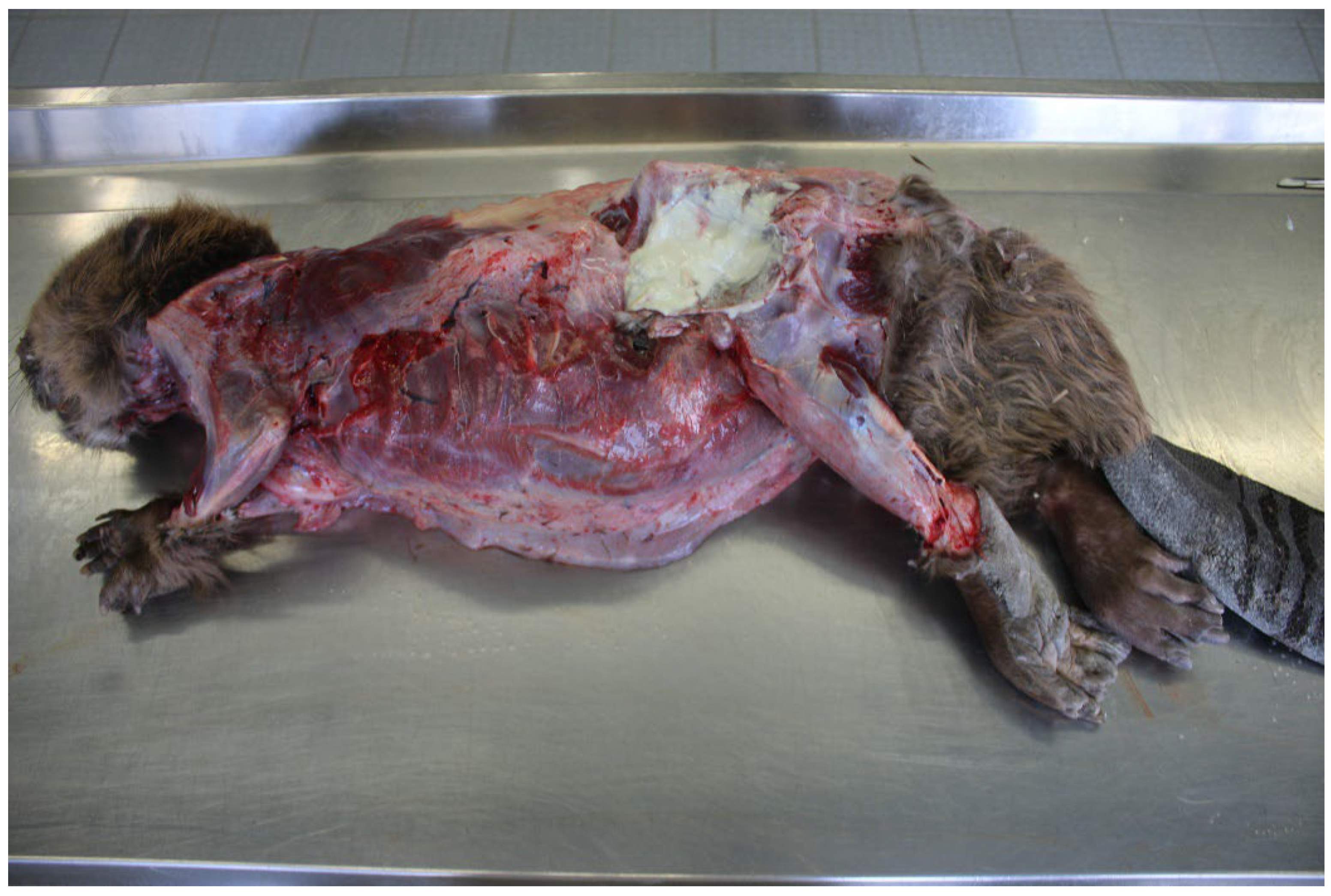
In general, all beavers that are found dead are sent to post-mortem examination for a passive wildlife health monitoring. An active health monitoring of beavers is not possible because beavers are strictly protected species and hunting, capturing and taking beavers from their natural habitat is strictly prohibited.

2.1.1. Beaver 1

The first beaver (Beaver 1) was found dead in the county of Main–Spessart in the federal state of Bavaria, Germany on 21 September 2015 and was submitted to LHL Giessen for post-mortem and microbiological examinations on the same day.

2.1.2. Beaver 2

The second beaver (Beaver 2) was found moribund at Wilhelmsruh on the Hevel in the county of Soest in the federal state of North Rhine–Westphalia, Germany on 7 April 2021. It was submitted to a local veterinarian and died two days later in the veterinary practice. Post-mortem and microbiological examinations were carried out at CVUA Westfalen in Arnsberg on 9 April 2021.

2.1.3. Beaver 3

The third beaver (Beaver 3) was detected dead in the urban district of Heilbronn in the federal state of Baden–Wuerttemberg, Germany on 8 June 2022 and was submitted the next day to CVUA Stuttgart for post-mortem and microbiological investigations.

2.2. Bacteriological Examinations

The lungs, liver, spleen, kidneys, and the small and large intestines of the beavers were submitted to bacteriological examinations. Aseptic cut surfaces of the organs were directly streaked onto the surface of sheep blood agar and selective MacConkey or Gassner agar (intestines), and subsequently incubated at 37 °C under atmospheric conditions for two days. In addition, all abscesses were sampled and material streaked on sheep blood agar and selective MacConkey agar. Grown colonies were taken directly from the agar plates and pure cultures were prepared for identification and characterization.

2.3. Identification and Characterization

Corynebacterium cultures were identified at the species level by MALDI-TOF MS (matrix-assisted laser desorption/ionization—time of flight mass spectrometry; Bruker Daltonics, Bremen, Germany) using an extended reference database [31]. The isolates were further compared by spectroscopic analysis using Fourier-transform infrared-spectroscopy (FT-IR; Bruker Optics, Ettlingen, Germany) according to Martel et al. [32].
Metabolic profiles were created using the GEN III OmniLog® ID System (Biolog, Hayward, CA, USA) according to the manufacturer’s instructions. The type strain C. ulcerans DSM 46325T (NCTC 7910T) was used for quality control and reference.
Phospholipase D activity was detected by streaking the C. ulcerans isolates at right angles to a centrally growing Staphylococcus (S.) aureus (WDCM 00034) and Rhodococcus (R.) equi (ATCC 33701) by typical inhibition of the staphylococcal and enhancement of the rhodococcal hemolysis, respectively [33].
In addition, these results were verified by partial sequencing of the rpoB-gene according to the recommendations of Khamis et al. [34], using the primers C2700F and C3130R for identification of the Corynebacterium spp. In addition, sequencing of the 16-23S internal transcribed spacer region (16-23S ITS) was performed using modified broad-range primers based on sequences provided by Johnson et al. [35] and Hunt et al. [36]: 1522F_mod TGCGGYTGGAWCACCTCCTT, 189R_mod TACTDAGATGTTTCAVTTC. Sequence data were evaluated by comparison with sequence entries in the GenBank using BLASTN [37].
Detection of the diphtheria toxin gene was performed by real-time PCR [38], and toxin production was verified by the Elek immunoprecipitation assay following the optimized modified protocol by Melnikov et al. [39].

2.4. Antimicrobial Susceptibility Testing

Antimicrobial susceptibility testing of the C. ulcerans isolates was performed by the broth microdilution method for determination of minimum inhibitory concentrations (MIC) to 14 different antimicrobials. The tests were carried out in commercially available antimicrobial microdilution plates for companion animals following the manufacturer’s instructions (Sifin Diagnostics, Berlin, Germany, according to guidelines of the German Veterinary Society (DVG) research group on antimicrobial resistance). The following 14 antimicrobials were tested (ranges given in mg/L): amoxicillin/clavulanic acid (0.063/0.031–16/8), ampicillin (0.125–8), cefovecin (0.25–4), cephalexin (0.5–16), chloramphenicol (1–16), clindamycin (0.031–1), enrofloxacin (0.016–2), erythromycin (0.023–4), gentamicin (0.063–4), oxacillin (0.063–2), penicillin G (0.063–4), pradofloxacin (0.004–1), tetracycline (0.063–8) and trimethoprim/sulfamethoxazole (0.25/4.75–2/38). Results (MIC values) of the antimicrobial susceptibility testing were interpreted using clinical breakpoints according to EUCAST [40] for broth microdilution testing. The type strain C. ulcerans DSM 46325T (NCTC 7910T) was used for quality control and reference.

2.5. Whole Genome Sequencing (WGS) Analysis

WGS of the three isolates was performed with Illumina paired-end sequencing (Illumina, San Diego, CA, USA) after DNA isolation using the Promega Maxwell system and library preparation with the Illumina DNA prep kit. WGS read data were checked for quality and absence of contamination with Illumina SAV software, fastqc [41] and kraken2 [42] before conducting in-depth analysis.
For genetic species confirmation, genomes were trimmed [43] and assembled with spades [44]. After performing assembly QC via QUAST [45], an average nucleotide identity (ANI) analysis was carried out with the tool PyANI (Application and Python module for whole-genome classification of microbes using Average Nucleotide Identity, Version v0.2, of the Leighton Pritchard Strathclyde Institute for Pharmacy and Biomedical Sciences University, Glasgow, Scotland) based on blast algorithm, as described in [23]. This involved pairwise comparison of the assemblies with public genomes of C. diphtheriae NCTC 11397T, C. belfantii FRC0043T, C. rouxii FRC0190T, C. pseudotuberculosis DSM 20689T (ATCC 19410T), C. silvaticum DSM 109166T (KL 0182T, CVUAS 4292T), C. ulcerans DSM 46325 T (NCTC 7910T) and C. epidermidicanis DSM 45586T, as a closely related outgroup of the C. diphtheriae group.
Multi-locus sequence typing (MLST) and core genome (cg) MLST were performed in Ridom SeqSphere+ (Ridom GmbH, Münster, Germany). MLST was conducted based on the seven genes atpA, dnaE, dnaK, fusA, leuA, odhA, rpoB described in [46,47]. Minimum spanning trees (MST) were constructed based on cgMLST allelic profiles of the sequenced C. ulcerans isolates, ignoring missing alleles during the pairwise profile comparisons and using two different ad hoc typing schemes:
(1)
A pan-genomic C. ulcerans/C. pseudotuberculosis scheme with 193 species-overlapping target loci, as previously described in [23]. The addition of isolate typing profiles for C. pseudotuberculosis and C. silvaticum enables differentiation of the most closely related species.
(2)
A C. ulcerans cgMLST scheme of 1211 target loci, previously described by Berger et al. [25], with the addition of other animal-based C. ulcerans isolate typing profiles.

3. Results

3.1. Post-Mortem and Bacteriological Examinations

3.1.1. Beaver 1 (County of Main–Spessart, Bavaria; 21 September 2015)

The beaver was a young male (without head, tail and skin) with a remaining body weight of 1.0 kg. The animal was cachectic and showed low-grade autolysis. Post-mortem examination of the inner organs revealed no special findings. Abscesses could not be found.
Bacteriological examination revealed a moderate growth (about 100 colonies) of C. ulcerans (151012433-002) and Staphylococcus aureus in the lungs and a low growth (about 10 colonies) of Yersinia enterocolitica in the colon.
The cause of the cachexia and death of this animal could not be identified.

3.1.2. Beaver 2 (County of Soest, North Rhine–Westphalia, 7 April 2021)

The second beaver was an old female with a body weight of 13.6 kg. This animal was also cachectic with atrophy of the coronary fat and pronounced edema were detected in the subcutaneous tissue of the ventral thoracic region and abdomen. Furthermore, post-mortem also revealed swollen lymph nodes and scars in the throat region. An abscess was detected in the subcutis in the region of the left hip and a further abscess at the third left rib fused with the left lung (Figure 1). Swab samples from these abscesses were taken for bacteriological examinations. The lungs showed a severe edema and black-colored areas in the tissue. Histologically, the lung revealed a multifocal granulomatous pneumonia with conidia demarcated in connective tissue typical of adiaspiromycosis, caused by the fungus Emmonsia crescens. Bacteriological testing showed severe growth (about 1000 colonies) of Streptococcus (S.) castoreus in the lungs, liver and abscess, accompanied by moderate growth (about 100 colonies) of C. ulcerans (S 477/6/21). The age, the bite wounds and the infections of the animal are to be considered as the cause of cachexia and death.

3.1.3. Beaver 3 (District of Heilbronn, Baden-Wuerttemberg, 8 June 2022)

The third beaver was a young male with a body weight of 16 kg. The nutritional condition of the beaver was poor, resulting in a loss of the coronary fat. The external examination of the body revealed previous injuries in the throat area and multiple abscesses in the lymph nodes localized in the inguinal region and on the cranial upper jaw. Swab samples from the lymph node abscesses and the inguinal region were taken for bacteriological examination. Black colored stipples in the tissue were verified by histo-pathological examinations as multiple granuloma-enclosing conidia that are typical of adiaspiromycosis (Emmonsia crescens). Bacteriological examinations showed severe growth (about 1000 colonies) of C. ulcerans (CVUAS 33950), accompanied by a moderate growth (about 100 colonies) of S. castoreus in the specimens taken from the abscesses. The injuries and the infections are considered the cause of the cachexia and subsequent death of the beaver.

3.2. Identification and Characterization of the Corynebacterium sp.

The three C. ulcerans isolates were unequivocally identifiable at species level by MALDI-TOF MS (Score 2.3–2.5) (metadata and single MALDI-TOF mass-spectra of the isolates used in this study are listed in the MALDI-UP catalogue [https://maldi-up.ua-bw.de] and are accessible on request) and FT-IR spectroscopy (Figure 2), as well as by partial sequencing of the 16-23S rRNA intergenic spacer region (NCBI BLAST: 100% query cover, differences of percent identity of 3.3–3.8% and 7.3–7.5% to the closely related species C. pseudotuberculosis and C. silvaticum, respectively). Evaluation of the partial rpoB gene sequences using NCBI BLAST revealed high similarity between C. ulcerans and C. silvaticum (NCBI BLAST: 100% query cover, differences of percent identities to C. silvaticum of 0.0–0.3%). However, a differentiation from C. pseudotuberculosis was unequivocal (100% query cover, difference of percent identity of 4.9%).
All of the three C. ulcerans isolates from the beavers produced phospholipase D. This could be demonstrated on sheep blood agar by inhibition of the hemolysis of S. aureus and a synergistic hemolysis with R. equi. These C. ulcerans isolates are classified as toxigenic since they carry and express the diphtheria toxin gene.

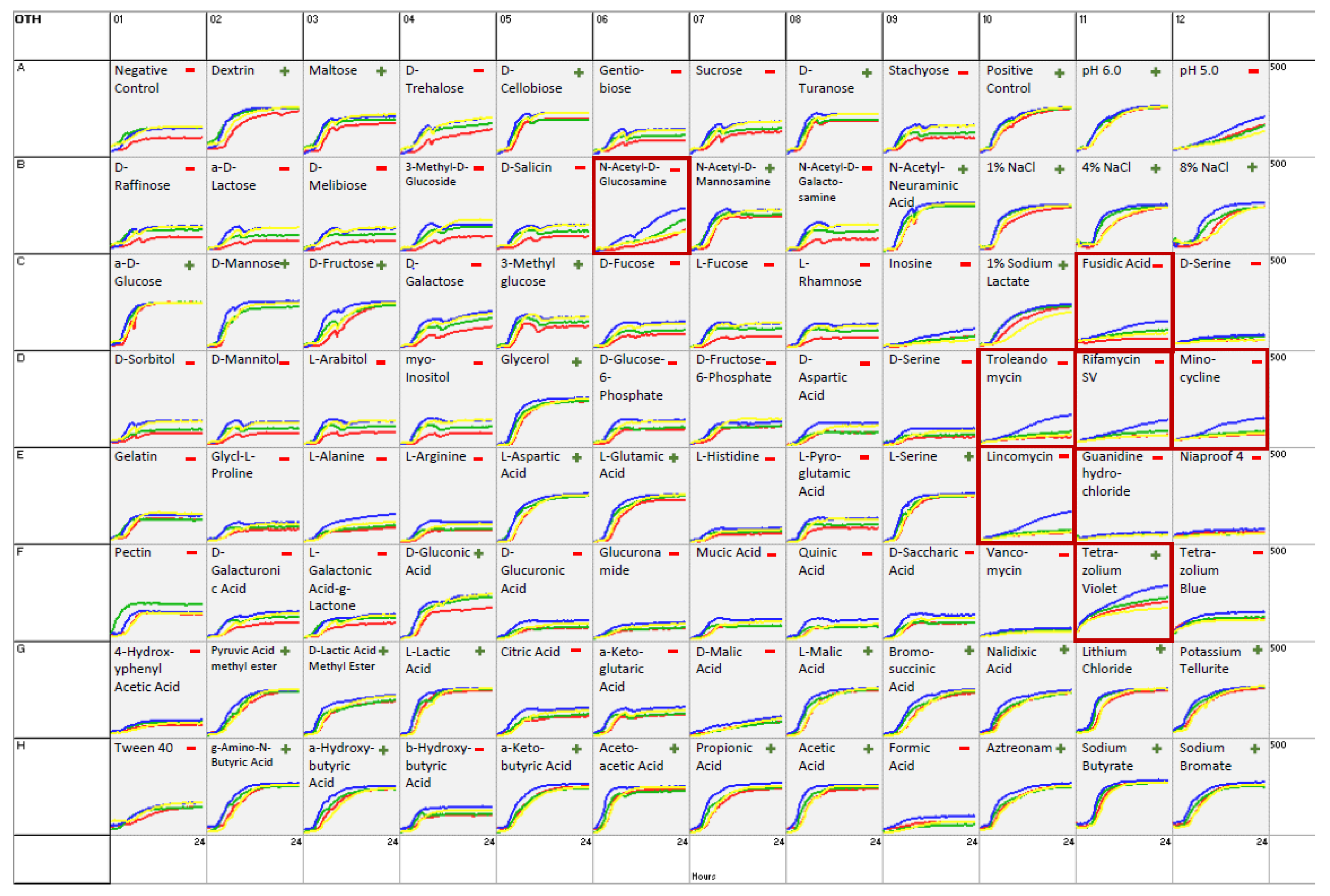
3.3. Biochemical Investigations

A broad analysis of biochemical identification properties was assessed for the three isolates under study, for which results are shown in Figure 3 and Supplementary Table S1. As reference strain C. ulcerans DSM 46325T (NCTC 7910T) was used. Differences in biochemical reactions between the beaver isolates and the reference strain become apparent. Despite not all members of the C. diphtheriae group having been included in the Omnilog database, the three C. ulcerans isolates and the reference strain C. ulcerans DSM 46325T (NCTC 7910T) were clearly identified at the species level by the Omnilog software, with a distance value of ≥0.500 (DIST 5.439–5.597).
Analysis of metabolic activities revealed positive results for 16 out of 94 biochemical reactions (Figure 3). The results did not differ between the tested isolates and the type strain for C. ulcerans DSM 46325T (NCTC 7910T) (Figure 3, red line). However, slight differences in metabolic activities could be detected. In seven reactions (N-acetyl-D-glucosamine, fusidic acid, troleandomycin, rifamycin SV, minocycline, lincomycin, and tetrazolium violet), isolate S 477/6/21 (Beaver 2) showed a slightly higher metabolic rate compared to the other isolates. Contrarily, isolate CVUAS 33950 (Beaver 3) metabolized pectin at a slightly higher activity level (Supplementary Table S1).

3.4. Antimicrobial Susceptibility Testing

The results of antimicrobial susceptibility testing are listed in Table 1. Consistently, all isolates were resistant to clindamycin (MIC > 2 mg/L) and susceptible to all other antimicrobials tested. No differences in resistance patterns were evident compared with the type strain DSM 46325T (NCTC 7910T).
However, when using the newly recommended EUCAST breakpoints for erythromycin (MIC > 0.06 mg/L), only isolate S 477/6/21 (Beaver 2) displayed a resistant phenotype (0.064 mg/L) [40], while type strain DSM 46325T (NCTC 7910T) was susceptible.

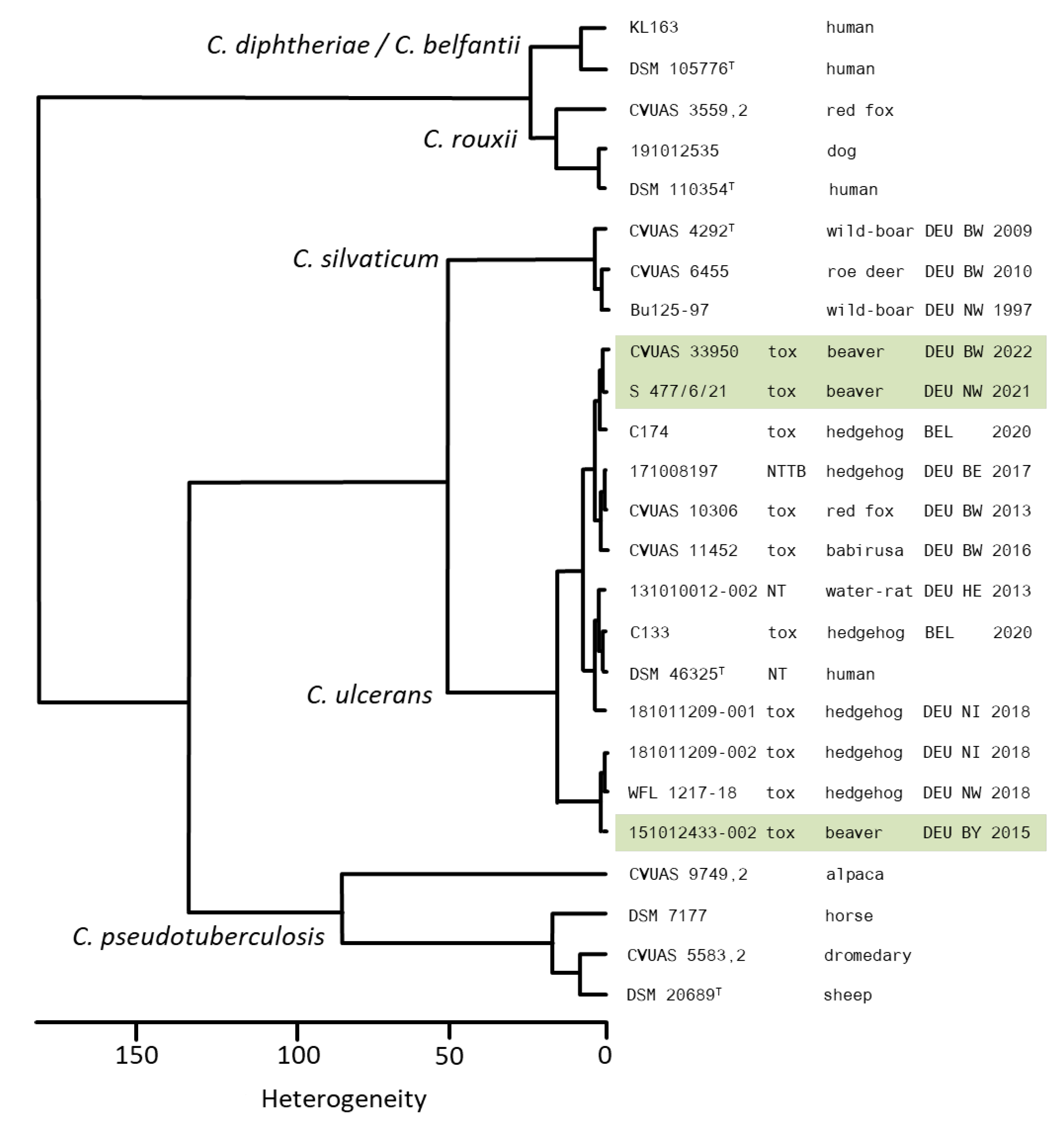
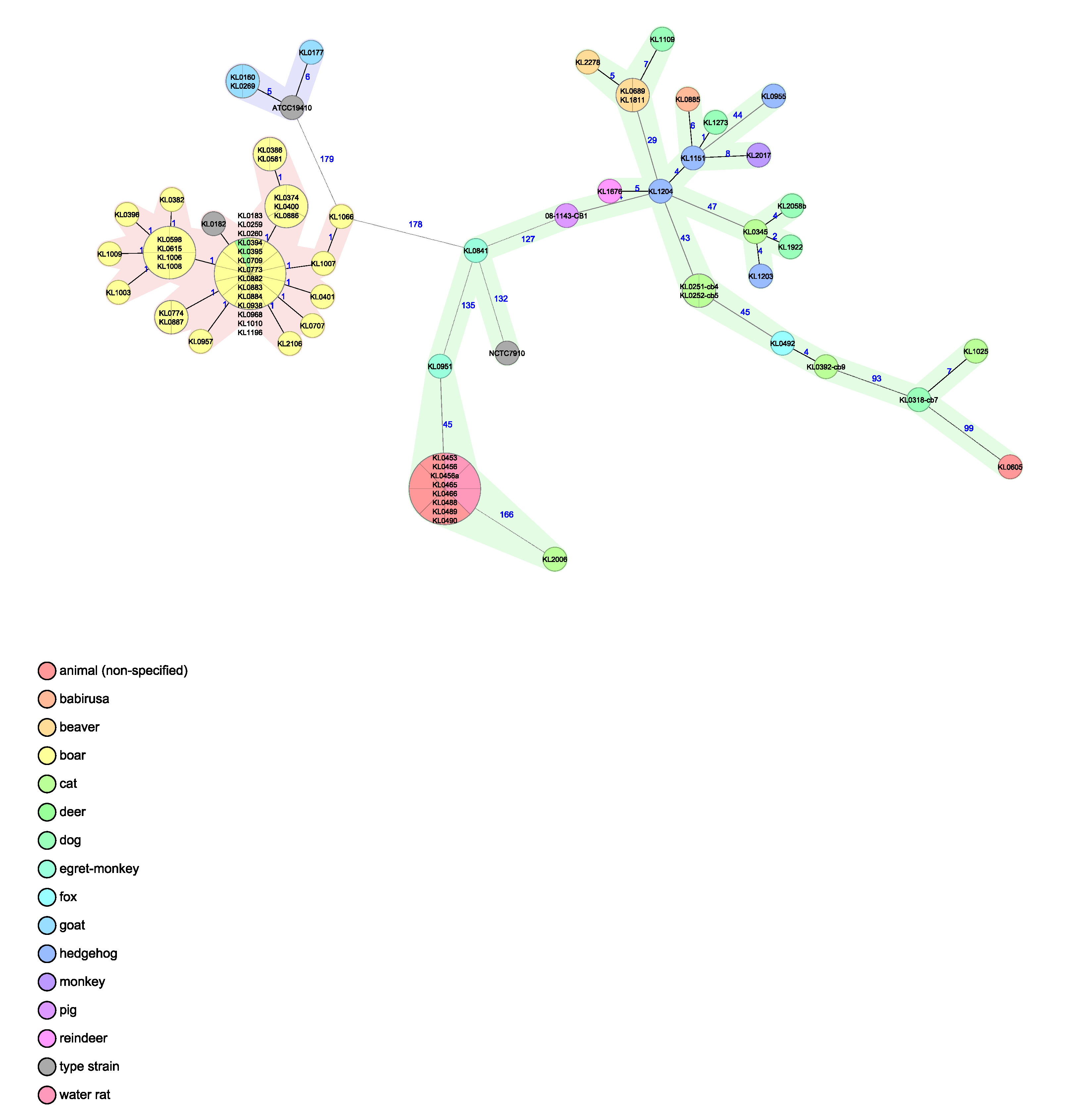
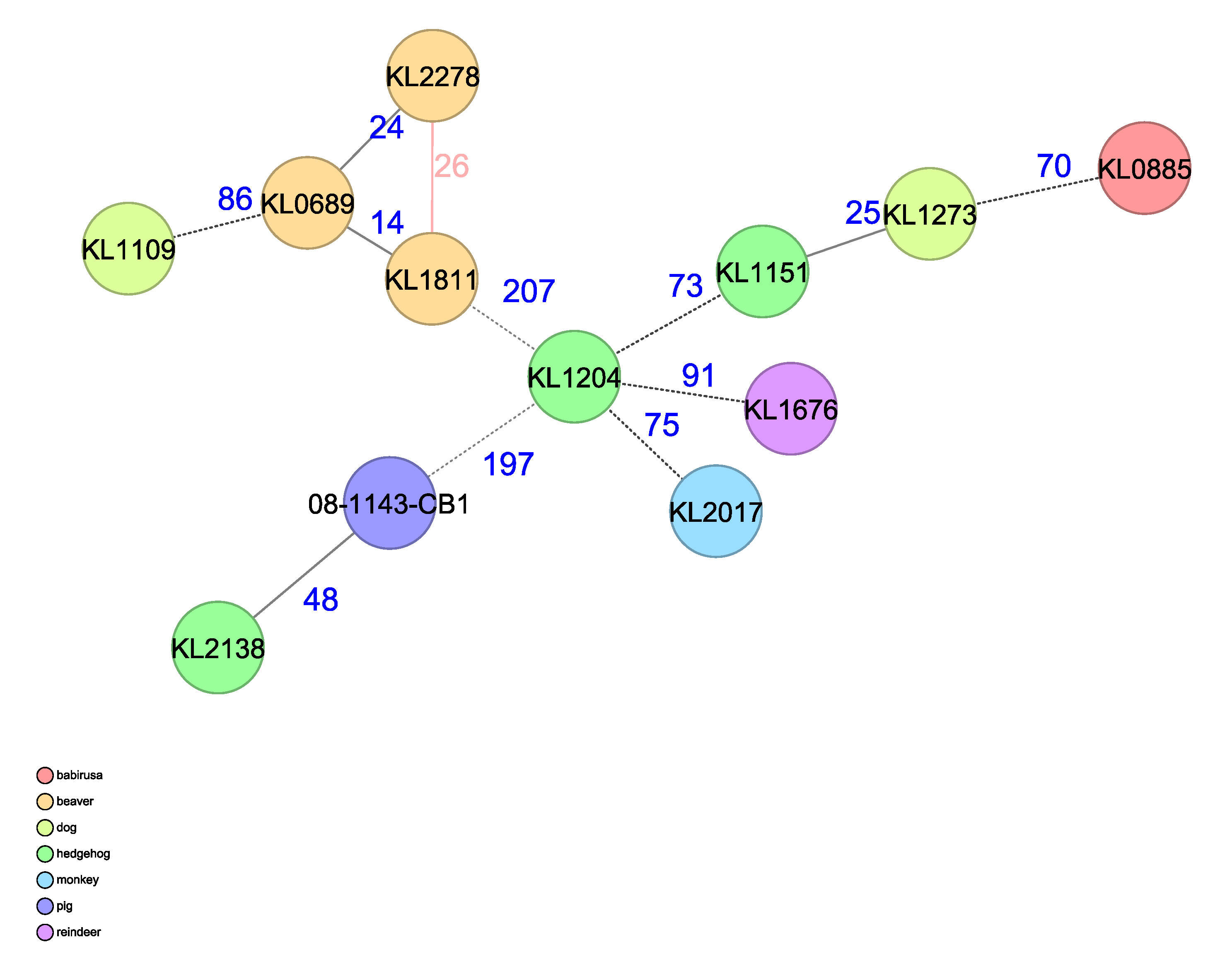
3.5. Whole Genome Sequencing (WGS) Analysis

WGS was carried out on the three C. ulcerans isolates and the resulting data were used for confirmation of the genetic species by ANI and for phylogenetic clustering by cgMLST, using two different schemes. ANI analysis in comparison with type strain genomes from the C. diphtheriae group confirmed the results of the MALDI-TOF MS, FT-IR and 16-23S ITS analyses. The three isolates were clearly classified as C. ulcerans, with ANI values of 98% compared with the C. ulcerans-type strain. These ANI values are above the threshold value of ~95–96% for taxonomic delineation of prokaryotic species [48], whereas ANI values for all other species were below 91% (Table 2).
MLST analysis based on seven housekeeping loci classified all three isolates as C. ulcerans sequence type (ST)- 332 (atpA: 42, dnaE: 33, dnaK: 78, fusA: 49, leuA: 48, odhA: 43, rpoB: 40).
CgMLST analysis was performed with two different ad hoc schemes to be able to classify the isolates from two different perspectives. For the generation of the pangenomic scheme 193 overlapping genes from C. ulcerans and C. pseudotuberculosis were used. These divide in the resulting species-specific MST branches, with the beavers’ isolates grouping in the branch of the species C. ulcerans but being separate in different branches from C. pseudotuberculosis, as well as from the closely related C. silvaticum. For comparison, genomic profiles from other animal-derived isolates of the three species, generated with the same scheme, were included in the analysis. The beaver-derived isolates clearly group on the branch of C. ulcerans (Figure 4). Thereby, the species classification can be clearly confirmed.
Using the cgMLST scheme, including 1211 core genes from C. ulcerans, generated and described in [25], the resulting MST shows the genetic relationship of the beaver-derived C. ulcerans isolates to each other and to other genomic profiles from animal-derived C. ulcerans isolates, added for comparison. The beavers’ isolates show thereby the highest similarity to each other with allelic differences (AD) of 14–26 and to the nearest relative C. ulcerans from another host species derived from a dog quantified with an AD of 86 (Figure 5).

4. Discussion

C. ulcerans occupies a special status among the corynebacteria belonging to the C. diphtheriae species complex due to its potential production of the diphtheria toxin, its broad spectrum of hosts ranging from livestock and companion animals to wild animals, and its pronounced zoonotic character [26,49,50]. The wide spectrum of susceptible animals has been comprehensively presented by Berger et al. [25]. C. ulcerans is considered to be an emerging human pathogen causing cases of diphtheria-like illness, which have outnumbered diphtheria cases caused by C. diphtheriae in Europe for the last two decades [26,27,28,31,32]. Of particular concern has become the transmission of C. ulcerans from companion animals, especially cats and dogs, to humans. Such infections can be severe with even lethal outcomes, which have been documented in numerous case reports and summarized in reviews [26,49]. In contrast, human-to-human transmission of C. ulcerans plays only a minor role for this pronounced zoonotic pathogen [51]. In parallel, cases of infections with toxigenic C. ulcerans displaying clinical signs of the respiratory tract and ulcerative skin lesions have recently been reported in dogs and cats [52]. Furthermore, hunting dogs are reported to represent a potential link for transmission of C. ulcerans from wild animals to humans and companion animals through direct contact with wildlife [53]. C. ulcerans infections have also been observed in a greater number of hedgehogs, who often live in close proximity to humans [25,32]. The increasing population of beavers in Germany might form a novel reservoir for C. ulcerans. However, no cases of C. ulcerans in other European countries have so far been reported. Therefore, the prevalence of C. ulcerans in beavers is generally unknown and thus the present study is only able to give an indication of the presence of pathogenic C. ulcerans in beavers. The role of established and new toxigenic and NTTB strains of animal and human origin has been recently and comprehensively evaluated by Prygiel et al. [21]. The close relationship between animal and human pathogenic corynebacteria isolates suggests that other animal pathogenic isolates may also be pathogenic to humans [24]. Thus, it is of great interest, from the One Health point of view, whether beaver populations represent a novel reservoir for pathogenic, zoonotic C. ulcerans. The three deceased beavers included in this study were spatio-temporally found widely separated from each other in the German federal states of Bavaria, North Rhine–Westphalia and Baden–Wuerttemberg, in which Eurasian beavers had been reintroduced and reestablished [54]. Although the source of toxigenic C. ulcerans in beavers remains hidden, beavers have to be considered as a further, previously undetected free-ranging species posing a reservoir for this zoonotic agent. Taking this into account, monitoring of wildlife on pathogenic bacteria and molecular epidemiology should be implemented as an indispensable tool of the One Health approach.
Detection of C. ulcerans in the lungs of Beaver 1 was not associated with major pathological changes, suggesting a colonization of the lungs as commensal, as is also described for the respiratory tract and oral cavity of dogs [55,56] and cats [57,58]. However, since this animal was submitted decapitated, we cannot rule out bacterial spread to this organ. Conversely, the other two beavers developed multiple abscesses in the subcutis and lymph nodes of the trunk and showed scarring of the skin in the throat area. These injuries are indicative of bite wounds resulting from ranking or territorial fights as also reported for other animal species like otters [59], water rats [60], squirrels [61] or hedgehogs in which C. ulcerans had also been detected [25,32]. In addition to these wildlife species, beavers also serve as a so-far-undiscovered new host. Thus, transmission of C. ulcerans from beavers to other semi-aquatic rodents like the invasive muskrats and nutrias living in the same freshwater habitats is conceivable. However, the role of beavers as a carrier and possible reservoir for C. ulcerans among semi-aquatic mammals currently remains speculative. Therefore, semi-aquatic wildlife should also be consistently monitored on pathogenic Corynbacterium spp.
Identification of corynebacteria at the species level in routine bacteriological laboratories is difficult because similar profiles can be retrieved by conventional phenotypical and even current test methods such as MALDI-TOF MS [52,62,63]. However, the GEN III OmniLog system, a fully automated system for the biochemical identification of microorganisms, unequivocally confirmed the placement of isolates to C. ulcerans. Although the OmniLog system does not always clearly identify coryneforms [64], the isolates tested and the reference strain were clearly identified as C. ulcerans. The same result was also reported by Berger et al. [25]. Despite minor differences, isolates from beavers represented a largely homogenous population within the variability range of C. ulcerans. The Omnilog software was additionally used to compare the metabolic curves of each of the 94 metabolic reactions per isolate and in comparison to the reference strain. For this purpose, the metabolic profile curves of the measured individual reactions were overlaid and the differences visually highlighted (Figure 3). These results show that the metabolic responses in terms of negative and positive reactions do not differ between the tested isolates and the reference strain. These results are consistent with those previously reported [25,65] (Supplementary Table S1). However, it can be seen that the isolate from Beaver 2 appears to be slightly more metabolically active than the other isolates or the reference strain. Interestingly, this is the same isolate that was exclusively resistant to erythromycin according to the current EUCAST recommendations on breakpoints. Susceptibility testing revealed sensitivity to erythromycin, except for one isolate (S 477/6/21, Beaver 2). In contrast, all isolates were revealed to be resistant to clindamycin. This antimicrobial susceptibility pattern agrees with previous studies, which report antimicrobial susceptibility to erythromycin for the majority of human and animal C. ulcerans isolates tested. In contrast, susceptibility to clindamycin has been reported from sensitive to intermediary to resistant [25,66,67].
The quality of MALDI-TOF MS results strongly relies on comprehensive and sophisticated databases representing all currently established Corynebacterium spp. of the C. diphtheriae group [23,31]. This is of even more importance, as closely related corynebacteria, too, must be clearly differentiated due to their different zoonotic potential [23,31,52,62]. In this context, the recent separation of isolates from the so-called wild boar cluster of C. ulcerans as the novel species C. silvaticum has to be considered [23]. This Corynebacterium sp. is most closely related to C. ulcerans based on 16S rRNA gene and rpoB gene sequences; thus, C. silvaticum strains were formerly classified as C. ulcerans [23,31]. In our study, however, C. ulcerans could unequivocally be differentiated from C. silvaticum using MALDI-TOF MS and FT-IR, supported by an extended spectral database, as described in the catalogue of the MALDI-User Platform (https://MALDI-UP.ua-bw.de (accessed on 13 June 2022)) [68]. Using extended and comprehensive spectral databases for MALDI-TOF MS and FT-IR analysis, precise and reproducible identification of the current corynebacteria can be carried out quickly and accurately [23,31,69]. The same applies to 16-23S ITS sequences, which allowed a clear identification of Corynebacterium spp. in this study.
Production of virulence factors, e.g., phospholipase D (PLD), presence of the tox gene, and production of the diphtheria toxin were verified for all three C. ulcerans isolates as recommended [60,62]. Whereas PLD is reportedly produced by all pathogenic C. ulcerans isolates [26,52], production of the diphtheria toxin, which is considered a main virulence factor [26,70], was found in only about 5% of the C. diphtheria and in 50–60% of the C. ulcerans isolates [28]. A recent study in France revealed a striking frequency in toxigenic isolates from dogs and cats among companion animals [71]. Strains of toxigenic as well as the NTTB phenotype in C. ulcerans in wild and exotic animal species have already been found in otters [69], water rats [60], squirrels [61] and hedgehogs [25,32]. These animal species have been reported to suffer from suppurative to necrosuppurative, ulcerative, superficial to deep skin lesions, and infestation of various inner organs like the lung and heart, irrespective of the production of the diphtheria toxin.
In addition to C. ulcerans, S. castoreus was isolated in abscesses from two animals and also, in one of these animals, in the liver and lungs. S. castoreus has been described as a separate, beta-hemolytic Streptococcus sp. belonging to the group A streptococci, which have previously been isolated from multiple bite wounds in a beaver [72]. There are few reports on the detection of S. castoreus exclusively in beavers, considering that this Streptococcus sp. is strictly host-specific [73,74]. In these reports, S. castoreus has also been isolated from suppurative lesions in association with bite wounds and systemic infections, as well as from apparently healthy beavers. However, S. castoreus has been assessed as being an opportunistic pathogen in beavers, primarily colonizing the normal microbiota of the oral, respiratory and genital mucosa. Finally, tissue lesions and concomitant colonization by opportunistic pathogenic bacteria such as Actinomyces, Corynebacterium or streptococci and Gram-negative obligate anaerobic bacteria might lead to severe poly-microbial suppurative inflammation and abscesses [74,75,76,77].
Overall, the number of beavers that are available for mircobiological investigations is very limited because impairment of beavers in their natural habitat is strictly prohibited. Thus, active monitoring on the health of beavers is not realizable, meaning that data are available from only a limited number of free-ranging beavers found dead. In the federal states of Hesse and Baden–Wuerttemberg, 17 and 115 animals have been subjected to post-mortem examinations in the last 10 years, respectively. However, in the federal state of North Rhine–Westphalia, this was the first beaver examined by post-mortem analysis.
Whole-genome sequencing has become a valuable tool for the molecular characterization of bacterial isolates as a basis of molecular epidemiology [78,79]. Evaluation of the genome sequence data of the C. ulcerans isolates from the three beavers revealed a genome size of approximately 2.5 Mbp and a GC content of 53.4%. This data is consistent with that previously reported for C. ulcerans isolates originating from animals and humans [80]. ANI and pangenomic cgMLST analysis clearly classify the isolates as C. ulcerans, which is consistent with MALDI-TOF MS, FT-IR and 16-23S ITS analysis.
WGS-derived MLST based on seven housekeeping loci allowed a classification of the beaver C. ulcerans isolates as ST-332. There are only a few reports on this sequence type, detected in hedgehogs [25], in a cat [47] and in humans [47,51,81,82]. Detection of ST-332 in animals and humans is an indication of a possible zoonotic transmission of this pathogen and emphasizes the importance of MLST and cgMLST for unravelling transmission routes of C. ulcerans [47]. A MLST based on C. ulcerans cgMLST analysis using 1211 core genes shows that the genetic distance between the C. ulcerans isolates from the beavers is smaller than that of the isolates from other animal host species. A similar cgMLST scheme in C. diphtheriae with a size range of 1500 alleles showed a comparable range of allelic distances like single nucleotide polymorphism (SNP) phylogenies; closely related isolates of C. diphtheriae and C. ulcerans showed distances of only a few alleles/SNPs, respectively [25,70,83]. Given this knowledge, we conclude that distances in this range (>20) for the beaver isolates are no indication of a close genetic relationship or even direct transmission.

5. Conclusions

Wildlife disease surveillance is an important instrument for the early detection of emerging diseases and zoonoses. Therefore, examinations of all wild animals found dead are a vital element of wildlife disease surveillance. In this context, we examined three Eurasian beavers and were able to detect C. ulcerans in this wild animal species for the first time. Based on WGS data, the isolates show close genetic but no clonal relationship to each other or to isolates originating from other host species. C. ulcerans isolates are known as zoonotic agents. In this respect, these isolates from beavers must be considered as potential zoonotic candidates, and beavers should be recognized as a heretofore unknown possible reservoir for pathogenic C. ulcerans. In order to cover the important and complex group of zoonotic corynebacteria, interdisciplinary cooperation with regard to the One Health approach is pivotal.
Further investigations on pathogenic C. ulcerans in wildlife supported by molecular epidemiology should be conducted to obtain deeper knowledge about the occurrence, epidemiology and impact of C. ulcerans in beavers and other semi-aquatic mammals, particularly considering the ever-increasing beaver population in Europe.

Supplementary Materials

The following supporting information can be downloaded at: https://www.mdpi.com/article/10.3390/pathogens12080979/s1, Supplementary Table S1 (Supplementary Data): Phenotypic characterization of the three C. ulcerans isolates from Eurasian beavers included in this study.

Author Contributions

Conceptualization, J.R., R.S. and C.P.; methodology, R.S., C.P., T.E., R.B., B.B., M.P., K.R., A.S., A.B., A.D. and J.R.; software, R.S., R.B., A.B., A.D. and J.R.; validation, R.S., R.B., A.B., A.D. and J.R.; formal analysis, R.S., C.P., T.E., R.B., B.B., M.P., K.R., A.S., A.B., A.D. and J.R.; investigation, R.S., R.B., B.B., M.P., K.R., A.B., A.D. and J.R.; resources, R.S., C.P., T.E., R.B., B.B., M.P., K.R., A.S., A.B., A.D. and J.R.; data curation, R.S., R.B., B.B., M.P., K.R., A.B., A.D. and J.R.; writing—original draft preparation, R.S.; writing—review and editing, R.S., C.P., T.E., R.B., B.B., M.P., K.R., A.S., A.B., A.D. and J.R.; visualization, R.B., M.P., A.D. and J.R.; supervision, R.S.; project administration, R.S., J.R. and C.P. All authors have read and agreed to the published version of the manuscript.

Funding

This research received no external funding.

Institutional Review Board Statement

Ethical review and approval were waived for this study because the animals were brought to us dead for post-mortem examinations.

Informed Consent Statement

Not applicable.

Data Availability Statement

WGS raw data are available at the short reads archive of the National Center of Biotechnology Information (NCBI) under bioproject PRJNA938404 (with the following biosample accessions for beaver 1: SAMN33434792, beaver 2: SAMN33434793, beaver 3: SAMN33434794).

Acknowledgments

We would like to thank Martin Dyk and Maja Hrubenja for their precise MALDI-TOF MS, FT-IR, PCR and DNA-sequence analyses. We thank Katharina Engel for her excellent assistance and support on OmniLog system and antimicrobial testing. We thank Sabine Lohrer, Jasmin Fräßdorf, Marion Lindermayer and Anne Könitzer for their excellent laboratory work on WGS analyses and Annika Sprenger for participation in MLST and cgMLST analysis.

Conflicts of Interest

The authors declare no conflict of interest.

References

  1. Nolet, B.A.; Rosell, F. Comeback of the beaver Castor fiber: An overview of old and new conservation problems. Biol. Conserv. 1998, 83, 165–173. [Google Scholar] [CrossRef]
  2. Halley, D.J.; Saveljev, A.P.; Rosell, F. Population and distribution of beavers Castor fiber and Castor canadensis in Eurasia. Mammal Rev. 2020, 51, 1–24. [Google Scholar] [CrossRef]
  3. Schwab, G.; Schmidbauer, M. Beaver (Castor fiber L., Castoridae) management in Bavaria. Denisia 2003, 9, 99–106. [Google Scholar]
  4. Wróbel, M. Population of Eurasian beaver (Castor fiber) in Europe. Glob. Ecol. Conserv. 2020, 23, e01046. [Google Scholar] [CrossRef]
  5. Batbold, J.; Batsaikhan, N.; Shar, S.; Hutterer, R.; Krystufek, B.; Yigit, N.; Mitsainas, G.; Palomo, L. Castor fiber (amended version of 2016 Assessment). The IUCN Red List of Threatened Species 2021. e.T4007A197499749. Available online: https://dx.doi.org/10.2305/IUCN.UK.2021-1.RLTS.T4007A197499749.en (accessed on 10 July 2023). [CrossRef]
  6. Maas, M.; Glorie, J.; Dam-Deisz, C.; de Vries, A.; Franssen, F.F.J.; Jaarsma, R.I.; Hengeveld, P.D.; Dierikx, C.M.; van der Giessen, J.W.B.; Opsteegh, M. Zoonotic Pathogens in Eurasian Beavers (Castor fiber) in the Netherlands. J. Wildl. Dis. 2022, 58, 404–408. [Google Scholar] [CrossRef]
  7. Centers for Disease Control and Prevention (CDC), National Center for Emerging and Zoonotic Infectious Diseases (NCE-ZID). Zoonotic Diseases. Available online: https://www.cdc.gov/onehealth/basics/zoonotic-diseases.html (accessed on 9 January 2023).
  8. Jones, K.E.; Patel, N.G.; Levy, M.A.; Storeygard, A.; Balk, D.; Gittleman, J.L.; Daszak, P. Global trends in emerging infectious diseases. Nature 2008, 451, 990–993. [Google Scholar] [CrossRef]
  9. Recht, J.; Schuenemann, V.J.; Sánchez-Villagra, M.R. Host Diversity and Origin of Zoonoses: The Ancient and the New. Animals 2020, 10, 1672. [Google Scholar] [CrossRef]
  10. Campbell-Palmer, R.; Rosell, F.; Naylor, A.; Cole, G.; Mota, S.; Brown, D.; Fraser, M.; Pizzi, R.; Elliott, M.; Wilson, K.; et al. Eurasian beaver (Castor fiber) health surveillance in Britain: Assessing a disjunctive reintroduced population. Vet. Rec. 2021, 188, e84. [Google Scholar] [CrossRef]
  11. Girling, S.J.; Naylor, A.; Fraser, M.; Campbell-Palmer, R. Reintroducing beavers Castor fiberto Britain: A disease risk analysis. Mammal Rev. 2019, 49, 300–323. [Google Scholar] [CrossRef]
  12. Howe, C.W. (Ed.) A Review of the Evidence on the Interactions of Beavers with the Natural and Human Environment in Relation to England, 1st ed.; Natural England: Peterborough, UK, 2020; pp. 193–202. [Google Scholar]
  13. Schulze, C.; Heuner, K.; Myrtennäs, K.; Karlsson, E.; Jacob, D.; Kutzer, P.; Große, K.; Forsman, M.; Grunow, R. High and novel genetic diversity of Francisella tularensis in Germany and indication of environmental persistence. Epidemiol. Infect. 2016, 144, 3025–3036. [Google Scholar] [CrossRef]
  14. Rosell, F.; Rosef, O.; Parker, H. Investigations of Waterborne Pathogens in Eurasian Beaver (Castor fiber) from Telemark County, Southeast Norway. Acta Vet. Scand. 2001, 42, 479–482. [Google Scholar] [CrossRef]
  15. You, M.H.; Kim, J.H.; Kim, D.Y.; Gomez, D.K.; Jung, T.S.; Park, S.C. Pleuritis and pericarditis associated with Klebsiella pneumoniae in a Eurasian beaver (Castor fiber). Korean J. Vet. Res. 2008, 48, 501–503. [Google Scholar]
  16. Platt-Samoraj, A.; Syczyło, K.; Bancerz-Kisiel, A.; Szczerba-Turek, A.; Giżejewska, A.; Szweda, W. Yersinia enterocolitica strains isolated from beavers (Castor fiber). Pol. J. Vet. Sci. 2015, 18, 449–451. [Google Scholar] [CrossRef]
  17. Girling, S.J.; Goodman, G.; Burr, P.; Pizzi, R.; Naylor, A.; Cole, G.; Brown, D.; Fraser, M.; Rosell, F.N.; Schwab, G.; et al. Evidence of Leptospira species and their significance during reintroduction of Eurasian beavers (Castor fiber) to Great Britain. Vet. Rec. 2019, 185, 482. [Google Scholar] [CrossRef]
  18. Lauková, A.; Strompfová, V.; Kandričáková, A.; Ščerbová, J.; Semedo-Lemsaddek, T.; Miltko, R.; Belzecki, G. Virulence factors genes in enterococci isolated from beavers (Castor fiber). Folia Microbiol. 2015, 60, 151–154. [Google Scholar] [CrossRef]
  19. Monecke, S.; Feßler, A.T.; Burgold-Voigt, S.; Krüger, H.; Mühldorfer, K.; Wibbelt, G.; Liebler-Tenorio, E.M.; Reinicke, M.; Braun, S.D.; Hanke, D.; et al. Staphylococcus aureus isolates from Eurasian Beavers (Castor fiber) carry a novel phage-borne bicomponent leukocidin related to the Panton-Valentine leukocidin. Sci. Rep. 2021, 11, 24394. [Google Scholar] [CrossRef]
  20. Campbell-Palmer, R.; Schwab, G.; Girling, S.; Lisle, S.; Gow, D. Managing Wild Eurasian Beavers: A Review of European Management Practices with Consideration to Application in Scotland; Scott, J., Ed.; Commissioned Report No. 812 ed.; Scottish Natural Heritage: Inverness, UK, 2015; pp. 29–34. [Google Scholar]
  21. Prygiel, M.; Polak, M.; Mosiej, E.; Wdowiak, K.; Formińska, K.; Zasada, A.A. New Corynebacterium Species with the Potential to Produce Diphtheria Toxin. Pathogens 2022, 11, 1264. [Google Scholar] [CrossRef]
  22. Schlez, K.; Eisenberg, T.; Rau, J.; Dubielzig, S.; Kornmayer, M.; Wolf, G.; Berger, A.; Dangel, A.; Hoffmann, C.; Ewers, C.; et al. Corynebacterium rouxii, a recently described member of the C. diphtheriae group isolated from three dogs with ulcerative skin lesions. Antonie Leeuwenhoek 2021, 114, 1361–1371. [Google Scholar] [CrossRef]
  23. Dangel, A.; Berger, A.; Rau, J.; Eisenberg, T.; Kampfer, P.; Margos, G.; Contzen, M.; Busse, H.-J.; Konrad, R.; Peters, M.; et al. Corynebacterium silvaticum sp. nov., a unique group of NTTB corynebacteria in wild boar and roe deer. Int. J. Syst. Evol. Microbiol. 2020, 70, 3614–3624. [Google Scholar] [CrossRef]
  24. Dover, L.G.; Thompson, A.R.; Sutcliffe, I.C.; Sangal, V. Phylogenomic Reappraisal of Fatty Acid Biosynthesis, Mycolic Acid Biosynthesis and Clinical Relevance among Members of the Genus Corynebacterium. Front. Microbiol. 2021, 12, 802532. [Google Scholar] [CrossRef]
  25. Berger, A.; Dangel, A.; Peters, M.; Mühldorfer, K.; Braune, S.; Eisenberg, T.; Szentiks, C.A.; Rau, J.; Konrad, R.; Hörmansdorfer, S.; et al. Tox-positive Corynebacterium ulcerans in hedgehogs, Germany. Emerg. Microbes Infect. 2019, 8, 211–217. [Google Scholar] [CrossRef] [PubMed]
  26. Hacker, E.; Antunes, C.; Mattos-Guaraldi, A.L.; Burkovski, A.; Tauch, A. Corynebacterium ulcerans, an emerging human pathogen. Futur. Microbiol. 2016, 11, 1191–1208. [Google Scholar] [CrossRef]
  27. Both, L.; Collins, S.; de Zoysa, A.; White, J.; Mandal, S.; Efstratiou, A. Molecular and Epidemiological Review of Toxigenic Diphtheria Infections in England between 2007 and 2013. J. Clin. Microbiol. 2015, 53, 567–572. [Google Scholar] [CrossRef] [PubMed]
  28. Gower, C.M.; Scobie, A.; Fry, N.K.; Litt, D.J.; Cameron, J.C.; Chand, M.; Brown, C.S.; Collins, S.; White, J.M.; Ramsay, M.; et al. The changing epidemiology of diphtheria in the United Kingdom, 2009 to 2017. Eurosurveillance 2020, 25, 1900462. [Google Scholar] [CrossRef] [PubMed]
  29. Martini, H.; Soetens, O.; Litt, D.; Fry, N.K.; Detemmerman, L.; Wybo, I.; Desombere, I.; Efstratiou, A.; Piérard, D. Diphtheria in Belgium: 2010–2017. J. Med. Microbiol. 2019, 68, 1517–1525. [Google Scholar] [CrossRef]
  30. Zakikhany, K.; Efstratiou, A. Diphtheria in Europe: Current problems and new challenges. Futur. Microbiol. 2012, 7, 595–607. [Google Scholar] [CrossRef]
  31. Rau, J.; Eisenberg, T.; Peters, M.; Berger, A.; Kutzer, P.; Lassnig, H.; Hotzel, H.; Sing, A.; Sting, R.; Contzen, M. Reliable differentiation of a non-toxigenic tox gene-bearing Corynebacterium ulcerans variant frequently isolated from game animals using MALDI-TOF MS. Vet. Microbiol. 2019, 237, 108399. [Google Scholar] [CrossRef]
  32. Martel, A.; Boyen, F.; Rau, J.; Eisenberg, T.; Sing, A.; Berger, A.; Chiers, K.; Van Praet, S.; Verbanck, S.; Vervaeke, M.; et al. Widespread Disease in Hedgehogs (Erinaceus europaeus) Caused by Toxigenic Corynebacterium ulcerans. Emerg. Infect. Dis. 2021, 27, 2686–2690. [Google Scholar] [CrossRef]
  33. Barksdale, L.; Linder, R.; Sulea, I.T.; Pollice, M. Phospholipase D activity of Corynebacterium pseudotuberculosis (Corynebacterium ovis) and Corynebacterium ulcerans, a distinctive marker within the genus Corynebacterium. J. Clin. Microbiol. 1981, 13, 335–343. [Google Scholar] [CrossRef]
  34. Khamis, A.; Raoult, D.; La Scola, B. rpoB Gene Sequencing for Identification of Corynebacterium Species. J. Clin. Microbiol. 2004, 42, 3925–3931. [Google Scholar] [CrossRef]
  35. Johnson, J.L. Similarity analysis of rRNAs. In Methods for General and Molecular Bacteriology, 2nd ed.; Gerhardt, P., Murray, R.G.E., Wood, W.A., Krieg, N.R., Eds.; American Society for Microbiology: Washington, DC, USA, 1994; pp. 683–700. [Google Scholar]
  36. Hunt, D.E.; Klepac-Ceraj, V.; Acinas, S.G.; Gautier, C.; Bertilsson, S.; Polz, M.F. Evaluation of 23S rRNA PCR Primers for Use in Phylogenetic Studies of Bacterial Diversity. Appl. Environ. Microbiol. 2006, 72, 2221–2225. [Google Scholar] [CrossRef]
  37. Pruitt, K.D.; Tatusova, T.; Brown, G.R.; Maglott, D.R. NCBI Reference Sequences (RefSeq): Current status, new features and genome annotation policy. Nucleic Acids Res. 2011, 40, D130–D135. [Google Scholar] [CrossRef]
  38. Schuhegger, R.; Kugler, R.; Sing, A. Pitfalls with Diphtheria-like Illness due to Toxigenic Corynebacterium ulcerans. Clin. Infect. Dis. 2008, 47, 288. [Google Scholar] [CrossRef]
  39. Melnikov, V.G.; Berger, A.; Sing, A. Detection of diphtheria toxin production by toxigenic corynebacteria using an optimized Elek test. Infection 2022, 50, 1591–1595. [Google Scholar] [CrossRef]
  40. EUCAST (European Committee on Antimicrobial Susceptibility Testing). Breakpoint Tables for Interpretation of MICs and Zone Diameters, Corynebacterium diphtheriae and C. ulcerans. Available online: https://www.eucast.org/clinical_breakpoints (accessed on 5 January 2023).
  41. Andrews, S. FASTQC. A Quality Control Tool for High Throughput Sequence Data; Braham Institute: Cambridge, UK, 2010. [Google Scholar]
  42. Wood, D.E.; Lu, J.; Langmead, B. Improved metagenomic analysis with Kraken 2. Genome Biol. 2019, 20, 257. [Google Scholar] [CrossRef]
  43. Bolger, A.M.; Lohse, M.; Usadel, B. Trimmomatic: A flexible trimmer for Illumina sequence data. Bioinformatics 2014, 30, 2114–2120. [Google Scholar] [CrossRef]
  44. Bankevich, A.; Nurk, S.; Antipov, D.; Gurevich, A.A.; Dvorkin, M.; Kulikov, A.S.; Lesin, V.M.; Nikolenko, S.I.; Pham, S.; Prjibelski, A.D.; et al. SPAdes: A new genome assembly algorithm and its applications to single-cell sequencing. J. Comput. Biol. 2012, 19, 455–477. [Google Scholar] [CrossRef]
  45. Gurevich, A.; Saveliev, V.; Vyahhi, N.; Tesler, G. QUAST: Quality assessment tool for genome assemblies. Bioinformatics 2013, 29, 1072–1075. [Google Scholar] [CrossRef]
  46. Bolt, F.; Cassiday, P.; Tondella, M.L.; DeZoysa, A.; Efstratiou, A.; Sing, A.; Zasada, A.; Bernard, K.; Guiso, N.; Badell, E.; et al. Multilocus Sequence Typing Identifies Evidence for Recombination and Two Distinct Lineages of Corynebacterium diphtheriae. J. Clin. Microbiol. 2010, 48, 4177–4185. [Google Scholar] [CrossRef]
  47. König, C.; Meinel, D.M.; Margos, G.; Konrad, R.; Sing, A. Multilocus Sequence Typing of Corynebacterium ulcerans Provides Evidence for Zoonotic Transmission and for Increased Prevalence of Certain Sequence Types among Toxigenic Strains. J. Clin. Microbiol. 2014, 52, 4318–4324. [Google Scholar] [CrossRef]
  48. Yoon, S.-H.; Ha, S.-M.; Lim, J.; Kwon, S.; Chun, J. A large-scale evaluation of algorithms to calculate average nucleotide identity. Antonie Leeuwenhoek 2017, 110, 1281–1286. [Google Scholar] [CrossRef] [PubMed]
  49. Dias, A.A.; Santos, L.S.; Sabbadini, P.S.; Santos, C.S.; Silva Junior, F.C.; Napoleão, F.; Nagao, P.E.; Villas-Bôas, M.H.; Hirata Junior, R.; Guaraldi, A.L. Corynebacterium ulcerans diphtheria: An emerging zoonosis in Brazil and worldwide. Rev. Saude Publica 2011, 45, 1176–1191. [Google Scholar] [CrossRef] [PubMed]
  50. Guaraldi, A.L.D.M.; Hirata, R.; Azevedo, V.a.D.C. Corynebacterium diphtheriae, Corynebacterium ulcerans and Corynebacterium pseudotuberculosis—General aspects. In Corynebacterium Diphtheriae and Related Toxigenic Species, 1st ed.; Burkovski, A., Ed.; Springer: Dordrecht, The Netherlands, 2014; pp. 15–37. [Google Scholar]
  51. Konrad, R.; Hörmansdorfer, S.; Sing, A. Possible human-to-human transmission of toxigenic Corynebacterium ulcerans. Clin. Microbiol. Infect. 2015, 21, 768–771. [Google Scholar] [CrossRef] [PubMed]
  52. Abbott, Y.; Efstratiou, A.; Brennan, G.; Hallanan, S.; Leggett, B.; Leonard, F.C.; Markey, B.K.; Tuite, C.; Fry, N.K. Toxigenic Corynebacterium ulcerans associated with upper respiratory infections in cats and dogs. J. Small Anim. Pract. 2020, 61, 554–560. [Google Scholar] [CrossRef] [PubMed]
  53. Katsukawa, C.; Komiya, T.; Umeda, K.; Goto, M.; Yanai, T.; Takahashi, M.; Yamamoto, A.; Iwaki, M. Toxigenic Corynebacterium ulcerans isolated from a hunting dog and its diphtheria toxin antibody titer. Microbiol. Immunol. 2016, 60, 177–186. [Google Scholar] [CrossRef] [PubMed]
  54. Frosch, C.; Kraus, R.H.S.; Angst, C.; Allgöwer, R.; Michaux, J.; Teubner, J.; Nowak, C. The Genetic Legacy of Multiple Beaver Reintroductions in Central Europe. PLoS ONE 2014, 9, e97619. [Google Scholar] [CrossRef]
  55. Dias, A.A.; Silva, F.C., Jr.; Pereira, G.A.; Souza, M.C.; Camello, T.C.; Damasceno, J.A.; Pacheco, L.G.; Miyoshi, A.; Azevedo, V.A.; Hirata, R., Jr.; et al. Corynebacterium ulcerans Isolated from an Asymptomatic Dog Kept in an Animal Shelter in the Metropolitan Area of Rio de Janeiro, Brazil. Vector-Borne Zoonotic Dis. 2010, 10, 743–748. [Google Scholar] [CrossRef]
  56. Katsukawa, C.; Kawahara, R.; Inoue, K.; Ishii, A.; Yamagishi, H.; Kida, K.; Nishino, S.; Nagahama, S.; Komiya, T.; Iwaki, M.; et al. Toxigenic Corynebacterium ulcerans Isolated from the Domestic Dog for the First Time in Japan. Jpn. J. Infect. Dis. 2009, 62, 171–172. [Google Scholar] [CrossRef]
  57. Berger, A.; Huber, I.; Merbecks, S.S.; Ehrhard, I.; Konrad, R.; Hörmansdorfer, S.; Hogardt, M.; Sing, A. Toxigenic Corynebacterium ulcerans in Woman and Cat. Emerg. Infect. Dis. 2011, 17, 1767–1769. [Google Scholar] [CrossRef]
  58. Vandentorren, S.; Guiso, N.; Badell, E.; Boisrenoult, P.; Micaelo, M.; Troché, G.; Lecouls, P.; Moquet, M.J.; Patey, O.; Belchior, E. Toxigenic Corynebacterium ulcerans in a fatal human case and her feline contacts, France, March 2014. Eurosurveillance 2014, 19, 20910. [Google Scholar] [CrossRef]
  59. Foster, G.; Patterson, T.; Howie, F.; Simpson, V.; Davison, N.; Efstratiou, A.; Lai, S. Corynebacterium ulcerans in free-ranging otters. Vet. Rec. 2002, 150, 524. [Google Scholar]
  60. Eisenberg, T.; Mauder, N.; Contzen, M.; Rau, J.; Ewers, C.; Schlez, K.; Althoff, G.; Schauerte, N.; Geiger, C.; Margos, G.; et al. Outbreak with clonally related isolates of Corynebacterium ulcerans in a group of water rats. BMC Microbiol. 2015, 15, 42. [Google Scholar] [CrossRef]
  61. Olson, M.E.; Goemans, I.; Bolingbroke, D.; Lundberg, S. Gangrenous dermatitis caused by Corynebacterium ulcerans in Rich-ardson ground squirrels. J. Am. Vet. Med. Assoc. 1988, 193, 367–368. [Google Scholar]
  62. Konrad, R.; Berger, A.; Huber, I.; Boschert, V.; Hörmansdorfer, S.; Busch, U.; Hogardt, M.; Schubert, S.; Sing, A. Matrix-assisted laser desorption/ionisation time-of-flight (MALDI-TOF) mass spectrometry as a tool for rapid diagnosis of potentially toxigenic Corynebacterium species in the laboratory management of diphtheria-associated bacteria. Eurosurveillance 2010, 15, 19699. [Google Scholar] [CrossRef]
  63. Zasada, A.A.; Mosiej, E. Contemporary microbiology and identification of Corynebacteria spp. causing infections in human. Lett. Appl. Microbiol. 2018, 66, 472–483. [Google Scholar] [CrossRef]
  64. Morgan, M.C.; Boyette, M.; Goforth, C.; Sperry, K.V.; Greene, S.R. Comparison of the Biolog OmniLog Identification System and 16S ribosomal RNA gene sequencing for accuracy in identification of atypical bacteria of clinical origin. J. Microbiol. Methods 2009, 79, 336–343. [Google Scholar] [CrossRef]
  65. Goodfellow, M.; Kämpfer, P.; Busse, H.J.; Trujillo, M.E.; Suzuki, K.; Ludwig, W.; Whitman, W.B. The Actinobacteria. In Bergey’s Manual of Systematic Bacteriology; Springer: New York, NY, USA, 2009; Volume 5. [Google Scholar] [CrossRef]
  66. Hirai-Yuki, A.; Komiya, T.; Suzaki, Y.; Ami, Y.; Katsukawa, C.; Takahashi, M.; Yamamoto, A.; Yamada, Y.K. Isolation and characterization of toxigenic Corynebacterium ulcerans from 2 closed colonies of cynomolgus macaques (Macaca fascicularis) in Japan. Comp. Med. 2013, 63, 272–278. [Google Scholar]
  67. Marosevic, D.V.; Berger, A.; Kahlmeter, G.; Payer, S.K.; Hörmansdorfer, S.; Sing, A. Antimicrobial susceptibility of Corynebacterium diphtheriae and Corynebacterium ulcerans in Germany 2011–17. J. Antimicrob. Chemother. 2020, 75, 2885–2893. [Google Scholar] [CrossRef]
  68. Rau, J.; Männig, A.; Hiller, E.; Mauder, N.; Wind, C.; Horlacher, S.; Kadlec, K.; Schwarz, S.; Contzen, M. MALDI-TOF mass spectrometry for reliable identification of bacteria—A validation based on Staphylococcaceae field isolates. Asp. Food Control. Anim. Health 2016, 2016, 1–46. [Google Scholar]
  69. Sting, R.; Geiger, C.; Rietschel, W.; Blazey, B.; Schwabe, I.; Rau, J.; Schneider-Bühl, L. Corynebacterium pseudotuberculosis Infections in Alpacas (Vicugna pacos). Animals 2022, 12, 1612. [Google Scholar] [CrossRef]
  70. Dangel, A.; Berger, A.; Konrad, R.; Sing, A. NGS-based phylogeny of diphtheria-related pathogenicity factors in different Corynebacterium spp. implies species-specific virulence transmission. BMC Microbiol. 2019, 19, 28. [Google Scholar] [CrossRef]
  71. Museux, K.; Arcari, G.; Rodrigo, G.; Hennart, M.; Badell, E.; Toubiana, J.; Brisse, S. Corynebacteria of the Diphtheriae Species Complex in Companion Animals: Clinical and Microbiological Characterization of 64 Cases from France. Microbiol. Spectr. 2023, 11, e0000623. [Google Scholar] [CrossRef] [PubMed]
  72. Lawson, P.A.; Foster, G.; Falsen, E.; Markopoulos, S.J.; Collins, M.D. Streptococcus castoreus sp. nov., isolated from a beaver (Castor fiber). Int. J. Syst. Evol. Microbiol. 2005, 55, 843–846. [Google Scholar] [CrossRef] [PubMed]
  73. Mühldorfer, K.; Rau, J.; Fawzy, A.; Heydel, C.; Glaeser, S.P.; van der Linden, M.; Kutzer, P.; Knauf-Witzens, T.; Hanczaruk, M.; Eckert, A.S.; et al. Streptococcus castoreus, an uncommon group A Streptococcus in beavers. Antonie Leeuwenhoek 2019, 112, 1663–1673. [Google Scholar] [CrossRef] [PubMed]
  74. Schulze, C.; Kutzer, P.; Winterhoff, N.; Engelhardt, A.; Bilk, S.; Teubner, J. Isolation and antimicrobial susceptibility of Streptococcus castoreus isolated from carcasses of European beavers (Castor fiber) in Germany. Berl. Munch. Tierarztl. Wochenschr. 2015, 128, 394–396. [Google Scholar] [PubMed]
  75. Collins, M.D.; Hutson, R.; Hoyles, L.; Falsen, E.; Nikolaitchouk, N.; Foster, G. Streptococcus ovis sp. nov., isolated from sheep. Int. J. Syst. Evol. Microbiol. 2001, 51, 1147–1150. [Google Scholar] [CrossRef]
  76. Sting, R.; Schwalm, A.; Contzen, M.; Roller, M.; Rau, J. Actinomycetes associated with abscess formation in a goat, a llama and two alpacas. Berl. Munch. Tierarztl. Wochenschr. 2020, 133. [Google Scholar] [CrossRef]
  77. Tóth, A.G.; Tóth, I.; Rózsa, B.; Dubecz, A.; Patai, A.V.; Németh, T.; Kaplan, S.; Kovács, E.G.; Makrai, L.; Solymosi, N. Canine Saliva as a Possible Source of Antimicrobial Resistance Genes. Antibiotics 2022, 11, 1490. [Google Scholar] [CrossRef]
  78. Payne, M.; Kaur, S.; Wang, Q.; Hennessy, D.; Luo, L.; Octavia, S.; Tanaka, M.M.; Sintchenko, V.; Lan, R. Multilevel genome typing: Genomics-guided scalable resolution typing of microbial pathogens. Eurosurveillance 2020, 25, 1900519. [Google Scholar] [CrossRef]
  79. Schürch, A.; Arredondo-Alonso, S.; Willems, R.; Goering, R. Whole genome sequencing options for bacterial strain typing and epidemiologic analysis based on single nucleotide polymorphism versus gene-by-gene–based approaches. Clin. Microbiol. Infect. 2018, 24, 350–354. [Google Scholar] [CrossRef]
  80. Schaeffer, J.; Huhulescu, S.; Stoeger, A.; Allerberger, F.; Ruppitsch, W. Draft genome sequences of six Corynebacterium ulcerans strains isolated from humans and animals in Austria, 2013 to 2019. Microbiol. Resour. Announc. 2020, 9, e00946-20. [Google Scholar] [CrossRef]
  81. Levi, L.I.; Barbut, F.; Chopin, D.; Rondeau, P.; Lalande, V.; Jolivet, S.; Badell, E.; Brisse, S.; Lacombe, K.; Surgers, L. Cutaneous diphtheria: Three case-reports to discuss determinants of re-emergence in resource-rich settings. Emerg. Microbes Infect. 2021, 10, 2300–2302. [Google Scholar] [CrossRef]
  82. Subedi, R.; Kolodkina, V.; Sutcliffe, I.; Simpson-Louredo, L.; Hirata, R., Jr.; Titov, L.; Mattos-Guaraldi, A.; Burkovski, A.; Sangal, V. Genomic analyses reveal two distinct lineages of Corynebacterium ulcerans strains. New Microbes New Infect. 2018, 25, 7–13. [Google Scholar] [CrossRef]
  83. Meinel, D.M.; Margos, G.; Konrad, R.; Krebs, S.; Blum, H.; Sing, A. Next generation sequencing analysis of nine Corynebacterium ulcerans isolates reveals zoonotic transmission and a novel putative diphtheria toxin-encoding pathogenicity island. Genome Med. 2014, 6, 113. [Google Scholar] [CrossRef]
Figure 1. Carcass of a Eurasian beaver showing an abscess in the subcutis in the region of the left hip detected during post-mortem examination. C. ulcerans could be isolated from the pus of this abscess.
Pathogens 12 00979 g001
Figure 2. Dendrogram of IR spectra including three beaver C. ulcerans isolates, in comparison to a set of further C. ulcerans isolates from wildlife and zoo-animals, C. diphtheriae, C. belfantii, C. rouxii, C. silvaticum and C. pseudotuberculosis, including type strains (T). C. ulcerans strains: 151012433-002 = Beaver 1 S 477/6/21 = Beaver 2, CVUAS 33950 = Beaver 3.
Pathogens 12 00979 g002
Figure 3. Metabolic GEN III OmniLog profiles of C. ulcerans: 151012433-002 (Beaver 1, yellow line); S 477/6/21 (Beaver 2, blue line); CVUAS 33950 (Beaver 3, green line); and DSM 46325T (NCTC 7910T, reference strain, red line), incubated at 30 °C for 24 h. Visual representation of the kinetics in growth curves as measured from OD values for different substrates; +: positive reaction, −: negative reaction; well A1: negative control; well A10: positive control; red boxes highlighting special substrate utilization of S 477/6/21 in wells B06, C11, D10, D11, D12, E10, F11.
Pathogens 12 00979 g003
Figure 4. MST of the cgMLST analysis of the three beaver C. ulcerans isolates compared with isolates from C. ulcerans, C. silvaticum and C. pseudotuberculosis, using a pangenomic scheme with 193 target loci. Samples are color-coded according to their host species and branches are highlighted with color according to the Corynebacterium spp.: light green = C. ulcerans, light red = C. silvaticum, light blue = C. pseudotuberculosis. NCTC 7910T = DSM 46325T (C. ulcerans), KL 0182T = DSM 109166T, CVUAS 4292T = (C. silvaticum) [23], ATCC 19410T = DSM 20689T (C. pseudotuberculosis); C. ulcerans strains: KL 0689 = 151012433-002 (Beaver 1), KL 1811 = S 477/6/21 (Beaver 2), KL 2278 = CVUAS 33950 (Beaver 3).
Pathogens 12 00979 g004
Figure 5. MST of the cgMLST analysis of the three beaver C. ulcerans isolates in comparison to isolates from C. ulcerans, using a species-specific cgMLST scheme with 1211 target loci. Allelic differences are indicated and samples are color-coded according to their host species. C. ulcerans strains: KL 0689 = 151012433-002 (Beaver 1), KL 1811 = S 477/6/21 (Beaver 2), KL 2278 = CVUAS 33950 (Beaver 3).
Pathogens 12 00979 g005
Table 1. Minimal inhibitory concentration (MIC) values (mg/L) for the three beaver C. ulcerans isolates and the type strain DSM 46325T (NCTC 7910T) determined by the broth microdilution method using microtiter plates.
AntimicrobialsMIC RangeMIC Results of C. ulcerans Isolates
Beaver 1
151012433-002
Beaver 2
S 477/6/21
Beaver 3
CVUAS 33950
DSM 46325T (NCTC 7910T)
AMC0.063/0.031–16/8≤0.063/0.031≤0.063/0.031≤0.063/0.031≤0.063/0.031
AMP0.125–8≤0.125≤0.125≤0.125≤0.125
CFV0.25–4≤0.25≤0.25≤0.25≤0.25
CEX0.5–16≤0.5≤0.5≤0.5≤0.5
CMP1–16≤1≤1≤1≤1
CLI0.031–1>2>2>2>2
ENR0.016–2=0.063=0.063=0.031=0.031
ERY0.023–4=0.047=0.064=0.023=0.047
GEN0.063–4=2=4=4=4
OXA0.063–2=1=1=1=1
PEN0.063–4≤0.063≤0.063≤0.063≤0.063
PRX0.004–1=0.031=0.016=0.008=0.008
TET0.063–8=0.25=0.25=0.25=0.25
T/S0.25/4.75–2/38=0.5/9.5=0.5/9.5≤0.25/4.25≤0.25/4.25
AMC: amoxicillin/clavulanic acid; AMP: ampicillin; CFV: cefovecin; CEX: cephalexin; CMP: chloramphenicol; CLI: clindamycin; ENR: enrofloxacin; ERY: erythromycin; GEN: gentamicin; OXA: oxacillin; PEN: penicillin G; PRX: pradofloxacin; TET: tetracycline; T/S: trimethoprim/sulfamethoxazole.
Table 2. Average nucleotide identity (ANI) values of WGS assemblies of the three beaver C. ulcerans isolates compared to public-type strain genomes.
Type Strain GenomeSpeciesBeaver 1
151012433-002
Beaver 2
S 477/6/21
Beaver 3
CVUA S33950
DSM 46325T (NCTC 7910T)C. ulcerans0.9880.9870.987
KL 0182T (DSM 109166 T, CVUAS 4292 T)C. silvaticum0.9040.9020.904
ATCC 19410T (DSM 20689T)C. pseudotuberculosis0.8450.8440.845
NCTC 11397TC. diphtheriae0.7420.7420.742
FRC 0190TC. rouxii0.7410.7410.740
FRC 0043TC. belfantii0.7400.7390.739
DSM 45586TC. epidermidicanis0.7320.7320.732
Disclaimer/Publisher’s Note: The statements, opinions and data contained in all publications are solely those of the individual author(s) and contributor(s) and not of MDPI and/or the editor(s). MDPI and/or the editor(s) disclaim responsibility for any injury to people or property resulting from any ideas, methods, instructions or products referred to in the content.

