Article

First Molecular Evidence of Babesia vogeli, Babesia vulpes, and Theileria ovis in Dogs from Kyrgyzstan

1
Department of Parasitology, Faculty of Veterinary Medicine, Sivas Cumhuriyet University, 58140 Sivas, Türkiye
2
Department of Public Health, Faculty of Health Sciences, Karamanoğlu Mehmetbey University, 70100 Karaman, Türkiye
3
Faculty of Veterinary Medicine, Kyrgyz-Turkish Manas University, Bishkek 720044, Kyrgyzstan
4
Department of Parasitology, Faculty of Veterinary Medicine, Firat University, 23119 Elazig, Türkiye
*
Author to whom correspondence should be addressed.
Pathogens 2023, 12(8), 1046; https://doi.org/10.3390/pathogens12081046
Submission received: 27 July 2023 / Revised: 11 August 2023 / Accepted: 14 August 2023 / Published: 15 August 2023
(This article belongs to the Special Issue Babesia and Babesiosis in Animals)

Abstract

:
Tick-borne parasitic diseases cause mild to severe infections among vertebrate hosts, including dogs. Species in the genus Babesia are important tick-borne pathogens and have worldwide distributions. Although there are data on the prevalence and distribution of Babesia species among dogs around the world, there is no information available in Kyrgyzstan, according to a literature review. In this study, 337 dogs were screened by nested PCR for the presence of the 18S small subunit ribosomal RNA (18S SSU rRNA) gene of piroplasm species. Overall prevalence was 6.23% (21/337) for Babesia/Theileria spp. DNA sequencing of positively tested samples revealed that eighteen samples were infected with Babesia vogeli (B. vogeli) (5.34%), two samples with B. vulpes (0.59%), and one sample with Theileria ovis (T. ovis) (0.29%). The phylogenetic analyses and nucleotide sequences in contrast with those present in GenBank revealed that two nucleotide substitutions (594th and 627th) were found between B. vogeli isolates, including ours, indicating that the mutation is relatively rare. The sequences of other pathogens obtained in this study confirmed 100% nucleotide identity with B. vulpes and T. ovis sequences in GenBank. To the best of our knowledge, B. vogeli, B. vulpes, and T. ovis were detected for the first time in dogs from Kyrgyzstan, and it is thought that results will contribute to the understanding of the epidemiology of canine tick-borne pathogens in the country.

1. Introduction

Babesiosis is a tick-borne protozoan disease of various vertebrate hosts, including humans and domestic animals caused by the genus Babesia (Phylum Apicomplexa, Class Piroplasmea, and Order Piroplasmida). The parasite is naturally transmitted by ixodid ticks, causing a hemolytic disease by invading the host erythrocytes. The clinical symptoms of the disease depend on species, even strains, and host related factors such as age, immune status, and the presence of co-infections [1]. While the species generally have host-specific behavior, piroplasm species known to be host-specific have been detected in other hosts in recent years. However, the epidemiological and clinical significance of this condition are not clear yet. For these reasons, studies have been carried out in recent years to investigate Babesia species in different host species and to understand the taxonomy and epidemiology of these species. This has led to an increased interest in the Babesia species in the world [2,3,4].
Canine babesiosis was described as caused by Babesia canis in 1895 in Italy for the first time [5]. Recently, the existence of three different species under B. canis (B. canis canis, B. canis vogeli, and B. canis rossi) has been revealed. There are those who consider them subspecies of B. canis due to their morphological similarities, as well as different species due to their geographical distribution, vector diversity, genetic characteristics, and differences in the clinical symptoms they cause [6]. Babesia vogeli is transmitted by Rhipicephalus sanguineus (R. sanguineus), and accepted as a globally distributed pathogen. Babesia canis is transmitted by Dermacentor reticulatus (D. reticulatus), and the parasite is prevalent in European countries with warm climates. The parasite has also been detected in Asian countries [6,7]. Babesia rossi, transmitted with Haemaphysalis lechi (Hae. lechi), is a species limited to Africa [8]. The clinical course of infections caused by these parasites, apart from their different vectors, also differs [9,10]. Babesia gibsoni, described in 1910 in India, is the second Babesia species introduced in dogs [11]. Canine Babesia species have been defined for many years according to the size of the agent in the erythrocyte. Accordingly, large parasites (3–5 µm) were named as B. canis and small parasites (1.5–2.5 µm) as B. gibsoni [10]. Although the trophozoite and meront forms of B. gibsoni are generally smaller than those of B. canis, they fall into the same cluster according to their molecular characterization [12]. Babesia vulpes and B. conradae are other small Babesia species infecting dogs [13]. In addition, several unclassified species of Babesia can infect dogs [14,15]. On the other hand, species such as the equine parasites B. cabballi and T. equi [2] and the cattle parasite T. annulata have been detected in dogs by molecular methods [3].
Different identification techniques, such as microscopic, serologic, and molecular, have been used for the detection of blood parasites such as Babesia and Theileria in hosts. Microscopic methods, such as blood smears stained with Giemsa stain, are much preferred by researchers because they are easy to perform and do not need expensive laboratory equipment and chemicals. However, since the diagnosis of the pathogens using this method is performed according to the morphological characteristics of the pathogens, species with similar morphological characteristics cannot be distinguished from each other. Furthermore, microscopic techniques are not eligible for the detection of chronic cases due to the low amount of pathogens in the blood samples [2,9]. Serological methods, especially immunofluorescent antibody testing, are used to diagnose piroplasmosis in hosts. However, in acute piroplasmosis infections, the low or absence of specific antibodies in the blood may cause failure to identify the disease [3]. Moreover, due to cross-reactions between Babesia species, serologic methods may fail to identify the species or genotype causing the disease [14]. The above-mentioned limitations of microscopic and serological methods in the diagnosis of piroplasmosis have led to the need for different methods in the diagnosis of the disease. Molecular-based methods have many advantages compared with microscopic and serologic methods, such as the detection of low amounts of pathogen DNA present in the samples, the identification of pathogen species levels, and the diagnosis of acute or chronic infections. In addition, DNA sequence analysis can detect the genetic diversity of pathogens and can also contribute to understanding the taxonomy of the piroplasm species [1,2,4,7,15].
To date, no record of canine babesiosis has been found in the literature in Kyrgyzstan. Recently, we reported the first molecular presence and prevalence of Dirofilaria immitis (D. immitis) and D. repens [16], canine and bovine hemotropic mycoplasma species [17,18], and canine hepatozoonozis in dogs from Kyrgyzstan [19]. On the other hand, the presence of T. orientalis, T. annulata, B. major [20,21], Anaplasma centrale (A. centrale), A. phagocytophilum-like 1, A. capra, and A. ovis were revealed in cattle and sheep [22,23]. In this study, we aimed to detect Babesia species infecting shelter dogs in Kyrgyzstan and to confirm the species of the parasite detected through sequencing. In our study, we also tried to reveal the existence of other species of piroplasm that can infect dogs based on the 18S SSU rRNA gene.

2. Materials and Methods

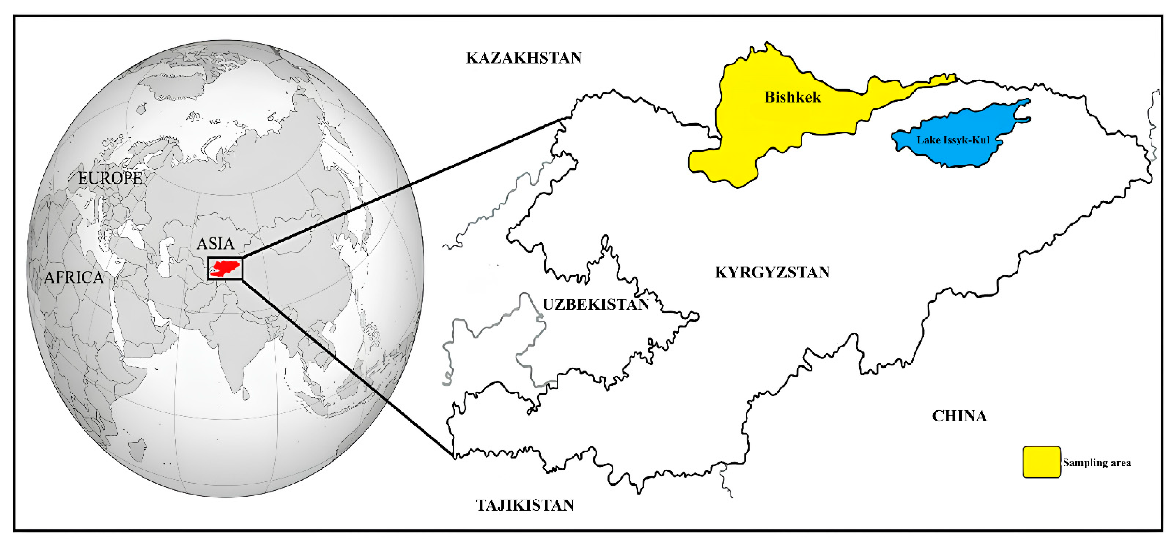
2.1. Study Area, Collection of Blood Samples, and DNA Extraction

The Republic of Kyrgyzstan is a country in Central Asia that is bordered by Uzbekistan, Kazakhstan, Tajikistan, and China. The country has seven managerial regions: Chuy, Jalal-Abad, Talas, Naryn, Batken, Issyk-Kul, and Osh. Bishkek, located in the Chuy region, is the country’s largest city and capital (Figure 1). Bishkek is located in the north of Kyrgyzstan, bordering Kazakhstan. The country has a continental climate, with hot summers and cold winters, and is surrounded by mountains. In this study, DNA samples were obtained from a study conducted by our team [16] and these DNA samples were stored in Sivas Cumhuriyet University Parasitology Department Laboratories under appropriate conditions. To summarize, the blood samples were collected from 337 shelter dogs in Bishkek between 2017 and 2019. In the sampling process, cooperation was made with the Kyrgyz-Turkish Manas University Veterinary Teaching Hospital. All dogs showed no clinical symptoms at first observation and were recorded as healthy or asymptomatic. Total DNA isolation from the blood samples was conducted using a commercial DNA isolation kit (PureLink Genomic DNA Kit, Invitrogen, Carlsbad, CA, USA).

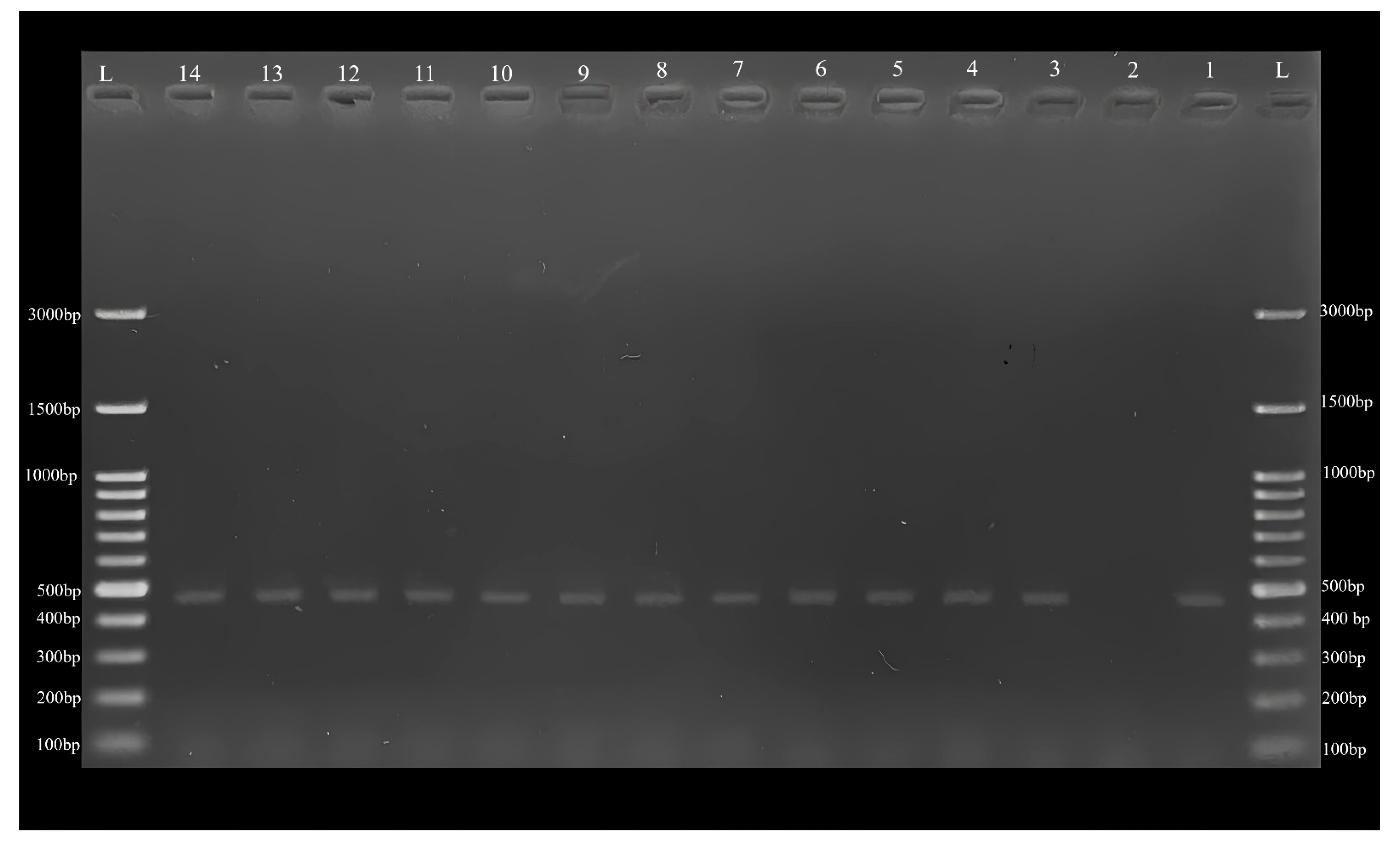
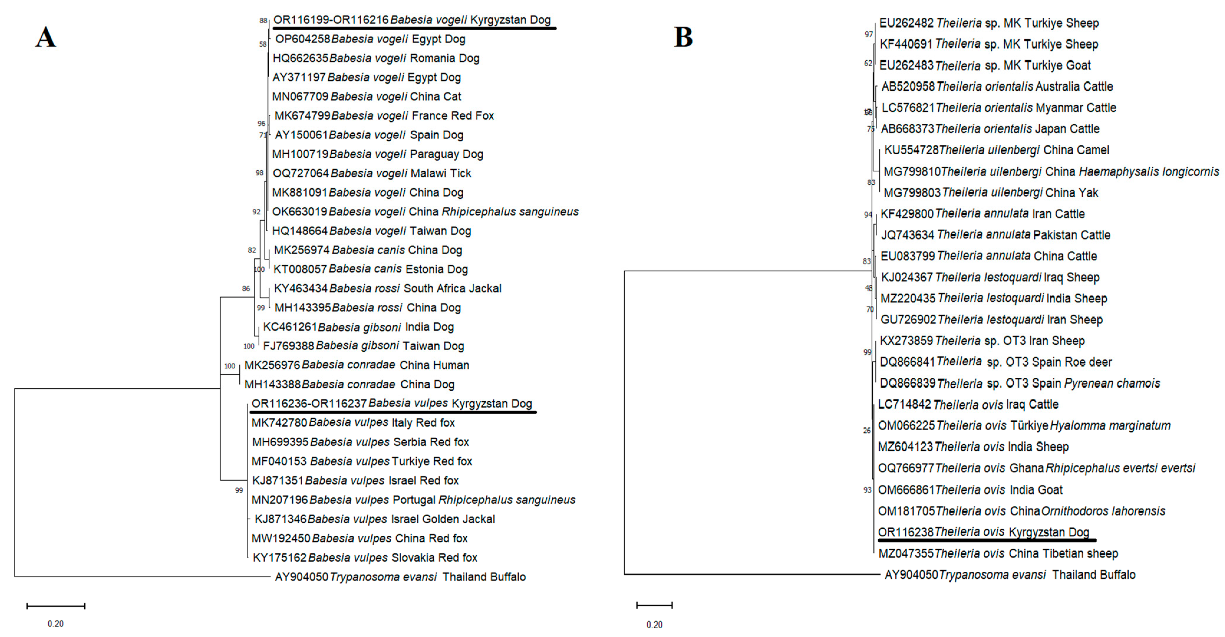
2.2. Polymerase Chain Reaction (PCR), Sequencing, and Phylogenetic Analyses

A total of 337 blood samples were analyzed by nested PCR in terms of piroplasm species. The names, sequence, amplicon sizes, and PCR conditions of primers used in this study are listed in Table 1.
PCR was carried out at 25 µL final volume, including 14.875 µL DNase-RNase-free sterile water (Qiagen®, Hilden, Germany), 2.5 µL 10× PCR buffer (Thermo Scientific™, Vilnus, Lithuania), 2.5 µL MgCl2 (25 mM) (Thermo Scientific™, Vilnus, Lithuania), 0.5 µL dNTP (10 mM) (Cat.No.: R0181, Thermo Scientific™, Vilnus, Lithuania), 0.125 µL of Taq DNA polymerase (5 U) (Cat.No.: EP0402, Thermo Scientific™, Vilnus, Lithuania), 1 µL (10 pmol/µL) of each of the primers, and 2.5 µL template DNA. For nested PCR, 1 µL of the first round of PCR product was used as template DNA. After PCR amplifications, the PCR product was loaded on the agarose gel and subjected to electrophoresis at 90 V for 60 min. The agarose gel was stained with an ethidium bromide solution for 20 min and checked for ~500 bp amplicons using a UV transilluminator.
In this study, all PCR positive samples were sequenced with BJ1 and BN2 primers. DNA sequences were carried out with an ABI 3730XL analyzer (Applied Biosystems, Foster City, CA, USA) using the BigDye Terminator v3.1 Cycle Sequencing Kit (Applied Biosystems, Foster City, CA, USA). Nucleotide sequence files were opened with FinchTV (version 1.4.0) software (Geospiza Inc., Seattle, Washington, DC, USA) for the determination of chromatogram quality. Sequences with low chromatogram quality were not used for the determination of the consensus sequences. MEGA-11 software (Version 11.0.13) was used to determine and assemble the consensus sequences [26]. The B. vogeli, B. vulpes, and T. ovis consensuses identified in this study were aligned with reference sequences of the mentioned pathogens present in the GenBank using the MUSCLE algorithm in the MEGA-11 software [26]. Further nucleotide identities between our sequence and sequences present in GenBank were determined by BLAST analyses.
After the determination of the consensus sequence of B. vogeli, B. vulpes, and T. ovis isolates identified in the current study, these sequences were uploaded to GenBank under the following accession numbers: B. vogeli OR116199-OR116216, B. vulpes OR116236-OR116237, and T. ovis OR116238. The lengths of consensus sequence recorded in the GenBank for B. vogeli, B. vulpes, and T. ovis were 450–455 bp, 481 bp, and 461 bp, respectively.
Phylogenetic trees were constructed using maximum likelihood analysis (ML) in the MEGA-11 software [26] for the determination of genetic relationships between pathogens identified in this study and those present in the GenBank. Before constructing the trees, the best-fit model for ML was determined as the Kimura-2 + G parameter model for Theileria species [27] using the Find Best-Fit Substitution Model in MEGA-11 [26]. Bootstrap values were performed with 1000 replicates.

3. Results

As a result of PCR analysis of 337 blood samples, 21 (6.23%) samples were found to be positive (Figure 2) for piroplasm species. All PCR positive samples were sequenced for species identifications of pathogens detected in this study.
In this study, 18 samples (5.34%) were found to be infected with B. vogeli using DNA sequence analyses. The partial sequence of the 18S SSU rRNA gene of B. vogeli isolates identified in this study showed 100% nucleotide identity among each other. Furthermore, BLAST analyses revealed that 98.03–100% nucleotide identities were present between our B. vogeli isolates and other B. vogeli isolates uploaded to GenBank from different parts of the world. Babesia vogeli isolates identified in this work shared 100% nucleotide identity with B. vogeli isolates detected from dogs in Egypt (AY371197, OP604258, and OP604259) and dogs in Romania (HQ662635 and JF461252). Phylogenetic analyses of the 18S SSU rRNA gene revealed that single nucleotide polymorphisms (SNPs) were present in two sites between B. vogeli isolates. The 594th and 627th nucleotides are cytosine in both our B. vogeli isolates and those from Egypt (AY371197, OP604258, and OP604259) and Romania (HQ662635 and JF461252). These nucleotides are thymine in B. vogeli isolates obtained from dogs in China (MK881091), Brazil (MW62732), Spain (AY150061), Paraguay (MH100719), and Taiwan (HQ148664), from ticks in China (OK663019) and Malawi (OQ727064), from red fox in France (MK674799), and from cat in China (MN067709).
The DNA of B. vulpes was detected in 2 out of 337 dog samples (0.59%) in this study. The 98.97–100% nucleotide identities were found between B. vulpes isolates obtained in this work and B. vulpes detected in red foxes, dogs, and tick species present in GenBank. B. vulpes 18S SSU rRNA gene sequences shared 100% nucleotide identity with each other and B. vulpes isolates identified from red foxes in Italy (MK742780), Poland (MH553357), Türkiye (MF040153), Slovakia (KY175167), and China (MW192450), and from dogs in Hungary (MH544242, MW805763), and from R. sanguineus in Portugal (MN207196).
Unexpectedly, T. ovis DNA was detected in one (0.29%) dog in the current study, according to DNA sequence analysis. The BLAST analysis showed that 100% nucleotide identity was present between T. ovis dog isolates identified in the current work and T. ovis isolates identified from R. evertsi evertsi (OQ766977) in Ghana, cattle (LC714842) in Iraq, goat (OM666861) in India, Ornithodoros lahorensis (O. lahorensis) (OM181705) and Tibetian sheep (MZ047355) in China, Hyalomma marginatum (Hy. marginatum) (OM066225) in Türkiye, and sheep (MZ604123) in India deposited to the GenBank from various parts of the world.
Phylogenetic trees revealed that our B. vogeli, B. vulpes, and T. ovis isolates were grouped with B. vogeli, B. vulpes, and T. ovis isolates, respectively, and placed in a different cluster with other Babesia (Figure 3A) and Theileria species (Figure 3B).

4. Discussion

Babesiosis is one of the most important tick-borne infectious diseases of dogs. Canine babesiosis is an infection that causes a wide range of symptoms in dogs, ranging from subclinical to severe. In most cases in dogs, the non-specific clinical manifestations include fever, lethargy, loss of appetite, jaundice, pale mucous membranes due to acute hemolysis, splenomegaly, and weight loss. In addition to these clinical symptoms, the diseases can cause death in cases of severe infection. The severity of the disease mainly depends on the pathogenicity of the species or subspecies [28,29]. Both the clinical signs are not fully descriptive in the diagnostic attempts of veterinarians, and the difficulties in identifying the species found in this lineage have been overcome by molecular methods. PCR and sequencing are widely used in diagnosis and in determining the phylogenetic positions of species [22,23,30]. Studies using these methods have provided more accurate information about the epidemiology of the disease as well as the identification of species. Disease agents, which were known in the past as two types as large and small species (B. canis and B. gibsoni), are now understood to include at least six species (B. canis canis, B. canis vogeli, B. canis rossi, B. gibsoni, B. vulpes, and B. condradae) [5,6,11,13]. In fact, some species of other vertebrates have been identified in dogs with these methods [4]. On the other hand, it is very important to know about the presence and prevalence of pathogens in order to know what diseases can emerge in each country. In this study, the investigation of piroplasm parasites in dogs from Kyrgyzstan was performed by PCR and sequence analysis for the first time. In PCR, we amplified the 18S SSU rRNA gene of Babesia species. PCR positive results were seen in 21 (6.23%) of 337 blood samples. As a result of the sequencing, we have revealed the presence of B. vogeli and B. vulpes in dogs in the country. Interestingly, the sequence results revealed the presence of T. ovis in dogs. It is a sheep protozoon parasite transmitted by ticks [31]. The results showed that babesiosis should be taken into account in dog health in the country, as well as contributing to the global epidemiology of the parasites. In addition, it may be thought that dogs may contribute to the epidemiology of T. ovis [4].
Babesia vogeli is a globally distributed tick-borne pathogen, and this species has been detected on five continents. This species can be referred to as the most common species of Babesia [14,32,33,34,35,36]. In this study, B. vogeli was detected in 18 (5.34%) of 337 blood samples. The prevalence of the parasite was 2.1% in Malaysia [37], 4.8% in Brazil [38], 5.1% in Egypt [39], 5.1% in Iraq [40], 8.5% in Bosnia and Herzegovina [41], 9.3% in Iran [42], and 10.8% in Nigeria [43]. When the studies given above were evaluated, it was seen that the prevalence of B. vogeli was quite different. It is possible to associate this with many factors, such as the characteristics of the geographical region, the size of the sampling, whether the animals studied are pets, shelter animals or stray animals, and the method used. The important aspect of these studies is that the parasite is usually first detected in the region. In our study, B. vogeli was introduced for the first time to dogs in Kyrgyzstan. It is thought that these results will contribute to the global epidemiology of the parasite. In this study, all B. vogeli isolates were sequenced, and the 98.03–100% nucleotide identities were determined between our B. vogeli isolates and other isolates identified from dogs, red foxes, cats, and various tick species. According to the nucleotide sequence comparison of the partial 18S SSU rRNA gene region of between B. vogeli isolates, SNPs were found in two sites. The 594th and 627th nucleotides are cytosine in both our B. vogeli isolates and B. vogeli isolates identified in Egypt (AY371197, OP604258, and OP604259) and Romania (HQ662635 and JF461252). These nucleotides are thymine in B. vogeli isolates in China (MK881091), Brazil (MW62732), Spain (AY150061), Paraguay (MH100719), Taiwan (HQ148664), China (OK663019), Malawi (OQ727064), France (MK674799), and China (MN067709) (Figure 3A). Studies revealed that the 18S SSU rRNA gene was highly conserved, with little intraspecific variation [44]. Therefore, two nucleotide substitutions in small parts of the 18S SSU rRNA gene of B. vogeli isolates, when evaluated together with the data in the GenBank, suggested that the pathogen may consist of two different genotypes.
Banet et al. [1] showed that T. annae, B. annae, B. microti-like piroplasm, B. cf. microti, and Babesia Spanish dog isolates are synonymous with B. vulpes. For this reason, the species detected in this study was named as B. vulpes [1]. Although it is stated that B. vulpes is mainly transmitted by Ixodes species, this issue has not been fully elucidated. Since I. hexoganus is more common in parasite-infected dogs, it is suggested that its vector may be this species [45]. In addition, parasite DNA has also been detected in tick species such as I. ricinus [46], D. reticulatus [47], and R. sanguineus [48]. Ixodes crenulatus has been identified as the dominant tick species in rodents [49]. The presence of D. marginatus. D. niveus, R. sanguineus, R. turanicus, Hae. punctata, Hae. sulcata, Hy. marginatum, Hy. anatolicum, Hy. scupense, and Hy. asiaticum have also been recorded in the country [50]. Babesia vulpes infects foxes and dogs [51]. However, there are many studies on the prevalence of B. vulpes in red foxes almost all over the world, and in these studies, the prevalence of B. vulpes in foxes has been detected at 72.00% in Spain [52], 69.20% in Portugal [53], 50.70% in Austria [54], 46.40% in Germany [55], and 30.70% in Poland [56]. While there is a high fox population in Kyrgyzstan [57], further studies on foxes may also find the parasite widespread. However, it is known that the parasite does not cause clinical infection in foxes, but it causes clinical disease in dogs [58]. There have been limited studies on the prevalence of B. vulpes in dogs. Studies are generally in the form of case reports. In this way, it has been reported in countries such as Canada [59], Spain [60], and Russia [61]. In North America, 9098 dogs were examined and B. vulpes was detected in only 0.52% of them [58]. In Spain, 153 dogs were investigated and T. annae (B. vulpes) was detected in 0.70% of the samples [62]. In this study, we detected B. vulpes in 0.59% of the 337 dogs examined. This prevalence, together with other data, shows that the prevalence of B. vulpes in dogs is quite low. As a result, as the detection of the parasite in the country will guide future studies, it has emerged that B. vulpes should be considered in clinical cases associated with babesiosis in dogs. The high nucleotide identities (98.97–100%) were seen between our B. vulpes isolates and B. vulpes identified from different hosts, such as red foxes, dogs, and tick species. This result shows that the 18S SSU rRNA gene can be used successfully in the diagnosis of B. vulpes among hosts, but the gene is not useful for the determination of the genetic diversity of the pathogen.
The improvement of molecular identification techniques and the widespread use of these for the detection of parasitic diseases, especially in the last two decades, have provided important contributions to research into the high specificity and sensitivity diagnosis of parasitic diseases, the determination of the genetic diversity of pathogens, and the detection of new species/strains/genotypes [15,22,23,30]. Furthermore, molecular techniques can provide unexpected species detection in different host species [63]. Normally, the BJ1 and BN2 primers used in this study were thought to be specific to Babesia species [25], but subsequent studies have shown that these primers also amplify Theileria species [64]. Therefore, all PCR-positive samples were sequenced with the primers, and T. ovis was detected in one dog sample in the current study. The T. ovis dog isolate identified in this study was recorded to the GenBank (Accession number: OR116238). This isolate was included among Genbank registered T. ovis isolates in the phylogenetic tree, as confirmed by sequence analysis (OR116238). The 100% nucleotide identity was detected between the T. ovis dog isolate detected in this study and T. ovis isolates identified from different hosts, including sheep and tick species detected in different parts of the world. No study has been found in Kyrgyzstan that has been conducted among sheep and goats, which are the hosts of this parasite, to which we compared our obtained sequence for the determination of genetic diversity between T. ovis isolates. Beforehand, T. ovis, T. buffeli (T. orientalis complex), and T. luwenshuni were detected by molecular methods in asymptomatic dogs in Iran [4]. The study in Iran was carried out on shepherd dogs, and they detected T. luwenshuni, T. buffeli, and T. ovis in 13% of the 52 dogs they examined [4]. This result is considerably higher than our result. In this case, the most decisive factor may be shepherd dogs. Additionally, T. annae (B. microti-like), Theileria sp. OT3, T. equi, and T. annulata have been detected in dogs [2,65,66,67]. Although these findings do not prove that dogs are hosts for the specified species, they may be interpreted as contributing to the epidemiology of these species. This is even more important considering that dogs are used in the management of herds, especially in species such as T. ovis, which is a parasite of sheep and goats.

5. Conclusions

To the best of our knowledge, this study presents the first molecular detection of B. vogeli, B. vulpes, and T. ovis in dogs from Kyrgyzstan. These results may contribute to better understanding the epidemiology of canine babesiosis in Kyrgyzstan. Moreover, the B. vogeli nucleotide sequence revealed that this species may consist of two genotypes (according to Genbank data), and we think this data might be clarified with further studies. However, there is a need to carry out studies involving wild Canidae and vector tick species in the country.

Author Contributions

Conceptualization, K.A. and N.D.; Methodology, K.A., U.E., O.F.S., M.F.A. and A.A.; Formal Analysis, K.A., N.D., U.E., O.F.S., M.F.A. and A.A.; Data Curation, K.A., U.E., O.F.S., M.F.A. and A.A.; Writing—Original Draft Preparation, K.A. and U.E.; Writing—Review and Editing, K.A. and N.D. All authors have read and agreed to the published version of the manuscript.

Funding

This research received no external funding.

Institutional Review Board Statement

The ethical approval of this study was obtained from the ethics committees of Kyrgyz-Turkish Manas University (Animal Experiment Ethic Committee, protocol no: 2017–06/02) and Sivas Cumhuriyet University Animal Experiments Local Ethics Committee (Animal Experiment Ethic Committee, protocol no: 03.08.2023–736).

Informed Consent Statement

Not applicable.

Data Availability Statement

Data available in a publicly accessible repository.

Acknowledgments

The authors would like to thank all veterinarians and technicians for their kind help during sample collection.

Conflicts of Interest

The authors declare no conflict of interest.

References

  1. Baneth, G.; Florin-Christensen, M.; Cardoso, L.; Schnittger, L. Reclassification of Theileria annae as Babesia vulpes sp. nov. Parasite. Vector. 2015, 8, 207–214. [Google Scholar] [CrossRef] [Green Version]
  2. Beck, R.; Vojta, L.; Mrljak, V.; Marinculić, A.; Beck, A.; Živičnjak, T.; Cacciò, S.M. Diversity of Babesia and Theileria species in symptomatic and asymptomatic dogs in Croatia. Int. J. Parasitol. 2009, 39, 843–848. [Google Scholar] [CrossRef]
  3. Bigdeli, M.; Rafie, S.M.; Namavari, M.M.; Jamshidi, S. Report of Theileria annulata and Babesia canis infections in dogs. Comp. Clin. Path. 2012, 21, 375–377. [Google Scholar] [CrossRef]
  4. Gholami, S.; Laktarashi, B.; Shiadeh, M.M.; Spotin, A. Genetic variability, phylogenetic evaluation and first global report of Theileria luwenshuni, T. buffeli, and T. ovis in sheepdogs in Iran. Parasitol. Res. 2015, 115, 2125–2130. [Google Scholar] [CrossRef] [PubMed]
  5. Piana, G.P.; Galli-Valerio, B. Su di un’ infezione del cane con parasiti endoglobulari. Mod. Zooiatr. 1895, 6, 163–169. [Google Scholar]
  6. Matijatko, V.; Torti, M.; Schetters, T.P. Canine babesiosis in Europe: How many diseases? Trends Parasitol. 2012, 28, 99–105. [Google Scholar] [CrossRef] [PubMed]
  7. Aktas, M.; Ozubek, S. A survey of canine haemoprotozoan parasites from Turkey, including molecular evidence of an unnamed Babesia. Comp. Immunol. Microbiol. Infect. Dis. 2017, 52, 36–42. [Google Scholar] [CrossRef]
  8. Uilenberg, G. Babesia—A historical overview. Vet. Parasitol. 2006, 138, 3–10. [Google Scholar] [CrossRef]
  9. Uilenberg, G.; Franssen, F.F.J.; Perie, N.M.; Spanjer, A.A.M. Three groups of Babesia canis distinguished and a proposal for nomenclature. Vet. Quart. 1989, 11, 33–40. [Google Scholar] [CrossRef] [Green Version]
  10. Solano-Gallego, L.; Baneth, G. Babesiosis in dogs and cats-expanding parasitological and clinical spectra. Vet. Parasitol. 2011, 181, 48–60. [Google Scholar] [CrossRef]
  11. Patton, W.S. Preliminary report on a new piroplasm (Piroplasma gibsoni sp. nov.) found in the blood of the hounds of the Madras Hunt and subsequently discovered in the blood of the jackal Canis aureus. Bull. Soc. Pathol. Exot. 1910, 3, 274–280. [Google Scholar]
  12. Schnittger, L.; Rodriguez, A.E.; Florin-Christensen, M.; Morrison, D.A. Babesia: A world emerging. Infect. Genet. Evol. 2012, 12, 1788–1809. [Google Scholar] [CrossRef]
  13. Penzhorn, B.L.; Oosthuizen, M.C. Babesia species of domestic cats: Molecular characterization has opened pandora’s box. Front. Vet. Sci. 2020, 7, 134. [Google Scholar] [CrossRef] [PubMed]
  14. Birkenheuer, A.J.; Levy, M.G.; Breitschwerdt, E.B. Development and evaluation of a seminested PCR for detection and differentiation of Babesia gibsoni (Asian genotype) and B. canis DNA in canine blood samples. J. Clin. Microbiol. 2003, 41, 4172–4177. [Google Scholar] [CrossRef] [Green Version]
  15. Ozubek, S.; Aktas, M. Molecular evidence of a new Babesia sp. in goats. Vet. Parasitol. 2017, 233, 1–8. [Google Scholar] [CrossRef] [PubMed]
  16. Aydın, M.F.; Altay, K.; Aytmirzakizi, A.; Dumanlı, N. First molecular detection of Dirofilaria immitis and D. repens in dogs from Kyrgyzstan. Acta Parasitol. 2020, 65, 949–953. [Google Scholar] [CrossRef]
  17. Altay, K.; Aydın, M.F.; Aytmirzakizi, A.; Jumakanova, Z.; Cunusova, A.; Dumanlı, N. First molecular evidence for Mycoplasma haemocanis and Candidatus Mycoplasma haematoparvum in asymptomatic shelter dogs in Kyrgyzstan. Kafkas Univ. Vet. Fak. Derg. 2020, 26, 143–146. [Google Scholar] [CrossRef]
  18. Altay, K.; Aydın, M.F.; Aytmirzakizi, A.; Jumakanova, Z.; Cunusova, A.; Dumanlı, N. First molecular detection and phylogenetic analysis of Mycoplasma wenyonii and Candidatus Mycoplasma haemobos in cattle in different parts of Kyrgyzstan. Biologia 2023, 78, 633–640. [Google Scholar] [CrossRef]
  19. Altay, K.; Aydın, M.F.; Aytmirzakizi, A.; Jumakanova, Z.; Cunusova, A.; Dumanlı, N. Molecular survey of hepatozoonosis in natural infected dogs: First detection and molecular characterisation of Hepatozoon canis in Kyrgyzstan. Kafkas Univ. Vet. Fak. Derg. 2019, 25, 77–81. [Google Scholar] [CrossRef]
  20. Aktaş, M.; Kısadere, İ.; Özübek, S.; Cihan, H.; Salıkov, R.; Cirak, V.Y. First molecular survey of piroplasm species in cattle from Kyrgyzstan. Parasitol. Res. 2019, 118, 2431–2435. [Google Scholar] [CrossRef] [PubMed]
  21. Ozubek, S.; Ulucesme, M.C.; Cirak, V.Y.; Aktas, M. Detection of Theileria orientalis genotypes from cattle in Kyrgyzstan. Pathogens 2022, 11, 1185. [Google Scholar] [CrossRef]
  22. Altay, K.; Erol, U.; Sahin, O.F.; Aytmirzakizi, A. First molecular detection of Anaplasma species in cattle from Kyrgyzstan; molecular identification of human pathogenic novel genotype Anaplasma capra and Anaplasma phagocytophilum related strain. Ticks Tick Borne Dis. 2022, 13, 101861. [Google Scholar] [CrossRef]
  23. Altay, K.; Erol, U.; Sahin, O.F.; Aytmirzakizi, A.; Temizel, E.M.; Aydin, M.F.; Dumanli, N.; Aktas, M. The detection and phylogenetic analysis of Anaplasma phagocytophilum-like 1, A. ovis and A. capra in sheep: A. capra divides into two genogroups. Vet. Res. Commun. 2022, 46, 1271–1279. [Google Scholar] [CrossRef] [PubMed]
  24. Oosthuizen, M.C.; Zweygarth, E.; Collins, N.E.; Troskie, M.; Penzhorn, B.L. Identification of a novel Babesia sp. from a sable antelope (Hippotragus niger Harris, 1838). J. Clin. Microbiol. 2008, 46, 2247–2251. [Google Scholar] [CrossRef] [PubMed] [Green Version]
  25. Casati, S.; Sager, H.; Gern, L.; Piffaretti, J.C. Presence of potentially pathogenic Babesia sp. for human in Ixodes ricinus in Switzerland. Ann. Agric. Environ. Med. 2006, 13, 65–70. [Google Scholar]
  26. Tamura, K.; Stecher, G.; Kumar, S. MEGA11: Molecular evolutionary genetics analysis version 11. Mol. Biol. Evol. 2021, 38, 3022–3027. [Google Scholar] [CrossRef] [PubMed]
  27. Kimura, M. A simple method for estimating evolutionary rate of base substitutions through comparative studies of nucleotide sequences. J. Mol. Evol. 1980, 16, 111–120. [Google Scholar] [CrossRef]
  28. Schetters, T.P.; Kleuskens, J.A.G.M.; Scholtes, N.; Gorenflot, A. Parasite localization and dissemination in the Babesia-infected host. Ann. Trop. Med. Parasitol. 1998, 92, 513–519. [Google Scholar] [CrossRef] [PubMed]
  29. Birkenheuer, A.J. Babesiosis. In Green’s Infectious Diseases of the Dog and Cat, 4th ed.; Greene, C.E., Ed.; Elsevier: St. Louis, MI, USA, 2012; pp. 771–784. [Google Scholar]
  30. Sahin, O.F.; Erol, U.; Altay, K. Buffaloes as new hosts for Anaplasma capra: Molecular prevalence and phylogeny based on gtlA, groEL, and 16S rRNA genes. Res. Vet. Sci. 2022, 152, 458–464. [Google Scholar] [CrossRef] [PubMed]
  31. Aktas, M.; Altay, K.; Dumanli, N. PCR-based detection of Theileria ovis in Rhipicephalus bursa adult ticks. Vet. Parasitol. 2006, 140, 259–263. [Google Scholar] [CrossRef]
  32. Criado, A.; Martinez, J.; Buling, A.; Barba, J.C.; Merino, S.; Jefferies, R.; Irwin, P.J. New data on epizootiology and genetics of piroplasms based on sequences of small ribosomal subunit and cytochrome b genes. Vet. Parasitol. 2006, 142, 238–247. [Google Scholar] [CrossRef]
  33. Criado-Fornelio, A.; Martinez-Marcos, A.; Buling-Sarana, A.; Barba-Carretero, J.C. Molecular studies on Babesia, Theileria and Hepatozoon in southern Europe. Part II: Phylogenetic analysis and evolutionary history. Vet. Parasitol. 2003, 114, 173–194. [Google Scholar] [CrossRef]
  34. Matjila, P.T.; Penzhorn, B.L.; Bekker, C.P.J.; Nijhof, A.M.; Jongejan, F. Confirmation of occurrence of Babesia canis vogeli in domestic dogs in South Africa. Vet. Parasitol. 2004, 122, 119–125. [Google Scholar] [CrossRef] [PubMed]
  35. Inokuma, H.; Yoshizaki, Y.; Shimada, Y.; Sakata, Y.; Okuda, M.; Onishi, T. Epidemiological survey of Babesia species in Japan performed with specimens from ticks collected from dogs and detection of new Babesia DNA closely related to Babesia odocoilei and Babesia divergens DNA. J. Clin. Microbiol. 2003, 41, 3494–3498. [Google Scholar] [CrossRef] [PubMed] [Green Version]
  36. Jefferies, R.; Ryan, U.M.; Muhlnickel, C.J.; Irwin, P.J. Two species of canine Babesia in Australia: Detection and characterization by PCR. J. Parasitol. 2003, 89, 409–412. [Google Scholar] [CrossRef] [PubMed] [Green Version]
  37. Prakash, B.K.; Low, V.L.; Vinnie-Siow, W.Y.; Tan, T.K.; Lim, Y.A.L.; Morvarid, A.R.; AbuBakar, S.; Sofian-Azirun, M. Detection of Babesia spp. in dogs and their ticks from Peninsular Malaysia: Emphasis on Babesia gibsoni and Babesia vogeli infections in Rhipicephalus sanguineus sensu lato (Acari: Ixodidae). J. Med. Entomol. 2018, 55, 1337–1340. [Google Scholar] [CrossRef]
  38. de Oliveira Carieli, E.P.; da Silva, V.C.L.; de Lima, E.R.; Dias, M.B.D.M.C.; Fukahori, F.L.P.; de Azevedo Rêgo, M.S.; Júnior, J.W.P.; de Cássia Peixoto Kim, P.; Leitão, R.S.C.S.; Mota, R.A.; et al. Parasitological and molecular detection of Babesia canis vogeli in dogs of Recife, Pernambuco and evaluation of risk factors associated. Semina. Ciências. Agrárias. 2016, 37, 163–171. [Google Scholar] [CrossRef] [Green Version]
  39. Selim, A.; Megahed, A.; Ben Said, M.; Alanazi, A.D.; Sayed-Ahmed, M.Z. Molecular survey and phylogenetic analysis of Babesia vogeli in dogs. Sci. Rep. 2022, 12, 6988. [Google Scholar] [CrossRef] [PubMed]
  40. Badawi, N.M.; Yousif, A.A. Babesia canis spp. in dogs in Baghdad province, Iraq: First molecular identifcation and clinical and epidemiological study. Vet. World. 2020, 13, 579–585. [Google Scholar] [CrossRef]
  41. Ćoralić, A.; Gabrielli, S.; Zahirović, A.; Stojanović, N.M.; Milardi, G.L.; Jažić, A.; Zuko, A.; Čamo, D.; Otašević, S. First molecular detection of Babesia canis in dogs from Bosnia and Herzegovina. Ticks Tick Borne Dis. 2018, 9, 363–368. [Google Scholar] [CrossRef]
  42. Khanmohammadi, M.; Zolfaghari-Emameh, R.; Arshadi, M.; Razmjou, E.; Karimi, P. Molecular identifcation and genotyping of Babesia canis in dogs from Meshkin Shahr County, Northwestern Iran. J. Arthropod Borne Dis. 2021, 15, 97–107. [Google Scholar] [CrossRef]
  43. Obeta, S.S.; Ibrahim, B.; Lawal, I.A.; Natala, J.A.; Ogo, N.I.; Balogun, E.O. Prevalence of canine babesiosis and their risk factors among asymptomatic dogs in the federal capital territory, Abuja, Nigeria. Parasite Epidemiol. Control. 2020, 11, e00186. [Google Scholar] [CrossRef]
  44. Mandal, M.; Banerjee, P.S.; Garg, R.; Ram, H.; Kundu, K.; Kumar, S.; Kumar, G.R. Genetic characterization and phylogenetic relationships based on 18S rRNA and ITS1 region of small form of canine Babesia spp. from India. Infect. Genet. Evol. 2014, 27, 325–331. [Google Scholar] [CrossRef]
  45. Camacho, A.T.; Pallas, E.; Gestal, J.J.; Guitián, F.J.; Olmeda, A.S.; Telford III, S.R.; Spielman, A. Ixodes hexagonus is the main candidate as vector of Theileria annae in Northwest Spain. Vet. Parasitol. 2003, 112, 157–163. [Google Scholar] [CrossRef]
  46. Lledó, L.; Giménez-Pardo, C.; Dominguez-Penafiel, G.; Sousa, R.; Gegúndez, M.I.; Casado, N.; Criado, A. Molecular detection of hemoprotozoa and Rickettsia species in arthropods collected from wild animals in the Burgos Province, Spain. Vector Borne Zoonotic Dis. 2010, 10, 735–738. [Google Scholar] [CrossRef] [Green Version]
  47. Hodžić, A.; Zörer, J.; Duscher, G.G. Dermacentor reticulatus, a putative vector of Babesia cf. microti (syn. Theileria annae) piroplasm. Parasitol. Res. 2017, 116, 1075–1077. [Google Scholar] [CrossRef] [PubMed] [Green Version]
  48. Iori, A.; Gabrielli, S.; Calderini, P.; Moretti, A.; Pietrobelli, M.; Tampieri, M.P.; Galuppi, R.; Cancrini, G. Tick reservoirs for piroplasms in central and northern Italy. Vet. Parasitol. 2010, 170, 291–296. [Google Scholar] [CrossRef] [PubMed]
  49. Akyshova, B.; Chen, Y.N.; Chen, J. Abundance of ectoparasitic ticks and mites (Acari: Ixodida, Mesostigmata, Trombidiformes) on rodents in the Alamedin Gorge of Kyrgyz Range, Kyrgyzstan. Syst. Appl. Acarol. 2022, 27, 1120–1131. [Google Scholar] [CrossRef]
  50. Aknazarov, B.; Jetigenov, E.; Atabekova, N.; Suerkulov, U.; Abdumanap, N. Spread of arthropod-borne infections in Kyrgyzstan. E3S Web Conf. 2023, 380, 01027. [Google Scholar] [CrossRef]
  51. Solano-Gallego, L.; Sainz, Á.; Roura, X.; Estrada-Peña, A.; Miró, G. A review of canine babesiosis: The European perspective. Parasit. Vectors. 2016, 9, 336. [Google Scholar] [CrossRef] [PubMed] [Green Version]
  52. Checa, R.; López-Beceiro, A.M.; Montoya, A.; Barrera, J.P.; Ortega, N.; Gálvez, R.; Marino, V.; González, J.; Olmeda, Á.S.; Fidalgo, L.E.; et al. Babesia microti-like piroplasm (syn. Babesia vulpes) infection in red foxes (Vulpes vulpes) in NW Spain (Galicia) and its relationship with Ixodes hexagonus. Vet. Parasitol. 2018, 252, 22–28. [Google Scholar] [CrossRef] [PubMed]
  53. Cardoso, L.; Cortes, H.C.E.; Reis, A.; Rodrigues, P.; Simões, M.; Lopes, A.P.; Vila-Viçosa, M.J.; Talmi-Frank, D.; Eyal, O.; Solano-Gallego, L.; et al. Prevalence of Babesia microti-like infection in red foxes (Vulpes vulpes) from Portugal. Vet. Parasitol. 2013, 196, 90–95. [Google Scholar] [CrossRef] [PubMed]
  54. Hodžić, A.; Mrowietz, N.; Cezanne, R.; Bruckschwaiger, P.; Punz, S.; Habler, V.E.; Tomsik, V.; Lazar, J.; Duscher, G.G.; Glawischnig, W.; et al. Occurrence and diversity of arthropod-transmitted pathogens in red foxes (Vulpes vulpes) in western Austria, and possible vertical (transplacental) transmission of Hepatozoon canis. Parasitology 2018, 145, 335–344. [Google Scholar] [CrossRef]
  55. Najm, N.A.; Meyer-Kayser, E.; Hoffmann, L.; Herb, I.; Fensterer, V.; Pfister, K.; Silaghi, C. A molecular survey of Babesia spp. and Theileria spp. in red foxes (Vulpes vulpes) and their ticks from Thuringia, Germany. Ticks Tick Borne Dis. 2014, 5, 386–391. [Google Scholar] [CrossRef]
  56. Mierzejewska, E.J.; Dwużnik, D.; Koczwarska, J.; Stańczak, Ł.; Opalińska, P.; Krokowska-Paluszak, M.; Wierzbicka, A.; Górecki, G.; Bajer, A. The red fox (Vulpes vulpes), a possible reservoir of Babesia vulpes, B. canis and Hepatozoon canis and its association with the tick Dermacentor reticulatus occurrence. Ticks Tick Borne Dis. 2021, 1, 101551. [Google Scholar] [CrossRef] [PubMed]
  57. McCarthy, J.L.; McCarthy, K.P.; Fuller, T.K.; McCarthy, T.M. Assessing variation in wildlife biodiversity in the Tien Shan Mountains of Kyrgyzstan using ancillary camera-trap photos. Mt. Res. Develop. 2010, 30, 295–301. [Google Scholar] [CrossRef]
  58. Barash, N.R.; Thomas, B.; Birkenheuer, A.J.; Breitschwerdt, E.B.; Lemler, E.; Qurollo, B.A. Prevalence of Babesia spp. and clinical characteristics of Babesia vulpes infections in North American dogs. J. Vet. Intern. Med. 2019, 33, 2075–2081. [Google Scholar] [CrossRef] [PubMed] [Green Version]
  59. Arsenault, A.C.; Foley, P.M.; Clancey, N.P. Babesia vulpes in a dog from Prince Edward Island, Canada. Can. Vet. J. 2022, 63, 589–592. [Google Scholar] [PubMed]
  60. Unterköfler, M.S.; Pantchev, N.; Bergfeld, C.; Wülfing, K.; Globokar, M.; Reinecke, A.; Fuehrer, H.P.; Leschnik, M. Case report of a fatal Babesia vulpes infection in a splenectomised dog. Parasitologia 2023, 3, 59–68. [Google Scholar] [CrossRef]
  61. Radyuk, E.; Karan, L. A case of Babesia vulpes infection in a dog in Russia. Vet. Parasitol. Reg. Stud. 2020, 22, 100467. [Google Scholar] [CrossRef]
  62. Tabar, M.D.; Francino, O.; Altet, L.; Sánchez, A.; Ferrer, L.; Roura, X. PCR survey of vectorborne pathogens in dogs living in and around Barcelona, an area endemic for leishmaniosis. Vet. Record. 2009, 164, 112–116. [Google Scholar] [CrossRef]
  63. Ozubek, S.; Aktas, M. Molecular and parasitological survey of ovine piroplasmosis, including the first report of Theileria annulata (Apicomplexa: Theileridae) in sheep and goats from Turkey. J. Med. Entomol. 2017, 54, 212–220. [Google Scholar] [CrossRef]
  64. Laus, F.; Spaterna, A.; Faillace, V.; Veronesi, F.; Ravagnan, S.; Beribé, F.; Cerquetella, M.; Meligrana, M.; Tesei, B. Clinical investigation on Theileria equi and Babesia caballi infections in Italian donkeys. BMC Vet. Res. 2015, 11, 100. [Google Scholar] [CrossRef] [PubMed] [Green Version]
  65. Matjila, P.T.; Leisewitz, A.L.; Oosthuizen, M.C.; Jongejan, F.; Penzhorn, B.L. Detection of a Theileria species in dogs in South Africa. Vet. Parasitol. 2008, 157, 34–40. [Google Scholar] [CrossRef] [PubMed]
  66. Fritz, D. A PCR study of piroplasms in 166 dogs and 111 horses in France (March 2006 to March 2008). Parasitol. Res. 2010, 106, 1339–1342. [Google Scholar] [CrossRef] [PubMed]
  67. Simões, P.B.; Cardoso, L.; Araújo, M.; Yisaschar-Mekuzas, Y.; Baneth, G. Babesiosis due to the canine Babesia microti-like small piroplasm in dogs-first report from Portugal and possible vertical transmission. Parasit. Vectors. 2011, 4, 50. [Google Scholar] [CrossRef] [Green Version]
Figure 1. Location of Kyrgyzstan in the world and sampling area.
Pathogens 12 01046 g001
Figure 2. Agarose-gel electrophoresis of positive samples obtained in this study. L. Ladder, 1. Positive control, 2. Negative control, 3–14. Positive samples.
Pathogens 12 01046 g002
Figure 3. Phylogenetic trees according to 18 SSU rRNA sequences of Babesia (A) and Theileria (B) species. The figure was created using the ML method. Bootstrap values were performed with 1000 replicates. The evolutionary history was performed using the ML method and the Kimura-2 + G model was used with Babesia and Theileria species, respectively [27]. Evolutionary analyses were conducted in MEGA-11 [26]. Babesia and Theileria species identified in this study are underlined in the phylogenetic tree. Trypanosoma evansi was added as outgroup. Underlined sequences were identified in this study.
Pathogens 12 01046 g003
Table 1. Information about the primers used in this study.
Primer NameSequence (5′-3′)Amplicon SizePCR ConditionsReferences
Outer PCRNbab_1FAAGCCATGCATGTCTAAGTATAAGCTTTT1600 bp−94 °C 5 min.
−94ºC 1 min., 56 °C 1 min., and 72 °C 1 min. (35 cycle)
−72 °C 7 min.
[24]
Nbab_1RCTTCTCCTTCCTTTAAGTGATAAGGTTCAC
Inner PCRBJ1GTCTTGTAATTGGAATGATGG~500 bp−94 °C 5 min.
−94 °C 1 min., 56 °C 1 min., and 72 °C 1 min. (35 cycle)
−72 °C 7 min.
[25]
BN2TAGTTTATGGTTAGGACTACG
Disclaimer/Publisher’s Note: The statements, opinions and data contained in all publications are solely those of the individual author(s) and contributor(s) and not of MDPI and/or the editor(s). MDPI and/or the editor(s) disclaim responsibility for any injury to people or property resulting from any ideas, methods, instructions or products referred to in the content.

