Article

Efficiency of Experimental Formulation Containing Duddingtonia flagrans and Pochonia chlamydosporia against Moniezia expansa Eggs

by
Giancarlo Bomfim Ribeiro
1,
Ially de Almeida Moura
2,
André Ricardo e Silva
3,
Jackson Victor de Araújo
3,
Caio Márcio de Oliveira Monteiro
4,
Júlia dos Santos Fonseca
5,
Ana Patrícia David de Oliveira
6 and
Wendell Marcelo de Souza Perinotto
1,*
1
Programa de Pós-Graduação Integrado em Zootecnia, Centro de Ciências Agrárias, Ambientais e Biológicas, Universidade Federal do Recôncavo da Bahia, Cruz das Almas 44380-000, BA, Brazil
2
Programa de Pós-Graduação em Ciência Animal, Universidade Estadual de Santa Cruz, Ilhéus 45662-900, BA, Brazil
3
Departamento de Veterinária, UFV, Universidade Federal de Viçosa, Viçosa 36570-900, MG, Brazil
4
Departamento de Biociências e Tecnologia, Instituto de Patologia Tropical e Saúde Pública, Universidade Federal de Goiás, Goiânia 74605-050, GO, Brazil
5
Programa de Pós-Graduação em Ciências Veterinárias, Departamento de Epidemiologia e Saúde Pública, Universidade Federal Rural do Rio de Janeiro, Seropédica 23897-000, RJ, Brazil
6
Instituto Federal de Educação Ciência e Tecnologia, Salvador 40301-015, BA, Brazil
*
Author to whom correspondence should be addressed.
Pathogens 2023, 12(8), 1028; https://doi.org/10.3390/pathogens12081028
Submission received: 14 July 2023 / Revised: 5 August 2023 / Accepted: 8 August 2023 / Published: 10 August 2023
(This article belongs to the Special Issue Advances in the Control of the Helminthosis in Domestic Animals)

Abstract

:
This study aimed to evaluate the effectiveness of the experimental formulation containing chlamydospores of Duddingtonia flagrans and Pochonia chlamydosporia fungi, against Moniezia expansa. Two experiments were carried out. The first experiment evaluated the in vitro efficacy using 1 g of the experimental formulation (V1) added to 100 M. expansa eggs and the control (V2) (without the fungal formulation). Intact eggs or eggs with alterations were counted in order to evaluate their effectiveness. The second experiment evaluated the action of the fungal formulation on M. expansa eggs after passing through the gastrointestinal tract of goats. Three groups were identified as B1, B2, and B3, which received 1.0, 1.5 g of experimental fungal formulation, and placebo, respectively. In experiment 1, all the eggs in V1 were subjected to the predatory action of fungi, while in V2, the eggs remained intact. In experiment 2, the reduction of eggs in groups B1 and B2 were 49% and 57% 24 h after ingestion, 60% and 63% 48 h after, and 48% and 58% 72 h after. The predatory capacity against M. expansa eggs shown in the tests demonstrated that experimental fungal formulation has the potential to be used on integrated helminth control programs.

Graphical Abstract

1. Introduction

Monieziosis is a disease caused by tapeworms from the genus Moniezia sp. The species Moniezia expansa is the most important for small ruminants within the genus, as it parasitizes the small intestine of these animals [1,2]. Adult animals are usually asymptomatic, but the young end up being the most affected, compromising growth and productivity [3,4]. Eventually, when infections involve numerous parasites, they can cause severe damage to the small intestine, constipation, intestinal obstruction, and death of animals, causing direct economic losses [5].
Anthelmintic resistance, common to all classes of helminths, is a global challenge [6]. Associated with the limitation of active principles acting against tapeworms, it can compromise the efficiency of parasite control [7,8,9]. These limitations pave the way for the development of new technologies that can be integrated into current breeding systems without compromising productivity and meeting the need for rational use of anthelmint products [10].
Biological control is one of the options to complement helminth control actions. Helminthophagous fungi have been shown to be effective in eliminating the free-living stages of helminths in pastures, decontaminating the environment, and reducing consequently the reinfection of animals [11,12,13]. These fungi act during the free-living stages of helminths [14] and exert their predatory action through different mechanisms of action, capturing and destroying the parasites [15]. Duddingtonia flagrans is known to produce a three-dimensional adhesive network responsible for capturing and eliminating nematodes [16].
Duddingtonia flagrans has been widely studied, and there are already commercial products which allow the implementation of biological control for parasites in animal production [12,17,18,19]. The association between different species of fungi is important to expand the spectrum of action of fungal formulations, especially if the fungi act on different targets of the life cycle of the parasites [20].
Previous studies involving Pochonia chlamydosporia proved its in vitro and in vivo ability to control helminths that affect domestic animals [20]. There is a classification system [21] that defines three types of ovicidal activity: type 1–The egg is not penetrated, but development ceases or causes the development of anomalous larvae; type 2–The hypha does not penetrate the egg, but both the shell and the embryo are enzymatically damaged; type 3–The hypha penetrates the egg, colonizing the interior of the egg and the embryo.
Its ovicidal activity on Taenia saginata eggs has already been documented [22]. As with the previous cestode, M. expansa needs intermediate hosts, and its eggs remain in the environment until ingested [23,24]. Thus, their eggs can be a suitable target for this fungus. Furthermore, the search for new formulations and the association of different mechanisms of action from different groups of fungi may represent advances to control these parasites [25].
Given this situation, this study was carried out to evaluate the ability of an experimental fungal formulation, containing D. flagrans and P. chlamydosporia chlamydospores, to reduce the number of M. expansa eggs before and after passing through the gastrointestinal tract of goats.

2. Materials and Methods

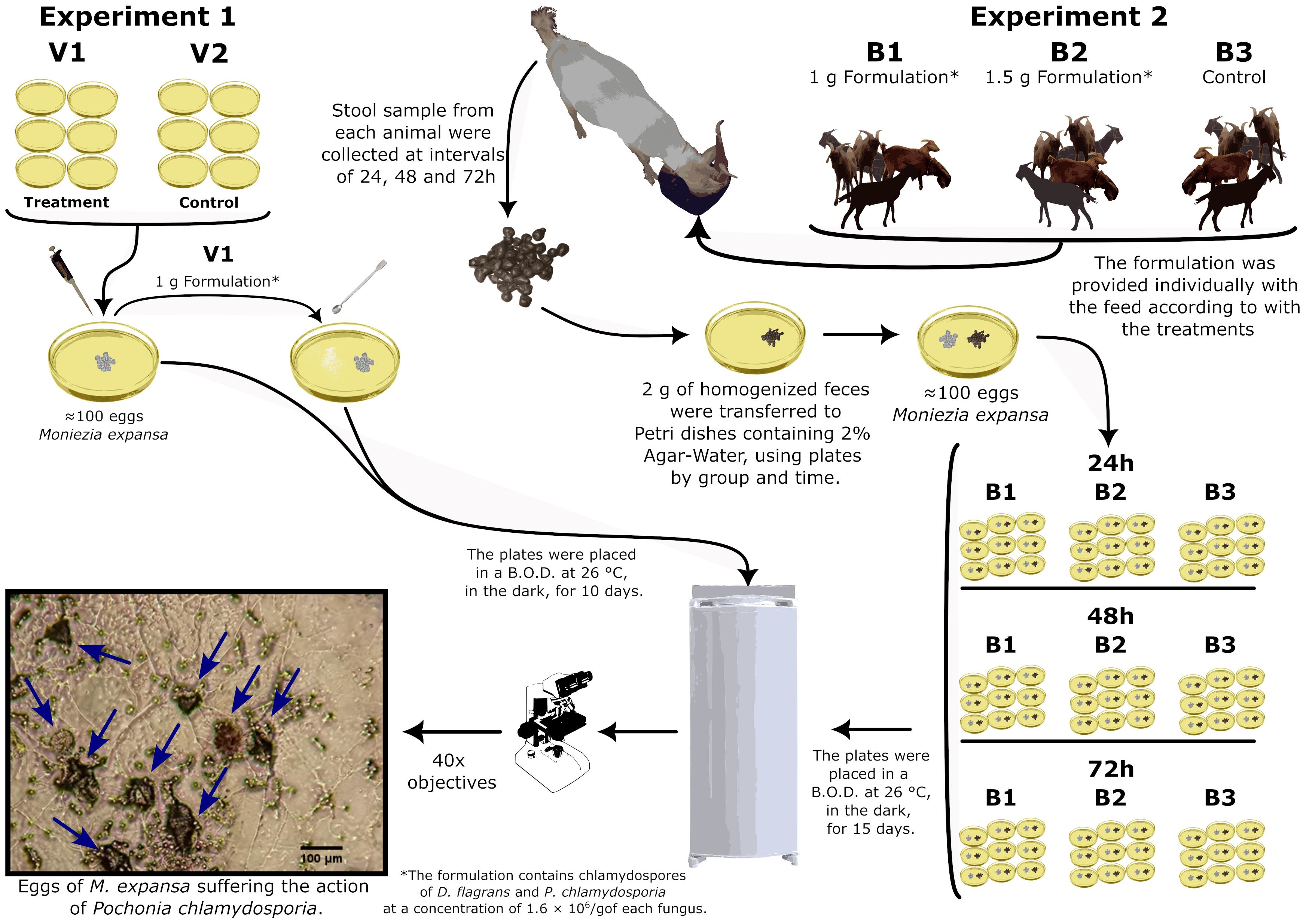
2.1. Obtaining Moniezia expansa Eggs

Moniezia expansa eggs were obtained by dissecting the uterus of adult parasites from the intestinal loops of goats slaughtered in the municipality of Socorro, São Paulo, Brazil, provided by the veterinary industry of the state of São Paulo.

2.2. Obtaining the Experimental Fungal Formulation

The experimental fungal formulation used in the study was produced and supplied by the veterinary industry of the state of São Paulo, Brazil. The formulation contained chlamydospores of D. flagrans and P. chlamydosporia at a concentration of 1.6 × 10⁶/g of each fungus.
These microorganisms are registered at the National Genetic Heritage Management System (SISGEN), registration number A8A8866, in the research line of Biological Control of nematodes by the researcher Jackson Victor de Araújo from the Federal University of Viçosa, MG-Brazil.

2.3. Goats Used in the Experiment

Eighteen goats belonging to the herds of the Federal University of Recôncavo da Bahia, crossbred Saanen and Anglo-Nubian of both sexes (males and females) with ages between 6 and 18 months and weighing around 30 kg, were used. This study was approved by the Ethics Committee in the Use of Animals (CEUA) of UFRB (protocol 23007.00006228/2019-16).

2.4. Experimental Tests

2.4.1. Experiment 1

Initially, we carried out a study to evaluate the effectiveness of the fungus before administration to the animals (without the action of the gastrointestinal tract and its development in the feces). For this purpose, 2% Agar–Water culture media were used in Petri dishes and two groups were created containing five Petri dishes each, with 100 eggs of M. expansa added to each plate. The groups were identified as V1 and V2. In group V1, 1 g of the fungal formulation was added, and V2 was the control without the addition of the formulation. The plates remained in a Biological Oxygen Demand (B.O.D.) incubator at 26 °C in the dark for 10 days. After this period, the integrity of the eggs was evaluated according to the parameters established by Lýsek [21].

2.4.2. Experiment 2

The evaluation using animals was carried out to verify the predatory capacity of the fungus after passing through the gastrointestinal tract of goats. The animals were weighed and treated with the anthelmintic ivermectin 0.08% at a dose of 1 mL for every 4 kg orally and 0.75 mL of albendazole 5% for every 10 kg orally, following the indicated prescription in the medicine leaflet. Fifteen days after deworming and confirming the absence of helminth eggs in the feces, the animals were raffled to determine their respective groups. Three groups containing six animals were formed. The goats in group B1 received 1 g of experimental fungal formulation per animal, those in group B2 received 1.5 g of experimental fungal formulation per animal, and the goats in group B3 received placebo (1.5 g of feed per animal). The treatments were performed with a single dose; the product was mixed with the feed and offered to the animals. The supply was carried out in individual troughs, and the animals were observed and released when they ingested all the offered content to guarantee that the whole product had been consumed.
After administration, stool samples were collected from each animal directly from the rectal ampulla, weighing approximately 5 g at intervals of 24, 48, and 72 h. Each sample was homogenized and later mixed to form a common sample by group and collecting time. Then, 2 g of feces were transferred to Petri dishes containing 2% Agar–Water, using plates by group and time.
All plates were added with 100 eggs of M. expansa. Therefore, ten aliquots of 10 μL each were obtained from 20 mL of an aqueous suspension of Moniezia eggs stored in a 40 mL cell culture bottle and quantified. Then, the mean number of eggs per sample was calculated, and from this mean, the suspensions were adjusted for 100 eggs for the Petri dish. Plates were set in a B.O.D. incubator at 26 °C in the dark for 15 days. Every three days, plates were inspected to verify the presence of characteristic conidia and conidiophores of the fungus, according to the classification proposed by Hoog [26], and also to check for egg predation. The integrity of the eggs was evaluated according to the parameters established by Lýsek [21].
To obtain the reduction value percentages, the following formula was used:
Reduction % = (Average number of eggs in the control group − Average number of eggs in the treated group)/(Average number of eggs in the control group) × 100

2.5. Statistical Analysis

The data were submitted to the Shapiro–Wilk normality test with a 95% confidence level. The Mann–Whitney test was performed to assess whether the number of intact eggs differed from one group to the other in experiment 1. In experiment 2, the variables were subjected to tests of homogeneity of variance (Levene with a 95% confidence interval) and subsequently to analysis of variance (ANOVA). Means were compared by Tukey’s test (p < 0.05). To calculate the effect size, we used squared gamma (ω2). All analyses were performed using the R software version 4.1.0.

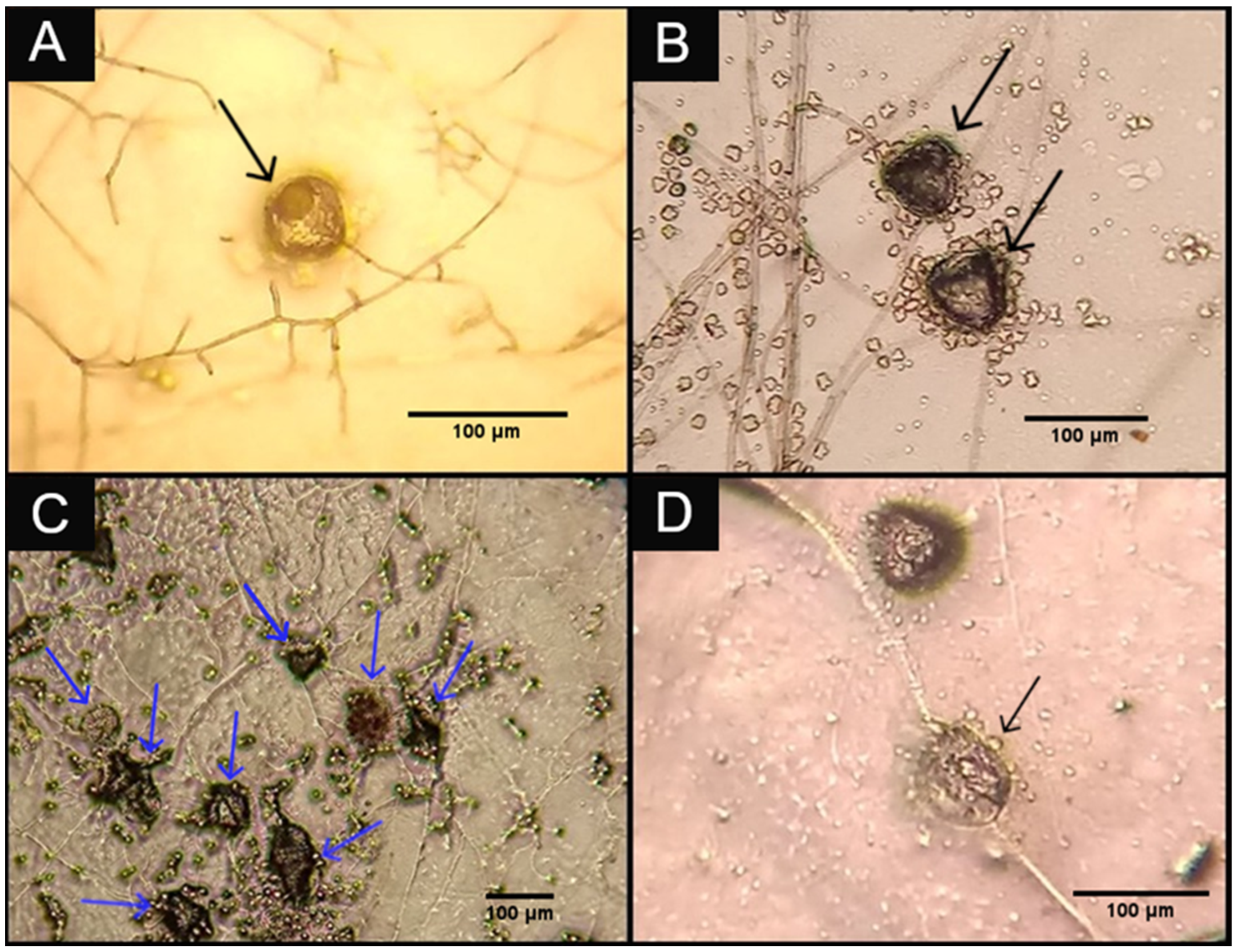
3. Results and Discussion

In experiment 1, the fungus developed in the Petri dishes of V1 and its hyphae colonized the eggs of M. expansa, causing structural changes in the eggs. Changes described by Lýsek [21] were observed on all eggs, and the result of the reduction test was 100%.
While evaluating the integrity of M. expansa eggs, the results showed that the number of eggs was lower in group V1 (Z = 2.9912, p = 0.0123, r = 0.86) than in V2. The median for V1 was 0, and for group V2, it was 113.
In experiment 2, the development of hyphae on the culture medium in the Petri dishes of groups B1 and B2 was verified, but hyphae of P. chlamydosporia were not verified in group B3. The experimental fungal formulation was not offered to the animals that provided the feces used in group B3, justifying the absence of the fungus in the plates of this group. Lytic effects on the eggs occurred even after passage through the gastrointestinal tract of goats (Figure 1).
The reduction of eggs after gastrointestinal transit was evaluated, and a significant effect was observed in the 24-h treatments (F = 30.32, p < 0.0001, ω2 = 0.685); 48 h (F = 38.81, p < 0.0001, ω2 = 0.737); 72 h (F = 31.32, p < 0.0001, ω2 = 0.692). The values assigned to each treatment and collecting time are shown in Table 1.
Fungal formulations that make use of associations can obtain synergistic effects with this. Furthermore, they can broaden their spectrum of action as long as the associated fungi act on different species of helminths [27]. D. flagrans is a larvae-predating nematophagous fungus that produces sticky three-dimensional traps. P. chlamydosporia is an opportunistic and ovicidal fungus that produces a structure called the oppressor capable of penetrating eggs [12,13,16,28].
The association of P. chlamydosporia and D. flagrans probably would not generate a synergistic effect to reduce the number of eggs of M. expansa specifically. As a result, D. flagrans did not demonstrate the ability to exert a type 3 lytic effect on helminth eggs, despite having a type 1 lytic effect on Fasciola hepatica eggs after 21 days of experiment [29]. However, Pochonia sp. could collaborate with the reduction of nematode eggs and probably reduce the number of recovered lavas; since acting alone or in association with D. flagrans and M. thaumasium, it reduced the number of cyathostomine larvae by 73.2%, 86.8%, and 77.3%, respectively [30]. However, when it was used as the only agent in field evaluations, it was unable to control the free-life stages of bovine gastrointestinal nematodes [20]. Therefore, considering the reduction of eggs, it is unlikely that D. flagrans contributed significantly to the results presented. Thus, the experimental fungal formulation used in this work acts against larvae and eggs of helminths. For each of the objectives in the present work, one of the two fungi was predominant (D. flagrans and P. chlamydosporia). The effect against larvae occurred mainly due to the action of D. flagrans, which has already been studied widely [12,13,31,32]. As for the ovicidal action, the preponderant role was played by P. chlamydosporia, as evidenced in the present study.
The ovicidal capacity of P. chlamydosporia has already been described for Dipylidium caninum (reduction of 92.2% and 88.4% for isolates VC1 and VC4), Taenia taeniaeformis (73.8% of type 3 lytic effect for isolate VC4), T. saginata (reduction of 54.2% and 42.4% for isolates VC1 and VC4), and Anoplocephala perfoliata (reduction of 71.17% for isolate VC1) [33,34,35]. The results for the reduction in the number of eggs are shown in Table 2. The treatments tested in the present work were able to reduce the number of eggs at all collecting times, and the results did not differ among themselves when observing the different treatments.
The study with Paecilomyces lilacinus demonstrated an ovicidal effect by showing a type 3 mechanism of action on Moniezia sp. eggs [36]. Likewise, type 3 effect on the eggs was observed in the present study, evidencing ovicidal action. These results demonstrated the ability of the fungus to colonize and prey on eggs in large proportions. This characteristic is considered crucial for biological control agents [37]. Thus, P. chlamydosporia is part of the fungi which perform against this tapeworm, expanding the range of options for biological control. However, the effectiveness of P. lilacinus appears to be lower than that of P. chlamydosporia. When two fungal isolates of Pochonia sp. (VC1 and VC4) showed a type 3 lytic effect in 71% and 74% of Oxyuris equi eggs, the effects for P. lilacinus were significantly smaller at 54% [38]. In addition, these fungi were evaluated for eggs of Ascaridia galli and Toxocara canis. In this case, Pochonia sp. also demonstrated a greater reduction in the number of viable eggs (86% and 67%), contrary to the reduction provided by Paecilomyces sp., which was lower (29% and 28%). These values correspond to the studied parasites obtained at the end of the experiment (42 days) [39]. Therefore, these results demonstrate the superiority of Pochonia sp. as an agent for biological control and the importance of knowing its effectiveness in vivo studies and on other helminths of veterinary interest.
In the present study, after passing through the gastrointestinal tract, there was a predatory effect on the eggs, but this reduction was lower than that observed in experiment 1. This difference may be the result of a lower amount of chlamydospores under the conditions of experiment 2. Significant losses of chlamydospores occur during the transit through the gastrointestinal tract. The exposure of chlamydospores to the conditions of such environment can generate relative losses of 89.7% of chlamydospores [40]. Furthermore, the amount of formulation used in experiment 1 (1 g) was either identical to or very close to the amount supplied to the animals in experiment 2. In this sense, the high concentration of chlamydospores in experiment 1 would justify the destruction of all eggs in experiment 1, V1 group.
As M. expansa needs intermediate hosts, the main hosts are oribatid mites, and their eggs remain in the environment until they are ingested [24]. The eggs show adaptive characteristics in relation to their intermediate host, the oribatid mites, and to the environment that are essential for their survival. The eggshell provides mechanical protection from the environment. The piriform apparatus that protects the embryo in the innermost layer of the egg is composed of keratin, and its submembrane located at the interface between the two anterior layers has a lipid coating that protects the egg from dissection [41]. As their eggs remain exposed and only develop after ingestion by a host, the fungus has the opportunity to exert its lytic effects. Given this scenario, egg reduction was attributed to the effects of this fungus, being that the present study is the first report of P. chlamydosporia ovicidal action on M. expansa eggs in an in vitro test and after passing through the gastrointestinal tract of goats.
Moniezia expansa eggs have a thick wall that requires mechanical breakdown of the membrane and enzymatic digestion of the piriform apparatus, which protects the embryo. The shell is resistant to a variety of proteolytic enzymes, amylases, and lipases [41]. These challenges must be overcome in order for the fungus to act. In the presence of eggs, metabolites are produced, such as glycoproteins, which help in the surface adhesion process. After adhesion, appressoria are produced at the end of the germ tubes, which perform physical and biochemical penetration functions [42]. The mechanical and enzymatic actions result in the rupture of the outer layer of the egg, removing the outer proteinaceous vitelline membrane and allowing the penetration of the hyphae [43].
Proteases produced by the fungus play a relevant role in the rupture of the eggshell. The crude extract of P. chlamydosporia composed of proteases was used on Ascaridia galli eggs and showed a reduction effect of 64.1% [44]. The crude extract reduced the hatching of Ancylostoma sp. eggs with a reduction of 76.8% [45]. M. expansa eggs are rich in keratin in the composition of their structures, such as the shell and the piriform apparatus. These structures are disrupted under the effect of proteases and mechanical action, effects very similar to those observed during infection of intermediate hosts [41]. These data demonstrate the importance of this class of enzymes in the role of the fungus on parasitized eggs. It is likely that these enzymes acted on M. expansa eggs and helped in the penetration and colonization process.
Pochonia chlamydosporia produces relevant structural changes in eggs exposed to hyphae, interfering with the development and hatching of larvae [46]. Extracellular enzymes (proteases, chitinases, phosphatases, and lipases) act on the eggs, and the hyphae of the fungus colonize the host tissues [47,48]. These are the mechanisms of action involved in the parasitism of the fungus on its hosts and are probably the same ones performed on M. expansa eggs.
The production of resistance spores called chlamydospores enables the survival and dissemination of this fungus; this structure has important glycogen reserves and has thick walls produced along the hyphae [49,50]. Due to their characteristics, chlamydospores are able to transit through the gastrointestinal tract of ruminants and maintain their predatory capacity when they are eliminated in the feces [28,40,51]. In addition to these capabilities, the production of numerous chlamydospores is what allows the development of products for biological control [52]. The mechanisms of action and the ability to survive the gastrointestinal tract allowed the fungus to remain viable and perform against M. expansa eggs.
Moniezia expansa infections rarely result in the death of animals, but they can lead to economic losses due to reduced growth rate, in addition to other factors [3,49]. The occurrence of infections is not uncommon, and the most susceptible animals are young, possibly the most affected by complications from the disease [53]. The development of control mechanisms that may allow reduction of the infection of these animals could lead to an improvement in the productivity indexes of the herds.
As an example of performance improvement, the animals that received 1 g of D. flagrans chlamydospores for each 10 kg of live weight in the Bioverm® commercial formulation resulted in an average weight gain of 300 kg in the treated group, against an average weight of 261 kg in the control group, leading to significant economic gains both by increasing weight gain and by reducing deworming costs [54]. The reduction of infective forms of nematodes produced a significant reduction in the OPG of the animals, and the reduction in parasitism improved the productive performance.
With the use of biological control, it becomes possible to reduce or ration the use of anthelmintics, which according to Castro [55], have been used irrationally over decades, generating an increasing resistance of parasites to the bases of chemical products available on the market. With the scarcity of bases due to the resistance generated over time, the control of helminths becomes increasingly difficult. Allied with this, according to Porto Filho [56], the residues left by the use of anthelmintics are of great importance in single health, with global health implications. These products leave residues in meat, milk, and derivatives, which reach the table directly, impacting the health of the consumer. In addition, in the application, workers are exposed to these residues due to a lack of guidance and adequate personal protective equipment (PPE). Such residues contaminate soil, water, and vegetation as a result of applications and also due to improper disposal of their packaging. Therefore, there are direct and indirect impacts on the health of the population, animal health, and environmental health, thus configuring a problem in One Health.
The proper use of anthelmintics and the reduction of their dependence have become a necessity to circumvent the consequences imposed in the current situation of anthelmintic resistance, which has been an obstacle to the control of parasites worldwide [57]. Therefore, alternative measures are imperative to control helminths that infect production animals.
In addition, the presence of residues in products of animal origin can pose a risk to public health [58]. The withdrawal period for certain anthelmintics makes their use unfeasible in certain production systems, such as dairy cattle. In some situations, there is non-compliance with the waiting period required for the proper use of the product, thus causing the presence of residues in foods of animal origin [59]. These residues have an impact on the health of herds, public health, and the environment, therefore causing a global health problem.
On the other hand, helminthophagous fungi do not cause anthelmintic resistance, do not contaminate soil and water, and do not produce chemical residues in meat, milk, or by-products derived from animals for human consumption [60]. In this sense, the use of products based on helminthophagous fungi can integrate helminth control systems, contributing to the reduction of the dependence on anthelmintics.
From another perspective, proglottids obtained from adult parasites from the intestinal loops of small ruminants can provide immature eggs. Similar to the study of Ascaris suum, the use of these forms led to controversial results, as the eggs of these parasites were protected from fungal action [39]. Considering this, obtaining naturally eliminated proglottids may be more reliable sources for future studies.
It is worth mentioning that biological control is not a substitute for anthelmintics. In situations of health problems, the latter is an excellent tool. The role of biological control is to reduce contamination of pastures and reinfection of animals, thus reducing the need to apply these drugs without affecting their production rates or even improving them [54]. In addition, favoring good management practices for these chemical compounds maintains satisfactory levels of effectiveness for their use [12].
The predatory capacity of M. expansa eggs verified in the present study showed that a experimental formulation containing D. flagrans and P. chlamydosporia fungi has the potential for application in integrated helminth control programs, with P. chlamydosporia being a species with ovicidal effect.

Author Contributions

G.B.R., A.R.e.S. and W.M.d.S.P. conceived and designed the study. G.B.R., A.P.D.d.O. and I.d.A.M. performed the data collection. G.B.R. performed statistical analyses. G.B.R., J.d.S.F., C.M.d.O.M., J.V.d.A. and W.M.d.S.P. wrote the article. All authors have read and agreed to the published version of the manuscript.

Funding

Foundation for Research Support of the State of Bahia (FAPESB), for the Master’s fellowship during this research.

Institutional Review Board Statement

This study was approved by the Ethics Committee in the Use of Animals (CEUA) of Federal University of Recôncavo da Bahia (protocol 23007.00006228/2019-16).

Informed Consent Statement

Not applicable.

Acknowledgments

We thank the Foundation for Research Support of the State of Bahia (FAPESB) for the Master’s fellowship during this research. We thank the Environmental and Biological Sciences from the Federal University of Recôncavo da Bahia. We also thank theveterinary industry of the state of São Paulo for donating the experimental fungal formulation and the M. expansa eggs used in the experiment.

Conflicts of Interest

The authors declare no conflict of interest.

References

  1. Bashtar, A.-R.; Hassanein, M.; Abdel-Ghaffar, F.; Al-Rasheid, K.; Hassan, S.; Mehlhorn, H.; AL-Mahdi, M.; Morsy, K.; Al-Ghamdi, A. Studies on Monieziasis of Sheep I. Prevalence and Antihelminthic Effects of Some Plant Extracts, a Light and Electron Microscopic Study. Parasitol. Res. 2011, 108, 177–186. [Google Scholar] [CrossRef]
  2. Mohamed, H.I.; Arafa, W.M.; El-Dakhly, K.M. Prevalence and Associated Risk Factors of Gastrointestinal Helminths and Coccidian Infections in Domestic Goats, Capra hircus, in Minya, Egypt. Beni-Suef Univ. J. Basic Appl. Sci. 2023, 12, 29. [Google Scholar] [CrossRef]
  3. Kelly, R.; Evans, M.; Sargison, N. Identifying Knowledge Gaps in Moniezia expansa Epidemiology: A Report of a Small Intestinal Torsion in a 5-Week-Old Lamb. N. Z. Vet. J. 2021, 69, 186–189. [Google Scholar] [CrossRef]
  4. Rahman, S.; Bulbul, K.H. Monieziosis: A Neglected Helminthic Disease in Ruminants. Anim. Dis. 2022; submitted. [Google Scholar]
  5. Iacob, O.C.; El-Deeb, W.M.; Paşca, S.-A.; Turtoi, A.-I. Uncommon Co-Infection Due to Moniezia expansa and Moniezia benedeni in Young Goats from Romania: Morphological and Histopathological Analysis. Ann. Parasitol. 2020, 66, 501–507. [Google Scholar] [CrossRef] [PubMed]
  6. Salgado, J.A.; Santos, C.d.P. Overview of Anthelmintic Resistance of Gastrointestinal Nematodes of Small Ruminants in Brazil. Rev. Bras. Parasitol. Vet. 2016, 25, 3–17. [Google Scholar] [CrossRef] [PubMed] [Green Version]
  7. Willy, M.L.L. Determinación de resistencia antihelmíntica (Moniezia expansa, Moniezia benedeni y Thysanosoma actioides) frente a albendazol y febendazol en ovino en tres rebaños de La Paz—Bolivia. REDVET Rev. Electrónica Vet. 2009, 10, 1–13. [Google Scholar]
  8. Amarante, A.F.T. Os Parasitas De Ovinos; Editora UNESP: São Paulo, Brasil, 2014; ISBN 978-85-68334-42-3. [Google Scholar]
  9. Kumar, G.; Selvakkumar, R. Fenbendazole and Praziquantel Resistance in Moniezia expansa in Jamunapari Goat Kids. J. Entomol. Zool. Stud. 2020, 8, 1124–1125. [Google Scholar]
  10. Vercruysse, J.; Charlier, J.; Dijk, J.V.; Morgan, E.R.; Geary, T.; Samson-Himmelstjerna, G.V.; Claerebout, E. Control of Helminth Ruminant Infections by 2030. Parasitology 2018, 145, 1655–1664. [Google Scholar] [CrossRef] [Green Version]
  11. Do Carmo, T.A.; Mena, M.O.; Cipriano, I.d.A.; de Favare, G.M.; Guelpa, G.J.; Pinto, S.D.C.; do Amarante, A.F.T.; de Araújo, J.V.; de Soutello, R.V.G. Biological Control of Gastrointestinal Nematodes in Horses Fed with Grass in Association with Nematophagus Fungi Duddingtonia flagrans and Pochonia chlamydosporia. Biol. Control 2023, 182, 105219. [Google Scholar] [CrossRef]
  12. Gives, P.M.; Arellano, M.E.L.; Marcelino, L.A.; Jenkins, S.O.; Guerrero, D.R.; Várgas, G.R.; Murillo, V.E.V. The Nematophagous Fungus Duddingtonia flagrans Reduces the Gastrointestinal Parasitic Nematode Larvae Population in Faeces of Orally Treated Calves Maintained under Tropical Conditions-Dose/Response Assessment. Vet. Parasitol. 2018, 263, 66–72. [Google Scholar] [CrossRef]
  13. Vilela, V.L.R.; Feitosa, T.F.; Braga, F.R.; Vieira, V.D.; Lucena, S.C.d.; Araújo, J.V.d. Control of Sheep Gastrointestinal Nematodes Using the Combination of Duddingtonia flagrans and Levamisole Hydrochloride 5%. Rev. Bras. De Parasitol. Veterinária 2018, 27, 26–31. [Google Scholar] [CrossRef] [PubMed] [Green Version]
  14. Castro, A.A.; Oliveira, C.R.C.; Anjos, D.H.S.; Ornelas, É.I.D. Potencial dos fungos nematófagos Arthrobotrys sp. E Monacrosporium thaumasium para o controle de larvas de ciatostomíneos de eqüinos (Nematoda: Cyathostominae). Rev. Bras. Parasitol. Vet. 2003, 12, 53–57. [Google Scholar]
  15. Braga, F.R.; Araújo, J.V.; Silva, A.R.; Carvalho, R.O.; Araujo, J.M.; Campos, A.K.; Tavela, A.O. Ação in vitro dos fungos nematófagos Duddingtonia flagrans (Duddington, 1955), Monacrosporium thaumasium (Drechsler, 1937) e Pochonia chlamydosporia (Gams & Zare, 2001) sobre ovos de Eurytrema coelomaticum (Giard & Billet, 1892). Arq. Inst. Biológico 2009, 76, 131–134. [Google Scholar]
  16. Wang, B.-B.; Zhang, N.; Gong, P.-T.; Li, J.-H.; Yang, J.; Zhang, H.-B.; Zhang, X.-C.; Cai, K.-Z. Morphological Variability, Molecular Phylogeny, and Biological Characteristics of the Nematophagous Fungus Duddingtonia flagrans. J. Basic Microbiol. 2019, 59, 645–657. [Google Scholar] [CrossRef] [PubMed]
  17. Buzatti, A.; Santos, C.P.; Fernandes, M.A.M.; Yoshitani, U.Y.; Sprenger, L.K.; Molento, M.B. Duddingtonia flagrans no controle de nematoides gastrintestinais de equinos em fases de vida livre. Arq. Bras. Med. Veterinária E Zootec. 2017, 69, 364–370. [Google Scholar] [CrossRef] [Green Version]
  18. Jobim, M.B.; Santurio, J.M.; De La Rue, M.L. Duddingtonia flagrans: Controle biológico de nematodeos de bovinos a campo. Ciênc. Rural 2008, 38, 2256–2263. [Google Scholar] [CrossRef] [Green Version]
  19. Rodrigues, J.V.F.; Braga, F.R.; Campos, A.K.; de Carvalho, L.M.; Araujo, J.M.; Aguiar, A.R.; Ferraz, C.M.; da Silveira, W.F.; Valadão, M.C.; Oliveira, T.d.; et al. Duddingtonia flagrans Formulated in Rice Bran in the Control of Oesophagostomum spp. Intestinal Parasite of Swine. Exp. Parasitol. 2018, 184, 11–15. [Google Scholar] [CrossRef]
  20. Fonseca, J.D.S.; Ferreira, V.M.; Freitas, S.G.d.; Vieira, Í.S.; Araújo, J.V.d. Efficacy of a Fungal Formulation with the Nematophagous Fungus Pochonia chlamydosporia in the Biological Control of Bovine Nematodiosis. Pathogens 2022, 11, 695. [Google Scholar] [CrossRef]
  21. Lýsek, H.; Fassatiová, O.; Cuervo Pineda, N.; Lorenzo Hernández, N. Ovicidal Fungi in Soils of Cuba. Folia Parasitol. 1982, 29, 265–270. [Google Scholar]
  22. Araújo, J.M.; Araújo, J.V.; Braga, F.R.; Carvalho, R.O.; Silva, A.R.; Campos, A.K. Interaction and Ovicidal Activity of Nematophagous Fungus Pochonia chlamydosporia on Taenia saginata Eggs. Exp. Parasitol. 2009, 121, 338–341. [Google Scholar] [CrossRef] [PubMed]
  23. Fonseca, J.d.S.; Altoé, L.S.C.; de Carvalho, L.M.; Soares, F.E.d.F.; Braga, F.R.; de Araújo, J.V. Nematophagous Fungus Pochonia chlamydosporia to Control Parasitic Diseases in Animals. Appl. Microbiol. Biotechnol. 2023, 107, 3859–3868. [Google Scholar] [CrossRef]
  24. Stunkard, H.W. The Life Cycle of Moniezia expansa. Science 1937, 86, 312. [Google Scholar] [CrossRef] [PubMed]
  25. Araújo, J.V.; Braga, F.R.; Mendoza-de-Gives, P.; Paz-Silva, A.; Vilela, V.L.R. Recent Advances in the Control of Helminths of Domestic Animals by Helminthophagous Fungi. Parasitologia 2021, 1, 168–176. [Google Scholar] [CrossRef]
  26. Hoog, G.S.d.; Oorschot, C.A.N.v.; Hijwegen, T. Taxonomy of the Dactylaria Complex II. Dissoconium Gen. Nov. and Cordana Preuss. Proc. Kon. Ned. Akad. Wet. Ser. C 1983, 86, 197–206. [Google Scholar]
  27. Li, S.; Wang, D.; Gong, J.; Zhang, Y. Individual and Combined Application of Nematophagous Fungi as Biological Control Agents against Gastrointestinal Nematodes in Domestic Animals. Pathogens 2022, 11, 172. [Google Scholar] [CrossRef]
  28. Braga, F.R.; Ferraz, C.M.; da Silva, E.N.; de Araújo, J.V. Efficiency of the Bioverm® (Duddingtonia flagrans) Fungal Formulation to Control In Vivo and In Vitro of Haemonchus contortus and Strongyloides papillosus in Sheep. 3 Biotech 2020, 10, 62–66. [Google Scholar] [CrossRef]
  29. Braga, F.R.; Araújo, J.V.; Campos, A.K.; Araújo, J.M.; Carvalho, R.O.; Silva, A.R.; Tavela, A.O. In Vitro Evaluation of the Action of the Nematophagous Fungi Duddingtonia Flagrans, Monacrosporium Sinense and Pochonia Chlamydosporia on Fasciola Hepatica Eggs. World J. Microbiol. Biotechnol. 2008, 24, 1559–1564. [Google Scholar] [CrossRef]
  30. Tavela, A.O.; Araújo, J.V.; Braga, F.R.; Araujo, J.M.; Magalhães, L.Q.; Silveira, W.F.; Borges, L.A. In Vitro Association of Nematophagous Fungi Duddingtonia Flagrans (AC001), Monacrosporium Thaumasium (NF34) and Pochonia Chlamydosporia (VC1) to Control Horse Cyathostomin (Nematoda: Strongylidae). Biocontrol Sci. Technol. 2012, 22, 607–610. [Google Scholar] [CrossRef]
  31. Vilela, V.L.R.; Feitosa, T.F.; Braga, F.R.; de Araújo, J.V.; de Oliveira Souto, D.V.; da Silva Santos, H.E.; da Silva, G.L.L.; Athayde, A.C.R. Biological Control of Goat Gastrointestinal Helminthiasis by Duddingtonia flagrans in a Semi-Arid Region of the Northeastern Brazil. Vet. Parasitol. 2012, 188, 127–133. [Google Scholar] [CrossRef] [Green Version]
  32. Da Silveira, W.F.; de Oliveira, G.D.; Braga, F.R.; de Carvalho, L.M.; Domingues, R.R.; da Silva, L.A.; Zanuncio, J.C.; de Araújo, J.V. Predation Rate of Nematophagous Fungi after Passing through the Gastrointestinal Tract of Goats. Small Rumin. Res. 2017, 147, 101–105. [Google Scholar] [CrossRef]
  33. Araújo, J.M.; Braga, F.R.; Araújo, J.V.; Soares, F.E.F.; Geniêr, H.L.A. Biological Control of Taenia saginata Eggs. Helminthologia 2010, 47, 189–192. [Google Scholar] [CrossRef] [Green Version]
  34. Braga, F.R.; Silva, A.R.; Carvalho, R.O.; Araújo, J.V.; Pinto, P.S.A. Ovicidal Activity of Different Concentrations of Pochonia chlamydosporia Chlamydospores on Taenia taeniaeformis Eggs. J. Helminthol. 2011, 85, 7–11. [Google Scholar] [CrossRef] [PubMed]
  35. Araujo, J.M.; Braga, F.R.; de Araújo, J.V.; Carvalho, R.O. Atividade dos fungos nematófagos Pochonia chlamydosporia e Paecilomyces lilacinus sobre cápsulas de ovos de Dipylidium caninum. Rev. Inst. Adolfo Lutz 2009, 68, 488–491. [Google Scholar] [CrossRef]
  36. Braga, F.R.; Araújo, J.V.; Araujo, J.M.; Carvalho, R.O.; Silva, A.R.; Campos, A.K.; Tavela, A.O. Ovicidal Activity of Paecilomyces lilacinus on Moniezia sp. Eggs. J. Helminthol. 2008, 82, 241–243. [Google Scholar] [CrossRef] [PubMed]
  37. Braga, F.R.; Araújo, J.V. Nematophagous Fungi for Biological Control of Gastrointestinal Nematodes in Domestic Animals. Appl. Microbiol. Biotechnol. 2014, 98, 71–82. [Google Scholar] [CrossRef]
  38. Braga, F.R.; Araújo, J.V.; Soares, F.E.F.; Tavela, A.O.; Araujo, J.M.; Carvalho, R.O.; Fernandes, F.M.; Queiroz, J.H. Enzymatic Analysis and in Vitro Ovicidal Effect of Pochonia Chlamydosporia and Paecilomyces Lilacinus on Oxyuris Equi Eggs of Horses. Biocontrol Sci. Technol. 2012, 22, 685–696. [Google Scholar] [CrossRef]
  39. Thapa, S.; Meyling, N.V.; Katakam, K.K.; Thamsborg, S.M.; Mejer, H. A Method to Evaluate Relative Ovicidal Effects of Soil Microfungi on Thick-Shelled Eggs of Animal-Parasitic Nematodes. Biocontrol Sci. Technol. 2015, 25, 756–767. [Google Scholar] [CrossRef]
  40. Ojeda-Robertos, N.F.; Torres-Acosta, J.F.; Ayala-Burgos, A.J.; Sandoval-Castro, C.A.; Valero-Coss, R.O.; Mendoza-de-Gives, P. Digestibility of Duddingtonia flagrans chlamydospores in ruminants: In Vitro and In Vivo studies. BMC Vet. Res. 2009, 5, 46–52. [Google Scholar] [CrossRef] [Green Version]
  41. Caley, J. In Vitro Hatching of the Tapeworm Moniezia expansa (Cestoda: Anoplocephalidae) and Some Properties of the Egg Membranes. Z. Parasitenk. 1975, 45, 335–346. [Google Scholar] [CrossRef]
  42. Lopez-Llorcz, L.V.; Olivares-Bernabeu, C.; Salinas, J.; Jansson, H.-B.; Kolattukudy, P.E. Pre-Penetration Events in Fungal Parasitism of Nematode Eggs. Mycol. Res. 2002, 106, 499–506. [Google Scholar] [CrossRef]
  43. Ward, E.; Kerry, B.R.; Manzanilla-López, R.H.; Mutua, G.; Devonshire, J.; Kimenju, J.; Hirsch, P.R. The Pochonia chlamydosporia Serine Protease Gene Vcp1 Is Subject to Regulation by Carbon, Nitrogen and Ph: Implications for Nematode Biocontrol. PLoS ONE 2012, 7, e35657. [Google Scholar] [CrossRef] [PubMed] [Green Version]
  44. Braga, F.R.; Araújo, J.V.; Araujo, J.M.; Frassy, L.N.; Tavela, A.O.; Soares, F.E.F.; Carvalho, R.O.; Queiroz, L.M.; Queiroz, J.H. Pochonia chlamydosporia Fungal Activity in a Solid Medium and Its Crude Extract against Eggs of Ascaridia galli. J. Helminthol. 2012, 86, 348–352. [Google Scholar] [CrossRef] [PubMed]
  45. Braga, F.R.; Araujo, J.M.; Silva, A.R.; de Araújo, J.V.; Carvalho, R.O.; de Freitas Soares, F.E.; de Queiroz, J.H.; Gênier, H.L.A. Ação ovicida do extrato bruto enzimático do fungo Pochonia chlamydosporia sobre ovos de Ancylostoma sp. Rev. Soc. Bras. Med. Trop. 2011, 44, 116–118. [Google Scholar] [CrossRef] [Green Version]
  46. Castro, L.S.; Martins, I.V.F.; Tunholi-Alves, V.M.; Amaral, L.S.; Pinheiro, J.; de Araújo, J.V.; de Oliveira Monteiro, C.M.; Tunholi, V.M. Susceptibility of Embryos of Biomphalaria tenagophila (Mollusca: Gastropoda) to Infection by Pochonia chlamydosporia (Ascomycota: Sordariomycetes). Arch. Microbiol. 2022, 204, 271. [Google Scholar] [CrossRef]
  47. Esteves, I.; Peteira, B.; Atkins, S.D.; Magan, N.; Kerry, B. Production of Extracellular Enzymes by Different Isolates of Pochonia Chlamydosporia. Mycol. Res. 2009, 113, 867–876. [Google Scholar] [CrossRef] [Green Version]
  48. Gouveia, A.d.S.; Monteiro, T.S.A.; Valadares, S.V.; Sufiate, B.L.; de Freitas, L.G.; de Oliveira Ramos, H.J.; de Queiroz, J.H. Understanding How Pochonia chlamydosporia Increases Phosphorus Availability. Geomicrobiol. J. 2019, 36, 747–751. [Google Scholar] [CrossRef]
  49. Dallemole-Giaretta, R.; de Freitas, L.G.; de Brito Caixeta, L.; Xavier, D.M.; Ferraz, S.; de Fátima Silva, C. Produção de clamidósporos de Pochonia chlamydosporia em diferentes substratos. Ciênc. Agrotec. 2011, 35, 314–321. [Google Scholar] [CrossRef] [Green Version]
  50. Youssar, L.; Wernet, V.; Hensel, N.; Yu, X.; Hildebrand, H.-G.; Schreckenberger, B.; Kriegler, M.; Hetzer, B.; Frankino, P.; Dillin, A.; et al. Intercellular Communication Is Required for Trap Formation in the Nematode-Trapping Fungus Duddingtonia flagrans. PLoS Genet. 2019, 15, e1008029. [Google Scholar] [CrossRef] [Green Version]
  51. Céspedes-Gutiérrez, E.; Aragón, D.M.; Gómez-Álvarez, M.I.; Cubides-Cárdenas, J.A.; Cortés-Rojas, D.F. Survival of the Nematophagous Fungus Duddingtonia flagrans to In Vitro Segments of Sheep Gastrointestinal Tract. Exp. Parasitol. 2021, 231, 108172. [Google Scholar] [CrossRef]
  52. Monteiro, T.S.A.; Gouveia, A.S.; Balbino, H.M.; Morgan, T.; Grassi de Freitas, L. Chapter 34—Duddingtonia. In Beneficial Microbes in Agro-Ecology; Amaresan, N., Senthil Kumar, M., Annapurna, K., Kumar, K., Sankaranarayanan, A., Eds.; Academic Press: Cambridge, MA, USA, 2020; pp. 683–694. ISBN 978-0-12-823414-3. [Google Scholar]
  53. Martins, N.S.; Dos Santos, C.C.; da Motta, S.P.; da Silva Moreira, A.; da Rosa Farias, N.A.; Ruas, J.L. Gastrointestinal Parasites in Sheep from the Brazilian Pampa Biome: Prevalence and Associated Factors. Braz. J. Vet. Med. 2022, 44, e001522. [Google Scholar] [CrossRef] [PubMed]
  54. Da Silva Santos Castelo Branco de Oliveira, L.; Dias, F.G.S.; Melo, A.L.T.; de Carvalho, L.M.; Silva, E.N.; de Araújo, J.V. Bioverm® in the Control of Nematodes in Beef Cattle Raised in the Central-West Region of Brazil. Pathogens 2021, 10, 548. [Google Scholar] [CrossRef]
  55. de Castro, L.L.D.; Sprenger, L.K.; Madrid, I.M.; de Oliveira, F.C.; de Oliveira, P.A.; de Castro, L.M.; Berne, M.E.A.; Leite, F.P.L. Efeito in vitro e in vivo de extratos de Eugenia Uniflora Em Nematódeos Gastrintestinais de ovinos. Ciênc. Anim. Bras./Braz. Anim. Sci. 2019, 20, 1–12. [Google Scholar] [CrossRef] [Green Version]
  56. Porto Filho, J.M.; Costa, R.G.; Araújo, A.C.P.; Albuquerque Júnior, E.C.; Cunha, A.N.; Cruz, G.R.B. da Determining Anthelmintic Residues in Goat Milk in Brazil. Rev. Bras. Saúde Prod. Anim. 2019, 20, e04102019. [Google Scholar] [CrossRef]
  57. Fissiha, W.; Kinde, M.Z. Anthelmintic Resistance and Its Mechanism: A Review. Infect. Drug Resist. 2021, 14, 5403–5410. [Google Scholar] [CrossRef]
  58. Lourenco, A.; Fraga-Corral, M.; De Colli, L.; Moloney, M.; Danaher, M.; Jordan, K. Determination of the Presence of Pathogens and Anthelmintic Drugs in Raw Milk and Raw Milk Cheeses from Small Scale Producers in Ireland. LWT 2020, 130, 109347. [Google Scholar] [CrossRef]
  59. Lobato, V.; Rath, S.; Reyes, F.G.R. Occurrence of Ivermectin in Bovine Milk from the Brazilian Retail Market. Food Addit. Contam. 2006, 23, 668–673. [Google Scholar] [CrossRef]
  60. Mendoza-de Gives, P.; Braga, F.R.; de Araújo, J.V. Nematophagous Fungi, an Extraordinary Tool for Controlling Ruminant Parasitic Nematodes and Other Biotechnological Applications. Biocontrol Sci. Technol. 2022, 32, 777–793. [Google Scholar] [CrossRef]
Figure 1. Images (A,B): Type 1 ovicidal effect on Moniezia expansa eggs (black arrows). Image (C): Moniezia expansa eggs whose integrity is compromised by Pochonia chlamydosporia and type 2 ovicidal effect (black arrows). Image (D): Moniezia expansa egg penetrated by Pochonia chlamydosporia hypha showing type 3 ovicidal effect (blue arrows). All images were obtained from the groups that received treatment with the experimental fungal formulation, groups B1 (total dose of 1 g of the formulation for each animal) and B2 (total dose of 1.5 g of the formulation for each animal).
Pathogens 12 01028 g001
Table 1. Mean ± standard deviation and standard error (SE) of the number of intact eggs of the respective groups: B1 (total dose of 1 g of the formulation for each animal); B2 (total dose of 1.5 g of the formulation for each animal); B3 (total dose of 1.5 g of placebo for each animal). Three collecting times (24, 48, and 72 h) following ingestion, after 15 days in B.O.D. incubator at 26 °C in the dark.
Mean of Intact Eggs
Group (n)24 h48 h72 h
B1 (9)59.0 a ± 16.3 (SE = 5.4)42.5 a ± 14.7 (SE = 4.9) 57.7 a ± 12.6 (SE = 6.41)
B2 (9)49.5 a ± 20.10 (SE = 6.7)39.1 a ± 17.9 (SE = 5.9)46.8 a ± 18.3 (SE = 6.1)
B3 (9)115.2 b ± 21.25 (SE = 10.4)105.0 b ± 20.6 (SE = 6.9)110.4 b ± 22.47 (SE = 7.49)
Means followed by different lower-case letters in the column show a significant difference of 5%.
Table 2. Percentage reduction of Moniezia expansa intact eggs using 1 g of the formulation for each animal in group B1 and 1.5 g of the formulation for each animal in group B2 at three collecting times (24, 48, and 72 h) following ingestion, after 15 days in B.O.D. incubator at 26 °C in the dark. The value of group B3 (Control), which received 1.5 g of placebo per animal and their values, were used to calculate the reduction.
Percentage Reduction of Eggs (%)
Group24 h48 h72 h
B149 a60 a48 a
B257 a63 a58 a
Means followed by different lower-case letters in the column show a significant difference of 5%.
Disclaimer/Publisher’s Note: The statements, opinions and data contained in all publications are solely those of the individual author(s) and contributor(s) and not of MDPI and/or the editor(s). MDPI and/or the editor(s) disclaim responsibility for any injury to people or property resulting from any ideas, methods, instructions or products referred to in the content.

