Article

Molecular Epidemiology of Streptococcus pneumoniae Detected in Hospitalized Pediatric Acute Respiratory Infection Cases in Central Vietnam

1
Department of Pediatric Infectious Diseases, Institute of Tropical Medicine, Nagasaki University, Nagasaki 852-8523, Japan
2
Graduate School of Biomedical Sciences, Nagasaki University, Nagasaki 852-8523, Japan
3
Center for Microbiology Research, Kenya Medical Research Institute, Nairobi 54840-00200, Kenya
4
Department of Bacteriology, National Institute of Hygiene and Epidemiology, Hanoi 100000, Vietnam
5
Department of Pediatrics, Khanh Hoa General Hospital, Nha Trang 650000, Vietnam
6
Khanh Hoa Health Service Department, Nha Trang 650000, Vietnam
*
Author to whom correspondence should be addressed.
Pathogens 2023, 12(7), 943; https://doi.org/10.3390/pathogens12070943
Submission received: 11 June 2023 / Revised: 14 July 2023 / Accepted: 15 July 2023 / Published: 17 July 2023
(This article belongs to the Section Bacterial Pathogens)

Abstract

:
Streptococcus pneumoniae is the major bacterial pathogen causing high pneumonia morbidity and mortality in children <5 years of age. This study aimed to determine the molecular epidemiology of S. pneumoniae detected among hospitalized pediatric ARI cases at Khanh Hoa General Hospital, Nha Trang, Vietnam, from October 2015 to September 2016 (pre-PCV). We performed semi-quantitative culture to isolate S. pneumoniae. Serotyping, antimicrobial susceptibility testing, resistance gene detection and multi-locus sequence typing were also performed. During the study period, 1300 cases were enrolled and 413 (31.8%) S. pneumoniae were isolated. School attendance, age <3 years old and prior antibiotic use before admission were positively associated with S. pneumoniae isolation. Major serotypes were 6A/B (35.9%), 19F (23.7%) and 23F (12.7%), which accounted for 80.3% of vaccine-type pneumococci. High resistance to Clarithromycin, Erythromycin and Clindamycin (86.7%, 85%, 78.2%) and the mutant drug-resistant genes pbp1A (98.1%), pbp2b (98.8%), pbp2x (99.6%) ermB (96.6%) and mefA (30.3%) were detected. MLST data showed high genetic diversity among the isolates with dominant ST 320 (21.2%) and ST 13223 (19.3%), which were mainly found in Vietnam. Non-typeables accounted for most of the new STs found in the study. Vaccine-type pneumococcus and macrolide resistance were commonly detected among hospitalized pediatric ARI cases.

1. Introduction

Acute respiratory infections (ARIs) are the leading causes of childhood morbidity and mortality worldwide. In particular, upper respiratory infections are very common but rarely life-threatening, while lower respiratory infections account for more severe illnesses. Streptococcus pneumoniae (S. pneumoniae) is the most common bacterial etiology causing ARIs that resulted in 346,345 deaths in children aged <5 years and 370,288 deaths in children aged 0–14 years old due to lower respiratory infections in 2019 [1,2].
The bacterium usually colonizes the nasopharynx of children at a young age, which is a prerequisite for infection [3,4]. Invasive pneumococcal disease (IPD) occurs when the bacterium enters a sterile site such as blood, cerebrospinal fluid pleural fluid, joint fluid or pericardial fluid. In contrast, non-invasive disease occurs without invasion of the bacteria causing otitis media, sinusitis and bronchitis [5].
IPD is usually caused by about 6–11 serotypes (1, 5, 6A, 6B, 14, 19F, 23F are most common globally) which have guided the design of pneumococcal conjugate vaccines (PCVs) and account for 49–88% of deaths occurring in Africa and Asia [6,7]. S. pneumoniae can be classified into >99 serotypes based on type-specific antisera reaction against the capsular polysaccharides, with a 100th serotype 10D from oral Streptococcus published so far [8]. PCV protection only occurs against the serotypes in the vaccine. Currently, the World Health Organization (WHO) recommends the use of PCV10 (Synflorix®) and PCV13 (Prevnar 13®) in children <5 years of age [9].
Vaccination of susceptible populations using the PCVs has played a key role in the reduction in vaccine serotypes, IPD, circulating penicillin non-susceptible and multi-drug-resistant pneumococci shortly after introduction, as seen in studies in the US, England, Wales and Brazil [10,11,12,13]. A reduction in vaccine serotype (VT) pneumococcal carriage was also seen in clinical trials carried out in Vietnam, looking at the most effective and suitable vaccine to implement in the area (either PCV 10 or PCV13) [14]. The widespread use of PCVs, however, alters the serotype distribution, leading to increased non-vaccine serotype (NVT) pneumococci prevalence. This serotype replacement has threatened the effectiveness of the current PCVs in the post-PCV era [8,15].
There is also a worldwide concern about increasing S. pneumoniae antimicrobial resistance and the emergence of multi-drug-resistant (MDR) isolates, which have made the treatment of pneumococcal diseases difficult [16]. The intra- and interspecies recombination of S. pneumoniae DNA contributes to changes in capsule composition, antibiotic resistance, molecular typing and virulence factors during natural transformation [17,18]. Structural alteration in penicillin-binding proteins (PBP) involved in the synthesis of the cell wall results in penicillin resistance. Of the six pbp genes identified in S. pneumoniae, three genes, pbp1a, pbp2b and pbp2x, are most often associated with penicillin resistance. Macrolides have also been used for the treatment of pneumococcal infections, and resistance in S. pneumoniae usually develops from target modification mediated by the ermB-encoded gene that leads to the methylation of 23S ribosomal RNA methylase and the mefA gene that encodes efflux pumps [19].
The global spread of antimicrobial resistance shows no sign of slowing down, becoming a significant threat to public health systems not only in developing countries but throughout the world [20]. As of 2022, Vietnam has not introduced PCVs into the routine childhood immunization program. This study, therefore, aimed to determine the pre-PCV pneumococcal carriage rate, serotype distribution, antimicrobial susceptibility and sequence typing of S. pneumoniae among hospitalized children having acute respiratory infections in Nha Trang, Vietnam.

2. Materials and Methods

2.1. Study Design

This was a hospital-based pediatric ARI surveillance from May 2015 to September 2016 in Khanh Hoa General Hospital (KHGH), Nha Trang City, Vietnam. KHGH is the only public provincial hospital that provides inpatient care for sick children in the area, with a bed capacity of 1000 [21].

2.2. Patients

The study participants were children more than one month old and under 15 years old who were admitted to the pediatric ward at KHGH during the study period with acute respiratory infections (ARI) defined by cough and/or difficulty in breathing. Written informed consent was received from the parents or guardians of the children. Clinical and demographic data were collected using a structured form that included symptoms and findings at hospitalization and other underlying characteristics. The study hospital setting, target population and patient enrollment at KHGH have been described previously [22].

2.3. Specimen Collection

Soon after admission, eligible cases were enrolled in the study and nasopharyngeal swab specimens were collected using a flexible nasopharyngeal flocked swab (Copan, Italy; Cat. No. 503CS01). The swab specimens were then stored in tubes of skim milk–tryptone–glucose–glycerin (STGG) medium and preserved at −80 °C.

2.4. Culture, Identification and DNA Extraction of S. pneumoniae

Nasopharyngeal swab samples were cultured and S. pneumoniae detection was conducted using a semi-quantitative culture method as previously described [21]. From the S. pneumoniae isolates, genomic DNA was extracted manually using the commercial kit (QIAamp DNA Mini kit Qiagen) according to the manufacturer’s instructions and used for the PCR assays.

2.5. Serotyping

Isolated S. pneumoniae serotyping was performed using a sequential triplex real-time PCR, as described by Pimenta et al. [23], where probes from the CDC S. pneumoniae detection protocol for the Asia region were used to target different serotypes. Positive and negative controls were included in all testing. When the serotype of the isolates could not be identified and was LytA-positive, the isolate was classified as non-typeable.

2.6. Antimicrobial Susceptibility Testing

The antimicrobial agents were selected based on commonly used antibiotics as well as standard treatment guidelines for the treatment of S. pneumoniae infection [24]. The agar dilution method was used to determine the minimum inhibitory concentrations (MICs) of the selected antimicrobials, which were Penicillin, Amoxicillin, Amoxicillin/Clavulanate (2:1), Ampicillin, Cefuroxime, Cefotaxime, Cefepime, Imipenem, Azithromycin, Erythromycin, Clarithromycin, Chloramphenicol, Clindamycin, Tetracycline, Ciprofloxacin. The results were interpreted according to the breakpoints of the Clinical and Laboratory Standards Institute (CLSI) criteria (2016) [25] for all antimicrobials except Ciprofloxacin, for which breakpoints from the European Committee on Antimicrobial Susceptibility Testing (EUCAST) 2015 were used [26]. S. pneumoniae ATCC 49619 was included as the quality control strain. S. pneumoniae isolates were classified as MDR if they were resistant to 3 or more classes of antibiotics [27].

2.7. Penicillin and Erythromycin Resistance Genes

PCR was used to detect alterations in the three penicillin-binding protein (PBP) genes that mediate β-lactam resistance (pbp1a, pbp2b and pbp2x) and genes associated with erythromycin resistance, ermB and mefA. Details of primers used and PCR conditions are as described in [28,29] and are given in Supplementary Table S1. PBP genotypes are represented as gPRSP (alterations in three pbp genes), gPISP (alterations in one or two pbp) and gPSSP (absence of alterations in the three pbp genes) according to the previously published scheme [30]. For all PCR experiments, positive and negative controls were included.

2.8. Multi-Locus Sequence Typing

The internal fragments of seven housekeeping genes, aroE, gdh, gki, recP, spi, xpt and ddl, were amplified by PCR, sequenced and analyzed using primer pairs described previously [31,32]. Commercial positive control and negative control were included in each experiment. Alleles and sequence types (STs) were determined by the PubMLST database (https://pubmlst.org/spneumoniae/ (accessed on 10 November 2022)). The sequences and STs that were not found in the database were submitted to the curator of the database.
The clustering of related STs was analyzed and visualized by GrapeTree software (stand-alone version), and clonal clusters (CCs) were assigned as groups sharing six identical housekeeping alleles. Profiles of international pneumococcal clones from PMEN (Pneumococcal Molecular Epidemiology Network, http://www.sph.emory.rdu/PMEN (accessed on 20 July 2022) and Global pneumococcal sequencing project, https://www.pneumogen.net/gps/pmen.html (accessed on 20 July 2022) were compared to assess the clones in the study.

2.9. Statistical Analysis

Data analysis was carried out using Stata Version 14.0. Demographic, clinical and social characteristics were compared among children colonized with S. pneumoniae and those not colonized using logistic regression to calculate odds ratios and 95% confidence intervals (CIs).

3. Results

3.1. Demographic and Clinical Characteristics of Study Participants
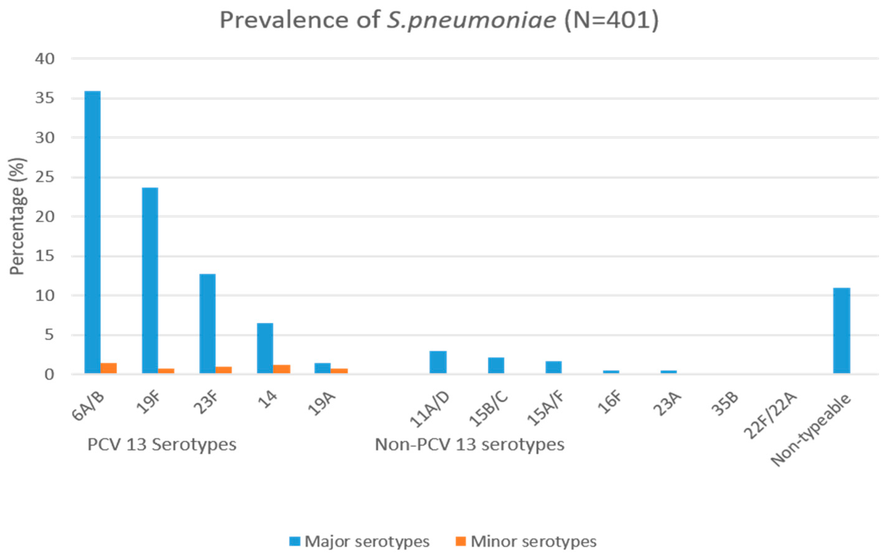
All hospitalized pediatric ARI cases from the target population in Nha Trang City were approached by the study doctors, and over 90% were successfully enrolled. During the study period, 1300 children having acute respiratory infections were enrolled in the study. Overall, more males (780, 60%) than females (520, 40%) were enrolled in the study. The majority of the children came from big families, as 692 (53.2%) had more than four members per household, and 671 (51.6%) attended daycare or school currently. There was no association between smokers in the household and positive pneumococcal detection. The age group with the majority of children was 0–1 years, with 825 (63.5%). Moreover, the number of children found to have pneumonia using the revised WHO Integrated Management of Childhood Illness (IMCI) pneumonia classification [33] was 116 (8.9%). Pneumonia was defined as having tachypnea and/or chest indrawing. Overall, S. pneumoniae was detected in 413 (31.8%) children, and in 23.9% of those with clinical pneumonia versus 32.0% of those without it (p = 0.551). Moreover, S. pneumoniae detection was higher in the age groups of 0–1 and 2–3 years (33.8% and 34.2%, respectively) and lowest among children older than 5 years (11.3%). S. pneumoniae carriage was positively associated with children ≤3 years old (OR 2.63, 95% CI 1.69–4.06; for 0–1 years, OR 4.03, 95% CI 1.99–8.16; for 2–3 years, OR 4.10, 95% CI 1.97–8.51) compared to children >5 years, those who attend daycare or school (OR 1.72, 95% CI 1.36–2.18) compared to those who do not, and those who reported antibiotics use prior to admission (OR 1.32, 95% CI 1.01–1.74) compared to those who did not, as shown in Table 1.

3.2. Serotyping

From 413 S. pneumoniae isolates, we serotyped 401 isolates, as twelve samples did not grow sufficiently upon revival. Twelve serogroups/serotypes were found. The prevalent VTs were 6A/B (144, 35.9%), 19F (95, 23.7%) and 23F (51, 12.7%). The most prevalent NVTs were 11A/D (12, 3.0%) and 15B/C (9, 2.2%). Serotypes detected together with others, i.e., minor serotypes, amounted to 21 (5.2%). PCV13 serotypes accounted for 80.3% of the serotypes amplified. Those we were unable to serotype were classified as non-typeable (n = 44, 11%), as shown in Figure 1.

3.3. Antimicrobial Susceptibility Testing

The MICs of 399 S. pneumoniae were determined. Fourteen isolates did not grow sufficiently. High antimicrobial resistance was seen for Clarithromycin (346, 86.7%), Erythromycin (339, 85.0%) and Clindamycin (312, 78.2%), while low resistance was observed for Penicillin (1, 0.3%), Ciprofloxacin (2, 0.5%), Cefotaxime (5, 1.3%), Imipenem (41, 10.3%) and Amoxicillin/Clavulanate (43, 10.8%). Resistance to Cefuroxime was the highest among β-lactams (71.9%), which is a second-generation cephalosporin, as shown in Table 2. Moreover, 48.6% of the isolates were MDR and the overall resistance to β-lactam and Macrolides was 18.4% and 85.9% respectively. The highest seen resistance to Erythromycin and Tetracycline was in serotype 14 (96.2%, 25/26) and 11A/D (91.7%, 11/12), respectively. Comparatively lower resistance rates were seen for Amoxicillin, among which the highest rate was in serotype 19F (34.7%, 33/95) and for Chloramphenicol in 6A/B (66%, 95/144) (Supplementary Table S2).

3.4. Penicillin and Erythromycin Resistance Genes

A total of 267 S. pneumoniae isolates, isolated between the 2015 October and 2016 May study period, were shipped from Vietnam to Nagasaki University for drug-resistant gene genotyping and MLST analysis. The remaining 132 samples were not able to ship to Nagasaki University in time due to the COVID-19 pandemic situation in Vietnam. A high proportion of PBP gene mutations was present, i.e., pbp1a (262, 98.1%), pbp2b (264, 98.8%) and pbp2x (266, 99.6%). The pbp genotype that was the most prevalent was the gPRSP (97%), and no isolate was gPSSP. Erythromycin resistance genes ermB were found to amount to 258 (96.6%) and mefA, 81 (30.3%), while isolates having both genes amounted to 75 (28.1%). Serotype 19F had the highest distribution of mefA and also the highest number of isolates having both ermB + mefA genes when compared to other serotypes. However, the ermB gene (except in 35B) and pbp genes were distributed across all serotypes (Supplementary Table S3).

3.5. Multi-Locus Sequence Typing

Among 267 S. pneumoniae isolates analyzed, three isolates were not successful in the assignment of STs. From 264 isolates, 52 STs were identified by MLST, of which 15 STs (ST 17845, 17846, 17847, 17848, 17849, 17850, 17851, 17852, 17853, 17854, 17855, 17856, 17857, 17859 and 17860) were newly assigned, as shown in Figure 2. The predominant STs were 320 (56, 21.2%), ST13223 (51, 19.3%), ST81 (21, 8%) and ST90 (15, 5.7%).
The genotyped pneumococci clustered into nine CCs and 17 singletons based on the single-locus variant (SLV) in the seven housekeeping genes. The five most prevalent CCs were CC90 (5 STs), CC1106 (5 STs) and CC271 (5 STs). Based on PMEN, four international resistance clones containing 42 isolates were identified. These are Spain23F-1 (ST81, 21, 8.3%), Spain6B-2 (ST90, 15, 5.9%), Taiwan19F-14 (ST236, 3, 1.2%) and Sweden15A-25 (ST63, 3, 1.2%), as shown in Table 3. The CCs that showed MDR patterns from the pneumococcal isolates were CC13223, CC90 and the singleton CC17860.
The distribution of STs among different serotypes showed that serotype 6A/B was dominant in ST 13223 and ST 90, with high ST diversity, while serotype 19F was majorly in ST 320. Non-typeable isolates had seven varieties of the most newly assigned ST17851 (n = 2), ST17852 (n = 4), ST17849 (n = 2), ST17853 (n = 1), ST17855 (n = 1), ST17859 (n = 1) and ST17860 (n = 2), while serotype 14 had nine isolates of newly assigned ST 17845 (Supplementary Table S4). Among isolates having both ermB and mefA (74, 29.1%), clone Taiwan19F-14 (2, 2.7%) was the only one present. Positive ermB and negative mefA (174, 68.5%) isolates had four clones distributed among them, i.e., Spain23F-1 (21, 12.1%), Spain6B-2 (15, 8.6%), Sweden15A-25 (3, 1.7%) and Taiwan19F-14 (1, 0.57%). Lastly, isolates that did not have the ermB gene but had the mefA gene (5, 2%) had no international clone present.

4. Discussion

The study covered the pre-PCV period (October 2015–September 2016) and focused on children with acute respiratory infections admitted to KHGH. From the study subjects, we identified children ≤3 years, those attending daycare or school and those who had taken antibiotics 14 days prior to admission to be positively associated with the detection of S. pneumoniae. The study also found high resistance to macrolides and high diversity of sequence types associated with multi-resistant pneumococcal international clones. This is the first comprehensive report on the population structure of S. pneumoniae using MLST in the study area.
In our study, S. pneumoniae was detected in 413 (31.8%) pediatric ARI cases, of whom 23.9% had clinical pneumonia and 32.0% did not. However, there was no significant difference between those with and without pneumonia, nor in each respiratory or systemic symptom between those with S. pneumoniae and those without. S. pneumoniae is a common pathogen causing pneumonia in hospitalized children [5]. Therefore, we could regard the detection of S. pneumoniae as colonization in many cases, but some may be the cause of respiratory symptoms in the children. A previous study that detected S. pneumoniae from nasopharyngeal samples using real-time PCR in children with radiologically confirmed pneumonia (RCP), other lower respiratory infections (LRTIs) and healthy controls reported that although the detection rates were similar in these groups, the median nasopharyngeal bacterial load of S. pneumoniae was substantially higher in children with RCP compared to other LRTIs and healthy controls [34]. We did not conduct the S. pneumoniae bacteria load real-time PCR testing in this study, so we were not able to confirm this previous finding. On the other hand, it is important to note that this assumption may not hold true in all cases, as reported in a recent Canadian study [35].
The proportion of serotypes detected were mainly those covered by the PCV 13 vaccine. The dominant VTs found in this study, 6A/B (35.9%), 19F (23.7%) and 23F (12.7%), are similar to a review study carried out on healthy children in the South East Asia region from 2001 to 2019, in which they found an overall pneumococcal prevalence of 36% (4139/11,501) and dominance of serotype 6A/B (12.9%), 23F (9.3%) and 19F (10.1%). Moreover, a study in hospitals in the UK and Ireland on community-acquired respiratory infections pre-PCV7 from 1999 to 2007 found the dominating serotypes in the respiratory tract to be 19F (83, 11.1%), 23F (72, 9.6%), 6B (52, 7.1%) and 3 (51, 6.8%) among children and adults [34]. This changed over time following the introduction of PCV, as reported in a study conducted from 2006 to 2018 that saw an overall decrease in all the dominant vaccine serotypes, with the exception of serotype 3 (13, 1.27%) [35]. In 2001, PCV7 was approved in Europe, and from 2006 to 2008, it was introduced into the national immunization programs of many European countries. In 2009, higher-valent PCVs were adopted, i.e., PCV10 and PCV13, replacing PCV7 from 2009 to 2011. This led to changes in circulating serotypes in the European region to the most frequently isolated 24F, 22F, 8 and 15A in countries that use PCV13, and serotypes 19A and 3 in countries that use PCV10 [36]. This highlights the need to continuously monitor the evolving serotypes and their impact on currently available pneumococcal vaccines.
Our study isolated NVTs 11A/D (3%), 15B/C (2.2%) and 15A/F (1.7%), which have been reported previously both in pre-PCV and post-PCV implementation. A review that focused on serotype distribution causing IPD in the post-PCV era from 2000 to 2015, reported that the predominant non-PCV13 serotypes seen were 22F, 12F, 33F, 24F, 15C, 15B, 23B, 10A and 38, which changed by rank per region of the world [37]. In addition, a study in Japan reported an 8% to 48.1% increase in NVTs from 2010 to 2017 [30], whereas in Denmark in 2014, 71% of IPD was caused by NVTs after the introduction of PCV vaccines. This increase was also reported in the US in 2011 where common NVTs were 33F, 22F, 12, 15B, 15C, 23A and 11 [38]. This trend is worrying, as serotype replacement and switching as a result of vaccine pressure, host dynamics or the environment have the ability to render the current vaccines ineffective. There is an increasing need to develop serotype-independent effective vaccines that can cover vulnerable populations and are capable of targeting all invasive S. pneumoniae in the future. Vietnam, however, has yet to start using the currently available PCVs. Based on our study data, further surveillance is necessary to see how much pneumococcal carriage and antibiotic resistance would decrease after PCV introduction in the study area.
In Vietnam, factors that may have led to the high antimicrobial resistance include unrestricted access to antimicrobials through private pharmacies and their irrational use in farming and animal rearing, leading to constant selective pressure and the spread of the global pandemic multi-resistant clones Taiwan19F-14 and Spain23F-1 from as early as 1993, which when seen are not easily lost [39,40]. A review article that reported on antimicrobial resistance in pediatric pneumococcal isolates globally from 2000 to 2020, before and after the introduction of any PCV, found that there were absolute reductions in resistant isolates over 10 years after any PCV implementation apart from resistance to macrolides and tetracycline [41]. Furthermore, a study conducted from 2016 to 2017 on clinical isolates causing IPD from 13 hospitals in Vietnam reported macrolide resistance of 94% (1234/1317) [42]. This would indicate the importance of following the resistance to macrolides and tetracycline even after PCV implementation.
Our study also found a low rate of β-lactam resistance and a high proportion of PBP gene mutations, signifying that pbp mutations did not mainly confer resistance to penicillin in our isolates. Fani et al. reported that other factors could be responsible for β-lactam resistance other than altered pbps alone [43]. Furthermore, Erythromycin-resistant S. pneumoniae genotypes are usually different over various geographic areas. The most common mechanism of resistance is usually ermB encoded in many parts of Asia such as Japan, Sri Lanka, China, Taiwan and South Korea as well as in Belgium (91.5%), France (90%), Spain (88.3%), Serbia (82.4%), Poland (80.8%) and Italy (55.8%). Meanwhile, mefA-encoded resistance was most common in Greece (66.2%), the US before PCV7, Finland (55.4%), the UK (70.8%), Australia (59.5%) and Germany (53.2%) [44]. Our study showed ermB to be the common mode of resistance for Erythromycin. Serotype 19F was found to have more resistance genes compared to other serotypes. This has been seen in other studies, such as one from Tunisia that reported ermB + mefA to be 64% in 19F isolates and one in Korea that reported 57% of both genes in 19F [45,46].
The sequence types predominant in the study were ST320, ST13223, ST81 and ST90. They were mostly found within MDR CCs and within three international clones, Spain23F-1, Spain6B-2 and Taiwan19F-14, responsible for the spread of MDR isolates globally, including in Vietnam [47,48]. Most of these isolates were resistant to Erythromycin, Tetracycline and Cefuroxime. CC271 isolates were associated with serotype 19F, which has been reported in North America and several other Asian countries [49,50,51,52]. However, the clonal spread of CC271 and serotype replacement within the CC has been reported, which is of great concern in the control of VTs [53]. An MDR CC that was uniquely predominant in this study was CC13223. This CC was in serotype 6A/B and resistant to Erythromycin, Cefuroxime, Chloramphenicol and Tetracycline. Non-typeable isolates from this study led to the discovery of more new ST varieties (seven different types) compared to other serotypes. This could highlight the importance of surveillance of non-typeable pneumococci for designing future pneumococcal vaccines as well as understanding the evolution of S. pneumoniae. In terms of serotyping, antimicrobial susceptibility testing and multi-locus sequence typing results, there was no major difference in trends of S. pneumoniae between the groups with or without pneumonia. The study also shows that apart from vaccine pressure when introduced, other factors such as antibiotic use, mobile genetic elements, capsule switching and age at colonization could play a critical role in the spread of MDR serotypes in PCV-naïve populations.
The study encountered some limitations; the surveillance data only covered one year at one hospital and only hospitalized pediatric ARI cases, making it difficult to generalize the results to the general population. Future studies with the inclusion of community samples will be important. Resistance genes and MLST analysis were carried out on 267 isolates from a total of 399. This, however, was not a shortcoming of the study, as there was little difference in serotype distribution and susceptibility testing results between the isolates that were analyzed and those that were not. We believe that the results from the 267 isolates from the October 2015 to May 2016 period can be representative of the overall isolates collected in this study. We were also unable to distinguish different serotypes within serogroups such as 6A/B, 11A/D and 15A/F and could not confirm non-typeables due to limitations of the serotyping method used. However, the main serotypes that are included in PCVs were identified.

5. Conclusions

Children three years old or younger, those attending daycare or school and those who used antibiotics 14 days prior to hospital admission were positively associated with pneumococcal colonization. PCV introduction would be beneficial in this area as high rates of VTs were detected in hospitalized pediatric ARI cases and were associated with antibiotic resistance.

Supplementary Materials

The following supporting information can be downloaded at: https://www.mdpi.com/article/10.3390/pathogens12070943/s1, Table S1: Primers used for amplification of penicillin-binding protein genes and macrolide resistance genes. Table S2: Antimicrobial resistance of S. pneumoniae against antimicrobial agents in different serotypes. Table S3: Resistance genes among different serotypes. Table S4: Sequence type distribution among different serotypes.

Author Contributions

Conceptualization, L.-M.Y. and P.W.; methodology, L.-M.Y., M.T. and P.W.; software, H.-H.L. and L.-M.Y.; validation, L.-M.Y. and M.T.; formal analysis, P.W. and H.-H.L.; investigation, H.-A.N., K.-A.L., M.-M.S. and P.W.; resources, H.-M.V. and L.-M.Y.; data curation, P.W.; writing, P.W., M.T. and L.-M.Y.; supervision, L.-M.Y.; project administration, H.-M.V., M.-X.B. and D.-A.D.; funding acquisition, L.-M.Y.; writing—review and editing, P.W., H.-H.L., M.T., M.-M.S. and L.-M.Y. All authors have read and agreed to the published version of the manuscript.

Funding

This research was supported by the Japan Program for Infectious Diseases Research and Infrastructure, Japan Agency for Medical Research and Development (AMED) under grant number JP21wm0125006 and partially by the Bill and Melinda Gates Foundation, grant number OPP1139859. P.W. was supported by a research student scholarship from the Japan International Cooperation Agency and Nagasaki University Graduate School of Biomedical Sciences.

Institutional Review Board Statement

The study was approved by the Ethical Review Board of the National Institute of Hygiene and Epidemiology (NIHE), Hanoi, Vietnam (IRB-VN 01057), and the Institute of Tropical Medicine, Nagasaki University, Nagasaki, Japan (09031837-3). Written informed consent was obtained from all subjects and their representatives.

Informed Consent Statement

Written informed consent was obtained from all subjects involved in the study.

Data Availability Statement

Novel sequences from the study were deposited in the PubMLST database (https://pubmlst.org/spneumoniae/ (accessed on 10 November 2022)).

Acknowledgments

We thank the children who participated and the parents who gave consent for this study. We also thank the staff of the National Institute of Hygiene and Epidemiology (NIHE), Khanh Hoa General Hospital (KHGH) and Khanh Hoa Health Service (KHHS), Nha Trang, Vietnam, for their support. Finally, we also thank the Program for Nurturing Global Leaders in Tropical and Emerging Communicable Diseases at the Graduate School of Biomedical Sciences, Nagasaki University, for the PhD training opportunity, and the entire staff of the Nagasaki University Department of Pediatric Infectious Diseases for their support.

Conflicts of Interest

The authors declare no conflict of interest.

References

  1. Safiri, S.; Mahmoodpoor, A.; Kolahi, A.A.; Nejadghaderi, S.A.; Sullman, M.J.M.; Mansournia, M.A.; Ansarin, K.; Collins, G.S.; Kaufman, J.S.; Abdollahi, M. Global burden of lower respiratory infections during the last three decades. Front. Public Health 2023, 10, 1028525. [Google Scholar] [CrossRef] [PubMed]
  2. Global Burden of Disease Collaborative Network Global Burden of Disease Collaborative Network. Global Burden of Disease Study 2019 (GBD 2019). Results. Institute for Health Metrics and Evaluation (IHME): Seattle, WA, USA, 2020. Available online: https://vizhub.healthdata.org/gbd-results/ (accessed on 7 June 2023).
  3. Bogaert, D.; De Groot, R.; Hermans, P.W.M. Streptococcus pneumoniae colonisation: The key to pneumococcal disease. Lancet Infect. Dis. 2004, 4, 144–154. [Google Scholar] [CrossRef] [PubMed]
  4. Weiser, J.N.; Ferreira, D.M.; Paton, J.C. Streptococcus pneumoniae: Transmission, colonization and invasion. Nat. Rev. Microbiol. 2018, 16, 355–367. [Google Scholar] [CrossRef]
  5. O’Brien, K.L.; Wolfson, L.J.; Watt, J.P.; Henkle, E.; Deloria-Knoll, M.; McCall, N.; Lee, E.; Mulholland, K.; Levine, O.S.; Cherian, T. Burden of disease caused by Streptococcus pneumoniae in children younger than 5 years: Global estimates. Lancet 2009, 374, 893–902. [Google Scholar] [CrossRef]
  6. Johnson, H.L.; Deloria-Knoll, M.; Levine, O.S.; Stoszek, S.K.; Hance, L.F.; Reithinger, R.; Muenz, L.R.; O’Brien, K.L. Systematic evaluation of serotypes causing invasive pneumococcal disease among children under five: The pneumococcal global serotype project. PLoS Med. 2010, 7, e1000348. [Google Scholar] [CrossRef] [PubMed] [Green Version]
  7. Song, J.Y.; Nahm, M.H.; Moseley, M.A. Clinical implications of pneumococcal serotypes: Invasive disease potential, clinical presentations, and antibiotic resistance. J. Korean Med. Sci. 2013, 28, 4–15. [Google Scholar] [CrossRef] [Green Version]
  8. Ganaie, F.; Saad, J.S.; McGee, L.; van Tonder, A.J.; Bentley, S.D.; Lo, S.W.; Gladstone, R.A.; Turner, P.; Keenan, J.D.; Breiman, R.F.; et al. A new pneumococcal capsule type, 10D, is the 100th serotype and has a large cps fragment from an oral streptococcus. MBio 2020, 11, e00937-20. [Google Scholar] [CrossRef]
  9. World Health Organization (WHO) Pneumococcal conjugate vaccines in infants and children under 5 years of age: WHO position paper—February 2019. Wkly. Epidemiol. Rec. 2019, 94, 85–104.
  10. Huang, S.S.; Platt, R.; Rifas-Shiman, S.L.; Pelton, S.I.; Goldmann, D.; Finkelstein, J.A. Post-PCV7 changes in colonizing pneumococcal serotypes in 16 Massachusetts communities, 2001 and 2004. Pediatrics 2005, 116, e408–e413. [Google Scholar] [CrossRef] [Green Version]
  11. Kyaw, M.H.; Lynfield, R.; Schaffner, W.; Craig, A.S.; Hadler, J.; Reingold, A.; Thomas, A.R.; Harrison, L.H.; Bennett, N.M.; Farley, M.M.; et al. Effect of Introduction of the Pneumococcal Conjugate Vaccine on Drug-Resistant Streptococcus pneumoniae. N. Engl. J. Med. 2006, 354, 1455–1463. [Google Scholar] [CrossRef]
  12. Ladhani, S.N.; Slack, M.P.E.; Andrews, N.J.; Waight, P.A.; Borrow, R.; Miller, E. Invasive pneumococcal disease after routine pneumococcal conjugate vaccination in children, England and Wales. Emerg. Infect. Dis. 2013, 19, 61–68. [Google Scholar] [CrossRef]
  13. Brandileone, M.C.C.; Almeida, S.C.G.; Bokermann, S.; Minamisava, R.; Berezin, E.N.; Harrison, L.H.; Andrade, A.L. Dynamics of antimicrobial resistance of Streptococcus pneumoniae following PCV10 introduction in Brazil: Nationwide surveillance from 2007 to 2019. Vaccine 2021, 39, 3207–3215. [Google Scholar] [CrossRef] [PubMed]
  14. Temple, B.; Nation, M.L.; Dai, V.T.T.; Beissbarth, J.; Bright, K.; Dunne, E.M.; Hinds, J.; Hoan, P.T.; Lai, J.; Nguyen, C.D.; et al. Effect of a 2+1 schedule of ten-valent versus 13-valent pneumococcal conjugate vaccine on pneumococcal carriage: Results from a randomised controlled trial in Vietnam. Vaccine 2021, 39, 2303–2310. [Google Scholar] [CrossRef] [PubMed]
  15. Løchen, A.; Croucher, N.J.; Anderson, R.M. Divergent serotype replacement trends and increasing diversity in pneumococcal disease in high income settings reduce the benefit of expanding vaccine valency. Sci. Rep. 2020, 10, 18977. [Google Scholar] [CrossRef] [PubMed]
  16. Nagaraj, S.; Kalal, B.S.; Manoharan, A.; Shet, A. Streptococcus pneumoniae serotype prevalence and antibiotic resistance among young children with invasive pneumococcal disease: Experience from a tertiary care center in South India. Germs 2017, 7, 78–85. [Google Scholar] [CrossRef] [Green Version]
  17. Shak, J.R.; Vidal, J.E.; Klugman, K.P. Influence of bacterial interactions on pneumococcal colonization of the nasopharynx. Trends Microbiol. 2013, 21, 129–135. [Google Scholar] [CrossRef] [Green Version]
  18. Hakenbeck, R.; Balmelle, N.; Weber, B.; Gardès, C.; Keck, W.; De Saizieu, A. Mosaic genes and mosaic chromosomes: Intra- and interspecies genomic variation of Streptococcus pneumoniae. Infect. Immun. 2001, 69, 2477–2486. [Google Scholar] [CrossRef] [Green Version]
  19. Noguchi, N.; Tano, J.; Nasu, Y.; Koyama, M.; Narui, K.; Kamishima, H.; Saito, T.; Tsuyuki, K.; Sasatsu, M. Antimicrobial susceptibilities and distribution of resistance genes for β-lactams and macrolides in Streptococcus pneumoniae isolated between 2002 and 2004 in Tokyo. Int. J. Antimicrob. Agents 2007, 29, 26–33. [Google Scholar] [CrossRef]
  20. Dadgostar, P. Antimicrobial resistance: Implications and costs. Infect. Drug Resist. 2019, 12, 3903–3910. [Google Scholar] [CrossRef] [Green Version]
  21. Nguyen, H.A.T.; Fujii, H.; Vu, H.T.T.; Parry, C.M.; Dang, A.D.; Ariyoshi, K.; Yoshida, L.M. An alarmingly high nasal carriage rate of Streptococcus pneumoniae serotype 19F non-susceptible to multiple beta-lactam antimicrobials among Vietnamese children. BMC Infect. Dis. 2019, 19, 241. [Google Scholar] [CrossRef] [Green Version]
  22. Yoshida, L.M.; Suzuki, M.; Yamamoto, T.; Nguyen, H.A.; Nguyen, C.D.; Nguyen, A.T.; Oishi, K.; Vu, T.D.; Le, T.H.; Le, M.Q.; et al. Viral Pathogens Associated with Acute Respiratory Infections in Central Vietnamese Children. Pediatr. Infect. Dis. J. 2010, 29, 75–77. [Google Scholar] [CrossRef] [PubMed] [Green Version]
  23. Pimenta, F.C.; Roundtree, A.; Soysal, A.; Bakir, M.; Du Plessis, M.; Wolter, N.; Von Gottberg, A.; McGee, L.; Da Gloria Carvalho, M.; Beall, B. Sequential Triplex Real-Time PCR Assay for Detecting 21 Pneumococcal Capsular Serotypes That Account for a High Global Disease Burden. J. Clin. Microbiol. 2013, 51, 647–652. [Google Scholar] [CrossRef] [PubMed] [Green Version]
  24. Cools, F.; Delputte, P.; Cos, P. The search for novel treatment strategies for Streptococcus pneumoniae infections. FEMS Microbiol. Rev. 2021, 45, fuaa072. [Google Scholar] [CrossRef] [PubMed]
  25. CLSI. M100S Performance Standards for Antimicrobial (CLSI), 26th ed.; Clinical and Laboratory Standards Institute: Wayne, PA, USA, 2015; ISBN 1562389238. [Google Scholar]
  26. EUCAST Clinical Breakpoint Table v. 5.0, Valid from 1 January 2015. pp. 1–77. Available online: http://www.eucast.org/clinical_breakpoints/ (accessed on 2 November 2022).
  27. Magiorakos, A.; Srinivasan, A.; Carey, R.B.; Carmeli, Y.; Falagas, M.E.; Giske, C.G.; Harbarth, S.; Hindler, J.F. Multidrug-resistant, extensively drug-resistant and pandrug-resistant bacteria: An international expert proposal for interim standarddefinitions for acquired resistance. Clin. Microbiol. Infect. 2011, 28, 250–290. [Google Scholar] [CrossRef] [Green Version]
  28. Nagai, K.; Shibasaki, Y.; Hasegawa, K.; Davies, T.A.; Jacobs, M.R.; Ubukata, K.; Appelbaum, P.C. Evaluation of PCR primers to screen for Streptococcus pneumoniae isolates and β-lactam resistance, and to detect common macrolide resistance determinants. J. Antimicrob. Chemother. 2001, 48, 915–918. [Google Scholar] [CrossRef]
  29. Ubukata, K.; Chiba, N.; Hasegawa, K.; Kobayashi, R.; Iwata, S.; Sunakawa, K. Antibiotic Susceptibility in Relation to Penicillin-Binding Protein Genes and Serotype Distribution of Streptococcus pneumoniae Strains Responsible for Meningitis in Japan, 1999 to 2002. Antimicrob. Agents Chemother. 2004, 48, 1488–1494. [Google Scholar] [CrossRef] [Green Version]
  30. Ubukata, K.; Takata, M.; Morozumi, M.; Chiba, N.; Wajima, T.; Hanada, S.; Shouji, M.; Sakuma, M.; Iwata, S. Effects of pneumococcal conjugate vaccine on genotypic penicillin resistance and serotype changes, Japan, 2010–2017. Emerg. Infect. Dis. 2018, 24, 2010–2020. [Google Scholar] [CrossRef] [Green Version]
  31. Ibarz Pavón, A.B.; Maiden, M.C.J. Multilocus sequence typing. Methods Mol. Biol. 2009, 551, 129–140. [Google Scholar] [CrossRef] [Green Version]
  32. Enright, M.; Spratt, B. A multilocus sequence typing scheme for Streptococcus pneumoniae: Identification of clones associated with serious invasive disease. Microbiology 1998, 144, 3049–3060. [Google Scholar] [CrossRef] [Green Version]
  33. WHO; UNICEF. Revised WHO Classification and Treatment of childhood Pneumonia at Health Facilities: Qiuck Reference Guide. Evidence for Technical Update of Pocket Book Recommendations. Geneva, WHO. 2012. Available online: https://apps.who.int/iris/bitstream/handle/10665/137332/WHO_FWC_MCA_14.9_eng.pdf (accessed on 15 July 2022).
  34. Vu, H.T.T.; Yoshida, L.M.; Suzuki, M.; Nguyen, H.A.T.; Nguyen, C.D.L.; Nguyen, A.T.T.; Oishi, K.; Yamamoto, T.; Watanabe, K.; Vu, T.D.; et al. Association between Nasopharyngeal Load of Streptococcus pneumoniae, Viral Coinfection, and Radiologically Confirmed Pneumonia in Vietnamese Children. Pediatr. Infect. Dis. J. 2011, 30, 11–18. [Google Scholar] [CrossRef] [Green Version]
  35. Pernica, J.M.; Inch, K.; Alfaraidi, H.; Van Meer, A.; Carciumaru, R.; Luinstra, K.; Smieja, M. Assessment of nasopharyngeal Streptococcus pneumoniae colonization does not permit discrimination between Canadian children with viral and bacterial respiratory infection: A matched-cohort cross-sectional study. BMC Infect. Dis. 2021, 21, 509. [Google Scholar] [CrossRef]
  36. Tin Tin Htar, M.; Christopoulou, D.; Schmitt, H.J. Pneumococcal serotype evolution in Western Europe. BMC Infect. Dis. 2015, 15, 419. [Google Scholar] [CrossRef] [PubMed] [Green Version]
  37. Balsells, E.; Guillot, L.; Nair, H.; Kyaw, M.H. Serotype distribution of Streptococcus pneumoniae causing invasive disease in children in the post-PCV era: A systematic review and meta-analysis. PLoS ONE 2017, e0177113. [Google Scholar] [CrossRef] [PubMed] [Green Version]
  38. Du, Q.; Shi, W.; Yu, D.; Yao, K. hu Epidemiology of non-vaccine serotypes of Streptococcus pneumoniae before and after universal administration of pneumococcal conjugate vaccines. Hum. Vaccines Immunother. 2021, 17, 5628–5637. [Google Scholar] [CrossRef]
  39. Mitchell, M.E.V.; Alders, R.; Unger, F.; Nguyen-Viet, H.; Le, T.T.H.; Toribio, J.A. The challenges of investigating antimicrobial resistance in Vietnam—What benefits does a One Health approach offer the animal and human health sectors? BMC Public Health 2020, 20, 213. [Google Scholar] [CrossRef] [PubMed] [Green Version]
  40. Hoa, N.Q.; Trung, N.V.; Larsson, M.; Eriksson, B.; Phuc, H.D.; Chuc, N.T.K.; Lundborg, C.S. Decreased Streptococcus pneumoniae susceptibility to oral antibiotics among children in rural Vietnam: A community study. BMC Infect. Dis. 2010, 10, 85. [Google Scholar] [CrossRef] [Green Version]
  41. Andrejko, K.; Ratnasiri, B.; Hausdorff, W.P.; Laxminarayan, R.; Lewnard, J.A. Antimicrobial resistance in paediatric Streptococcus pneumoniae isolates amid global implementation of pneumococcal conjugate vaccines: A systematic review and meta-regression analysis. Lancet Microbe 2021, 2, e450–e460. [Google Scholar] [CrossRef]
  42. Vu, T.V.D.; Choisy, M.; Do, T.T.N.; Nguyen, V.M.H.; Campbell, J.I.; Le, T.H.; Nguyen, V.T.; Wertheim, H.F.L.; Pham, N.T.; Nguyen, V.K.; et al. Antimicrobial susceptibility testing results from 13 hospitals in Viet Nam: VINARES 2016–2017. Antimicrob. Resist. Infect. Control 2021, 10, 78. [Google Scholar] [CrossRef]
  43. Fani, F.; Leprohon, P.; Légaré, D.; Ouellette, M. Whole genome sequencing of penicillin-resistant Streptococcus pneumoniae reveals mutations in penicillin-binding proteins and in a putative iron permease. Genome Biol. 2011, 12, R115. [Google Scholar] [CrossRef] [Green Version]
  44. Zhou, X.; Liu, J.; Zhang, Z.; Cui, B.; Wang, Y.; Zhang, Y.; Xu, H.; Cheng, G.; Liu, Y.; Qin, X. Characterization of Streptococcus pneumoniae Macrolide Resistance and Its Mechanism in Northeast China over a 20-Year Period. Microbiol. Spectr. 2022, 10, e00546-22. [Google Scholar] [CrossRef]
  45. Safari, D.; Kuo, L.C.; Huang, Y.T.; Liao, C.H.; Sheng, W.H.; Hsueh, P.R. Increase in the rate of azithromycin-resistant Streptococcus pneumoniae isolates carrying the erm(B) and mef(A) genes in Taiwan, 2006–2010. BMC Infect. Dis. 2015, 14, 704. [Google Scholar] [CrossRef] [Green Version]
  46. Bae, S.; Lee, K. Distribution of capsular serotypes and macrolide resistance mechanisms among macrolide-resistant Streptococcus pneumoniae isolates in Korea. Diagn. Microbiol. Infect. Dis. 2009, 63, 213–216. [Google Scholar] [CrossRef]
  47. Bogaert, D.; Ha, N.T.; Sluijter, M.; Lemmens, N.; De Groot, R.; Hermans, P.W.M. Molecular epidemiology of pneumococcal carriage among children with upper respiratory tract infections in Hanoi, Vietnam. J. Clin. Microbiol. 2002, 40, 3903–3908. [Google Scholar] [CrossRef] [PubMed] [Green Version]
  48. Parry, C.M.; Duong, N.M.; Zhou, J.; Mai, N.T.H.; Diep, T.S.; Thinh, L.Q.; Wain, J.; Chau, N.V.V.; Griffiths, D.; Day, N.P.J.; et al. Emergence in Vietnam of Streptococcus pneumoniae resistant to multiple antimicrobial agents as a result of dissemination of the multiresistant Spain23F-1 clone. Antimicrob. Agents Chemother. 2002, 46, 3512–3517. [Google Scholar] [CrossRef] [PubMed]
  49. Bowers, J.R.; Driebe, E.M.; Nibecker, J.L.; Wojack, B.R.; Sarovich, D.S.; Wong, A.H.; Brzoska, P.M.; Hubert, N.; Knadler, A.; Watson, L.M.; et al. Dominance of multidrug resistant CC271 clones in macrolide-resistant Streptococcus pneumoniae in Arizona. BMC Microbiol. 2012, 12, 12. [Google Scholar] [CrossRef] [PubMed]
  50. Liang, Z.; Fu, J.; Li, L.; Yi, R.; Xu, S.; Chen, J.; Ye, X.; McGrath, E. Molecular epidemiology of Streptococcus pneumoniae isolated from pediatric community-acquired pneumonia in pre-conjugate vaccine era in Western China. Ann. Clin. Microbiol. Antimicrob. 2021, 20, 4. [Google Scholar] [CrossRef]
  51. Del Grosso, M.; Northwood, J.G.E.; Farrell, D.J.; Pantosti, A. The macrolide resistance genes erm(B) and mef(E) are carried by Tn2010 in dual-gene Streptococcus pneumoniae isolates belonging to clonal complex CC271. Antimicrob. Agents Chemother. 2007, 51, 4184–4186. [Google Scholar] [CrossRef] [Green Version]
  52. Dzaraly, N.D.; Mohd Desa, M.N.; Muthanna, A.R.; Masri, S.N.; Taib, N.M.; Suhaili, Z.; Sulaiman, N.; Baharin, N.H.Z.; Shuan, C.Y.; Ariffin, Z.; et al. Antimicrobial susceptibility, serotype distribution, virulence profile and molecular typing of piliated clinical isolates of pneumococci from east coast, Peninsular Malaysia. Sci. Rep. 2021, 11, 8220. [Google Scholar] [CrossRef]
  53. Scherer, E.M.; Beall, B.; Metcalf, B. Serotype-Switch Variant of pneumoniae Sequence Type 271. Emerg. Infect. Dis. 2021, 27, 1689–1692. [Google Scholar] [CrossRef]
Figure 1. Serotype distribution among 401 S. pneumoniae isolates among hospitalized children having acute respiratory infections aged 1 month to <15 years old.
Pathogens 12 00943 g001
Figure 2. An eBURST diagram showing the pneumococcal sequence (STs) from 264 carriage isolates in Vietnam. CC, clonal complex.
Pathogens 12 00943 g002
Table 1. Clinical and demographic characteristics of children enrolled in hospital-based surveillance from October 2015 to September 2016.
CharacteristicAll n (%)Pneumococcal +ve n (%)Pneumococcal −ve n (%)Odds Ratio (95% CI)
Total (n)1300413 (31.8)887 (68.2)
Age
0–1 yr.825 (63.5)279 (33.8)546 (66.2)4.03 (1.99–8.16)
2–3 yrs.316 (24.3)108 (34.2)208 (65.8)4.10 (1.97–8.51)
4–5 yrs.79 (6.1)17 (21.5)62 (78.5)2.16 (0.90–5.20)
>5 yrs.80 (6.2)9 (11.3)71 (88.7)Reference
Sex
Male780 (60.0)250 (32.1)530 (67.9)1.03 (0.81–1.31)
Female520 (40.0)163 (31.4)357 (68.6)Reference
Day-care or school attendance
Yes671 (51.6)251 (37.4)420 (62.6)1.72 (1.36–2.18)
No629 (48.4)162 (25.8)467 (74.2)Reference
Prior antibiotic use a
Yes950 (73.1)317 (33.4)633 (66.6)1.32 (1.01–1.74)
No350 (26.9)96 (27.4)254 (72.6Reference
From a big family (>4 members)
Yes692 (53.2)211 (30.5)481 (69.5)0.88 (0.69–1.11)
No608 (46.8)202 (33.2)406 (66.8)Reference
Smoker(s) in the household (n = 1273)
Yes403 (31.7)275 (31.6)595 (68.4)1.03 (0.94–1.12)
No870 (68.3)128 (31.8)275 (68.2)Reference
Wheeze
Yes666 (51.2)206 (30.9)460 (69.1)0.92 (0.73–1.17)
No634 (48.8)207 (32.6)427 (67.4)Reference
Cough *
Yes1298 (99.8)412 (31.7)886 (68.2)0.47 (0.29–7.45)
No2 (0.2)1 (50.0)1 (50.0)Reference
Crackle upon lung examination
Yes284 (21.8)88 (31.0)196 (69.0)0.95 (0.72–1.27)
No1016 (78.2)325 (31.9)691 (68.0)Reference
Danger signs b
Yes27 (2.1)10 (37.0)17 (63.0)1.27 (0.59–2.75)
No1273 (97.9)403 (31.7)870 (68.3)Reference
Pneumonia
Yes116 (8.9)34 (29.3)82 (70.7)0.88 (0.58–1.33)
No1184 (91.1)379 (32.0)805 (68.0)Reference
Difficulty breathing c
Yes129 (9.9)38 (29.5)91 (70.5)0.89 (0.59–1.32)
No1171 (90.1)375 (32.0)796 (68.0)Reference
Any virus detection d
Yes851 (65.5)276 (32.4)575 (67.6)1.09 (0.85–1.39)
No449 (34.5)137 (30.5)312 (69.5)Reference
Duration of hospitalization
Range (min–max)0–22 days
0–5 days894 (68.8)289 (32.3)605 (67.7)Reference
6–10 days374 (28.8)118 (31.6)256 (68.4)0.96 (0.74–1.25)
>10 days32 (2.4)6 (18.8)26 (81.2)0.48 (0.19–1.19)
a Those who did not know antibiotic history were added to the group of no antibiotic use 14 days prior to admission. * Fisher’s exact test, Chi-square was used on variables without the sign. b Variables such as unable to drink, altered consciousness, convulsions, lethargy, poor sucking, toxic appearance, irritability, hypothermia and bulging fontanel were all put together to show the danger signs. c Included those observed by doctor as having difficulty breathing, those having tachypnea based on respiratory rate and those having chest indrawing. d Viruses detected from these participants were influenza A virus, influenza B virus, respiratory syncytial virus, rhino virus, adeno virus, boca virus, human metapneumovirus, human parainfluenza virus (type 1, 2, 3 and 4), human coronavirus (229E and OC43).
Table 2. Antimicrobial susceptibility of 399 pneumococcal isolates to 13 common antimicrobial agents.
AntimicrobialsSusceptible
n (%)
Intermediate
n (%)
Resistance
n (%)
MIC (µg/mL) Breakpoints S-R
* Penicillin (parenteral non-meningitis)394 (98.7)4 (1)1 (0.3)≤2–≥8
Amoxicillin (non-meningitis)271 (67.9)66 (16.5)62 (15.5)≤2–≥8
Ampicillin42 (10.5)281 (70.4)76 (19.0)≤0.25–≥8
Amoxicillin/Clavulanate290 (72.7)66 (16.5)43 (10.8)≤2/1–≥8/4
Cefuroxime (parenteral)74 (18.5)38 (9.5)287 (71.9)≤0.5–≥2
* Cefotaxime (non-meningitis)368 (92.2)26 (6.5)5 (1.3)≤1–≥4
Imipenem232 (58.1)126 (31.6)41 (10.3)≤0.12–≥1
Erythromycin55 (13.8)5 (1.3)339 (85.0)≤0.25–≥1
Clarithromycin48 (12.0)5 (1.3)346 (86.7)≤0.25–≥1
Chloramphenicol270 (67.7)0129 (32.3)≤4–≥8
Clindamycin84 (21.1)3 (0.8)312 (78.2)≤0.25–≥1
Tetracycline74 (18.5)21 (5.3)304 (76.2)≤1–≥4
Ciprofloxacin28 (7.0)369 (92.5)2 (0.5)≤0.25–≥4
* Penicillin and Cefotaxime, CLSI 2016 non-meningitis breakpoints were applied. All were interpreted using CLSI 2016, except Ciprofloxacin, for which EUCAST 2015 was used. MIC, minimum inhibitory concentrations.
Table 3. Clonal complexes, sequence types, PMEN, resistance patterns and serotype distribution in 254 S. pneumoniae isolates.
CC/SingletonsNo. of Isolates (%)No. of STsSequence Types (STs)PMENResistance Patterns *Serotypes
CC (9)
9023 (8.7)58465, 1624, 90, 9332, 95Spain6B-2ERY + CXM + TE + C6A/B
11066 (2.2)517855, 17854, 1106, 14138, 17853 ERY + CXM + TE23F(1), 11A/D (1), 16F(1), NT (3)
27165 (24.6)5236, 283, 320, 4467271Taiwan19F-14ERY + CXM + TE19F(62), 14(1), 19A(1), 6A/B(1)
8129 (11)42395, 81, 6796, 83Spain23F-1ERY + CXM23F(26), 15B/C (3)
88015 (5.7)43176, 880, 17847, 9116 ERY + CXM + TE23F(13), 19F(1) 11A/D(1)
178498 (3)417849, 17846, 1593, 15730 ERY + CXM + TE19F(1), 6A/B(1) NT (6)
6312 (4.5)417850, 2675, 63, 782Sweden15A-25ERY + TE14(6), 15A/F(6)
1322352 (19.7)213223, 9650 ERY + CXM + TE + C6A/B
19612 (0.8)21961, 17848 TE15B/C
Singletons (17)
3194 (1.5)1singleton ERY19A
89661 (0.4)1singleton ERY + CXM + TE22F/A
178459 (3.4)1singleton ERY + CXM + TE14
178561 (0.4)1singleton ERY + CXM + TE6A/B
178571 (0.4)1singleton 015B/C
178512 (0.8)1singleton CXM + TENT
48521 (0.4)1singleton ERY + CXM + TE19A
1667 (2.7)1singleton ERY + CXM + TE11A/D
8554 (1.5)1singleton ERY + CXM6A/B
4482 (0.8)1singleton ERY + CXM14 (1), 16F (1)
44175 (1.9)1singleton ERY + CXM + TE6A/B
178526 (2.3)1singleton ERY + CXM + TENT(4), 6A/B(2)
15182 (0.8)1singleton ERY + CXM6A/B
15451 (0.4)1singleton 015B/C
178592 (0.8)1singleton ERY + CXM + TE11A/D, NT
5581 (0.4)1singleton CXM + TE35B
178603 (1.1)1singleton ERY + CXM + TE + AMP6A/B(1), NT(2)
* Resistance pattern shown to represent different antimicrobial classes of drugs. ERY: Erythromycin, C: Chloramphenicol, TE: Tetracycline, CXM: Cefuroxime, AMP: Ampicillin, PMEN: Pneumococcal Molecular Epidemiology Network.
Disclaimer/Publisher’s Note: The statements, opinions and data contained in all publications are solely those of the individual author(s) and contributor(s) and not of MDPI and/or the editor(s). MDPI and/or the editor(s) disclaim responsibility for any injury to people or property resulting from any ideas, methods, instructions or products referred to in the content.

