Article

Helminths in Invasive Raccoons (Procyon lotor) from Southwest Germany

1
Bacteriology and Mycology, Institute for Infectious Diseases and Zoonoses, Department of Veterinary Sciences, LMU Munich, 85764 Oberschleißheim, Germany
2
Parasitology Unit, Institute of Biology, University of Hohenheim, 70599 Stuttgart, Germany
3
IDEXX Laboratories, Vet Med Labor GmbH, 70806 Kornwestheim, Germany
4
Wildlife Research Unit, Agricultural Centre Baden-Wuerttemberg (LAZBW), 88326 Aulendorf, Germany
5
Aulendorf State Veterinary Diagnostic Centre (STUA), 88326 Aulendorf, Germany
*
Author to whom correspondence should be addressed.
Pathogens 2023, 12(7), 919; https://doi.org/10.3390/pathogens12070919
Submission received: 14 June 2023 / Revised: 2 July 2023 / Accepted: 6 July 2023 / Published: 8 July 2023

Abstract

:
As hosts of numerous zoonotic pathogens, the role of raccoons needs to be considered in the One Health context. Raccoons progressively expand their range as invasive alien species in Europe. This study aimed to investigate the intestinal helminth fauna of raccoons in Baden-Wuerttemberg, Germany, as no such screening had ever been conducted there. In total, we obtained 102 animals from hunters in 2019 and 2020. Intestinal helminths were retrieved using the SSCT (segmented sedimentation and counting technique) and identified morphologically and by PCR-based Sanger sequencing. Fecal samples were assessed using the ELISA PetChekTM IP assay (IDEXX, Germany) and flotation technique. The artificial digestion method was employed for analyzing muscle tissue. We detected species of four nematode genera (Baylisascaris procyonis, Toxocara canis, Capillaria spp., and Trichuris spp.), three cestode genera (Atriotaenia cf. incisa/procyonis, Taenia martis, and Mesocestoides spp.), and three trematode genera (Isthmiophora hortensis/melis, Plagiorchis muris, and Brachylaima spp.). Echinococcus spp. and Trichinella spp. were not found. The invasive behavior and synanthropic habits of raccoons may increase the infection risk with these helminths in wildlife, domestic and zoo animals, and humans by serving as a connecting link. Therefore, it is crucial to initiate additional studies assessing these risks.

1. Introduction

Ecosystems and life on earth, in general, are severely threatened by human-induced alterations that have led to a global climate crisis and the beginning of the sixth mass extinction event [1,2]. The convergence of both events leads to mutually adverse effects for One Health, partly attributable to the escalating environmental pollution and distribution of (non-)zoonotic pathogens [3,4,5,6]. The environment, wildlife, humans, and their domesticated animals are intricately interconnected through their diverse roles in maintaining and transmitting parasitic infectious diseases [2,5,7]. Considering the bidirectional transmission of parasites, with a predominant flow from wildlife to domestic animals, and the reverse direction, it is imperative to view at these pathogens and their respective hosts from a One Health perspective [5,7,8].
The raccoon, an extensively discussed mesocarnivore in this context, is recognized as a host of numerous pathogens and progressively expands its range as an invasive alien species (IAS) in Asia and Europe [9,10,11,12,13,14]. Since 2016, raccoons have been listed as IAS on the Union list of the Regulation (EU) No 1143/2014. In Germany, raccoons are found in different population densities across all 16 federal states, with two primary distribution areas in central and northeastern Germany. By the mid-21st century, raccoons are anticipated to populate most of the country [12,13]. The hunting bag of raccoons, especially in the northeastern area of Baden-Wuerttemberg (BW), has increased considerably. The number of hunted raccoons during the 2010/11 hunting season was 339, while in 2020/21, this figure had increased to 4015 [15]. According to Fischer et al. [12], the hunting bag data represents roughly 10% of the population. Based on this estimate, the total number of raccoons in BW is approximately 40,000. Genetic analyses have revealed that the raccoon population in BW primarily originates from Hessian raccoons, with some contribution from populations in the Harz and Rhineland-Palatinate regions [13,16,17].
Raccoons, being opportunistic generalists, inhabit diverse environments, frequently favoring locations close to water resources [18,19,20]. From an epidemiological standpoint, their synanthropic lifestyle holds particular significance as it contributes to reduced territorial ranges and increased population densities. This is primarily attributed to the benefits of abundant food resources and diverse shelter options [10,19,21,22]. Overall, raccoons can negatively impact native wildlife species by competing with and preying on them. Furthermore, raccoons pose a threat to the health of wildlife, domestic animals, and the public as reservoirs for various pathogens [14,21,23,24]. Raccoons defecate in specific places called latrines, which play a crucial epidemiological role, particularly in the life cycles of parasites, as many wild mammals and birds frequently visit and feed on undigested seeds found in raccoon feces [25,26]. Individual latrines are often used by various raccoons, which can increase the risk of intraspecies transmission [26]. The habitual proximity of latrines to humans and domestic animals poses an infection risk, particularly in urban areas [14,25,27]. Spatial overlaps of distinct species may enable pathogens to increase their abundance, broaden their host range and expand their geographic range [28].
Due to the synanthropic behavior of raccoons as an IAS, it is important to conduct investigations on infectious pathogens, particularly helminths. Raccoons have been extensively studied for their parasite infestations in their native range, with at least 100 different species of helminths from 65 distinct genera being documented globally [10,29]. A recently published study suggests that raccoons in Germany may also harbor a diverse range of metazoan parasites [10]. Furthermore, the occurrence and epidemiology of parasites can vary regionally due to different environmental factors [10,30]. However, only studies with limited oversight on parasite occurrence in raccoons in Germany, particularly in the southern regions, are available so far. Hence, we conducted a study to investigate the presence of intestinal helminths in raccoons of BW, considering that raccoons are significant hosts for various zoonotic helminths [7,14,16,31]. Such studies are necessary to better understand the raccoons’ role in pathogen transmission and its potential as an epidemiological link between infections in wild animals, domestic animals, and humans [3,6,7,9,14,19].

2. Materials and Methods

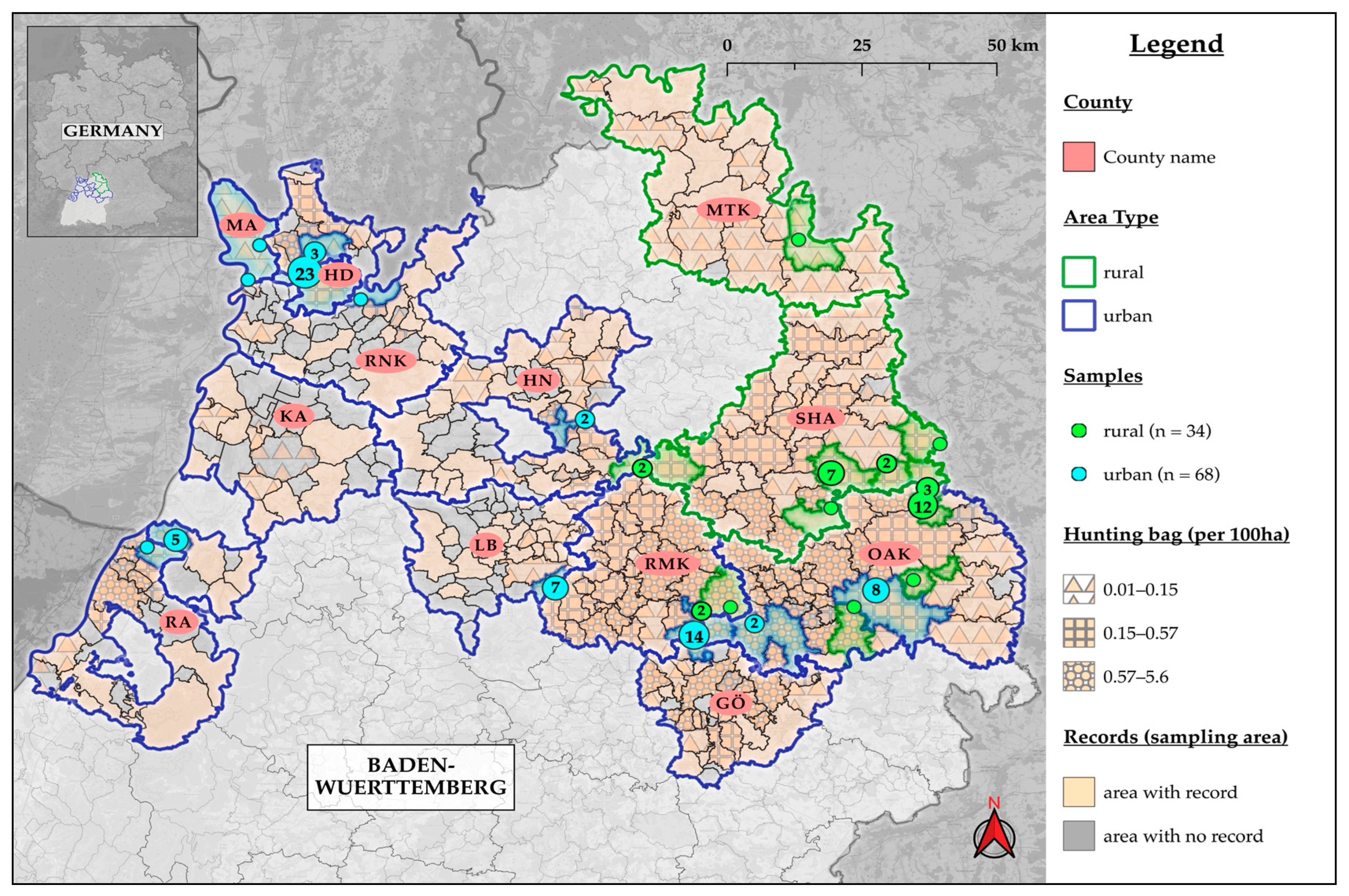
2.1. Sample Collection

We collected 102 free-ranging raccoons to analyze them for intestinal helminths. The collection of animals was between August 2019 and November 2020 during the hunting season for raccoons in BW (§10 DVO JWMG 2015, of 25 February 2018), except for one individual that had already been road-killed in May. The sampling area covered 27 municipalities in 12 counties in the northern part of BW. These counties included the City of Heidelberg (HD) and Mannheim (MA) as well as the districts Ostalbkreis (OAK; Ellenberg, Westhausen, Aalen, Lorch, Essingen, Schwäbisch Gmünd), Schwäbisch Hall (SHA; Fichtenau, Frankenhardt, Stimpfach, Bühlerzell, Kreßberg, Mainhardt), Rems-Murr-Kreis (RMK; Alfdorf, Plüderhausen), Karlsruhe (KA; Rheinstetten), Ludwigsburg (LB; Remseck am Neckar), Main-Tauber-Kreis (MTK; Weikersheim), Heilbronn (HN; Weinsberg, Wüstenrot), Rhein-Neckar-Kreis (RNK; Schriesheim, Dossenheim, Neckargemünd), Rastatt (RA; Durmersheim), Göppingen (GÖ; Börtlingen, Birenbach) (Figure 1). More than ten raccoons were collected in each of four counties (HD, OAK, SHA, RMK), five to nine from KA and LB, and less than five animals each from MTK, GÖ, HN, RNK, MA, and RA. The municipalities in which the hunting sites are located were classified as rural or urban areas based on the Federal Office for Building and Regional Planning (BBSR) classification [32,33]. The carcasses were stored at −20 °C until necropsy. No ethical approval was required for this study.

2.2. Necropsy

The necropsy of the carcasses was executed under BSL3 conditions at the Parasitology Unit of the University of Hohenheim. We evaluated the carcasses as fresh, minimally, or moderately autolytic. Heavily autolytic raccoons or incomplete animals were excluded. The animals’ weight, length, and sex were documented. Their age was determined as either adult (>1-year-old) or juvenile (<1-year-old) based on various factors such as tooth development and wear, weight and length, reproductive status, and hunting date [9].
We removed the small intestine and opened it longitudinally. Macroscopically visible helminths were collected and preselected in nematodes, cestodes, and trematodes. The smaller helminths were extracted by the segmented sedimentation and counting technique (SSCT) and detected by microscopy [34]. Nematodes and cestodes were stored in distilled water and then rapidly frozen at −80 °C. Trematodes were preserved in 80% ethanol. Fecal samples from the rectum were collected and stored at −80 °C for five days and subsequently transferred to −20 °C. The diaphragm pillar samples of the 101 dissected raccoons were examined for Trichinella spp. during necropsy by the compression method. Additionally, from a randomly selected 52 out of 101 raccoons, muscle samples of about 5 to 10 g of the diaphragm pillar and upper foreleg were collected and stored at −20 °C for further examination by the artificial digestion method at Aulendorf State Veterinary Diagnostic Centre (STUA, Germany, Aulendorf).

2.3. Parasitic Examinations

We identified nematode and cestode species by morphological molecular methods, whereas trematodes were assigned to a species exclusively based on morphology.
For morphological assessment and species identification, the trematodes were stained using Malzacher stain [35]. The prefixed trematodes were placed in alcoholic borax carmine, according to Grenacher, for 10 to 15 min, which stained the organs red. The stain was removed, and distilled water was applied to the trematodes for a differentiation step. As soon as the parenchyma was still slightly pink and the organs were red, the distilled water was removed, and astral blue was added. The predefined exposure time of ten minutes proved to be too long for small trematodes and was reduced to four minutes. The subsequent differentiation step also took longer and was adjusted in time to 15 to 20 min. Once the blue color had faded, the process continued. The stained objects were then dehydrated in an ascending ethanol series (50%, 70%, 90%, 95%, and 100%) for 10 min each. Trematodes were then placed in wintergreen oil, a methyl salicylic acid ester, for 30 min to make them more transparent. Finally, the trematodes were transferred on a slide into the inclusion medium, resin (Entellan® new). After at least two days of hardening, the objects were measured and characterized according to Schell, S.C. [36] and Seo et al. [37] using photo-binoculars (ZEISS Axio Imager 2; Carl Zeiss Microscopy GmbH, Jena, Germany).
At STUA Aulendorf, 3 to 5 g of the collected feces per raccoon were tested for parasite eggs and larvae using the in-house flotation method followed by microscopical examination. A zinc chloride/saline solution (ZnCl2/NaCl) with a density of 1.35 g/mL was used for processing. Furthermore, 3–6 g per raccoon of the collected 52 muscle samples (diaphragm pillar, upper foreleg) were examined for Trichinella spp. at STUA Aulendorf using the artificial digestion methods according to the EU Regulation 2015/1375. To increase the test’s sensitivity, the samples were digested for 60 min and then sedimented for 40 min in the first step and 105 min in the second step. Then, 12 mL of sediment were diluted with 10 mL of water and examined for Trichinella spp. using the trichinoscope at 40–100 times magnification. In addition, 67 randomly selected fecal samples were tested for ascarids, hookworms, and whipworms using the ELISA-based PetChekTM IP test kit (IDEXX, Kornwestheim, Germany) [38].
The intensity of infection, based on the number of parasite stadia found by visual examination during necropsy, was divided into three infestation groups: minor (≤5 cestodes, ≤5 nematodes, ≤5 trematodes), moderate (≤30 cestodes, ≤15 nematodes, ≤10 trematodes), and heavy infestation (>30 cestodes, >15 nematodes, >10 trematodes).

2.4. Nematode and Cestode Identification by PCR

2.4.1. Sample Preparation

Small pieces of tissue (0.3–0.5 mm3) from each of the collected nematodes and cestodes were transferred to 200 μL PCR tubes containing 20–25 μL of 0.02 M NaOH and heated to 99 °C for 15 min [39]. The resulting lysate could be used directly as a template for the subsequent PCR assays.

2.4.2. DNA Amplification and Sequencing

For species identification, the complete or partial mitochondrial (mt) NADH dehydrogenase subunit-1 (nad1) of the nematode and cestode samples were analyzed, and in the case of cestodes, an additional fragment of the mt cytochrome-c-oxidase subunit-1 (cox1). Nested PCRs were carried out with all samples; the primer combinations can be seen in Table 1.
The reaction volume for each sample was 25 µL for the first PCR and 50 µL for the nested PCR. The reaction mixtures consisted of 10 mM Tris–HCl (pH 8.3), 50 mM KCl, 2 mM MgCl2, 200 µM of each dNTP, 0.2 µM of each forward and reverse primer, and Taq polymerase (0.625 U for the first PCR, 1.25 U for nested PCR). As a template for the first PCR, 1 µL of the lysate was added to the reaction mixture, and for the nested, 1 µL of the primary PCR product was added. The PCRs were performed with an initial denaturation step at 95 °C (5 min), followed by 35 cycles of denaturation at 95 °C (30 s), annealing at 50–55 °C (30 s), elongation at 72 °C (1 min), and a final elongation step at 72 °C (5 min).
PCR results were detected on a 1.5% agarose gel stained with GelRedTM. PCR products were purified with the High Pure PCR Purification Kit (Roche, Mannheim, Germany) according to the manufacturer’s instructions and sent to Microsynth Seqlab GmbH (Göttingen, Germany) for sequencing. The electropherograms obtained by Sanger sequencing were analyzed using GENtle (Cologne, Germany) [40]. Sequences were compared with reference entries in the NCBI GenBank for species identification using the NCBI basic local alignment search tool (BLAST).

2.5. Statistics and Graphics
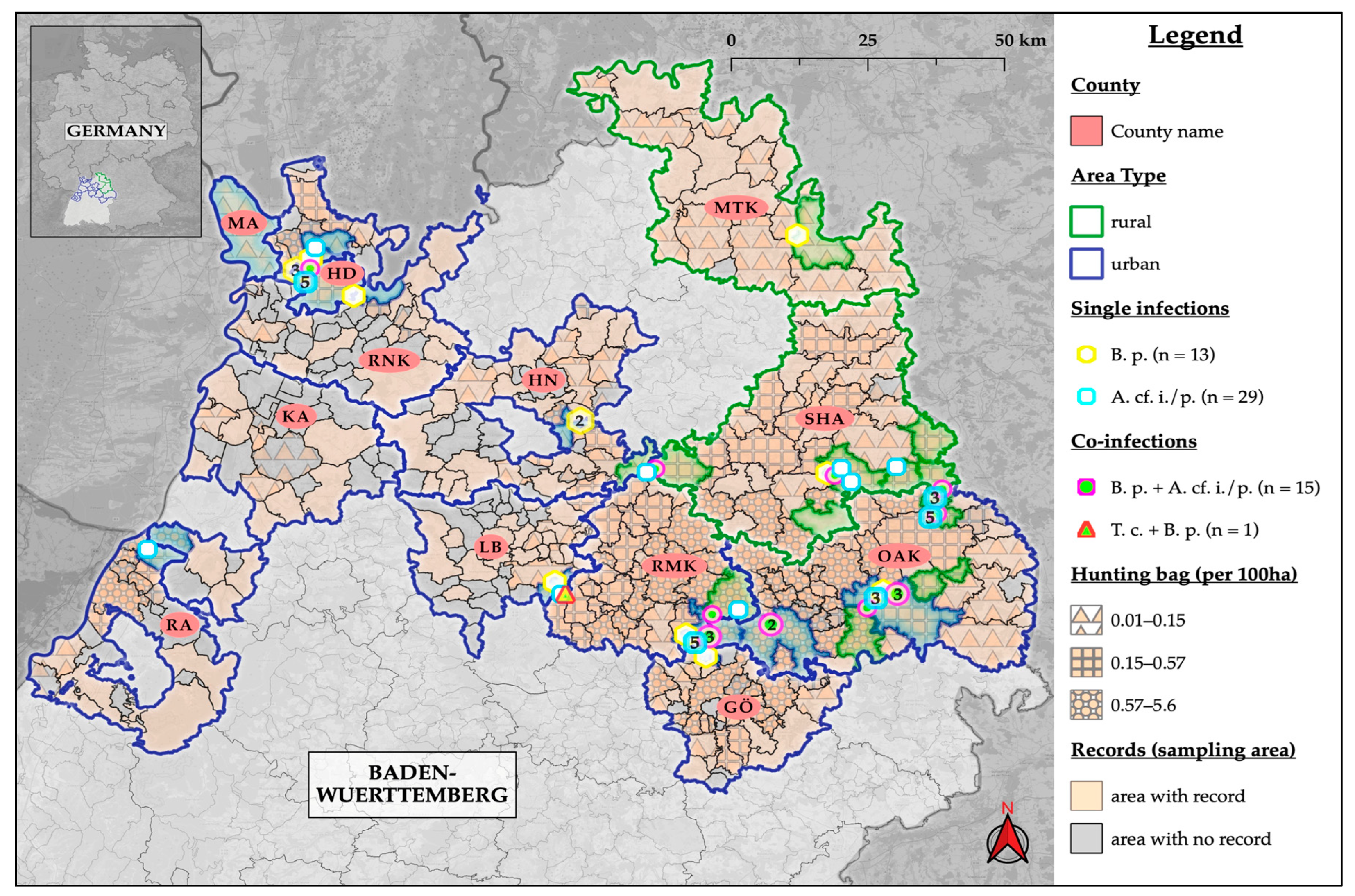
Microsoft® Office Excel 16.63 was utilized for data management, graphics, and tables. QGIS 3.22 was used to create maps based on data on the occurrence of raccoons in each municipality provided by the Wildlife Research Center of the Agricultural Center Baden-Württemberg (WFS; LAZBW, Aulendorf, Germany). Statistical analyses were carried out using R (version 4.1.2) and RStudio (1 March 2023), but only for sample sizes ≥ 10 to ensure statistical validity. The prevalence of helminths in raccoons was calculated for the entire sample, as well as proportionally for age, sex, age–sex cohort, hunting year, and area type (rural vs. urban), along with the 95% Clopper–Pearson confidence intervals. Fisher’s exact test was used to determine any dependencies between the categories and a positive result for significant correlations. Differences in infestation levels within the category types (sex, age, area type) were calculated with the Kruskal–Wallis rank sum test. The Mann–Whitney U (MWU) test was used to compare body weight and length values between negative and positive results. A significance level of p ≤ 0.05 with a Bonferroni α adjustment was set for the statistical tests, and any significant effects are denoted with an asterisk (*).

3. Results

We found that 72.3% of the 101 raccoons (n = 73; 95% CI: 62.5–80.7) were positive for at least one helminth parasite in the examinations performed (Table 2). Cestodes represented the most common helminth group with 46.5% positive raccoons (n = 47; 95% CI: 36.5–56.7), ahead of nematodes with 31.7% (n = 32; 95% CI: 22.8–41.7) and trematodes with 15.8% positive individuals (n = 16; 95% CI: 9.3–24.4). Helminths detected in at least four raccoons are listed in Table 2. During the necropsy, 102 animals were examined, of which 80.4% (n = 82) were determined to be fresh, 12.7% (n = 13) were classified as minimally autolytic, and 5.9% (n = 6) were moderately autolytic. None of the collected animals was found to be heavily autolytic. The intestine of raccoon WB40 (male, adult, rural, Kreßberg, SHA) was incomplete due to hunting. Therefore, this raccoon was excluded from this study. This individual was examined exclusively by flotation; no parasites were found. When the sampled communities were classified as either urban or rural, it was found that 67.3% (n = 68) of the 101 raccoons were from urban areas, while 32.7% (n = 33) were from rural areas (Table 2 and Figure 1). Although detailed morphometric data were collected during the necropsy, they will not be further discussed since no significant correlation with parasite infection was identified.

3.1. Nematodes

Nematodes were detected and identified in the intestines of 31.7% of 101 raccoons by the SSCT following PCR, the flotation technique, and PetChekTM IP (IDEXX, Kornwestheim, Germany). Of these, 29 raccoons (28.7%; 95% CI: 20.1–38.6) were infected with Baylisascaris (B.) procyonis, one of them also with Toxocara (T.) canis (0.99%; 95% CI: 0.03–5.4) (Figure 2). Additionally, one raccoon with Capillaria spp. (0.99%; 95% CI: 0.03–5.4) and two with Trichuris spp. (1.98%; 95% CI: 0.2–7) were found.
By visual examination during necropsy and the SSCT, nematodes were identified in 29 raccoons. For each of the 29 infected raccoons, at least one of the examined nematodes could be assigned to a parasite species by sequencing. Baylisascaris procyonis was found in 28 animals and T. canis in one individual (WB64, male, adult, urban, Remseck am Neckar, LB). Ascarid eggs found in the T. canis-positive raccoon (WB64) were identified as B. procyonis by sequencing. Hence, raccoon WB64 had a dual infection with T. canis and B. procyonis. All nad1 fragments showed 100% identity to the B. procyonis sequence JF951366 [41], except for the one nematode of WB64 (195 bp), which showed 99% similarity to a reference sequence from T. canis MN635719 [42].
Flotation detected ascarid eggs in ten raccoons that were also positive for other B. procyonis stadia. A single infection with Capillaria spp. was recorded exclusively in the flotation for WB67 (male, adult, urban, HD). Moreover, 24 of the 67 raccoons tested for ascarids with PetChekTMIP ELISA (IDEXX, Kornwestheim, Germany) were also positive for B. procyonis in the PCR assays. Only 8 of the 24 B. procyonis-positive raccoons were thus confirmed in the ELISA. In addition, Trichuris spp. were detected in two raccoons exclusively by PetChekTMIP ELISA (IDEXX, Kornwestheim, Germany). One of these (WB19, rural) was a juvenile female raccoon from Frankenhardt, SHA, and the other was a juvenile male individual from Lorch, OAK (WB96, urban).
No Trichinella spp. were found in the examinations using the artificial digestion or compression method. No significant dependencies of the tested categories with Fisher’s exact test between the positive and negative nematode groups could be found.

3.2. Cestodes

A total of 46.5% cestode-positive raccoons were exclusively detected by visual examination during necropsy and the SSCT (Table 2). The cestodes were determined based on morphological characteristics, and to verify these we used the described PCR tests with subsequent sequencing. Of the 60 cestode specimens analyzed via sequencing, 38 yielded sequences that could be evaluated.
We detected Atriotaenia (A.) cf. incisa/procyonis in 43.6% (n = 44; 95% CI: 33.7–53.8) of the 101 examined raccoons (Table 2 and Figure 2). The morphological examination allowed the identification of the cestodes as Atriotaenia spp. This was performed using written descriptions of the morphology of Atriotaenia spp., such as the characteristics of the scoleces [43]. No cestode other than A. incisa or A. procyonis, which resembles this genus, is known to infect raccoons. Of the 38 yielded gene sequences, 36 were identical to each other, including those of the morphologically determined cestodes. Consequently, all 36 identical sequences were assigned to either A. incisa or A. procyonis. The comparison of the most frequently encountered sequences with the references in the NCBI GenBank did not allow species identification. The approximately 730 bp-long cox1 gene fragment showed the highest similarity with 82.8% to a Dipylidium caninum sequence (NC021145). Therefore, the complete nad1 gene was also analyzed from some of these samples. This 900 bp-long sequence had the highest match of 87.9% with Taenia crassiceps (NC002547). Since genetic determination was not successful and it is almost impossible to distinguish morphologically between A. procyonis and A. incisa, the name A. cf. incisa/procyonis is used here [43]. The complete nad1 and partial cox1 gene A. cf. incisa/procyonis gene sequences were submitted to the NCBI GenBank under the Accession Numbers OR001745 for nad1 and OR039311 for cox1. Cestodes in the total and A. cf. incisa/procyonis-positive raccoons originated more frequently from rural than urban areas with a significance of p = 0.02. No other significant dependencies of the tested categories with Fisher’s exact test between the positive and negative cestode groups could be found.
Taenia (T.) martis was found in 1.98% (n = 2; 95% CI: 0.2–7) of the 101 raccoons, of which WB37 (juvenile, male, urban, Aalen, OAK) was also infected with A. cf. incisa/procyonis. The other T. martis-positive raccoon was a male adult individual from a rural area (Bühlerzell, SHA; WB30). Species identification was performed by molecular means. Small fragments of the cox1 (118 bp) and nad1 (51 bp) genes were obtained. They were 100% congruent with the reference sequence of T. martis AB731758 [39]. Although the fragments were short, both could thus be clearly assigned to this species. Furthermore, we found Mesocestoides spp. in one (0.99%; 95% CI: 0.03–5.4) of the A. cf. incisa/procyonis-positive raccoons (WB93, female, adult, rural, SHA, Frankenhardt). Four scoleces were isolated from this raccoon and could be identified as Mesocestoides spp. based on their size and characteristic morphology. Neither the nested PCR with the cox1 nor the nad1 primers yielded evaluable results. Cestodes from two male adult raccoons (WB18 (rural, Frankenhardt, SHA) and WB94 (urban, Lorch, OAK)) could not be attributed morphologically or genetically to a specific species. No Echinococcus spp. were detected by the SSCT.

3.3. Trematodes

Trematodes were detected in 15.8% of the raccoons (n = 16; 95% CI: 9.3–24.4) by the SSCT (n = 14) and flotation (n = 2). Adult trematodes of half of the 14 positive raccoons were morphologically assigned to a genus or specific species, while this was not possible for the other 7 due to the poor condition of the worms. Two trematode infections were exclusively detected through egg flotation (WB52 and WB75), and the causative species, therefore, were not determined. The trematodes of 4% of the examined raccoons (n = 4; 95% CI: 1.1–9.8) were assigned to Isthmiophora (I.) hortensis/melis. Without genetic differentiation, I. melis and I. hortensis cannot be distinguished from each other. Two raccoons were male and adult (WB08, WB100) and two were male juvenile individuals (WB09, WB102; Table 2). Raccoons WB08 and WB09 originated from KA (Rheinstetten) and WB100 (Ellenberg), and WB102 (Lorch) from OAK. The latter two raccoons had a heavy infestation of I. hortensis/melis, with 30 individuals each. Raccoon WB10 (male, adult, urban) from Rheinstetten, KA was infected with Plagiorchis muris (0.99%; 95% CI: 0.03–5.4). In addition, Brachylaima spp. were detected in 1.98% of raccoons (n = 2; 95% CI: 0.2–7). One individual originated from Aalen, OAK (male, juvenile, urban, WB36) and the other animal from Weinsberg, HN (female, adult, urban, WB74). No significant dependencies of the tested categories with Fisher’s exact test between the positive and negative trematode groups could be found.

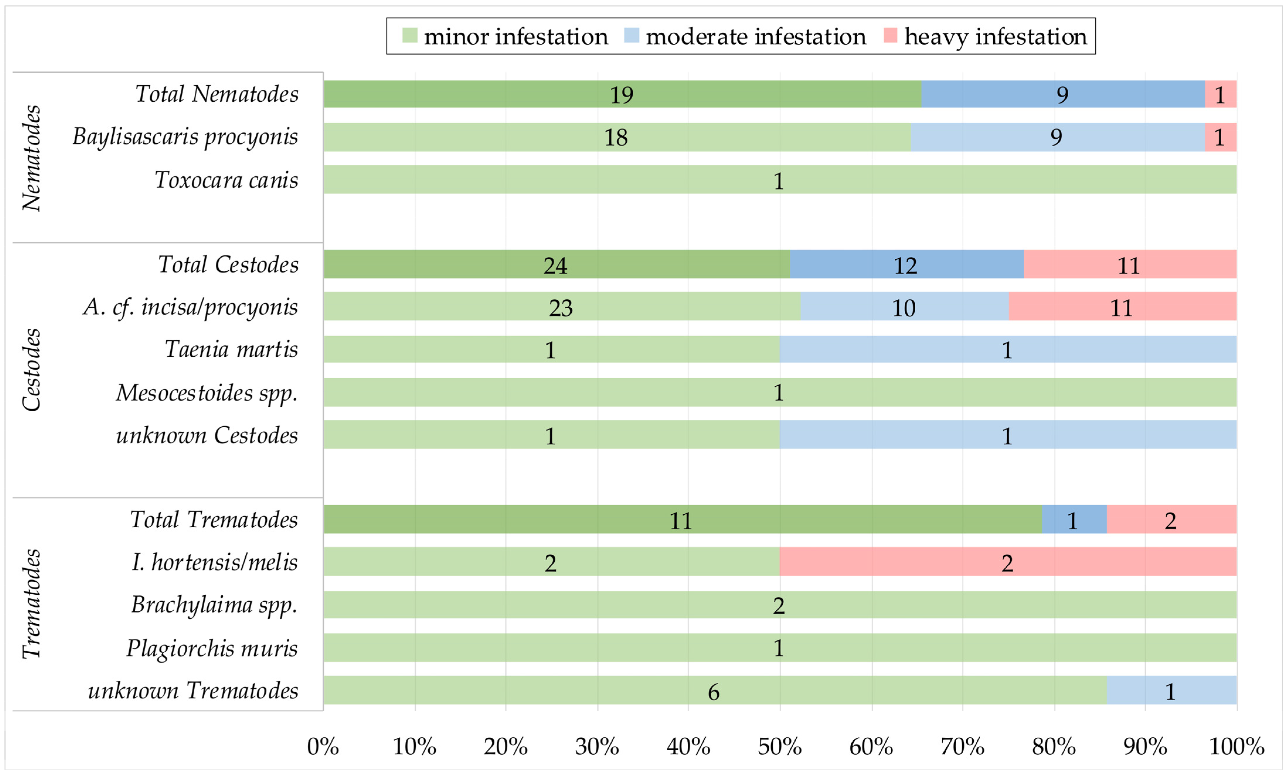
3.4. Intensity of Infestation and Co-Infections

Based on visual examination during necropsy and the SSCT, the infestation levels of the individual helminths could be determined (Figure 3). Thus, of the 29 raccoons infested with nematodes, 65.5% had minor (n = 19; 95% CI: 45.7–82.1), 31% had moderate (n = 9; 95% CI: 15.3–50.8), and 3.4% had a heavy infestation (n = 1; 95% CI: 0.1–17.8). Of the 47 cestode-positive raccoons, 51.1% were minorly (n = 24; 95% CI: 36.1–65.9), 25.5% were moderately (n = 12; 95% CI: 13.9–40.3), and 23.4% were heavily infested (n = 11; 95% CI: 12.3–38.0). Of the 14 raccoons with trematodes, 78.6% had a minor infestation (n = 13; 95% CI: 49.2–95.3), 7.1% were moderately (n = 1; 95% CI: 0.2–33.9), and 14.3% were heavily infested (n = 2; 95% CI: 1.8–42.8) (Figure 3). No significant differences in infestation levels within the categories (sex, age, area type) were seen with the Kruskal–Wallis rank sum test.
Co-infections were detected in 32.9% of the 73 positive raccoons (n = 24; 95% CI: 22.3–44.9) (Table 3). The largest proportion, 21.9% of the positive raccoons (n = 16; 95% CI: 13.1–33.1), had a dual infection with nematodes and cestodes (Table 3). In two cases each, we found dual infections with nematodes and cestodes and with cestodes and trematodes. One raccoon, WB36 (female, juvenile, urban, Aalen, OAK), had a triple infection with all three parasite groups (Table 3).

4. Discussion

This study aimed to systematically investigate the occurrence of helminths of raccoons in BW, as no such screening had been carried out in the southwestern region of Germany. We found that 72.3% of 101 free-ranging raccoons were positive for at least one helminth parasite in the examinations performed (Table 2). Among the helminths detected, there were positive results for species of four nematode genera (B. procyonis, T. canis, Capillaria spp., and Trichuris spp.), three cestode genera (A. cf. incisa/procyonis, T. martis and Mesocestoides spp.), and three trematode genera (I. hortensis/melis, Plagiorchis muris, and Brachylaima spp.). The detected parasite diversity and presence of six potentially zoonotic helminths are relevant considering the One Health aspect. According to our findings, the raccoons’ propensity for spreading and synanthropic habits may lead to environmental pollution in BW with pathogens, including helminths [9]. Consequently, raccoons may serve as a transmission link between various animal groups, including wildlife, domestic and zoo animals, and humans, thereby elevating the risk of infection [3,6,12,14].

4.1. Methodological Limitations

The acquisition of raccoons was exclusively conducted in the northern half of BW, where the population density and hunting activity are highest (Figure 1). Therefore, our results are not representative of the entire region of southwestern Germany. The spatial and temporal distribution of raccoon carcasses and their submission may be biased due to complex biological factors (e.g., raccoon population dynamics) and human-related factors (e.g., hunters’ commitment in respective areas) [44]. To provide an overview of helminths in the raccoon population of BW, we restricted our sample size to approximately 100 animals. Based on a hunting bag of 4015 raccoons during the 2019/2020 hunting season, the estimated raccoon population in BW is estimated to be 40,000 animals [13,15]. With a sample size of 101 raccoons, only 3.3% of the hunting bag and 0.3% of the estimated population in BW were represented [9]. Higher numbers are required for accurate prevalence and population estimates. Two-thirds of the sampled raccoons were from urban areas, which is not surprising given the high raccoon densities in such areas and the densely populated sample area (Figure 1) [9]. The categorization of raccoons into urban and rural areas is a simplified classification, as it only considers the hunting community and not the individual raccoon’s spatial usage [9]. Home range sizes can vary based on factors such as season, habitat and area type (urban > rural), sex (male > female), and age (adult > juvenile) [21,23].
Helminth prevalence may be underestimated due to the natural decomposition processes of intestinal parasites. The freezing and thawing of the carcasses and samples may have damaged or even destroyed less persistent parasites such as flukes, small tapeworms [45], and some Trichinella spp. [46], leading to an underestimation of the overall infestation rate. For some cestodes and trematodes, the genera were determined exclusively by morphological and parasitological examinations. In this context, it should be mentioned that these are not completely reliable, and a genetic identification is preferred. To detect intestinal nematodes in raccoons, we used a combination of three methodological approaches (visual examination/SSCT, flotation, and ELISA-based PetChekTM IP) to obtain the most accurate picture and prevalence of the nematode fauna [8,47,48], of which the ELISA test was only performed in 67 of the 101 raccoons. The intestinal collection of nematodes is the gold standard to obtain reliable prevalence values, including prepatent infections [8,48]. Prepatent infections, however, are not detected in fecal egg flotations, which can lead to an underestimation of the true prevalence [8,27,48]. In addition, male-only infections, insufficient fecal material, irregular shedding, and fluctuating amounts of eggs, as is the case in many intestinal helminths, e.g., Baylisascaris spp., can lead to false-negative results in the flotation [8,27]. Of the B. procyonis-positive raccoons by visual examination and the SSCT, only 35.7% (10/28) were also positive by egg flotation; thus, 64.3% of the positive samples remained undetected. The species of ascarid eggs in the flotation was not corroborated by PCR in the samples with B. procyonis individuals detected by visual examination and the SSCT.
The ELISA-based PetChekTM IP assay detects antigens of (pre-)patent stadia of ascarids, hookworms, and whipworms in feces; however, it has not yet been validated for detecting Baylisascaris spp. [8,38]. The eight raccoons positive for ascarids detected by this ELISA were also positive for B. procyonis in the PCR assays. Thus, it is reasonable to assume that the ELISA is able to identify B. procyonis. However, 16 samples with B. procyonis could not be confirmed by this ELISA.
Hence, it is evident that the selection of methods can significantly impact the prevalence, which must be considered when evaluating them.

4.2. Nematodes

4.2.1. Baylisascaris procyonis

Baylisascaris procyonis was found in 28.7% of 101 examined raccoons, making it the second most prevalent helminth species in this study. Previously reported prevalence values for B. procyonis in Germany vary significantly, ranging from 0% to 95% depending on the geographical region [10,16,25,49,50]. Raccoon populations in central Germany, including urban areas, especially exhibit remarkably high prevalence rates [10,25,49]. Prevalence rates in neighboring countries show considerable differences but tend to be lower than those in Germany [18,47,51,52]. The prevalence variations of B. procyonis in endemic areas are influenced by the migrated raccoons from founder populations, with additional ecological or geographical factors potentially playing a role [16,50]. The population growth and spatial expansion led to the mixing of founder populations and consequently to the spread of B. procyonis [17,30]. It is described that in BW, raccoons from the Hesse, Harz, and Rhineland-Palatinate regions had mixed, with a high prevalence of B. procyonis reported in Hessian and Harz raccoons [13,16,17,53]. Given that the raccoon population is in the early stages of spreading in BW, it can be assumed that B. procyonis prevalence rates will continue to increase. A total of 64.3% of the 28 raccoons that tested positive in the visual examinations and SSCT showed a mild infestation, 32.1% had a moderate infestation, and a heavy infection was observed in only 3.7% of the raccoons (Figure 3). These results contradict previous studies from Germany, which often reported heavy or moderate infestations [10,25]. However, it should be noted that the categorization criteria for infestation severity may differ between studies. Although many publications identified influencing factors, such as age, sex, land use, area type or season [8,20,22,54,55,56], this could not be detected in the present study.
The genus Baylisascaris comprises ten species, with B. procyonis being considered the most pathogenic in intermediate and accidental hosts [22,25]. Baylisascaris procyonis is an emerging zoonotic pathogen with an indirect life cycle involving raccoons as the final hosts, typically resulting in subclinical infections [2,57,58,59]. In addition, there are more than 130 intermediate or accidental hosts (rodents, birds, domestic animals, wild carnivores, primates, and humans) of this parasite, which can occasionally develop severe clinical signs resulting from visceral, ocular, or neural larva migrans syndrome [27,57,58]. Raccoons infected with B. procyonis can excrete over 200,000 EPG (eggs per gram of feces), which become infectious after 14 days and can remain infective in the environment for years under suitable moisture conditions [25,55,57,58]. Raccoons defecating on communal latrines, which are visited by various potential intermediate hosts and are an essential and persistent source of B. procyonis infections, lead to environmental contamination with this parasite [25,26,27]. Furthermore, it should be noted that domestic dogs not only serve as intermediate hosts for B. procyonis but can also act as alternative final hosts [4,8,27,58]. Therefore, in areas with high B. procyonis prevalence, dogs may pose a potential public health risk and may extend the range of environmental contamination [5,8,58]. Additionally, it should be noted that B. procyonis can negatively impact the populations of potentially endangered intermediate hosts, contributing to biodiversity loss [5,8,25,27,53,58]. In many zoos, B. procyonis infections are of increasing concern for various animal species [52,60,61]. The exposure to B. procyonis in zoological facilities appears to be real in BW, as indicated by the detection of B. procyonis eggs in the latrines of one out of six raccoon enclosures tested using the flotation method (Reinhardt, N. P.; unpublished). Zoological institutions in raccoon distribution areas should be aware of the presence of B. procyonis and implement prevention measures accordingly [25,52,60,61]. Baylisascaris procyonis, with its potential to cause severe or fatal infections, particularly in children, poses a public health risk [27] that is underestimated in Germany, according to Peter et al. [10]. Nevertheless, cases of Baylisascariasis in humans are rare both in Europe and North America [27,58]. However, serological investigations indicate that subclinical infections may frequently occur in adults in B. procyonis areas, suggesting many unreported cases [27,62].
In summary, B. procyonis poses a potential One Health threat as it can have a negative health impact on humans and a variety of domestic, zoo, and wildlife species [4,8,27,58,59,61]. In a preventive approach, high raccoon densities should be considered a risk factor; therefore, identifying B. procyonis risk areas is highly beneficial [16,27].

4.2.2. Toxocara canis, Capillaria spp. and Trichuris spp.

We detected T. canis in the intestine of one raccoon, which is the second description of this nematode in raccoons and the first detection by PCR and Sanger sequencing. Davidson et al. [63] morphologically identified eggs of Toxocara spp. and Toxascaris spp. in the feces of two Norwegian raccoons. The ascarid eggs collected from the T. canis-positive raccoon in the flotation were examined using PCR, revealing the presence of B. procyonis and no positive result for T. canis eggs. This could be attributed to a non-patent T. canis infection or the absence of T. canis eggs in the sample used for the flotation and PCR analysis. Consequently, the raccoon’s status as a final host for T. canis could not be confirmed with certainty. Nevertheless, the possibility should be considered that raccoons occurring in close proximity to domestic dogs or red foxes (Vulpes vulpes) may be involved in disseminating T. canis. In dogs, T. canis is one of the most common intestinal parasites and is also pathogenic to humans [64]. Fecal examinations in Germany revealed a prevalence of 6.1% in dogs (N = 24,677), with a higher prevalence observed in animals up to six months, being twice as high [65]. In German red foxes, prevalence rates of 43.8% (N = 80) have been reported, making it one of the most commonly detected endoparasites in this species [11]. An infection with Toxocara spp. in intermediate hosts follows a nearly equivalent course to Baylisascaris spp., although the latter more frequently leads to severe or fatal neural damage [25,27]. Toxocariasis is the most common helminthic zoonosis in industrialized countries, with a human seroprevalence of 6.2% in Europe and 4.8% in Germany [27,64,66]. Peak values of 32.7% were recorded in Ireland and 27.4% in France [66].
We detected eggs of Capillaria spp. in one raccoon and Trichuris spp. using the ELISA PetChekTM IP assay (IDEXX, Kornwestheim, Germany) in two raccoons’ fecal samples. Both genera have been previously detected in raccoons in Germany, albeit with low prevalence [10,31,47].
No Trichinella spp. were found in this study. In Germany, several investigations have been conducted on Trichinella spp. in raccoons, but only immunological detection in meat juice has been successful [10,67,68]. This nematode is globally distributed and is regularly found in carnivores and wild boars in Germany [67,68,69]. Trichinella spp. have been detected in raccoons in Europe, North America, and Japan [67,68].

4.3. Cestodes

In this study, A. cf. incisa/procyonis was the most commonly detected helminth, with a prevalence of 43.6% among the 101 examined raccoons. The species allocation is uncertain. In total, five species of Atriotaenia have been described, four from American procyonids, mustelids, and bats [70]. Relevant here are A. procyonis from North American raccoons and A. incisa from European badgers. Both species are morphologically indistinguishable and possibly conspecific [43]. In the absence of any gene sequence data prior to our study, we are not in a position to decide whether the taxon we found in our raccoons had been translocated from North America with its host (as with B. procyonis), or whether the indigenous badger parasite described as A. incisa has expanded its host range to raccoons. Genetic data from North America and Europe will be necessary to draw a conclusion from this. The parasite burden in the affected raccoons is higher compared to all the other parasites found, with 25% of the 44 positive raccoons (n = 11; 95% CI: 13.2–40.3) being heavily infected and 22.7% (n = 10; 95% CI: 11.5–37.8) moderately infected (Figure 3). These high parasite burdens are consistent with previous investigations in Germany [43]. In Europe, Atriotaenia spp. has been repeatedly detected in raccoons, with the recent prevalence ranging from 12.0% (N = 175) in Germany to 19.7% (N = 234) in Poland [10,18]. In comparison, the prevalence detected here is more than twice as high. The high prevalence and infestation rates found in this and previous studies in Germany suggest that raccoons are important hosts of this parasite [10]. Significantly higher prevalence rates of up to 86.9% (N = 35) have been reported from raccoons in North America [29,71,72]. There have been relatively few studies on A. cf. incisa/procyonis, so little is known about its life cycle and pathogenicity [73]. Cyclophyllidean cestodes require intermediate hosts for their development, in the case of Atriotaenia sp. most probably coprophagous beetles [72]. Due to raccoons consuming beetles throughout the year, the likelihood of infection is relatively high [20]. Statistically, it was determined that a significantly higher proportion of A. cf. incisa/procyonis-positive raccoons originated from rural areas. This could be associated with a presumed higher abundance of intermediate hosts in rural areas and the increased utilization of anthropogenic food sources by raccoons in urban areas, which can decrease the diversity of parasites with indirect life cycles [71,74].
We identified T. martis for the first time in raccoons. Taenia martis is widely distributed in Europe and commonly infects the intestines of various species of Mustelidae [75,76]. In southern Germany, prevalence rates of T. martis were found to be 36% (N = 47) in stone martens (Martes foina) and 48% (N = 437) in muskrats (Ondatra zibethicus) [77]. Whether raccoons could be a new possible final host of T. martis with patent infections is unclear since both individuals were immature and only about 3 cm long. However, the fact that the parasite was found in two raccoons from different counties (SHA, OAK) may indicate a broader distribution in the raccoon population of BW. Other taeniids such as Hydatigera sp., T. crassiceps, and T. pisiformis have already been detected in raccoons [71,78,79]. Taenia martis is considered an emerging infectious pathogen because it causes eye and brain infections in humans and can induce severe to fatal infections in nonhuman primates [75,76,79]. It should be noted that out of four described human cases worldwide, three have occurred in Germany [79].
Mesocestoides spp. could only be detected at the genus level in one raccoon in this study. The accurate species identification of Mesocestoides spp. is difficult due to taxonomic uncertainties within this genus and should, at best, be performed both morphologically and molecularly [80]. In German raccoons, a prevalence of 2.6% (N = 234) to 3.5% (N = 762) has been described so far [10,50]. In comparison, a study from Poland showed that a higher prevalence of 67% (N = 55) can occur [47]. This may indicate that raccoons could likely be final hosts of Mesocestoides spp. This genus is distributed worldwide in various carnivorous final hosts and, in individual cases, can also be transmitted to humans [79,80]. Common second intermediate hosts are small mammals, birds, reptiles, and amphibians [47,80], which are also essential sources of feed for raccoons [20].
Comparing the prevalence of the cestode genera found, it should be noted that cestodes using small mammals as intermediate hosts (T. martis, Mesocestoides spp.) are much less common. Another important zoonotic parasite with such a heteroxenous life cycle, Echinococcus spp., was not detected. By now, no Echinococcus sp. has been described in raccoons, either in North America or elsewhere. Considering the high prevalence of Echinococcus multilocularis in red foxes in our study area, the involvement of raccoons in the life cycle of this parasite appears to be negligible [19,81].

4.4. Trematodes

Raccoons have been found to harbor numerous species of trematodes. The use of water-rich environments makes infections with digenetic trematodes likely because raccoons in these habitats may feed on insects, amphibians, mollusks, and fish, all of which are known intermediate hosts of trematodes [20,82]. The low number of trematode infections observed in this study is consistent with the low infection rates in Hessian and Bavarian raccoons, reported by Peter et al. [10]. Several explanations exist, such as the possibility that raccoons serve only as accidental hosts or that trematodes pass through their digestive tract without maturing. Differences in habitat use, such as less use of or access to water-rich areas and thus less contact with potentially infected intermediate hosts, may also play a role. In addition, there are methodological limitations, as described earlier, since flukes quickly decompose through freezing and thawing processes, rendering them undetectable.
Among the examined raccoons, 4% were found to harbor the species I. hortensis/melis. In this project, the species I. melis and I. hortensis were grouped due to their close morphological resemblance, making it impossible to differentiate them without using genetic techniques [83]. Morphologically identifying Isthmiophora spp. is challenging because they can vary in morphology within and between species depending on the host [84]. Unlike other representatives in the Echinostomatidae family, Isthmiophora spp. exclusively infest mammals’ intestinal tract during their adult phase [85]. So far, over 30 species of vertebrates, including humans, have been identified as final hosts [84,85]. Both I. melis and I. hortensis have been found in the intestines of raccoons, wild boars (Sus scrofa), raccoon dogs (Nyctereutes procyonoides), and other wild animals [84,85]. In central Europe, the prevalence of Isthmiophora spp. infections in raccoons has been reported to range from 4.8% (N = 62; Czechia) to 50.3% (N = 175; Germany and Poland) [18].
In 1% of the examined raccoons, another potentially human pathogenic trematode species, Plagiorchis muris, commonly found in freshwater ecosystems across Europe, was identified based on its morphology [86]. Plagiorchis muris has previously been detected in raccoons, including in Germany, with a prevalence of 0.9% (N = 234) [10,78,86]. This parasite is typically found in the small intestine of rodents, birds, reptiles, insectivores, and omnivores [87]. Although human infections have been described, the risk for humans is minimal due to the trophic transmission primarily through raw fish [87].
Morphologically, Brachylaima spp. were also identified in almost 2% of the raccoons. This genus has been frequently described in raccoons, with a prevalence of 3.8% (N = 234) in central Germany [10,82].

5. Conclusions

In summary, according to our findings, the raccoons’ propensity for spreading and synanthropic habits may lead to environmental pollution in BW with pathogens, including helminths [9]. Consequently, raccoons may serve as a transmission link between various animal groups, including wildlife, domestic and zoo animals, and humans, thereby elevating the risk of helminthic infections [3,6,12,14]. The detection of the parasite variety and abundance of potentially zoonotic helminths, including B. procyonis, T. canis, T. martis, Mesocestoides spp., Plagiorchis muris, and I. hortensis/melis are relevant considering One Health aspects. The relatively low infestation levels with individual helminths and the co-infections of various helminth groups, compared to the main distribution areas of raccoons in Germany, provide evidence for the raccoon’s early stage of spread in BW [10,49]. Despite this, the present study shows a relatively high diversity of helminth fauna in raccoons from BW, indicating an existing interaction in the life cycles of these helminths in BW. Anthropogenic alterations, along with host- and pathogen-specific factors, influence the dynamics of environmental pathogen pollution and helminthic transmissions among wildlife species, domestic animals, and humans [3,6,9]. Urbanization and the climate crisis, in particular, can have a significant impact on the epidemiology of helminths, such as B. procyonis, and the occurrence and spread of intermediate or final hosts, such as raccoons [2,4,5,19,22,88]. As raccoons continue to expand their population in BW, they are likely to integrate into new distribution areas and serve as new hosts for native parasites and pathogens [9,10,12,15,18].
Therefore, in regions with high biodiversity, endangered species, and human populations, it could be advisable to implement measures to deter, control, or eradicate raccoons to minimize their potential negative impact as IAS and their contribution to pathogen pollution [9,21,24]. However, in this regard, it should be considered that increasing mortality through hunting will raise the turnover rates of the population and disease transmission due to high offspring rates or migrating raccoons, which may be counterproductive [20]. Additionally, in alignment with the One Health approach, the four fundamental elements of prediction, prevention, diagnosis, and intervention are imperative [4]. Prevention measures (e.g., hygiene concepts, public education, anthelminthic management in pets, baiting programs, and raccoon management) are essential to mitigate the negative impact of helminth infections in raccoons on both human and animal health [2,8,25,82]. The regular removal of latrines and heat-based sterilization methods of the surroundings should be employed in raccoon-inhabited areas to minimize environmental contamination with helminth eggs [8,25,27].
Our results highlight the importance of wildlife disease surveillance systems as part of a One Health approach [2,3,5,6,9,54]. Exploring risk factors in wildlife final and intermediate hosts and the environment will provide a better understanding of the epidemiology of helminths, the role of wildlife, and the potential negative consequences of infectious pathogens [4,54]. This exploratory study provides the framework for selective prevalence studies with larger sample sizes, latrine monitoring, or research on intermediate hosts in the study area to obtain a more profound insight into the infection situation, environmental contamination, e.g., with B. procyonis, and infection risk for humans and animals with helminths. Moreover, similar studies in other parts of Germany and Europe would improve our understanding of the differences in the occurrence of helminths in raccoons.

Author Contributions

Conceptualization: N.P.R., R.K.S., U.M. and J.A.; supervision: R.K.S. and U.M.; resources: R.K.S., U.M., E.G. and L.K.; validation: M.W., T.R. and E.G.; methodology: M.W., T.R., E.G. and L.K.; investigation: N.P.R., J.H., M.W. and L.K.; visualization, data curation, project administration, formal analysis and writing—original draft preparation: N.P.R.; writing—review and editing: R.K.S., U.M., M.W., T.R., J.H., E.G., J.A. and L.K. All authors have read and agreed to the published version of the manuscript.

Funding

This research received no external funding.

Institutional Review Board Statement

Not applicable.

Informed Consent Statement

Not applicable.

Data Availability Statement

The datasets generated during and/or analyzed during the current study are available from the corresponding author on reasonable request.

Acknowledgments

All authors would like to acknowledge the institutes and persons involved for their support and help with this project. Our particular thanks go to the StabLab (LMU) for their help with the statistical evaluation and to all hunters participating in the project and the Federal Hunting Association Baden-Wuerttemberg. We thank Norbert Peter for his opinion on some photographs of ill-preserved trematodes. My special appreciation goes to the Hanns-Seidel-Foundation e.V., Munich, and the Federal Ministry of Education and Research (BMBF) for funding me as a doctoral thesis stipend.

Conflicts of Interest

The authors declare no conflict of interest.

References

  1. Dirzo, R.; Ceballos, G.; Ehrlich, P.R. Circling the drain: The extinction crisis and the future of humanity. Philos. Trans. R. Soc. B. 2022, 377, 20210378. [Google Scholar] [CrossRef] [PubMed]
  2. Gordon, C.A.; McManus, D.P.; Jones, M.K.; Gray, D.J.; Gobert, G.N. The Increase of Exotic Zoonotic Helminth Infections: The Impact of Urbanization, Climate Change and Globalization. Adv. Parasitol. 2016, 91, 311–397. [Google Scholar] [CrossRef]
  3. Yon, L.; Duff, J.P.; Agren, E.O.; Erdelyi, K.; Ferroglio, E.; Godfroid, J.; Hars, J.; Hestvik, G.; Horton, D.; Kuiken, T.; et al. Recent Changes in Infectious Diseases in European Wildlife. J. Wildl. Dis. 2019, 55, 3–43. [Google Scholar] [CrossRef]
  4. Louis, M.E. Baylisascaris procyonis, a Zoonotic Threat Requiring an Integrated One Health Approach. Master’s Thesis, North Carolina State University, Releigh, NC, USA, 2020. [Google Scholar]
  5. Thompson, R.C. Parasite zoonoses and wildlife: One health, spillover and human activity. Int. J. Parasitol. 2013, 43, 1079–1088. [Google Scholar] [CrossRef] [PubMed]
  6. Daszak, P.; Cunningham, A.A.; Hyatt, A.D. Emerging Infectious Diseases of Wildlife–Threats to Biodiversity and Human Health. Science 2000, 287, 443–449. [Google Scholar] [CrossRef] [PubMed]
  7. Otranto, D.; Deplazes, P. Zoonotic nematodes of wild carnivores. Int. J. Parasitol. Parasites Wildl. 2019, 9, 370–383. [Google Scholar] [CrossRef]
  8. Sapp, S.G.H. Baylisascaris procyonis Infection Dynamics and Transmission among Wildlife, Domestic Animal, and Human Hosts. Ph.D. Thesis, University of Georgia, Athens, GA, USA, 2018. [Google Scholar]
  9. Reinhardt, N.P.; Köster, J.; Thomas, A.; Arnold, J.; Fux, R.; Straubinger, R.K. Bacterial and Viral Pathogens with One Health Relevance in Invasive Raccoons (Procyon lotor, Linne 1758) in Southwest Germany. Pathogens 2023, 12, 389. [Google Scholar] [CrossRef]
  10. Peter, N.; Dörge, D.D.; Cunze, S.; Schantz, A.V.; Skaljic, A.; Rueckert, S.; Klimpel, S. Raccoons contraband—The metazoan parasite fauna of free-ranging raccoons in central Europe. Int. J. Parasitol. Parasites Wildl. 2023, 20, 79–88. [Google Scholar] [CrossRef] [PubMed]
  11. Waindok, P.; Raue, K.; Grilo, M.L.; Siebert, U.; Strube, C. Predators in northern Germany are reservoirs for parasites of One Health concern. Parasitol. Res. 2021, 120, 4229–4239. [Google Scholar] [CrossRef]
  12. Fischer, M.L.; Sullivan, M.J.P.; Greiser, G.; Guerrero-Casado, J.; Heddergott, M.; Hohmann, U.; Keuling, O.; Lang, J.; Martin, I.; Michler, F.-U.; et al. Assessing and predicting the spread of non-native raccoons in Germany using hunting bag data and dispersal weighted models. Biol. Invasions 2016, 18, 57–71. [Google Scholar] [CrossRef]
  13. Fischer, M.L.; Hochkirch, A.; Heddergott, M.; Schulze, C.; Anheyer-Behmenburg, H.E.; Lang, J.; Michler, F.U.; Hohmann, U.; Ansorge, H.; Hoffmann, L.; et al. Historical Invasion Records Can Be Misleading: Genetic Evidence for Multiple Introductions of Invasive Raccoons (Procyon lotor) in Germany. PLoS ONE 2015, 10, e0125441. [Google Scholar] [CrossRef] [PubMed]
  14. Beltrán-Beck, B.; García, F.J.; Gortázar, C. Raccoons in Europe: Disease hazards due to the establishment of an invasive species. Eur. J. Wildl. Res. 2012, 58, 5–15. [Google Scholar] [CrossRef]
  15. Deutscher Jagdverband, e.V. Infografik_Jahresjagdstrecke_Waschbär_2020/2021. Available online: https://www.jagdverband.de/sites/default/files/2022-02/2022-01_Infografik_Jahresjagdstrecke_Waschbaer_2020_2021.jpg (accessed on 14 June 2023).
  16. Heddergott, M.; Steinbach, P.; Schwarz, S.; Anheyer-Behmenburg, H.E.; Sutor, A.; Schliephake, A.; Jeschke, D.; Striese, M.; Muller, F.; Meyer-Kayser, E.; et al. Geographic Distribution of Raccoon Roundworm, Baylisascaris procyonis, Germany and Luxembourg. Emerg. Infect. Dis. 2020, 26, 821–823. [Google Scholar] [CrossRef] [PubMed]
  17. Fischer, M.L.; Salgado, I.; Beninde, J.; Klein, R.; Frantz, A.C.; Heddergott, M.; Cullingham, C.I.; Kyle, C.J.; Hochkirch, A.; Leung, B. Multiple founder effects are followed by range expansion and admixture during the invasion process of the raccoon (Procyon lotor) in Europe. Divers. Distrib. 2017, 23, 409–420. [Google Scholar] [CrossRef]
  18. Biedrzycka, A.; Popiolek, M.; Zalewski, A. Host-parasite interactions in non-native invasive species are dependent on the levels of standing genetic variation at the immune locus. BMC Evol. Biol. 2020, 20, 43. [Google Scholar] [CrossRef]
  19. Mackenstedt, U.; Jenkins, D.; Romig, T. The role of wildlife in the transmission of parasitic zoonoses in peri-urban and urban areas. Int. J. Parasitol. Parasites Wildl. 2015, 17, 71–79. [Google Scholar] [CrossRef]
  20. Michler, B.A. Koproskopische Untersuchungen zum Nahrungsspektrum des Waschbären Procyon lotor (Linné, 1758) im Müritz-Nationalpark (Mecklenburg-Vorpommern) unter spezieller Berücksichtigung des Artenschutzes und des Endoparasitenbefalls. Ph.D. Thesis, Technische Universität Dresden, Dresden, Germany, 2017. [Google Scholar]
  21. Mazzamuto, M.V.; Panzeri, M.; Bisi, F.; Wauters, L.A.; Preatoni, D.; Martinoli, A. When management meets science: Adaptive analysis for the optimization of the eradication of the Northern raccoon (Procyon lotor). Biol. Invasions 2020, 22, 3119–3130. [Google Scholar] [CrossRef]
  22. French, S.K.; Pearl, D.L.; Peregrine, A.S.; Jardine, C.M. Baylisascaris procyonis infection in raccoons: A review of demographic and environmental factors influencing parasite carriage. Vet. Parasitol. Reg. Stud. Rep. 2019, 16, 100275. [Google Scholar] [CrossRef]
  23. Fiderer, C.; Göttert, T.; Zeller, U. Spatial interrelations between raccoons (Procyon lotor), red foxes (Vulpes vulpes), and ground-nesting birds in a Special Protection Area of Germany. Eur. J. Wildl. Res. 2019, 65, 14. [Google Scholar] [CrossRef]
  24. Salgado, I. Is the raccoon (Procyon lotor) out of control in Europe? Biodivers Conserv. 2018, 27, 2243–2256. [Google Scholar] [CrossRef]
  25. Bauer, C. Baylisascariosis–Infections of animals and humans with ‘unusual’ roundworms. Vet. Parasitol. 2013, 193, 404–412. [Google Scholar] [CrossRef] [PubMed]
  26. Hirsch, B.T.; Prange, S.; Hauver, S.A.; Gehrt, S.D. Patterns of latrine use by raccoons (Procyon lotor) and implication for Baylisascaris procyonis transmission. J. Wildl. Dis. 2014, 50, 243–249. [Google Scholar] [CrossRef]
  27. Graeff-Teixeira, C.; Morassutti, A.L.; Kazacos, K.R. Update on Baylisascariasis, a Highly Pathogenic Zoonotic Infection. Clin. Microbiol. Rev. 2016, 29, 375–399. [Google Scholar] [CrossRef]
  28. Barbet, A.F.; Al-Khedery, B.; Stuen, S.; Granquist, E.G.; Felsheim, R.F.; Munderloh, U.G. An emerging tick-borne disease of humans is caused by a subset of strains with conserved genome structure. Pathogens 2013, 2, 544–555. [Google Scholar] [CrossRef] [PubMed]
  29. Weinstein, S.B.; van Wert, J.C.; Kinsella, M.; Tkach, V.V.; Lafferty, K.D. Infection at an ecotone: Cross-system foraging increases satellite parasites but decreases core parasites in raccoons. Ecology 2019, 100, e02808. [Google Scholar] [CrossRef] [PubMed]
  30. Duscher, T.; Hodžić, A.; Glawischnig, W.; Duscher, G.G. The Raccoon Dog (Nyctereutes procyonoides) and the Raccoon (Procyon lotor)–their Role and Impact of Maintaining and Transmitting Zoonotic Diseases in Austria, Central Europe. Parasitol. Res. 2017, 116, 1411–1416. [Google Scholar] [CrossRef] [PubMed]
  31. Popiolek, M.; Szczesna-Staskiewicz, J.; Bartoszewicz, M.; Okarma, H.; Smalec, B.; Zalewski, A. Helminth Parasites of an Introduced Invasive Carnivore Species, the Raccoon (Procyon lotor L.), From the Warta Mouth National Park (Poland). J. Parasitol. 2011, 97, 357–360. [Google Scholar] [CrossRef]
  32. Bundesinstitut für Bau-Stadt-und Raumforschung (BBSR). Raumabgrenzungen und Raumtypen des BBSR. Available online: https://www.bbsr.bund.de/BBSR/DE/forschung/raumbeobachtung/Raumabgrenzungen/deutschland/gemeinden/Raumtypen2010_vbg/Raumtypen2010_LageSied.html#doc2826692bodyText2 (accessed on 14 June 2023).
  33. Bundesinstitut für Bau-Stadt-und Raumforschung (BBSR). Raumtypen 2010: Besiedelung und Lage. Available online: https://www.bbsr.bund.de/BBSR/DE/forschung/raumbeobachtung/Raumabgrenzungen/deutschland/gemeinden/Raumtypen2010_vbg/raumTypenBesiedlLage_2020.csv?__blob=publicationFile&v=2 (accessed on 14 June 2023).
  34. Umhang, G.; Woronoff-Rhen, N.; Combes, B.; Boué, F. Segmental Sedimentation and Counting Technique (SSCT): An adaptable method for qualitative diagnosis of Echinococcus multilocularis in fox intestines. Exp. Parasitol. 2011, 128, 57–60. [Google Scholar] [CrossRef]
  35. Malzacher, P. Eine neue Färbung für zoologische Totalpräparate: Astrablau-Boraxkarmin. Mikrokosmos 1972, 61, 181–182. [Google Scholar]
  36. Schell, S.C. How to Know the Trematodes; William, C., Ed.; Brown Company Publishers: Dubuque, IA, USA, 1970. [Google Scholar]
  37. Seo, S.B.; Rim, H.J.; Lee, C.W. Studies on the Parasitic Helminths of Korea: I. Trematodes of Rodents. Korean J. Parasitol. 1964, 2, 20–26. [Google Scholar] [CrossRef]
  38. Elsemore, D.A.; Geng, J.; Cote, J.; Hanna, R.; Lucio-Forster, A.; Bowman, D.D. Enzyme-linked immunosorbent assays for coproantigen detection of Ancylostoma caninum and Toxocara canis in dogs and Toxocara cati in cats. J. Vet. Diagn. Investig. 2017, 29, 645–653. [Google Scholar] [CrossRef] [PubMed]
  39. Nakao, M.; Lavikainen, A.; Yanagida, T.; Ito, A. Phylogenetic systematics of the genus Echinococcus (Cestoda: Taeniidae). Int. J. Parasitol. 2013, 43, 1017–1029. [Google Scholar] [CrossRef] [PubMed]
  40. Manske, M. GENtle, a Free Multi-Purpose Molecular Biology Tool. Ph.D. Thesis, University of Cologne, Cologne, Germany, 2006. [Google Scholar]
  41. Xie, Y.; Zhang, Z.; Niu, L.; Wang, Q.; Wang, C.; Lan, J.; Deng, J.; Fu, Y.; Nie, H.; Yan, N.; et al. The Mitochondrial Genome of Baylisascaris procyonis. PLoS ONE 2011, 6, e27066. [Google Scholar] [CrossRef] [PubMed]
  42. Inegbenosun, C.U.; Isaac, C.; Anika, F.U.; Aihebholoria, O.P. Prevalence of intestinal parasites in animal hosts and potential implications to animal and human health in Edo, Nigeria. J. Vet. Sci. 2023, 24, e8. [Google Scholar] [CrossRef] [PubMed]
  43. Priemer, J.; Lux, E. Atriotaenia incisa (Cestoda), a parasite of the badger, Meles meles, and the raccoon, Procyon lotor, in Brandenburg, Germany. Can. J. Zool. 1994, 72, 1848–1853. [Google Scholar] [CrossRef]
  44. Giacinti, J.A.; Pearl, D.L.; Ojkic, D.; Jardine, C.M. Comparison of Two Surveillance Components for Investigating the Epidemiology of Canine Distemper Virus in Raccoons (Procyon lotor). J. Wildl. Dis. 2021, 57, 104–115. [Google Scholar] [CrossRef]
  45. Munscher, E.C. Physical and Health Assessment of a Population of Raccoon (Procyon lotor) in Northeastern Florida. Master Thesis, University of North Florida, Jacksonville, FL, USA, 2006. [Google Scholar]
  46. Pozio, E. Factors affecting the flow among domestic, synanthropic and sylvatic cycles of Trichinella. Vet. Parasitol. 2000, 93, 241–262. [Google Scholar] [CrossRef]
  47. Karamon, J.; Kochanowski, M.; Cencek, T.; Bartoszewicz, M.; Kusyk, P. Gastrointestinal helminths of raccoons (Procyon lotor) in western Poland (Lubuskie province)—with particular regard to Baylisascaris procyonis. Bull. Vet. Inst. Pulawy 2014, 58, 547–552. [Google Scholar] [CrossRef]
  48. Page, L.K.; Gehrt, S.D.; Titcombe, K.K.; Robinson, N.P. Measuring prevalence of raccoon roundworm (Baylisascaris procyonis): A comparison of common techniques. Wildl. Soc. Bull. 2005, 33, 1406–1412. [Google Scholar] [CrossRef]
  49. Rentería-Solís, Z.; Birka, S.; Schmäschke, R.; Król, N.; Obiegala, A. First detection of Baylisascaris procyonis in wild raccoons (Procyon lotor) from Leipzig, Saxony, Eastern Germany. Parasitol. Res. 2018, 117, 3289–3292. [Google Scholar] [CrossRef]
  50. Schwarz, S.; Sutor, A.; Mattis, R.; Conraths, F.J. The raccoon roundworm (Baylisascaris procyonis)—No zoonotic risk for Brandenburg? Berl. Und Münchener Tierärztliche Wochenschr. 2015, 128, 34–38. [Google Scholar]
  51. Duscher, G.G.; Frantz, A.C.; Kuebber-Heiss, A.; Fuehrer, H.P.; Heddergott, M. A potential zoonotic threat: First detection of Baylisascaris procyonis in a wild raccoon from Austria. Transbound. Emerg. Dis. 2020, 00, 1–4. [Google Scholar] [CrossRef] [PubMed]
  52. Al-Sabi, M.N.S.; Chriél, M.; Hansen, M.S.; Enemark, H.L. Baylisascaris procyonis in wild raccoons (Procyon lotor) in Denmark. Vet. Parasitol. Reg. Stud. Rep. 2015, 1–2, 55–58. [Google Scholar] [CrossRef] [PubMed]
  53. Osten-Sacken, N.; Heddergott, M.; Schleimer, A.; Anheyer-Behmenburg, H.E.; Runge, M.; Horsburgh, G.J.; Camp, L.; Nadler, S.A.; Frantz, A.C. Similar yet different: Co-analysis of the genetic diversity and structure of an invasive nematode parasite and its invasive mammalian host. Int. J. Parasitol. 2018, 48, 233–243. [Google Scholar] [CrossRef] [PubMed]
  54. French, S.K.; Pearl, D.L.; Shirose, L.; Peregrine, A.S.; Jardine, C.M. Demographic and Environmental Factors Associated with Baylisascaris procyonis Infection of Raccoons (Procyon lotor) in Ontario, Canada. J. Wildl. Dis. 2020, 56, 328–337. [Google Scholar] [CrossRef] [PubMed]
  55. Weinstein, S.B. Baylisascaris procyonis Demography and Egg Production in a California Raccoon Population. J. Parasitol. 2016, 102, 622–628. [Google Scholar] [CrossRef]
  56. Jardine, C.M.; Pearl, D.L.; Puskas, K.; Campbell, D.G.; Shirose, L.; Peregrine, A.S. The impact of land use, season, age, and sex on the prevalence and intensity of Baylisascaris procyonis infections in raccoons (Procyon lotor) from Ontario, Canada. J. Wildl. Dis. 2014, 50, 784–791. [Google Scholar] [CrossRef]
  57. Kazacos, K.R. Baylisascaris procyonis and Related Species. In Parasitic Diseases of Wild Mammals; Samuel, W.M., Pybus, M.J., Kocan, A.A., Eds.; Iowa State University Press: Ames, IA, USA, 2001; Volume 2, pp. 301–341. [Google Scholar]
  58. Kazacos, K.R.; Jelicks, L.A.; Tanowitz, H.B. Baylisascaris larva migrans. Handb. Clin. Neurol. 2013, 114, 251–262. [Google Scholar] [CrossRef]
  59. Sorvillo, F.; Ash, L.R.; Berlin, O.G.W.; Morse, S.A. Baylisascaris procyonis: An Emerging Helminthic Zoonosis. Emerg. Infect. Dis. 2002, 8, 355–359. [Google Scholar] [CrossRef]
  60. Zimmerman, D.M.; Dangoudoubiyam, S.; Kazacos, K.R. Serological Diagnosis of Baylisascaris procyonis in Primates Using a Human Elisa Test. J. Zoo. Wildl. Med. 2019, 50, 414–420. [Google Scholar] [CrossRef]
  61. Louis, M.M.; Minter, L.J.; Flowers, J.R.; Stoskopf, M.K.; Kennedy-Stoskopf, S. Raccoon roundworm prevalence (Baylisascaris procyonis) at the North Carolina Zoo, USA. PeerJ 2020, 8, e9426. [Google Scholar] [CrossRef]
  62. Weinstein, S.B.; Lake, C.M.; Chastain, H.M.; Fisk, D.; Handali, S.; Kahn, P.L.; Montgomery, S.P.; Wilkins, P.P.; Kuris, A.M.; Lafferty, K.D. Seroprevalence of Baylisascaris procyonis Infection among Humans, Santa Barbara County, California, USA, 2014–2016. Emerg. Infect. Dis. 2017, 23, 1397–1399. [Google Scholar] [CrossRef]
  63. Davidson, R.K.; Oines, O.; Hamnes, I.S.; Schulze, J.E. Illegal Wildlife Imports More than Just Animals—Baylisascaris procyonis in Raccoons (Procyon lotor) in Norway. J. Wildl. Dis. 2013, 49, 986–990. [Google Scholar] [CrossRef]
  64. Chen, J.; Liu, Q.; Liu, G.H.; Zheng, W.B.; Hong, S.J.; Sugiyama, H.; Zhu, X.Q.; Elsheikha, H.M. Toxocariasis: A silent threat with a progressive public health impact. Infect. Dis. Poverty 2018, 7, 59. [Google Scholar] [CrossRef]
  65. Barutzki, D.; Schaper, R. Results of Parasitological Examinations of Faecal Samples from Cats and Dogs in Germany between 2003 and 2010. Parasitol. Res. 2011, 109, 45–60. [Google Scholar] [CrossRef] [PubMed]
  66. Strube, C.; Raulf, M.K.; Springer, A.; Waindok, P.; Auer, H. Seroprevalence of human toxocarosis in Europe: A review and meta-analysis. Adv. Parasitol. 2020, 109, 375–418. [Google Scholar] [CrossRef] [PubMed]
  67. Cybulska, A.; Kornacka, A.; Popiołek, M.; Bień-Kalinowska, J.; Moskwa, B. Use of meat juice from raccoons (Procyon lotor) collected from Central Europe for immunological detection of Trichinella spp. Vet. Parasitol. 2020, 297, 109066. [Google Scholar] [CrossRef] [PubMed]
  68. Stope, M. Wild raccoons in Germany as a reservoir for zoonotic Agents. Eur. J. Wildl. Res. 2019, 65, 94. [Google Scholar] [CrossRef]
  69. Mayer-Scholl, A.; Reckinger, S.; Schulze, C.; Nöckler, K. Study on the occurrence of Trichinella spp. in raccoon dogs in Brandenburg, Germany. Vet. Parasitol. 2016, 231, 102–105. [Google Scholar] [CrossRef] [PubMed]
  70. Gomez-Puerta, L.A.; Ticona, D.S.; Lopez-Urbina, M.T.; Gonzalez, A.E. A new species of Atriotaenia (Cestoda: Anoplocephalidae) from the hog-nosed skunk Conepatus chinga (Carnivora: Mephitidae) in Peru. J. Parasitol. 2012, 98, 806–809. [Google Scholar] [CrossRef]
  71. Kelley, S.W.; Horner, N.V. The Prevalence of Cestodes in Raccoons (Procyon lotor) from North-Central Texas. Comp. Parasitol. 2008, 75, 292–298. [Google Scholar] [CrossRef]
  72. Gallati, W.W. Life History, Morphology and Taxonomy of Atriotaenia (Ershovia) procyonis (Cestoda: Linstowiidae), a Parasite of the Raccoon. J. Parasitol. 1959, 45, 363–377. [Google Scholar] [CrossRef] [PubMed]
  73. Torres, J.; Miquel, J.; Motjé, M. Helminth parasites of the eurasian badger (Meles meles L.) in Spain: A biogeographic approach. Parasitol. Res. 2001, 87, 259–263. [Google Scholar] [CrossRef] [PubMed]
  74. Monello, R.; Gompper, M. Effects of resource availability and social aggregation on the species richness of raccoon endoparasite infracommunities. Oikos 2011, 120, 1427–1433. [Google Scholar] [CrossRef]
  75. Brunet, J.; Benoilid, A.; Kremer, S.; Dalvit, C.; Lefebvre, N.; Hansmann, Y.; Chenard, M.P.; Mathieu, B.; Grimm, F.; Deplazes, P.; et al. First case of human cerebral Taenia martis cysticercosis. J. Clin. Microbiol. 2015, 53, 2756–2759. [Google Scholar] [CrossRef]
  76. Brunet, J.; Pesson, B.; Chermette, R.; Regnard, P.; Grimm, F.; Deplazes, P.; Ferreira, X.; Sabou, M.; Pfaff, A.W.; Abou-Bacar, A.; et al. First case of peritoneal cysticercosis in a non-human primate host (Macaca tonkeana) due to Taenia martis. Parasites Vectors 2014, 7, 422. [Google Scholar] [CrossRef]
  77. Frank, B.; Zeyhle, E. Larval cestodes in muskrats (Ondatra zibethicus). Nachr. Dtsch. Pflanzenschutzd. 1981, 33, 166–170. [Google Scholar]
  78. Matoba, Y.; Yamada, D.; Asano, M.; Oku, Y.; Kitaura, K.; Yagi, K.; Tenora, F.; Asakawa, M. Parasitic helminths from feral raccoons (Procyon lotor) in Japan. Helminthologia 2006, 43, 139–146. [Google Scholar] [CrossRef]
  79. Deplazes, P.; Eichenberger, R.M.; Grimm, F. Wildlife-transmitted Taenia and Versteria cysticercosis and coenurosis in humans and other primates. Int. J. Parasitol. Parasites Wildl. 2019, 9, 342–358. [Google Scholar] [CrossRef]
  80. Varcasia, A.; Sanna, D.; Casu, M.; Lahmar, S.; Dessì, G.; Pipia, A.P.; Tamponi, C.; Gaglio, G.; Hrckova, G.; Otranto, D.; et al. Species delimitation based on mtDNA genes suggests the occurrence of new species of Mesocestoides in the Mediterranean region. Parasit. Vectors 2018, 11, 619. [Google Scholar] [CrossRef]
  81. Schmidberger, J.; Kratzer, W.; Stark, K.; Gruner, B.; Echinococcosis Working, G. Alveolar echinococcosis in Germany, 1992–2016. An update based on the newly established national AE database. Infection 2018, 46, 197–206. [Google Scholar] [CrossRef] [PubMed]
  82. Ching, H.L.; Leighton, B.J.; Stephen, C. Intestinal parasites of raccoons (Procyon lotor) from southwest British Columbia. Can. J. Vet. Res. 2000, 64, 107–111. [Google Scholar] [PubMed]
  83. Kostadinova, A.; Gibson, D.I. Isthmiophora Lühe, 1909 and Euparyphium Dietz, 1909 (Digenea: Echinostomatidae) re-defined, with comments on their nominal species. Syst. Parasitol. 2002, 52, 205–217. [Google Scholar] [CrossRef] [PubMed]
  84. Hildebrand, J.; Adamczyk, M.; Laskowski, Z.; Zalesny, G. Host-dependent morphology of Isthmiophora melis (Schrank, 1788) Luhe, 1909 (Digenea, Echinostomatinae)—Morphological variation vs. molecular stability. Parasit. Vectors 2015, 8, 481. [Google Scholar] [CrossRef]
  85. Choe, S.; Na, K.J.; Kim, Y.; Jeong, D.H.; Yang, J.J.; Eom, K.S. Infections of Two Isthmiophora Species (Digenea: Echinostomatidae) in Wild Mammals from Republic of Korea with Their Morphological Descriptions. Korean J. Parasitol. 2019, 57, 647–656. [Google Scholar] [CrossRef]
  86. Romeo, C.; Cafiso, A.; Fesce, E.; Martinez-Rondan, F.J.; Panzeri, M.; Martinoli, A.; Cappai, N.; Defilippis, G.; Ferrari, N. Lost and found: Helminths infecting invasive raccoons introduced to Italy. Parasitol. Int. 2021, 83, 102354. [Google Scholar] [CrossRef]
  87. Hong, S.J.; Woo, H.-C.; Chai, J.-Y. A Human Case of Plagiorchis muris (Tanabe, 1922: Digenea) Infection in the Republic of Korea: Freshwater Fish as a Possible Source of Infection. J. Parasitol. 1996, 82, 647–649. [Google Scholar] [CrossRef]
  88. Jenkins, E.J.; Schurer, J.M.; Gesy, K.M. Old problems on a new playing field: Helminth zoonoses transmitted among dogs, wildlife, and people in a changing northern climate. Vet. Parasitol. 2011, 182, 54–69. [Google Scholar] [CrossRef]
Figure 1. Map of raccoon sampling sites [9]. The illustration depicting the cumulative areas where raccoons were observed (records) based on survey records and hunting bag data of 2019 obtained from the Wildlife Research Unit at Agricultural Centre Baden-Wuerttemberg (LAZBW, Aulendorf, Germany). The hunting bag describes the number of raccoons hunted per 100 ha. MA (Mannheim), HD (Heidelberg), RNK (Rhein-Neckar-Kreis), RA (Rastatt), KA (Kreis Karlsruhe), HN (Kreis Heilbronn), LB (Kreis Ludwigsburg), RMK (Rems-Murr-Kreis), GÖ (Kreis Göppingen), MTK (Main-Tauber-Kreis), SHA (Kreis Schwäbisch Hall), OAK (Ostalbkreis).
Pathogens 12 00919 g001
Figure 2. Map of helminth locations. The illustration depicting the cumulative areas where raccoons were observed (records) based on survey records and hunting bag data of 2019 obtained from the Wildlife Research Unit at Agricultural Centre Baden-Wuerttemberg (LAZBW, Aulendorf, Germany). The hunting bag describes the number of raccoons hunted per 100 ha. MA (Mannheim), HD (Heidelberg), RNK (Rhein-Neckar-Kreis), RA (Rastatt), KA (Kreis Karlsruhe), HN (Kreis Heilbronn), LB (Kreis Ludwigsburg), RMK (Rems-Murr-Kreis), GÖ (Kreis Göppingen), MTK (Main-Tauber-Kreis), SHA (Kreis Schwäbisch Hall), OAK (Ostalbkreis). B. p., Baylisascaris procyonis; A. cf. i./p., Atriotaenia cf. incisa/procyonis; T. c., Toxocara canis.
Pathogens 12 00919 g002
Figure 3. Infestation intensity of helminths based on morphological examination. The numbers represent raccoon counts per infestation level. A. cf. incisa/procyonis, Atriotaenia cf. incisa/procyonis; I. hortensis/melis, Isthmiophora hortensis/melis.
Pathogens 12 00919 g003
Table 1. PCR primers used to detect Cestodes and Nematodes.
Target Gene PrimerPrimer-SequencePCR Product Length
Cestodes 1
nad1first PCRnad1 Forward5′-TGATGATTTGTCTAGTC-3′∼990 bp
nad1 Reverse5′-TTCTTGAAGTTAACAGC-3′
nested PCRnad1 Fornest5′-GATTTGTCTAGTCATAGATG-3′∼980 bp
nad1 Revnest5′-CTTGAAGTTAACAGCATCACG-3′
cox1first PCRcox1H Forward5′-WATAAAGGTTTRTTATTTGCTATG-3′∼930 bp
cox1H Reverse5′-ATCHAWTAAGCATGATGCAAAAGG-3′
nested PCRcox1H Fornest5′-TATGTTTTCAATAGTBTGTTTAGG-3′∼890 bp
cox1H Revnest5′-CATGATGCAAAAGGCAAAWAAACC-3′
Nematodes 2
nad1first PCRToxo Forward5′-ATTGCTTTTATTACTTTGTATGAGC-3′∼480 bp
Toxo Reverse5′-GCAAATAAATTACCAACAAACTC-3′
nested PCRToxo Fornest5′-CCTAATAAGGTTAGTTTTAT-3′∼380 bp
Toxo Revnest5′-AAAAACAAAATATGTTAACATG-3′
1 Dumendiak, S., unpublished; 2 Wassermann, M., unpublished. Fornest, forward primers for nested PCR; Revnest, reverse primers for nested PCR.
Table 2. Results. The table shows the absolute and relative results with 95% confidence intervals according to the individual categories. The relative frequency describes the proportion of positive results within each category.
% (n, CI 95%)
ParametersRaccoons (n)Helminth PositivesNematodesB. procyonisCestodesA. cf. incisa/procyonisTrematodesI. hortensis/melis
Total10172.3 (73; 62.5–80.7) 31.7 (32; 22.8–41.7)28.7 (29; 20.1–38.6)46.5 (47; 36.5–56.7)43.6 (44; 33.7–53.8)15.8 (16; 9.3–24.4)4 (4; 1.1–9.8)
Years
201932.7 (33; 23.7–42.7)69.7 (23; 51.3–84.4)24.2 (8; 11.1–42.3)21.2 (7; 9.0–38.9)48.5 (16; 30.8–66.5)45.5 (15; 28.1–63.6)18.2 (6; 7.0–35.5)6.1 (2; 0.7–20.2)
202067.3 (68; 57.3–76.3) 73.5 (50; 61.4–83.5)35.3 (24; 23.7–47.2)32.4 (22; 21.5–44.8)45.6 (31; 33.5–58.1)42.6 (29; 30.7–55.2)14.7 (10; 7.3–25.4)2.9 (2; 0.4–10.2)
Area
rural32.7 (33; 23.7–42.7)75.8 (25; 55.6–87.1) 27.3 (9; 13.3–45.5)24.2 (8; 11.1–42.3)63.6 (21; 45.1–79.6) *57.6 (19; 39.2–74.5) *12.1 (4; 3.4–28.2)3 (1; 0.1–15.8)
urban67.3 (68; 57.3–76.3) 70.6 (48; 58.3–81.0)33.8 (23; 22.8–46.3)30.9 (21; 20.2–43.3)38.2 (26; 26.7–50.8) *36.8 (25; 25.4–49.3) *17.6 (12; 9.5–28.8)4.4 (3; 0.9–12.4)
Sex
38.6 (39; 29.1–48.8)69.2 (27; 52.4–83.0)25.6 (10; 13.0–42.1)23.1 (9; 11.1–39.9)51.3 (20; 34.8–67.6)51.3 (20; 34.8–67.6)15.4 (6; 5.9–30.5)0 (0; 0–9.0)
61.4 (62; 51.2–70.9)74.2 (46; 61.5–84.5)35.5 (22; 23.7–48.7)32.3 (20; 20.9–45.3)43.5 (27; 31.0–56.7)38.7 (24; 26.6–51.9)16.1 (10; 8.0–27.7)6.5 (4; 1.8–15.7)
Age
>1 a54.5 (55; 44.2–64.4)80.0 (44; 67.0–89.6)34.5 (19; 22.2–48.6)32.7 (18; 20.7–46.7)58.2 (32; 44.1–71.3)52.7 (29; 38.8–66.3)14.5 (8; 6.5–26.7)3.6 (2; 0.4–12.5)
<1 a45.5 (46; 35.6–55.8)63.0 (29; 47.5–76.8)28.3 (13; 16.0–43.5)23.9 (11; 12.6–38.8)32.6 (15; 19.5–48.0)32.6 (15; 19.5–48.0)17.4 (8; 7.8–31.4)4.4 (2; 0.5–14.8)
Age–Sex Class
>1 a + ♀15.8 (16; 9.3–24.4)81.2 (13; 54.4–96.0) 31.2 (5; 11.0–58.7)31.2 (5; 11.0–58.7)68.8 (11; 41.3–89.0)68.8 (11; 41.3–89.0)12.5 (2; 1.6–38.3)0 (0; 0–20.6)
>1 a + ♂38.6 (39; 29.1–48.8)79.5 (31; 61.5–89.2)35.9 (14; 21.2–52.8)33.3 (13; 19.1–50.2)53.8 (21; 37.2–69.9)46.2 (18; 30.1–62.8)15.4 (6; 5.9–30.5)5.1 (2; 0.6–17.3)
<1 a + ♀22.8 (23; 15.0–32.2)60.9 (14; 38.5–80.3)21.7 (5; 7.5–43.7)17.4 (4; 5.0–38.8)39.1 (9; 19.7–61.5)39.1 (9; 19.7–61.5)17.4 (4; 5.0–38.8)0 (0; 0–14.8)
<1 a + ♂22.8 (23; 15.0–32.2)65.2 (15; 41.7–83.6)34.8 (8; 16.4–57.3)30.4 (7; 13.2–52.9)26.1 (6; 10.2–48.8)26.1 (6; 10.2–48.8)17.4 (4; 5.0–38.8)8.7 (2; 1.1–28.0)
* significant relationships calculated with Fisher’s exact test with a significance level of p ≤ 0.025. a, year. B. procyonis, Baylisascaris procyonis; A. cf. incisa/procyonis, Atriotaenia cf. incisa/procyonis; I. hortensis/melis, Isthmiophora hortensis/melis; ♀, female; ♂, male.
Table 3. Co-infections of nematodes, cestodes, and trematodes. This table shows the absolute and relative co-infection results with 95% confidence intervals for the individual categories. The relative frequency describes the proportion of co-infections within the positive raccoons of each category.
% (n Positives, CI 95%)
Parametersn PositivesCoinfectionsNem/CestNem/TremCest/TremNem/Cest/Trem
Total7332.9 (24; 22.3–44.9)21.9 (16; 13.1–33.1)2.7 (2; 0.3–9.5)2.7 (2; 0.3–9.5)1.4 (1; 0–7.4)
Years
20192334.8 (8; 16.4–57.3)17.4 (4; 5.0–38.8)0 (0; 0–14.8)4.3 (1; 0.1–21.9)4.3 (1; 0.1–21.9)
20205032 (16; 19.5–46.7)24 (12; 13.1–38.2)4 (2; 0.5–13.7)2 (1; 0.1–10.6)0 (0; 0–7.1)
Area
rural2540 (10; 21.1–61.3)28 (7; 12.1–49.4)0 (0; 0–13.7)5.9 (2; 0.7–19.7)0 (0; 0–13.7)
urban4829.2 (14; 17.0–44.1)18.8 (9; 9.0–32.6)4.2 (2; 0.5–14.3)0 (0; 0–5.3)2.1 (1; 0.1–11.1)
Sex
2733.3 (9; 16.5–54.0)25.9 (7; 11.1–46.3)3.7 (1; 0.1–19.0)3.7 (1; 0.1–19.0)0 (0; 0–12.8)
4632.6 (15; 19.5–48.0)19.6 (9; 9.4–33.9)2.2 (1; 0.1–11.5)2.2 (1; 0.1–11.5)2.2 (1; 0.1–11.5)
Age
>1 a4436.4 (16; 22.4–52.2)27.3 (12; 15.0–42.8)4.5 (2; 0.6–15.5)2.3 (1; 0.1–12.0)0 (0; 0–8.0)
<1 a2927.6 (8; 12.7–47.2)13.8 (4; 3.9–31.7)0 (0; 0–11.9)3.4 (1; 0.1–17.8)3.4 (1; 0.1–17.8)
Age–Sex Class
>1 a + ♀1338.5 (5; 13.9–68.4)30.8 (4; 9.1–61.4)7.7 (1;0.2–36.0)0 (0; 0–24.7)0 (0; 0–24.7)
>1 a + ♂3135.5 (11; 19.2–54.6)25.8 (8; 11.9–44.6)3.2 (1; 0.1–16.7)3.2 (1; 0.1–16.7)0 (0; 0–11.2)
<1 a + ♀1428.6 (4; 8.4–58.1)21.4 (3; 4.7–50.8)0 (0; 0–23.2)7.1 (1; 0.2–33.9)0 (0; 0–23.2)
<1 a + ♂1526.7 (4; 7.8–55.1)6.7 (1; 0.2–31.9)0 (0; 0–21.8)0 (0; 0–21.8)6.7 (1; 0.2–31.9)
Nem, nematodes; Cest, cestodes; Trem, trematodes; a, year; ♀, female; ♂, male.
Disclaimer/Publisher’s Note: The statements, opinions and data contained in all publications are solely those of the individual author(s) and contributor(s) and not of MDPI and/or the editor(s). MDPI and/or the editor(s) disclaim responsibility for any injury to people or property resulting from any ideas, methods, instructions or products referred to in the content.

