Systematic Review

A Systematic Meta-Analysis of Global Sarcocystis Infection in Sheep and Goats

1
Key Laboratory of Livestock Infectious Diseases in Northeast China, Ministry of Education, Key Laboratory of Zoonosis, College of Animal Science and Veterinary Medicine, Shenyang Agricultural University, Dongling Road 120, Shenyang 110866, China
2
Key Laboratory of Ruminant Infectious Disease Prevention and Control (East), Ministry of Agriculture and Rural Affairs, Shenyang 110866, China
3
Department of Pediatrics, School of Medicine, University of California, La Jolla, San Diego, CA 92093, USA
*
Authors to whom correspondence should be addressed.
These authors contributed equally to this work.
Pathogens 2023, 12(7), 902; https://doi.org/10.3390/pathogens12070902
Submission received: 14 April 2023 / Revised: 21 June 2023 / Accepted: 27 June 2023 / Published: 2 July 2023

Abstract

:
Sarcocystosis is an intracellular parasitic disease caused by Sarcocystis spp. that has a worldwide prevalence. Symptoms of the disease include diarrhea and muscle pain. The disease poses a threat to the health of animals. The aim of this review is to investigate the global prevalence of Sarcocystis infection in sheep and goats during 2013–2022. We searched five databases: Web of Science, Science Direct, PubMed, Scopus, and Google Scholar. A total of 36 articles containing 44 datasets met the criteria and were included in the study. The total infection rates of Sarcocystis in sheep and goats were 66.3% (95% CI, 51.79–79.38%) and 52.1% (95% CI, 29.45–74.23%), respectively. It was found that Sarcocystis species tend to have a host species preference. Coinfection of S. tenella and S. arieticanis often occurred in sheep, and goats were frequently infected with S. capracanis. Age and sex were identified as risk factors for Sarcocystis infection in sheep and goats. The infection rates of female and male animals were significantly different, with females having a higher infection rate. Age-adjusted analysis showed that infection rates in animals older than one year were higher than in animals younger than one year. This study unveiled the global distribution of Sarcocystis and sheds light on its transmission in sheep and goats.

1. Introduction

Sarcocystosis is an intracellular parasitic disease with a worldwide distribution. Sarcocystis, the causative agent of the disease, is a two-host protozoan that can infect virtually all warm-blooded animals. There are more than 200 species of Sarcocystis, but only 26 species have known life cycles [1]. The definitive hosts of Sarcocystis are mostly predatory carnivores, while the intermediate hosts are mostly herbivores and omnivores. The intermediate hosts accidentally consume water or food contaminated with oocysts or sporocysts containing sporozoites. Upon digestion by gastric acid, the sporocysts containing sporozoites egress, migrate to the blood circulation, and travel to the muscles to form cysts. When the definitive hosts eat the intermediate hosts that harbor tissue cysts, the liberated bradyzoites migrate to the lamina propria of the small intestine and develop into male and female gametes. Male and female gametes fuse to develop into oocysts that are excreted in feces [2].
A variety of methods are now used to detect Sarcocystis, including visual inspection, microscopic examination of muscle squash, pepsin digestion, indirect immunofluorescence, histology, and polymerase chain reaction (PCR), including PCR, multiplex PCR, PCR-RFLP, and nested PCR [3,4,5,6,7,8]. The four genes that are commonly used for species identification are 18s rRNA, 28s rRNA, mitochondrial COX1 (cytochrome c oxidase subunit I), and ribosomal transcriptional space 1 (ITS-1) [9]. Among these, 18s rRNA and 28s rRNA are highly conserved and are often used for intraspecific identification. Mitochondrial COX1 and ITS-1 have strong specificity and are often used for interspecies identification [10,11]. Since there are many species of Sarcocystis and the structures of some species are difficult to distinguish, more accurate detection results require a combination of two or more methods. PCR in combination with cyst wall microscopy have commonly been used for species identification [9,12].
Sarcocystis tenella, Sarcocystis arieticanis, Sarcocystis gigantea, and Sarcocystis medusiformis are known to frequently infect sheep. The former two are pathogenic, transmitted by canids, and form small tissue cysts, while the latter two are nonpathogenic, transmitted by felids, and form large cysts that are visible to the naked eye [2,9]. Symptoms of S. tenella infection in sheep include anorexia, weight loss, fever, anemia, hair loss, abortion, premature birth, neurologic signs, myositis, and death [2]. Sarcocystis capracanis, Sarcocystis hircicanis, and Sarcocystis moulei are the species that frequently infect goats. The definitive hosts of the former two are canids, and those of the latter are felids.
The economic importance of Sarcocystis in sheep and goats is underrated, as abortion and weight loss are often undiagnosed. In addition to health-related problems, Sarcocystis can result in low wool production [13]. To understand the global prevalence of Sarcocystis in sheep and goats, we conducted a meta-analysis, taking advantage of literature published in the last 10 years. We explored factors that influence Sarcocystis infection in goats and sheep, including age and sex. This review strives to elucidate the worldwide distribution and risk factors for Sarcocystis infection in goats and sheep, thus shedding light on the significant impact and transmission of this neglected parasitic disease.

2. Materials and Methods

2.1. Article Search

The research was conducted strictly following PRISMA checklist (http://prisma-statement.org/PRISMAStatement/Checklist). A PRISMA flow diagram was created (Figure 1). In the five international databases (Web of science, Science di-rect, Pubmed, Scopus, and Google scholar), the following entries were used for article search, “sarcocystis spp.”, “sarcocystosis”, “sarcocysts”, “prevalence”, “epidemiology”, “frequency”, “occurrence”, “sheep”, “ovis ammon aries”, “ovis aries”, “ovis gmelini musimon”, “ovis aries musimon”, “goat”, “goats”, “capra”, “capras”. Retrieval was performed using “OR” and “AND” as Boolean operators.

2.2. Inclusion and Exclusion Criteria

Studies were included if they were sheep- or goat-related, descriptive studies or analytic cross-sectional studies, published between 2013 and 2022, written in English, containing data obtained through microscopy, serology, and/or molecular tools, and containing sample size and number of positive animals. The studies were excluded if they were case reports, letters, or reviews. Studies that evaluated infections in humans or animals other than sheep and goats and those that involved experimental infections were excluded. Surveys lacking total sample size, prevalence, or full texts were all excluded.

2.3. Data Screening

After excluding duplicate articles and articles that had not been published in the last 10 years, the remaining articles were preliminarily scanned for titles and abstracts. Relevant data were extracted using an Excel spreadsheet containing the following items: first author, sampling time, sample size, infection rate, animal age, animal sex, data acquisition method, sampling site, and identified Sarcocystis species.

2.4. Data Extraction Formula

(((((((sheep[Title/Abstract]) OR (Ovis aries[Title/Abstract])) OR (Ovis ammon aries[Title/Abstract])) OR (Ovis gmelini musimon[Title/Abstract])) OR (Ovis aries musimon[Title/Abstract])) OR ((goats[Title/Abstract]) OR (Capras[Title/Abstract]))) AND ((((Prevalence[Title/Abstract]) OR (Epidemiology[Title/Abstract])) OR (Frequency[Title/Abstract])) OR (Occurrence[Title/Abstract]))) AND (((sarcocystis spp.[Title/Abstract]) OR (sarcocystosis[Title/Abstract])) OR (sarcocysts[Title/Abstract])).

2.5. Statistical Analysis
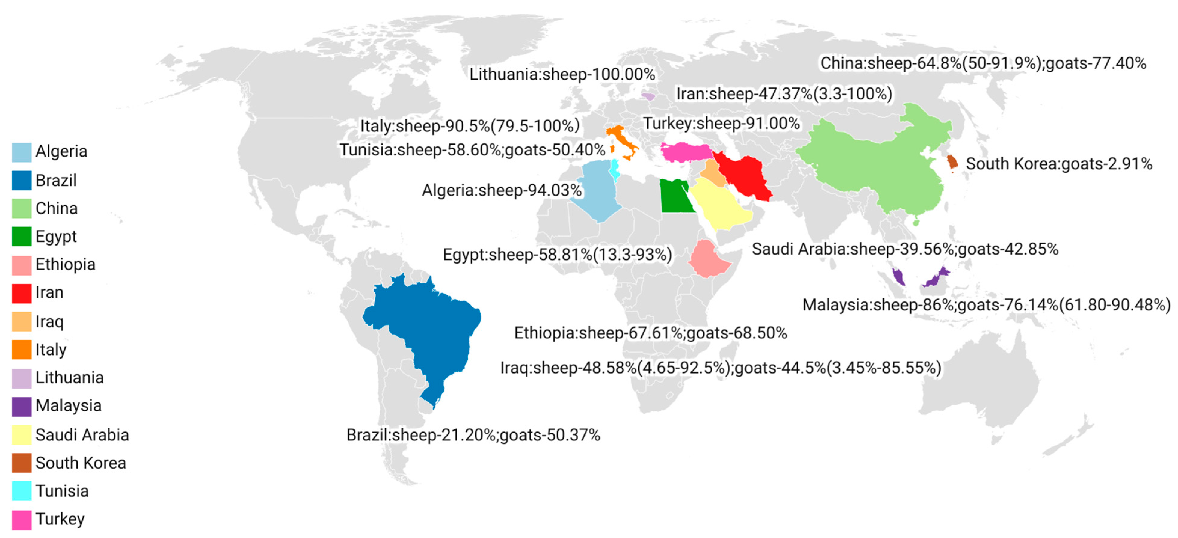
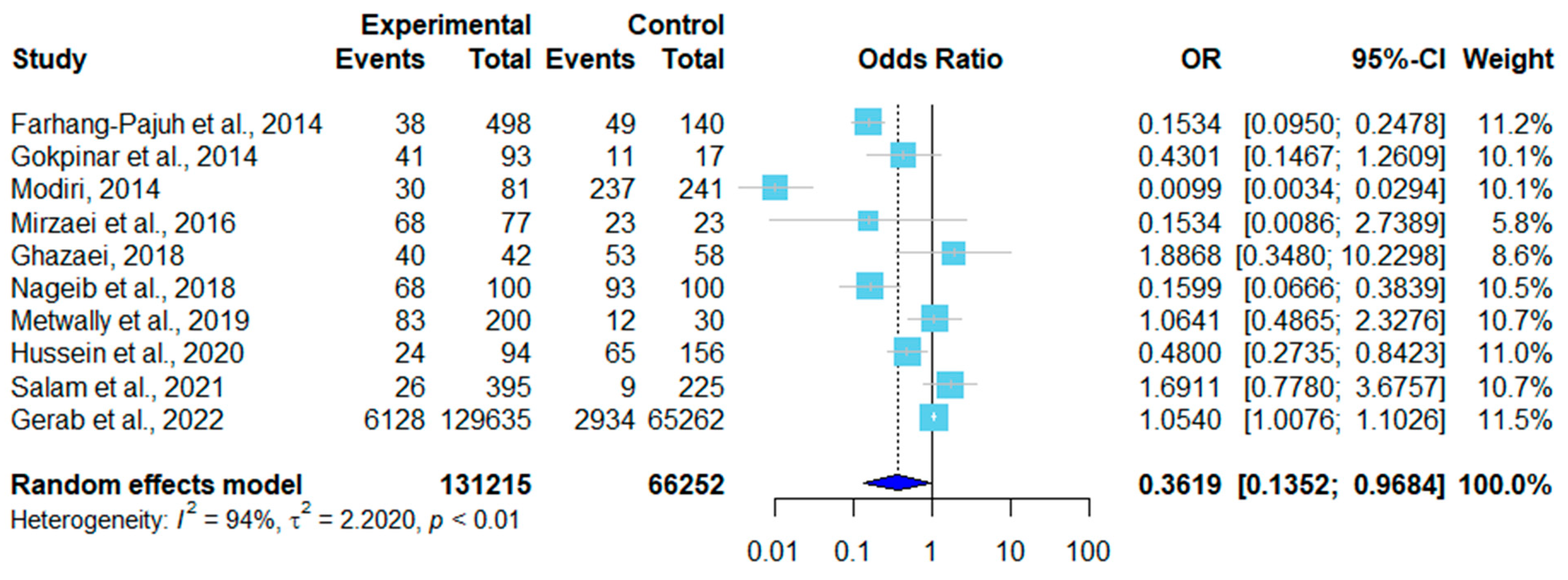
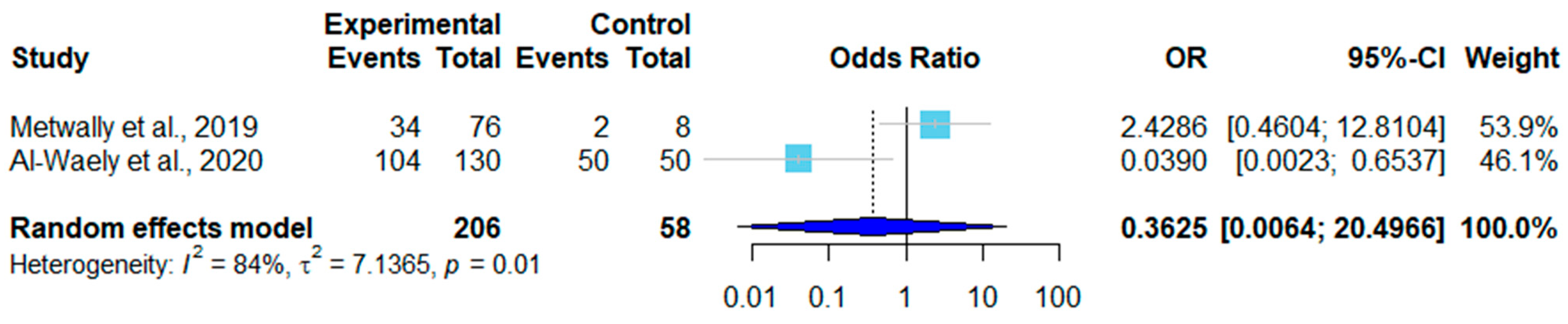
In this study, R software was used to calculate the infection rates in sheep and goats. The I2 value was used to determine whether the meta-analysis should use a fixed or random effects model. The heterogeneity was divided into three grades of low, medium, and high, based on I2 values of 25%, 50%, and 75%, respectively. A fixed effects model was used for low heterogeneity, and a random effects model was used for medium and high heterogeneity. The prevalence was analyzed according to sex and age.

3. Results

Five international databases were searched, which yielded a total of 601 articles, among which 18 articles were retrieved from Science Direct, 108 from Scopus, 49 from PubMed, 212 from Web of Science, and 214 from Google Scholar (Figure 1). A total of 166 duplicate articles were deleted, including 139 that were removed using EndNote X9 software, and 27 were deleted manually. Based on our time scope (2013–2022), 222 articles were excluded. After checking the abstracts and titles, 116 irrelevant articles were weeded out. The full texts of the remaining 59 articles were downloaded and carefully read. In the end, only 35 articles were included in this study. Forty sets of data were obtained, including 30 sets on sheep [5,9,13,14,15,16,17,18,19,20,21,22,23,24,25,26,27,28,29,30,31,32,33,34,35,36,37,38,39,40] and 10 on goats (Figure 2, Table 1 and Table 2) [16,17,36,37,38,41,42,43,44,45]. Meta analysis data can be downloaded from Supplementary Material.
The infection rate of 201,603 sheep was 70.08% (95% CI, 0.5615–0.8234), and the infection rate of 29,078 goats was 52.1% (95% CI, 0.2945–0.7423), as shown in Figure 3 and Figure 4. Of the thirty studies in sheep, fifteen utilized the tissue squash method, nine used molecular techniques (PCR), two used histopathology, three used digestion, and one used Percoll gradient centrifugation. Sarcocystis tenella was identified in 18 studies, S. arieticanis infection in sheep was found in 13 studies, and 12 studies were mixed infections of S. tenella and S. arieticanis (Table 1).
Five of the ten studies on goats used tissue squash, four used PCR, and one used digestion. In six studies, the species was identified as S. capracanis (Table 2).
There were 12 sex-related datasets on sheep. Two were excluded due to a lack of male controls. In six of the remaining ten, females had higher infection rates than males (Table 3). A forest plot was drawn according to the odds ratio of infection in male and female sheep (Figure 5). In the random effects model, OR = 0.36, 95% CI (0.14–0.97) (Figure 4), I2 = 94%, and p < 0.01, indicating that there were significant differences in infection rates in sheep in different studies (Figure 5). Of the three sex-related datasets on goats, two showed a higher infection rate in male goats than in female goats (Table 4). As shown in Figure 6, a forest plot was drawn according to the odds ratio of infection in male and female goats. The random effects model showed that OR = 0.66, 95% CI (0.24–1.82), I2 = 73%, and p = 0.02, indicating that the infection rates of goats varied significantly among studies (Figure 6).
There were ten age-related datasets on sheep, among which six showed that the infection rate increased with age (Table 5). A forest plot was drawn according to the age-adjusted infection rates. In the random effects model, OR = 0.66, 95% CI (0.32–1.39), I2 = 73%, and p < 0.01, which means there was a significant difference in the rates of sheep infections among different studies (Figure 7). Goats had three age-related datasets, and the data were divided into two groups of ≤1-year-old and >1-year-old. Only two studies met the criteria. The infection rate of goats older than one year of age was higher than that of those younger than one year of age (Table 6). In the random effects model, OR = 0.66 and 95% CI (0.32–1.39). A forest plot showed I2 = 84% and p = 0.01, which means the age-adjusted infection rates varied significantly among studies (Figure 8).

4. Discussion

Sarcocystis is an intracellular protozoan with a global distribution. Infection is often asymptomatic in ruminants, but in severe cases, infection can lead to loss of appetite, anemia, abortion, premature birth, or difficulty breathing and death [13]. Sarcocystosis causes hair loss and a reduction in milk production in sheep. Sarcocystis costs the Spanish sheep industry an estimated EUR 20 million per year [46]. As a result, epidemiological studies of Sarcocystis in sheep and goats provide guidance to the farming industry and public health safety. Recently, two meta-analysis studies have been conducted on Sarcocystis infection in ruminants in China [47] and Iran [48]. The infection rates of Sarcocystis in ruminants in China and Iran were reported to be 65% and 74.4%, respectively. In another meta-analysis study, the global prevalence of Sarcocystis in cattle was reported to be 62.7% [49]. In our study, we focused on sheep and goats and compiled data obtained from 14 countries worldwide (Figure 2).
This article compiled and analyzed data from 30 studies on Sarcocystis in sheep from 13 countries (three from Italy, one from Malaysia, one from Tunisia, nine from Iran, three from China, one from Brazil, five from Egypt, two from Iraq, and one from each of the following countries: Saudi Arabia, Ethiopia, Lithuania, Turkey, and Algeria) [5,9,13,14,15,16,17,18,19,20,21,22,23,24,25,26,27,28,29,30,31,32,33,34,35,36,37,38,39,40]. In addition, we included ten studies on Sarcocystis in goats from eight countries (two from Malaysia, two from Iraq, and one from each of the following countries: Tunisia, China, Brazil, South Korea, Saudi Arabia, and Ethiopia) [16,17,36,37,41,42,43,44,45]. The highest (100%) infection rate in sheep was found in Italy, Lithuania, and Iran [15,18,21,39], and the lowest (1.25%) infection rate was found in Iran [50]. Infection rates were associated with the sampling site and detection method. The highest (90.48%) infection rate in goats was reported in Malaysia [41], and the lowest (2.91%) infection rate was found in South Korea [45]. In Iran, one study that took samples from the diaphragm and esophagus of sheep found the infection rate to be 100% [18], while another study that took brain samples found the infection rate to be 1.25% [50]. In a study published by Kutty et al. in 2015, the same samples were subjected to different detection methods [41]. The infection rates were 52.38% (55/105), 43.8% (46/105), and 90.48% (95/105) using microscopic examination of muscle squash, H&E staining, and PCR, respectively [41].
Among the included articles, 13 reported the infection rate of sarcocystosis by sex (Table 3 and Table 4). Seven of the thirteen studies showed that females had higher infection rates than males, while the other six studies showed the opposite. Modiri et al. (2014) found that female sheep (98.34%, 237/241) had a higher infection rate than male sheep (37.03%, 30/81) [23]. Studies that showed a higher infection rate in male animals often had a larger sampling size of males than females. Salam et al. (2021) found that the infection rate difference between male goats (50.7%, 491/17809) and female goats (49.3%, 478/9911) [51] was not significant. However, with the same sampling size of female and male animals, Gerab et al. (2022) found that female sheep (93%, 93/100) had a higher infection rate than male sheep (68%, 68/100) [30]. Some scholars believed that the higher infection rate in females was because female animals often had compromised immunity during gestation and delivery, which increased their risk of infection [11].
Among the included studies, we found that in most articles, the infection rate was higher in animals older than one year (59.42%) as compared to those younger than one year (26.82%) (Table 5 and Table 6). This result is related to the increase in the probability of infection as animals age [52]. While a few studies have shown higher rates of infection in younger animals, this may be due to smaller sample sizes of adult animals.
The current study is subject to a few limitations. First, we are unclear as to how the animals were raised. Animals raised in different environments will likely have different infection rates. Second, different detection methods offer varying sensitivity and specificity, which will affect the infection rate [44]. The Percoll gradient centrifugation method was less widely employed in the detection of Sarcocystis. Third, the obtained data resulted from one-time surveys and may not represent the infection rates at other times. Fourth, the infection rates of Sarcocystis in sheep and goats were not reported in many other countries around the world.
The PCR detection method is fast, accurate, and suitable for routine laboratory practice. Compared with conventional PCR, nested PCR and PCR-RFLP have more steps and are more suitable for distinguishing species [14]. Microscopic examination of tissue pellets is among the most commonly used detection techniques, and complete cysts can be visualized under a microscope. However, it requires trained hands and is time-consuming and labor-intensive, which limits the number of samples that can be processed. The accuracy is in question because sometimes cysts are easy to miss [2].
The histopathological method is time-consuming and laborious, but the internal structure of the cyst can be clearly seen. This method is more suitable for the study of the entire cyst structure. In the digestion method, the cysts in the tissue are digested, and the release of bradyzoites from the cysts facilitates the detection of the parasite. However, because the cyst wall is dissolved, the species of Sarcocystis cannot be distinguished, and a complete cyst cannot be obtained [2]. The Percoll gradient centrifugation method has a high cost and is not suitable for epidemiological detection, but it is more suitable for purifying cysts. With this method, the morphology of the obtained cysts will not change [40].
This study is the first statistical analysis of the global prevalence of Sarcocystis in sheep and goats in the past 10 years (2013–2022). We investigated risk factors, including age and sex, for Sarcocystis infection in sheep and goats. The data analysis provided here enriches our understanding of the distribution and transmission of Sarcocystis in sheep and goats on a global scale.

5. Conclusions

The total infection rates of Sarcocystis in sheep and goats worldwide were 70.08% (95% CI, 0.5615–0.8234) and 52.1% (95% CI, 0.2945–0.7423), respectively. Age and sex were identified as risk factors for Sarcocystis infection. Female sheep and goats had higher infection rates than male sheep and goats. In terms of age, animals older than one year of age had higher infection rates than those younger than one year of age.

Supplementary Materials

The following supporting information can be downloaded at: https://www.mdpi.com/article/10.3390/pathogens12070902/s1, File S1: meta analysis data.

Author Contributions

Conceptualization, Y.F., X.S., and N.Y.; methodology, R.G. and X.S.; software, R.G., validation, X.Z., X.L., and M.L.; formal analysis, Y.F., R.G., and X.S.; resources, N.Y.; data curation, Y.F.; writing—original draft preparation, R.G., Y.F., and T.J.; writing—review and editing, N.Y. and T.J.; supervision, N.Y. and T.J.; project administration, N.Y.; funding acquisition, N.Y. All authors have read and agreed to the published version of the manuscript.

Funding

Shenyang Young and the middle-aged Scientific and Technological Innovation Talent Support Program (grant number RC210291), Key Laboratory for Prevention and Control of Avian Influenzas and Other Major Poultry Diseases, Ministry of Agriculture and Rural Affairs, P.R., China, and Key Laboratory of Livestock Disease Prevention of Guangdong Province (grant number YDWS202209) provided funding.

Institutional Review Board Statement

Not applicable.

Informed Consent Statement

Not applicable.

Data Availability Statement

The data presented in this study are available upon request from the corresponding author.

Conflicts of Interest

The authors declare no conflict of interest.

References

  1. Rosenthal, B.M. Zoonotic Sarcocystis. Res. Vet. Sci. 2021, 136, 151–157. [Google Scholar] [CrossRef] [PubMed]
  2. Dubey, J.P.; Calero-Bernal, R.; Rosenthal, B.M.; Speer, C.A.; Fayer, R. Sarcocystosis of Animals and Humans; CRC Press: Boca Raton, FL, USA, 2015. [Google Scholar]
  3. Zeng, H.; Van Damme, I.; Kabi, T.W.; Oba, B.; Gabril, S. Sarcocystis species in bovine carcasses from a Belgian abattoir: A cross-sectional study. Parasites Vectors 2021, 14, 271. [Google Scholar] [CrossRef]
  4. Sheikhi, M.; Salahi-Moghaddam, A.; Asl, M.N.; Farahani, A.; Shamseddin, J. A survey on the Frequency of Sarcocystis in Bandar Abbas, Iran in 2019–2020. Gene Cell Tissue 2020, 7, e109990. [Google Scholar] [CrossRef]
  5. Dahmani, A.; Aissi, M.; Zenia, S.; Harhoura, K.; Saadi, A. Morphometric Study of Microscopic Cysts of Sarcocystis Sp. in Sheep Carcasses. Folia Vet. 2020, 64, 38–46. [Google Scholar] [CrossRef]
  6. Xue, S.; Xiang, C.R.; Ma, C.F.; Yan, W.C. Serological investigation of Sarcocystis infection in dairy cattle in parts of Henan Province. Adv. Vet. Med. 2022, 43, 142–144. [Google Scholar]
  7. Xue, R.; Han, L.F.; Qian, W.F.; He, B.; Yan, W.C. Tissue squash and molecular identification of Sarcocystis in pigs in Luoyang City. J. Henan Univ. Sci. Technol. Nat. Sci. Ed. 2018, 39, 6. [Google Scholar]
  8. Moré, G.; Schares, S.; Maksimov, A.; Conraths, F.J.; Venturini, M.C.; Schares, G. Development of a multiplex real time PCR to differentiate Sarcocystis spp. affecting cattle. Vet. Parasitol. 2013, 197, 85–94. [Google Scholar] [CrossRef]
  9. Hu, J.J.; Huang, S.; Wen, T.; Esch, W.G.; Liang, Y.; Li, H.L. Sarcocystis spp. in domestic sheep in Kunming City, China: Prevalence, morphology, and molecular characteristics. Parasite. 2017, 24, 30. [Google Scholar] [CrossRef] [Green Version]
  10. Prakas, P.; Oksanen, A.; Butkauskas, D.; Sruoga, A.; Liaugaudaitė, S. Identification and intraspecific genetic diversity of Sarcocystis rileyi from ducks, Anas spp., in Lithuania and Finland. J. Parasitol. 2014, 100, 657–661. [Google Scholar] [CrossRef]
  11. Sudan, V.; Shanker, D.; Paliwal, S.; Sruoga, A.; Liaugaudaitė, S. Phylogenetics of Sarcocystis fusiformis isolates based on 18S rRNA and cox 1 genes. Microb. Pathog. 2021, 159, 105144. [Google Scholar] [CrossRef]
  12. Ma, C.L.; Ye, Y.L.; Wen, T.; Huang, Z.M.; Song, J.L. Prevalence and morphological and molecular characteristics of Sarcocystis bertrami in horses in China. Parasite 2020, 27, 1. [Google Scholar] [CrossRef] [Green Version]
  13. Dessi, G.; Tamponi, C.; Pasini, C.; Porcu, F.; Meloni, L.; Cavallo, L.; Sini, M.F.; Knoll, S.; Scala, A.; Varcasia, A. A survey on Apicomplexa protozoa in sheep slaughtered for human consumption. Parasitol. Res. 2022, 121, 1437–1445. [Google Scholar] [CrossRef] [PubMed]
  14. Pipia, A.P.; Varcasia, A.; Zidda, A.; Dessi, G.; Panzalis, R.; Tamponi, C.; Marrosu, R.; Tosciri, G.; Sanna, G.; Dore, F.; et al. Cross-sectional investigation on sheep sarcosporidiosis in Sardinia, Italy. Vet. Parasitol. Reg. Stud. Rep. 2016, 3, 13–17. [Google Scholar] [CrossRef] [Green Version]
  15. Bacci, C.; Vismarra, A.; Passeri, B.; Dessi, G.; Panzalis, R.; Tamponi, C.; Marrosu, R.; Tosciri, G.; Sanna, G.; Dore, F.; et al. Detection of Toxoplasma gondii and Sarcocystis tenella in indigenous Cornigliese sheep in Italy using serological and molecular methods. Small Rumin. Res. 2016, 135, 13–16. [Google Scholar] [CrossRef]
  16. Latif, B.; Kutty, K.M.; Muslim, A.; Hussaini, J.; Omar, E.; Heo, C.C.; Rossle, N.F.; Abdullah, S.; Kamarudin, M.A.; Zulkarnain, M.A. Light microscopy and molecular identification of Sarcocystis spp. in meat producing animals in Selangor, Malaysia. Trop. Biomed. 2015, 32, 444–452. [Google Scholar] [PubMed]
  17. Amairia, S.; Amdouni, Y.; Rouatbi, M.; Rjeibi, M.R.; Awadi, S.; Gharbi, M. First detection and molecular identification of Sarcocystis spp. in small ruminants in North-West Tunisia. Transbound. Emerg. Dis. 2018, 65, 441–446. [Google Scholar] [CrossRef] [PubMed]
  18. Rad, H.; Nourani, H.; Razmi, G. Histopathological, ultrastructural and molecular examination of Sarcocystis spp. in sheep of Mashhad area, Khorasan Razavi province, Iran. Iran. J. Vet. Sci. Technol. 2020, 12, 1–9. [Google Scholar]
  19. Pestechian, N.; Yousefi, H.A.; Kalantari, R.; Jafari, R.; Khamesipour, F.; Keshtkar, M.; Esmaeilifallah, M. Molecular and Microscopic Investigation of Sarcocystis Species Isolated from Sheep Muscles in Iran. J. Food Qual. 2021, 2021, 5562517. [Google Scholar] [CrossRef]
  20. Farhang-Pajuh, F.; Yakhchali, M.; Mardani, K. Molecular determination of abundance of infection with Sarcocystis species in slaughtered sheep of Urmia, Iran. Vet. Res. Forum Int. Q. J. 2014, 5, 181–186. [Google Scholar]
  21. Rahdar, M.; Kardooni, T. Molecular identification of Sarcocystis spp. in sheep and cattle by PCR-RFLP from southwest of Iran. Jundishapur J. Microbiol. 2017, 10, e12798. [Google Scholar] [CrossRef] [Green Version]
  22. Shahabi, S.; Dehbashi, N.; Sarkari, B.; Arefkhah, N.; Sedaghat, B.; Savardashtaki, A. Detection and phylogenetic analysis of Sarcocystis moulei and Sarcocystis spp. (Sarcocystidae: Apicomplexa) from slaughtered sheep in southwest Iran. J. Parasit. Dis. 2022, 46, 215–219. [Google Scholar] [CrossRef]
  23. Modiri, D.; Hosainvosughi. Survey of contamination rate sheep to Sarcocystis in slaughterhouse Boroujerd City and studied by PCR. Adv. Environ. Biol. 2014, 8, 424–428. [Google Scholar]
  24. Kalantari, N.; Khaksar, M.; Ghaffari, S.; Hamidekish, S.M. Molecular Analysis of Sarcocystis Spp. Isolated from Sheep (Ovis aries) in Babol Area, Mazandaran Province, Northern Iran. Iran. J. Parasitol. 2016, 11, 73–80. [Google Scholar]
  25. Ghazaei, C. Evaluation of Sarcocyst Parasite Strains in Carcasses Obtained from Ardabil Meat Industrial Group. Int. J. Mol. Clin. Microbiol. 2018, 8, 950–956. [Google Scholar]
  26. Mirzaei, M.; Rezaei, H. The role of sheep in the epidemiology of Sarcocystis spp. in Tabriz area northwest of Iran. J. Parasit. Dis. Off. Organ Indian Soc. Parasitol. 2016, 40, 285–288. [Google Scholar] [CrossRef] [Green Version]
  27. Dong, H.; Su, R.; Wang, Y.; Tong, Z.; Zhang, L.; Yang, Y.; Hu, J. Sarcocystis species in wild and domestic sheep (Ovis ammon and Ovis aries) from China. BMC Vet. Res. 2018, 14, 377. [Google Scholar] [CrossRef]
  28. Sun, Y.; Ju, J.; Su, X.; Xie, C.; Li, Y.; Kang, M. Infection survey and morphological characteristics of Sarcocystis spp. in naturally infected Tibetan sheep from Qinghai in northwestern China. Parasitol. Int. 2021, 80, 102219. [Google Scholar] [CrossRef]
  29. Portella, L.P.; Cadore, G.C.; Sangioni, L.A.; Alves, M.E.M.; Chemeris, R.; Brum, L.P.; Voge, F.S.F. Molecular detection of protozoa of the Sarcocystidae family in sheep from the State of Rio Grande do Sul, Brazil. Ciência Rural 2016, 46, 1613–1617. [Google Scholar] [CrossRef]
  30. Gerab, R.A.; Edris, A.B.M.; Lamada, H.M.; Elrais, A.M. Prevalence and Distribution of Sarcocystis in Buffaloes and Sheep in Egypt. J. Adv. Vet. Res. 2022, 12, 302–307. [Google Scholar]
  31. Hussein, N.M. Morphological and Molecular characterization of Sarcocystis arieticanis from the Heart Muscles of Domestic sheep, Ovis aries, in Qena, Upper Egypt. J. Adv. Vet. Res. 2020, 10, 73–80. [Google Scholar]
  32. El-Morsey, A.; Abdo, W.; Sultan, K.; Elhawary, N.M.; AbouZaid, A.A. Ultrastructural and Molecular Identification of the sarcocysts of Sarcocystis tenella and Sarcocystis arieticanis Infecting Domestic Sheep (Ovis aries) from Egypt. Acta Parasitol. 2019, 64, 501–513. [Google Scholar] [CrossRef]
  33. El-Morsey, A.; Abdo, W.; Zaid, A.A.A.; Sorour, S.S.G. Morphologic and molecular identification of three macroscopic Sarcocystis species infecting domestic sheep (Ovis aries) and cattle (Bos taurus) in Egypt. Parasitol. Res. 2021, 120, 637–654. [Google Scholar] [CrossRef]
  34. Nageib, B.; Kuraa, H. Microscopical and serological studies with ultrastructure description of Sarcocystis species in sheep in Assiut. Assiut Vet. Med. J. 2018, 64, 46–55. [Google Scholar]
  35. Abdullah, S.H. Investigation of Sarcocystis spp. in slaughtered cattle and sheep by peptic digestion and histological examination in Sulaimani Province, Iraq. Vet. World 2021, 14, 468–474. [Google Scholar] [CrossRef]
  36. Salam, S.M.; Salih Mustafa, B.H. Identification of Sarcocystis Species “Macrocystis” by Visual and Molecular Technique in Sheep and Goats—Sulaymaniyah Slaughterhouse. J. Anim. Poult. Prod. 2021, 12, 331–337. [Google Scholar]
  37. Metwally, D.M.; Al-Damigh, M.A.; Al-Turaiki, I.M.; Khadragy, M.F. Molecular characterization of Sarcocystis species isolated from sheep and goats in Riyadh, Saudi Arabia. Animals 2019, 9, 256. [Google Scholar] [CrossRef] [PubMed] [Green Version]
  38. Mekibib, B.; Abdisa, D.; Denbarga, Y.; Abebe, R. Muscular Sarcocystis infection in ruminants slaughtered at Municipality abattoir and selected Hotels in Hawassa city, southern Ethiopia: Prevalence and associated risk factors. Vet. Parasitol. Reg. Stud. Rep. 2019, 18, 100333. [Google Scholar] [CrossRef]
  39. Januskevicius, V.; Januskeviciene, G.; Prakas, P.; Butkauskas, D.; Petkevicius, S. Prevalence and intensity of Sarcocystis spp. infection in animals slaughtered for food in Lithuania. Vet. Med. 2019, 64, 149–157. [Google Scholar] [CrossRef] [Green Version]
  40. Gokpinar, S.; Yildiz, K.; Gurcan, I.S. Prevalence and Concentration of Sarcocystis spp. Microscopic Cysts in Sheep Muscles Using Percoll Gradient Centrifugation. Isr. J. Vet. Med. 2014, 69, 16–19. [Google Scholar]
  41. Kutty, M.K.; Latif, B.; Muslim, A.; Hussaini, J.; Daher, A.M.; Heo, C.C.; Abdullah, S. Detection of sarcocystosis in goats in Malaysia by light microscopy, histology, and PCR. Trop. Anim. Health Prod. 2015, 47, 751–756. [Google Scholar] [CrossRef]
  42. Hu, J.J.; Liu, T.T.; Liu, Q.; Esch, G.W.; Chen, J.Q.; Huang, S.; Wen, T. Prevalence, morphology, and molecular characteristics of Sarcocystis spp. in domestic goats (Capra hircus) from Kunming, China. Parasitol. Res. 2016, 115, 3973–3981. [Google Scholar] [CrossRef]
  43. Bittencourt, M.V.; Meneses, I.D.S.; Ribeiro-Andrade, M.; de Jesus, R.F.; de Araújo, F.R.; Gondim, L.F.P. Sarcocystis spp. in sheep and goats: Frequency of infection and species identification by morphological, ultrastructural, and molecular tests in Bahia, Brazil. Parasitol. Res. 2016, 115, 1683–1689. [Google Scholar] [CrossRef]
  44. Al-Waely, T.N.; Abd AL-Amery, A.M. Prevalence of Sarcocystosis in Goats (Capra hircus) at Wasit Province, Iraq. Plant Arch. 2020, 20, 8939–8944. [Google Scholar]
  45. Hong, E.J.; Sim, C.; Chae, J.S.; Kim, H.C.; Park, J.; Choi, K.S.; Yu, D.H.; Park, C.H.; Yoo, J.G.; Park, B.K. Ultrastructural and molecular identification of Sarcocystis tenella (Protozoa, Apicomplexa) in naturally infected Korean native goats. Vet. Med. 2016, 61, 374–381. [Google Scholar] [CrossRef] [Green Version]
  46. Martínez-Navalón, B.; Anastasio-Giner, B.; Cano-Fructuoso, M.; Sanchez-Martínez, P.; Llopis-Morant, A.; Perez-Castarlenas, B.; Goyena, E.; Berriatua, E. Sarcocystis infection: A major cause of carcass condemnation in adult sheep in Spain. Span. J. Agric. Res. 2012, 10, 388–392. [Google Scholar] [CrossRef]
  47. Zhu, Z.; Ying, Z.; Feng, Z.; Liu, Q.; Liu, J. The Occurrence and Meta-Analysis of Investigations on Sarcocystis Infection among Ruminants (Ruminantia) in Mainland China. Animals 2022, 13, 149. [Google Scholar] [CrossRef] [PubMed]
  48. Anvari, D.; Narouei, E.; Hosseini, M.; Narouei, M.R.; Siyadatpanah, A.; Shariatzadeh, S.A.; Pagheh, A.S.; Gholami, S.; Sarvi, S.; Sargazi, D.; et al. Sarcocystosis in ruminants of Iran, as neglected food-borne disease: A systematic review and meta-analysis. Acta Parasitol. 2022, 65, 555–568. [Google Scholar] [CrossRef]
  49. Shams, M.; Shamsi, L.; Asghari, A.; Motazedian, M.H.; Mohammadi-Ghalehbin, B.; Omidian, M.; Nazari, N.; Sadrebazzaz, A. Molecular epidemiology, species distribution, and zoonotic importance of the neglected meat-borne pathogen Sarcocystis spp. in cattle (Bos taurus): A global systematic review and meta-analysis. Acta Parasitol. 2022, 67, 1055–1072. [Google Scholar] [CrossRef] [PubMed]
  50. Salehi, M.; Bahari, P.; Vatanchian, M. First Molecular Identification of Sarcocystis ovicanis (Protozoa, Apicomplexa) in the Brain of Sheep in Iran. Iran. J. Parasitol. 2014, 9, 286–291. [Google Scholar] [PubMed]
  51. Okita, F.O.; Obadiah, H.I.; Gyegweh, K.T.; Okonkwo, A.A.; Azatyom, J.A. Survey of Sarcocystis species infection in slaughtered goats in Makurdi metropolis. Int. J. Infect. Dis. Ther. 2017, 2, 4–8. [Google Scholar]
  52. Kaur, M.; Singh, B.B.; Sharma, R.; Gill, J.P.S. Pervasive environmental contamination with human feces results in high prevalence of zoonotic Sarcocystis infection in pigs in the Punjab, India. J. Parasitol. 2016, 102, 229–232. [Google Scholar] [CrossRef] [PubMed]
Figure 1. Data Selection Flow Chart.
Pathogens 12 00902 g001
Figure 2. The global prevalence of Sarcocystis spp. infection in sheep and goats. Created with Datawrapper.
Pathogens 12 00902 g002
Figure 3. Forest plot of infection rates in sheep [5,9,13,14,15,16,17,18,19,20,21,22,23,24,25,26,27,28,29,30,31,32,33,34,35,36,37,38,39,40]. The proportion column calculated the ratio of the number of positive samples (events) to the total number of samples (total). The weight each study carried in the data analysis is listed in the last column.
Pathogens 12 00902 g003
Figure 4. Forest plot of infection rates in goats [16,17,36,37,38,41,42,43,44,45]. The proportion column calculated the ratio of the nuber of positive samples (events) to the total number of samples (total). The weight each study carried in the data analysis is listed in the last column.
Pathogens 12 00902 g004
Figure 5. Forest plot of the infection rates in male and female sheep in different countries [20,23,25,26,30,31,34,36,37,40]. The experimental column compiled the number of positive samples over the total number of samples from male sheep. The control group compiled the number of positive samples over the total number of samples from female sheep. OR represents the odds ratio, which is the ratio of odds of infection in male and female sheep. OR = 1 is not statistically significant. The weight each study carried is shown in the last column.
Pathogens 12 00902 g005
Figure 6. Forest plot of the infection rates in male and female goats in different countries [36,37,44]. The experimental column compiled the number of positive samples over the total number of samples from male goats. The control group compiled the number of positive samples over the total number of samples from female goats. OR (odds ratio) was the ratio of odds of infection in males and females. OR = 1 is not statistically significant. The weight each study carried is shown in the last column.
Pathogens 12 00902 g006
Figure 7. Forest plot of the age-adjusted infection rates in sheep from different studies [25,26,31,34,37,40]. The experimental group contained the number of positive samples and the total number of samples from sheep less than one year of age. In the control group, the number of positive samples and the total number of samples from sheep older than one year of age were included. OR (odds ratio) is the ratio of odds of infection in sheep less than and more than one year of age. OR = 1 is not statistically significant. The weight of each study is shown in the last column.
Pathogens 12 00902 g007
Figure 8. Forest plot of the age-adjusted infection rates in goats from different studies [37,44]. The experimental group contained the number of positive samples and the total number of samples from goats less than one year of age. The control group contained the number of positive samples and the total number of samples from goats older than one year of age. OR (odds ratio) is the ratio of odds of infection in goats less than and more than one year of age. OR = 1 is not statistically significant. The weight of each study is shown in the last column.
Pathogens 12 00902 g008
Table 1. 32 datasets of infection in sheep from different countries.
No.CountrySampling TimePositive/TotalInfection RateDetection Method *TissueSpeciesReference (year)
1Iran2011.2–2012.1235/63836.83%PCR-RFLPEsophagus
Diaphragm
Skeletal muscle
S. gigantea
S. medusiformis
[20] (2014)
2Iranunknown267/32282.91%Digestion methodEsophagus
Diaphragm
unknown[23] (2014)
3Turkey2011.2–2012.191/10091.00%Percoll gradient centrifugationSkeletal muscleS. tenella
S. arieticanis
[40] (2014)
4Malaysia2013.2–2013.1037/4386.00%Digestion methodTongue
Heart
Diaphragm
Esophagus
Skeletal muscle
S. ovicanis[16] (2015)
5Italy201383/11279.50%Nested PCRHeartS. tenella
S. arieticanis
[14] (2016)
6Italy2014.3–2014.424/24100%Conventional PCRHeartS. tenella[15] (2016)
7Brazilunknown17/8021.20%Conventional PCRHeartS. tenella
S. arieticanis
[29] (2016)
8Iran2013.9–2013.1050/15033.30%Tissue squash methodIntra-abdominal
Muscle
Diaphragm
S. gigantea
S. moulei
Sarcocystis spp.
[24] (2016)
9Iran2013.4–2013.1021/6203.30%Tissue squash methodEsophagus
Diaphragm
unknown[26] (2016)
10Iranunknown25/25100%PCR-RFLPHeart
Tongue
Diaphragm
Skeletal muscle
S. tenella
S. capracanis
[21] (2017)
11China2015.3–2015.1179/8691.90%Tissue squash methodEsophagus
Diaphragm
Skeletal muscle
Tongue
Heart
S. tenella
S. arieticanis
[9] (2017)
12Iranunknown211/25084.40%Tissue squash methodIntervertebral muscle
Esophagus
Heart
Tongue
S. arieticamis
S. tenella
[25] (2018)
13Tunisia2016.5–2016.6116/19858.6%Conventional PCRNeckS. tenella[17] (2018)
14China2014.3–2017.10335/63852.51%Tissue squash methodHeartS. tenella
S. arieticanis
[27] (2018)
15Egyptunknown93/10093.00%Tissue squash methodEsophagus
Diaphragm Tongue
Skeletal muscle Heart
S. arieticanis
S. tenella
[34] (2018)
16Saudi Arabia2016.3–2017.191/23039.56%Digestion method Tongue
Heart
Skeletal muscle
Diaphragm
Esophagus
S. tenella[37] (2019)
17Ethiopia2016.11–2017.5119/17667.61%Tissue squash methodHeart
Esophagus
unknown[38] (2019)
18Lithuania2012–201461/61100.00%Tissue squash methodEsophagus
Diaphragm
Heart
Neck
Jaw
Back
unknown[39] (2019)
19Egypt2017.1–2018.2152/17586.80%Tissue squash methodEsophagus
Diaphragm
Skeletal muscle
Tongue
Heart
S. tenella
S. arieticanis
[32] (2019)
20Algeriaunknown315/33594.03%Histopathological methodEsophagus
Diaphragm
S. arieticanis
S. tenella
[5] (2020)
21Iran2018.10–2019.5200/200100%Conventional PCREsophagusS. gigantea
S. tenella
S. arieticanis
[18] (2020)
22Egypt2018.4–2019.352/11047.27%Tissue squash methodHeart S. arieticanis[31] (2020)
23Iranunknown125/15083.33%Conventional PCRHeartS. gigantea
S. tenella
[19] (2021)
24China2017.2–2018.11577/115550.00%Tissue squash methodHeart
Diaphragm
Esophageal
S. gigantea[28] (2021)
25Egypt2018.1–2019.633/25013.20%Histopathologic examinationEsophagus
Tongues
Diaphragm
Abdominal muscle

Skeletal muscle
Heart
S. gigantea
S. medusiformis
[33] (2021)
26Iraq2020.5–2020.774/8092.50%Tissue squash methodDiaphragm
Esophagus
S. tenella
S. arieticanis
[35] (2021)
27Iraq2020.8–2021.19062/194,8974.65%Tissue squash methodEsophagus
Diaphragm
Heart
S. gigantea
S. medisiformis
[36] (2021)
28Italy2019.4–2019.7127/13892.00%Conventional PCRHeartS. tenella[13] (2022)
29Egypt2020.7–2021.6161/20080.50%Tissue squash methodEsophagus
Tongue
Heart
Masseter
Skeletal muscle
unknown[30] (2022)
30Iran2019.3–2019.510/6016.66%Tissue squash methodMuscles #S. moulei[22] (2022)
* All PCR methods targeted the 18s rRNA gene, # Specific muscle type unknown.
Table 2. 10 datasets of infection in goats in different countries.
No.CountrySampling TimePositive/TotalInfection RateDetection Method *TissueSpeciesReference (year)
1Malaysia2014.1–2014.295/10590.48%Conventional PCRSkeletal muscleS. capracanis[41] (2015)
2Malaysia2013.2–2013.1055/8961.80%Tissue squash methodTongue
Heart
Diaphragm
Esophagus
Skeletal muscle
S. capracanis[16] (2015)
3South Korea2014.1–2014.83/1032.91%Conventional PCRHeartS. tenella[45] (2016)
4China2014.7–2015.9174/22577.40%Tissue squash methodEsophagus
Diaphragm
Skeletal muscle
Tongue
Heart
S. capracanis
S. hircicanis
[42] (2016)
5Brazil2012.1–2013.6137/27050.37%Conventional PCRTongue
Esophagus
Heart
S. capracanis[43] (2016)
6Tunisia2016.5–2016.661/12150.4%Conventional PCRNeckS. capracanis[17] (2018)
7Saudi Arabia2016.3–2017.136/8442.85%Tissue squash methodTongue
Heart
Skeletal muscle
Diaphragm
Esophagus
S. capracanis[37] (2019)
8Ethiopia2016.11–2017.5124/18168.50%Tissue squash methodHeart
Esophagus
unknown[38] (2019)
9Iraq2019.10–2020.3156/18085.55%Digestion
method
Esophagus
Diaphragm
unknown[44] (2020)
10 Iraq2020.8–2021.1969/27,7203.45%Tissue squash methodEsophagus
Diaphragm
Heart
S. moulei[36] (2021)
* All PCR methods targeted the 18s rRNA gene.
Table 3. Infection rates of male and female sheep in different countries.
Reference (year)CountryRegionTotalMaleInfected%FemaleInfected%
[20] (2014)IranUrmia638498387.631404935
[23] (2014)IranBoroujerd322813037.0324123798.34
[40] (2014)TurkeyKirikkale100776888.32323100
[26] (2016)IranTabriz620395266.5822594.0
[25] (2018)IranArdabil250942426.961566573.03
[34] (2018)EgyptAssiut100424095.2585391.4
[37] (2019)Saudi ArabiaRiyadh2302008341.5301240
[31] (2020)EgyptQena110934144.08171164.70
[36] (2021)IraqSulaymaniyah194,897129,635612867.665,262293432.4
[30] (2022)EgyptTanta20010068681009393
Table 4. Infection rates of male and female goats in different countries.
Reference (year)CountryRegionTotalMaleInfected%FemaleInfected%
[37] (2019)Saudi ArabiaRiyadh84643046.8720630
[44] (2020)IraqWasit1801048379.80767193.42
[36] (2021)IraqSulaymaniyah27,72017,80949150.7991147849.3
Table 5. Age-adjusted infection rates in sheep in different countries.
Reference (year)Country, RegionNo. of Positive SamplesPositivity (%)Age Groups (year)No. of Samples Age-Adjusted No. of Positive Samples Age-Adjusted Positivity (%)
[40] (2014)Turkey,
Kirikkale
91911
2
3
4
63
21
12
4
53
21
12
4
84.12
100
100
100
[26] (2016)Iran, Tabriz355.64<1
1–3
>3
96
423
101
6
18
11
6.25
4.25
10.89
[25] (2018)Iran, Ardabil8935.6<1
>1
97
153
24
65
24.74
42.48
[34] (2018)Egypt, Assiut93930.5–2
2–4
≥4
40
41
19
38
38
17
95
92.7
89.5
[38] (2019)Ethiopia,
Hawassa
17667.611.5–2.517611967.61
[37] (2019)Saudi Arabia, Riyadh9541.30<1
>1
168
62
73
22
43.45
35.48
[32] (2019)Egypt,
Gharbia
15286.8>217515286.8
[31] (2020)Egypt, Qena5247.27<1
>1
41
69
11
41
26.82
59.42
[28] (2021)China, Qinghai57750>1115557750
[30] (2022)Egypt, Tanta16180.51–2
3–5
100
100
68
93
68
93
Table 6. Age-adjusted infection rates in goats from different studies.
Reference (year)Country, RegionNo. of Positive SamplesPositivity (%)Age Groups (year)No. of SamplesAge-Adjusted No. of Positive SamplesAge-Adjusted Positivity (%)
[37] (2019)Saudi Arabia,
Riyadh
3642.85<1
>1
76
8
34
2
44.73
25
[38] (2019)Ethiopia,
Hawassa
12468.501.5–2.518112468.50
[44] (2020)Iraq, Wasit15485.55<1
1–2
>7
130
41
9
104
41
9
80
100
100
Disclaimer/Publisher’s Note: The statements, opinions and data contained in all publications are solely those of the individual author(s) and contributor(s) and not of MDPI and/or the editor(s). MDPI and/or the editor(s) disclaim responsibility for any injury to people or property resulting from any ideas, methods, instructions or products referred to in the content.

