Article

Diversity and Phylogeny of Cattle Ixodid Ticks and Associated Spotted Fever Group Rickettsia spp. in Tunisia

1
Laboratory of Microbiology, National School of Veterinary Medicine of Sidi Thabet, University of Manouba, Manouba 2010, Tunisia
2
Ministry of National Defense, General Directorate of Military Health, Veterinary Service, Tunis 1008, Tunisia
3
Laboratory of Parasitology, National School of Veterinary Medicine of Sidi Thabet, University of Manouba, Manouba 2010, Tunisia
4
Department of Basic Sciences, Higher Institute of Biotechnology of Sidi Thabet, University of Manouba, Manouba 2010, Tunisia
*
Authors to whom correspondence should be addressed.
Pathogens 2023, 12(4), 552; https://doi.org/10.3390/pathogens12040552
Submission received: 16 January 2023 / Revised: 28 March 2023 / Accepted: 1 April 2023 / Published: 3 April 2023
(This article belongs to the Special Issue Tick-Borne Bacteria in Africa: From Diagnosis to Control)

Abstract

:
Tick-borne rickettsioses are mainly caused by obligate intracellular bacteria belonging to the spotted fever group (SFG) of the Rickettsia genus. So far, the causative agents of SFG rickettsioses have not been detected in cattle ticks from Tunisia. Therefore, the aim of this study was to investigate the diversity and phylogeny of ticks associated with cattle from northern Tunisia and their associated Rickettsia species. Adult ticks (n = 338) were collected from cattle in northern Tunisia. The obtained ticks were identified as Hyalomma excavatum (n = 129), Rhipicephalus sanguineus sensu lato (n = 111), Hyalomma marginatum (n = 84), Hyalomma scupense (n = 12) and Hyalomma rufipes (n = 2). After DNA extraction from the ticks, 83 PCR products based on the mitochondrial 16S rRNA gene were sequenced and a total of four genotypes for Rh. sanguineus s.l., two for Hy. marginatum and Hy. excavatum and only one for Hy. scupense and Hy. rufipes were recorded, with the occurrence of one, two and three novel genotypes, respectively, for Hy. marginatum, Hy. excavatum and Rh. sanguineus s.l. mitochondrial 16S rRNA partial sequences. The tick DNA was tested for the presence of Rickettsia spp. by using PCR measurements and sequencing targeting three different genes (ompB, ompA and gltA). Of the 338 analyzed ticks, 90 (26.6%), including 38 (34.2%) Rh. sanguineus s.l., 26 (20.1%) Hy. excavatum, 25 (29.8%) Hy. marginatum and one (50%) Hy. rufipes tick, were positive for Rickettsia spp. Based on 104 partial sequences of the three analyzed genes, the BLAST analysis and phylogenetic study showed the infection of Hy. excavatum, Hy. marginatum and Rh. sanguineus s.l. tick specimens with R. massiliae, R. aeschlimannii and R. sibirica subsp. mongolitimonae and one Hy. rufipes tick specimen with R. aeschlimannii. In addition, coinfection with R. massiliae and R. aeschlimannii was reported in one Hy. marginatum and one Rh. sanguineus s.l. tick specimen, while a coinfection with R. massiliae and R. sibirica subsp. mongolitimonae was recorded in one Rh. sanguineus s.l. tick specimen. In conclusion, our study reports, for the first time in Tunisia, the infection of cattle ticks belonging to Hyalomma and Rhipicephalus genera with zoonotic Rickettsia species belonging to the SFG group.

1. Introduction

Ticks are obligate blood-feeding arthropods. They transfer pathogenic bacteria, protozoa and viruses to their vertebrate hosts, such as humans and wild and domestic animals [1]. Different categories of tick-borne diseases, namely babesiosis, ehrlichiosis, anaplasmosis, Lyme disease, Rocky Mountain spotted fever and Crimean Congo hemorrhagic fever, pose a serious threat to animal and human health [1]. Several tick species tolerate and reproduce better in hot and humid climatic conditions [2], in particular the genus Rhipicephalus, which has been introduced in several geographical areas around the world, being assisted by its strong capacity to adapt and spread as a vector and its economic impact given its wide distribution, vector capacity, blood-sucking habits and the proportion of cattle that it affects [3,4]. In fact, globally, these ticks affect 80% of the world’s cattle population and are associated with staggering economic losses [5].
The genus Rickettsia (family Rickettsiaceae; order Rickettsiales) is formed by four groups: the typhus group, the spotted fever group (SFG), the Rickettsia bellii group and the Rickettsia canadensis group [6], causing rickettsioses in vertebrate hosts, including humans and domestic and wild animals [7,8]. These particular vector-borne diseases are transmitted by lice and fleas and constitute a major problem of exceptional importance due to the high morbidity and low mortality rates in humans and animals, as well as their impact on animal production [8]. This situation is enraged by climate change. These changes influence the vectors that transmit pathogens and create conditions that encourage the emergence and re-emergence of numerous diseases, including those spread by ticks. Currently, cattle are the subject of several investigations, since it has been demonstrated that they can act as hosts or reservoirs, like other ruminants, for emerging and re-emerging bacterial infections, including those due to the genera of Anaplasma [9,10], Borrelia [11], Bartonella [12,13], Coxiella [14] and Rickettsia [15].
The molecular typing of these infectious agents is crucial to better understand ecological niches and identify circulating strains [16]. Therefore, the sequence analysis of PCR-amplified fragments targeting genes encoding Rickettsia-specific outer membrane proteins (ompB, ompA), the citrate synthase (gltA) and the ribosomal 16S rRNA gene has become one of the most reliable approaches for the identification of Rickettsia species [16,17]. In Tunisia, cases of infections by several Rickettsia species have already been reported, such as R. conorii, which was the first case of Mediterranean spotted fever (MSF) in humans in 1910 [18], and more recently mentioned by Znazen et al. [18] and Khrouf et al. [19]. Furthermore, several other SFG pathogenic rickettsiae, including R. helvetica, R. africae and R. aeschlimannii, have been revealed in camels and their associated ticks of the Hyalomma genus located in the south and center of Tunisia [13,20]. Additionally, R. massiliae DNA has been detected in Rhipicephalus sanguineus sensu lato ticks collected from dogs [21] and camels [13], and recently from small ruminants reared in the north of the country [22]. So far, the causative agents of SFG rickettsioses have not been detected in cattle ticks in Tunisia. Therefore, such studies would be essential in order to contribute to the knowledge of the current epidemiological situation of rickettsiosis in the country. We investigated the diversity and phylogeny of cattle ticks and associated Rickettsia species. As a matter of fact, the Rickettsia infection prevalence was evaluated overall according to potential risk factors. Moreover, genotyping and a phylogenetic analysis of the revealed ticks and Rickettsia spp. isolates were also carried out using different discriminative gene fragments.

2. Materials and Methods

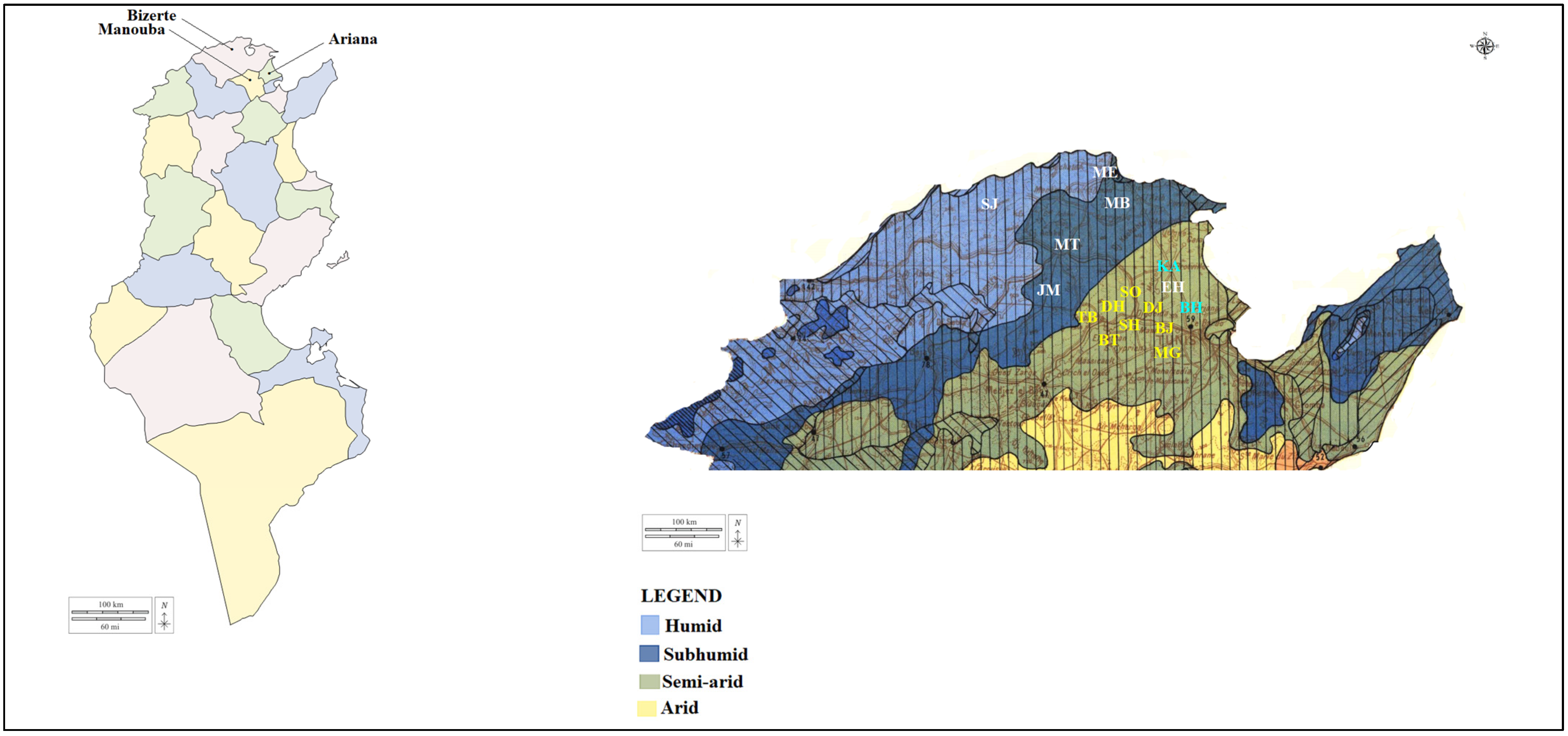
2.1. Study Regions, Tick Collection and Morphological Identification

Between June and September 2019 and 2020, ticks were randomly collected from 254 apparently healthy cattle (189 females and 65 males) reared in 45 farms located in three Tunisian governorates (Bizerte, Ariana and Manouba) belonging to two bioclimatic zones (subhumid and higher semi-arid) (Figure 1). The minimal required number of tick samples was estimated according to the following formula: N = 1.962 ∗ Pexp (1-Pexp)/d2 [23]. The expected prevalence (Pexp) of infection was determined according to previous reports on tick-borne bacterial infections in ticks infesting African ruminants (Pexp = 30%) with a confidential interval of 95% [21,23]. Here, d corresponds to the accepted absolute error (d) of 5%. According to this formula, a total of 322 samples were required in this study (107 ticks from each governorate (n = 3), along with 7 specimens from each herd (n = 45)).
The farms visited were small, enclosing an average of twenty heads of cattle, with traditional and poorly maintained dwellings. The cattle analyzed were aged from 6 months to 15 years and mainly belonged to the Friesian Pie Noire and Holstein breeds. Despite the use of an acaricide treatment, almost all animals surveyed were infested with ticks, particularly in the mammary region and the inner surface of the ears. Only unfed and partially engorged ticks were manually collected from different preferred sites of animal bodies (ears, neck, udder and external genitalia) and separately categorized according to the examined cattle. The obtained specimens were morphologically identified using the taxonomic key used by Walker [24], and then classified according to tick species, life stage and gender. Each tick specimen was individually conserved in a tube containing 70% ethanol and stored at −20 °C.

2.2. Total DNA Extraction and Tick DNA Amplification

Each identified tick was washed with sterile water, dried and crushed individually using an automated TissueLyser LT system (Qiagen, Hilden, Germany). Genomic DNA extraction was performed from each tick sample using the DNeasy tissue kit (Qiagen, Hilden, Germany). The obtained DNA extracts were stored at −20 °C. The DNA extraction efficiency was validated by PCR amplification targeting the ribosomal RNA subunit (mitochondrial 16S rRNA) gene using the tick-specific primers TQ16S+1F and TQ16S-2R, as described by Black and Piesman [25] (Table 1).

2.3. Molecular Detection of Rickettsia Species

Firstly, a nested PCR targeting a fragment (425 bp) of the rickettsial outer membrane protein B (ompB) gene tick DNA samples was performed in order to identify all Rickettsia species. For further characterization, nested and single PCRs were carried out, respectively, on the outer membrane protein A (ompA) and the citrate synthase protein (gltA) gene fragments (532 and 381 bp, respectively). The PCR tests were performed in an automated DNA thermal cycler. The thermal cycling profiles were as described by Oteo, Portillo, [27] and Regnery, Olson, [28] respectively.
The PCR reactions were performed in a final volume of 50 µL composed of 0.125 U/µL of Taq DNA polymerase (Biobasic Inc., Markham, Canada), 1× PCR buffer, 0.2 mM of dNTP, 1.5 mM of MgCl2, 3 μL of genomic DNA (50–150 ng) in the first PCR and 1 μL in the second PCR (for nested PCR), 0.5 μM of the primers and autoclaved water. An electrophoresis phase in 1.5% agarose gels stained with ethidium bromide was performed to visualize the PCR products under UV transillumination.

2.4. Statistical Analysis

The exact confidence intervals (CI) for prevalence rates at the 95% level were estimated. To study the potential influence of abiotic factors (geographic sites and bioclimatic areas) and factors related to the ticks (species and gender) on the molecular prevalence of Rickettsia species, a chi-square test or Fisher’s exact test was performed using Epi Info 6.01 (CDC, Atlanta, GA, USA) with a threshold value of 0.05.

2.5. DNA Sequencing, Sequence Alignment and Phylogenetic Study

Selected positive PCR products obtained after mitochondrial 16S rRNA, ompB, ompA and gltA PCR tests were selected and purified using the GF-1 Ambi Clean kit (Vivantis, Oceanside, CA, USA) according to the manufacturer’s instructions. The purified DNA amplicons were sequenced in both directions, using the same primers as for the single mitochondrial 16S rRNA and gltA PCRs and the second PCR of each nested PCR amplification targeting ompA and ompB genes. The Big Dye Terminator cycle sequencing ready reaction kit (Applied Biosystems, Foster City, CA, USA) and an ABI3730XL automated DNA sequencer (Macrogen Europe, Amsterdam, The Netherlands) were employed.
The chromatograms were evaluated with Chromas Lite v 2.01 (http://www.technelysium.com.au/chromas_lite.html (accessed on 3 September 2022)). The raw sequences were determined on both forward and reverse strands in order to achieve maximal data accuracy. The complementary strands of each sequenced product were manually assembled using the DNAMAN software program (Version 5.2.2; Lynnon Biosoft, Que., Canada). The overlapping parts were selected after the automatic removal of primer region sequences. The nucleotide sequences of the mitochondrial 16S rRNA as well as of the three genes ompB, ompA and gltA of Rickettsia spp. were used to calculate the genotype diversity (Gd), the nucleotide diversity (Pi) and the average number of nucleotide differences (k), using DnaSP version 5.10 software (http://www.ub.edu/dnasp/ (accessed on 3 September 2022)).
Sequence similarities were calculated using the CLUSTAL W method [29] after multiple sequence alignments. A BLAST analysis was performed to assess the level of similarity with previously reported sequences (http://blast.ncbi.nlm.nih.gov/ (accessed on 25 September 2022)). By using the DNAMAN software program, genetic distances among the operational taxonomic units were computed using the maximum composite likelihood method [30] and were used to construct neighbor-joining trees [31]. Using a bootstrapping process with 1000 iterations, the statistical support for the internal branches of the trees was evaluated [32].

3. Results

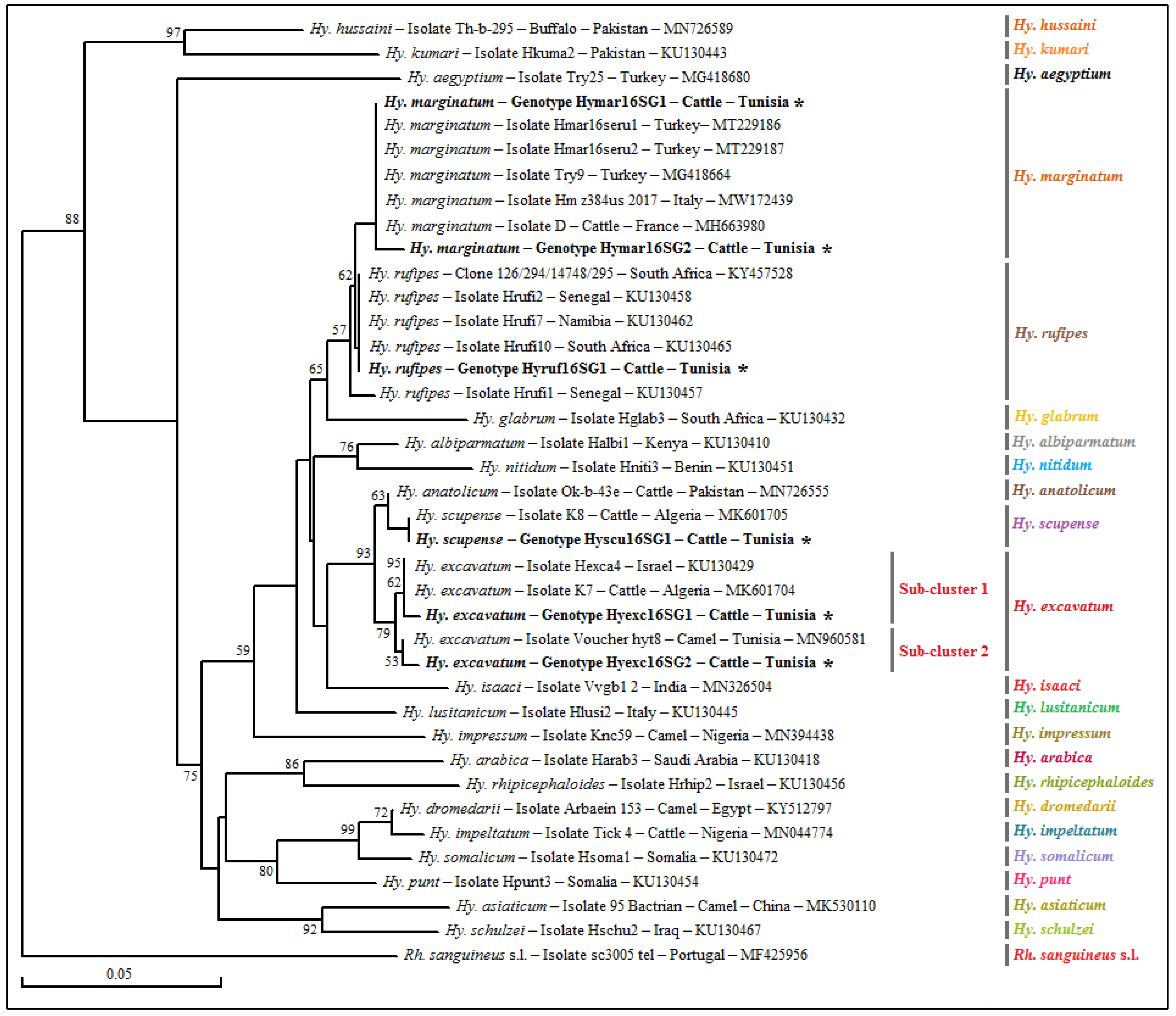
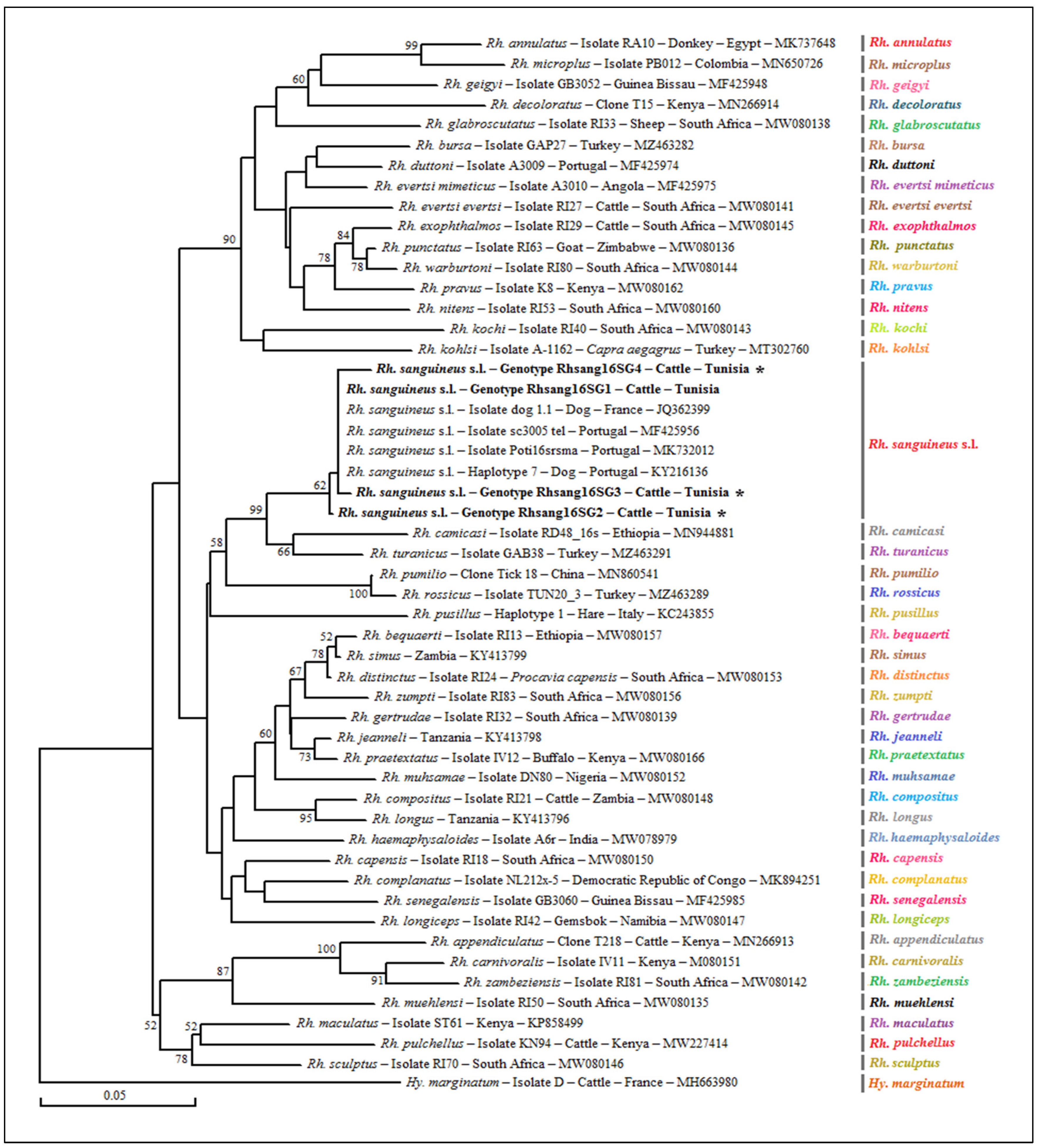
3.1. Morphological and Molecular Identification of Ticks and Phylogenetic Analysis

3.1.1. Efficiency of DNA Isolation and Distribution of Collected Ticks

A total of 338 adult ticks (70 females and 268 males) were collected from cattle situated in the Bizerte, Manouba and Ariana governorates, comprising higher semi-arid area (31.9%) and subhumid (22.8%) areas (Table 2). The tick DNA extracts were tested using a single PCR based on mitochondrial 16S rRNA and validated in all samples (100%). The morphological diagnosis using the diagnostic key used by Walker et al. [24] and molecular identification involving sequencing and a BLAST analysis of a partial sequence of the mitochondrial 16S rRNA gene of 83 ticks showed that the 338 collected ticks belong to the two genera Hyalomma (n = 227) and Rhipicephalus (n = 111), particularly to the species Hy. excavatum (n = 129), Rh. sanguineus sensu lato (n = 111), Hy. marginatum (n = 84), Hy. scupense (n = 12) and Hy. rufipes (n = 2) (Table 2).

3.1.2. Genotyping and Phylogenetic Analysis of Selected Tick Specimens

In order to confirm the results of the morphological identification of the analyzed ticks and to genetically characterize the isolates of each revealed species, the sequencing of 320 bp of the mitochondrial 16S rRNA gene was carried out on 83 randomly selected positive (n = 56) and negative (n = 27) ticks for the Rickettsia genus. Four species of the Hyalomma genus, namely Hy. marginatum, Hy. excavatum, Hy. scupense and Hy. rufipes, and one species of the Rhipicephalus, genus namely Rh. sanguineus s.l., were identified from the BLAST analysis (Table 3, Tables 5 and 6 and Tables S1–S4). Based on this partial sequence, we accurately selected genotypes that differed from each other by at least one mutation at the nucleotide sequence belonging to the mitochondrial 16S rRNA gene.
The genetic diversity analysis performed using DnaSP version 5.10.01 software on a 272 bp sequence of the mitochondrial 16S rRNA gene identified two different genotypes for Hy. marginatum named Hymar16SG1 and Hymar16SG2, isolated from 24 specimens with genotype diversity (Gd) equal to 0.159. The percentage of GC was 47.8%. The nucleotide diversity (Pi) and the average number of nucleotide differences (k) were estimated, respectively, at 0.00059 and 0.159 by noting the presence of a single mutational position between the two revealed genotypes, sharing 99.63% similarity in terms of nucleotide sequences (Table 4). The first genotype (Hymar16SG1) was found to be different from those published in GenBank and is, therefore, considered a new genetic variant (Table 3 and Table S1). The second genotype (Hymar16SG2) was found to be identical to isolate D of the Hy. marginatum tick specimen infesting cattle in France (GenBank accession number MH663980) (Figure 2). The alignment of partial sequences of the mitochondrial 16S rRNA gene of Hy. excavatum revealed two different genotypes, named Hyexc16SG1 and Hyexc16SG2, isolated from 31 tick specimens, with genotype diversity (Gd) equal to 0.452. The percentage of GC was 49.3%. The nucleotide diversity (Pi) and average number of nucleotide differences (k) were estimated, respectively, at 0.00502 and 1.355 by noting the presence of three mutational positions between the two revealed genotypes, sharing 98.89% similarity in terms of nucleotides (Table 4). The two genotypes (Hyexc16SG1 and Hyexc16SG2) were found to be different from all those published in GenBank and are, therefore, considered novel genetic variants (Table 5 and Table S2). The only 16S rRNA sequence revealed from Hy. rufipes is represented by the Hyruf16SG1 genotype. This genotype is identical to the South African isolate Hrufi10 (GenBank accession number KU130465) (Table 5).
The phylogenetic analysis based on the alignment of Tunisian genotypes belonging to the three revealed tick species, with different sequences of several Hyalomma species obtained from GenBank, generated several clusters (Figure 2). The Hy. marginatum cluster is formed by several isolates from different Mediterranean countries such as Italy, France and Turkey (Figure 2). The first genotype (Hymar16SG1) was found to be identical to all these isolates, while the second (Hymar16SG2) is genetically close. Moreover, the Hy. excavatum cluster is formed of two subclusters with a node robustness equal to 79% (Figure 2). The first genotype (Hyexc16SG1) was assigned to the first subcluster, with a Hy. excavatum isolate from Algeria (MK601704), and the second genotype (Hyexc16SG2) was clustered with an isolate collected from a Tunisian dromedary (GenBank accession number MN960581) in the second subcluster. The cluster representing the Hy. rufipes species is composed of the Hyruf16SG1 genotype revealed in the present study and those isolated from other tick specimens of Hy. rufipes from several African countries such as Senegal, Namibia and South Africa (Figure 2).
The genetic diversity analysis carried out using the software program DnaSP version 5.10.01 made it possible to identify four different genotypes for Rh. sanguineus s.l. named Rhsang16SG1–Rhsang16SG4, isolated from 15 tick specimens, with a diversity of genotypes (Gd) equal to 0.467. The percentage of GC was 48.5%. The nucleotide diversity (Pi) and average number of nucleotide differences (k) were estimated, respectively, at 0.00189 and 0.514 by noting the presence of three mutational positions between the four revealed genotypes, sharing 99.6 to 99.3% nucleotide similarity (Table 4). The first genotype (Rhsang16SG1) was found to be identical to isolate dog 1.1 from a tick of the Rh. sanguineus s.l. complex collected from a dog in France (GenBank accession number JQ362399). The remaining three genotypes (Rhsang16SG2–Rhsang16SG4) were found to be different from all of those published in GenBank and are, therefore, considered new genetic variants (Table 4 and Table 6). The phylogenetic analysis based on the alignment of our Rh. sanguineus s.l. sequences showed a similarity with those of Rhipicephalus spp. published in GenBank. The cluster of Rh. sanguineus s.l. was composed of several isolates from southern Mediterranean countries such as Portugal and France. The Rhsang16SG1 genotype was phylogenetically the closest, while the Rhsang16SG2 genotype was the most distant (Figure 3).

3.2. Molecular Prevalence of Rickettsia spp.

According to our ompB PCR results, the overall infection rate of Rickettsia spp. was 26.6% (90/338). The Rickettsia infection rates were similar between higher semi-arid (31.9%) and subhumid (22.8%) areas and the low difference between the infection rates was statistically not significant (p = 0.063) (Table 2). Additionally, the infection prevalence rates between the governorates were similar, estimated at 22.8%, 32.7% and 28% in cattle ticks located, respectively, in farms from the governorates of Bizerte, Manouba and Ariana, showing a statistically non-significant difference (p = 0.157) (Table 2). The four tick species showed distinct infection rates and the difference was statistically significant (p = 0.022). Indeed, the highest rate was estimated in Hy. rufipes (50%) followed by Rh. sanguineus s.l. (34.2%), then finally Hy. marginatum and Hy. excavatum, with similar rates estimated at 29.8% and 20.1%, respectively (Table 2). Additionally, a statistically non-significant difference was recorded between the prevalence rates in the two sexes (p = 0.186), with rates estimated at 32.8% and 25% in female and male ticks, respectively (Table 2).

3.3. Rickettsia Species Identification

In order to identify and genetically characterize the revealed Rickettsia species, at least one of the three partial sequences of the analyzed genes (ompB, ompA and gltA) was sequenced for the 66 samples positive for Rickettsia spp. selected for sequencing (i.e., 22 Hy. excavatum, 22 Rh. sanguineus s.l., 21 Hy. marginatum and one Hy. rufipes). Partial sequences (n = 104) of the three analysed genes were obtained and deposited in GenBank under the accession numbers OQ123608–OQ123654 for ompB partial sequences, OQ123655-OQ123684 for ompA partial sequences and OQ123685–OQ123711 for gltA partial sequences.
Three Rickettsia species were identified in ticks positive for Rickettsia spp. selected for the genetic analysis, namely R. aeschlimannii, R. sibirica subsp. mongolitimonae and R. massiliae (Table 7). Based on the analysis of the three genes, coinfection by R. massiliae and R. aeschlimannii was reported in two ticks of Hy. marginatum (Hyma72 and Hyma336) and two Rh. sanguineus s.l. ticks (Rhsa73 and 273), while a coinfection with R. massiliae and R. sibirica subsp. mongolitimonae was only recorded in one specimen of the Rh. sanguineus s.l. complex (Rhsa284) (Table 3, Table 5 and Table 6).

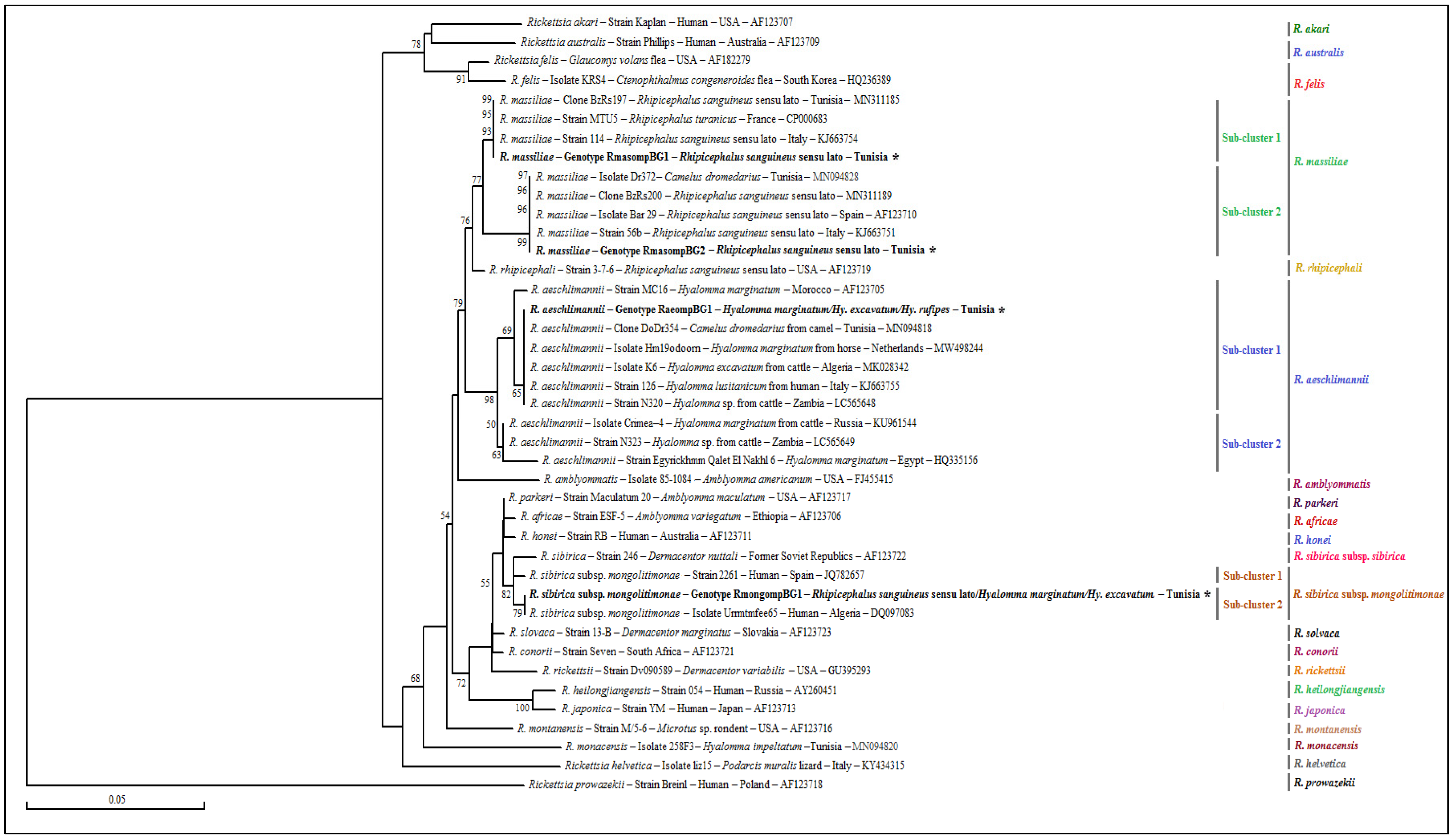
3.4. Genotyping and Phylogenetic Analysis

Based on all revealed sequences of the three analysed genes, we precisely selected the genotypes that differed from each other by at least one mutation at the nucleotide sequence level.

3.4.1. Rickettsia spp. ompB Partial Sequences

The sequencing of ompB partial sequence (382 bp) was performed on 47 cattle tick samples belonging to Hy. marginatum (n = 14), Hy. excavatum (n = 18), Hy. rufipes (n = 1) and Rh. sanguineus s.l. (n = 14). The BLAST analysis showed that 17 Hy. excavatum, 13 Hy. marginatum and one Hy. rufipes tick were infected with R. aeschlimannii. In addition, two ticks belonging to Hy. excavatum and Hy. marginatum species were positive for R. sibirica subsp. mongolitimonae. Furthermore, the sequencing of fourteen samples of Rh. sanguineus s.l. ticks showed that 13 samples were found to be infected with R. massiliae and only one tick specimen was positive for R. sibirica subsp. mongolitimonae (Table 7).
The genetic diversity analysis carried out using DnaSP version 5.10.01 software on a 382 bp of the ompB gene made it possible to identify two different genotypes for 13 R. massiliae isolates, named RmasompBG1 and RmasompBG2, with genotype diversity (Gd) equal to 0.385. The GC rate was 51.3%. The nucleotide diversity (Pi) and average number of nucleotide differences (k) were estimated, respectively, at 0.00503 and 1.923 by noting the presence of 5 mutational positions between the two different revealed genotypes, sharing 98.69% nucleotide similarity (Table 4). These two genotypes were precisely isolated from thirteen specimens of Rh. sanguineus s.l. ticks (Table 6). The RmasompBG1 genotype was found to be identical to the MTU5 strain isolated from a human in France (GenBank accession number CP000683), and the RmasompBG1 genotype was identical to the Bar29 strain isolated from a Rh. sanguineus s.l. tick specimen located in Spain (GenBank accession number AF123710).
The sequence alignment of R. aeschlimannii revealed a single genotype named RaeompBG1 isolated from specimens belonging to Hy. marginatum, Hy. excavatum and Hy. rufipes. This genotype was identical to the DoDr354 clone belonging to R. aeschlimannii isolated from a Hyalomma dromedarii tick specimen infesting a Tunisian camel (GenBank accession number MN094818). The alignment of partial sequences belonging to R. sibirica subsp. mongolitimonae made it possible to select a single genotype named RmongompBG1 infecting one Hy. marginatum, one Hy. excavatum specimen and one Rh. sanguineus s.l. tick. This genotype was found to be identical to the pathogenic isolate Urrmtmfee65 of R. sibirica subsp. mongolitimonae infecting a human from Algeria (GenBank accession number DQ097083).
The phylogenetic analysis based on the alignment of our ompB genotypes belonging to the three revealed species, with different partial sequences of several classified Rickettsia species obtained from GenBank, generated various clusters (Figure 4). The R. massiliae cluster is formed of two subclusters genetically close to the R. rhipicephali cluster, with a robustness node equal to 77% (Figure 4). The first genotype (RmasompBG1) was assigned to the first subcluster with those isolated from strain MTU5 infecting a human in France (GenBank accession number CP000683), with clone BzRs197 and strain 114 both isolated from Rh. sanguineus s.l. ticks, respectively, in Tunisia and Italy (GenBank accession numbers MN311185 and KJ663754, respectively). The second genotype was classified to the second subcluster, with isolate Dr372 infecting a camel in Tunisia (GenBank accession numbers MN094828) and several isolates and strains infecting Rh. sanguineus s.l. ticks from several Mediterranean countries, such as Tunisia, Spain and Italy (Figure 4). The R. aeschlimannii cluster is formed by two subclusters with a robustness, node equal to 98% (Figure 4). The only revealed genotype (RaeompBG1) was assigned to the first subcluster, containing several isolates and strains infecting various Hyalomma ticks species parasitizing a human in Italy and a cattle and a horse, respectively, in Russia and the Netherlands (Figure 4). Finally, the cluster representing the R. sibirica subsp. Mongolitimonae subspecies is composed of two subclusters, with a robustness node equal to 82%, the second of which is formed by the RmongompBG1 genotype, revealed in the present study in one Hy. excavatum tick and the pathogenic isolate (Urrmtmfee65) of R. sibirica subsp. mongolitimonae infecting an Algerian human (GenBank accession number DQ097083) (Figure 4).

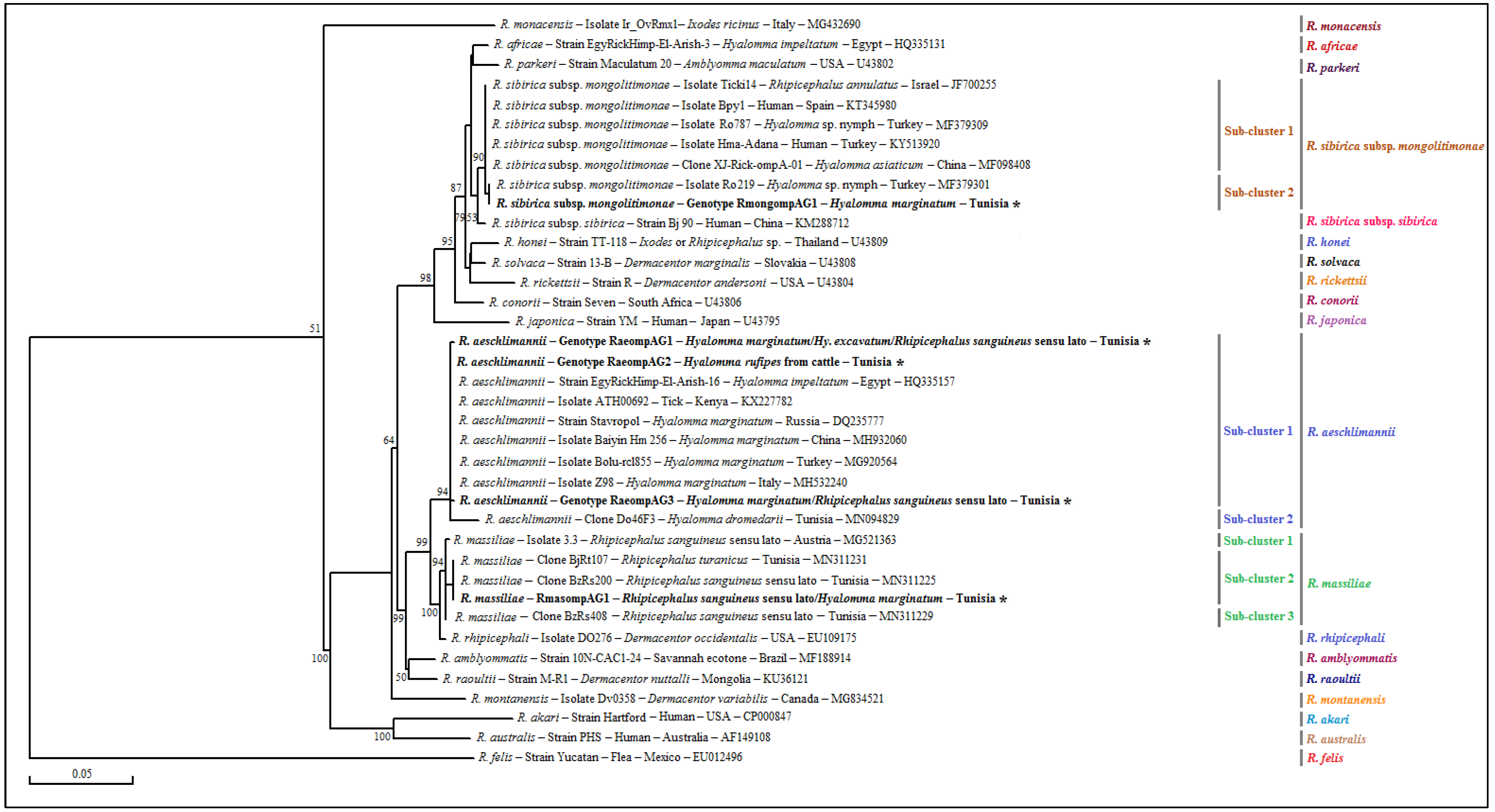
3.4.2. Rickettsia spp. ompA Partial Sequences

The sequencing of a 490 bp fragment of the ompA gene, which corresponds to the 532 bp amplified sequence without the forward and reverse primer sequences, confirmed the presence of R. massiliae, R. aeschlimannii and R. sibirica subsp. mongolitimonae. The obtained results affirmed that Hyalomma ticks, precisely 7 Hy. marginatum, 10 Hy. excavatum and one Hy. rufipes, were tested positive for R. aeschlimannii. However, only one specimen of Hy. marginatum was found positive for R. massiliae (Table 7). It was noted that only one tick specimen of the Hy. marginatum species was recorded as being positive for R. sibirica subsp. mongolitimonae. However, for Rhipicephalus sanguineus s.l., eight were positive for R. massiliae and only one tick specimen of this complex was positive for R. aeschlimannii (Table 7).
The alignment of sequences Isolated from R. massiliae allowed us to select a single genotype named RmasompAG1 infecting eight Rh. sanguineus s.l. ticks and one Hy. marginatum tick specimen. This genotype was found to be identical to the BzRs200 clone of R. massiliae isolated from a Rh. sanguineus s.l. tick specimen infecting a Tunisian goat (GenBank accession number MN311225).
The genetic diversity analysis performed using the software DnaSP version 5.10.01 on a partial sequence of 490 bp of the ompA gene made it possible to identify three different R. aeschlimannii genotypes named RaeompAG1, RaeompAG2 and RaeompAG3 isolated from seven Hy. marginatum specimens, with ten others belonging to Hy. Excavatum species and one Rh. sanguineus s.l. specimen, with the diversity of the genotypes (Gd) estimated at 0.542. The percentage of GC was 53.8%. The nucleotide diversity (Pi) and the average number of nucleotide differences (k) were estimated, respectively, at 0.00123 and 0.605 by noting the presence of two mutational positions between the three different revealed genotypes. Our genotypes shared 99.8–99.6% nucleotide similarity. The RaeompAG1 genotype was found to be identical to the Z98 isolate of R. aeschlimannii isolated from Hy. marginatum in Italy (GenBank accession number MH532240). The two other genotypes RaeompAG2 and RaeompAG3 were different from all other sequences published in GenBank and were considered as new genetic variants (Table 4, Table 5 and Table 6).
The only sequence belonging to R. sibirica subsp. mongolitimonae infecting one Hy. marginatum tick specimen allowed us to select a single genotype named RmongompAG1. The BLAST analysis showed that the latter was identical to isolate Ro219 infecting a Hyalomma nymph tick collected in Turkey (GenBank accession number MF379301).
The phylogenetic analysis based on the alignment of our Tunisian genotypes with different partial sequences of the ompA gene of several Rickettsia species obtained from GenBank generated several clusters (Figure 5). The R. massiliae cluster comprises three subclusters with a robustness node equal to 94%. The RmasompAG1 genotype revealed in the present study was assigned to the second subcluster along with those of R. massiliae clones BjRt107 and BzRs200 isolated from Rh. turanicus and Rh. sanguineus s.l. ticks infesting goats in Tunisia (GenBank accession numbers MN311231 and MN311225). The R. aeschlimannii cluster is relatively heterogeneous, being composed of three different subclusters, with a robustness node equal to 94%. The three revealed genotypes (RaeompAG1, RaeompAG2 and RaeompAG1) identified in our study were present in the first subcluster, with a multitude of isolates and strains infecting tick specimens mainly of Hy. marginatum species from several countries around the world. In the end, the cluster representing the subspecies R. sibirica subsp. mongolitimonae, which is genetically close to the cluster of R. sibirica subsp. sibirica, is formed by two subclusters with a robustness node equal to 90%, the second of which is formed by the RmongompAG1 genotype revealed in this study and the isolate Ro219 infecting a Hyalomma sp. tick in Turkey (GenBank accession number MF379301) (Figure 5).

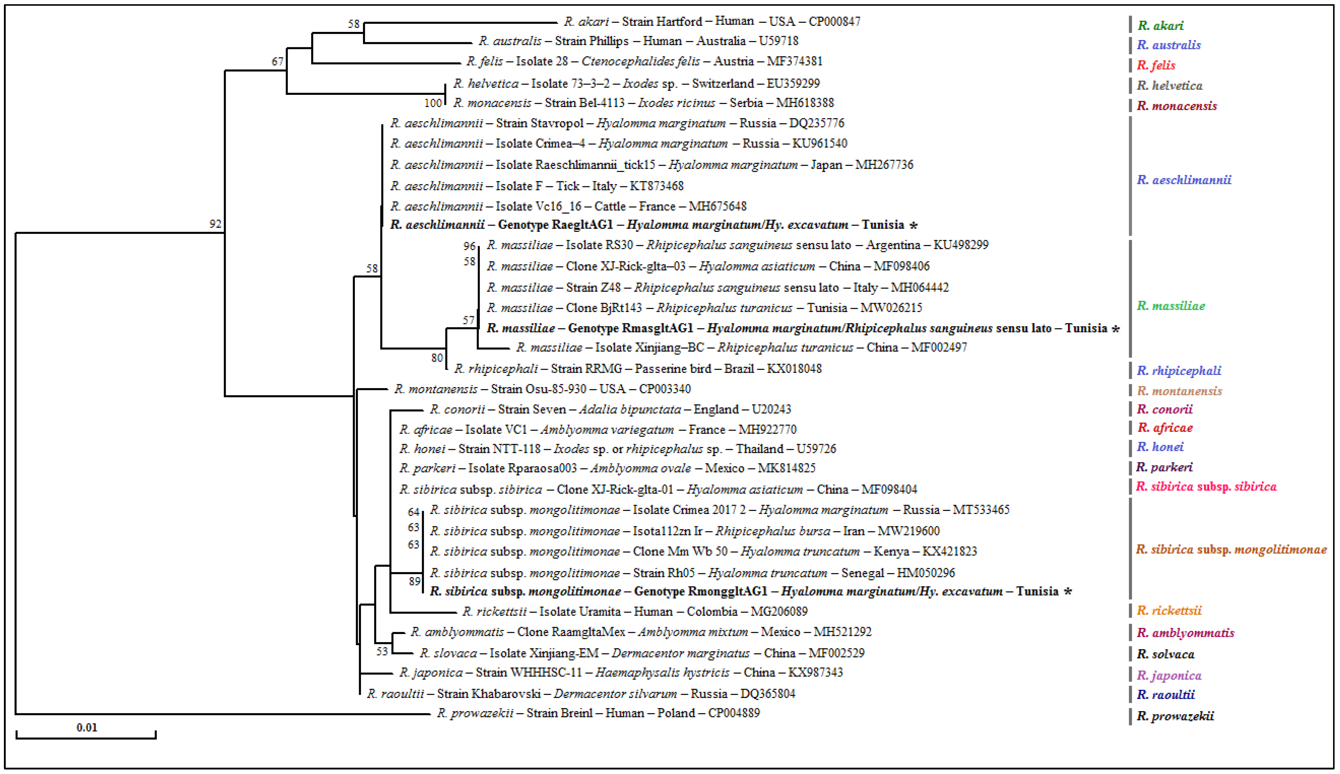
3.4.3. Rickettsia spp. gltA Partial Sequences

The sequencing of a 341 bp fragment of the gltA gene, which corresponds to the 381 bp amplified sequence without the forward and reverse primer sequences, revealed infections with R. aeschlimannii, R. sibirica subsp. mongolitimonae and R. massiliae (Table 3, Table 5 and Table 6). The BLAST analysis confirmed the infection of 4 Hy. excavatum and 6 Hy. marginatum with R. aeschlimannii. In addition, one Hy. marginatum tick tested positive for R. massiliae and two ticks of Hy. excavatum and Hy. marginatum specimens were found to be infected with R. sibirica subsp. mongolitimonae. However, 15 Rh. sanguineus s.l. ticks were found to be infected with R. massiliae (Table 7).
The alignment of sequences belonging to R. massiliae revealed a single genotype named RmasgltAG1 infecting 1 and 15 specimens belonging, respectively, to Hy. marginatum and Rh. sanguineus s.l. This genotype was found to be identical to the R. massiliae clone BjRt143 isolated from one Rh. turanicus tick infecting a goat from Tunisia (GenBank accession number MW026215). The sequence analysis of R. aeschlimannii isolates identified a single genotype named RaegltAG1 infecting six Hy. marginatum and four Hy. excavatum tick specimens. This genotype was 100% identical to the R. aeschlimannii isolate Vc16_16 infecting cattle in France (GenBank accession number MH675648) (Table 3, Table 5 and Table 6). The only sequence belonging to R. sibirica subsp. mongolitimonae infecting a Hy. excavatum tick specimen allowed us to select a single genotype named RmonggltAG1. The BLAST analysis showed that this genotype was 100% identical to the Crimea 2017/2 isolate of R. sibirica subsp. mongolitimonae infecting one Hy. marginatum specimen from Russia (GenBank accession number MT533465).
The phylogenetic tree based on the gltA gene revealed that the RmasgltAG1 genotype clustered in the R. massiliae cluster with strains infecting Rh. sanguineus s.l. ticks from Italy and Argentina, Hyalomma asiaticum ticks from China and Rh. turanicus tick specimens infesting small ruminants in Tunisia (Figure 6). For the gltA gene, the R. aeschlimannii cluster is homogeneous and the single genotype (RaegltAG1) revealed in the present study is included with several isolates and strains infecting Hy. marginatum specimens from several worldwide countries and with an isolate of R. aeschlimannii infecting cattle in France. The cluster representing the subspecies R. sibirica subsp. mongolitimonae was relatively homogeneous, containing several isolates infecting ticks of Hy. truncatum species from African countries and Hy. marginatum species located in Russia (Figure 6).

4. Discussion

Ticks of the Ixodidae family are, along with mosquitoes, the most relevant vectors of pathogens, with veterinary and medical importance worldwide [33]. The majority of these pathogens appear in tropical countries, which cause the rising of the incidence of tick-borne diseases (TBDs), due to increased interactions between pathogens, hosts and vectors, related directly to global changes [34]. However, epidemiological studies on these diseases are very limited in Tunisia [22].
To date, despite the large cattle population in Tunisia, screening studies for Rickettsia bacteria in cattle ticks are very few. Therefore, the present study aimed to detect and characterize ticks of cattle reared in traditional farms located in northern Tunisia and their associated Rickettsia species.
In the present study, three hundred and thirty-eight ticks were collected from cattle in northern Tunisia, most of them belonging to the Hyalomma genus (227/338), with precisely four species (Hy. excavatum, Hy. marginatum, Hy. scupense and Hy. rufipes), while only one Rhipicephalus species was identified as Rh. sanguineus s.l. (111/338). The overall prevalence of Rickettsia spp. was 26.6% (90/338). The tick species Hy. rufipes was the most infected (50%), followed by Rh. sanguineus s.l. (34.2%) and Hy. marginatum (29.8%). The weakest infection rate was recorded in Hy. excavatum specimens (20.1%). Indeed, these results concur with those of a recent study conducted on the infection of cattle ticks in Cameroon, reporting a high rate of infection with Rickettsia spp. (50%) in Hy. rufipes ticks, suggesting that it could be considered as one of the main vectors of Rickettsia spp. in this country [35]. Moreover, according to Cicculli et al. [36], Rickettsia spp. were detected in Hy. marginatum ticks collected from cattle in France (15.5%), suggesting that this tick species has an important role in the transmission of these pathogens. Furthermore, it is interesting to note that other species, such as Hy. dromedarii (6%) and Hy. impeltatum (8%), are considered potential vectors of Rickettsia spp. in camel herds in Tunisia [13]. Additionally, Pesquera et al. [37], as well as Ehlers et al. [38], reported that Rhipicephalus ticks, more specifically Rh. microplus, are potentially involved in the transmission of Rickettsia bacteria to cattle located in Madagascar and the Comoros Islands.
Additionally, the infection rate of Rickettsia spp. is less important in Rh. sanguineus s.l. compared to those found in other species of the Hyalomma genus, in agreement with various reports that have considered that this species of tick is often less infected by Rickettsia spp. compared to species belonging to Hyalomma and Amblyomma genera [35,39,40].
Even if the incidence of TBDs is rising, scarce data on ticks and TBDs in ruminants are available. In Tunisia, previous studies carried out on the detection of the DNA of Rickettsia bacteria in ticks collected from small ruminants [22] made it possible to identify the infection rates in Rh. turanicus (23.4%) and Rh. sanguineus s.l. (9.5%) specimens. This provides evidence that Rhipicephalus spp. could be among the main vectors of Rickettsia species in northern Tunisia [22]. Our results are also consistent with those reported by Khrouf et al. [21], who suggested the potential incrimination of ticks of the Rhipicephalus genus infesting dogs and sheep from central Tunisia in the transmission of Rickettsia species.
In the present study, the sequencing of three different DNA fragments of ompB, ompA and gltA genes revealed the presence of three species of Rickettsia, namely R. aeschlimannii, R. sibirica subsp. mongolitimonae and R. massiliae. The identification of R. aeschlimannii in ticks of the Hyalomma genus collected from cattle is consistent with the results of previous studies confirming that Hy. marginatum and Hy. excavatum are the main vectors of this zoonotic agent [41,42,43,44]. R. sibirica subsp. mongolitimonae was also detected in Hy. excavatum and Rh. sanguineus s.l. ticks infesting cattle in northern Tunisia. However, this pathogen presents a great topic of interest, since it has been associated with human infections in France [45], South Africa [46], Greece [47] and Spain [48]. Several hypotheses suggest that the tick vectors of this pathogen primarily include species of the Hyalomma genus. On the other hand, in addition to R. aeschlimannii [49], it seems that other species of Hyalomma genus such as Hy. excavatum and even species belonging to other genera such as Rh. sanguineus s.l. seem to play a role as vectors of this zoonotic bacterium in Mediterranean countries. This diversity of potential tick vectors found in the north of the country could be related to the biotope. Indeed, the latter is characterized by dense vegetation, which offers high protection to ticks and due to its structure prevents the rapid movement of cattle, thereby facilitating their infestation [50]. Moreover, the presence of other animals such as dogs and small ruminants, as well as climate change over the years causing longer periods of drought have led to the abundance and diversification of tick species that infest cattle in these investigated regions [51].
The phylogenetic analysis of our isolates belonging to R. aeschlimannii species infecting ticks of the Hyalomma genus showed an almost perfect homology with those published in GenBank. Indeed, the analysis of ompB and gltA partial sequences proved that our isolates are similar to those previously detected in camels in Tunisia [13] and cattle in France [36] and in isolates identified in Hy. marginatum ticks from Italy [52]. This finding leads us to suggest that these two animal species as well as their associated tick species, essentially of the Hyalomma genus, are probably incriminated in the transmission cycle of R. aeschlimannii in the Mediterranean context.
Based on the phylogenetic analysis of ompB, ompA and gltA partial sequences, low genetic diversity was observed among R. sibirica subsp. mongolitimonae genotypes identified in this study. In particular, the RmonompBG1, RmonompAG1 and RmonggltAG1 genotypes of ompB, ompA and gltA genes, respectively, were 100% similar to those isolated from the Urrmtmfee 65, Ro219 and Crimea 2017/2 strains of R. sibirica subsp. mongolitimonae respectively infecting a human in Algeria, who developed fairly severe symptoms, including an inoculation sore on the leg, fever and lymphangitis extending from the sore to an enlarged and painful lymph node in the groin [53], as well as a tick of Hyalomma genus in Turkey [54] and another tick specimen from Hy. marginatum in Russia [55]. This finding leads us to suggest that the R. sibirica subsp. mongolitimonae isolates revealed in the present study could have zoonotic potential with their transmission ensured, in part, by ticks of the Rh. sanguineus s.l. complex and those of the Hyalomma genus, namely Hy. excavatum and Hy. marginatum. Despite the fact that the R. sibirica subsp. mongolitimonae is apparently associated with Hyalomma subspecies ticks in North Africa [56], further epidemiological and experimental studies are needed to confirm this hypothesis.
Additionally, R. massiliae DNA has been detected in tick specimens belonging to Rh. sanguineus s.l, Hy. marginatum and Hy. excavatum, thereby confirming its presence for the first time in cattle ticks in Tunisia. This SFG Rickettsia was identified in many ticks of the Rhipicephalus genus, such as Rh. sanguineus s.l., Rh. turanicus and Ixodes ricinus ticks in several European countries, infesting domestic and wild hosts such as dogs, cats, horses, red foxes and asymptomatic humans [57]. The sequence analysis revealed that the R. massiliae isolates showed low genetic diversity. In addition, the genotypes identified based on the partial sequences of the ompB gene showed perfect similarity to those isolated from the MTU5 strain of R. massiliae detected in a human from France [58] and to the Bar29 strain from Rh. sanguineus s.l. ticks located in Spain [59]. Furthermore, based on the partial sequence alignment of ompA gene, we found that the only revealed genotype (RmasompAG1) was identical to that of the BzRs200 clone of R. massiliae isolated from the Rh. sanguineus s.l. tick specimen infecting a Tunisian goat [22]. Based on the gltA gene, the only revealed genotype (RmasgltAG1) presented a perfect identity to that previously identified from an isolate of R. massiliae infecting one Rh. turanicus specimen collected from a goat in Tunisia [22]. Similarly, R. massiliae was also identified in Rh. turanicus and Rh. sanguineus s.l. from Algeria [60], Italy [31], Cyprus [49] and Greece [47]. Based on the analysis of the three genes, the sequence similarity between different isolates and strains of R. massiliae infecting humans and several tick species of Hyalomma and Rhipicephalus genera indicates a possible increased risk of rickettsioses for cattle and even for humans who cohabit in the studied regions.

5. Conclusions

In conclusion, the present study provides a molecular survey on Rickettsia spp. in cattle ticks from the north of Tunisia. Three Rickettsia species (R. sibirica subsp. mongolitimonae, R. aeschlimannii and R. massiliae), which are potential or validated human pathogens, were detected and characterized. However, further research studies are necessary to evaluate the pathogenicity of our revealed Rickettsia isolates and to confirm the role of each tick species investigated in this study in the transmission of these pathogens to humans and to different animal species in Tunisia.

Supplementary Materials

The following supporting information can be downloaded at: https://www.mdpi.com/article/10.3390/pathogens12040552/s1, Table S1: Designations and information on the origins and mitochondrial 16S rRNA genotypes of the remaining Tunisian isolates of Hyalomma marginatum ticks infesting cattle; Table S2: Designations and information on the origins and mitochondrial 16S rRNA genotypes of the remaining Tunisian isolates of Hyalomma excavatum ticks infesting cattle; Table S3: Designations and information on the origins and mitochondrial 16S rRNA genotypes of Tunisian isolates of Hyalomma scupense ticks infesting cattle; Table S4: Designations and information on the origins and mitochondrial 16S rRNA genotypes of the remaining Tunisian isolates of Rhipicephalus sanguineus sensu lato ticks infesting cattle.

Author Contributions

Conceptualization, M.B.S.; methodology, M.B.S.; software, M.K. and M.B.S.; validation, M.K. and M.B.S.; formal analysis, M.K. and M.B.S.; investigation, M.K., H.B., R.S., R.A. and M.B.S.; resources, M.K., R.S. and R.A.; data curation, M.D., M.M. and M.B.S.; writing—original draft preparation, M.K., and M.B.S.; writing—review and editing, L.M., H.B., R.S. and M.B.S.; visualization, L.M. and M.B.S.; supervision, M.B.S.; project administration, L.M. and M.B.S.; funding acquisition, L.M. and M.B.S. All authors have read and agreed to the published version of the manuscript.

Funding

This work was supported by the research laboratory “Laboratoire d’épidémiologie d’infections enzootiques des herbivores en Tunisie” (LR02AGR03), and the research projects “Screening and Molecular Characterization of Pathogenic and Zoonotic Bacteria of Medical and Economic Interest in Cattle and Camel Ticks in Tunisia” (19PEJC07-22) and “Study of the Bacterial Microbiota in Ticks with a Medical and Economic Impact in Tunisia: Contribution to the Control of Vector-Borne Bacterial Diseases” (P2ES2020-D4P1), all of which were funded by the Ministry of Higher Education and Scientific Research of Tunisia.

Institutional Review Board Statement

The present investigation was performed in compliance with the current ethical standards and animal welfare of the European guidelines. Informed oral consent was affirmed by all animal owners who participated in the study. The ticks were removed by competent veterinarians without animal suffering.

Informed Consent Statement

Informed consent was obtained from livestock owners before including their animals in this study.

Data Availability Statement

Not applicable.

Acknowledgments

The authors are thankful to the practicing veterinarians and their technicians for their help and contributions in facilitating the access to the farms.

Conflicts of Interest

The authors declare no conflict of interest.

References

  1. Garcia, K.; Weakley, M.; Do, T.; Mir, S. Current and Future Molecular Diagnostics of Tick-Borne Diseases in Cattle. Vet. Sci. 2022, 9, 241. [Google Scholar] [CrossRef] [PubMed]
  2. Kohli, S.; Atheya, U.K.; Thapliyal, A. Prevalence of Theileriosis in Cross-Bred Cattle: Its Detection through Blood Smear Examination and Polymerase Chain Reaction in Dehradun District, Uttarakhand, India. Vet. World 2014, 7, 168–171. [Google Scholar] [CrossRef] [Green Version]
  3. García, D.I.D.; Agatón, F.T.; Rosario-Cruz, R. Evaluación Económica Del Control de Garrapatas Rhipicephalus microplus En México/Economic Evaluation of Tick (Rhipicephalus microplus) Control in Mexico. CIBA Rev. Iberoam. Las Cienc. Biológicas Agropecu. 2016, 5, 43–52. [Google Scholar]
  4. Lagunes, R.L.; Domínguez-García, D.; Quiroz, H.; Martínez-Velázquez, M.; Rosario-Cruz, R. Potential Effects on Rhipicephalus microplus Tick Larvae Fed on Calves Immunized with a Subolesin Peptide Predicted by Epitope Analysis. Trop. Biomed. 2016, 33, 726–738. [Google Scholar] [PubMed]
  5. Hurtado, O.J.B.; Giraldo-Ríos, C. Economic and Health Impact of the Ticks in Production Animals. In Ticks and Tick-Borne Pathogens; Abubakar, M., Perera, P.K., Eds.; IntechOpen: London, UK, 2018. [Google Scholar]
  6. Merhej, V.; Raoult, D. Rickettsial Evolution in the Light of Comparative Genomics. Biol. Rev. 2010, 86, 379–405. [Google Scholar] [CrossRef]
  7. de la Fuente, J.; Antunes, S.; Bonnet, S.; Cabezas-Cruz, A.; Domingos, A.G.; Estrada-Peña, A.; Johnson, N.; Kocan, K.M.; Mansfield, K.L.; Nijhof, A.M.; et al. Tick-Pathogen Interactions and Vector Competence: Identification of Molecular Drivers for Tick-Borne Diseases. Front. Cell Infect. Microbiol. 2017, 7, 114. [Google Scholar] [CrossRef] [PubMed] [Green Version]
  8. Merhej, V.; Angelakis, E.; Socolovschi, C.; Raoult, D. Genotyping, Evolution and Epidemiological Findings of Rickettsia Species. Infect. Genet. Evol. 2014, 25, 122–137. [Google Scholar] [CrossRef]
  9. Alanazi, A.D.; Alanazi, A.D.; Nguyen, V.L.; Alyousif, M.S.; Manoj, R.R.S.; Alouffi, A.S.; Donato, R.; Sazmand, A.; Mendoza-Roldan, J.A.; Dantas-Torres, F.; et al. Ticks and Associated Pathogens in Camels (Camelus dromedarius) from Riyadh Province, Saudi Arabia. Parasit. Vectors 2020, 13, 110. [Google Scholar] [CrossRef] [Green Version]
  10. Azmat, M.; Ijaz, M.; Farooqi, S.H.; Ghaffar, A.; Ali, A.; Masud, A.; Saleem, S.; Rehman, A.; Ali, M.M.; Mehmood, K.; et al. Molecular Epidemiology, Associated Risk Factors, and Phylogenetic Analysis of Anaplasmosis in Camel. Microb. Pathog. 2018, 123, 377–384. [Google Scholar] [CrossRef]
  11. Ben Said, M.; Belkahia, H.; Alberti, A.; Abdi, K.; Zhioua, M.; Daaloul-Jedidi, M.; Messadi, L. First Molecular Evidence of Borrelia Burgdorferi Sensu Lato in Goats, Sheep, Cattle and Camels in Tunisia. Ann. Agric. Environ. Med. 2016, 23, 442–447. [Google Scholar] [CrossRef]
  12. Bahari, A.; Azami, S.; Goudarztalejerdi, A.; Karimi, S.; Esmaeili, S.; Chomel, B.B.; Sazmand, A. Molecular Detection of Zoonotic Pathogens in the Blood and Tissues of Camels (Camelus dromedarius) in Central Desert of Iran. Yale J. Biol. Med. 2021, 94, 249–258. [Google Scholar] [PubMed]
  13. Selmi, R.; Ben Said, M.; Ben Yahia, H.; Abdelaali, H.; Messadi, L. Molecular Epidemiology and Phylogeny of Spotted Fever Group Rickettsia in Camels (Camelus dromedarius) and Their Infesting Ticks from Tunisia. Transbound. Emerg. Dis. 2019, 67, 733–744. [Google Scholar] [CrossRef] [PubMed]
  14. Larson, P.S.; Espira, L.; Grabow, C.; Wang, C.A.; Muloi, D.; Browne, A.S.; Deem, S.L.; Fèvre, E.M.; Foufopoulos, J.; Hardin, R.; et al. The Sero-Epidemiology of Coxiella burnetii (Q Fever) across Livestock Species and Herding Contexts in Laikipia County, Kenya. Zoonoses Public Health 2019, 66, 316–324. [Google Scholar] [CrossRef] [Green Version]
  15. Selmi, R.; Belkahia, H.; Sazmand, A.; Ben Said, M.; Messadi, L. Epidemiology and Genetic Characteristics of Tick-Borne Bacteria in Dromedary Camels of the World. Acta Trop. 2022, 234, 106599. [Google Scholar] [CrossRef]
  16. Ereqat, S.; Nasereddin, A.; Al-Jawabreh, A.; Azmi, K.; Harrus, S.; Mumcuoglu, K.; Apanaskevich, D.; Abdeen, Z. Molecular Detection and Identification of Spotted Fever Group Rickettsiae in Ticks Collected from the West Bank, Palestinian Territories. PLoS Negl. Trop. Dis. 2016, 10, e0004348. [Google Scholar] [CrossRef]
  17. Roux, V.; Rydkina, E.; Eremeeva, M.; Raoult, D. Citrate Synthase Gene Comparison, a New Tool for Phylogenetic Analysis, and Its Application for the Rickettsiae. Int. J. Syst. Bacteriol. 1997, 47, 252–261. [Google Scholar] [CrossRef] [Green Version]
  18. Znazen, A.; Khrouf, F.; Elleuch, N.; Lahiani, D.; Marrekchi, C.; M’Ghirbi, Y.; Ben Jemaa, M.; Bouattour, A.; Hammami, A. Multispacer Typing of Rickettsia Isolates from Humans and Ticks in Tunisia Revealing New Genotypes. Parasit. Vectors 2013, 6, 367. [Google Scholar] [CrossRef] [Green Version]
  19. Khrouf, F.; Sellami, H.; Elleuch, E.; Hattab, Z.; Ammari, L.; Khalfaoui, M.; Souissi, J.; Harrabi, H.; M’ghirbi, Y.; Tiouiri, H.; et al. Molecular Diagnosis of Rickettsia Infection in Patients from Tunisia. Ticks Tick Borne Dis. 2016, 7, 653–656. [Google Scholar] [CrossRef]
  20. Demoncheaux, J.-P.; Socolovschi, C.; Davoust, B.; Haddad, S.; Raoult, D.; Parola, P. First Detection of Rickettsia aeschlimannii in Hyalomma dromedarii Ticks from Tunisia. Ticks Tick Borne Dis. 2012, 3, 398–402. [Google Scholar] [CrossRef]
  21. Khrouf, F.; M’Ghirbi, Y.; Znazen, A.; Ben Jemaa, M.; Hammami, A.; Bouattour, A. Detection of Rickettsia in Rhipicephalus sanguineus Ticks and Ctenocephalides felis Fleas from Southeastern Tunisia by Reverse Line Blot Assay. J. Clin. Microbiol. 2013, 52, 268–274. [Google Scholar] [CrossRef] [Green Version]
  22. Belkahia, H.; Selmi, R.; Zamiti, S.; Daaloul-Jedidi, M.; Messadi, L.; Ben Said, M. Zoonotic Rickettsia Species in Small Ruminant Ticks from Tunisia. Front. Vet. Sci. 2021, 8, 676896. [Google Scholar] [CrossRef] [PubMed]
  23. Thrusfield, M. Veterinary Epidemiology; Blackwell Publishing Professional: Ames, IA, USA, 2005; Volume 47, ISBN 0-632-06397-1. [Google Scholar]
  24. Walker, A.R.; International Consortium on Ticks and Tick-Borne Diseases. Ticks of Domestic Animals in Africa: A Guide to Identification of Species; Bioscience Reports: Edinburgh, UK, 2003; ISBN 9780954517304. [Google Scholar]
  25. Black, W.C.; Piesman, J. Phylogeny of Hard- and Soft-Tick Taxa (Acari: Ixodida) Based on Mitochondrial 16S RDNA Sequences. Proc. Natl. Acad. Sci. USA 1994, 91, 10034–10038. [Google Scholar] [CrossRef] [PubMed] [Green Version]
  26. Choi, Y.-J.; Jang, W.-J.; Kim, J.-H.; Ryu, J.-S.; Lee, S.-H.; Park, K.-H.; Paik, H.-S.; Koh, Y.-S.; Choi, M.-S.; Kim, I.-S. Spotted Fever Group and Typhus Group Rickettsioses in Humans, South Korea. Emerg. Infect. Dis. 2005, 11, 237–244. [Google Scholar] [CrossRef] [PubMed]
  27. Oteo, J.A.; Portillo, A.; Santibáñez, S.; Blanco, J.R.; Pérez-Martínez, L.; Ibarra, V. Cluster of Cases of Human Rickettsia felis Infection from Southern Europe (Spain) Diagnosed by PCR. J. Clin. Microbiol. 2006, 44, 2669–2671. [Google Scholar] [CrossRef] [Green Version]
  28. Regnery, R.L.; Olson, J.G.; Perkins, B.A.; Bibb, W. Serological Response to “Rochalimaea henselae” Antigen in Suspected Cat-Scratch Disease. Lancet 1992, 339, 1443–1445. [Google Scholar] [CrossRef]
  29. Pearson, W.R. An Introduction to Sequence Similarity (“Homology”) Searching. Curr. Protoc. Bioinform. 2013, 42, 3.1.1–3.1.8. [Google Scholar] [CrossRef] [Green Version]
  30. Schloss, P.D.; Westcott, S.L. Assessing and Improving Methods Used in Operational Taxonomic Unit-Based Approaches for 16S RRNA Gene Sequence Analysis. Appl. Environ. Microbiol. 2011, 77, 3219–3226. [Google Scholar] [CrossRef] [Green Version]
  31. Desper, R.; Gascuel, O. Getting a Tree Fast: Neighbor Joining, FastME, and Distance-Based Methods. Curr. Protoc. Bioinform. 2006, 15, 6.3.1–6.3.28. [Google Scholar] [CrossRef] [PubMed]
  32. Mort, M.E.; Soltis, P.S.; Soltis, D.E.; Mabry, M.L. Comparison of Three Methods for Estimating Internal Support on Phylogenetic Trees. Syst. Biol. 2000, 49, 160–171. [Google Scholar] [CrossRef]
  33. Dantas-Torres, F.; Otranto, D. Best Practices for Preventing Vector-Borne Diseases in Dogs and Humans. Trends Parasitol. 2016, 32, 43–55. [Google Scholar] [CrossRef]
  34. Battisti, E.; Zanet, S.; Boraso, F.; Minniti, D.; Giacometti, M.; Duscher, G.G.; Ferroglio, E. Survey on Tick-Borne Pathogens in Ticks Removed from Humans in Northwestern Italy. Vet Parasitol. Reg. Stud. Rep. 2019, 18, 100352. [Google Scholar] [CrossRef] [PubMed]
  35. Vanegas, A.; Keller, C.; Krüger, A.; Manchang, T.K.; Hagen, R.M.; Frickmann, H.; Veit, A.; Achukwi, M.D.; Krücken, J.; Poppert, S. Molecular Detection of Spotted Fever Group Rickettsiae in Ticks from Cameroon. Ticks Tick Borne Dis. 2018, 9, 1049–1056. [Google Scholar] [CrossRef]
  36. Cicculli, V.; Capai, L.; Quilichini, Y.; Masse, S.; Fernández-Alvarez, A.; Minodier, L.; Bompard, P.; Charrel, R.; Falchi, A. Molecular Investigation of Tick-Borne Pathogens in Ixodid Ticks Infesting Domestic Animals (Cattle and Sheep) and Small Rodents (Black Rats) of Corsica, France. Ticks Tick Borne Dis. 2019, 10, 606–613. [Google Scholar] [CrossRef]
  37. Pesquera, C.; Portillo, A.; Palomar, A.M.; Oteo, J.A. Investigation of Tick-Borne Bacteria (Rickettsia spp., Anaplasma spp., Ehrlichia spp. and Borrelia spp.) in Ticks Collected from Andean Tapirs, Cattle and Vegetation from a Protected Area in Ecuador. Parasit. Vectors 2015, 8, 46. [Google Scholar] [CrossRef] [PubMed] [Green Version]
  38. Ehlers, J.; Krüger, A.; Rakotondranary, S.J.; Ratovonamana, R.Y.; Poppert, S.; Ganzhorn, J.U.; Tappe, D. Molecular Detection of Rickettsia spp., Borrelia spp., Bartonella spp. And Yersinia pestis in Ectoparasites of Endemic and Domestic Animals in Southwest Madagascar. Acta Trop. 2020, 205, 105339. [Google Scholar] [CrossRef]
  39. Chisu, V.; Leulmi, H.; Masala, G.; Piredda, M.; Foxi, C.; Parola, P. Detection of Rickettsia hoogstraalii, Rickettsia helvetica, Rickettsia massiliae, Rickettsia slovaca and Rickettsia aeschlimannii in Ticks from Sardinia, Italy. Ticks Tick Borne Dis. 2017, 8, 347–352. [Google Scholar] [CrossRef]
  40. Chitanga, S.; Chibesa, K.; Sichibalo, K.; Mubemba, B.; Nalubamba, K.S.; Muleya, W.; Changula, K.; Simulundu, E. Molecular Detection and Characterization of Rickettsia Species in Ixodid Ticks Collected from Cattle in Southern Zambia. Front. Vet Sci. 2021, 8, 684487. [Google Scholar] [CrossRef]
  41. Azagi, T.; Klement, E.; Perlman, G.; Lustig, Y.; Mumcuoglu, K.Y.; Apanaskevich, D.A.; Gottlieb, Y. Francisella-like Endosymbionts and Rickettsia Species in Local and Imported Hyalomma Ticks. Appl. Environ. Microbiol. 2017, 83, 18. [Google Scholar] [CrossRef] [Green Version]
  42. Kumsa, M.K.; Chambon, A.; Yan, M.C.; Maiter, S. Catching the Shimmers of the Social: From the Limits of Reflexivity to Methodological Creativity. Qual. Res. J. 2014, 15, 419–436. [Google Scholar] [CrossRef]
  43. Leulmi, H.; Aouadi, A.; Bitam, I.; Bessas, A.; Benakhla, A.; Raoult, D.; Parola, P. Detection of Bartonella tamiae, Coxiella burnetii and Rickettsiae in Arthropods and Tissues from Wild and Domestic Animals in Northeastern Algeria. Parasit. Vectors 2016, 9, 27. [Google Scholar] [CrossRef] [Green Version]
  44. Parola, P.; Paddock, C.D.; Socolovschi, C.; Labruna, M.B.; Mediannikov, O.; Kernif, T.; Abdad, M.Y.; Stenos, J.; Bitam, I.; Fournier, P.-E.; et al. Update on Tick-Borne Rickettsioses around the World: A Geographic Approach. Clin. Microbiol. Rev. 2013, 26, 657–702. [Google Scholar] [CrossRef] [PubMed] [Green Version]
  45. Fournier, P.-E. Evidence of Infection in Humans with Rickettsia helvetica in Eastern France. Emerg. Infect. Dis. 2000, 6, 389–392. [Google Scholar] [CrossRef] [PubMed]
  46. Pretorius, A.-M.; Birtles, R.J. Rickettsia mongolotimonae Infection in South Africa. Emerg. Infect. Dis. 2004, 10, 126–129. [Google Scholar] [CrossRef]
  47. Germanakis, A.; Chochlakis, D.; Angelakis, E.; Tselentis, Y.; Psaroulaki, A. Skin Lesions and Inoculation Eschars at the Tick Bite Site in Spotted Fever Group Rickettsioses: Experience from a Patient Series in Eastern Crete, Greece. Dermatology 2014, 228, 332–337. [Google Scholar] [CrossRef]
  48. Aguirrebengoa, K.; Portillo, A.; Santibáñez, S.; Marín, J.J.; Montejo, M.; Oteo, J.A.; Aguirrebengoa, K.; Portillo, A.; Santibáñez, S.; Marín, J.J.; et al. Human Rickettsia sibirica mongolitimonae Infection, Spain. Emerg. Infect. Dis. 2008, 14, 528–529. [Google Scholar] [CrossRef]
  49. Chochlakis, D.; Ioannou, I.; Sandalakis, V.; Dimitriou, T.; Kassinis, N.; Papadopoulos, B.; Tselentis, Y.; Psaroulaki, A. Spotted Fever Group Rickettsiae in Ticks in Cyprus. Microb. Ecol. 2011, 63, 314–323. [Google Scholar] [CrossRef]
  50. Boulkaboul, A. Parasitisme Des Tiques (Ixodidae) Des Bovins à Tiaret, Algérie. Rev. D’élevage Médecine Vétérinaire Pays Trop. 2003, 56, 157. [Google Scholar] [CrossRef] [Green Version]
  51. Awa, D.N.; Adakal, H.; Luogbou, N.D.D.; Wachong, K.H.; Leinyuy, I.; Achukwi, M.D. Cattle Ticks in Cameroon: Is Rhipicephalus (Boophilus) microplus Absent in Cameroon and the Central African Region? Ticks Tick Borne Dis. 2015, 6, 117–122. [Google Scholar] [CrossRef]
  52. Chisu, V.; Foxi, C.; Mannu, R.; Satta, G.; Masala, G. A Five-Year Survey of Tick Species and Identification of Tick-Borne Bacteria in Sardinia, Italy. Ticks Tick Borne Dis. 2018, 9, 678–681. [Google Scholar] [CrossRef]
  53. Fournier, P.-E.; Zhu, Y.; Yu, X.; Raoult, D. Proposal to Create Subspecies of Rickettsia sibirica and an Emended Description of Rickettsia sibirica. Ann. N. Y. Acad. Sci. 2006, 1078, 597–606. [Google Scholar] [CrossRef]
  54. Orkun, Ö.; Çakmak, A. Molecular Identification of Tick-Borne Bacteria in Wild Animals and Their Ticks in Central Anatolia, Turkey. Comp. Immunol. Microbiol. Infect. Dis. 2019, 63, 58–65. [Google Scholar] [CrossRef] [PubMed]
  55. Alieva, E.E.; Bondarenko, E.I.; Maliy, K.D.; Shvalov, A.N.; Verbenets, E.A.; Gafarova, M.T. The Role of Rhipicephalus sanguineus Ticks Parasitizing Dogs in the Spread of Tick-Borne Rickettsial Pathogens in the City of Sevastopol. New. Microbes. New. Infect. 2020, 36, 100704. [Google Scholar] [CrossRef] [PubMed]
  56. Kernif, T.; Socolovschi, C.; Bitam, I.; Raoult, D.; Parola, P. Vector-Borne Rickettsioses in North Africa. Infect Dis Clin N. Am. 2012, 26, 455–478. [Google Scholar] [CrossRef] [PubMed]
  57. Chisu, V.; Masala, G.; Foxi, C.; Socolovschi, C.; Raoult, D.; Parola, P.; Chisu, V.; Masala, G.; Foxi, C.; Socolovschi, C.; et al. Rickettsia conorii israelensis in Rhipicephalus sanguineus Ticks, Sardinia, Italy. Ticks Tick Borne Dis. 2014, 5, 446–448. [Google Scholar] [CrossRef]
  58. Blanc, G.; Ogata, H.; Robert, C.; Audic, S.; Claverie, J.M.; Raoult, D.; Blanc, G.; Ogata, H.; Robert, C.; Audic, S.; et al. Lateral Gene Transfer between Obligate Intracellular Bacteria: Evidence from the Rickettsia massiliae Genome. Genome. Res. 2007, 17, 1657–1664. [Google Scholar] [CrossRef] [Green Version]
  59. Roux, V.; Raoult, D. Phylogenetic Analysis of Members of the Genus Rickettsia Using the Gene Encoding the Outer-Membrane Protein ROmpB (OmpB). Int. J. Syst. Evol. Microbiol. 2000, 50, 1449–1455. [Google Scholar] [CrossRef] [Green Version]
  60. Abdelkadir, K.; Palomar, A.M.; Portillo, A.; Oteo, J.A.; Ait-Oudhia, K.; Khelef, D. Presence of Rickettsia aeschlimannii, “Candidatus Rickettsia Barbariae” and Coxiella burnetii in Ticks from Livestock in Northwestern Algeria. Ticks Tick Borne Dis. 2019, 10, 924–928. [Google Scholar] [CrossRef]
Figure 1. Map of Tunisia showing investigated governorates. Legend: The districts of the governorate of Bizerte are written in white, those of the governorate of Manouba in yellow and those of the governorate of Ariana in blue. Abbreviations: SJ: Sejnane; ME: Metline; MB: Menzel Bourguiba; MT: Mateur; JM: Joumine; EH: El Mabtouh; KA: Kalâat El Andalous; BH: Bach Hamba; SO: Sidi Othmen; DH: Dhniba; TB: Tebourba; BT: El Battan; MG: Mornaguia; BJ: Bjaoua; SH: Sanhaja; DJ: Djedeida.
Pathogens 12 00552 g001
Figure 2. Phylogenetic tree representing partial sequences (320 bp) of the mitochondrial 16S rRNA gene isolated from analyzed tick specimens belonging to Hy. marginatum, Hy. excavatum and Hy. rufipes with those of the Hyalomma species published in GenBank using the neighbor-joining method. Legend: Branche-related numbers represent the bootstrap percentages over 1000 iterations supporting the nodes (only percentages greater than 50% are shown). The host, genotype, strain, isolate or clone, country of origin and GenBank accession number are indicated. The sequences of Rickettsia spp. newly obtained in this study are represented in bold and marked with an asterisk. A partial sequence of the mitochondrial 16S rRNA gene isolated from a Rh. sanguineus s.l. tick was added as an out-group sequence. Note: Our GenBank accession numbers related to each genotype present in the tree are shown in Table 3 and Table 5.
Pathogens 12 00552 g002
Figure 3. A phylogenetic analysis of partial sequences (320 bp) of the mitochondrial 16S rRNA gene isolated from the revealed tick specimens of the Rhipicephalus sanguineus sensu lato complex with those of other Rhipicephalus species published in GenBank using the neighbor-joining method. Legend: Branche-related numbers represent the bootstraps rate over 1000 iterations supporting the nodes (only percentages greater than 50% are shown). The host, strain, isolate or clone, country of origin and GenBank accession number are indicated. The sequences of Rh. sanguineus s.l. newly obtained in this study are represented in bold and marked with an asterisk. A partial sequence of the mitochondrial 16S rRNA gene isolated from the Hy. marginatum tick was added as an out-group sequence. Note: Our GenBank accession numbers related to each genotype are shown in Table 6.
Pathogens 12 00552 g003
Figure 4. Phylogenetic tree of Rickettsia species inferred with partial ompB sequences (382 bp) of Rickettsia spp. obtained in this study, with selected sequences representative of the Rickettsia genus. Legend: Numbers over the branches indicate the percentage of replicated trees in which the associated taxa clustered together in the bootstrap test (1000 replicates, only percentages greater than 50% are represented). The partial ompB sequences representative of different Rickettsia spp. genotypes obtained in this study are indicated in bold and marked with an asterisk. The host or vector, genotype, strain or isolate name, country of origin and GenBank accession number are indicated. One R. prowazekii ompB partial sequence was added as an out-group. Note: Our GenBank accession numbers related to each genotype are shown in Table 3, Table 5 and Table 6.
Pathogens 12 00552 g004
Figure 5. Neighbor-joining tree based on the alignment of partial ompA sequences (490 bp) using the neighbor-joining method showing the novel obtained sequences from Tunisian cattle ruminant ticks. Legend: Bootstrap values (1000 replicates) are indicated in each node (only percentages greater than 50% are shown). The genotypes of Rickettsia spp. obtained in the present study are indicated in bold and marked with an asterisk. The host or vector, genotype, strain or isolate name, country of origin and GenBank accession number are represented. One R. felis ompA partial sequence was added as an out-group. Note: Our GenBank accession numbers related to each genotype are shown in Table 3, Table 5 and Table 6.
Pathogens 12 00552 g005
Figure 6. Phylogenetical relationships based on nucleotide multiple alignments of partial Rickettsia spp. gltA sequences (341 bp). Legend: Numbers over the branches indicate the percentages of replicated trees in which the associated taxa clustered together in the bootstrap test (1000 replicates, only percentages greater than 50% are represented). The only R. massiliae gltA genotype revealed in this study from positive samples is represented in bold and marked with an asterisk. The host or vector, genotype, sequence type, strain or isolate name, country of origin and GenBank accession number are indicated. One R. prowazekii gltA partial sequence was added as an out-group. Note: Our GenBank accession numbers related to each genotype are shown in Table 3, Table 5 and Table 6.
Pathogens 12 00552 g006
Table 1. Primers used for the identification or genetic characterization of Rickettsia species infecting ticks collected from cattle.
Assays (Reference)Target GenesPrimersSequences (5′-3′)Amplicon Size (bp)
Single PCR 1 [25]
16S rRNATQ16S+1FCTGCTCAATGATTTTTTAAATTGCTGTGG324
TQ16S-2RACGCTGTTATCCCTAGAG
Nested PCR 2 [26]
First PCRompBrompB_OFGTAACCGGAAGTAATCGTTTCGTAA511
rompB ORGCTTTATAACCAGCTAAACCACC
Second PCR rompB_SFG_IFGTTTAATACGTGCTGCTAACCAA425
rompB SFG-IRGGTTTGGCCCATATACCATAAG
Semi-nested PCR 3 [27]
First PCRompARr190.70pATGGCGAATATTTCTCCAAAA631
Rr190.701nGTTCCGTTAATGGCAGCATCT
Second PCR Rr190.70pATGGCGAATATTTCTCCAAAA532
Rr190.602nAGTGCAGCATTCGCTCCCCCT
Single PCR 3 [28]
gltARpCS.877pGGGGGCCTGCTCACGGCGG381
RpCS.1258nATTGCAAAAAGTACAGTGAACA
Abbreviations: 1 Single PCR based on a mitochondrial 16S rRNA gene allowing the selection of tick samples with DNA extraction efficiency; 2 nested PCR based on the ompB gene allowing detection or the characterization after the sequencing of Rickettsia species; 3 single and semi-nested PCR based on gltA and ompA genes, respectively, allowing the characterization after sequencing of Rickettsia species.
Table 2. Molecular prevalence results for Rickettsia spp. according to tick species, tick gender, bioclimatic area, governorate and district.
FactorsNumber (%)Positive (% ± C.I. 1)p-Value (Chi2)
Tick species
Hyalommaexcavatum129 (38.2)26 (20.1 ± 0.06)0.022 * (11.39)
Rhipicephalus sanguineus sensu lato111 (32.8)38 (34.2 ± 0.08)
Hyalommamarginatum84 (24.9)25 (29.8 ± 0.09)
Hyalommascupense12 (3.6)0 (0)
Hyalommarufipes2 (0.6)1 (50.0 ± 0.69)
Tick gender
Male268 (79.3)67 (25.0 ± 0.05)0.186 (1.75)
Female70 (20.7)23 (32.8 ± 0.10)
Bioclimatic area
Subhumid197 (58.3)45 (22.8 ± 0.05)0.063 (3.45)
Higher semi-arid141 (41.7)45 (31.9 ± 0.07)
Governorate
Bizerte197 (58.3)45 (22.8 ± 0.05)0.157 (3.70)
Manouba116 (34.3)38 (32.7 ± 0.08)
Ariana25 (7.4)7 (28.0 ± 0.17)
District
Menzel Bourguiba11328 (24.7 ± 0.08)0.696 (0.152)
Sidi Othman3911 (28.2 ± 0.14)
Mornaguia3510 (28.5 ± 0.15)
Tebourba3113 (41.9 ± 0.17)
Kalaât El Andalous204 (20.0 ± 0.17)
Djedeida196 (21.5 ± 0.21)
Bach Hamba160 (0)
Dhniba144 (28.5 ± 0.24)
Battan123 (25.0 ± 0.26)
Mabtouh101 (10 ± 0.19)
Sejnane91 (11.1 ± 0.2)
Metline64 (66.66 ± 0.38)
Bjaoua53 (60.0 ± 0.43)
Sanhaja52 (40.0 ± 0.43)
Joumine30 (0)
Mateur10 (0)
Total33890 (26.6 ± 0.05)
Abbreviations: 1 C.I.: 95% confidence interval; * statistically significant, p < 0.05.
Table 3. Designation and information on the origins and genotypes of Tunisian isolates of Rickettsia spp. isolated from Hy. marginatum ticks infesting cattle.
Sample
(District)
Morp. Id.BLAST 1 (GenBank 2, Genotype)BLAST 3 (GenBank 2, Genotype)
ompBompAgltA
Hyma83
(Tebourba)
Hy. sp.100% Hy. marg
(OQ109189, Hymar16SG1)
100% R. aesch
(OQ123608, RaeompBG1)
100% R. aesch
(OQ123655, RaeompAG1)
-
Hyma161
(Jdaida)
Hy. marg100% Hy. marg
(OQ109190, Hymar16SG1)
100% R. aesch
(OQ123609, RaeompBG1)
--
Hyma173
(Mornaguia)
Hy. marg100% Hy. marg
(OQ109191, Hymar16SG1)
100% R. aesch
(OQ123610, RaeompBG1)
-100% R. aesch
(OQ123685, RaegltAG1)
Hyma151
(Sidi Othmen)
Hy. marg100% Hy. marg
(OQ109192, Hymar16SG1)
100% R. aesch
(OQ123611, RaeompBG1)
--
Hyma108
(Dhniba)
Hy. marg100% Hy. marg
(OQ109193, Hymar16SG1)
100% R. aesch
(OQ123612, RaeompBG1)
-100% R. aesch
(OQ123686, RaegltAG1)
Hyma109
(Dhniba)
Hy. marg100% Hy. marg
(OQ109194, Hymar16SG1)
100% R. aesch
(OQ123613, RaeompBG1)
--
Hyma334
(Metline)
Hy. marg100% Hy. marg
(OQ109195, Hymar16SG1)
100% R. aesch
(OQ123614, RaeompBG1)
--
Hyma113
(Dhniba)
Hy. marg100% Hy. marg
(OQ109196, Hymar16SG1)
100% R. aesch
(OQ123615, RaeompBG1)
100% R. aesch
(OQ123656, RaeompAG1)
-
Hyma333
(Metline)
Hy. marg-100% R. aesch
(OQ123616, RaeompBG1)
--
Hyma140
(Sidi Othmen)
Hy. marg100% Hy. marg
(OQ109197, Hymar16SG1)
100% R. aesch
(OQ123617, RaeompBG1)
-100% R. aesch
(OQ123687, RaegltAG1)
Hyma96
(Jdaida)
Hy. marg100% Hy. marg
(OQ109198, Hymar16SG1)
100% R. aesch
(OQ123618, RaeompBG1)
-100% R. aesch
(OQ123688, RaegltAG1)
Hyma209
(Jdaida)
Hy. marg100% Hy. marg
(OQ109199, Hymar16SG1)
-100% R. aesch (OQ123657, RaeompAG1)-
Hyma68
(Battan)
Hy. marg99.6% Hy. marg
(OQ109200, Hymar16SG2)
-100% R. aesch
(OQ123658, RaeompAG1)
-
Hyma336
(Metline)
Hy. marg100% Hy. marg
(OQ109201, Hymar16SG1)
-100% R. mas
(OQ123676, RmasompAG1)
-
Hyma25
(Sejnane)
Hy. marg--100% R. aesch
(OQ123659, RaeompAG1)
-
Hyma5
(K. El Andalous)
Hy. marg100% Hy. marg
(OQ109202, Hymar16SG1)
-100% R. aesch
(OQ123660, RaeompAG1)
-
Hyma174
(Mornaguia)
Hy. marg100% Hy. marg
(OQ109203, Hymar16SG1)
-99.8% R. aesch
(OQ123661, RaeompAG2)
-
Hyma72
(Tebourba)
Hy. marg100% Hy. marg
(OQ109204, Hymar16SG1)
--100% R. mas
(OQ123696, RmasgltAG1)
Hyma198
(Tebourba)
Hy. sp.100% Hy. marg
(OQ109205, Hymar16SG1)
100% R. aesch
(OQ123619, RaeompBG1)
-100% R. aesch
(OQ123689, RaegltAG1)
Hyma226
(Tebourba)
Hy. sp.100% Hy. marg
(OQ109206, Hymar16SG1)
100% R. sib subsp. mong
(OQ123639, RmongompBG1)
100% R. sib subsp. mong (OQ123675, RmongompAG1)100% R. sib subsp. mong
(OQ123694, RmonggltAG1)
Hyma156
(Sidi Othmen)
Hy. marg-100% R. aesch
(OQ123620, RaeompBG1)
--
Abbreviations: Morp. Id.: morphologically identified tick species; 1 BLAST analysis for mitochondrial 16S rRNA partial sequence of ticks; 2 GenBank accession number; 3 BLAST analysis for ompB, ompA and gltA partial sequences of Rickettsia spp.; Hy. marg: Hy. marginatum; Hy. exc: Hy. excavatum; R. aesch: Rickettsia aeschlimannii; R. mas: Rickettsia massiliae; R. sib subsp. mong: Rickettsia sibirica subsp. mongolitimonae; -: not sequenced.
Table 4. Genetic diversity found within mitochondrial 16S rRNA partial sequences isolated from selected ticks for molecular identification and ompB, ompA and gltA partial sequences isolated from Rickettsia spp. infecting ticks.
GeneTick or Rickettsia SpeciesSize (pb)NVSGC%GGdPik
Mito 16S rRNAHy. scupense27312048.71000
Hy. marginatum27224147.820.1590.000590.159
Hy. rufipes2721047.81000
Rh. sanguineus s.l.27215348.540.4670.001890.514
Hy. excavatum27031349.320.4520.005021.355
ompBR. massiliae38213551.320.3850.005031.923
R. aeschlimannii38231051.31000
R. sibirica subsp. mongolitimonae3823051.31000
ompAR. massiliae4909054.01000
R. aeschlimannii49120253.830.5420.001230.605
R. sibirica subsp. mongolitimonae4901053.31000
gltAR. massiliae34116049.01000
R. aeschlimannii3419049.21000
R. sibirica subsp. mongolitimonae3412048.41000
Abbreviations: Mito 16S rRNA = Mitochondrial 16S rRNA; N = number of analyzed sequences; VS = number of variable sites; GC% = percentage in GC; G = number of genotypes; Gd = genotypic diversity; Pi = nucleotide diversity; k = average number of nucleotide differences.
Table 5. Designations and information on the origins and genotypes of Tunisian isolates of Rickettsia spp. isolated from Hy. excavatum and Hy. rufipes ticks infesting cattle.
Sample
(District)
Morp. Id.BLAST 1 (GenBank 2, Genotype)BLAST 3 (GenBank 2, Genotype)
ompBompAgltA
Hyex143
(Sidi Othmen)
Hy. sp.99.2% Hy. exc
(OQ109213, Hyexc16SG1)
-99.8% R. aesch
(OQ123662, RaeompAG2)
-
Hyex206
(Sanhaja)
Hy. sp.99.2% Hy. exc
(OQ109214, Hyexc16SG1)
100% R. aesch
(OQ123621, RaeompBG1)
99.8% R. aesch
(OQ123663, Raeomp AG2)
-
Hyex90
(Battan)
Hy. exc99.6% Hy. exc
(OQ109215, Hyexc16SG2)
100% R. aesch
(OQ123622, RaeompBG1)
--
Hyex167
(Mornaguia)
Hy. exc99.6% Hy. exc
(OQ109216, Hyexc16SG2)
100% R. aesch
(OQ123623, RaeompBG1)
--
Hyex141
(Sidi Othmen)
Hy. exc99.2% Hy. exc
(OQ109217, Hyexc16SG1)
100% R. aesch
(OQ123624, RaeompBG1)
--
Hyex115
(Tebourba)
Hy. exc99.2% Hy. exc
(OQ109218, Hyexc16SG1)
100% R. aesch
(OQ123625, RaeompBG1)
--
Hyex16
(K. El Andalous)
Hy. exc99.6% Hy. exc
(OQ109219, Hyexc16SG2)
100% R. aesch
(OQ123626, RaeompBG1)
-100% R. aesch
(OQ123690, RaegltAG1)
Hyex48
(M. Bourguiba)
Hy. exc99.6% Hy. exc
(OQ109220, Hyexc16SG2)
100% R. aesch
(OQ123627, RaeompBG1)
-100% R. aesch
(OQ123691, RaegltAG1)
Hyex78
(Battan)
Hy. exc99.6% Hy. exc
(OQ109221, Hyexc16SG2)
100% R. aesch
(OQ123628, RaeompBG1)
-100% R. aesch
(OQ123692, RaegltAG1)
Hyex171
(Mornaguia)
Hy. exc99.6% Hy. exc
(OQ109222, Hyexc16SG2)
100% R. aesch
(OQ123629, RaeompBG1)
--
Hyex250
(M. Bourguiba)
Hy. exc99.2% Hy. exc
(OQ109223, Hyexc16SG1)
100% R. aesch
(OQ123630, RaeompBG1)
99.8% R. aesch
(OQ123664, RaeompAG2)
-
Hyex237
(Sidi Othmen)
Hy. exc99.2% Hy. exc
(OQ109224, Hyexc16SG1)
100% R. sib subsp. mong
(OQ123640, RmongompBG1)
-100% R. sib subsp. mong (OQ123695, RmonggltAG1)
Hyex129
(Sidi Othmen)
Hy. exc99.2% Hy. exc
(OQ109225, Hyexc16SG1)
100% R. aesch
(OQ123631, RaeompBG1)
--
Hyex211
(Jdaida)
Hy. exc99.2% Hy. exc
(OQ109226, Hyexc16SG1)
100% R. aesch
(OQ123632, RaeompBG1)
99.8% R. aesch
(OQ123665, RaeompAG2)
-
Hyex195
(Mornaguia)
Hy. exc99.2% Hy. exc
(OQ109227, Hyexc16SG1)
100% R. aesch
(OQ123633, RaeompBG1)
99.8% R. aesch
(OQ123666, RaeompAG2)
100% R. aesch
(OQ123693, RaegltAG1)
Hyex175
(Mornaguia)
Hy. exc99.2% Hy. exc
(OQ109228, Hyexc16SG1)
100% R. aesch
(OQ123634, RaeompBG1)
99.8% R. aesch
(OQ123667, RaeompAG2)
-
Hyex188
(Mornaguia)
Hy. exc99.2% Hy. exc
(OQ109229, Hyexc16SG1)
100% R. aesch
(OQ123635, RaeompBG1)
99.8% R. aesch
(OQ123668, RaeompAG2)
-
Hyex164
(Mornaguia)
Hy. exc99.2% Hy. exc
(OQ109230, Hyexc16SG1)
-99.8% R. aesch
(OQ123669, RaeompAG2)
-
Hyex177
(Mornaguia)
Hy. exc99.2% Hy. exc
(OQ109231, Hyexc16SG1)
100% R. aesch
(OQ123636, RaeompBG1)
99.8% R. aesch
(OQ123670, RaeompAG2)
-
Hyex170
(Mornaguia)
Hy. exc99.2% Hy. exc
(OQ109232, Hyexc16SG1)
-99.8% R. aesch
(OQ123671, RaeompAG2)
-
Hyex148
(Sidi Othmen)
Hy. exc99.2% Hy. exc
(OQ109233, Hyexc16SG1)
100% R. aesch
(OQ123637, RaeompBG1)
--
Hyru97
(Jdaida)
Hy. sp.100% Hy. ruf
(OQ109244, Hyruf16SG1)
100% R. aesch
(OQ123638, RaeompBG1)
100% R. aesch
(OQ123672, RaeompAG3)
-
Abbreviations: Morp. Id.: morphologically identified tick species; 1 BLAST analysis for mitochondrial 16S rRNA partial sequence of ticks; 2 GenBank accession number; 3 BLAST analysis for ompB, ompA and gltA partial sequences of Rickettsia spp.; Hy. marg: Hy. marginatum; Hy. exc: Hy. excavatum; Hy. ruf: Hy. rufipes; R. aesch: Rickettsia aeschlimannii; R. mas: Rickettsia massiliae; R. sib subsp. mong: Rickettsia sibirica subsp. mongolitimonae; -: not sequenced.
Table 6. Designations and information on the origins and genotypes of Tunisian isolates of Rickettsia spp. isolated from Rhipicephalus sanguineus sensu lato ticks infesting cattle.
SampleMorp. Id.BLAST 1 (GenBank 2, Genotype)BLAST 3 (GenBank 2, Genotype)
ompBompAgltA
Rhsa275
(M. Bourguiba)
Rh. sang s.l.100% Rh. sang s.l.
(OQ109257, Rhsang16SG1)
100% R. mas
(OQ123642, RmasompBG1)
100% R. mas (OQ123677, RmasompAG1)100% R. mas
(OQ123697, RmasgltAG1)
Rhsa282
(M. Bourguiba)
Rh. sang s.l.100% Rh. sang s.l.
(OQ109258, Rhsang16SG1)
100% R. mas
(OQ123643, RmasompBG2)
-100% R. mas
(OQ123698, RmasgltAG1)
Rhsa77
(Bjaoua)
Rh. sang s.l.100% Rh. sang s.l.
(OQ109259, Rhsang16SG1)
100% R. mas
(OQ123644, RmasompBG2)
100% R. mas
(OQ123678, RmasompAG1)
100% R. mas
(OQ123699, RmasgltAG1)
Rhsa1
(K. El Andalous)
Rh. sang s.l.99.6% Rh. sang s.l.
(OQ109260, Rhsang16SG2)
100% R. mas
(OQ123645, RmasompBG2)
100% R. mas
(OQ123679, RmasompAG1)
100% R. mas
(OQ123700, RmasgltAG1)
Rhsa73
(Bjaoua)
Rh. sang s.l.99.6% Rh. sang s.l.
(OQ109261, Rhsang16SG3)
100% R. mas
(OQ123646, RmasompBG1)
99.8% R. aesch
(OQ123673, RaeompAG2)
100% R. mas
(OQ123701, RmasgltAG1)
Rhsa9
(K. El Andalous)
Rh. sang s.l.100% Rh. sang s.l.
(OQ109262, Rhsang16SG1)
100% R. mas
(OQ123647, RmasompBG2)
-100% R. mas
(OQ123702, RmasgltAG1)
Rhsa284
(Mornaguia)
Rh. sang s.l.99.6% Rh. sang s.l.
(OQ109263, Rhsang16SG4)
100% R. sib subsp. mong (OQ123641, RmongompBG1)100% R. mas
(OQ123680, RmasompAG1)
100% R. mas
(OQ123703, RmasgltAG1)
Rhsa252
(Sanhaja)
Rh. sang s.l.100% Rh. sang s.l.
(OQ109264, Rhsang16SG1)
100% R. mas
(OQ123648, RmasompBG1)
-100% R. mas
(OQ123704, RmasgltAG1)
Rhsa273
(M. Bourguiba)
Rh. sang s.l.99.6% Rh. sang s.l.
(OQ109265, Rhsang16SG3)
100% R. mas
(OQ123649, RmasompBG2)
99.8% R. aesch (OQ123674, RaesompAG1)100% R. mas
(OQ123705, RmasgltAG1)
Rhsa203
(Tebourba)
Rh. sang s.l.100% Rh. sang s.l.
(OQ109266, Rhsang16SG1)
-100% R. mas
(OQ123681, RmasompAG1)
100% R. mas
(OQ123706, RmasgltAG1)
Rhsa122
(Tebourba)
Rh. sang s.l.--100% R. mas
(OQ123682, RmasompAG1)
-
Rhsa121
(Tebourba)
Rh. sang s.l.--100% R. mas
(OQ123683, RmasompAG1)
100% R. mas
(OQ123707, RmasgltAG1)
Rhsa254
(Sanhaja)
Rh. sang s.l.--100% R. mas
(OQ123684, RmasompAG1)
-
Rhsa119
(Tebourba)
Rh. sang s.l.---100% R. mas
(OQ123708, RmasgltAG1)
Rhsa120
(Tebourba)
Rh. sang s.l.---100% R. mas
(OQ123709, RmasgltAG1)
Rhsa57
(M. Bourguiba)
Rh. sang s.l.---100% R. mas
(OQ123710, RmasgltAG1)
Rhsa268
(Mornaguia)
Rh. sp.100% Rh. sang s. l. (OQ109267, Rhsang16SG1)100% R. mas
(OQ123650, RmasompBG2)
--
Rhsa261
(M. Bourguiba)
Rh. sang s.l.100% Rh. sang s.l.
(OQ109268, Rhsang16SG1)
--100% R. mas
(OQ123711, RmasgltAG1)
Rhsa303
(M. Bourguiba)
Rh. sang s.l.-100% R. mas
(OQ123651, RmasompBG2)
--
Rhsa293
(M. Bourguiba)
Rh. sang s.l.-100% R. mas
(OQ123652, RmasompBG2)
--
Rhsa322
(M. Bourguiba)
Rh. sang s.l.-100% R. mas
(OQ123653, RmasompBG2)
--
Rhsa327
(M. Bourguiba)
Rh. sang s.l.-100% R. mas
(OQ123654, RmasompBG2)
--
Abbreviations: Morp. Id.: morphologically identified tick species; 1 BLAST analysis for mitochondrial 16S rRNA partial sequence of ticks; 2 GenBank accession number; 3 BLAST analysis for ompB, ompA and gltA partial sequences of Rickettsia spp.; Hy. marg: Hy. marginatum; Hy. exc: Hy. excavatum; R. aesch: Rickettsia aeschlimannii; R. mas: Rickettsia massiliae; R. sib subsp. mong: Rickettsia sibirica subsp. mongolitimonae.
Table 7. Rickettsia species identified by sequencing partial ompB, ompA and gltA gene sequences infecting cattle ticks.
Tick SpeciesompB PCR Positive/
Sequenced
ompA PCR Positive/SequencedgltA PCR Positive/SequencedRickettsia spp.
Hyalomma excavatum17104R. aeschlimannii
000R. massiliae
101R. sibirica subsp. mongolitimonae
Hyalomma marginatum1375R. aeschlimannii
011R. massiliae
111R. sibirica subsp. mongolitimonae
Rhipicephalus sanguineus sensu lato020R. aeschlimannii
13815R. massiliae
100R. sibirica subsp. mongolitimonae
Hyalomma rufipes110R. aeschlimannii
000R. massiliae
000R. sibirica subsp. mongolitimonae
Total473027Rickettsia spp.
Disclaimer/Publisher’s Note: The statements, opinions and data contained in all publications are solely those of the individual author(s) and contributor(s) and not of MDPI and/or the editor(s). MDPI and/or the editor(s) disclaim responsibility for any injury to people or property resulting from any ideas, methods, instructions or products referred to in the content.

