Article

Candidatus Rickettsia andeanae’ and Probable Exclusion of Rickettsia parkeri in Ticks from Dogs in a Natural Area of the Pampa Biome in Brazil

1
Setor de Medicina Veterinária Preventiva, Departamento de Medicina Veterinária, Escola de Veterinária e Zootecnia, Universidade Federal de Goiás, Rod. Goiânia—Nova Veneza, km 8, Campus Samambaia, Goiânia 74690-900, GO, Brazil
2
Departamento de Medicina Veterinária Preventiva e Saúde Animal, Faculdade de Medicina Veterinária e Zootecnia, Universidade de São Paulo, Av. Prof. Orlando Marques de Paiva, 87, Cidade Universitária, São Paulo 05508-270, SP, Brazil
3
Instituto Federal de Educação, Ciência e Tecnologia Farroupilha, Campus Alegrete, Alegrete 97541-000, RS, Brazil
4
Facultad de Medicina Veterinaria y Zootecnia, Fundación Universitaria San Martin, Bogotá 110110, DC, Colombia
*
Authors to whom correspondence should be addressed.
Pathogens 2023, 12(3), 446; https://doi.org/10.3390/pathogens12030446
Submission received: 15 February 2023 / Revised: 6 March 2023 / Accepted: 9 March 2023 / Published: 12 March 2023
(This article belongs to the Special Issue Emerging Infections in Domestic Animals)

Abstract

:
Spotted fever illness caused by the tick-borne pathogen Rickettsia parkeri has emerged in the Pampa biome in southern Brazil, where the tick Amblyomma tigrinum is implicated as the main vector. Because domestic dogs are commonly parasitized by A. tigrinum, this canid is also a suitable sentinel for R. parkeri-associated spotted fever. Herein, we investigate rickettsial infection in ticks, domestic dogs and small mammals in a natural reserve of the Pampa biome in southern Brazil. The ticks A. tigrinum, Amblyomma aureolatum and Rhipicephalus sanguineus were collected from dogs. Molecular analyses of ticks did not detect R. parkeri; however, at least 34% (21/61) of the A. tigrinum ticks were infected by the non-pathogenic agent ‘Candidatus Rickettsia andeanae’. Serological analyses revealed that only 14% and 3% of 36 dogs and 34 small mammals, respectively, were exposed to rickettsial antigens. These results indicate that the study area is not endemic for R. parkeri rickettsiosis. We tabulated 10 studies that reported rickettsial infection in A. tigrinum populations from South America. There was a strong negative correlation between the infection rates by R. parkeri and ‘Candidatus R. andeanae’ in A. tigrinum populations. We propose that high infection rates by ‘Candidatus R. andeanae’ might promote the exclusion of R. parkeri from A. tigrinum populations. The mechanisms for such exclusion are yet to be elucidated.

Graphical Abstract

1. Introduction

South America has experienced the emergence and re-emergence of tick-borne spotted fever illness affecting humans and domestic dogs. In Brazil, the reported cases of tick-borne spotted fever are associated with different species of Rickettsia, such as Rickettsia rickettsii, Rickettsia parkeri sensu stricto (s.s.) and the Rickettsia parkeri strain, Atlantic rainforest [1,2,3,4,5].
Rio Grande do Sul, the southernmost state in Brazil, is the only part of the country where the Pampa biome can be found. The Pampa biome has a temperate climate with four well-defined seasons and rainfall distributed throughout the year, and is located within the Temperate Zone [6]. At least 14 confirmed cases of tick-borne spotted fever were reported from 2007 to September 2022 in Rio Grande do Sul, where the possible etiological agent involved is R. parkeri [7], either R. parkeri s.s. [4] or R. parkeri strain Atlantic rainforest [8]. In the Pampa biome, the species Amblyomma triste and Amblyomma tigrinum are commonly found on domestic and wild Carnivora, and these ixodid species are considered the main vectors of R. parkeri s.s. [4,9,10,11]. In Argentina, Uruguay and the Brazilian Pampa Biome, R. parkeri s.s. has been described as the main causative agent of spotted fever rickettsiosis [4,10,11,12].
The bacterium ‘Candidatus Rickettsia andeanae’, although classified as a spotted fever group agent, is not known to cause human illness [13]. It has been proposed that the infection of ‘Candidatus R. andeanae’ in a tick population might interfere with R. parkeri development in ticks [13].
Recent studies have indicated an increase in the known distribution area of R. parkeri s.s. in the Pampa biome and suggest that this pathogen may be much more common in this region than previously reported [14,15,16,17,18]. Epidemiological studies have indicated the domestic dog as being a highly suitable indicator of the R. parkeri-associated spotted fever in southern Brazil [8,19]. Therefore, the aim of this study was to investigate rickettsial infection in ticks, small mammals and domestic dogs in a natural reserve of the Pampa biome in Rio Grande do Sul, Brazil.

2. Materials and Methods
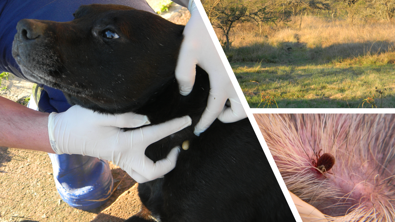
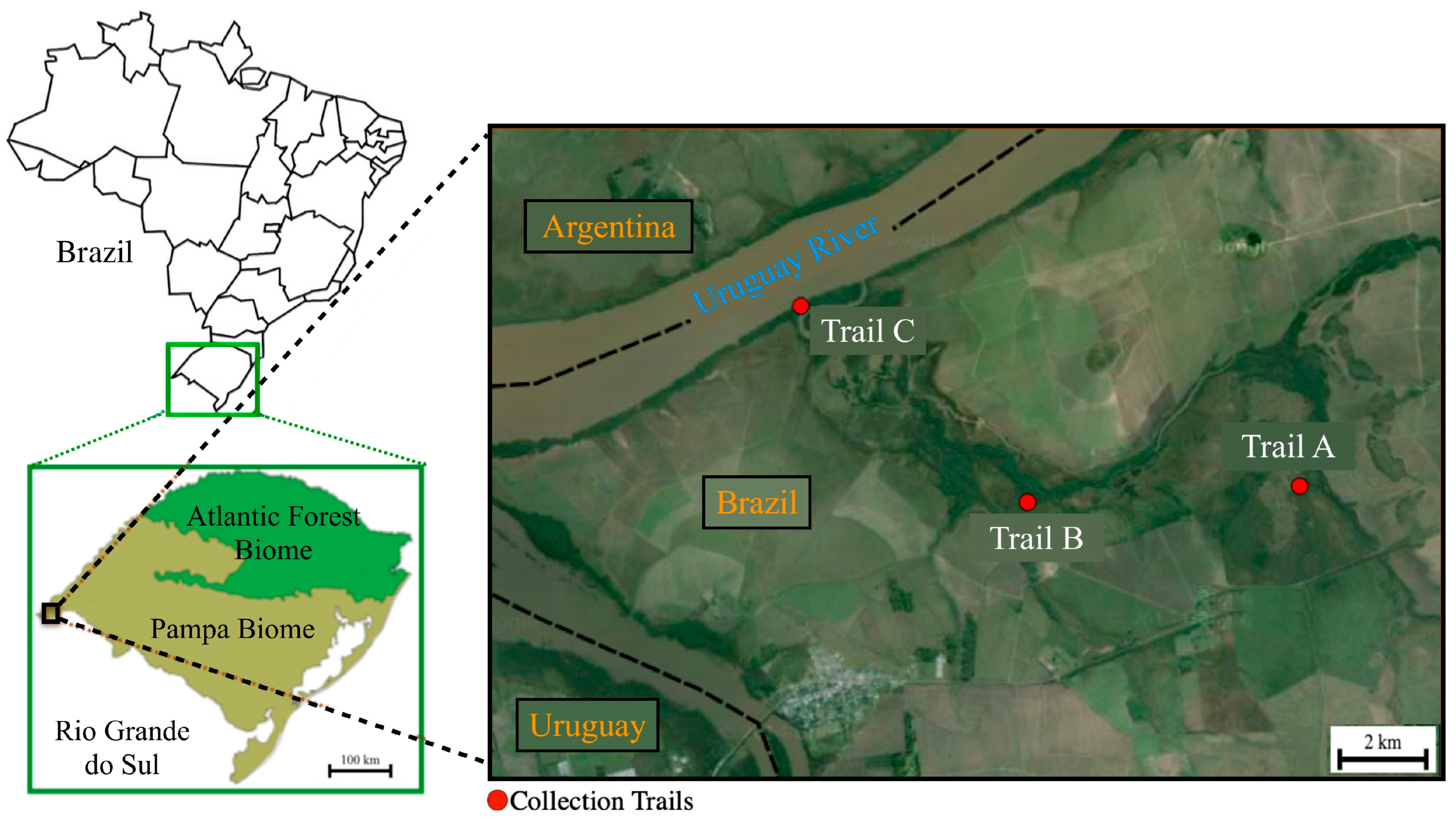
2.1. Study Area

This study was performed in Espinilho State Park “Parque Estadual do Espinilho” (PEE) (30°12′25″ S; 57°33′18″ W) and in the rural area surrounding the Park. The Park has an area of 1617.14 ha and is located in the municipality of Barra do Quaraí, the westernmost city in the southern region of Brazil. The PEE is a natural Reserve of the Pampa biome, which in Brazil is restricted to the state of Rio Grande do Sul. The climate of the PEE region is Cfa type according Koppen-Geiger, characterized by a humid temperate climate with hot summers, and an average annual temperature of 23.4 °C and annual precipitation of 1300 mm. The municipality of Barra do Quaraí had an estimated population of 4238 inhabitants in 2021 and a territorial area of 105,593.7 ha. (https://www.ibge.gov.br/cidades-e-estados/rs/barra-do-quarai.html, accessed on 01 July 2022).
According to the PEE Management Plan, it is estimated that 32 species of mammals inhabit the region [20], 10 of which are on the list of endangered species of wild fauna of the State of Rio Grande do Sul, such as Alouatta caraya (black howler monkey), Leopardus munoai (pampas cat), Lepardus geoffroyi (Geoffroy’s cat), Herpailurus yaguarondi (jaguarundi), Chrysocyon brachyurus (maned wolf), Nasua nasua (South American coati), Ozotoceros bezoarticus (pampas deer) and Wilfredomys oenax (greater Wilfred’s mouse).
Three field campaigns were performed, the first in June 2013, the second in October 2013, and the third in January 2014. In each one, we sampled domestic dogs, small mammals and ticks. Field campaigns and animal procedures described here have been authorized by the Chico Mendes Institute for Biodiversity (ICMBio Permit No. 38502-1), the Institutional Animal Care and Use Committee (IACUC) of the School of Veterinary Medicine and Animal Science of the University of São Paulo (protocol 2908/2013), and State Secretary for the Environment of the Rio Grande do Sul (protocol 05/2013 registration No. 428).

2.2. Domestic Dogs

During the three field campaigns in PEE and in the surrounding rural area, 36 individual dogs had blood serum samples collected. In the first field campaign, 33 of these dogs were examined for tick infestations. During the second and third field campaigns, 17 and 20 of the first campaign-dogs, respectively, were re-examined for tick infestations, resulting in a total of 70 canine examinations during the study. Individual canine examinations for ticks were performed as described next [21]. Briefly, it consisted of a 3–4 min examination period of the entire canine body, from which the collected ticks were put in plastic tubes and transported alive to the laboratory.
For calculation of the number of sampled dogs to be tested in serological analysis, we adopted a 3.16:1 ratio (number of humans/number of dogs) for rural area, following Soto et al. [22]. The sample size is considered 10% of the expected prevalence, based on a previous study that evaluated canine seroreactivity to spotted fever group (SFG) rickettsiae [23], with 95% accuracy. Thus, the formula of Arya et al. [24] was adopted, as follows:
n = [(z²)P(1 − P)]/d², where n = sample size; z = z statistic for the level of confidence; P = expected prevalence; and d = allowable error. This formula allowed for at least 28 dogs to be sampled in the present study.

2.3. Small Mammals

Field captures of wild small mammals were attempted in three trails (A, B and C) within the PEE (Figure 1). For this purpose, we used a total of 75 Shermman (25 in each trail) and five Tomahawk live-traps (two in trail A, two in trail B, and one in trail C) baited with banana, bacon, peanut butter, apple, and ham.
In each of the three campaigns, the traps were used during four consecutive nights. Concomitantly, three stations of pitfall traps were set up by using five buckets of 42.5 cm diameter and 60 cm height connected to a plastic fence (of least 30 m long and 50 cm high) in each station [25]. The total sampling effort was 960 trap-nights for live traps and 180 trap-nights for pitfall traps. Captured mammals were taxonomically identified following previous studies [26,27], submitted to anesthesia by using ketamine and xylazine, and then examined for tick infestations. Each animal was bled through intracardiac or tail vein route, and the blood sample was centrifuged for obtaining sera to be tested by serological analysis. A numbered earring (fish and small animal tag size 1; National Band and Tag, Newport, KY, USA) was applied to each animal. After returning from anesthesia, the small mammals were released at the same trapping site.

2.4. Host-Seeking Ticks

In each of the three campaigns, collection of host-seeking ticks from ground vegetation consisted of passing a cotton flannel (75 × 100 cm) by one investigator through a 50–100 m trail, as described in [21,28]. In parallel, we used the visual search method, as described in [29]. Collected ticks were transported alive to the laboratory.

2.5. Tick Identification and Rickettsial Infection in Ticks

Taxonomic identification of ticks relied on standard morphological keys [30,31,32]. Adult ticks that arrived alive at the laboratory were stored in a −80 °C freezer until they were tested for isolation of rickettsiae in Vero cell culture, through the technique of shell vial, as previously described in [33].
Other frozen or alcohol-preserved adults and the body remnants of ticks processed by the shell vial technique had their DNA extracted through the guanidine isothiocyanate and phenol/chloroform technique [34]. Unengorged nymphal ticks were processed individually by boiling [21,35]. Tick DNA samples were tested by a TaqMan real-time PCR assay, targeting a 147 bp fragment of the rickettsial gltA gene [33,36]. Once the sample showed a positive result, the amplification of larger fragments of rickettsial genes was attempted by two conventional PCR assays. One used primers CS-78 and CS-323 targeting fragment 401 bp of the rickettsial gltA gene [33]; the second assay, with primers Rr190.70 F and Rr190.701R, targeted a fragment (≈632 bp) of the SFG rickettsial 190 kDa outer membrane protein gene (ompA) [37]. PCR products were DNA sequenced in an ABI automated sequencer (model ABI 3500 Genetic Analyzer; Applied Biosystems/Thermo Fisher Scientific Foster City, CA, USA) with the same primers used for PCR. The resultant sequences were submitted for BLAST analysis to infer closest identities to available sequences of Rickettsia spp. in GenBank.
Samples that were negative when using the real-time PCR assay were tested by conventional PCR, targeting a fragment (≈460 bp) of the tick mitochondrial 16S rRNA gene [38] with the purpose of validating the viability of the extracted DNA. If no product was amplified by this PCR assay from any of the tick DNA samples, the tick sample was discarded. In addition, we used this PCR assay to generate 16S rRNA gene partial DNA sequences to confirm the morphological identification of some tick species that were found infected by rickettsiae.

2.6. Serology

Sera samples from dogs, rodents and marsupial were tested by immunofluorescence assay (IFA), using crude antigens of the following six Rickettsia species from Brazil: R. rickettsii strain Taiaçu, R. parkeri s.s. strain At24, Rickettsia amblyommatis strain Ac37, Rickettsia rhipicephali strain HJ5, Rickettsia felis strain Pedreira, and Rickettsia bellii strain Mogi, as described elsewhere [23]. For this purpose, each serum was initially tested at the 1:64 dilution in phosphate buffered saline (PBS). Slides were incubated with fluorescein isothiocyanate-labelled rabbit anti-dog IgG (Sigma, St Louis, MO, USA), goat anti-mouse IgG (Sigma), sheep anti-opossum IgG (CCZ, São Paulo, Brazil) and rabbit anti-guinea pig IgG (Sigma) for canine, rodent, marsupial and Cavia aperea sera, respectively, as previously reported [21]. Serum samples that were reactive at the 1:64 dilution were titrated in in two-fold increments to determine the endpoint IgG titers to each of the six Rickettsia antigens. If a serum reacted to a Rickettsia species with an endpoint titer that was at least 4-fold higher than the endpoint titers to the other five Rickettsia species, the highest titer was considered probably homologous to the first Rickettsia species or to a very closely related genotype [23,39]. A negative control serum (previously shown to be non-reactive) and a positive control serum (known reactive serum) were included at the 1:64 dilution in each IFA slide.

2.7. Correlation of Rickettsial Infection in A. tigrinum Populations

Based on the results of rickettsial detection in A. tigrinum ticks of the present study, we surveyed previous studies reporting R. parkeri and ‘Candidatus R. andeanae’ infections in different A. tigrinum populations from South America. The data were tabulated, and a Spearman test was applied to test the correlation between the infection rates of two Rickettsia species in A. tigrinum. Analyses were performed by using the R software (R Core Team, 2022, Vienna, Austria).

3. Results

3.1. Ticks from Domestic Dogs

Since the three field campaigns (June 2013, October 2013, January 2014) encompassed a study period of seven months, all data were pooled for presentation. Among a total of 70 canine examinations throughout the study, 30% (21/70) revealed tick infestations. The collected ticks were identified as adults of Amblyomma aureolatum (18 females and 3 males on eight dogs, 11% infestation rate), adults of A. tigrinum (47 females and 27 males on 10 dogs, 14% infestation), and adults of Rhipicephalus sanguineus s.s. (12 females and 11 males on 10 dogs, 14% infestation).

3.2. Ticks from Wild Mammals and Vegetations

During the whole study period, a total of 34 small mammals of four species [one Didelphidae marsupial, Cryptonanus chacoensis (Chacoan gracile opossum); two Cricetidae rodents, Akodon azarae (Azara’s grass mouse) and Oligoryzomys nigripes (pygmy rice rat); and one Caviidae rodent, Cavia aperea (Brazilian guinea pig)] were captured, none of them showed tick infestation. This single C. aperea specimen was a fortuitous collection because it was removed from the mouth of a domestic dog inside the PEE during our field work. The only tick collected from a wild mammal was an adult male of Amblyomma ovale, collected from a Cerdocyon thous (crab-eating fox) that was found to be run over in front of the PEE entrance.
From the vegetation, we collected five tick specimens that were identified as A. aureolatum (one nymph, two adult males) and two A. tigrinum (one nymph, one adult male).

3.3. Rickettsial Infection in Ticks

Isolation of rickettsiae in Vero cell culture was attempted individually with three adult specimens of A. tigrinum collected from dogs. However, no isolate was obtained.
A total of 102 tick specimens were tested by real-time PCR, resulting in the detection of rickettsial DNA in 51 samples, giving an overall infection rate of 50%. Considering each tick species and stage separately, rickettsial DNA was detected in 5% (1/20) of A. aureolatum adults, and in 82% (50/61) of A. tigrinum adults (Table 1). These ticks were further tested by conventional PCR, targeting fragments of the gltA and ompA rickettsial genes. PCR products generated reliable DNA sequences from 22 ticks. By BLAST analysis, the gltA (350 bp without primer sequences) and ompA (588 bp without primer sequences) fragments that were amplified from one adult of A. aureolatum and 21 adults of A. tigrinum were 100% identical to the available sequences of ‘Candidatus R. andeanae’ in GenBank (JN180849 and KF179352, respectively). Based on rickettsial confirmation by DNA sequences, the infection rate by ‘Candidatus R. andeanae’ in A. tigrinum ticks was at least 34% (21/61). The 21 ‘Candidatus R. andeanae’-infected ticks include the three specimens that were processed for isolation of rickettsiae in Vero cell culture.

3.4. Serology

A total of 70 individual serum samples (36 dogs and 34 small mammals) were tested by IFA, employing six rickettsial antigens. The proportions of reactive sera to rickettsial antigens were 7% for R. parkeri and R. rhipicephali, 9% for R. rickettsii, 6% for R. amblyommatis and R. felis, and 3% for R. bellii (Table 2). Among 36 dogs, five (14%) reacted to R. parkeri (endpoint titers: 512–8192) and R. rickettsii (128–8192), four (11%) to R. amblyommatis (64–8192), R. rhipicephali (256–1024) and R. felis (128-4096) and only two (6%) to R. bellii (64–128). Regarding the small mammals, only one C. aperea showed seroreactivity to R. rickettsii (endpoint titer: 128) and R. rhipicephali (64), whereas none of the 21 A. azarae, the one C. chacoensis and the 11 O. nigripes were reactive to any rickettsial antigen (Table 2).
Four dogs had endpoint titers to R. parkeri that were at least 4-fold higher than the endpoint titers to the other five Rickettsia species, indicating a possible homologous reaction to R. parkeri or a closely related genotype (Table 2).

3.5. Accession Numbers

DNA representative sequences generated in this study have been deposited in GenBank under the accession numbers KX434746–KX434747 for the 16S rRNA gene of A. aureolatum and A. tigrinum, KX434742–KX434743 for the gltA gene, and KX434737–KX434738 for the ompA gene of ‘Candidatus R. andeanae’. Voucher tick specimens have been deposited in the tick collection ‘Coleção Nacional de Carrapatos Danilo Gonçalves Saraiva’ (CNC), University of São Paulo, Brazil, under the accession numbers CNC 3322 and CNC 3332–3333.

3.6. Correlation of Rickettsial Infection in A. tigrinum Populations

With the inclusion of the present study, we tabulated 10 studies that reported rickettsial infection in A. tigrinum populations from South America (Table 3). In four studies, only R. parkeri was detected, and in four other studies, only ‘Candidatus R. andeanae’ was detected. Two other studies reported the presence of the two rickettsiae at disproportionate infection rates in the same A. tigrinum population. There was a strong negative correlation (rho: −0.76) between the ranked infection rates by R. parkeri and ‘Candidatus R. andeanae’ in populations of A. tigrinum. This is, when there is a high rate of infection by ‘Candidatus R. andeanae’, the prevalence of R. parkeri tends to be low or null, and vice versa.

4. Discussion

Through the sampling of domestic dogs, small mammals and ticks within and around a natural area of the Pampa biome in southern Brazil, we detected the following three tick species infesting the dogs: A. aureolatum, A. tigrinum and R. sanguineus s.s. The former two species were found to harbor the SFG agent ‘Candidatus R. andeanae’, revealed by DNA sequences in 34% (21/61) of the A. tigrinum ticks and 5% (1/20) of the A. aureolatum adult ticks. Surprisingly, no tick was found infesting small mammals, although this vertebrate group has been reported as hosts for immature stages of A. aureolatum and A. tigrinum [46]. This negative result could be related to low sampling, season or/and to host preference of the local populations of A. aureolatum and A. tigrinum. For instance, in an Atlantic Forest area of southeastern Brazil, immature stages of A. aureolatum were reported on several passerine birds, but never in the small mammals that were trapped concomitantly in the same sites [47]. In a study with A. tigrinum in Argentina, birds were reported as frequent hosts for both larvae and nymphs, whereas Caviidae rodents were preferred hosts for nymphs, and Cricetidae rodents for larvae [48]. Further studies with ticks in the PEE should consider the evaluation of birds and a larger sample of rodents, especially Caviidae, for a longer sampling period to gather information on host usage of A. aureolatum and A. tigrinum in a preserved region of the Pampa biome.
Molecular analyses of ticks revealed the SFG agent ‘Candidatus R. andeanae’ in A. tigrinum ticks at a high infection rate, at least 34%. Notably, no R. parkeri was detected in these ticks. This result contrasts with two recent studies in the Brazilian Pampa biome, which reported absence of ‘Candidatus R. andeanae’, and R. parkeri infection rates of 28 and 46% of the A. tigrinum ticks [4,17]. Noteworthy, while the present study was conducted in a more preserved area of Pampa (the PEE), the previous two studies were conducted in a spotted fever-endemic area, where the Pampa has been highly degraded due to anthropic alterations (agriculture, cattle farms). The distance between this endemic area and the present study area is ≈250 Km. A rickettsial survey among A. tigrinum ticks from degraded areas of the Pampa biome in Uruguay also failed to detect ‘Candidatus R. andeanae’ and, at the same time, reported a R. parkeri-infection rate of 67% [14]. On the other hand, in a more preserved area of the Pampa biome in Argentina (Esteros del Iberá ecoregion), ‘Candidatus R. andeanae’ was detected in 100% of the A. tigrinum ticks [45]. These preliminary data suggest that the presence of the human pathogen R. parkeri under high infection rates in populations of A. tigrinum of the Pampa biome could be associated with environmental degradation of anthropic origin, a condition yet to be evaluated in further studies. Interestingly, previous studies in southeastern Brazil demonstrated that the endemic areas for Brazilian spotted fever (caused by R. rickettsii) were associated with forest degradation, which might have favored the growth of tick vector populations (either A. aureolatum or Amblyomma sculptum) and/or the encounter of the tick with vertebrate hosts that act as rickettsial amplifiers [47,49].
Our serological analyses showed that, at most, 14% of the dogs were exposed to SFG antigens, including R. parkeri, for which four dogs presented this agent as the possible antigen involved in a homologous reaction (PAIHR) (Table 2). In contrast, 100% of 18 dogs were seroreactive to R. parkeri (12 dogs showing R. parkeri as the PAIHR) in a spotted fever-endemic area of the Pampa biome, where 46% of the A. tigrinum ticks were infected by R. parkeri [17]. In another study in Brazil, where 92 (86%) out of 104 maned wolves (C. brachyurus) were infested by A. tigrinum and at least 4% of the ticks were infected by R. parkeri, 65 (83%) out of 78 maned wolves were seroreactive to R. parkeri, with 30 individuals showing R. parkeri as the PAIHR [42]. Thus, our canine serological results are congruent with the lack of detection of R. parkeri in the sampled ticks and, at the same time, suggest that R. parkeri might be present in the study area at very low infection rates in the tick populations. This statement might also be related to the fact that all but one of the small mammals were serologically non-reactive to SFG rickettsiae.
Through the analysis of infection rates by R. parkeri and ‘Candidatus R. andeanae’ in 10 populations of A. tigrinum (Table 3), there was a significant negative correlation between the tick infection by these two SFG agents. While R. parkeri is a recognized human pathogen transmitted by ticks [50], ‘Candidatus R. andeanae’ is likely an endosymbiont, possibly not tick-transmitted. This statement relies on recent studies with infected Amblyomma maculatum ticks, which showed that ‘Candidatus R. andeanae’ was not detected in the tick salivary glands [51] and was not efficiently transmitted to the host skin during tick feeding [52]. Our results are corroborated by studies with North American populations of A. maculatum, in which R. parkeri-infection rates tended to be inversely related to the prevalence of infection by ‘Candidatus R. andeanae’ [50]. In fact, when >40% of the A. maculatum ticks were infected by ‘Candidatus R. andeanae’, the R. parkeri-infection rate was zero. On the other hand, when infection rates by ‘Candidatus R. andeanae’ were between 0 and <5%, the tick populations showed R. parkeri-infection rates ranging from 8 to 52% [13]. As proposed for A. maculatum [13], high infection rates by ‘Candidatus R. andeanae’ might promote the exclusion of R. parkeri from A. tigrinum populations. The mechanisms for such exclusion are yet to be elucidated.
Transstadial and transovarial perpetuations are the main mechanisms for maintaining SFG rickettsiae in tick populations; however, several studies with different Rickettsia and tick species have reported decreasing rates of transstadial passage and/or transovarial transmission of one rickettsial agent in ticks that were previously infected by another rickettsial agent [53,54,55,56,57]. Although this interference effect has not been evaluated between ‘Candidatus R. andeanae’ and R. parkeri, it is possible that these two agents interfere with each other through transstadial and/or transovarial perpetuation in A. tigrinum ticks, as has been demonstrated between Rickettsia peacockii and R. rickettsii in Dermacentor andersoni [53], between Rickettsia montanensis and R. rhipicephali in Dermacentor variabilis [54], between R. amblyommatis and R. rickettsii in Amblyomma americanum [55], between R. amblyommatis and R. parkeri in A. americanum [56], and between R. bellii and R. rickettsii in Amblyomma dubitatum [57]. Indeed, the mechanisms of such interference, related to competition between Rickettsia species within a tick, have not yet been elucidated.
Although we did not test antigens of ‘Candidatus R. andeanae’ for serological evaluation of animals in the present study, this agent belongs to the SFG and is phylogenetically closer to R. rhipicephali and R. amblyommatis than to the remaining Rickettsia species tested as antigens in the present study [58]. Therefore, had the dogs and small mammals been infected by ‘Candidatus R. andeanae’ in the study area, it is likely that at least some of them would have presented endpoint titers higher to R. amblyommatis or R. rhipicephali than to the remaining rickettsial antigens. On the other hand, our results are in accordance with the above-mentioned studies that reported that ‘Candidatus R. andeanae’ is not tick-transmitted. Finally, we failed to isolate rickettsia in Vero cell cultures that were inoculated with ‘Candidatus R. andeanae’-infected A. tigrinum ticks. This result is also congruent with previous studies that reported unsuccessful isolation or limited propagation without stability of ‘Candidatus R. andeanae’ in mammal cell lines, including Vero [58,59,60], which reinforces the possible role of this agent as a tick endosymbiont.

5. Conclusions

Despite the recent emergence of R. parkeri s.s.—transmitted by A. tigrinum—as a major agent of spotted fever illness in the South American Pampa, the present study provides epidemiological data to support that the PEE, a natural Pampa reserve, is not endemic for R. parkeri rickettsiosis. This statement is supported by the lack of detection of R. parkeri-infected ticks and the relatively low R. parkeri serological reactiveness of domestic dogs and small mammals in the sampled areas. Based on studies that evaluated rickettsial infection in 10 populations of A. tigrinum from South America, we detected a strong negative correlation between the infection rates by R. parkeri and the non-pathogenic agent ‘Candidatus R. andeanae’. Hence, our findings that at least 34% of the A. tigrinum ticks from the PEE were infected by ‘Candidatus R. andeanae’ might be the primary factor contributing to the non-endemic status of the area for R. parkeri rickettsiosis. This condition might be related to the preserved landscape of the PPE as a Pampa reserve, since more degraded areas of this biome have experienced high infection rates of R. parkeri in A. tigrinum ticks, which have resulted in high canine exposure to R. parkeri and human spotted fever illness.

Author Contributions

Conceptualization, F.S.K. and M.B.L.; methodology, F.S.K., L.C.B., F.G., T.F.M., J.S., G.L.M. and M.B.L.; formal analysis, F.S.K., G.T.P., G.P. and M.B.L.; investigation, F.S.K., L.C.B., T.F.M., J.S. and M.B.L.; resources, F.S.K. and M.B.L.; data curation, F.S.K., F.G., T.F.M. and M.B.L.; writing—original draft preparation, F.S.K., G.T.P. and M.B.L.; writing—review and editing, F.S.K., L.C.B., F.G., T.F.M., J.S., G.L.M., G.P. and M.B.L.; visualization, F.S.K. and M.B.L.; supervision, M.B.L.; project administration, F.S.K.; funding acquisition, F.S.K. and M.B.L. All authors have read and agreed to the published version of the manuscript.

Funding

This research was funded by Fundação de Amparo à Pesquisa do Estado de São Paulo (FAPESP process 2012/21915-4), Conselho Nacional de Desenvolvimento Científico e Tecnológico (CNPq process 474307/2013-1), and Coordenação de Aperfeiçoamento de Pessoal de Nível Superior (CAPES Finance Code 001).

Institutional Review Board Statement

The animal study protocol was approved by Chico Mendes Institute for biodiversity (ICMBio Permit No. 38502-1), the Institutional Animal Care and Use Committee (IACUC) of the Faculty of Veterinary Medicine of the University of São Paulo (protocol 2908/2013), and State Secretary for the Environment of the Rio Grande do Sul (protocol 05/2013 registration number 428).

Informed Consent Statement

Not applicable.

Data Availability Statement

The data presented in this study are available within the article.

Conflicts of Interest

The authors declare no conflict of interest.

References

  1. Rodriguez-Morales, A.J.; Bonilla-Aldana, D.K.; Idarraga-Bedoya, S.E.; Garcia-Bustos, J.J.; Cardona-Ospina, J.A.; Faccini-Martínez, Á.A. Epidemiology of zoonotic tick-borne diseases in Latin America: Are we just seeing the tip of the iceberg? F1000Research 2018, 7, 1988. [Google Scholar] [CrossRef]
  2. Labruna, M.B.; Kamakura, O.; Moraes-Filho, J.; Horta, M.C.; Pacheco, R.C. Rocky Mountain spotted fever in dogs, Brazil. Emerg. Infect. Dis. 2009, 15, 458–460. [Google Scholar] [CrossRef]
  3. Faccini-Martínez, Á.A.; Krawczak, F.S.; Oliveira, S.V.; Labruna, M.B.; Angerami, R.N. Rickettsioses in Brazil: Distinct diseases and new paradigms for epidemiological surveillance. Rev. Soc. Bras. Med. Trop. 2021, 54, e07322020. [Google Scholar] [CrossRef] [PubMed]
  4. Weck, B.; Dall’Agnol, B.; Souza, U.; Webster, A.; Stenzel, B.; Klafke, G.; Martins, J.R.; Reck, J. Spotted fever group Rickettsia in the pampa biome, Brazil, 2015–2016. Emerg. Infect. Dis. 2016, 22, 2014–2016. [Google Scholar] [CrossRef] [Green Version]
  5. Moo-Llanes, D.A.; Oca-Aguilar, A.C.M.; Romero-Salas, D.; Sánchez-Montes, S. Inferring the Potential Distribution of an Emerging Rickettsiosis in America: The Case of Rickettsia parkeri. Pathogens 2021, 10, 592. [Google Scholar] [CrossRef] [PubMed]
  6. Roesch, L.F.W.; Vieira, F.C.B.; Pereira, V.A.; Schunemann, A.L.; Teixeira, I.F.; Senna, A.J.T.; Stefenon, V.M. The Brazilian Pampa: A fragile biome. Diversity 2009, 1, 182–198. [Google Scholar] [CrossRef] [Green Version]
  7. Brasil. Ministério da Saúde. Ministério da Saúde. Casos confirmados de febre maculosa. Brasil, Grandes Regiões e Unidades Federadas. 2007 a 2022*. Sistema de Informação de Agravos de Notificação. Available online: https://www.gov.br/saude/pt-br/assuntos/saude-de-a-a-z/f/febre-maculosa/situacao-epidemiologica/casos-confirmados-de-febre-maculosa-brasil-grandes-regioes-e-unidades-federadas-infeccao-2007-2022/view (accessed on 24 November 2022).
  8. Krawczak, F.S.; Binder, L.C.; Oliveira, C.S.; Costa, F.B.; Moraes-Filho, J.; Martins, T.F.; Sponchiado, J.; Melo, G.L.; Gregori, F.; Polo, G.; et al. Ecology of a tick-borne spotted fever in southern Brazil. Exp. Appl. Acarol. 2016, 70, 219–229. [Google Scholar] [CrossRef]
  9. Evans, D.E.; Martins, J.R.; Guglielmone, A.A. A review of the ticks (Acari, Ixodida) of Brazil, their hosts and geographic distribution—1. The State of Rio Grande do Sul, Southern Brazil. Mem. Inst. Oswaldo Cruz. 2000, 95, 453–470. [Google Scholar] [CrossRef] [Green Version]
  10. Nava, S.; Elshewany, Y.; Eremeeva, M.E.; Sumner, J.W.; Mastropaolo, M.; Paddock, C.D. Rickettsia parkeri in Argentina. Emerg. Infect. Dis. 2008, 14, 1894–1897. [Google Scholar] [CrossRef] [PubMed]
  11. Venzal, J.M.; Portillo, A.; Estrada-Peña, A.; Castro, O.; Cabrera, P.A.; Oteo, J.Á. Rickettsia parkeri in Amblyomma triste from Uruguay. Emerg. Infect. Dis. 2004, 10, 1493–1495. [Google Scholar] [CrossRef]
  12. Romer, Y.; Seijo, A.C.; Crudo, F.; Nicholson, W.L.; Varela-Stokes, A.; Lash, R.R.; Paddock, C.D. Rickettsia parkeri rickettsiosis, Argentina. Emerg. Infect. Dis. 2011, 17, 1169–1173. [Google Scholar] [CrossRef] [PubMed]
  13. Paddock, C.D.; Denison, A.M.; Dryden, M.W.; Noden, B.H.; Lash, R.R.; Abdelghani, S.S.; Evans, A.E.; Kelly, A.R.; Hecht, J.A.; Karpathy, S.E.; et al. High prevalence of “Candidatus Rickettsia andeanae” and apparent exclusion of Rickettsia parkeri in adult Amblyomma maculatum (Acari: Ixodidae) from Kansas and Oklahoma. Ticks Tick Borne Dis. 2015, 6, 297–302. [Google Scholar] [CrossRef] [Green Version]
  14. Lado, P.; Castro, O.; Labruna, M.B.; Venzal, J.M. First molecular detection of Rickettsia parkeri in Amblyomma tigrinum and Amblyomma dubitatum ticks from Uruguay. Ticks Tick Borne Dis. 2014, 5, 660–662. [Google Scholar] [CrossRef] [PubMed]
  15. Souza, U.; Dall’Agnol, B.; Michel, T.; Webster, A.; Weck, B.; Doyle, R.; Kasper, C.B.; Soares, J.; Martins, J.R.; Trigo, T.C.; et al. Molecular survey of Rickettsia spp. in the Neotropical deer tick Haemaphysalis juxtakochi from Brazilian pampa. Parasitol Res. 2018, 117, 3293–3298. [Google Scholar] [CrossRef] [PubMed]
  16. Weck, B.; Dall’Agnol, B.; Souza, U.; Webster, A.; Stenzel, B.; Klafke, G.; Martins, J.R.; Reck, J. Rickettsia parkeri in Amblyomma dubitatum ticks in a spotted fever focus from the Brazilian Pampa. Acta Trop. 2017, 28, 182–185. [Google Scholar] [CrossRef]
  17. Weck, B.; Krawczak, F.S.; Costa, F.B.; Dall’Agnol, B.; Marcili, A.; Reck, J.; Labruna, M.B. Rickettsia parkeri in the Pampa biome of southern Brazil: Isolation, molecular characterization, and serological evidence of canine infection. Vet. Parasitol. Reg. Stud. Rep. 2020, 22, 100448. [Google Scholar] [CrossRef]
  18. Dall’Agnol, B.; Souza, U.A.; Weck, B.; Trigo, T.C.; Jardim, M.M.A.; Costa, F.B.; Labruna, M.B.; Peters, F.B.; Favarini, M.O.; Mazim, F.D.; et al. Rickettsia parkeri in free-ranging wild canids from Brazilian Pampa. Transbound. Emerg. Dis. 2018, 65, e224–e230. [Google Scholar] [CrossRef] [PubMed]
  19. Saito, T.B.; Cunha-Filho, N.A.; Pacheco, R.C.; Ferreira, F.; Pappen, F.G.; Farias, N.A.; Larsson, C.E.; Labruna, M.B. Canine infection by rickettsiae and ehrlichiae in southern Brazil. Am. J. Trop. Med. Hyg. 2008, 79, 102–108. [Google Scholar] [CrossRef]
  20. Rio Grande do Sul. Plano de Manejo do Parque Estadual do Espinilho; Secretaria Estadual do Meio Ambiente, Departamento de Florestas e Áreas Protegidas: Porto Alegre, Brazil, 2009; pp. 1–238. Available online: https://www.sema.rs.gov.br/parque-estadual-do-espinilho (accessed on 7 February 2023).
  21. Krawczak, F.S.; Binder, L.C.; Sobotyk, C.; Costa, F.B.; Gregori, F.; Martins, T.F.; Pádua, G.T.; Sponchiado, J.; Melo, G.L.; Polo, G.; et al. Rickettsial infection in ticks from a natural area of Atlantic Forest biome in southern Brazil. Exp. Appl. Acarol. 2022, 88, 371–386. [Google Scholar] [CrossRef]
  22. Soto, F.R.M.; Ferreira, F.; Pinheiro, S.R.; Nogari, F.; Risseto, M.R.; de Souza, O.; Amaku, M. Dinâmica populacional canina no Município de Ibiúna-SP estudo retrospectivo. Braz. J. Vet. Res. Anim. Sci. 2006, 43, 178. [Google Scholar] [CrossRef] [Green Version]
  23. Labruna, M.B.; Horta, M.C.; Aguiar, D.M.; Cavalcante, G.T.; Pinter, A.; Gennari, S.M.; Camargo, L.M.A. Prevalence of Rickettsia infection in dogs from the urban and rural areas of Monte Negro Municipality, Western Amazon, Brazil. Vector-Borne Zoonotic Dis. 2007, 7, 249–255. [Google Scholar] [CrossRef]
  24. Arya, R.; Antonisamy, B.; Kumar, S. Sample size estimation in prevalence studies. Indian J. Pediatr. 2012, 79, 1482–1488. [Google Scholar] [CrossRef] [PubMed]
  25. Umetsu, F.; Naxara, L.; Pardini, R. Evaluating the efficiency of pitfall traps for sampling small mammals in the neotropics. J. Mammal. 2006, 87, 757–765. [Google Scholar] [CrossRef]
  26. Bonvicino, C.R.; Oliveira, J.A.; D’Andrea, P.S. Guia dos Roedores do Brasil, Com Chaves Para Gêneros Baseadas em Caracteres Externos. Organização Pan-Americana da Saúde. 2008. 120p. Available online: http://www.fiocruz.br/ioc/media/livro%20roedores.pdf (accessed on 24 November 2022).
  27. Melo, G.L.; Sponchiado, J.; Machado, A.F.; Cáceres, N.C. Small-mammal community structure in a South American deciduous Atlantic Forest. Community Ecol. 2011, 12, 58–66. [Google Scholar] [CrossRef]
  28. Oliveira, P.R.; Borges, L.M.F.; Lopes, C.M.L.; Leite, R.C. Population dynamics of the free-living stages of Amblyomma cajennense (Fabricius, 1787) (Acari: Ixodidae) on pastures of Pedro Leopoldo, Minas Gerais State, Brazil. Vet. Parasitol. 2000, 92, 295–301. [Google Scholar] [CrossRef]
  29. Terassini, F.A.; Barbieri, F.S.; Albuquerque, S.; Szabó, M.P.J.; Camargo, L.M.A.; Labruna, M.B. Comparison of two methods for collecting free-living ticks in the Amazonian Forest. Ticks Tick Borne Dis. 2010, 1, 194–196. [Google Scholar] [CrossRef]
  30. Martins, T.F.; Onofrio, V.C.; Barros-Battesti, D.M.; Labruna, M.B. Nymphs of the genus Amblyomma (Acari: Ixodidae) of Brazil: Descriptions, redescriptions, and identification key. Ticks Tick-Borne Dis. 2010, 1, 75–99. [Google Scholar] [CrossRef]
  31. Barros-Battesti, D.M.; Arzua, M.; Bechara, G.H. Carrapatos de Importância Médico-Veterinária da Região Neotropical: Um Guia Ilustrado para Identificação de Espécies; Vox/ICTTD-3/ Butantan: São Paulo, Brazil, 2006; pp. 1–223. [Google Scholar]
  32. Nava, S.; Beati, L.; Venzal, J.M.; Labruna, M.B.; Szabó, M.P.J.; Petney, T.; Saracho-Bottero, M.N.; Tarragona, E.L.; Dantas-Torres, F.; Silva, M.M.S.; et al. Rhipicephalus sanguineus (Latreille, 1806): Neotype designation, morphological re-description of all parasitic stages and molecular characterization. Ticks Tick-Borne Dis. 2018, 9, 1573–1585. [Google Scholar] [CrossRef] [PubMed]
  33. Labruna, M.B.; Whitworth, T.; Horta, M.C.; Bouyer, D.H.; McBride, J.W.; Pinter, A.; Popov, V.; Gennari, S.M.; Walker, D.H. Rickettsia species infecting Amblyomma cooperi ticks from an area in the State of São Paulo, Brazil, where Brazilian Spotted Fever is endemic. J. Clin. Microbiol. 2004, 42, 90–98. [Google Scholar] [CrossRef] [PubMed] [Green Version]
  34. Sangioni, L.A.; Horta, M.C.; Vianna, M.C.B.; Gennari, S.M.; Soares, R.M.; Galvão, M.A.M.; Schumaker, T.T.S.; Ferreira, F.; Vidotto, O.; Labruna, M.B. Rickettsial infection in animals and Brazilian spotted fever endemicity. Emerg. Infect. Dis. 2005, 11, 265–270. [Google Scholar] [CrossRef] [PubMed]
  35. Horta, M.C.; Labruna, M.B.; Pinter, A.; Linardi, P.M.; Schumaker, T.T.S. Rickettsia infection in five areas of the state of São Paulo, Brazil. Mem. Inst. Oswaldo Cruz 2007, 102, 793–801. [Google Scholar] [CrossRef] [PubMed] [Green Version]
  36. Guedes, E.; Leite, R.C.; Prata, M.C.A.; Pacheco, R.C.; Walker, D.H.; Labruna, M.B. Detection of Rickettsia rickettsii in the tick Amblyomma cajennense in a new Brazilian spotted fever-endemic area in the state of Minas Gerais. Mem. Inst. Oswaldo Cruz 2005, 100, 841–845. [Google Scholar] [CrossRef] [Green Version]
  37. Roux, V.; Fournier, P.E.; Raoult, D. Differentiation of spotted fever group rickettsiae by sequencing and analysis of restriction fragment length polymorphism of PCR-amplified DNA of the gene encoding the protein rOmpA. J. Clin. Microbiol. 1996, 34, 2058–2065. [Google Scholar] [CrossRef] [PubMed] [Green Version]
  38. Mangold, A.J.; Bargues, M.D.; Mas-Coma, S. Mitochondrial 16S rDNA sequences and phylogenetic relationships of species of Rhipicephalus and othertick genera among Metastriata (Acari: Ixodidae). Parasitol. Res. 1998, 84, 478–484. [Google Scholar] [CrossRef] [PubMed]
  39. Szabó, M.P.; Nieri-Bastos, F.A.; Spolidorio, M.G.; Martins, T.F.; Barbieri, A.M.; Labruna, M.B. In vitro isolation from Amblyomma ovale (Acari: Ixodidae) and ecological aspects of the Atlantic rainforest Rickettsia, the causative agent of a novel spotted fever rickettsiosis in Brazil. Parasitology 2013, 140, 719–728. [Google Scholar] [CrossRef]
  40. Romer, Y.; Nava, S.; Govedic, F.; Cicuttin, G.; Denison, A.M.; Singleton, J.; Kelly, A.J.; Kato, C.Y.; Paddock, C.D. Rickettsia parkeri rickettsiosis in different ecological regions of Argentina and its association with Amblyomma tigrinum as a potential vector. Am. J. Trop. Med. Hyg. 2014, 91, 1156–1160. [Google Scholar] [CrossRef] [PubMed] [Green Version]
  41. Tomassone, L.; Conte, V.; Parrilla, G.; De Meneghi, D. Rickettsia infection in dogs and Rickettsia parkeri in Amblyomma tigrinum ticks, Cochabamba Department, Bolivia. Vector Borne Zoonotic Dis. 2010, 10, 953–958. [Google Scholar] [CrossRef] [Green Version]
  42. Arrais, R.C.; Paula, R.C.; Martins, T.F.; Nieri-Bastos, F.A.; Marcili, A.; Labruna, M.B. Survey of ticks and tick-borne agents in maned wolves (Chrysocyon brachyurus) from a natural landscape in Brazil. Ticks Tick-Borne Dis. 2021, 12, 101639. [Google Scholar] [CrossRef]
  43. Saracho Bottero, M.N.; Tarragona, E.L.; Nava, S. Spotted fever group rickettsiae in Amblyomma ticks likely to infest humans in rural areas from northwestern Argentina. Medicina (Buenos Aires) 2015, 75, 391–395. [Google Scholar]
  44. Abarca, K.; Lopez, J.; Acosta-Jamett, G.; Martinez-Valdebenito, C. Detection of Rickettsia andeanae in two regions of Chile. Rev. Chilena Infectol. 2013, 30, 388–394. [Google Scholar] [CrossRef] [Green Version]
  45. Eberhardt, A.T.; Fernandez, C.; Fargnoli, L.; Beldomenico, P.M.; Monje, L.D. A putative novel strain of Ehrlichia infecting Amblyomma tigrinum associated with Pampas fox (Lycalopex gymnocercus) in Esteros del Iberá ecoregion, Argentina. Ticks Tick-Borne Dis. 2020, 11, 101318. [Google Scholar] [CrossRef] [PubMed]
  46. Nava, S.; Venzal, J.M.; González-Acuña, D.; Martins, T.F.; Guglielmone, A.A. Ticks of the Southern Cone of America: Diagnosis, Distribution and Hosts with Taxonomy, Ecology and Sanitary Importance; Elsevier: London, UK, 2017. [Google Scholar]
  47. Ogrzewalska, M.; Saraiva, D.G.; Moraes-Filho, J.; Martins, T.F.; Costa, F.B.; Pinter, A.; Labruna, M.B. Epidemiology of Brazilian spotted fever in the Atlantic Forest, state of São Paulo, Brazil. Parasitology 2012, 139, 1283–1300. [Google Scholar] [CrossRef] [PubMed]
  48. Nava, S.; Mangold, A.J.; Guglielmone, A.A. The natural hosts of larvae and nymphs of Amblyomma tigrinum Koch, 1844 (Acari: Ixodidae). Vet. Parasitol. 2006, 140, 124–132. [Google Scholar] [CrossRef] [PubMed]
  49. Luz, H.R.; Costa, F.B.; Benatti, H.R.; Ramos, V.N.; Serpa, M.C.; Martins, T.F.; Acosta, I.C.L.; Ramirez, D.G.; Muñoz-Leal, S.; Ramirez-Hernandez, A.; et al. Epidemiology of capybara-associated Brazilian spotted fever. PLoS Negl. Trop. Dis. 2019, 13, e0007734. [Google Scholar] [CrossRef] [PubMed] [Green Version]
  50. Paddock, C.D.; Goddard, J. The evolving medical and veterinary importance of the Gulf Coast tick (Acari: Ixodidae). J. Med. Entomol. 2015, 52, 230–252. [Google Scholar] [CrossRef] [Green Version]
  51. Budachetri, K.; Browning, R.B.; Adamson, S.W.; Dowd, S.E.; Chao, C.C.; Ching, W.M.; Karim, S. An insight into the microbiome of Amblyomma maculatum (Acari: Ixodidae). J. Med. Entomol. 2014, 51, 119–129. [Google Scholar] [CrossRef]
  52. Grasperge, B.J.; Morgan, T.W.; Paddock, C.D.; Peterson, K.E.; Macaluso, K.R. Feeding by Amblyomma maculatum (Acari: Ixodidae) enhances Rickettsia parkeri (Rickettsiales: Rickettsiaceae) infection in the skin. J. Med. Entomol. 2014, 51, 855–863. [Google Scholar] [CrossRef] [Green Version]
  53. Burgdorfer, W.; Hayes, S.F.; Mavros, A.J. Nonpathogenic rickettsiae in Dermacentor andersoni: A limiting factor for the distribution of Rickettsia rickettsii. In Rickettsiae and Rickettsial Diseases; Burgdorfer, W., Anacker, R.L., Eds.; Academic Press: New York, NY, USA, 1981; pp. 585–594. [Google Scholar]
  54. Macaluso, K.R.; Sonenshine, D.E.; Ceraul, S.M.; Azad, A.F. Rickettsial infection in Dermacentor variabilis (Acari: Ixodidae) inhibits transovarial transmission of a second Rickettsia. J. Med. Entomol. 2002, 39, 809–813. [Google Scholar] [CrossRef] [Green Version]
  55. Levin, M.L.; Schumacher, L.B.M.; Snellgrove, A. Effects of Rickettsia amblyommatis infection on the vector competence of Amblyomma americanum ticks for Rickettsia rickettsii. Vector-Borne Zoonotic Dis. 2018, 18, 579–587. [Google Scholar] [CrossRef]
  56. Wright, C.L.; Sonenshine, D.E.; Gaff, H.D.; Hynes, W.L. Rickettsia parkeri transmission to Amblyomma americanum by cofeeding with Amblyomma maculatum (Acari: Ixodidae) and potential for spillover. J. Med. Entomol. 2015, 52, 1090–1095. [Google Scholar] [CrossRef]
  57. Sakai, R.K.; Costa, F.B.; Ueno, T.E.; Ramirez, D.G.; Soares, J.F.; Fonseca, A.H.; Labruna, M.B.; Barros-Battesti, D.M. Experimental infection with Rickettsia rickettsii in an Amblyomma dubitatum tick colony, naturally infected by Rickettsia bellii. Ticks Tick Borne Dis. 2014, 5, 917–923. [Google Scholar] [CrossRef] [PubMed]
  58. Paddock, C.D.; Fournier, P.E.; Sumner, J.W.; Goddard, J.; Elshenawy, Y.; Metcalfe, M.G.; Loftis, A.D.; Varela–Stokes, A. Isolation of Rickettsia parkeri and identification of a novel spotted fever group Rickettsia sp. from Gulf Coast ticks (Amblyomma maculatum) in the United States. Appl. Environ. Microbiol. 2010, 76, 2689–2696. [Google Scholar] [CrossRef] [PubMed] [Green Version]
  59. Luce–Fedrow, A.; Wright, C.; Gaff, H.D.; Sonenshine, D.E.; Hynes, W.L.; Richards, A.L. In vitro propagation of Candidatus Rickettsia andeanae isolated from Amblyomma maculatum. FEMS Immunol. Med. Microbiol. 2011, 64, 74–81. [Google Scholar] [CrossRef] [PubMed] [Green Version]
  60. Ferrari, F.A.; Goddard, J.; Moraru, G.M.; Smith, W.E.; Varela-Stokes, A.S. Isolation of “Candidatus Rickettsia andeanae” (Rickettsiales: Rickettsiaceae) in embryonic cells of naturally infected Amblyomma maculatum (Ixodida: Ixodidae). J. Med. Entomol. 2013, 50, 1118–1125. [Google Scholar] [CrossRef] [PubMed]
Figure 1. Locations of trails A, B and C within the Espinilho State Park in the state of Rio Grande do Sul, southern Brazil, where animals and ticks were sampled in the present study.
Pathogens 12 00446 g001
Table 1. Molecular detection of rickettsial DNA in ticks collected in Barra do Quaraí municipality, state of Rio Grande do Sul, Brazil, from June 2013 to January 2014.
Tick SpeciesTick StageSourceNo. Ticks with Rickettsial DNA/No. Tested Ticks (%)Rickettsia Species Identified by DNA Sequencing
Amblyomma aureolatumNymphsVegetation0/1 (0)
AdultsVegetation/Dogs1/20 (5)Candidatus R. andeanae’
Amblyomma ovaleAdultsCerdocyon thous0/1 (0)
Amblyomma tigrinumAdultsVegetation/Dogs50/61 (82) *Candidatus R. andeanae’
Rhipicephalus sanguineusAdultsDogs0/19 (0)
TOTAL 51/102 (50)
* Among the 50 PCR-positive ticks, reliable sequences of ‘Candidatus R. andeanae’ were generated from only 21 ticks, giving an infection rate of at least 34%.
Table 2. Seroreactivity to six Rickettsia species of animals from Barra do Quaraí municipality, state of Rio Grande do Sul, a non-endemic area for Brazilian spotted fever, from June 2013 to January 2014.
Dogs and Small Mammal Species (No. Tested Specimens)No. Seroreactive Animals to Each Rickettsia Species a
(% Seroreactivity)
No. Animals with PAIHR b
R.paR.riR.amR.rhR.feR.be
Dogs (36)5 (14)5 (14)4 (11)4 (11)4 (11)2 (6)4 R.pa
Akodon azarae (21)000000
Cavia aperea (1)01 (100)01 (100)00
Cryptonanus chacoensis (1)000000
Oligoryzomys nigripes (11)000000
TOTAL (70)5 (7)6 (9)4 (6)5 (7)4 (6)2 (3)
a R.pa, Rickettsia parkeri; R.ri, Rickettsia rickettsii; R.am, Rickettsia amblyommatis; R.rh, Rickettsia rhipicephali; R.fe, Rickettsia felis; R.be, Rickettsia bellii. b PAIHR, possible antigen involved in a homologous reaction. A homologous reaction was determined when the endpoint titer to a Rickettsia species was at least four-fold higher than the endpoint titers observed for the other five Rickettsia species. In this case, the Rickettsia species (or a very closely related genotype) involved in the highest endpoint titer was considered the PAIHR.
Table 3. Infection rates by Rickettsia parkeri and ‘Candidatus Rickettsia andeanae’ reported for adult ticks of different populations of Amblyomma tigrinum in South America.
No. Tested TicksInfection Rates (%) of A. tigrinum Adult TicksCountryReference
R. parkeriCa. R. andeanae’
6670Uruguay[14]
13460Brazil[17]
47280Brazil[4]
8130Argentina[40]
41542 aBolivia[41]
635 b17 bBrazil[42]
44048Argentina[43]
31065Chile[44]
120100Argentina[45]
61034BrazilThis study
a The 2% value refers to a single adult specimen of A. tigrinum that was reported to be infected by Rickettsia aeschlimannii; however, because this rickettsial agent has been restricted to the Old World and is genetically close to ‘Candidatus R. andeane’, we are tentatively considering this report as probably the latter agent until the presence of the former agent is confirmed in South America. b Values refer to minimal infection rates, as ticks were tested in pools.
Disclaimer/Publisher’s Note: The statements, opinions and data contained in all publications are solely those of the individual author(s) and contributor(s) and not of MDPI and/or the editor(s). MDPI and/or the editor(s) disclaim responsibility for any injury to people or property resulting from any ideas, methods, instructions or products referred to in the content.

