Article

Potassium Phosphonate Induces Resistance in Sweet Chestnut against Ink Disease Caused by Phytophthora Species

1
Department of Agricultural Sciences, University of Sassari, Viale Italia 39A, 07100 Sassari, Italy
2
Phytophthora Science and Management, Centre for Climate Impacted Terrestrial Ecosystems, Harry Butler Institute, Murdoch University, Perth, WA 6150, Australia
3
National Biodiversity Future Center S.c.a.r.l. (NBFC), Palazzo Steri, Piazza Marina 61, 90133 Palermo, Italy
*
Author to whom correspondence should be addressed.
Pathogens 2023, 12(3), 365; https://doi.org/10.3390/pathogens12030365
Submission received: 5 January 2023 / Revised: 13 February 2023 / Accepted: 20 February 2023 / Published: 22 February 2023
(This article belongs to the Special Issue Prevention and Management of Tree Diseases)

Abstract

:
Ink disease, caused by Phytophthora spp., represents a serious threat to sweet chestnuts throughout their distribution area. Among the control strategies, new perspectives have been offered by using potassium phosphonate, which indirectly controls Phytophthora diseases by acting on both host physiology and host-pathogen interactions. In this study, we tested in planta the effectiveness of trunk injection with K-phosphonate against seven different Phytophthora species associated with ink disease. For the two most aggressive species, P. cinnamomi and P. ×cambivora, the treatments were repeated at two different environmental conditions (a mean temperature of 14.5 °C vs. 25 °C) and tree phenology stages. The results obtained in this study demonstrated that K-phosphonate could contain the development of Phytophthora infection in phloem tissues. However, its effectiveness varied based on the concentration applied and the Phytophthora species tested. A concentration of 280 g/L of K-phosphonate was the most effective, and in some cases, callus formation around the necrotic lesion was detected. Overall, this study broadens the knowledge of endotherapic treatments with K-phosphonate as an effective measure for managing chestnut ink disease. Interestingly, the increase in mean temperature had a positive impact on the development of P. cinnamomi lesions on chestnut phloem tissues.

1. Introduction

Sweet chestnut (Castanea sativa Mill.) forest stands and orchards are part of the traditional and historical European landscape, covering more than 2.5 million ha, and mainly distributed among France and Italy, followed by Spain, Portugal, and Switzerland [1]. Historically, chestnut stands have represented a vital economic resource of the agroforestry ecosystems, particularly in European rural areas, producing timber, firewood, forage, tasty edible fruits, and secondary products such as pasture, hay, mushrooms, tannins, and honey [1,2,3]. In addition, chestnut woodlands play a huge ecological and social role, providing several ecosystem services, such as protection against fire and soil erosion, biodiversity preservation, and conservation of cultural values and recreation, among others [4].
Over the last decades, however, the productivity and sustainability of chestnut stands have been threatened by several factors, including socio-economic changes, climatic reverses, fire, and the introduction and re-emergence of pests and pathogens, which have led to the abandonment of chestnut cultivation in many areas [5,6,7,8,9]. Among diseases, those caused by Cryphonectria parasitica (Murr.) Barr (chestnut blight), and Phytophthora spp. (ink disease) have negatively impacted chestnut woodlands across Europe [10,11,12]. While the incidence of chestnut blight has been mitigated due to the natural spread of hypovirulence, which has maintained the disease at low severity in most regions [13], ink disease (Figure 1) still represents a serious phytosanitary problem that demands effective means to control [9]. Ink disease management is complex because its incidence is strictly related to climatic and site conditions. These include abundant rainfall and mild temperatures, waterlogging, and poor soil fertility, all exacerbated by anthropogenic activities like soil compaction and disturbance by tillage practices, vehicle movement along roads, movement of contaminated substrates, and planting of infested nursery stock [14,15,16,17,18]. The situation may be even more complex, as in addition to P. cinnamomi Rands and P. ×cambivora (Petri) Buisman, the main species associated with ink disease, other Phytophthora species may be recovered from symptomatic chestnut trees. These include P. cactorum (Lebert and Cohn) J. Schröt., P. castanetorum T. Jung, Horta Jung, Bakonyi and Scanu, P. cryptogea Pethybr. and Laff., P. gonapodyides (H.E. Petersen) Buisman, P. megasperma Drechsler, P. nicotianae Breda de Haan, P. plurivora T. Jung and T.I. Burgess, P. pseudosyringae T. Jung and Delatour, P. sansomeana E.M. Hansen and Reeser and P. syringae (Kleb.) Kleb. [9,12,19,20,21,22]. Therefore, disease control requires accurate diagnosis and analysis of the site and environmental conditions, other than selecting rigorous control methods.
Several systemic fungicides are active against Phytophthora, such as Metalaxyl, Fosetyl-Al, Dimethomorph, and Copper Hydroxide [23,24]. However, their use in forests and natural environments is restricted or prohibited in most European countries. In addition, there are several studies on biocontrol with natural antagonists (both fungi and bacteria), biofumigation with Brassicaceae, or breeding with resistant clones of C. sativa. However, their effectiveness in the field is still too low to control the disease [9,25]. Among the main control strategies, treatments with phosphonate (formerly named phosphite) are the most common and successful methods for controlling Phytophthora on woody trees in horticulture and natural ecosystems [26,27,28,29,30,31,32,33]. Phosphonates (phosphoric acid) are fully systemic fungicides, i.e., they are xylem- and phloem-translocated with both downward and upward movement in the host [34]. The precise mechanism of action of phosphonates is complex and not fully understood. However, both direct effects on pathogen growth and sporulation and indirect effects by stimulating host plant defenses have been demonstrated by several studies [35,36,37,38]. While phosphonate enhances fine root formation in treated woody trees, it does not seem to alter the abundance and composition of edaphic, floristic, and endophytic bacteria in trees or the balance between different microorganisms, including Phytophthora species [33,39], although this needs further study. Phosphonates can be applied by foliar spray, soil drenches, trunk painting, and injection [40,41]. Several commercial phosphonate formulations are used on woody trees, including potassium, sodium, and ammonium phosphonate [34,42]. Stem injection with K-phosphonate (K2HPO3) has been previously tested to control ink disease on sweet chestnut [43,44,45]. These studies were focused on the efficacy against either P. ×cambivora or P. cinnamomi, while very little is known about the effect of endotherapic treatments against other Phytophthora species associated with ink disease and the influence of the temperature in field experiments. Therefore, the present study was undertaken to (i) identify Phytophthora species previously obtained from ink-diseased chestnut trees; (ii) test the effectiveness of K-phosphonate against several Phytophthora species associated with ink disease; (iii) detect the most appropriate K-phosphonate concentration for stem injection and its phytotoxicity threshold; and (iv) evaluate whether environmental temperature has any effects on control treatments and the development of Phytophthora infection.

2. Materials and Methods

2.1. Collection and Identification of Phytophthora Isolates

Isolates of Phytophthora were sourced from the culture collection of the University of Sassari, Italy, and directly from ink-diseased chestnut trees in Sardinia (Table 1). Isolations were made by direct isolation from the typical flame-shaped necrotic lesions on phloem tissues and/or by baiting the rhizosphere soil samples of symptomatic trees [21]. The modified Synthetic Mucor Agar (SMA), a selective medium for Phytophthora, was used in both isolation methods [46]. Any growing colonies developed on SMA were subcultured and stored on carrot agar [47] (CA) at 20 °C. Isolates obtained were first grouped based on morphological analyses, and then representative morphotypes (8) were subjected to molecular analyses as described by Scanu and Webber [48]. DNA extraction, amplification, purification, sequencing, and analysis of the ITS sequences were performed as described by Scanu et al. [49]. All sequences were deposited at GenBank (http://www.ncbi.nlm.nih.gov/), and accession numbers are given in Table 1, together with all isolates’ information. All species were obtained from ink-diseased trees in chestnut stands in Sardinia, although two isolates (P. cinnamomi and P. ×cambivora) were derived from declining Q. suber trees (Table 1). Stock cultures were maintained on CA tubes at 12 °C at the culture collection of the University of Sassari. All isolates were passaged (inoculated and re-isolated) through host plant material prior to their use in experiments to ensure their pathogenicity was not lost during culture storage.

2.2. Study Site and Experimental Design

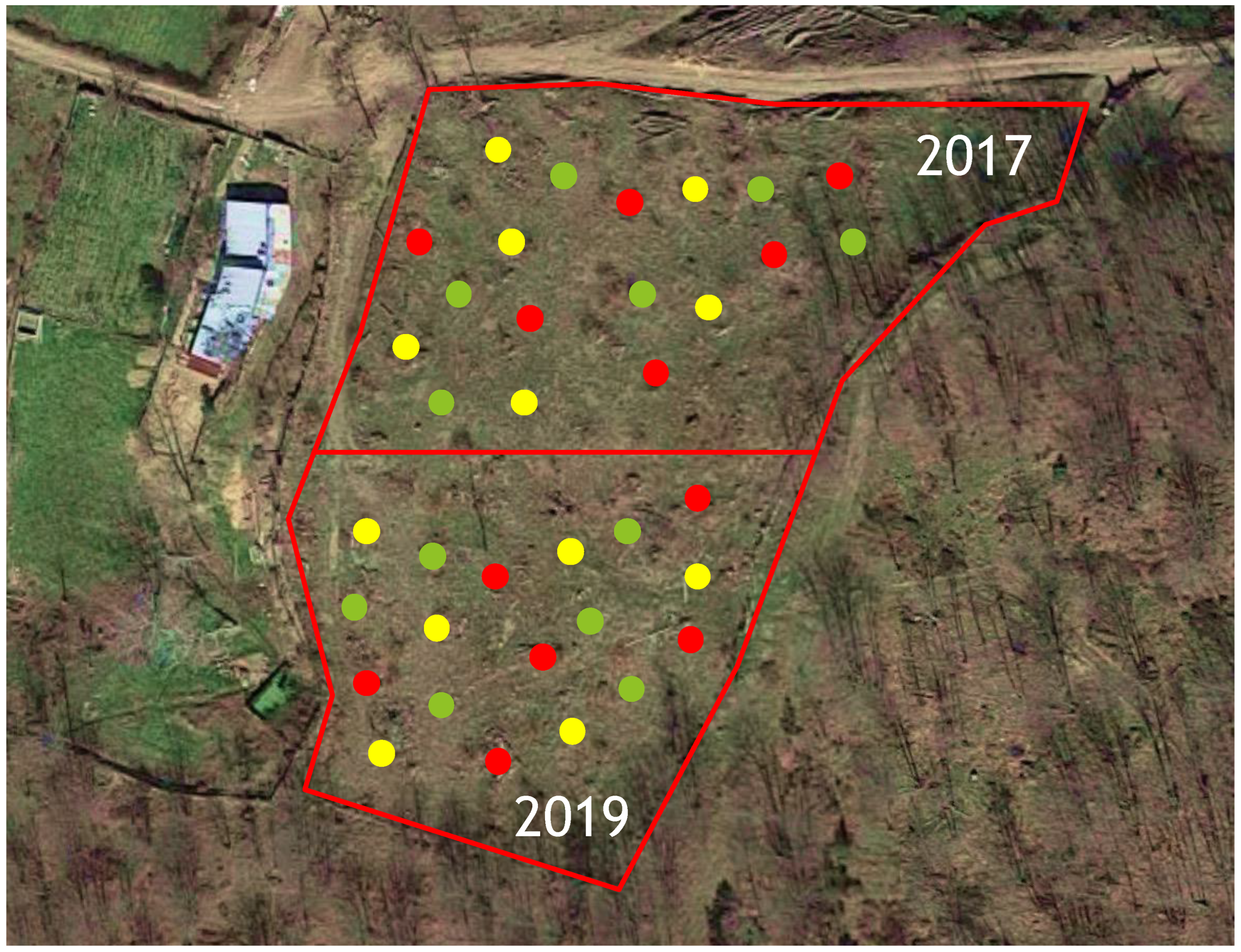
The experimental trials were conducted in two neighboring sweet chestnut stands located along a mountain slope in the Gennargentu area of Sardinia, Italy (40°01′06″ N, 9°14′40″ E; 1080 m a.s.l.). Both stands consisted of newly cut coppices with 3-year-old resprouts (in the following called “stems”), which were considered healthy as they did not show any specific symptoms of chestnut blight or ink disease. In experiment 1, in each coppice, twenty-one stems were selected, while in experiments 2 and 3, twelve stems were chosen, with each having at least 7 cm in diameter at the base. These were marked for K-phosphonate treatment (the main plot factor) and arranged in a split-plot design with six replicate blocks (coppices). A total of 60 coppices were randomly selected: 24 for experiment 1, and 18 each for experiments 2 and 3. Seven Phytophthora species, artificially inoculated in the stems (below), were randomly assigned as a subplot within each of the coppice main plots. The experiments were undertaken during September 2016 (experiment 1) in one chestnut stand and during August 2017 and May 2019 (experiments 2 and 3) in a second stand (Figure 2).

2.3. Stem Injection of K-Phosphonate

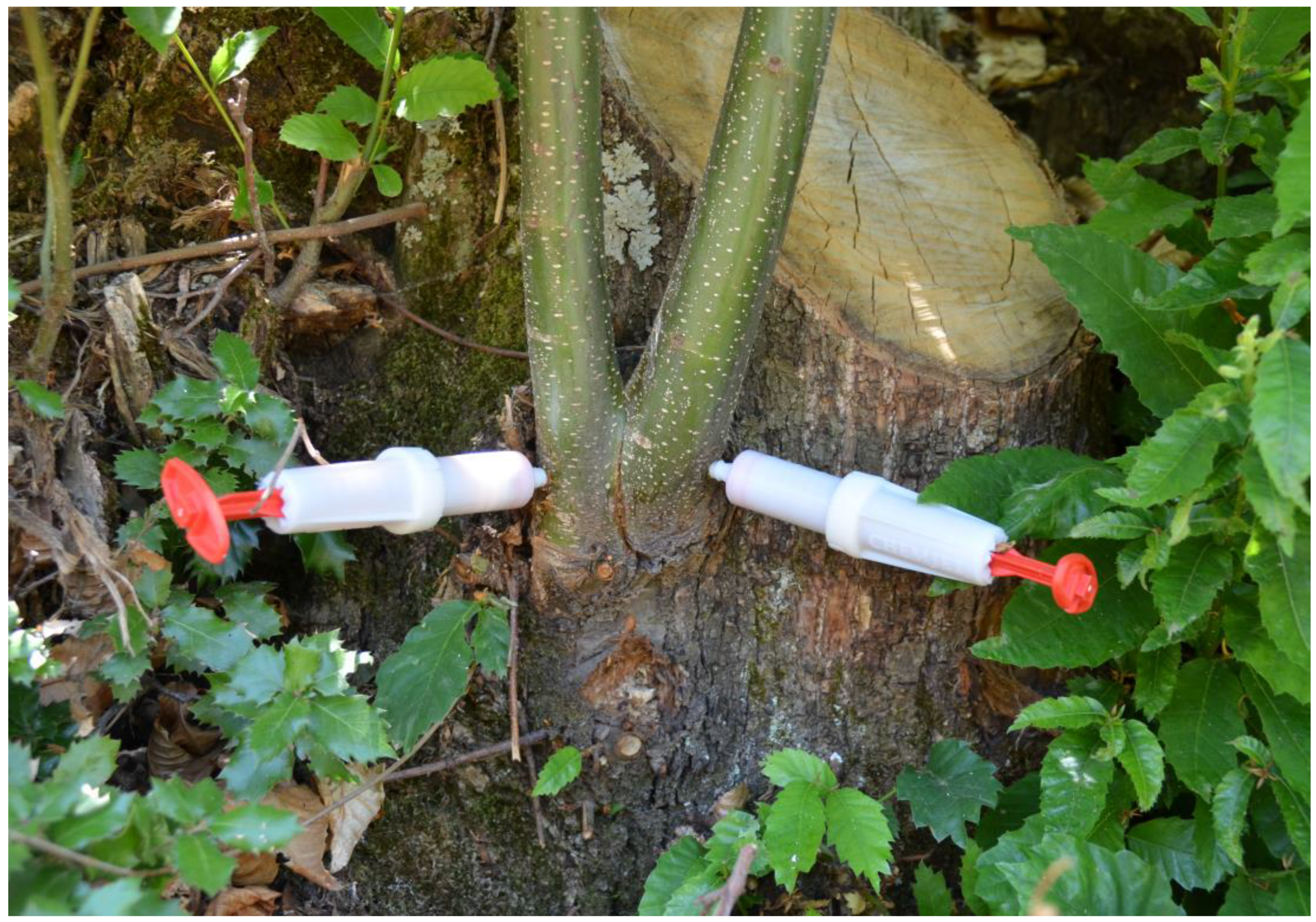
The selected chestnut stems were injected with aqueous phosphonate solutions made from a 70% commercial formulation (KalexTM, Alba Milagro, Parabiago, Italy), containing 700 g/L of potassium phosphonate (K2HPO3), adjusted to pH 4–5. Three different concentrations of K-phosphonate were tested, including 10% (70 g/L), 20% (140 g/L), and 40% (280 g/L) diluted with deionized water. The 10% concentration was tested only in the first experiment. K-phosphonate was injected at the base of each selected stem. A hole was drilled at the bottom of the stems through the outer bark layer into the sapwood with a 5.5 mm drill bit. Then the K-phosphonate solutions were injected using 20-mL spring-loaded tree syringes (Chemjet® Tree Injector, Chemjet Europe and Middle East, UK), one per stem, that lock tightly into the stem (Figure 3). For control treatments, the same procedure was followed, but 20 mL of sterile water was injected instead of K-phosphonate. Stems in the same coppice received the same K-phosphonate concentration, and there were six replicate coppices for each K-phosphonate concentration. One syringe per stem was used, and the time for uptake of the solution varied from 5 to 30 min. After injection, the hole was sealed with healing resin (Arbokol, Kollant, Vigonovo (VE), Italy) for pruning wounds to avoid any fungal infection. The foliage and stems of treated coppice were monitored for phytotoxicity for up to 2 days. Those individuals showing symptoms like pale brown discoloration of leaf margins and tip necrosis were excluded from the trials.

2.4. Stem Inoculation of Phytophthora

Two days after K-phosphonate treatments, chestnut stems were artificially inoculated with isolates of different Phytophthora species (Table 1), following the experimental design. While the first experiment included seven different Phytophthora spp., P. castanetorum, P. cinnamomi, P. gonapodyides, P. megasperma, P. plurivora, P. pseudosyringae, and P. ×cambivora, the second and third experiments focused only on P. ×cambivora and P. cinnamomi. Stem inoculations were made between 80 and 120 cm above the collar following the method used by Scanu and Webber [48]. After sterilizing the bark with 70% ethanol, a 7-mm-diameter hole was punched through the bark to the wood surface with a steel cork borer. The same-sized plug was taken from the edge of a Phytophthora colony actively growing on a 90-mm Petri dish of CA and used as inoculum by inserting it into the hole, replacing the bark plug. Moist cotton wool was placed over the wounds, covered with a 5 × 5 cm piece of aluminium foil, and sealed with an adhesive PVC tape. In the first experiment, made in September 2016, only one isolate per Phytophthora species was used, while in the second and third experiments, made in August 2017 and May 2019, respectively, two isolates of the most aggressive and main Phytophthora species associated with ink disease, P. ×cambivora, and P. cinnamomi [9], were used (Table 1). Within each chestnut coppice, each isolate was inoculated twice into individual stems, approximately 50 cm apart, to give a total of 12 inoculated stems (three replicate stems for each Phytophthora isolate). After 35 days, the inoculated stems were harvested and transferred to the laboratory to analyze the developing necrotic lesions. The periderm of each stem was destructively removed with a drawknife to expose the phloem. Each lesion was outlined and recorded on tracing paper, and then scanned on an Epson Perfection V30 photo scanner. The lesion area was calculated using APS Assess 2.0 (image analysis software for disease quantification; The American Phytopathological Society, St. Paul, MN, USA). Re-isolation of all the inoculated Phytophthora species onto SMA was attempted from the lesion margins. The cultures obtained were compared with the isolates used for the inoculation.
During the experiments, the temperatures were recorded with two data loggers (EL-USB-2-LCD Data logger, Lascar Electronics, Erie, PA, USA), which were placed in two different coppices. The temperature data, scanned every 15 min, were downloaded and analyzed with the software EasyLog Graph Version 7.4.0.0 (Lascar Electronic, Erie, PA, USA).

2.5. Statistical Analyses

Statistical analyses were performed using the software R and RStudio (R Core Team, 2017). R: A language and environment for statistical computing (R Foundation for Statistical Computing, Vienna, Austria). Linear mixed models (LMMs), which are strongly recommended when unbalanced samples occur, were used and fitted using Restricted Maximum Likelihood estimation (REML) [50]. Models were fitted separately for each pathogen using the lmer function from the “lme4” package in R [51]. Before the analysis, all data were explored for heteroscedasticity and normal distribution. In LMMs, the K-phosphonate treatment (i.e., control, 140 g/L and 280 g/L) was considered a fixed factor, whereas the stem nested within each selected coppice was selected as a random factor to account for the split-plot experimental design. Statistical differences among mean values of the lesion areas for each Phytophthora species were assessed using Analysis of Variance (ANOVA), followed by the Fisher’s protected least significant difference (LSD) test and Tukey HSD test (Honestly Significative Difference). Differences in mean values with p ≤ 0.05 were considered significant.

3. Results

3.1. Isolation and Identification of Phytophthora Species

Details of the Phytophthora species used in this study are provided in Table 1. Only P. ×cambivora was isolated from necrotic lesions detected on phloem tissues on chestnut trees, while P. castanetorum, P. gonapodyides, P. megasperma, and P. pseudosyringae were obtained from rhizosphere soil samples of symptomatic mature trees of sweet chestnut using the baiting method. An isolate of P. cinnamomi and P. plurivora were detected in chestnut seedlings in a nursery and newly planted chestnut saplings, respectively. While one isolate each of P. cinnamomi and P. ×cambivora were obtained from the rhizosphere soil of declining Q. suber trees. All morphological characters and ITS sequences matched those of the corresponding Phytophthora species (Table 1).

3.2. Experiment 1

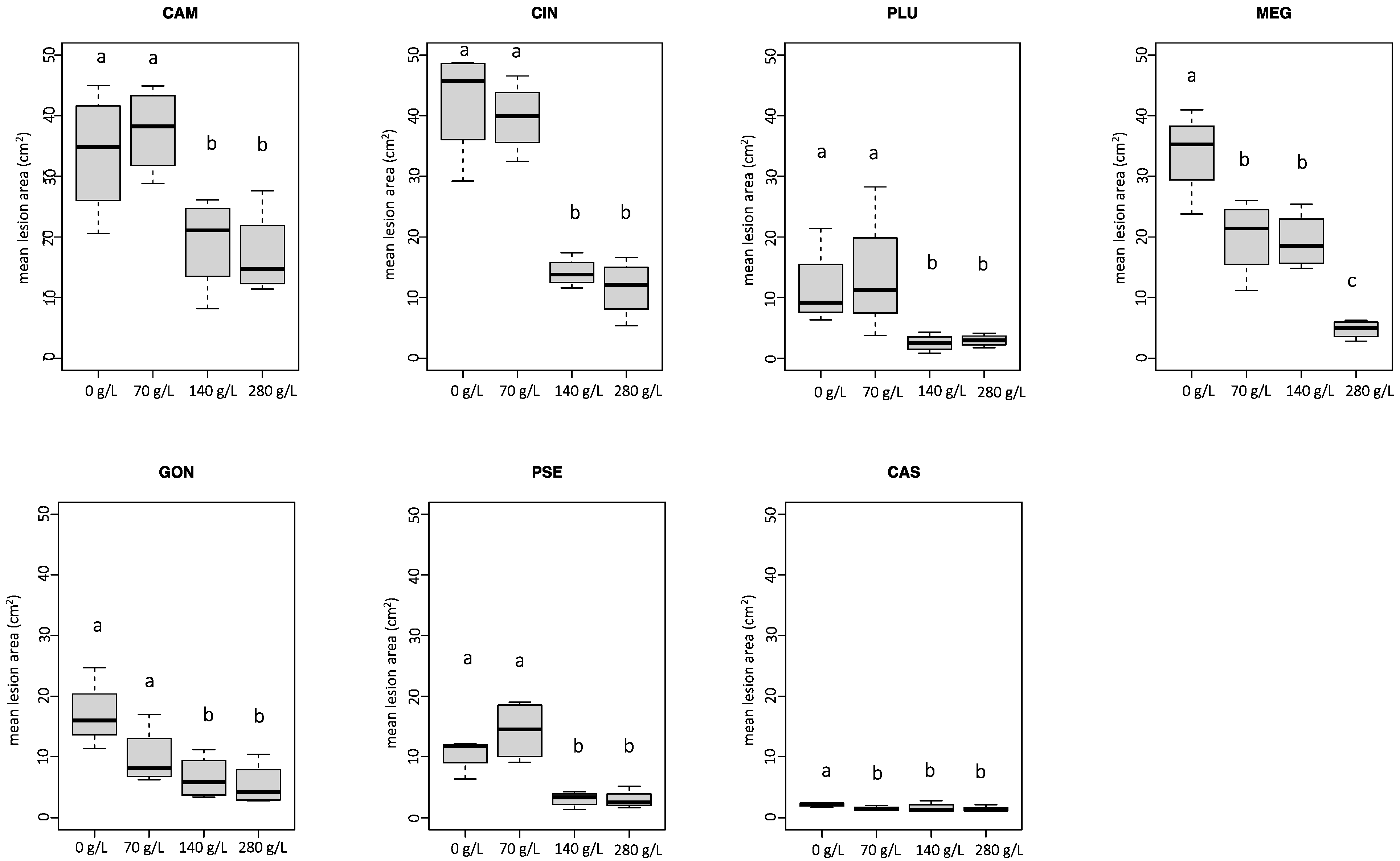
On all chestnut stems inoculated with Phytophthora, a characteristic necrotic lesion developed. The mean lesions caused by all Phytophthora species on treated stems with 140 g/L and 280 g/L of K-phosphonate concentrations were consistently smaller than those that developed on the control inoculations (Figure 4). In contrast, lesion areas did not differ significantly between stems injected with 70 g/L and controls, with the exception of P. megasperma and P. castanetorum. In some cases (P. plurivora, P. pseudosyringae, and P. ×cambivora), were even larger on average than controls (Figure 4). For P. megasperma, the mean lesion developed on stems treated with 280 g/L of K-phosphonate concentration was significantly different from that on stems treated with 70 g/L and 140 g/L concentrations. Phytophthora ×cambivora and P. cinnamomi were shown to be the most aggressive species in colonizing phloem tissues on untreated controls, with a mean lesion area of 33.8 cm2 and 42.4 cm2, respectively. Interestingly, P. megasperma showed to be as much aggressive as P. ×cambivora and P. cinnamomi. No phytotoxicity was observed in stems injected with K-phosphonate.
There was 100% pathogen re-isolation from lesions developed on both K-phosphonate-treated and untreated stems, except for P. castanetorum, which showed 40% re-isolation from around the inoculation points. During the trial, the temperature ranged from a minimum of 1.5 °C to a maximum of 30 °C, with a mean temperature of 13.3 °C.

3.3. Experiment 2

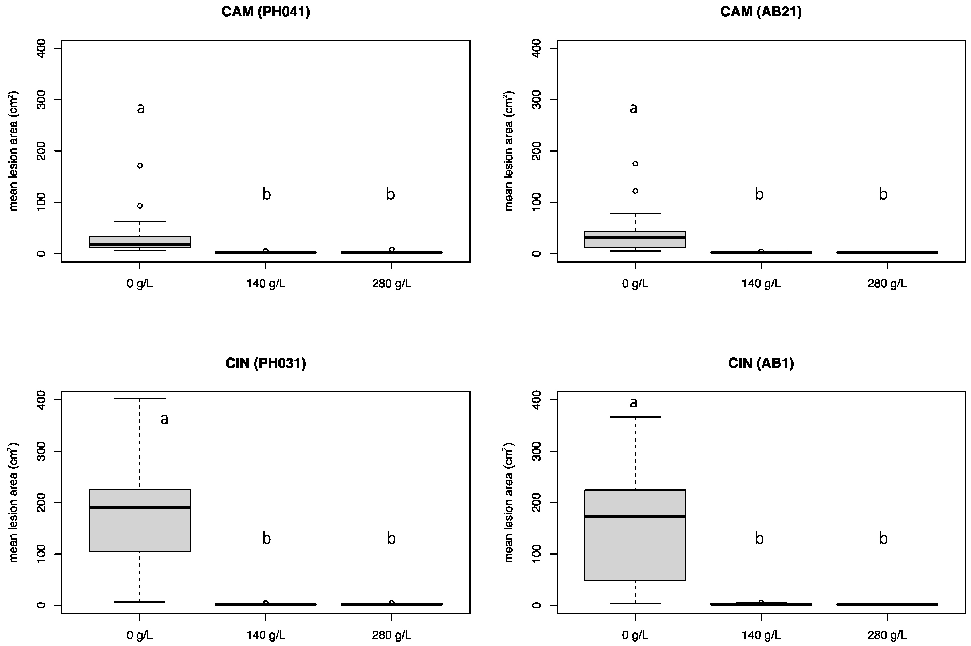
Based on the results obtained in the first experiment, two isolates of each of the two most aggressive Phytophthora species (P. ×cambivora and P. cinnamomi) (Table 1) and the two most effective K-phosphonate concentrations (140 g/L and 280 g/L) were used in experiment 2. Both K-phosphonate concentrations significantly (p < 0.05) reduced the development of necrotic lesions caused by all Phytophthora isolates (Figure 5 and Figure 6). No statistical differences were found between the two K-phosphonate concentrations. Phytophthora cinnamomi caused extensive necrotic lesions on untreated stems, with a mean lesion area of 171.7 cm2, which was, on average, almost five times larger than those caused by P. ×cambivora (mean value 34.5 cm2). Some phytotoxicity was observed in stems injected with 280 g/L of K-phosphonate.
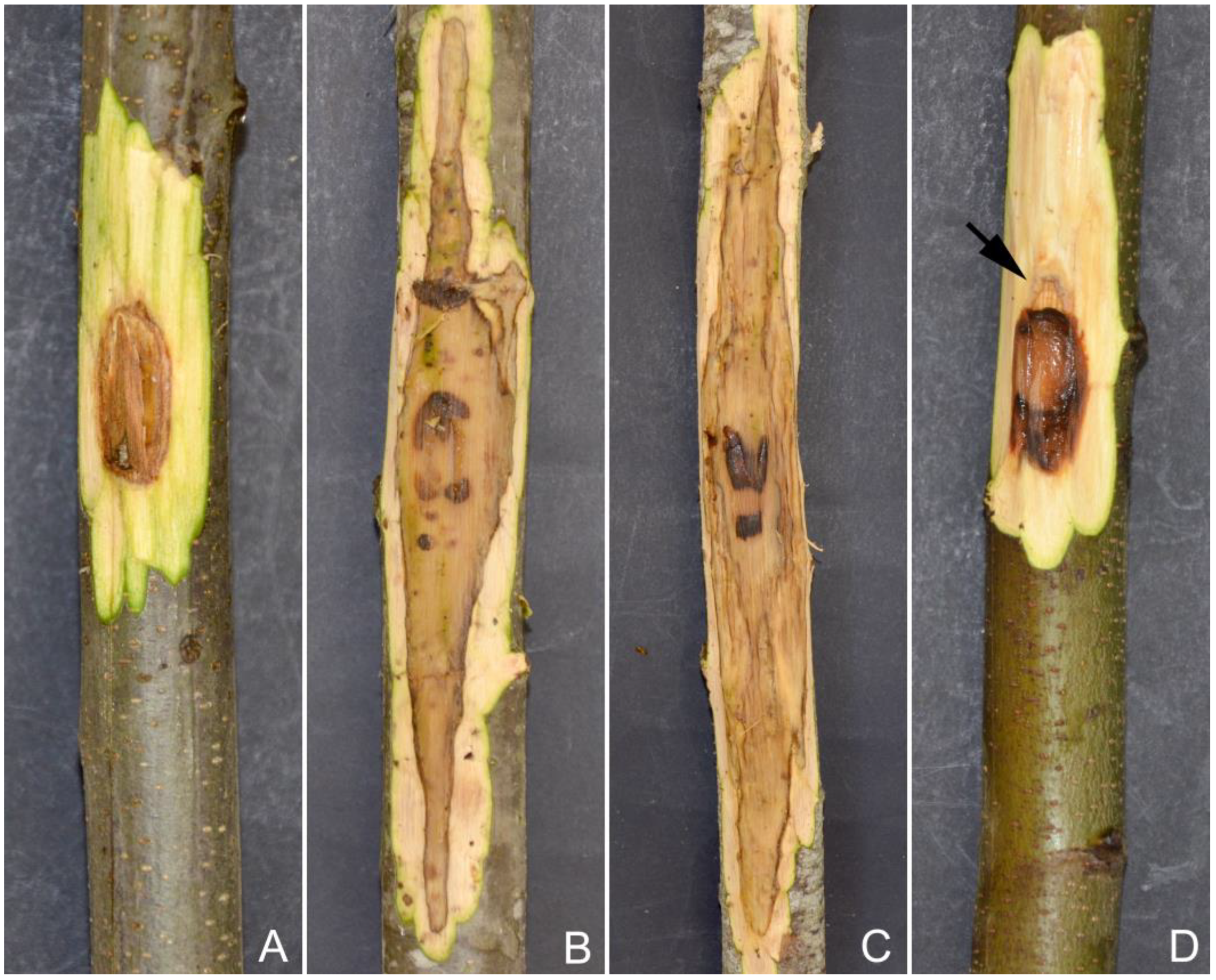
All Phytophthora isolates were readily re-isolated from the necrotic lesions visible in the phloem tissues of both treated and untreated stems. In some cases, stems treated with 280 g/L of K-phosphonate only developed limited discoloration around the inoculation point and did not yield any Phytophthora when isolation was attempted. The stems treated with the highest concentration showed callus formation, which tended to confine the necrotic lesion, particularly the upward development (Figure 6D). During the experiment, the recorded temperatures ranged from a minimum of 12 °C to a maximum of 37 °C, with a mean temperature of 25 °C.

3.4. Experiment 3

Similarly to experiment 2, both K-phosphonate concentrations were able to significantly reduce (p < 0.05) the development of necrotic lesions caused by both P. ×cambivora and P. cinnamomi isolates (Figure 7). The 280 g/L concentration of K-phosphonate was more effective than the 140 g/L concentration in reducing the growth of P. cinnamomi (96.2% vs. 95.3%) and P. ×cambivora (96.3% vs. 91.3%) on injected stems, compared with untreated stems. However, no statistical differences were found between necrotic lesions developed on stems injected with both concentrations, demonstrating their effectiveness. On control stems, P. ×cambivora was equally aggressive and caused lesions similar in size to those produced by P. cinnamomi, which were, on average, almost 50 cm2 in size. Phytotoxicity was not detected in any of the treated chestnut stems.
All Phytophthora isolates were readily re-isolated from the necrotic lesions visible in the phloem tissues of both treated and untreated stems. During the trial, the temperature ranged from a minimum of 1 °C to a maximum of 32 °C, with a mean temperature of 14.5 °C.

4. Discussion

The management of Phytophthora diseases in forest ecosystems still represents a challenge since scarce field experimental data about control strategies is available. This study provides further evidence of the efficacy of phosphonates in suppressing Phytophthora infection in woody trees [34,35,36,37,38,39,40,41,42,43,44,45]. Trunk injection with K-phosphonate significantly reduced the development of necrotic lesions on phloem tissues of chestnut trees artificially inoculated with a range of Phytophthora species associated with ink disease.
Phosphonate has been widely used in agricultural, forest, and natural settings to directly control Phytophthora by inhibiting its growth and sporulation and especially to stimulate host defense responses [23,29]. The direct effect of K-phosphonate in planta against Phytophthora is difficult to demonstrate [52]. In contrast, the containment of Phytophthora infection through a stimulated coordinated response following phosphonate injection was shown in this study by the formation of callus tissue around the margins of almost all lesions (Figure 6). in contrast no callus was observed in untreated stems, which is consistent with previous studies [38,53]. K-phosphonate may increase the percentage of healing attributable to its dual ability to slow the growth of the pathogen while enhancing the growth of the host plant and compartmentalizing lesions caused by Phytophthora [23]. Similar compartmentalization of necrotic lesions has been noted for Banksia brownii infected by P. cinnamomi following treatment with potassium phosphonate [51]. A transcriptomics analysis would be helpful to determine whether the phosphonate treatments are linked with the expression of microbial genes that might inhibit Phytophthora, as demonstrated previously [38].
K-phosphonate can be applied in different ways to protect chestnut trees, including soil drenches, foliar sprays, trunk paints, and injection [29,44,54]. However, trunk injection increases the efficiency of K-phosphonate to contain disease development as it allows the application of much higher concentrations than foliar and stem sprays. Indeed in the first experiment, the lowest concentration of K-phosphonate (70 g/L) did not significantly reduce the development of necrotic lesions for almost all Phytophthora species tested, compared to the untreated controls. Furthermore, once injected, K-phosphonate is rapidly translocated through the xylem and phloem up and down the tree [55]. This was also shown in this study, in the second experiment, when symptoms of phytotoxicity such as yellowing and browning were observed on leaves after 48 h from the treatment, indicating the systemic action of K-phosphonate [29]. Although it was only associated with small-sized treated stems and only detected in individuals treated with a 280 g/L concentration, phytotoxicity remains a concern. It must be balanced against the improvement in the health of most trees treated with K-phosphonate [55]. Trunk injection may also be considered invasive for the tree since the drill wounds can allow the entry of pathogenic bacteria or other fungi, i.e., C. parasitica on sweet chestnut. However, in all experiments, treated trees remained healthy and the wounds were successfully compartmentalized. Another disadvantage is that the absorption of K-phosphonate can vary among the physiological conditions of the treated trees and depend on climate variables, which can influence a plant’s transpiration rate [44]. Tree phenology may be another factor that can regulate the efficiency of the translocation of phosphonate to the roots [56,57]. However, the effectiveness of the treatments did not change in our experiments. For deciduous trees in temperate climates, summer injections are ideal for upward movement and should be avoided in late autumn and winter [58]. Most registered phosphonate products on woody trees usually recommend applying two annual trunk injections, which seem unsustainable in extensive forests due to the labor-intensive and costly nature of the injections. Previous studies reported that the effectiveness of K-phosphonate can last from 2 to 6 years, depending on the host plant and Phytophthora species involved in the pathosystem [29]. Further research is needed to determine how long phosphonate remains active in treated chestnut trees.
Despite the effectiveness of K-phosphonate in controlling Phytophthora diseases and its low environmental impact, its use is regulated differently worldwide. K-phosphonate is considered a systemic fungicide in Australia, the USA, and South Africa, where it is widely used in agriculture, agroforestry, and forestry [23,29]. In most European countries, including Italy, K-phosphonate has until now been regulated by Reg. CE n. 369/2013 and registered as fertilizers [59], thus with almost no restrictions. Since scientific evidence shows that phosphonates have no nutritional role in the plant to justify their use as fertilizer, recently, the European Directive EU 1009/2019 has restricted their use, with a ban on their use in both conventional and organic agriculture. Therefore, any new formulation based on phosphonate will be registered as a pesticide/fungicide on target crops/plants [60]. Currently in Italy K-phosphonate is registered on seven fruit bearing trees, tomato and grape. Therefore, at least in Europe, using K-phosphonate to control chestnut ink disease will depend on country-based regulations. In addition, treatments of K-phosphonate must also consider the Maximum Residue Limit (MRL) recently issued by EU Regulation 2021/1807, which increases the limit for chestnut fruits to 1500 mg/kg. Considering the efficacy of K-phosphonate against other important pathogens on sweet chestnut, such as Gnomoniopsis castanea [60], further research focused on residue values after treatments is needed. Previous studies reported lower values than those issued by the EU regulation 2021/1807 [59,60].
Because of these new limitations on the use of phosphonates, valid alternative treatments must be researched in the future. Silicate-based mulch could be a valuable alternative to phosphonates, especially where there might be evidence of resistant isolates [61,62]. Recently, Ca chelate is another potential product that can be utilized to stimulate plant defense responses against plant pathogens, particularly P. cinnamomi [63]. Green pesticides like those based on cinnamate anion and bioactive metabolites produced by fungi have shown a strong inhibition rate comparable to some fungicides against Phytophthora spp., including P. cinnamomi and P. ×cambivora [64,65]. Biocontrol beneficial microorganisms such as Proteobacteria (e.g., Bacillus spp.), Actinobacteria (e.g., Streptomyces spp.), Gammaproteobacteria (e.g., the fluorescent Pseudomonas), and fungi (e.g., non-pathogenic Fusarium spp. and Trichoderma spp.) have shown promising results against several foliar and soilborne diseases [66,67]. The antagonistic role of these microorganisms against Phytophthora deserves further investigation [68].
An additional outcome of this study is that P. ×cambivora and P. cinnamomi were confirmed to be the most aggressive Phytophthora species in colonizing phloem tissues on sweet chestnut [9,11,12]. Lesion development by P. cinnamomi may have been due to the higher temperatures during experiment 2, where, with a mean temperature of 25 °C, P. cinnamomi caused extensive lesions (av. 2 cm length per day) on untreated stems. This is consistent with the optimum temperature for pathogen growth in vitro, which is shown to be around 27 °C [49]. In a scenario of climate change, with an expected rise in the mean temperature in the following decades [69], the spread and impact of P. cinnamomi on chestnut stands will most likely increase in Mediterranean areas [70,71]. In contrast, the aggressiveness of P. ×cambivora was not affected by the increase in mean temperature during the trials. Although less aggressive, some Phytophthora species caused significant necrotic lesions on untreated stems, such as P. plurivora and P. megasperma. For the latter, only the highest K-phosphonate concentration of 280 g/L significantly reduced the lesion progress, suggesting some tolerance in this species. Development of resistance to phosphonate after prolonged exposure has been observed in some Phytophthora species [60], including P. crassamura, a close relative of P. megasperma [72]. This suggests that caution should be taken when relying on phosphonate as the only means to control Phytophthora diseases [73]. Phytophthora castanetorum, P. gonapodyides, and P. pseudosyringae were shown to be weak colonizers of phloem tissues. However, these species are known for their prevalent soilborne lifestyle; therefore, their involvement in chestnut ink disease may be more related to root infection [9]. Due to the complexity of ink disease, stringent hygiene protocols should be followed to prevent its spread before, during, and after treatment operations. Ink disease incidence is also strictly related to climatic and site conditions and human activities [15,18]. Therefore, any possible strategy should be part of an integrated management program to mitigate chestnut ink disease [74,75].

5. Conclusions

Overall, this study broadens the knowledge of using K-phosphonate to control chestnut ink disease caused by Phytophthora species [43,44,45]. Although field experiments are often time-consuming and expensive, this study provides a reliable and practical control treatment useful for sweet chestnut growers who have ink disease. The efficacy of trunk injection varied based on the concentration applied and the Phytophthora species tested, while it was not affected by tree phenology or environmental temperature during the treatments [44,54,55]. Although K-phosphonate has a low environmental impact, its availability on the market can be locally uncertain depending on the differences in categorization and authorization for its use [57,58].
This work also highlights the potential for some species to become serious pathogens on sweet chestnut in Mediterranean regions under the current climate change scenario. Climate change, in particular a rise in mean temperatures, extreme precipitation regimes, and severe droughts, could intensify ink disease incidences and further destabilize chestnut stands [71,76]. In the absence of alternative methods to control Phytophthora disease in forests, trunk injection with K-phosphonate remains a valid solution to mitigate the emergence of P. cinnamomi and, overall, reduce the impact of ink disease in chestnut stands.

Author Contributions

Conceptualization, G.E.S.J.H. and B.S.; methodology, A.B., G.E.S.J.H. and B.S.; software, A.B.; validation, A.B., S.S. and B.S.; formal analysis, A.B.; resources, G.E.S.J.H. and B.S.; data curation, A.B.; writing—original draft preparation, A.B.; writing—review and editing, S.S., G.E.S.J.H. and B.S.; supervision, G.E.S.J.H. and B.S.; project administration, B.S.; funding acquisition, B.S. All authors have read and agreed to the published version of the manuscript.

Funding

This research was funded by the project CASTANET, within the PSR Sardegna program, and by the RESTART-UNINUORO project “Azioni per la valorizzazione delle risorse agroforestali della Sardegna centrale/Actions for the valorisation of agroforestry resources in central Sardinia”, Regione Autonoma della Sardegna, D.G.R. N. 29/1 del 7.06.2018, fondi FSC 2014–2020. This study was also supported by the “fondo di Ateneo per la ricerca 2020,” an internal funding source provided by the University of Sassari.

Institutional Review Board Statement

Not applicable.

Informed Consent Statement

Not applicable.

Data Availability Statement

Not applicable.

Acknowledgments

The authors are grateful to Ciriaco Loddo and Paola Pintore for logistical support in the field. The authors’ thanks also go to Nicola Schianchi and Giuseppe Commisso for helping with field work and Roberto Mannu for his invaluable advice on the statistical analysis of data.

Conflicts of Interest

The authors declare no conflict of interest.

References

  1. Conedera, M.; Tinner, W.; Krebs, P.; de Rigo, D.; Caudullo, G. Castanea sativa in Europe: Distribution, habitat, usage and threats. Eur. Atlas For. Tree Species 2016, 78–79. [Google Scholar] [CrossRef]
  2. Pereira-Lorenzo, S.; Costa, R.; Ramos-Cabrer, A.M.; Ribeiro, C.; Serra da Silva, C.; Manzano, G.; Barreneche, T. Variation in grafted European chestnut and hybrids by microsatellites reveals two main origins in the Iberian Peninsula. Tree Genet. Genomes 2010, 6, 701–715. [Google Scholar] [CrossRef]
  3. Pezzi, G.; Maresi, G.; Conedera, M.; Ferrari, C. Woody species composition of chestnut stands in the Northern Apennines: The result of 200 years of changes in land use. Landsc. Ecol. 2011, 26, 1463–1476. [Google Scholar] [CrossRef]
  4. Roces-Díaz, J.V.; Vayreda, J.; Banqué-Casanovas, M.; Cusó, M.; Anton, M.; Bonet, J.A.; Brotons, L.; De Cáceres, M.; Herrando, S.; de Aragón, J.M.; et al. Assessing the distribution of forest ecosystem services in a highly populated Mediterranean region. Ecol. Indic. 2018, 93, 986–997. [Google Scholar] [CrossRef] [Green Version]
  5. Prospero, S.; Forster, B. Chestnut gall wasp (Dryocosmus kuriphilus) infestations: New opportunities for the chestnut blight fungus Cryphonectria parasitica. New Dis. Rep. 2011, 23, 2044-0588. [Google Scholar] [CrossRef] [Green Version]
  6. Visentin, I.; Gentile, S.; Valentino, D.; Gonthier, P.; Tamietti, G.; Cardinale, F. Gnomoniopsis castanea sp. nov. (Gnomoniaceae, Diaporthales) as a causal agent of nut rot in sweet chestnut. J. Plant Pathol. 2012, 94, 411–419. [Google Scholar] [CrossRef]
  7. Krebs, P.; Tinner, W.; Conedera, M. Del castagno e della castanicoltura nelle contrade insubriche: Tentativo di una sintesi eco-storica. Arch. Stor. Ticin. 2014, 4–37. [Google Scholar] [CrossRef]
  8. Seijo, F.; Millington, J.D.; Gray, R.; Sanz, V.; Lozano, J.; García-Serrano, F.; Sangüesa-Barreda, G.; Camarero, J.J. Forgetting fire: Traditional fire knowledge in two chestnut forest ecosystems of the Iberian Peninsula and its implications for European fire management policy. Land Use Policy 2015, 47, 130–144. [Google Scholar] [CrossRef] [Green Version]
  9. Jung, T.; Pérez-Sierra, A.; Durán, A.; Horta Jung, M.; Balci, Y.; Scanu, B. Canker and decline diseases caused by soil-and airborne Phytophthora species in forests and woodlands. Pers. Mol. Phylogeny Evol. Fungi 2018, 40, 182–220. [Google Scholar] [CrossRef] [Green Version]
  10. Rigling, D.; Prospero, S. Cryphonectria parasitica, the causal agent of chestnut blight: Invasion history, population biology and disease control. Mol. Plant Pathol. 2018, 19, 7–20. [Google Scholar] [CrossRef] [PubMed] [Green Version]
  11. Vettraino, A.M.; Natili, G.; Anselmi, N.; Vannini, A. Recovery and pathogenicity of Phytophthora species associated with a resurgence of ink disease in Castanea sativa in Italy. Plant Pathol. 2001, 50, 90–96. [Google Scholar] [CrossRef] [Green Version]
  12. Vettraino, A.M.; Morel, O.; Perlerou, C.; Robin, C.; Diamandis, S.; Vannini, A. Occurrence and distribution of Phytophthora species in European chestnut stands, and their association with Ink Disease and crown decline. Eur. J. Plant Pathol. 2005, 111, 169–180. [Google Scholar] [CrossRef]
  13. Bryner, S.F.; Rigling, D.; Brunner, P.C. Invasion history and demographic pattern of Cryphonectria hypovirus 1 across European populations of the chestnut blight fungus. Ecol. Evol. 2012, 2, 3227–3241. [Google Scholar] [CrossRef]
  14. Fonseca, T.F.; Abreu, C.G.; Parresol, B.R. Soil compaction and chestnut ink disease. For. Pathol. 2004, 34, 273–283. [Google Scholar] [CrossRef] [Green Version]
  15. Martins, L.; Castro, J.; Macedo, W.; Marques, C.; Abreu, C. Assessment of the spread of chestnut ink disease using remote sensing and geostatistical methods. Eur. J. Plant Pathol. 2007, 119, 159–164. [Google Scholar] [CrossRef]
  16. Jung, T.; Orlikowski, L.; Henricot, B.; Abad-Campos, P.; Aday, A.G.; Aguín Casal, O.; Bakonyi, J.; Cacciola, S.O.; Cech, T.; Chavarriaga, D.; et al. Widespread Phytophthora infestations in European nurseries put forest, semi-natural and horticultural ecosystems at high risk of Phytophthora diseases. For. Pathol. 2016, 46, 134–163. [Google Scholar] [CrossRef] [Green Version]
  17. Vannini, A.; Vettraino, A.M. Ink disease in chestnuts: Impact on the European chestnut. For. Snow Landsc. Res. 2001, 76, 345–350. [Google Scholar]
  18. Vannini, A.; Natili, G.; Thomidis, T.; Belli, C.; Morales-Rodriguez, C. Anthropogenic and landscape features are associated with ink disease impact in Central Italy. For. Pathol. 2021, 51, 1–8. [Google Scholar] [CrossRef]
  19. Černý, K.; Gregorová, B.; Strnadová, V.; Tomšovský, M.; Holub, V.; Gabrielová, Š. Phytophthora cambivora causing ink disease of sweet chestnut recorded in the Czech Republic. Czech Mycol. 2008, 60, 265–274. [Google Scholar] [CrossRef] [Green Version]
  20. Perlerou, C.; Tziros, G.; Vettraino, A.M.; Diamandis, S. Phytophthora cryptogea causing ink disease of Castanea sativa newly reported in Greece. Plant Pathol. 2010, 59, 799. [Google Scholar] [CrossRef]
  21. Scanu, B.; Linaldeddu, B.T.; Franceschini, A. First report of Phytophthora pseudosyringae associated with ink disease of Castanea sativa in Italy. Plant Dis. 2010, 94, 1068. [Google Scholar] [CrossRef]
  22. Jung, T.; Jung, M.H.; Cacciola, S.O.; Cech, T.; Bakonyi, J.; Seress, D.; Mosca, S.; Schena, L.; Seddaiu, S.; Pane, A.; et al. Multiple new cryptic pathogenic Phytophthora species from Fagaceae forests in Austria, Italy and Portugal. IMA Fungus 2017, 8, 219–244. [Google Scholar] [CrossRef] [Green Version]
  23. Garbelotto, M.; Schmidt, D. Phosphonate controls sudden oak death pathogen for up to 2 years. Calif. Agric. 2009, 63, 10–17. [Google Scholar] [CrossRef] [Green Version]
  24. González, M.; Romero, M.Á.; Serrano, M.S.; Sánchez, M.E. Fosetyl-aluminium injection controls root rot disease affecting Quercus suber in southern Spain. Eur. J. Plant Pathol. 2020, 156, 101–109. [Google Scholar] [CrossRef]
  25. Morales-Rodríguez, C.; Vettraino, A.M.; Vannini, A. Efficacy of biofumigation with Brassica carinata commercial pellets (BioFence) to control vegetative and reproductive structures of Phytophthora cinnamomi. Plant Dis. 2016, 100, 324–330. [Google Scholar] [CrossRef] [Green Version]
  26. Shearer, B.L.; Tippett, J.T. Jarrah Dieback: The dynamics and management of Phytophthora cinnamomi in the Jarrah (Eucalyptus marginata) forest of South-Western Australia. Research Bulletin n°3. 1989. Department of Conservation and Land management, Western Australia. Available online: https://scholar.google.com/scholar_lookup?title=Jarrah+Dieback:+The+Dynamics+and+Management+of+Phytophthora+cinnamomi+in+the+Jarrah+(Eucalyptus+marginata)+Forest+of+South-Western+Australia&author=Shearer,+B.L.&author=Tippett,+J.T.&publication_year=1989 (accessed on 1 December 2022).
  27. Fernandez-Escobar, R.; Gallego, F.J.; Benlloch, M.; Membrillo, J.; Infante, J.; Perez de Algaba, A. Treatment of oak decline using pressurized injection capsules of antifungal materials. Eur. J. Plant Pathol. 1999, 29, 29–38. [Google Scholar] [CrossRef]
  28. Pilbeam, R.A.; Colquhoun, I.J.; Shearer, B.; Hardy, G.S.J. Phosphite concentration: Its effect on phytotoxicity symptoms and colonisation by Phytophthora cinnamomi in three understorey species of Eucalyptus marginata forest. Australas. Plant Pathol. 2000, 29, 86–95. [Google Scholar] [CrossRef]
  29. Hardy, G.E.S.; Barrett, S.; Shearer, B.L. The future of phosphite as a fungicide to control the soilborne plant pathogen Phytophthora cinnamomi in natural ecosystems. Australas. Plant Pathol. 2001, 30, 133–139. [Google Scholar] [CrossRef]
  30. Tynan, K.M.; Wilkinson, C.J.; Holmes, J.M.; Dell, B.; Colquhoun, I.J.; McComb, J.A.; Hardy, G.S.J. The long-term ability of phosphite to control Phytophthora cinnamomi in two native plant communities of Western Australia. Aust. J. Bot. 2001, 49, 761–770. [Google Scholar] [CrossRef]
  31. Smith, S.E.; Smith, A.F.; Jakobsen, I. Mycorrhizal fungi can dominate phosphate supply to plants irrespective of growth responses. Plant Physiol. 2003, 133, 16–20. [Google Scholar] [CrossRef] [Green Version]
  32. Shearer, B.L.; Fairman, R.G. A stem injection of phosphite protects Banksia species and Eucalyptus marginata from Phytophthora cinnamomi for at least four years. Australas. Plant Pathol. 2007, 36, 78–86. [Google Scholar] [CrossRef]
  33. Barrett, S.; Rathbone, D. Long-term phosphite application maintains species assemblages, richness and structure of plant communities invaded by Phytophthora cinnamomi. Austral Ecol. 2018, 43, 360–374. [Google Scholar] [CrossRef]
  34. Guest, D.; Grant, B. The complex action of phosphonates as antifungal agents. Biol. Rev. 1991, 66, 159–187. [Google Scholar] [CrossRef]
  35. Smillie, R.; Grant, B.; Guest, D. The Mode of Action of Phosphite: Evidence for both direct and indirect modes of action on 3 Phytophthora spp. in plants. Phytopath. 1989, 79, 921–926. [Google Scholar] [CrossRef] [Green Version]
  36. Wilkinson, C.J.; Holmes, J.M.; Dell, B.; Tynan, K.M.; McComb, J.A.; Shearer, B.L.; Colquhoun, I.J.; Hardy, G.E.S. Effect of phosphite on in planta zoospore production of Phytophthora cinnamomi. Plant Pathol. 2001, 50, 587–593. [Google Scholar] [CrossRef]
  37. Dalio, R.J.D.; Fleischmann, F.; Humez, M.; Osswald, W. Phosphite Protects Fagus Sylvatica Seedlings towards Phytophthora plurivora via local toxicity, priming and facilitation of pathogen recognition. PLoS ONE 2014, 9, e87860. [Google Scholar] [CrossRef] [Green Version]
  38. Kasuga, T.; Hayden, K.J.; Eyre, C.A.; Croucher, P.J.; Schechter, S.; Wright, J.W.; Garbelotto, M. Innate resistance and phosphite treatment affect both the pathogen’s and host’s transcriptomes in the tanoak-Phytophthora ramorum pathosystem. J. Fungi 2021, 7, 198. [Google Scholar] [CrossRef]
  39. Solla, A.; Moreno, G.; Malewski, T.; Jung, T.; Klisz, M.; Tkaczyk, M.; Siebyla, M.; Pérez, A.; Cubera, E.; Hrynyk, H.; et al. Phosphite spray for the control of oak decline induced by Phytophthora in Europe. For. Ecol. Manag. 2021, 485, 118938. [Google Scholar] [CrossRef]
  40. Whiley, A.W.; Hargreaves, P.A.; Pegg, K.G.; Doogan, V.J.; Ruddle, L.J.; Saranah, J.B.; Langdon, P.W. Changing sink strengths influence translocation of phosphonate in avocado (Persea americana Mill.) trees. Aust. J. Agric. Res. 1995, 46, 1079–1090. [Google Scholar] [CrossRef]
  41. Nyoni, M.; Lotze, E.; Mazzola, M.; Wessels, J.P.B.; McLeod, A. Evaluating different approaches in the application of phosphonates for the control of apple root diseases. Austr. Plant Pathol. 2019, 48, 461–472. [Google Scholar] [CrossRef]
  42. Achary, V.; Ram, B.; Manna, M.; Datta, D.; Bhatt, A.; Reddy, M.; Agrawal, P. Phosphite: A novel P fertilizer for weed management and pathogen control. Plant Biotech. J. 2017, 15, 1493–1508. [Google Scholar] [CrossRef] [Green Version]
  43. Lim, T.M. Trunk injection with phosphorous acid for controlling Phytophthora on chestnuts: Early promising results. In Proceedings of the 9th Biennial Conference. Australasian Plant Pathology Society, Hobart, Australia, 4–8 July 1993; p. 25. [Google Scholar]
  44. Gentile, S.; Valentino, D.; Tamietti, G. Control of ink disease by trunk injection of potassium phosphite. J. Plant Pathol. 2009, 91, 565–571. [Google Scholar]
  45. Dal Maso, E.; Cocking, J.; Montecchio, L. An enhanced trunk injection formulation of potassium phosphite against chestnut ink disease. Arboric. J. 2017, 39, 125–141. [Google Scholar] [CrossRef]
  46. Brasier, C.M.; Beales, P.A.; Kirk, S.A.; Denman, S.; Rose, J. Phytophthora kernoviae sp. nov., an invasive pathogen causing bleeding stem lesions on forest trees and foliar necrosis of ornamentals in the UK. Mycol. Res. 2005, 109, 853–859. [Google Scholar] [CrossRef] [Green Version]
  47. Brasier, C.M. Physiology of Reproduction in Phytophthora. PhD. Thesis, University of Hull, Hull, UK, 1967; p. 220. [Google Scholar]
  48. Scanu, B.; Webber, J.F. Dieback and mortality of Nothofagus in Britain: Ecology, pathogenicity and sporulation potential of the causal agent Phytophthora pseudosyringae. Plant Pathol. 2016, 65, 26–36. [Google Scholar] [CrossRef]
  49. Scanu, B.; Hunter, G.C.; Linaldeddu, B.T.; Franceschini, A.; Maddau, L.; Jung, T.; Denman, S. A taxonomic re-evaluation reveals that Phytophthora cinnamomi and P. cinnamomi var. parvispora are separate species. For. Pathol. 2014, 44, 1–20. [Google Scholar] [CrossRef]
  50. Pinheiro, J.C.; Bates, D.M. Mixed-Effects Models in S and S-Plus; Springer: New York, NY, USA, 2000; pp. 3–56. [Google Scholar]
  51. Bates, D.; Mächler, M.; Bolker, B.M.; Walker, S.C. Fitting linear mixed-effects models using lme4. J. Stat. Softw. 2015, 67, 1–48. [Google Scholar] [CrossRef]
  52. Dann, E.; McLeod, A. Phosphonic acid: A long-standing and versatile crop protectant. Pest Manag. Sci. 2020, 77, 2197–2208. [Google Scholar] [CrossRef]
  53. Shearer, B.L.; Fairman, R.G.; Grant, M.J. Effective concentration of phosphite in controlling Phytophthora cinnamomi following stem injection of Banksia species and Eucalyptus marginata. For. Pathol. 2006, 36, 119–135. [Google Scholar] [CrossRef]
  54. Scott, P.; Bader, M.K.F.; Williams, N.M. Foliar phosphite application has minor phytotoxic impacts across a diverse range of conifers and woody angiosperms. Physiol. Plant. 2016, 158, 124–134. [Google Scholar] [CrossRef]
  55. Horner, I.J.; Hough, E.G.; Horner, M.B. Forest efficacy trials on phosphite for control of kauri dieback. N. Z. Plant Prot. 2015, 68, 7–12. [Google Scholar] [CrossRef] [Green Version]
  56. Guest, D.I.; Pegg, K.C.; Whiley, A.-W. Control of Phytophthora diseases of trees crops using trunk-injected phosphonates. Horticult. Rev. 1995, 17, 299–330. [Google Scholar]
  57. Pegg, K.G. Trunk injection methodology. Australas. Plant Pathol. 1990, 19, 142–143. [Google Scholar]
  58. Tattar, T.; Tattar, S. Evidence for the downward movement of materials injected into trees. Arboric. Urban For. 1999, 25, 325–332. [Google Scholar] [CrossRef]
  59. Manghi, M.C.; Masiol, M.; Calzavara, R.; Graziano, P.L.; Peruzzi, E.; Pavoni, B. The use of phosphonates in agriculture. Chemical, biological properties and legislative issues. Chemosphere 2021, 283, 131187. [Google Scholar] [CrossRef]
  60. Bastianelli, G.; Morales-Rodríguez, C.; Caccia, R.; Turco, S.; Rossini, L.; Mazzaglia, A.; Thomidis, T.; Vannini, A. Use of phosphonate salts to control chestnut ‘Brown Rot’ by Gnomoniopsis castaneae in fruit orchards of Castanea sativa. Agronomy 2022, 12, 2434. [Google Scholar] [CrossRef]
  61. Dann, E.K.; Le, D.P. Effects of silicon amendment on soilborne and fruit diseases of avocado. Plants 2017, 6, 51. [Google Scholar] [CrossRef] [Green Version]
  62. Hunter, S.; Williams, N.; McDougal, R.; Scott, P.; Garbelotto, M. Evidence for rapid adaptive evolution of tolerance to chemical treatments in Phytophthora species and its practical implications. PLoS ONE 2018, 13, 15. [Google Scholar] [CrossRef]
  63. Khdiar, M.Y.; Burgess, T.I.; Barber, P.A.; Hardy, G.E.S. Calcium chelate is as effective as phosphite in controlling Phytophthora root rot in glasshouse trials. Plant Pathol. 2022, 72, 112–119. [Google Scholar] [CrossRef]
  64. Bugatti, V.; Vertuccio, L.; Zara, S.; Fancello, F.; Scanu, B.; Gorrasi, G. Green pesticides based on cinnamate anion incorporated in layered double hydroxides and dispersed in pectin matrix. Carbohydr. Polym. 2019, 209, 356–362. [Google Scholar] [CrossRef]
  65. Evidente, A.; Maddau, L.; Scanu, B.; Andolfi, A.; Masi, M.; Motta, A.; Tuzi, A. Sphaeropsidones, phytotoxic dimedone methyl ethers produced by Diplodia cupressi: A structure−activity relationship study. J. Nat. Prod. 2011, 74, 757–763. [Google Scholar] [CrossRef]
  66. Raaijmakers, J.M.; Paulitz, T.C.; Steinberg, C.; Alabouvette, C.; Moënne-Loccoz, Y. The rhizosphere: A playground and battlefield for soilborne pathogens and beneficial microorganisms. Plant Soil 2009, 321, 341–361. [Google Scholar] [CrossRef] [Green Version]
  67. Ritika, B.; Utpal, D. An overview of fungal and bacterial biopesticides to control plant pathogens/diseases. Afr. J. Microbiol. Res. 2014, 8, 1749–1762. [Google Scholar] [CrossRef] [Green Version]
  68. Moon, J.H.; Won, S.J.; Maung, C.E.H.; Choi, J.H.; Choi, S.I.; Ajuna, H.B.; Ahn, Y.S. Bacillus velezensis CE 100 inhibits root rot diseases (Phytophthora spp.) and promotes growth of Japanese cypress (Chamaecyparis obtusa Endlicher) seedlings. Microorganisms 2021, 9, 821. [Google Scholar] [CrossRef]
  69. IPCC Global Warming of 1.5 °C: An IPCC Special Report on the Impacts of Global Warming of 1.5 °C Above Pre-Industrial Levels and Related Global Greenhouse Gas Emission Pathways, in the Context of Strengthening the Global Response to the Threat of Climate Change, Sustainable Development, and Efforts to Eradicate Poverty. Available online: https://www.ipcc.ch/sr15/ (accessed on 1 December 2022).
  70. Brasier, C.M.; Scott, J.K. European oak declines and global warming: A theoretical assessment with special reference to the activity of Phytophthora cinnamomi. Bulletin OEPP/EPPO Bull. 1994, 24, 221–234. [Google Scholar] [CrossRef]
  71. Dorado, F.J.; Alías, J.C.; Chaves, N.; Solla, A. Warming Scenarios and Phytophthora cinnamomi Infection in Chestnut (Castanea sativa Mill.). Plants 2023, 12, 556. [Google Scholar] [CrossRef]
  72. Scanu, B.; Linaldeddu, B.T.; Deidda, A.; Jung, T. Diversity of phytophthora species from declining mediterranean maquis vegetation, including two new species, Phytophthora crassamura and P. ornamentata sp. nov. PLoS ONE 2015, 10, e0143234. [Google Scholar] [CrossRef] [Green Version]
  73. Dobrowolski, M.P.; Shearer, B.L.; Colquhoun, I.J.; O’Brien, P.A.; Hardy, G.E.S. Selection for decreased sensitivity to phosphite in Phytophthora cinnamomi with prolonged use of fungicide. Plant Pathol. 2008, 57, 928–936. [Google Scholar] [CrossRef]
  74. Vettraino, A.M.; Franceschini, S.; Natili, G.; Paganini, R.; Alicicco, D.; Vuono, G.; Vannini, A. Integrated Control Protocol (ICP) of ink disease of chestnut in central Italy: Principles and future perspectives. Acta Hortic. 2010, 866, 425–430. [Google Scholar] [CrossRef]
  75. Vannini, A.; Morales-Rodríguez, C. Integrated disease management in tree nut cultivation. In Achieving Sustainable Cultivation of Tree Nuts; Serdar, ü., Fulbright, D., Eds.; Burleigh Dodds Science Pub: Cambridge, UK, 2019; p. 530. [Google Scholar]
  76. Burgess, T.I.; Scott, J.K.; Mcdougall, K.L.; Stukely, M.J.; Crane, C.; Dunstan, W.A.; Brigg, F.; Andjic, V.; White, D.; Rudman, T.; et al. Current and projected global distribution of Phytophthora cinnamomi, one of the world’s worst plant pathogens. Glob. Chang. Biol. 2017, 23, 1661–1674. [Google Scholar] [CrossRef] [Green Version]
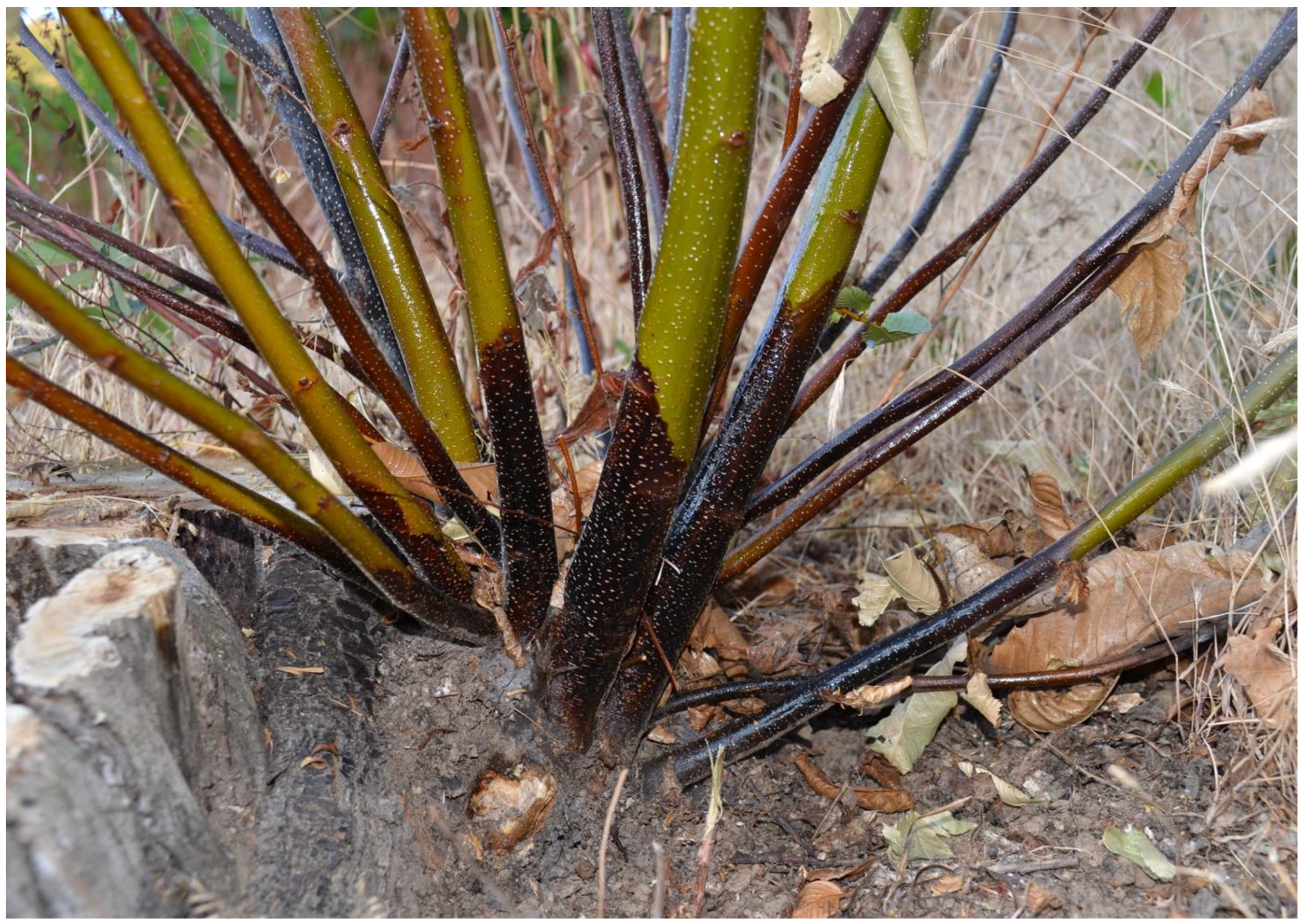
Figure 1. Ink-diseased chestnut stump with new resprouts showing the typical flame-shaped necrotic lesions on the outer bark caused by P. ×cambivora.
Pathogens 12 00365 g001
Figure 2. Experimental site with 36 coppices in which the stems were injected with 0 g/L (green dots), 140 g/L (yellow dots) and 280 g/L (red dots) of K-phosphonate in 2017 and 2019.
Pathogens 12 00365 g002
Figure 3. Chestnut resprouts are injected at the base of the stem with K-phosponate using Chemjet® Tree syringes.
Pathogens 12 00365 g003
Figure 4. Mean lesion area (cm2) caused by Phytophthora ×cambivora (CAM), P. cinnamomi (CIN), P. plurivora (PLU), P. megasperma (MEG), P. gonapodyides (GON), P. pseudosyringae (PSE), and P. castanetorum (CAS) on chestnut stems treated with four different concentrations of K2HPO3 (0 g/L, 70 g/L, 140 g/L, and 280 g/L). Bars represent the standard error. Box plots with the same letters did not show statistically significant differences on the HSD Tukey test for p ≤ 0.05.
Pathogens 12 00365 g004
Figure 5. Mean lesion area (cm2) caused by Phytophthora ×cambivora (PH041, AB21) and P. cinnamomi (PH031, AB1) on chestnut stems treated with three different concentrations of K2HPO3 (0 g/L, 140 g/L, and 280 g/L). Bars represent the standard error. Box plots with the same letters did not show statistically significant differences on the HSD Tukey test for p ≤ 0.05.
Pathogens 12 00365 g005
Figure 6. Lesion developed on treated and untreated chestnut stems: a contained lesion area caused by Phytophthora ×cambivora on a stem previously injected with K-phosphonate (280 g/L) (A), a lesion caused by P. ×cambivora and P. cinnamomi in control treatments (B,C), a contained lesioned area caused by P. cinnamomi on a stem previously injected with K-phosphonate (280 g/L) showing callus formation (arrow) around the inoculation point (D).
Pathogens 12 00365 g006
Figure 7. Mean lesion area (cm2) caused by Phytophthora ×cambivora (PH041, AB21) and P. cinnamomi (PH031, AB1) on chestnut stems treated with three different concentrations of K2HPO3 (0 g/L, 140 g/L, and 280 g/L). Bars represent the standard error. Box plots with the same letters did not show statistically significant differences on the HSD Tukey test for p ≤ 0.05.
Pathogens 12 00365 g007
Table 1. Details of Phytophthora isolates associated with ink disease and used in this study for stem inoculation.
Phytophthora sp.Isolate CodesYearHostSubstrateGenBank
Accessions
Experiment
P. castanetorumP142014Castanea sativaRhizosphere soilMF0361891
P. cinnamomiPH0312008Castanea sativaRootOP9181171, 2, 3
P. cinnamomiAB12016Quercus suberRhizosphere soilOP9181182, 3
P. gonapodyidesPH0382009Castanea sativaRhizosphere soilOQ1767291
P. megaspermaPH1782010Castanea sativaRhizosphere soilKP8634911
P. plurivoraPH0892010Castanea sativaRhizosphere soilOP9181161
P. pseudosyringaePH0432009Castanea sativaRootOP9181151
P. ×cambivoraPH0412009Castanea sativaCollarOP9181131, 2, 3
P. ×cambivoraAB212016Quercus suberRhizosphere soilOP9181142, 3
Disclaimer/Publisher’s Note: The statements, opinions and data contained in all publications are solely those of the individual author(s) and contributor(s) and not of MDPI and/or the editor(s). MDPI and/or the editor(s) disclaim responsibility for any injury to people or property resulting from any ideas, methods, instructions or products referred to in the content.