Article

The In Vitro Anticoccidial Activity of Some Herbal Extracts against Eimeria spp. Oocysts Isolated from Piglets

1
Department of Parasitology and Parasitic Disease, Faculty of Veterinary Medicine, University of Agricultural Sciences and Veterinary Medicine of Cluj-Napoca, 3-5 Mǎnǎştur Street, 400372 Cluj-Napoca, Romania
2
Department of Bromatology, Hygiene, Nutrition, Faculty of Pharmacy, “Iuliu Haţieganu” University of Medicine and Pharmacy, 6 Pasteur Street, 400349 Cluj-Napoca, Romania
3
Department of Pharmaceutical Technology and Biopharmaceutics, Faculty of Pharmacy, “Iuliu Haţieganu” University of Medicine and Pharmacy, 12 Ion Creangǎ Street, 400010 Cluj-Napoca, Romania
4
Department of Pharmaceutical Botany, Faculty of Pharmacy, “Iuliu Haţieganu” University of Medicine and Pharmacy, 23 Gheorghe Marinescu Street, 400337 Cluj-Napoca, Romania
5
Department of Infectious Diseases, Faculty of Veterinary Medicine, University of Agricultural Sciences and Veterinary Medicine of Cluj-Napoca, 3-5 Mǎnǎştur Street, 400372 Cluj-Napoca, Romania
6
Academy of Agricultural and Forestry Sciences Gheorghe Ionescu-Siseşti (A.S.A.S.), 61 Mǎrǎşti Bulevard, 011464 Bucharest, Romania
*
Author to whom correspondence should be addressed.
These authors contributed equally to this work.
Pathogens 2023, 12(2), 258; https://doi.org/10.3390/pathogens12020258
Submission received: 24 November 2022 / Revised: 2 February 2023 / Accepted: 3 February 2023 / Published: 6 February 2023
(This article belongs to the Special Issue Research on Coccidian Parasites in Livestock)

Abstract

:
Coccidiosis in pigs seldom results in important economic losses. However, it can influence growth rates in weaners and it is an important hygiene indicator in swine farms. Certain herbs, along with their extracts, have been used over the course of history in traditional medicine. The aim of this study was to evaluate the in vitro anticoccidial effects of Allium sativum L. (garlic), Artemisia absinthium L. (wormwood), Coriandrum sativum L. (coriander), Cucurbita pepo L. (pumpkin), Satureja hortensis L. (summer savory), and Calendula officinalis L. (marigold) against Eimeria suis and Eimeria debliecki oocysts. The stock solution of oocysts (58% E. suis + 42% E. debliecki) was incubated for three days, before adding the tested solutions. The unsporulated Eimeria spp. oocysts were then placed in a 3 mL well plate and incubated for 96 h at 27 °C, in a suspension containing serial dilutions of alcoholic plant extracts (5%, 2.5%, 1.25%, 0.625%, and 0.312%). The percentage of sporulated and destroyed oocysts was recorded every 24 h for 96 h. All extracts had a good in vitro anticoccidial effect against oocysts of Eimeria spp. compared with the control groups. Oocyst sporulation was significantly (p = 0.05) inhibited by the 5% marigold extract (3.6% sporulated oocysts). The same extract had the highest lytic effect on oocysts (65.2% destroyed oocysts). Our results prove that the most effective alcoholic plant extract is the marigold extract, followed, in order of efficiency, by the wormwood, coriander, garlic, pumpkin, and summer savory extracts. This study represents a preliminary contribution for establishing a new generation of natural disinfectants aimed at destroying Eimeria oocysts in the context of swine contamination.

1. Introduction

Coccidiosis in certain livestock (birds and ruminants), caused by Eimeria spp., has a grave economic impact. In pigs, on the other hand, it is considered less significant, as natural infections are only sporadically related to clinical disease [1]. Infections with Eimeria spp. are common in pigs worldwide, clinically affecting weaners and fatteners, manifested through diarrhea and weight loss. On the other hand, Eimeria infections can serve as an indicator of the overall hygiene within a farm. The lack of productivity, due to subclinical infections, is noticeable when the animals are in a heightened state of production (lactation, gestation). Ruminants and sows, particularly during the periparturient period, displayed an increase in the coproelimination of oocysts [1,2,3]. Globally, the prevalence rates for swine eimeriosis can vary between 5.6 and 77.7%, dependent on age and production types [3,4,5,6]. Resistance to anticoccidial compounds has seen an increase due to the commercial availability of products to the general public, without requiring a veterinarian’s prescription. One major concern is represented by new restrictions on the use of anticoccidial drugs along with the absence of an accessible or scalable anticoccidial vaccine for piglets [7].
Pigs are commonly infected by eight Eimeria species including Eimeria spinose, Eimeria porci, Eimeria debliecki, Eimeria neodebliecki, Eimeria perminuta, Eimeria polita, Eimeria scabra, and Eimeria suis, which can be differentiated by oocyst morphology [8,9]. Infected swine shed thousands of unsporulated oocysts, which within 5–13 days (depending on the Eimeria species) and under appropriate environmental conditions (humidity and temperature, as well as oxygen availability) will sporulate and become infectious [8,9]. The wall of Eimeria oocyst is a robust structure that is resistant to a variety of physical and chemical threats. Therefore, a critical point to destroy this pathogen is the disturbance of the sporulation process [10].
In swine farms worldwide, cleaning and disinfection are mandatory procedures used as preventative or precautionary measures against bacterial and parasitic infections [11]. Disinfection efficacy was evaluated by either the sporulation inhibition or destruction of the oocysts [12]. Several studies were carried out to assess the effects of physical (ozone, irradiation), chemical (chromium compounds, phenol, ethanol, formaldehyde, etc.), and commercial disinfectants (Preventol, Neopredisan, Dettol, TH4, Virkon®S, and others) against the sporulation of Eimeria oocysts [12,13,14,15,16,17]. Junior et al. (2007) [12] mention that protozoan oocysts are very resistant to most commercially available disinfectants.
Lastly, an increased interest in safe and effective alternatives aimed at controlling coccidiosis has led to the use of plant extracts, essential oils, and traditional medicinal products, in organic swine farms in particular [18,19,20]. Furthermore, extracts and essential oils from Artemisia absinthium, Allium sativum, and Satureja hortensis were evaluated for their activity on the sporulation of Eimeria species, and demonstrated, in vitro, a strong anticoccidial effect [21,22,23]. In organic swine farming, the use of chemicals is prohibited; therefore, plant extracts are a sustainable alternative for controlling parasitic diseases in livestock [24].
The aim of this study was to evaluate the effects of the alcoholic extracts from Allium sativum L. (garlic), Artemisia absinthium L. (wormwood), Coriandrum sativum L. (coriander), Cucurbita pepo L. (pumpkin), Satureja hortensis L. (summer savory), and Calendula officinalis L. (marigold) on the sporulation of Eimeria suis and Eimeria debliecki oocysts, isolated from piglets.

2. Materials and Methods

2.1. Eimeria spp. Oocysts Isolation

Oocysts used in this study were isolated from fresh feces of naturally infected piglets, aged between 2 and 4 months, from two free-range farms (Cluj county, Romania). The oocysts were purified by flotation, in a saturated sugar solution, and concentrated as described by Mircean et al. (2011) [25]. The obtained oocysts suspension was mixed with 2.5% potassium dichromate in equal volumes, resulting the stock suspension of oocysts (SOS) [25,26]. The McMaster method was used to establish the number of oocysts/mL. The SOS was preincubated for 3 days at 27 °C, because the sporulation time of Eimeria species of swine (5–13 days) differs from other animal species being much longer [8]. The experimental protocol started with the addition of the distilled water, ethanol, and alcoholic plant extracts (APEs).
Furthermore, the Eimeria species were identified by the morphology of sporulated oocysts [8]. For this, 5 mL of oocysts suspension was incubated for 14 days, and microphotographs were taken with Olympus DP72 camera (Olympus Corporation, Tokyo, Japan). The morphology and size of 50 oocysts were observed and measured under microscope (Olympus BX61, Olympus Corporation, Tokyo, Japan) using a 400× magnification.

2.2. Alcoholic Plant Extracts

The aerial parts of wormwood, marigold, and summer savory, along with coriander fruits, garlic bulbs, and pumpkin seeds, were used. The plant extracts were obtained by adding 10 g of the powdered plants to 100 mL of 70° alcohol (10%). All extractions and chemical analyses were performed at the “Iuliu Hațieganu” University of Medicine and Pharmacy Cluj-Napoca as previously described by Băieș et al. [27]. High-performance liquid chromatography coupled with mass spectrometry (LC/MS) were used for the analysis of major chemical compounds (poliphenols, sterols, tocopherols, sesquiterpene lactones, methoxylated flavones, and sulfoxide). The equipment, techniques, and methods used for analysis of alcoholic plant extracts have already been detailed in a previous publication [27].

2.3. Experimental Design

The experiment was performed in 24-well cell culture plates with 5 replicates for each experimental variant. Experimental variants are presented in Table 1. Two controls (ethanol = positive control, and potassium dichromate = negative control) and six APE (AS = A. sativum, AA = A. absinthium, CS = C. sativum, CP = C. pepo, CO = C. officinalis and SH = S. hortensis) were evaluated (Table 1). In each well, 1 mL of stock pre-incubated oocysts suspension (n = 15,000) and 1 mL of either APE, ethanol, or distilled water were placed, according to the experimental variant (Table 1). Next, 10% APE (the initial concentration) was tested in five, two-fold serial dilutions as follows: 5% = 50 mg/mL; 2.5% = 25 mg/mL; 1.25% = 12.5 mg/mL; 0.625% = 6.25 mg/mL; and 0.312% = 3.12 mg/mL, which represented the final concentrations of APE. Additionally, ethanol was used in five different concentrations (35%, 17.5%, 8.75%, 4.375%, and 2.187%) that corresponded with the ethanol concentration in the diluted plant extracts. The negative control was performed in five wells [20,22,23].
Afterward, the plates were incubated (Binder incubator BD 260) at 27 °C for 96 h. Every 24 h (4 times in total), one hundred oocysts/well were counted under a light microscope, recording the number of unsporulated, sporulated, and destroyed oocysts (Figure 1). Furthermore, the lethal concentration (LC50) for each APE was calculated [28,29].

2.4. Statistical Analysis and Ontologies

The mean and standard deviation of the mean were calculated for the number of sporulated and destroyed oocysts at 24, 48, 72, and 96 h. The ANOVA statistic was then used to compare the experimental groups with the control groups, and the experimental groups among themselves. A value of p ≤ 0.05 was considered statistically significant. The percentage of destroyed oocysts was adjusted using Abbott’s formula (destroyed oocysts in experimental variant − destroyed oocysts in corresponding ethanol variant)/(100 − destroyed oocysts in corresponding ethanol variant) × 100 [30]. The lethal concentration LC50 was calculated using the Microsoft Excel program.
The ontologies/pathogens, diseases, medicinal plants, and chemical compounds used in the experiment were described in Table S1, in accordance with the PPILOW project data management plan.

3. Results

3.1. Analysis of Plant Extracts

The major biological compounds identified by biochemical analysis of the 10% alcoholic plant extracts were as follows: poliphenols and sterols for A. absinthium; poliphenols, tocopherols, and sulfoxide for A. sativum; tocopherols and sterols for C. pepo; poliphenols, tocopherols, sterols, methoxylated flavones, and sesquiterpene lactones for C. sativum; poliphenols, tocopherols, and sterols for C. officinalis; and poliphenols, tocopherols, sterols, and methoxylated flavones for S. hortensis. These compounds were presented in detail in the supplementary material in Table S2, the data being previously published by Băieș et al. [27].

3.2. In Vitro Antiparasitic Activity of APE against Eimeria spp. Oocysts

The stock oocysts suspension contained Eimeria debliecki (42%) and Eimeria suis (58%) identified by their morphology.
Eimeria debliecki oocysts are of ovoid or ellipsoid shape, lacking micropyle and residuum, with a smooth, colorless, or lightly yellow wall. The dimension of these oocysts was 19.2 (16.2–23.9) × 14.1 (11.7–17.6) μm. The sporocysts measured 11.5 (6.9–16.1) × 5.1 (3.7–7.3) μm with the presence of a sporocyst residuum. Sporulation time varied between 5 and 7 days.
Eimeria suis oocysts are ellipsoidal without micropyle and oocyst residuum. Their size was 18.1 (15.6–22.7) × 13.8 (12.5–17.7) μm. The oocysts wall resembled that of E. debliecki, while sporocysts were generally shorter and stumpy, measuring 8.6 (6.8–10.9) × 5.9 (4.5–7.1) μm. Sporulation time was about 5–6 days.
The starting time of the experimental protocol was considered when the distilled water, ethanol, and APE were added, recording the results from this point onward. The percentages of sporulated and destroyed oocysts were calculated for control and experimental groups at 24, 48, 72, and 96 h (Tables S3 and S4).
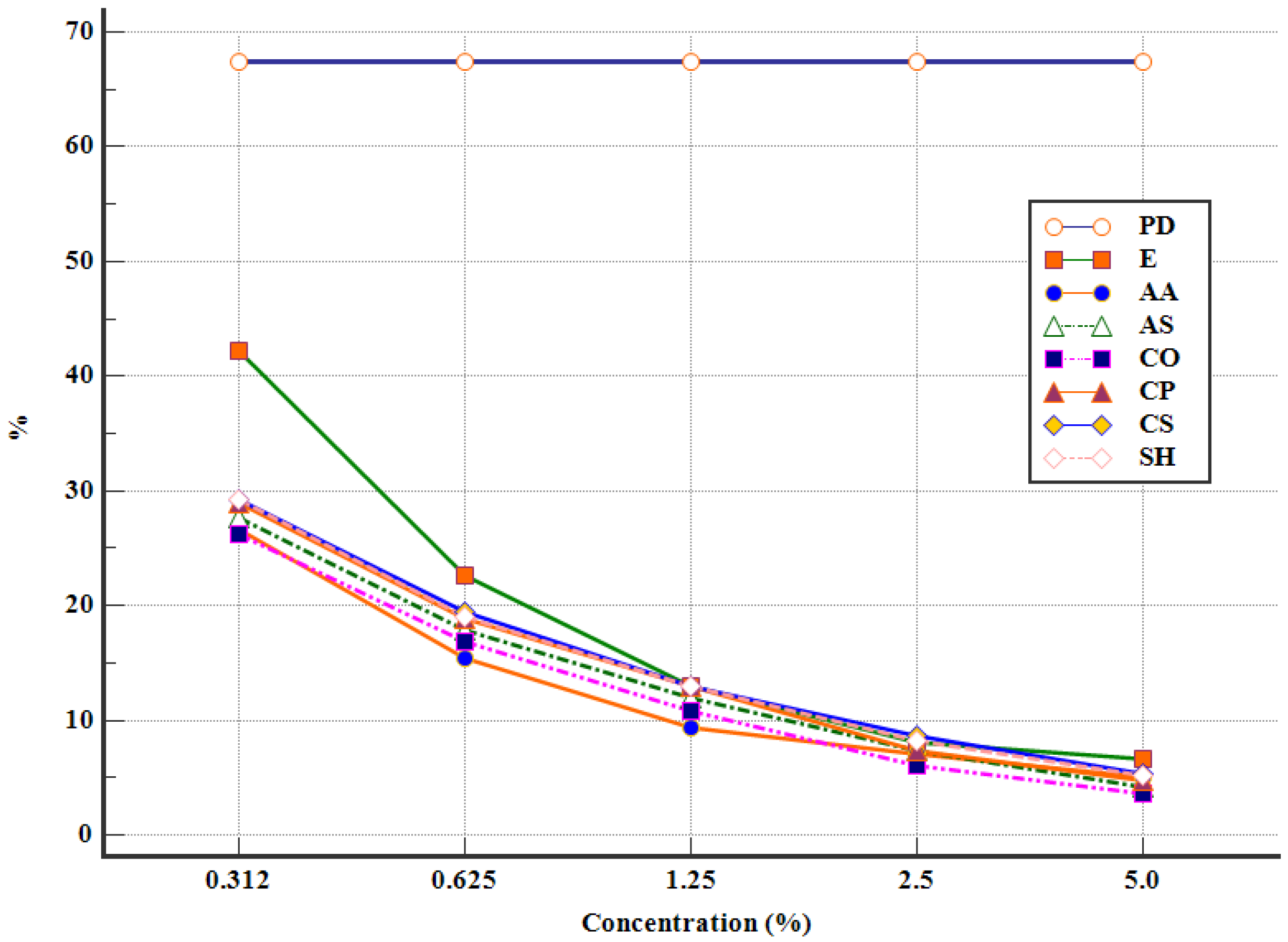
The sporulation process began after 48 h of incubation, in all experimental variants. The lowest percentages of sporulated oocysts were recorded for CO 5 (3.6 ± 0.3%), AS 5 (4.2 ± 0.75%), and AA 5 (5.0 ± 0.79%), after 96 h of incubation. The concentration of the plant extract was inversely proportionate with the percentage of sporulated oocysts, in all of the studied plants (Figure 2).
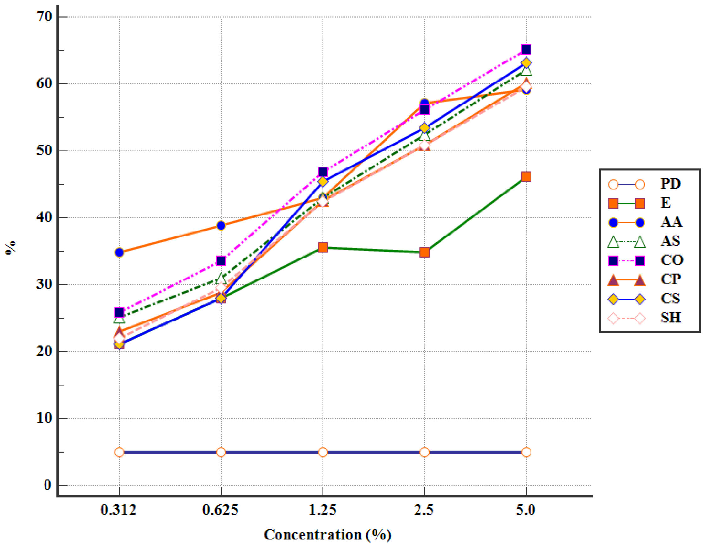
The efficacy of the APEs increased, following 72 h of incubation, resulting in a high number of destroyed oocysts for AA 2.5 (32.12 ± 3.81%), CO 2.5 (29.72 ± 3.14%), and CO 5 (27.5 ± 2.18%) (Table 2). Furthermore, after 96 h of incubation, the highest percentage of destroyed oocysts was noticed in CO 5 (35.01 ± 1.93%), followed by AA 2.5 (33.82 ± 4.87%) and CO 2.5 (32.51 ± 1.72%) (Table 2). Similar results were obtained with CS 5 (31.27 ± 4.42%), AS 5 (29.38 ± 3.16%), and CS 2.5 (28.08 ± 3.29%) (Table 2). On the other hand, the lowest percentage of destroyed oocysts, after 96 h of incubation, was observed in CP 0.625 (0.8 ± 0.12%), SH 0.312 (0.95 ± 0.2%), and SH 0.625 (1.89 ± 0.3%) (Table 2). A general and summarized outlook on the efficacy of APEs after 96 h of incubation can be observed in Figure 2 and Figure 3.
The lethal concentration (LC50) of each APE was presented in Table 3. For the first two days of incubation, LC50 was not calculated, since none of the APEs managed to destroy at least 50% of the Eimeria spp. oocysts. After 72 h of incubation, the lowest lethal concentration of APE was recorded by C. officinalis (LC50 = 24.55 mg/mL) followed, in order, by C. sativum, A. sativum, A. absinthium, C. pepo, and S. hortensis (Table 3). Furthermore, after 96 h of incubation, C. officinalis (LC50 = 16.98 mg/mL) induced the lowest LC50 value, followed by A. absinthium, C. sativum, A. sativum, C. pepo, and S. hortensis (Table 3).
All the APEs affected the sporulation of Eimeria spp. oocysts, as well as their integrity, in a dose-dependent manner. The marigold alcoholic extract, at a concentration of 50 mg/mL (5%), was the most effective of the APEs.
The results are statistically significant (p ≤ 0.05) between ethanol and APEs, for all studied plants, depending on the concentration of the extract (Tables S3 and S4). Statistically relevant values were also observed between the experimental groups at different concentrations, with the anticoccidial efficiency as a variable (Table S3, Table S4 and Table 2). We performed the ponderation both with potassium dichromate and with ethanol, the final results representing the real effectiveness of the APEs (Table 2).

4. Discussion

There is a constant and inherent requirement in finding alternative options for disinfectants, with an antiparasitic effect in livestock. The overall aim is to use molecules that are deemed safe for animals, with little to no spillover effect on the environment. The current trend involves using products based on natural compounds that have a proven antiparasitic activity, with very few side effects. Although swine eimeriosis is of low pathogenicity compared to isosporosis, our results can be extrapolated to other species where the disease is more clinically relevant (birds, rabbits, ruminants), and possibly even in piglet isosporosis.
This study evaluated the in vitro anticoccidial activity of the alcoholic extract of garlic, wormwood, coriander, pumpkin, marigold, and summer savory on the sporulation of oocysts of Eimeria suis and Eimeria debliecki. In swine, the inhibition of the sporulation process is a common criterion for evaluating anticoccidial properties [31]. Therefore, the method used in this study is cost-effective and time-efficient. Due to a lack of studies on Eimeria oocysts in pigs, we have focused our attention, in large part, on existing reports on broiler chicken coccidiosis. Phytochemicals may possess an anti-sporulation property by interfering with the physiological process necessary for sporulation, namely, preventing the access of oxygen and thus inhibiting the enzyme responsible for sporulation [19]. Biological compounds such as polyphenols, tocopherols, flavonoids, sesquiterpene lactones, and sulfoxide have demonstrated strong anticoccidial properties, both in vitro and in vivo, and can, therefore, be used as substitutes for commercial disinfectants [32,33,34,35].
Garlic is rich in organosulfur compounds (e.g., allicin, diallyl sulfide, and diallyl trisulfide), which are key components due to their antiparasitic effects [36]. Of these, allicin has exhibited a clear effect on the development of Eimeria tenella sporozoites in cell cultures with almost complete inhibition at a dose of 1.8 mg/mL [21]. Allicin also has a strong antiparasitic effect on other protozoa such as Giardia spp., Leishmania spp., Trichomonas vaginalis, etc. [37,38]. In another study performed on cell cultures, the garlic essential oil reached a maximum inhibition of 70% on E. tenella sporozoites after 24 h at a concentration of 50 μg/mL [18]. Gadelhaq et al. (2018) [20] concluded that garlic powder displayed no effect on oocysts sporulation, and the action is attributed to the toughness of the oocyst wall, acting as a stumbling block. All of the aforementioned criteria underlined the necessity for an investigation into the garlics’ ability to inhibit sporulation, as well as to destroy Eimeria oocysts. A different study reported the inhibitory effect of aqueous garlic extract on sporulation through the supposed anticoccidial effect of the organosulfur compound [39]. In the current study, garlic demonstrated a good activity even at the minimum concentration of 3.12 mg/mL in both inhibiting sporulation and destroying oocysts. The garlic extract demonstrated a strong efficacy at all tested concentrations, an effect comparable with C. officinalis, A. absinthium, and C. sativum. This effect can mainly be attributed to sulfoxide and polyphenols, which are in a fairly high concentration in this plant.
The main bioactive components of wormwood are artemisinin and quercetin [40]. Numerous experimental studies have been performed on the efficacy of A. absinthium and Artemisia annua in coccidiosis with positive results on the inhibitory effect on the sporulation of Eimeria spp. [22,41]. Reports also suggest that artemisinin could be effective against toxoplasmosis, giardiasis, isosporosis, babesiosis, and leismaniosis [37]. The in vitro antiparasitic effect of the wormwood alcoholic extract and essential oil against Eimeria oocysts has been demonstrated, with a lethal concentration of under 1 mg/mL [19,23]. In the present study, the wormwood ethanolic extract is the second most effective extract, at all concentrations used. The anticoccidial effect is probably due to the polyphenols found in abundance in this plant. Although the mechanism of these compounds is poorly understood, their inhibitory effect on embryogenesis is high. The minimum lethal concentration of the ethanolic extract of A. absinthium was 3.12 mg/mL.
Coriander is an aromatic plant with medicinal attributes and antiparasitic activity. The phytochemical screening of C. sativum showed that it contained essential oils, flavonoids, fatty acids, isocoumarin sterols, and phenolic compounds [42,43,44]. Coriander dried fruits contain about 1% essential oil, with linalool as the main active component [43]. A herbal formula containing C. sativum, among other plants, was successfully used as a natural anticoccidial molecule against eimeriosis [45]. Coriander essential oil demonstrated significant antiprotozoal effects against Leismania amazonensis and Leismania infantum amastigotes and promastigotes [46,47]. Aqueous and alcoholic coriander extracts have a weak in vitro effect against Cryptosporidium [48]. Boros et al., 2021, [49] showed that C. sativum ethanolic extracts completely inhibited the mobility of Trichinella spiralis and Trichinella britovi larvae. In vitro, coriander essential oil exhibited a strong anthelmintic efficacy against ovine gastrointestinal nematodes by inhibiting egg hatching, larval development, and motility [50,51]. In the present study, the coriander extract had the third highest effectiveness in inhibiting sporulation and destroying oocysts due to its rich polyphenol concentration. It must, however, be noted that their concentration in C. sativum is lower compared to that found in wormwood.
Pumpkin seeds contain several active constituents: essential fatty acids, amino acids, phytosterols (β-sitosterol), minerals, vitamins, and cucurbitin. The latter is the main chemically active compound in pumpkin seeds responsible for its antiparasitic effect [52,53]. Elhadi et al. (2013) [54] proved that cucurbitacin E and cucurbitacin L have a strong efficacy against Giardia lamblia, in vitro, at a concentration of 5 μg/mL after 5 days. Pumpkin is known for its antihelmintic properties, and while studies on the effect against protozoa are few, they show a strong activity against Plasmodium spp. [30]. Salman et al. (2022) [55] reported that pumpkin seeds, at concentrations of 200 μg/mL, displayed a significant inhibition of the number of Blastocystis (the most common protozoa in humans) throughout the incubation period, and the concentration of 400 μg/mL had the strongest antiparasitic effect. In an in vitro study, it was noted that the concentration of 5 and 10 mg/mL of ethanolic extract in pumpkin seeds had a complete inhibitory effect on the growth of Histomonas meleagridis, Tetratrichomonas gallinarum, and Blastocystis spp., after 48 h of incubation [56]. In the present study, the ethanolic pumpkin seed extract demonstrated a weak activity on inhibiting the sporulation of Eimeria oocysts at all studied concentrations (3.125–50 mg/mL), possibly due to the lack of flavonoid, polyphenols, and sesquiterpene lactones which are known for their anticoccidial effect.
Summer savory essential oil contains p-cymene, γ-terpinene, carvacrol, and thymol. Its disinfectant property is attributed to thymol [57]. Felici et al. (2020) [58] demonstrated the antiparasitic effect of saponins, thymol, and carvacrol on Eimeria spp. sporozoites in cell cultures. Additionally, carvacrol and thymol produced the destruction of Eimeria oocysts [23]. Thyme, belonging to the same family as summer savory, exhibited a strong anticoccidial effect in vitro [59]. In the current study, the S. hortensis ethanolic extract both destroyed and inhibited the development of oocysts, at all tested concentrations. Although the plants’ content is rich in polyphenols and methoxylated flavones, this extract had the lowest efficacy in inhibiting the sporulation as well as destroying oocysts, results comparable to the control group treated with ethanol.
C. officinalis is a medicinal plant rich in carotenoids, coumarins, volatile oils, flavonoids, saponins, sterols, and phenolic acids, which are known as biologically active compounds with multiple applications in phytotherapy [60,61]. Nikmehr et al. (2014) [62] demonstrated that marigold methanolic extract inhibited the growth of Leismania major promastigotes and amastigotes, while also reducing the number of macrophage amastigotes. The antihelmintic activity of certain bioactive principles (triterpenoid saponins, glycosides of oleanic acid) isolated from marigold, against free-living stages of Heligmosomoides bakery and Heligmosomoides polygyrus was already reported on [63,64]. The essential oil of C. officinalis killed L1-2 larvae of Strongiloides papillosus but had no effect on the embryogenesis of A. suum eggs [65]. In the present study, the ethanolic marigold extract displayed the strongest anticoccidial activity, compared to the other plants studied. The effectiveness of C. officinalis is probably attributed to the high concentration in poliphenols and tocopherols.
The unsporulated oocyst is the environmentally resistant form of Eimeria spp., while their sporulated forms represent the infective stage of the parasite. Despite the high anticoccidial activity of ethanol, we considered that the activity of various alcoholic plant extracts (APEs) can still be objectively quantified. Ethanol was used as a control in order to correctly assess the effectiveness of the APEs. Gadelhaq et al. (2018) [25] reported that 70% ethanol completely inhibited the sporulation of Eimeria oocysts after 48 h of incubation. Moreover, we also wanted to assess the antiprotozoal effect of APEs, using different concentrations, while also entertaining the possibility of using them as a disinfectant. Ethanol alone has a disinfectant activity, yet, used in combination with plants it potentiates their effects, which offers added value to plant extracts. The organic requirements restrict the use of chemicals. Therefore, natural plant products may represent an effective solution for pathogen control in organic swine farming [66]. In the present study, the stock solution of oocysts was previously incubated for three days to reduce, as much as possible, the contact time between the oocysts and the tested solutions; thus, the disinfecting action of the plant extracts is similar to that used in field conditions.
The screening performed on the six alcoholic plant extracts tested confirms the antiparasitic activity (the highest percentage of destroyed oocysts as well as the lowest percentage of sporulated oocysts) following contact with all APEs used in a dose-dependent manner, at a concentration ranging between 3.125 and 50 mg/mL. Our results prove that the most effective APE is marigold followed, in order, by wormwood, coriander, garlic, pumpkin, and summer savory. This in vitro study concludes that all alcoholic plant extracts induced a pronounced anticoccidial effect, which in turn is directly proportionate to the concentration of the plant extract.

5. Conclusions

This study is one of the few performed on Eimeria spp. oocysts isolated from piglets. Statistical analysis showed that all plant extracts were effective in inhibiting the sporulation of both E. suis and E. debliecki oocysts as well as destroying them, while a minor, statistically non-significant percentage of oocysts remained sporulated. The alcoholic extracts of C. officinalis, A. absinthium, and C. sativum were the most potent and obtained the lowest LC50 values.
As our in vitro results demonstrated that the APEs at higher concentrations had a dual effect, both inhibitory and destructive, their use as disinfectants in livestock shelters seems encouraging. In order to obtain the strongest anticoccidial effect, the implementation of herbal formulas which contain the most effective alcoholic plant extracts is needed.
Further investigation on the isolation, purification, toxicity, and mechanism of action of the aforementioned major compounds (of tested plants) is required.

Supplementary Materials

The following supporting information can be downloaded at: https://www.mdpi.com/article/10.3390/pathogens12020258/s1, Table S1: Ontologies/pathogens, diseases, medicinal plants, and chemical compounds used in experiment; Table S2: The LC/MS analysis of chemical compounds in alcoholic plant extracts (10%); Table S3: The percentage of sporulated oocysts (mean ± SDM) from the experimental groups and controls; Table S4: The percentage of destroyed oocysts (mean ± SDM) from the experimental groups and controls.

Author Contributions

Conceptualization, M.-H.B., A.G., M.S. and V.C.; methodology, M.-H.B., Z.B. and V.-D.C.; software, A.G.; validation, L.F., L.V., A.-M.V., G.C. and M.S.; formal analysis, A.C.-P., G.C. and M.S.; investigation, M.-H.B., L.V. and A.-M.V.; resources, M.S. and V.C.; data curation, M.-H.B., A.G. and A.-M.V.; writing—original draft preparation, M.-H.B., A.G. and A.C.-P.; writing—review and editing, M.-H.B., A.G., A.C.-P., L.F., L.V., A.-M.V., G.C., M.S. and V.C.; visualization, A.G. and M.S.; supervision, M.S. and V.C.; project administration, M.S. and V.C.; funding acquisition, M.S. and V.C. All authors have read and agreed to the published version of the manuscript.

Funding

This research was supported by University of Agricultural Sciences and Veterinary Medicine of Cluj-Napoca (USAMV Cluj-Napoca), and by the project PPILOW. The project PPILOW has received funding from the European Union’s Horizon 2020 research and innovation program under grant agreement N°816172.

Institutional Review Board Statement

This study was conducted in accordance with the Declaration of Helsinki and approved by the Ethics Committee of the University of Agricultural Sciences and Veterinary Medicine of Cluj-Napoca, Romania (Permit no. 231/02.11.2020).

Informed Consent Statement

Not applicable.

Data Availability Statement

Not applicable.

Acknowledgments

We would like to acknowledge the University of Agricultural Sciences and Veterinary Medicine of Cluj-Napoca and the “Iuliu Haţieganu” University of Medicine and Pharmacy of Cluj-Napoca for their support. Anne Collin (INRAE, Université de Tours, BOA, 37380 Nouzilly, France) is acknowledged for advice on ontologies/pathogens, diseases, medicinal plants, and chemical compounds (ATOL, AHOL, OPL, IPNI, and ChEBI references).

Conflicts of Interest

The authors declare no conflict of interest.

References

  1. Sharma, D.; Singh, N.K.; Singh, H.; Joachim, A.; Rath, S.S.; Blake, D.P. Discrimination, molecular characterisation and phylogenetic comparison of porcine Eimeria spp. in India. Vet. Parasitol. 2018, 255, 43–48. [Google Scholar] [CrossRef] [PubMed]
  2. Daugschies, A.; Imarom, S.; Ganter, M.; Bollwahn, W. Prevalence of Eimeria spp. in Sows at Piglet-producing Farms in Germany. J. Vet. Med. Ser. B 2004, 51, 135–139. [Google Scholar] [CrossRef]
  3. Abdu, S.; Gashaw, A. Production system dynamism and parasitic interaction of swine in and around Holetta, Ethiopia. Ethiop. Vet. J. 2010, 14, 71–82. [Google Scholar]
  4. Karamon, J.; Ziomko, I.; Cencek, T. Prevalence of Isospora suis and Eimeria spp. in suckling piglets and sows in Poland. Vet. Parasitol. 2007, 147, 171–175. [Google Scholar] [CrossRef] [PubMed]
  5. Kagira, J.M.; Githigia, S.M.; Nganga, J.C.; Kanyari, P.W.N.; Maingi, N.; Gachohi, J. Prevalence of gastrointestinal protozoa and association with risk factors in free-range pigs in Kenya. J. Protozool. Res. 2010, 20, 1–9. [Google Scholar]
  6. Zhang, W.J.; Xu, L.H.; Liu, Y.Y.; Xiong, B.Q.; Zhang, Q.L.; Li, F.C.; Zhao, J. Prevalence of coccidian infection in suckling piglets in China. Vet. Parasitol. 2012, 190, 51–55. [Google Scholar] [CrossRef]
  7. McDonald, V.; Shirley, M.W. Past and future: Vaccination against Eimeria. Parasitology 2009, 136, 1477–1489. [Google Scholar] [CrossRef]
  8. Eckert, J.; Braun, R.; Shirley, M.W.; Coudert, P. COST 89/820: Biotechnology: Guidelines on Techniques in Coccidiosis Research; European Commission: Luxembourg, 1995; pp. 103–118.
  9. Joachim, A.; Schwarz, L. Coccidia of swine: Eimeria species, Cystoisospora (syn. Isospora) suis. In Encyclopedia of Parasitology; Mehlhorn, H., Ed.; Springer: Berlin/Heidelberg, Germany, 2015. [Google Scholar]
  10. Mai, K.; Sharman, P.A.; Walker, R.A.; Katrib, M.; Souza, D.D.; McConville, M.J.; Wallach, M.G.; Belli, S.I.; Ferguson, D.J.; Smith, N.C. Oocyst wall formation and composition in coccidian parasites. Mem. Inst. Oswaldo Cruz 2009, 104, 281–289. [Google Scholar] [CrossRef] [PubMed]
  11. Hao, X.X.; Li, B.M.; Zhang, Q.; Lin, B.Z.; Ge, L.P.; Wang, C.Y.; Cao, W. Disinfection effectiveness of slightly acidic electrolysed water in swine barns. J. Appl. Microbiol. 2013, 115, 703–710. [Google Scholar] [CrossRef]
  12. Junior, J.S.G.; Bogado, A.L.G.; Da Cunha, T.C.B.; Garcia, J.L. In vitro evaluation of the disinfection efficacy on Eimeria tenella unsporulated oocysts isolated from broilers. Rev. Bras. Parasitol. Veterinária 2007, 16, 67–71. [Google Scholar]
  13. Bajwa, R.S.; Gill, B.S. Effect of irradiation (gamma rays) on the biology of Eimeria tenella oocysts. Ann. Rech. Vet. 1977, 8, 181–186. [Google Scholar]
  14. Williams, R.B. Laboratory tests of phenolic disinfectants as oocysticides against the chicken coccidium Eimeria tenella. Vet. Rec. 1997, 141, 447–448. [Google Scholar] [CrossRef] [PubMed]
  15. Daugschies, A.; Böse, R.; Marx, J.; Teich, K.; Friedhoff, K.T. Development and application of a standardized assay for chemical disinfection of coccidia oocysts. Vet. Parasitol. 2002, 103, 299–308. [Google Scholar] [CrossRef]
  16. Liou, C.T.; Wang, J.S.; Ooi, H.K. Effect of ozone treatment on Eimeria colchici oocysts. J. Parasitol. 2002, 88, 159–162. [Google Scholar] [CrossRef]
  17. Li, M.H.; Ooi, H.K. Effect of chromium compounds on sporulation of Eimeria piriformis oocysts. Exp. Anim. 2008, 57, 79–83. [Google Scholar] [CrossRef]
  18. Sidiropoulou, E.; Skoufos, I.; Marugan-Hernandez, V.; Giannenas, I.; Bonos, E.; Aguiar-Martins, K.; Tzora, A. In vitro anticoccidial study of oregano and garlic essential oils and effects on growth performance, fecal oocyst output, and intestinal microbiota in vivo. Front. Vet. Sci. 2020, 7, 420. [Google Scholar] [CrossRef]
  19. Zaman, M.A.; Iqbal, Z.; Abbas, R.Z.; Ehtisham-ul-Haque, S. In vitro Efficacy of Herbal Extracts against Eimeria tenella. Int. J. Agric. Biol. 2015, 17, 848–850. [Google Scholar] [CrossRef]
  20. Gadelhaq, S.M.; Arafa, W.M.; Abolhadid, S.M. In vitro activity of natural and chemical products on sporulation of Eimeria species oocysts of chickens. Vet. Parasitol. 2018, 251, 12–16. [Google Scholar] [CrossRef] [PubMed]
  21. Alnassan, A.A.; Thabet, A.; Daugschies, A.; Bangoura, B. In vitro efficacy of allicin on chicken Eimeria tenella sporozoites. Parasitol. Res. 2015, 114, 3913–3915. [Google Scholar] [CrossRef]
  22. Fatemi, A.; Razavi, S.M.; Asasi, K.; Torabi Goudarzi, M. Effects of Artemisia annua extracts on sporulation of Eimeria oocysts. Parasitol. Res. 2015, 114, 1207–1211. [Google Scholar] [CrossRef] [PubMed]
  23. Remmal, A.; Achahbar, S.; Bouddine, L.; Chami, N.; Chami, F. In vitro destruction of Eimeria oocysts by essential oils. Vet. Parasitol. 2011, 182, 121–126. [Google Scholar] [CrossRef] [PubMed]
  24. Papatsiros, V.G. Impact of animal health management on organic pig farming in Greece. Biotechnol. Anim. Husb. 2011, 27, 115–125. [Google Scholar] [CrossRef]
  25. Mircean, V.; Cozma, V.; Gyorke, A. Diagnostic Coproscopic in Bolile Parazitare la Animale (Coproparasitological Diagnostic in Parasitic Diseases in Animals); Risoprint: Cluj-Napoca, Romania, 2011; pp. 23–35. [Google Scholar]
  26. Felici, M.; Tugnoli, B.; Piva, A.; Grilli, E. In Vitro Assessment of Anticoccidials: Methods and Molecules. Animals 2021, 11, 1962. [Google Scholar] [CrossRef] [PubMed]
  27. Bǎieş, M.H.; Gherman, C.; Boros, Z.; Olah, D.; Vlase, A.M.; Cozma-Petrut, A.; Györke, A.; Miere, D.; Vlase, L.; Crişan, G.; et al. The Effects of Allium sativum L., Artemisia absinthium L., Cucurbita pepo L., Coriandrum sativum L., Satureja hortensis L. and Calendula officinalis L. on the Embryogenesis of Ascaris suum Eggs during an In Vitro Experimental Study. Pathogens 2022, 11, 1065. [Google Scholar] [CrossRef] [PubMed]
  28. Debbou-Iouknane, N.; Nerín, C.; Amrane-Abider, M.; Ayad, A. In vitro anticoccidial effects of Olive Leaf (Olea europaea L. var. Chemlal) extract against broiler chickens Eimeria oocysts. Vet. Zootech. 2021, 79, 1–8. [Google Scholar]
  29. Hamidi, M.R.; Jovanova, B.; Panovska, T.K. Toxicological evaluation of the plant products using Brine Shrimp (Artemia salina L.) model. Maced Pharm Bull 2014, 60, 9–18. [Google Scholar] [CrossRef]
  30. Ramírez, C.; Ibarra, F.; Pérez, H.I.; Manjarrez, N.; Salgado, H.J.; Ortega, L. Assessment and determination of LC50 of carvacrol and salicylic acid analogues with acaricide activity in larvae and adult ticks of Rhipicephalus (Boophilus) microplus. Parasite Epidemiol. Control 2016, 1, 72–77. [Google Scholar] [CrossRef]
  31. Habibi, H.; Firouzi, S.; Nili, H.; Razavi, M.; Asadi, S.L.; Daneshi, S. Anticoccidial effects of herbal extracts on Eimeria tenella infection in broiler chickens: In vitro and in vivo study. J Parasit. Dis. 2016, 40, 401–407. [Google Scholar] [CrossRef] [PubMed]
  32. Allen, P.C.; Danforth, H.D.; Augustine, P.C. Dietary modulation of avian coccidiosis. Int. J. Parasitol. 1998, 28, 1131–1140. [Google Scholar] [CrossRef]
  33. Mo, P.; Ma, Q.; Zhao, X.; Cheng, N.; Tao, J.; Li, J. Apoptotic effects of antimalarial artemisinin on the second generation merozoites of Eimeria tenella and parasitized host cells. Vet. Parasitol. 2014, 206, 297–303. [Google Scholar] [CrossRef]
  34. Alhotan, R.A.; Abudabos, A. Anticoccidial and antioxidant effects of plants derived polyphenol in broilers exposed to induced coccidiosis. Environ. Sci. Pollut. Res. 2019, 26, 14194–14199. [Google Scholar] [CrossRef]
  35. Nahed, A.; Abd El-Hack, M.E.; Albaqami, N.M.; Khafaga, A.F.; Taha, A.E.; Swelum, A.A.; Elbestawy, A.R. Phytochemical control of poultry coccidiosis: A review. Poult. Sci. 2022, 101, 101542. [Google Scholar]
  36. Lanzotti, V. The analysis of onion and garlic. J. Chromatogr. A 2006, 1112, 3–22. [Google Scholar] [CrossRef] [PubMed]
  37. Bauri, R.K.; Tigga, M.N.; Kullu, S.S. A review on use of medicinal plants to control parasites. Indian J. Nat. Prod. Resour. 2015, 6, 268–277. [Google Scholar]
  38. Ahmed, S.A. In vitro effects of aqueous extracts of garlic (Allium sativum) and onion (Allium cepa) on Trichomonas vaginalis. Parasitol. Unit. J. 2010, 3, 45–54. [Google Scholar]
  39. Udo, E.J.; Abba, A.M. Comparative Study of in-vitro anticoccidial efficacy of Allium sativum and Carica papaya. J. Zool. 2018, 2, 10–14. [Google Scholar]
  40. Batiha, G.E.S.; Olatunde, A.; El-Mleeh, A.; Hetta, H.F.; Al-Rejaie, S.; Alghamdi, S.; Rivero-Perez, N. Bioactive compounds, pharmacological actions, and pharmacokinetics of wormwood (Artemisia absinthium). Antibiotics 2020, 9, 353. [Google Scholar] [CrossRef]
  41. Titilincu, A.; Santha, B.; Cozma, V. Effects of Polioel 3 on sporulation and infectivity of Eimeria oocysts. Lucr. Ştiinłifice Med. Vet. 2008, 41, 372–378. [Google Scholar]
  42. Seidavi, A.; Tavakoli, M.; Slozhenkina, M.; Gorlov, I.; Hashem, N.M.; Asroosh, F.; Swelum, A.A. The use of some plant-derived products as effective alternatives to antibiotic growth promoters in organic poultry production: A review. Environ. Sci. Pollut. Res. 2011, 28, 47856–47868. [Google Scholar] [CrossRef]
  43. Aćimović, M.G.; Kostadinović, L.M.; Puvača, N.M.; Popović, S.J.; Urošević, M.I. Phytochemical constituents of selected plants from Apiaceae family and their biological effects in poultry. Food Feed Res. 2016, 43, 35–41. [Google Scholar] [CrossRef] [Green Version]
  44. Momin, A.H.; Acharya, S.S.; Gajjar, A.V. Coriandrum sativum-review of advances in phytopharmacology. Int. J. Pharm. Sci. Rev. Res. 2012, 3, 1233. [Google Scholar]
  45. Pop, L.M.; Varga, E.; Coroian, M.; Nedișan, M.E.; Mircean, V.; Dumitrache, M.O.; Györke, A. Efficacy of a commercial herbal formula in chicken experimental coccidiosis. Parasites Vectors 2019, 12, 343. [Google Scholar] [CrossRef] [PubMed]
  46. Monzote, L.; Herrera, I.; Satyal, P.; Setzer, W.N. In-vitro evaluation of 52 commercially-available essential oils against Leishmania amazonensis. Molecules 2019, 24, 1248. [Google Scholar] [CrossRef] [PubMed]
  47. Rondon, F.C.; Bevilaqua, C.M.; Accioly, M.P.; Morais, S.M.; Andrade-Junior, H.F.; Machado, L.K.; Rodrigues, A.C.M. In vitro effect of Aloe vera, Coriandrum sativum and Ricinus communis fractions on Leishmania infantum and on murine monocytic cells. Vet. Parasitol. 2011, 178, 235–240. [Google Scholar] [CrossRef]
  48. Obiad, H.M.; Al-Alousi, T.I.; Al-Jboori, A.H. An epidemiologic study on Cryptosporidium spp. in Kirkuk city with some trials for in vitro treating the parasite. In the Second Scientific Conference; Science College, Tikrit University: Tikrit, Iraq, 2012. [Google Scholar]
  49. Boros, Z.; Baies, M.H.; Gherman, C.; Cozma, V. The effects of Artemisia absinthium (wormwood), Allium sativum (garlic), Cucurbita pepo (pumpkin), and Coriandrum sativum (coriander) on Trichinella spiralis and Trichinella britovi larvae, in vitro study. Sci Parasitol. 2021, 22, 70–78. [Google Scholar]
  50. Macedo, I.T.F.; Oliveira, L.M.B.D.; Camurça-Vasconcelos, A.L.F.; Ribeiro, W.L.C.; Santos, J.M.L.D.; Morais, S.M.D.; Bevilaqua, C.M.L. In vitro effects of Coriandrum sativum, Tagetes minuta, Alpinia zerumbet and Lantana camara essential oils on Haemonchus contortus. Rev. Bras. Parasitol. Vet. 2013, 22, 463–469. [Google Scholar] [CrossRef]
  51. Helal, M.A.; Abdel-Gawad, A.M.; Kandil, O.M.; Khalifa, M.M.; Cave, G.W.; Morrison, A.A.; Elsheikha, H.M. Nematocidal effects of a coriander essential oil and five pure principles on the infective larvae of major ovine gastrointestinal nematodes in vitro. Pathogens 2020, 9, 740. [Google Scholar] [CrossRef]
  52. Kinyua, C.M. Modelling and Application of Response Surface Methodology for Optimization of Weight Gain of Eight Weeks Old Kenbro Served with Pumpkin (Cucurbita pepo L.) Seeds Extract. Ph.D. Thesis, Chuka University, Chuka, Kenya, 2019. [Google Scholar]
  53. Etewa, S.; Abaza, S. Herbal medicine and parasitic diseases. Parasitol. United J. 2011, 4, 3–14. [Google Scholar]
  54. Elhadi, I.M.; Koko, W.S.; Dahab, M.M.; El Imam, Y.M.; El Mageed, M.A. Antigiardial activity of some Cucurbita species and Lagenaria siceraria. Lab. Anim. 2013, 3, 8. [Google Scholar]
  55. Salman, S.S.; Ardalan, N.M. Evaluation of Amygdalin (B17) and Cucurbita pepo (Pumpkin seed) Activity Against Blastocystis from Diarrheic Patients in Baghdad, Iraq: In Vitro Study. Baghdad Sci. J. 2022, 19, 0016–0016. [Google Scholar]
  56. Grabensteiner, E.; Liebhart, D.; Arshad, N.; Hess, M. Antiprotozoal activities determined in vitro and in vivo of certain plant extracts against Histomonas meleagridis, Tetratrichomonas gallinarum and Blastocystis sp. Parasitol. Res. 2008, 103, 1257–1264. [Google Scholar] [CrossRef] [PubMed]
  57. Mahboubi, M.; Kazempour, N. Chemical composition and antimicrobial activity of Satureja hortensis and Trachyspermum copticum essential oil. Iran. J. Microbiol. 2011, 3, 194. [Google Scholar] [PubMed]
  58. Felici, M.; Tugnoli, B.; Ghiselli, F.; Massi, P.; Tosi, G.; Fiorentini, L.; Grilli, E. In vitro anticoccidial activity of thymol, carvacrol, and saponins. Poult. Sci. 2020, 99, 5350–5355. [Google Scholar] [CrossRef] [PubMed]
  59. Isakakroudi, N.; Talebi, A.; Allymehr, M.; Tavassoli, M. Effects of essential oils combination on sporulation of turkey (Meleagris gallopavo) Eimeria oocysts. Arch. Razi Inst. 2018, 73, 113–120. [Google Scholar]
  60. Sausserde, R.; Kampuss, K. Composition of carotenoids in calendula (Calendula officinalis L.) flowers. In Proceedings of the 9th Baltic Conference on Food Science and Technology “Food for Consumer Well-Being”, Jelgava, Latvia, 8–9 May 2014; pp. 13–18. [Google Scholar]
  61. Ashwlayanvd, K.A.; Verma, M. Therapeutic potential of Calendula officinalis. Pharm. Pharmacol. Int. 2018, 6, 149–155. [Google Scholar]
  62. Nikmehr, B.; Ghaznavi, H.; Rahbar, A.; Sadr, S.; Mehrzadi, S. In vitro anti-leishmanial activity of methanolic extracts of Calendula officinalis flowers, Datura stramonium seeds, and Salvia officinalis leaves. Chin. J. Nat. Med. 2014, 12, 423–427. [Google Scholar] [CrossRef]
  63. Doligalska, M.; Jóźwicka, K.; Kiersnowska, M.; Mroczek, A.; Pączkowski, C.; Janiszowska, W. Triterpenoid saponins affect the function of P-glycoprotein and reduce the survival of the free-living stages of Heligmosomoides bakeri. Vet. Parasitol. 2011, 179, 144–151. [Google Scholar] [CrossRef]
  64. Szakiel, A.; Ruszkowski, D.; Grudniak, A.; Kurek, A.; Wolska, K.I.; Doligalska, M.; Janiszowska, W. Antibacterial and antiparasitic activity of oleanolic acid and its glycosides isolated from marigold (Calendula officinalis). Planta Med. 2008, 74, 1709–1715. [Google Scholar] [CrossRef]
  65. Boyko, O.; Brygadyrenko, V. Nematicidal activity of essential oils of medicinal plants. Folia Oecologica 2021, 48, 42–48. [Google Scholar] [CrossRef]
  66. Von Borell, E.; Sørensen, J.T. Organic livestock production in Europe: Aims, rules and trends with special emphasis on animal health and welfare. Livest. Prod. Sci. 2004, 90, 3–9. [Google Scholar] [CrossRef]
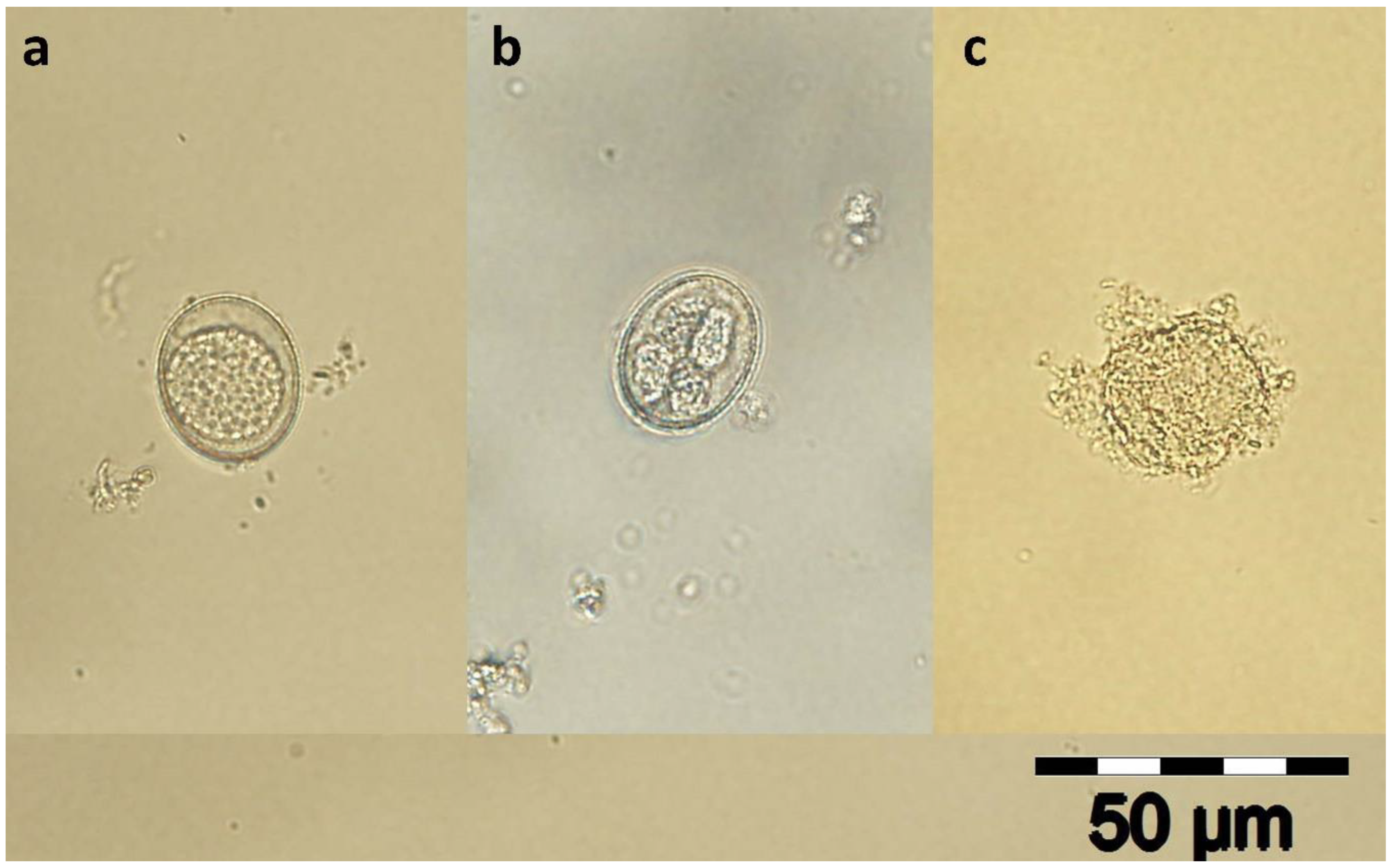
Figure 1. Unsporulated (a), sporulated (b), and destroyed oocyst of Eimeria suis (400×). The concentration of 50 mg/mL of each APE produces complete oocyst wall destruction (c).
Pathogens 12 00258 g001
Figure 2. Sporulated oocysts after 96 h of incubation (PD—potassium dichromate, E—ethanol, AA—A. absinthium, AS—A. sativum, CO—C. officinalis, CP—C. pepo, CS—C. sativum, SH—S. hortensis).
Pathogens 12 00258 g002
Figure 3. Destroyed oocysts after 96 h of incubation (PD—potassium dichromate, E—ethanol, AA—A. absinthium, AS—A. sativum, CO—C. officinalis, CP—C. pepo, CS—C. sativum, SH—S. hortensis).
Pathogens 12 00258 g003
Table 1. Experimental design.
GroupsConcentration (%)AbbreviationsContent/Well
Potassium
dichromate
0.625PD1 mL SOS (0.5 mL OS + 0.5 mL 2.5% PD) + 1 mL DW
Ethanol35E 351 mL SOS + 1 mL 70% E
17.5E 17.51 mL SOS + 1 mL 35% E
8.75E 8.751 mL SOS + 1 mL 17.5% E
4.375E 4.3751 mL SOS + 1 mL 8.75% E
2.187E 2.1871 mL SOS + 1 mL 4.37% E
Alcoholic plant extracts5AS 5, AA 5, CS 5, CP 5, SH 5, CO 51 mL SOS + 1 mL 10% APE
2.5AS 2.5, AA 2.5, CS 2.5, CP 2.5, SH 2.5, CO 2.51 mL SOS + 1 mL 5% APE
1.25AS 1.25, AA 1.25, CS 1.25, CP 1.25, SH 1.25, CO 1.251 mL SOS + 1 mL 2.5% APE
0.625AS 0.625, AA 0.625, CS 0.625, CP 0.625, SH 0.625 CO 0.6251 mL SOS + 1 mL 1.25% APE
0.312AS 0.312, AA 0.312, CS 0.312, CP 0.312, SH 0.312, CO 0.3121 mL SOS + 1 mL 0.625% APE
OS—oocysts suspension, SOS—stock oocysts suspension (oocysts suspension mixed with potassium dichromate in equal volumes), PD—potassium dichromate, DW—distilled water, E—ethanol, APEs—alcoholic plant extract, AS—A. sativum, AA—A. absinthium, CP—C. pepo, CS—C. sativum, SH—S. hortensis, CO—C. officinalis.
Table 2. The percentage of destroyed oocysts (mean ± SDM) from the experimental groups using Abbot formula.
Time (Hours)AS 5AA 5CS 5CP 5SH 5CO 5
2416.44 ± 5.22 a9.23 ± 3.05 a18.5 ± 3.59 a15.6 ± 2.88 a15.03 ± 2.72 a18.94 ± 1.93 a
4816.12 ± 6.38 a6.98 ± 2.04 a17.74 ± 3.34 a13.05 ± 2.29 a14.87 ± 2.54 a18.06 ± 2.77 a
7223.78 ± 5.47 a13.2 ± 3.09 a23.97 ± 3.74 a21.97 ± 2.98 a20.36 ± 2.88 a27.5 ± 2.18 a
9629.38 ± 3.16 ab23.96 ± 3.71 ab31.27 ± 4.42 ab25.36 ± 2.38 ab24.21 ± 2.45 b35.01 ± 1.93 a
AS 2.5AA 2.5CS 2.5CP 2.5SH 2.5CO 2.5
2415.94 ± 4.66 a16.03 ± 4.82 a18.96 ± 3.68 a15.2 ± 2.92 a14.1 ± 2.54 a18.68 ± 2.07 a
4814.72 ± 4.47 a23.46 ± 4.79 a18.78 ± 3.49 a12.47 ± 2.94 a13.03 ± 2.17 a17.8 ± 2.55 a
7225.79 ± 6.68 a32.12 ± 3.81 a25.6 ± 3.24 a22.07 ± 2.82 a22.64 ± 3.27 a29.72 ± 3.14 a
9626.45 ± 6.66 a33.82 ± 4.87 a28.08 ± 3.29 a24.02 ± 3.28 a24.15 ± 2.79 a32.51 ± 1.72 a
AS 1.25AA 1.25CS 1.25CP 1.25SH 1.25CO 1.25
246.22 ± 3.28 a9.44 ± 3.39 a9.6 ± 2.29 a4.99 ± 1.34 a4.49 ± 1.02 a8.5 ± 1.51 a
485.21 ± 3.21 a6.85 ± 2.54 a8.39 ± 2.7 a3.97 ± 1.12 a3.72 ± 0.99 a8.39 ± 1.81 a
7211.05 ± 3.17 ab11.14 ± 2.93 ab11.65 ± 2.23 ab7.42 ± 1.83 ab5.97 ± 1.19 b14.18 ± 1.94 a
9610.46 ± 4.28 a10.41 ± 2.3 a14.31 ± 2.52 a10.23 ± 1.73 a9.93 ± 1.88 a16.31 ± 1.76 a
AS 0.625AA 0.625CS 0.625CP 0.625SH 0.625CO 0.625
244.77 ± 2.32 a8.3 ± 3.8 a8.84 ± 2.32 a3.73 ± 1 a3.21 ± 0.8 a7.6 ± 1.41 a
483.33 ± 1.96 a12.97 ± 3.24 a10.43 ± 2.78 a4.31 ± 1.29 a3.81 ± 1.05 a9.74 ± 1.87 a
724.14 ± 2.22 ac14.17 ± 3.05 a5.57 ± 1.88 ac1.34 ± 0.46 c3.9 ± 0.94 bc8.71 ± 1.58 ab
963.82 ± 2.17 bc14.81 ± 2.99 a5.94 ± 1.36 bc0.8 ± 0.12 c1.89 ± 0.3 c7.49 ± 1.34 b
AS 0.312AA 0.312CS 0.312CP 0.312SH 0.312CO 0.312
242.4 ± 1.48 ab11.01 ± 4.16 a5.22 ± 1.89 ab1.24 ± 0.42 b1.47 ± 0.38 b4.75 ± 1.16 ab
483.58 ± 2.07 b14.89 ± 3.44 a6.57 ± 1.54 b1.85 ± 0.51 b2.1 ± 0.62 b6.55 ± 1.32 b
723.31 ± 2.04 bc14.92 ± 3.69 a1.89 ± 0.65 bc0.54 ± 0.12 c0.5 ± 0.09 c6.57 ± 1.31 b
965.06 ± 3.22 bc17.26 ± 3.09 a1.9 ± 0.65 bc2.06 ± 0.58 bc0.95 ± 0.2 c5.74 ± 1 b
SDM—standard deviation of mean, AS (A. sativum), AA (A. absinthium), CP (C. pepo), CS (C. sativum), SH (S. hortensis), CO (C. officinalis). Values with no common superscript in a column within an experiment were significantly different (p ≤ 0.05).
Table 3. The LC50 of each APE after 72 and 96 h of incubation.
Time (Hours)AS (mg/mL)AA (mg/mL)CS (mg/mL)CP (mg/mL)SH (mg/mL)CO (mg/mL)
7228.8431.6228.1833.1135.4824.55
9621.8818.6220.4223.4423.9916.98
AS (A. sativum), AA (A. absinthium), CP (C. pepo), CS (C. sativum), SH (S. hortensis), CO (C. officinalis).
Disclaimer/Publisher’s Note: The statements, opinions and data contained in all publications are solely those of the individual author(s) and contributor(s) and not of MDPI and/or the editor(s). MDPI and/or the editor(s) disclaim responsibility for any injury to people or property resulting from any ideas, methods, instructions or products referred to in the content.