Review

Development of Novel Anti-Leishmanials: The Case for Structure-Based Approaches

by
Mohini Soni
1,2 and
J. Venkatesh Pratap
1,2,*
1
Biochemistry and Structural Biology Division, CSIR-Central Drug Research Institute, Sector-10, Jankipuram Extension, Sitapur Road, Lucknow 226031, India
2
Academy of Scientific and Innovative Research (AcSIR), Ghaziabad 201002, India
*
Author to whom correspondence should be addressed.
Pathogens 2022, 11(8), 950; https://doi.org/10.3390/pathogens11080950
Submission received: 6 May 2022 / Revised: 16 August 2022 / Accepted: 17 August 2022 / Published: 22 August 2022
(This article belongs to the Special Issue Leishmania & Leishmaniasis)

Abstract

:
The neglected tropical disease (NTD) leishmaniasis is the collective name given to a diverse group of illnesses caused by ~20 species belonging to the genus Leishmania, a majority of which are vector borne and associated with complex life cycles that cause immense health, social, and economic burdens locally, but individually are not a major global health priority. Therapeutic approaches against leishmaniasis have various inadequacies including drug resistance and a lack of effective control and eradication of the disease spread. Therefore, the development of a rationale-driven, target based approaches towards novel therapeutics against leishmaniasis is an emergent need. The utilization of Artificial Intelligence/Machine Learning methods, which have made significant advances in drug discovery applications, would benefit the discovery process. In this review, following a summary of the disease epidemiology and available therapies, we consider three important leishmanial metabolic pathways that can be attractive targets for a structure-based drug discovery approach towards the development of novel anti-leishmanials. The folate biosynthesis pathway is critical, as Leishmania is auxotrophic for folates that are essential in many metabolic pathways. Leishmania can not synthesize purines de novo, and salvage them from the host, making the purine salvage pathway an attractive target for novel therapeutics. Leishmania also possesses an organelle glycosome, evolutionarily related to peroxisomes of higher eukaryotes, which is essential for the survival of the parasite. Research towards therapeutics is underway against enzymes from the first two pathways, while the third is as yet unexplored.

1. Background

Leishmaniasis is caused by the protozoan parasites consisting of ~20 Leishmania species, transmitted through the bites of promastigote-infected female phlebotomine sandflies during a blood meal. In the mammalian host, promastigotes are engulfed by phagocytic cells, transform into amastigotes within the cells, rapidly multiply and become a new source of infection back to the vector through the next blood meal [1]. The three main forms of the diseases have distinct symptoms and differ in degrees of severity. Visceral leishmaniasis (VL), also known as kala-azar (in the Indian subcontinent) [2], is the most lethal form of the disease, resulting in fatality if untreated. The clinical symptoms and signs of VL include irregular bouts of fever, weight loss, enlargement of the spleen and liver, and anemia; cutaneous leishmaniasis (CL), the most common form of leishmaniasis, mainly causes skin lesions and induces ulcers, which could leave life-long scars with serious disabilities; mucocutaneous leishmaniasis (ML) leads to the partial or total destruction of tissues in the nose, mouth, and throat [3]. The symptoms of leishmaniasis thus vary, ranging in pathogenesis from moderate in CL to extreme in VL. The disease affects many countries worldwide with Algeria, Afghanistan, Bolivia, Brazil, China, Colombia, Eritrea, Ethiopia, India, Iraq, Kenya, Libya, Pakistan, Somalia, South Sudan, Sudan, Syrian Arab Republic, Tunisia, and Yemen accounting for ~90% of the infections (Figure 1) [3].
Leishmaniasis has also been reported in some regions of Mexico, Central America, and South America, and some cases of cutaneous leishmaniasis have been noticed in Texas and Oklahoma in the USA (Figure 2) [3]. The epidemiology of leishmaniasis depends on the characteristics of the parasite and sand fly species, the local ecological features of the infection sites, the history of parasitic infection in the human population, and human physiology [3].
Although several drugs are available for chemotherapy of leishmaniasis, almost all of them have significant drawbacks (Table 1) [4]. Most anti-leishmanial treatments require multiple doses, for a prolonged period of time, contributing not only to the difficulty in the management of clinical leishmaniasis but also to the development of drug resistance. (Table 1) [5,6].
Most anti-leishmanial drug therapies are cumbersome and not always effective [20] and vaccines are still in the developmental stage. Alternative therapies such as the application of cytokines and immune modulators have also been investigated together with chemotherapy [21,22,23]. Significantly, these alternative approaches are also in the preliminary stage and do not ensure protection against the disease [24]. The developing vaccines are mostly prophylactic and can be broadly classified into three categories: live attenuated Leishmania vaccines e.g., ascorbic acid-deficient live mutants of Leishmania donovani; dead parasite vaccine (Leishmania major (ALM) + BCG vaccine); or defined vaccines such as DNA vaccines gP63 for visceral leishmaniasis. It is thus imperative that intensive focused research on rationale-driven candidates and target-based drug discovery be initiated towards obtaining novel therapeutics against leishmaniasis, utilizing approaches including structure-based drug design (SBDD) and ligand-based drug design (LBDD) approaches.
In structure- and ligand-based drug-discovery approaches, two components, small chemical inhibitors and target proteins and enzymes, are the central players. The major modules of SBDD comprise (i) docking the chemical inhibitors from chemical inhibitor libraries to the identified target proteins followed by (ii) experimental investigations to understand the effects of the selected docked inhibitors on the enzymatic activities in a multi well or high-throughput format. In the absence of a target protein 3D structure, structure–activity relationships (SARs)-based series of analogs could be synthesized and rescreened for the generation of potent lead compounds against the target protein. The LBDD strategy is used when SBDD is not possible due to the unavailability of the target protein structure for the fast and cost-efficient discovery of lead molecules (Figure 3).
The first step in an SBDD is the identification and preparation of the target protein’s three-dimensional structure (experimentally elucidated or computationally modelled) for in silico screening (Figure 3), followed by the docking of libraries of small molecules to the target protein using appropriate software and web-based tools and ranking. The interaction between protein and candidate molecules predicted via docking in SBDD provides details of the orientation and surface complementation of the interacting atoms. After the ligand is docked to the target protein, SBDD effectively refines the protein-ligand structure to improve ligand binding within the protein active site, in a process defined as lead-optimization. Additionally, molecular dynamics simulations may also be performed to understand and optimize the relative orientations of the interacting atoms to achieve a more favorable configuration (Figure 3). On the other hand, LBDD is used predominantly when the structure of the target protein is unknown (or cannot be modelled) but the effects of the chemical modulator on the target protein activity are known. LBDD primarily employs structure-activity relationships (SARs), where several analogs of the natural modulator would be designed with similar structure and chemical properties. If many modulators are known for the target protein, utilizing machine learning (ML) algorithms could be helpful for virtual screening in obtaining potent analogs. Scaffold hopping, which is extensively used in medicinal chemistry to generate several iso-functional compounds with distinct core moieties, could also be utilized in LBDD. The final step of any drug discovery (common to SBDD and LBDD) is to understand the pharmacokinetics and pharmacodynamics of the lead compounds which includes absorption, distribution, metabolism, excretion, and toxicity (ADMET) parameters (Figure 3) [25,26,27,28].
SBDD approaches have been successful in the identification of a novel inhibitor compound and lead molecules against a variety of diseases including infectious diseases, with some of them reaching clinical trials and getting FDA approvals. These approaches to the discovery of therapeutics against NTDs including the development of novel anti -leishmanials have made remarkable progress [28,29,30]. SBDD approaches using the structures of leishmanial Cysteine Protease B (CPB; essential for parasite survival in the host), Type 2 NADH dehydrogenase [NDH2] (a mitochondrial enzyme that converts NADH to ubiquinone and is crucial for parasite survival), Nucleoside Diphosphate Kinase (crucial for maintaining intracellular NTP levels), and Topoisomerase-II (catalyzes single-stranded breaks in DNA during gene replication and transcription) have led to the identification of novel inhibitors (Figure 4) [28,31,32,33,34,35,36]. It is pertinent, however, to emphasize that these studies have not yet resulted in the development of candidates that have reached any clinical trial stage but are primary steps towards a rationale-driven approach for novel anti-leishmanials. As Leishmania diverged early, significant differences are observed between leishmanial and mammalian homologs, whether in the individual proteins or in the whole pathways. For example, the Adenosine methyl decarboxylase (S-AdometDC) involved in the synthesis of polyamines, is an essential enzyme for all cellular organisms including Leishmania. Surprisingly, kinetoplastids including Leishmania have an additional enzyme, the AdometDC-like protein (ADL), which forms a hetero-oligomer with AdometDC, substantially increasing the efficacy. Leishmania donovani ADL also appears to bind to substrates while other trypanosomatid homologs have not been reported to bind to any substrates [37,38], indicative of subtle differences within close evolutionary related homologs as well. A similar distinction among close homologs is also observed in the C-terminal Coiled-coil (CC) oligomerization domain of the actin-interacting protein coronin that plays a role in actin-dynamics. Structurally, the Leishmania donovani and Trypanosoma brucei CC domains have a novel topology, oligomeric association, and an asymmetry, compared with other coronin CC structures, though the magnitudes of the asymmetry are different [39,40].

2. Emergence of Artificial Intelligence (AI) in Anti-leishmanial Drug Discovery

Artificial intelligence (AI) is a machine learning algorithm used to predict, analyze, interpret, model, and decide the further steps to handle very complex and bulky information in an efficient, fast, and reliable manner [41]. AI in medicine has modified the working model of health services from appointment scheduling, translating clinical data, and diagnosis, to decreasing healthcare costs [42,43,44]. Moreover, in the era of omics research, the emergence of AI would help in the analysis of high volumes of data and also provide virtual assistance during the entire research program [45,46,47,48]. Though AI enhances the therapeutic discovery processes significantly, an overemphasis on AI is also not recommended, as its drawbacks include susceptibility to security risks, inaccuracies in calculations being subsequently carried over, overlooking social variables, and human surveillance is still a necessity [49]. In drug-discovery research, AI contributes to quantitative structure-activity and property relationship (QSAR), structure-based modeling, de novo molecular design, prediction of physiochemical properties such as solubility, and partition coefficient of drug and chemical synthesis prediction [50,51,52]. Methodologically, AI works through machine learning (ML) mode by employing deep learning (DL) which finally constructs artificial neural networks (ANNs) [53] employing various algorithms, models, and approaches such as random forest, k-nearest neighbors (KNN), support vector machines (SVM), multilayer perceptron (MLP), learning vector quantization (LVQ), multi pass LVQ, linear discriminant analysis (LDA), artificial neural networks (ANN), Viola–Jones algorithm, and gradient boosting approaches [54].One of the best examples of an AI system is ‘AlphaFold Protein Structure Database’ which has been developed by DeepMind and EMBL-EBIand models the 3D structure from the amino acid sequences. This database covers the complete human proteome and 47 other species including Leishmania [55]. AI in anti-leishmanial drug discovery is still an emerging field and at an early phase that certainly requires extensive exploration at various levels such as predicting the protein structure, studying the interaction with inhibitors, projecting the favorable structure–activity relationship, mining the possible pharmacological features, suggesting the facile chemical synthesis processes, modeling the future cost analysis for actual drug discoveries, prediction of clinical trial failure, listing the adverse effects of the future drug, suggesting post-treatment prognosis situations, and advising of alternative medical treatments. AI in leishmaniasis research has contributed to critical aspects including infection diagnosis from microscopic images, peptide-fingerprints-based prediction and modeling of enzyme classes, pyruvate kinase inhibitors designing, prognosis features of unresponsive patients, and alternative chemotherapy prediction [56,57,58,59,60].

3. Parasite-Specific Essential Metabolic Pathways and Possible Drug Targets

Rationale-driven novel therapeutic approaches focus primarily on proteins and enzymes of metabolic pathways. As Leishmania is an eukaryote as are humans, the chosen pathway has to be either absent in humans or significantly diverged (sequence, tertiary structure, active site architecture and composition) for effective therapeutics. A recent review details various biochemical pathways which are crucial for the Leishmania parasite [61]. The rest of this review, for brevity, focuses on three key leishmanial metabolic pathways which could be plausible contenders for future anti-leishmanials: the folate biosynthesis pathway which is also parasite specific; the purine salvage pathway, as Leishmania cannot synthesize purines de novo and are dependent on the host for their purine requirements; and the peroxisomal import pathway. While the purine salvage and folate biosynthesis pathways have received considerable attention in relation to the development of anti-leishmanials, the peroxisomal import machinery is as yet unexplored.

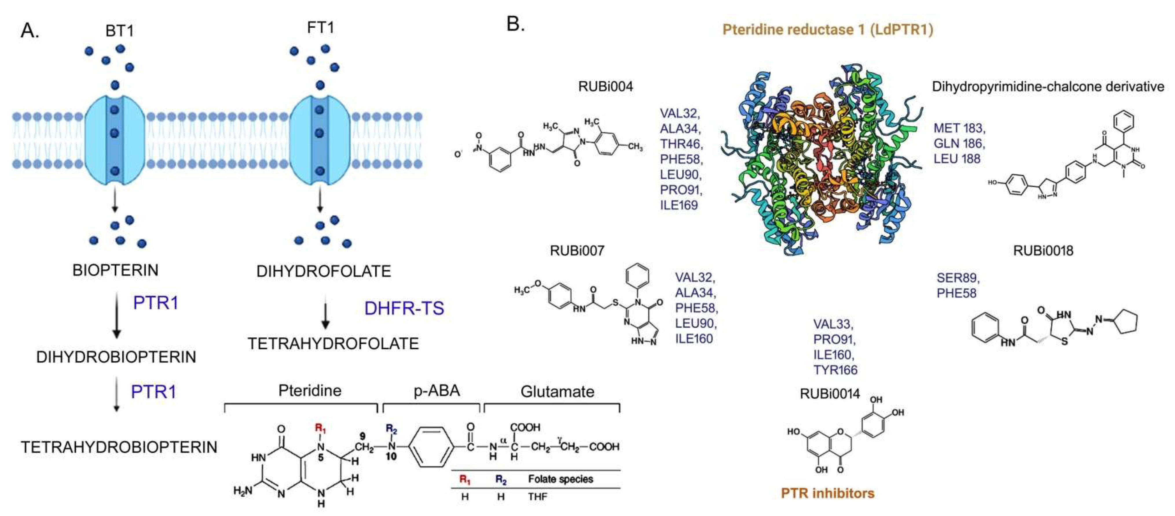
3.1. Folate Biosynthesis Pathway

Folic acid plays an essential role in key metabolic inter conversions in all forms of life which involve or result in the transfer of C1 (one carbon) units [62]. Folates, alternate carbon sources that donate or accept one carbon in many metabolic pathways, are heterocyclic derivatives of pterins and pteroic acid (conjugated with p-aminobenzoic acid pABA and glutamates (Figure 5A), that act as co-factors in hydroxylation, oxidoreduction, and C1 transfer reactions, and also have a significant role in many biological processes including DNA replication and amino acid metabolism [63,64].Surprisingly, while prokaryotes (and certain lower eukaryotes) can synthesize folate and folic acid de novo as they have catalytic enzymes for folate biosynthesis [62,65,66,67], higher eukaryotes including mammals lack these enzymes and pathways and are dependent on dietary resources via membrane-associated transport [62,65,66,67].
The multistep folate synthesis starts with the GTP cyclohydrolase I (GTPCHI) catalyzed pterin ring synthesis from GTP as a precursor, mainly in plants and prokaryotes [42,43]. In the fourth and penultimate step, dihydropteroate synthase mediates pABA attachment with pterin and after the completion of the reaction cycle, each catalyzed by a specific enzyme, GTP is converted into 7,8-dihydrofolate, reduced by dihydrofolate reductase (DHFR) to produce 5,6,7,8-tetrahydrofolate (folate or vitamin B9) [62,65,66,67]. In contrast, pterin ring synthesis in higher eukaryotes (including humans) occurs in three steps, starting with the precursor GTP, via the formation of 7,8-dihydroneopterin triphosphate (H2NTP), 6-pyruvoyl-tetrahydropterin (PTP), and finally into tetrahydrobiopterin (BH4) catalyzed by GTP cyclohydrolase I (in humans coded by the gene GCH1), 6-pyruvoyl-tetrahydropterin synthase (coded by PTS), and sepiapterin reductase (coded by SPR) enzymes, respectively [62,65,66,67]. Historically, the activity of biopterin as a cofactor for amino acid hydroxylases in any organism was identified by its requirement for the growth of the trypanosomatid insect trypanosome, Crithidia fasciculate [68,69,70,71,72]. Prokaryotes and many eukaryotes can synthesize both unconjugated and conjugated pteridines, but kinetoplastid protozoans require exogenous pteridines for their survival [73]. Leishmania acquire folates by active transport, using two different transporters that were identified by functional studies of mutants resistant to methotrexate (MTX) [71].
The three transporter and catalytic enzymes of folate biosynthesis in Leishmania, summarized in Table 2 are: (i) pterin reductase, (ii) thymidylate synthase (TS), and (iii) dihydrofolate reductase (DHFR) (Figure 5A) [73]. Gene deletion studies show that deletion of either PTR1 or DHFR-TS is lethal for the viability of Leishmania, especially in the intracellular environment of the host where folate and thymidilate rescue routes are insufficient. Additionally, PTR1 knockout Leishmania was hypersensitive to MTX and could not survive in a medium lacking pterins [74]. Thus, a dual inhibition approach of PTR1 and DHFR-TS would be promising for the development of potential drugs for the treatment of leishmaniasis.
Pterinreductase (PTR1) is a conserved dehydrogenase enzyme, with its catalytic site formed by Tyr, Lys, Ser, and Asn residues [68,69,73]. However, the serine in the leishmanial PTR1 catalytic site is replaced by an aspartate. To date, there are 14 PTR1 experimental structures reported for Leishmania: (12 from Leishmania major and one each of Leishmania tarentolae and Leishmania donovani) [77]. SBDD approaches with leishmanial PTR1 as the target protein have led to the discovery of novel inhibitors that combine the features of dihydropyrimidine and chalcone derivatives against PTR1 with improved efficacies (Figure 5B).
The other two main enzymes related to folate metabolism, dihydro folate reductase (DHFR), and thymidine synthase (TS) are commonly found fused as a bifunctional enzyme in some protozoa including Leishmania, unlike the monomeric enzymes found in bacteria and mammals [78,79,80,81]. Experimental structures of DHFR-TS from Leishmania are still unknown, although preliminary crystallographic data of the Leishmania major DHFR-TS complex have been reported, while structures of Trypanosoma homologs that share a (65–70%) sequence identity are available [78,79,80,81]. The human DHFR has ~35% sequence identity. The structures indicate that the bifunctional enzyme has the N-terminal DHFR domain and the C-terminal TS domain, with the linker region significantly shorter (three amino acids, compared to the long linkers in apicomplexa) [78,79,80,81]. The active site of the human enzyme is comparatively smaller and less hydrophobic, suggesting that more lipophilic inhibitors could potentially form stronger interactions with the protozoan enzymes. Such findings might precisely guide the discovery of new potential inhibitors against DHFR with a highly lethal capacity for parasites. Recently, research works have reported the development of molecules with activity against PTR1 and DHFR-TS of Leishmania, utilizing cheminformatics features to determine the binding mode and the possible activity in such enzymes [78,79,80,81,82,83,84].

3.2. The Purine Salvage Pathway

Unlike their mammalian and insect hosts, Leishmania completely lack de novo nucleotide biosynthesis pathways, are entirely dependent on their salvage pathway from hosts to fulfill their purine requirement, and are considered as obligate purine auxotrophs [85]. The Leishmania purine salvage pathway has three major components: extracellular nucleotidases (ecto-nucleotidase) that hydrolyze extracellular nucleotides into nucleosides to maintain the extracellular nucleoside pool, and also finally facilitate their transportation inside the parasite; transporter systems responsible for transporting purines through the cellular membrane; and catalytic enzymes for further processing of the salvaged purines, as required (Table 3) [86].

3.2.1. Extracellular Nucleotide Metabolism in Leishmania by Ecto-Nucleotidase

The nucleobases-specific transporter proteins in Leishmania usually require nucleosides instead of nucleotides. The pool of nucleosides is maintained by parasite-specific membrane-bound ecto-nucleotidases, enzymes which hydrolyze nucleotides into nucleosides (Figure 6) [87]. It has been shown that the most virulent Leishmania species (Leishmania amazonensis) efficiently hydrolyzes higher amounts of ATP, ADP, and AMP (Figure 6) [87]. Nucleotide hydrolysis by parasitic ecto-nucleotidases enzymes (membrane-bound) can modulate the host’s immune response effectively which finally results in the establishment of infection. Parasitic (including leishmanial) ecto-nucleotidases mediated hydrolysis of ATP and nucleotides disrupt the ‘danger signal’ (marked by the accumulation of excessive ATP and pro inflammatory cytokines at the site of infection) for many immune cells and prohibit the activation of any Th2 or pro inflammatory host immune responses [88]. Ecto-5’ nucleotidase (known as CD73 invertebrates) in Leishmania could also hydrolyze the ATP to supply the nucleosides [88]. Interestingly, Leishmania donovani possesses an exclusive ecto-3’ nucleotidase (Ld3’NT/NU) that could hydrolyze both nucleotides and nucleic acids including RNA and ssDNA [89,90] that are projected as putative drug targets [86].

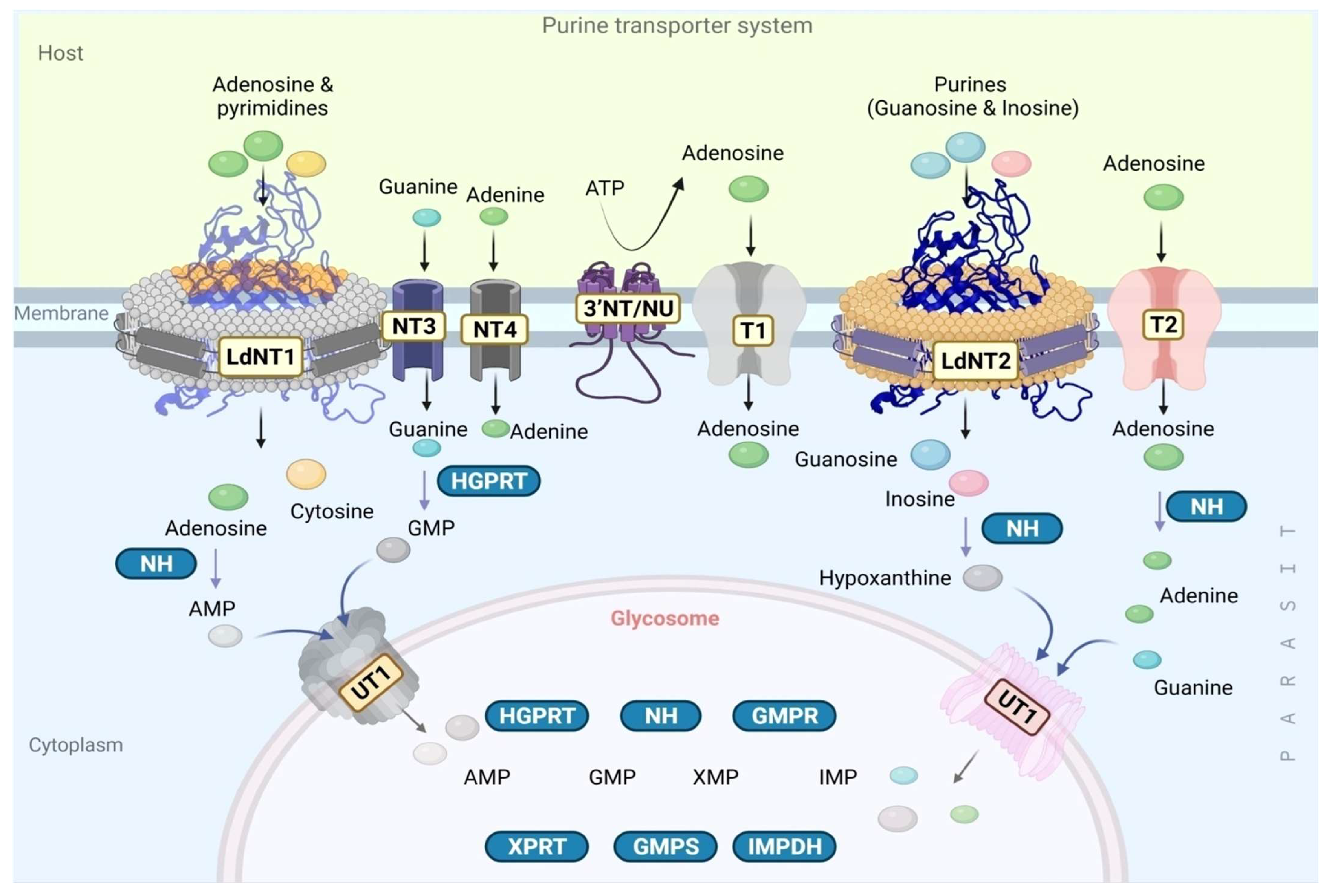
3.2.2. The Purine Transporter Systems

Leishmania parasites are decorated with specific transporters for the uptake of nucleobases primarily on the plasma and glycosomal membranes (Figure 6) [91,92,93,94,95,96,97,98]. Nucleobase transporters (NTs) NT1 and NT2, are permease proteins that exclusively transport inosine and guanosine. In addition, two supportive transporters NT3 and NT4 assist the transportation of nucleobases across the parasite membrane from the host. Moreover, it has been shown that in infectious amastigotes, two additional transporters T1 and T2 also carry adenosines with high affinity [85]. After import into the cytoplasm, the cytoplasmic salvage pathway enzymes including nucleoside amino hydrolases (NH) [for conversion to AMP, hypoxanthine, and adenine], HGPRT convert guanine into GMP [91]. Subsequently, AMP, hypoxanthine, and GMP enter the glycosome through an unknown transporter -1 (UT1) [91], which can directly transport adenine and guanine to the glycosome without any cytoplasmic enzymatic reactions. Interestingly, these transporters are pH-dependent (both neutral and acidic) and could switch the kind of nucleosides for transportation, which facilitate their survival even inside the acidic environment of phagolysosome [91]. Finally, glycosome-specific salvage pathway enzymes use these precursors or intermediates and catalyze the production of nucleotides (nucleoside mono-, di-, and triphosphates), fulfilling the parasite’s requirements (Figure 6) [91,92,93,94,95,96,97,98]. Comparative sequence studies of Leishmania and human nucleoside transporters reveal that 16 amino acids are conserved [96]. These conserved amino acids are hypothesized to be important for structure, based on structural analyses with other permeases. Ab initio computational modeling studies indicate that LdNT1, which comprises 11 transmembrane domains, undergoes a significant movement during its function with the transmembrane helices adopting inward-open and outward-close conformations [99], stabilized by two additional aromatic residues Phe 48 (TM1) and Trp 75 (TM2). Significantly, changes in the non-conserved amino acids could alter the functioning of NTs, as evinced by the point mutations G183D (TM5) and C337Y (TM7), which reduced the adenosine transportation by ~20 fold [99,100], opening a new window for precise structural and functional investigations of each amino acid in relation to drug discovery [99,100]. Structure-based identification of small inhibitors which could either block, freeze, or interlock the confirmation of these transporters or switch the substrate selectivity (or kinetics) and hinder purine transportation might be a novel idea to target LdNT1. These hypotheses await the elucidation of NT structures for further validation.

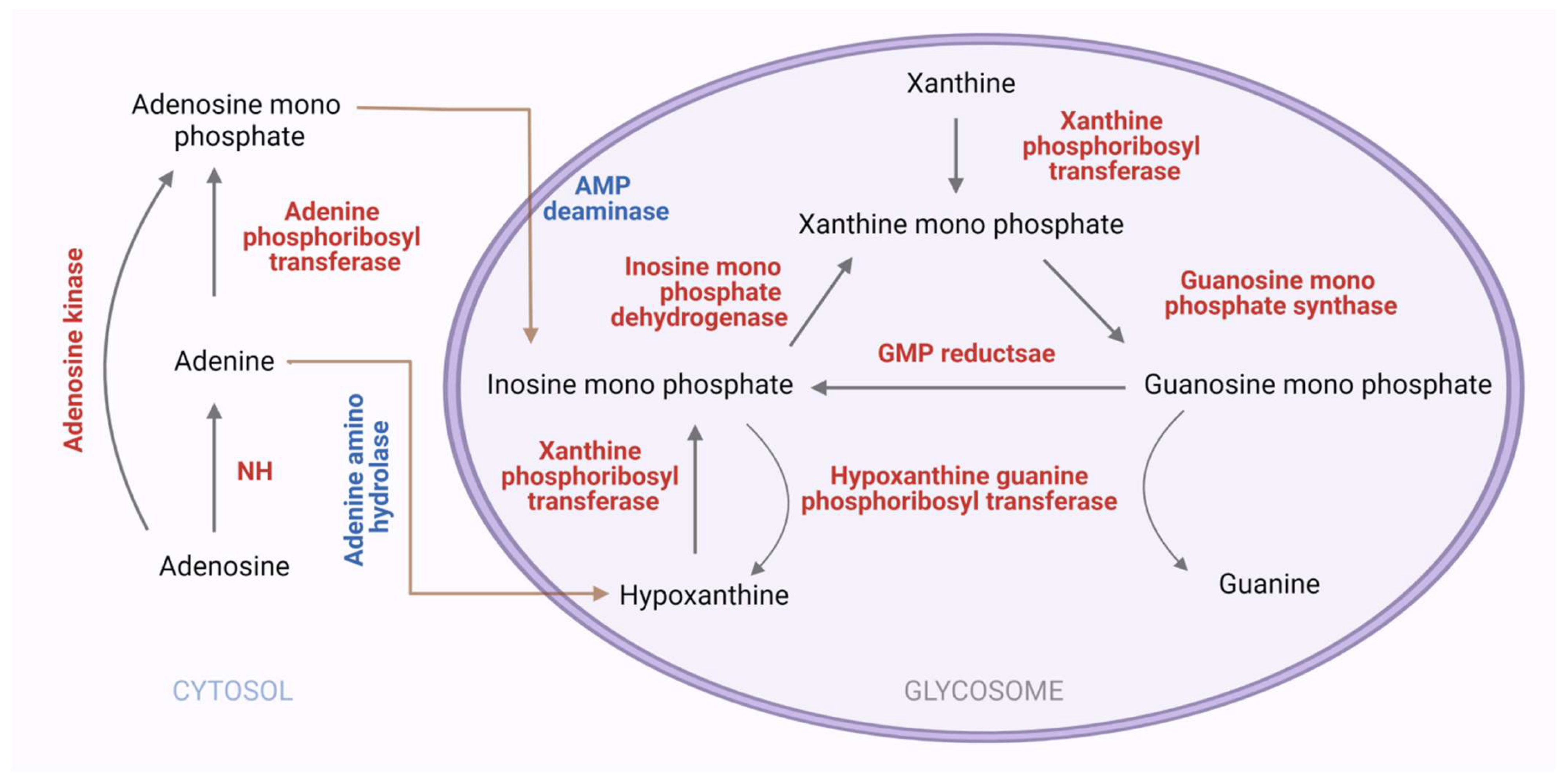
3.2.3. Purine Salvage Enzymes

The major enzymes of the purine salvage pathway in Leishmania are Phosphoribosyltranferases (PRTs) which convert dephosphorylated purines into corresponding nucleoside monophosphates [91]. Leishmania expresses three different PRTs: adenine-phosphoribosyltransferase (APRT) (EC2.4.2.7); hypoxanthine-guanine phosphoribosyltransferase (HGPRT) (EC2.4.2.8); and xanthine phosphoribosyltransferase (XPRT) (EC 2.4.2.22). In Leishmania, two distinct routes are found for adenine salvage via adenine phosphoribosyltransferase (APRT) or adenine amino hydrolase (AAH), converting adenine to hypoxanthine and AMP, respectively (Figure 7). Genetic deletion studies of APRT and AAH individually and in combination, confirm the importance of these enzymes with AAH playing a dominant role in purine salvage, with the hypoxanthine subsequently salvaged by HGPRT or XPRT [91]. The majority of purine flux into nucleotides occurs via two main routes, either HGPRT or XPRT, despite superficial complexity and convolutedness. HGPRT catalyzes the conversion of hypoxanthine to inosine monophosphate (IMP) and guanine to guanosine monophosphate (GMP). The crystal structures of Leishmania donovani APRT in complex with the substrate adenine and product AMP and sulfate and citrate ions that appear to mimic the binding of phosphate moieties, reveal differences in the binding poses with the active site pocket in a more open conformation to accommodate the larger AMP ligand [101]. Whereas AMP adopts a single conformation, adenine binds in two mutually exclusive orientations: one an orientation providing adenine-specific hydrogen bonds orientation and the other postulated for positioning adenine for the enzymatic reaction [101]. The structure of Leishmania donovani HGPRT, when compared with the human homologs (PDB ID: 1HMP), reveals contrasting modes of oligomer association involving distinct regions of the proteins, which could be valuable in rational drug discovery approaches [102]. XPRT acts on two substrates, XMP and diphosphates, forming xanthine and 5-phospho-alpha-D-ribose1-diphosphate. Among all PRT enzymes of the purine salvage pathway in Leishmania, XPRT (xanthine phosphoribosyltransferase) is unique in its substrate specificity and its nonexistence in humans, so it is an interesting protein not only for drug designing but also to understand the molecular determinants of its substrate specificity. A computational model of Leishmania donovani XPRT hypothesized that residues Ile 209, Tyr 208, and Glu 215 are important for the altered substrate specificity [102,103]. The enzymes of the purine salvage pathway are attractive therapeutics in Leishmania parasite, though the structures for a majority of these enzymes are yet unknown.

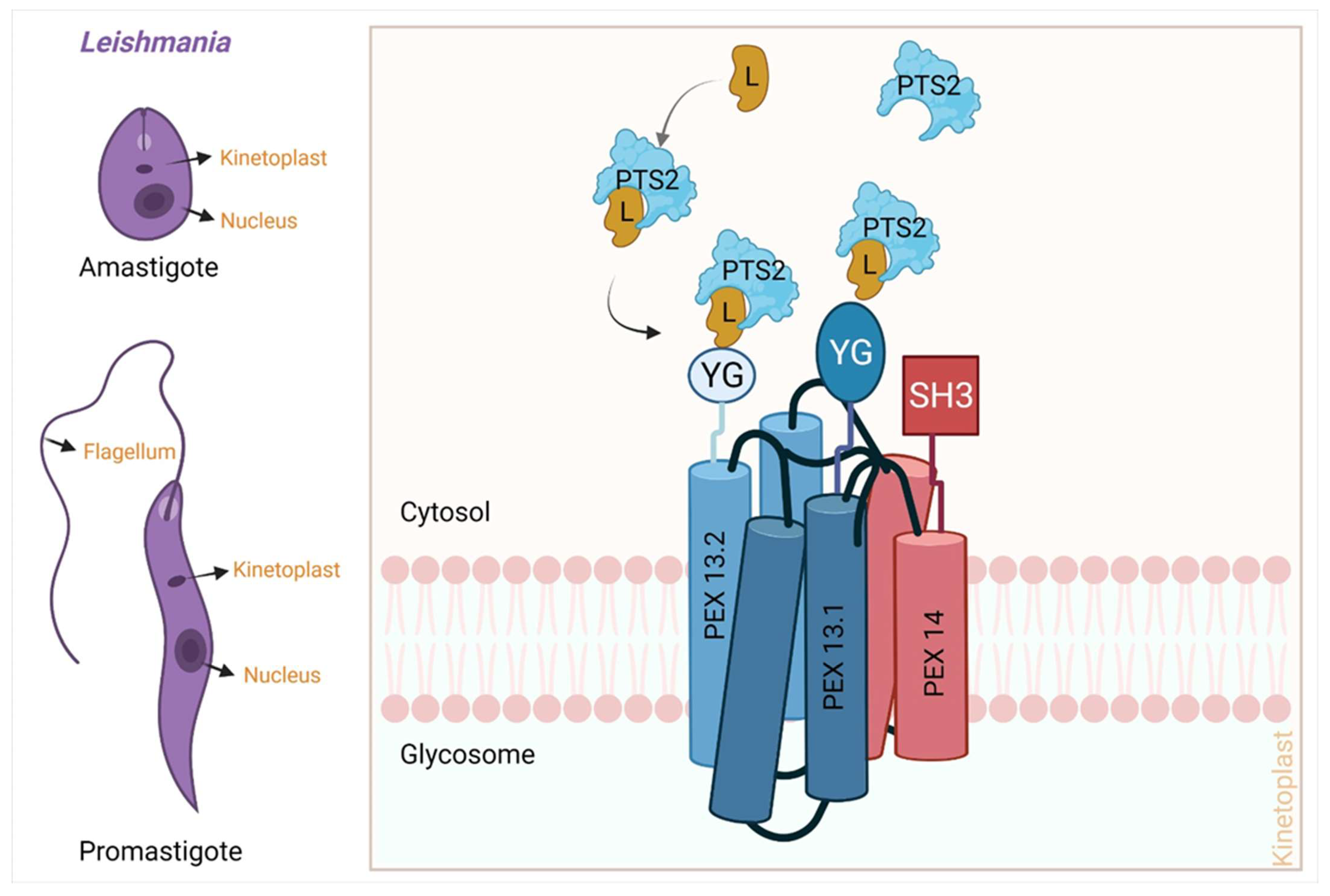
3.3. Peroxisomal Import Pathway

Peroxisomes are organelles that are found in almost all higher organisms from yeast to humans. Peroxisomes are surrounded by a single phospholipid bilayer membrane. The peroxisomal matrix harbors various enzymes that are involved in various biochemical processes including beta-oxidation, gluconeogenesis, and the pentose pathway. In yeast and plants, beta-oxidation of fatty acid takes exclusively in the peroxisome, while in mammals, the beta-oxidation of long-chain fatty acid takes place in the peroxisomes and shorter chain beta-oxidation occurs in mitochondria [104]. In plants, certain steps of photorespiration also take place in peroxisomes [104]. Dysfunction of peroxisomal proteins leads to various diseases in humans such as the Zellweger syndrome [105,106,107,108], neonatal adrenoleukodystrophy (NALD) [109], and infantile Refsum disease [110,111]. Kinetoplastids including Leishmania and Trypanosma possess the organelle glycosomes that are primarily derived from the peroxisome [112]. Glycosomes contain enzymes related to glucose metabolism and other related pathways such as the purine salvage pathway [113], and PEX proteins play a major role in the biogenesis of peroxisome. To date, around 34 peroxins are known of which functions of 19 are directly linked with peroxisome biogenesis [114,115,116]. It has been shown that during peroxisome biogenesis, the peroxisome either transports the peroxisomal protein into its membrane or destines the protein to the ER which eventually would make the membrane of the peroxisome [114,115,116]. Characterization of proteins of this pathway in Leishmania (or other kinetoplastids) are in infancy and consequently, there are no inhibitors identified against any of them. Table 4 summarizes the structural and inhibitor details of proteins of the leishmanial peroxisome import pathway.
The peroxisome import can be divided into the following stages (Figure 8):
(a)
Recognition of cargo in the cytosol
(b)
Loading of cargo or receptor complex onto the peroxisomal membrane
(c)
Cargo translocation over the membrane
(d)
Release of cargo complex into the peroxisomal membrane
(e)
Receptor recycling
Newly synthesized peroxisomal matrix proteins are transported to their desired location with the help of targeting signal sequences known as a peroxisomal targeting sequence (PTS). Two types of PTS are found, a conserved C-terminal PTS-1 (peroxisomal target sequence 1) and the N-terminal nona peptide PTS-2 (peroxisomal target sequence-2). The presence of the PTSs in any protein sequence does not always imply their peroxisomal targeting. Upon synthesis of glycosomal enzymes in the cytosol, they are recognized by two cytosolic receptors, PEX5 and PEX7. PEX5 recognizes PTS1 through its C-terminal tetra tricopeptide repeat (TPR), while PEX7 recognizes PTS-2. As PEX5 also contains a PEX7 binding region, the ultimate import depends on PEX5.The cargo-loaded receptors dock at the glycosomal membrane by binding to PEX14. PEX5-PEX14 binding results in the formation of a dynamic transient import pore, which allows translocation of the enzymes into the glycosomal lumen [112,117,118,119]. The peroxisomal import mechanism of Leishmania (Figure 8) involves the cytoplasmic PTS containing proteins binding with specific cargo protein [120]. Briefly, the peroxisomal transport signal-containing proteins which are initially present in the cytoplasm first bind with the specific ligand protein (L). This cargo-loaded ligand (L) protein has an affinity to bind PTS-proteins and facilitates the cargo delivery at the PEX receptors. Then PEX13 and 14 control the import of these cargo proteins into the peroxisome [119]. The glycosomal membrane-associated Leishmania donovani protein PEX14 plays a crucial role in protein import from the cytosol to the glycosomal matrix and consists of three domains: an N-terminal domain where the signaling molecule binds, a trans membrane domain, and an 84-residue coiled-coil domain (CC) that is responsible for oligomerization [120]. As seen in Table 4, no structures for any of the components of this important pathway from Leishmania are available, though a few trypanosomatid homologs are known [120]. Recently, a crystallization note of the CC domain of LdPEX14 has appeared in the literature [121], and any structural deviations and its critical functional values could be a great opportunity for SBDD [31,104]. The potential value of this pathway for a therapeutic approach thus depends on the structural elucidation of the components.

4. Conclusions

Leishmaniasis, a seriously debilitating NTD is still a major issue due to limited disease management, with high global morbidity. Current drugs such liposomal amphotericin B, paromomycin, and oral miltefosine have contained the disease to a certain extent, but have associated inadequacies including drug resistance, necessitating immediate discovery of novel anti leishmanials. As Leishmania majorly depends on the human host for their basic metabolic requirements, it has adapted several unique features and protein machinery which ultimately ensure their survival even under the adverse environment of the human host cells. Research is constantly underway to delineate suitable drug targets and is mainly focused on the protein related to important metabolic pathways, precursor transportation through glycosome, and adaptation machinery. Here, we have discussed three metabolic pathways and listed crucial enzymes, which are putative targets for therapeutic approaches. Some of these proteins have already been characterized in SBDD and novel inhibitors identified with potential activity inhibition and cytotoxicity (Figure 3). Recently, extensive work has been performed to filter the crucial drug targets by applying SBDD, LBDD, and other approaches, and has successfully discovered putative inhibitors. However, in vitro and in vivo validations need further exploration as they could provide an effective targeted future anti-leishmanial drug with minimal side effects. Structural characterization of leishmanial proteins precludes a rationale driven approach, despite the whole genome sequencing of Leishmania being completed in 2005 [122]. Leishmania major has 36 chromosomes with a haploid genome size of the 32.8 megabase, consisting of 911 predicted RNA genes, 39 pseudogenes, and 8272 protein-coding genes. In contrast, a query word search of ‘Leishmania’ in the Protein Data Bank (PDB) (in July 2022), resulted in only 632 entries. The corresponding numbers are higher for Trypanosoma where ~1378 structures are reported. These numbers reflect that the major obstacle in the application of SBDD in Leishmania is the unavailability of tertiary or quaternary structures of many leishmanial proteins, which might play critical roles in various metabolic pathways for housekeeping functions and survival. However, the computational prediction of protein structures using artificial intelligence and machine learning approaches as in AlphaFold2 and RoseTTA fold enable SBDD approaches to be carried forward in the absence of experimental structural data [123]. The robust and cohesive approach involving the characterization of the ~8000 leishmanial proteins on fast-track mode would pave the way for the development of novel anti-leishmanials, and also provide insights into parasite biology and their evolution. Advancements in the AI and machine learning areas also complement the anti-leishmanial drug discovery field and minimize human error with a more focused strategy.

Author Contributions

Conceptualization, M.S. and J.V.P.; methodology, M.S.; investigation, M.S.; resources, J.V.P.; data curation, M.S.; writing—original draft preparation, M.S.; writing—review and editing, M.S. and J.V.P.; visualization, M.S.; supervision, J.V.P.; project administration, J.V.P.; funding acquisition, J.V.P. All authors have read and agreed to the published version of the manuscript.

Funding

This research received no external funding.

Institutional Review Board Statement

Not applicable.

Informed Consent Statement

Not applicable.

Data Availability Statement

Not applicable.

Acknowledgments

The authors acknowledge Shakil Ahmed and Niti Kumar, CSIR-CDRI & KP Chang, Rosalind Franklin University, for their critical comments. MS acknowledges the Council of Scientific and Industrial Research, India (CSIR) for providing the Senior Research fellowship (31/004(1328)/2019-EMR-I) and the Academy of Scientific and Industrial Research (AcSIR), India, for PhD enrollment (10BB18A04012). The review bears the CSIR–CDRI communication number 10418.

Conflicts of Interest

The authors declare no conflict of interest.

Abbreviations

Neglected tropical disease (NTD), Visceral Leishmaniasis (VL), Cutaneous Leishmaniasis (CL), Mucocutaneous Leishmaniasis (ML), Structure-based drug design (SBDD), Ligand-Based Drug Design (LBDD), Pseudo-structure–activity relationship (PSAR), Quantitative Structure–activity Relationship (QSAR), Pteridine reductase 1 (PTR1), Cysteine proteases type B enzymes (CPB), Type 2 NADH dehydrogenase (NDH2), Topoisomerase 1 (Top1), Nucleobase transporter (NT), Unknown transporter (UT), Transmembrane domain (TM), Phospho ribosyltranferase (PRT), Adenosine mono phosphate (AMP), Guanosine mono phosphate (GMP), Xanthosine mono phosphate (XMP), Adenine phospho ribosyltransferase (APRT), Hypoxanthine-guanine phosphoribosyltransferase (HGPRT), Xanthine phosphoribosyltransferase (XPRT), Adenosine kinase (AK), Adenine aminohydrolase (AAH), Guanine deaminase (GDA), Adenylo succinate synthetase (ADSS), Adenylo succinate lyase (ASL), Adenine Mono Phosphate deaminase (AMPDA), Inosine monophosphate dehydrogenase (IMPDH), Guanosine mono phosphate synthase (GMPS), Guanosine mono phosphate reductase (GMPR), Nucleoside hydrolase (NH), Adenosine (ADO), Adenine (ADE), Coiled coil (CC).

References

  1. Van Griensven, J.; Ritmeijer, K.; Lynen, L.; Diro, E. Visceral leishmaniasis as an AIDS defining condition: Towards consistency across WHO guidelines. PLoS Negl. Trop. Dis. 2014, 8, e2916. [Google Scholar] [CrossRef] [PubMed] [Green Version]
  2. Zijlstra, E.E. Biomarkers in Post-kala-azar Dermal Leishmaniasis. Front. Cell. Infect. Microbiol. 2019, 9, 228. [Google Scholar] [CrossRef] [PubMed] [Green Version]
  3. World Health Organization = Organisation mondiale de la, S., Weekly Epidemiological Record 2021, 96, 35 [full issue]. Weekly Epidemiological Record = Relevé Épidémiologique Hebdomadaire 2021, 96, 401–420. Available online: https://apps.who.int/iris/handle/10665/352020 (accessed on 5 May 2022).
  4. Kedzierski, L.; Sakthianandeswaren, A.; Curtis, J.M.; Andrews, P.C.; Junk, P.C.; Kedzierska, K. Leishmaniasis: Current treatment and prospects for new drugs and vaccines. Curr. Med. Chem. 2009, 16, 599–614. [Google Scholar] [CrossRef] [PubMed]
  5. Andrade-Neto, V.V.; Cunha Junior, E.F.; Faioes, V.D.S.; Martins, T.P.; Silva, R.L.; Leon, L.L.; Santos, E.C.T. Leishmaniasis treatment: Update of possibilities for drug repurposing. Front. Biosci. 2018, 23, 967–996. [Google Scholar]
  6. Roatt, B.M.; de Oliveira Cardoso, J.M.; De Brito, R.C.F.; Coura-Vital, W.; de Oliveira Aguiar-Soares, R.D.; Reis, A.B. Recent advances and new strategies on leishmaniasis treatment. Appl. Microbiol. Biotechnol. 2020, 104, 8965–8977. [Google Scholar] [CrossRef]
  7. Chakraborty, A.K.; Majumder, H.K. Mode of action of pentavalent antimonials: Specific inhibition of type I DNA topoisomerase of Leishmania donovani. Biochem. Biophys. Res. Commun. 1988, 152, 605–611. [Google Scholar] [CrossRef]
  8. Marsden, P.D. The discovery of urea stibamine. Rev. Soc. Bras. Med. Trop. 1986, 19, 115. [Google Scholar] [CrossRef] [Green Version]
  9. Mehrizi, T.Z.; Rezayat, S.M.; Ardestani, M.S.; Shahmabadi, H.E.; Ramezani, A. A Review Study about the Effect of Chitosan Nanocarrier on Improving the Efficacy of Amphotericin B in the Treatment of Leishmania from 2010 to 2020. Curr. Drug Deliv. 2021, 18, 1234–1243. [Google Scholar] [CrossRef]
  10. Dutcher, J.D. The Discovery and Development of Amphotericin B. Dis. Chest 1968, 54 (Suppl. 1), 296–298. [Google Scholar] [CrossRef]
  11. Noyman, Y.; Levi, A.; Ben Amitai, D.; Reiss-Huss, S.; Sabbah, F.; Hodak, E.; Mimouni, T.; Friedland, R. Treating pediatric cutaneous Leishmania tropica with systemic liposomal amphotericin B: A retrospective, single-center study. Dermatol. Ther. 2022, 35, e15185. [Google Scholar] [CrossRef]
  12. Apa, H.; Devrim, I.; Bayram, N.; Deveci, R.; Demir-Özek, G.; Cartı, U. Liposomal amphotericin B versus pentavalent antimony salts for visceral Leishmania in children. Turk. J. Pediatr. 2013, 55, 378. [Google Scholar]
  13. Moen, M.D.; Lyseng-Williamson, K.A.; Scott, L.J. Liposomal amphotericin B: A review of its use as empirical therapy in febrile neutropenia and in the treatment of invasive fungal infections. Drugs 2009, 69, 361–392. [Google Scholar] [CrossRef]
  14. Bulté, D.; Van Bockstal, L.; Dirkx, L.; Kerkhof, M.V.D.; De Trez, C.; Timmermans, J.-P.; Hendrickx, S.; Maes, L.; Caljon, G. Miltefosine enhances infectivity of a miltefosine-resistant Leishmania infantum strain by attenuating its innate immune recognition. PLoS Neglected Trop. Dis. 2021, 15, e0009622. [Google Scholar] [CrossRef]
  15. Muschiola, C.; Berger, M.R.; Schuler, B.; Scherf, H.R.; Garzon, F.T.; Zeller, W.J.; Unger, C.; Eibl, H.J.; Schmähl, D. Alkyl phosphocholines: Toxicity and anticancer properties. Lipids 1987, 22, 930–934. [Google Scholar] [CrossRef]
  16. Coser, E.M.; Ferreira, B.A.; Branco, N.; Yamashiro-Kanashiro, E.H.; Lindoso, J.A.L.; Coelho, A.C. Activity of paromomycin against Leishmania amazonensis: Direct correlation between susceptibility in vitro and the treatment outcome in vivo. Int. J. Parasitol. Drugs Drug Resist. 2020, 14, 91–98. [Google Scholar] [CrossRef]
  17. Carter, C.H. Paromomycin (Humatin) in the treatment of intestinal amebiasis: A preliminary dose-range study. Antibiot. Med. Clin. Ther. 1959, 6, 586–588. [Google Scholar]
  18. Maia, A.C.R.G.; Porcino, G.N.; Detoni, M.L.; Quellis, L.R.; Emídio, N.B.; Marconato, D.G.; Messias, W.F.; Soldati, L.L.; Faria-Pinto, P.; Capriles, P.V.D.S.Z.; et al. Leishmania infantum amastigote nucleoside triphosphate diphosphohydrolase 1 (NTPDase 1): Its inhibition as a new insight into mode of action of pentamidine. Exp. Parasitol. 2019, 200, 1–6. [Google Scholar] [CrossRef]
  19. Sands, M.; Kron, M.A.; Brown, R.B. Pentamidine: A Review. Clin. Infect. Dis. 1985, 7, 625–6344. [Google Scholar] [CrossRef]
  20. Kwofie, S.K.; Broni, E.; Dankwa, B.; Enninful, K.S.; Kwarko, G.B.; Darko, L.; Durvasula, R.; Kempaiah, P.; Rathi, B.; Iii, W.A.M.; et al. Outwitting an Old Neglected Nemesis: A Review on Leveraging Integrated Data-Driven Approaches to Aid in Unraveling of Leishmanicides of Therapeutic Potential. Curr. Top. Med. Chem. 2020, 20, 349–366. [Google Scholar] [CrossRef]
  21. Halder, A.K.; Dias Soeiro Cordeiro, M.N. Advanced in Silico Methods for the Development of Anti- Leishmaniasis and Anti-Trypanosomiasis Agents. Curr. Med. Chem. 2020, 27, 697–718. [Google Scholar] [CrossRef]
  22. Oliveira, L.F.; Schubach, A.O.; Martins, M.M.; Passos, S.L.; Oliveira, R.V.; Marzochi, M.C.; Andrade, C.A. Systematic review of the adverse effects of cutaneous leishmaniasis treatment in the New World. Acta Trop. 2011, 118, 87–96. [Google Scholar] [CrossRef]
  23. Singh, K.; Garg, G.; Ali, V. Current Therapeutics, Their Problems and Thiol Metabolism as Potential Drug Targets in Leishmaniasis. Curr. Drug Metab. 2016, 17, 897–919. [Google Scholar] [CrossRef]
  24. Ghorbani, M.; Farhoudi, R. Leishmaniasis in humans: Drug or vaccine therapy? Drug Des. Dev. Ther. 2018, 12, 25–40. [Google Scholar] [CrossRef] [Green Version]
  25. Fischer, A.; Smieško, M.; Sellner, M.; Lill, M.A. Decision Making in Structure-Based Drug Discovery: Visual Inspection of Docking Results. J. Med. Chem. 2021, 64, 2489–2500. [Google Scholar] [CrossRef]
  26. Sheppard, D.; Lipkin, M.; Harris, C.; Catana, C.; Stouten, P. Strategies for Small Molecule Library Design. Curr. Pharm. Des. 2014, 20, 3314–3322. [Google Scholar] [CrossRef]
  27. Pennington, L.D.; Muegge, I. Holistic drug design for multiparameter optimization in modern small molecule drug discovery. Bioorganic Med. Chem. Lett. 2021, 41, 128003. [Google Scholar] [CrossRef]
  28. Sarnpitak, P.; Mujumdar, P.; Taylor, P.; Cross, M.; Coster, M.; Gorse, A.-D.; Krasavin, M.; Hofmann, A. Panel docking of small-molecule libraries—Prospects to improve efficiency of lead compound discovery. Biotechnol. Adv. 2015, 33, 941–947. [Google Scholar] [CrossRef] [Green Version]
  29. Eder, J.; Sedrani, R.; Wiesmann, C. The discovery of first-in-class drugs: Origins and evolution. Nat. Rev. Drug Discov. 2014, 13, 577–587. [Google Scholar] [CrossRef]
  30. Srivastava, S.; Shankar, P.; Mishra, J.; Singh, S. Possibilities and challenges for developing a successful vaccine for leishmaniasis. Parasites Vectors 2016, 9, 277. [Google Scholar] [CrossRef] [Green Version]
  31. Ferreira, L.L.G.; Andricopulo, A.D. Chemoinformatics Strategies for Leishmaniasis Drug Discovery. Front. Pharmacol. 2018, 9, 1278. [Google Scholar] [CrossRef] [Green Version]
  32. Njogu, P.; Guantai, E.M.; Pavadai, E.; Chibale, K. Computer-Aided Drug Discovery Approaches against the Tropical Infectious Diseases Malaria, Tuberculosis, Trypanosomiasis, and Leishmaniasis. ACS Infect. Dis. 2015, 2, 8–31. [Google Scholar] [CrossRef] [PubMed]
  33. Rashid, U.; Sultana, R.; Shaheen, N.; Hassan, S.F.; Yaqoob, F.; Ahmad, M.J.; Iftikhar, F.; Sultana, N.; Asghar, S.; Yasinzai, M.; et al. Structure based medicinal chemistry-driven strategy to design substituted dihydropyrimidines as potential antileishmanial agents. Eur. J. Med. Chem. 2016, 115, 230–244. [Google Scholar] [CrossRef] [PubMed]
  34. Casgrain, P.-A.; Martel, C.; McMaster, W.R.; Mottram, J.C.; Olivier, M.; Descoteaux, A. Cysteine Peptidase B Regulates Leishmania mexicana Virulence through the Modulation of GP63 Expression. PLoS Pathog. 2016, 12, e1005658. [Google Scholar] [CrossRef] [PubMed] [Green Version]
  35. Marreiros, B.C.; Sena, F.V.; Sousa, F.M.; Oliveira, A.S.F.; Soares, C.M.; Batista, A.P.; Pereira, M.M. Structural and Functional insights into the catalytic mechanism of the Type II NADH:quinone oxidoreductase family. Sci. Rep. 2017, 7, 42303. [Google Scholar] [CrossRef] [PubMed] [Green Version]
  36. Mamidala, R.; Majumdar, P.; Jha, K.K.; Bathula, C.; Agarwal, R.; Chary, M.T.; Majumder, H.K.; Munshi, P.; Sen, S. Identification of Leishmania donovani Topoisomerase 1 inhibitors via intuitive scaffold hopping and bioisosteric modification of known Top 1 inhibitors. Sci. Rep. 2016, 6, 1–13. [Google Scholar]
  37. Stevanović, S.; Perdih, A.; Senćanski, M.; Glišić, S.; Duarte, M.; Tomás, A.M.; Sena, F.V.; Sousa, F.M.; Pereira, M.M.; Solmajer, T. In Silico Discovery of a Substituted 6-Methoxy-quinalidine with Leishmanicidal Activity in Leishmania infantum. Molecules 2018, 23, 772. [Google Scholar] [CrossRef] [PubMed] [Green Version]
  38. Mishra, A.; Singh, N.; Agnihotri, P.; Mishra, S.; Singh, S.P.; Kolli, B.K.; Chang, K.P.; Sahasrabuddhe, A.A.; Siddiqi, M.I.; Pratap, J.V. Discovery of novel inhibitors for Leishmania nucleoside diphosphatase kinase (NDK) based on its structural and functional characterization. J. Comput. Mol. Des. 2017, 31, 547–562. [Google Scholar] [CrossRef] [PubMed]
  39. Nayak, A.R.; Karade, S.S.; Srivastava, V.K.; Rana, A.K.; Gupta, C.; Sahasrabuddhe, A.A.; Pratap, J. Structure of Leishmania donovani coronin coiled coil domain reveals an antiparallel 4 helix bundle with inherent asymmetry. J. Struct. Biol. 2016, 195, 129–138. [Google Scholar] [CrossRef]
  40. Parihar, P.S.; Singh, A.; Karade, S.S.; Sahasrabuddhe, A.A.; Pratap, J.V. Structural insights into kinetoplastid coronin oligomerization domain and F-actin interaction. Curr. Res. Struct. Biol. 2021, 3, 268–276. [Google Scholar] [CrossRef]
  41. Nayarisseri, A.; Khandelwal, R.; Tanwar, P.; Madhavi, M.; Sharma, D.; Thakur, G.; Speck-Planche, A.; Singh, S.K. Artificial Intelligence, Big Data and Machine Learning Approaches in Precision Medicine & Drug Discovery. Curr. Drug Targets 2021, 22, 631–655. [Google Scholar] [CrossRef]
  42. Thakur, A.; Mishra, A.P.; Panda, B.; Rodríguez, C.S.; Gaurav, I.; Majhi, B. Application of Artificial Intelligence in Pharmaceutical and Biomedical Studies. Curr. Pharm. Des. 2020, 26, 3569–3578. [Google Scholar] [CrossRef]
  43. Yu, K.H.; Beam, A.L.; Kohane, I.S. Artificial intelligence in healthcare. Nat. Biomed. Eng. 2018, 2, 719–731. [Google Scholar] [CrossRef]
  44. Miller, D.D.; Brown, E.W. Artificial Intelligence in Medical Practice: The Question to the Answer? Am. J. Med. 2018, 131, 129–133. [Google Scholar] [CrossRef]
  45. Dias, R.; Torkamani, A. Artificial intelligence in clinical and genomic diagnostics. Genome Med. 2019, 11, 70. [Google Scholar] [CrossRef] [Green Version]
  46. Mann, M.; Kumar, C.; Zeng, W.-F.; Strauss, M.T. Artificial intelligence for proteomics and biomarker discovery. Cell Syst. 2021, 12, 759–770. [Google Scholar] [CrossRef]
  47. Biswas, N.; Chakrabarti, S. Artificial Intelligence (AI)-Based Systems Biology Approaches in Multi-Omics Data Analysis of Cancer. Front. Oncol. 2020, 10, 588221. [Google Scholar] [CrossRef]
  48. Xu, Y.; Su, G.-H.; Ma, D.; Xiao, Y.; Shao, Z.-M.; Jiang, Y.-Z. Technological advances in cancer immunity: From immunogenomics to single-cell analysis and artificial intelligence. Signal Transduct. Target. Ther. 2021, 6, 312. [Google Scholar] [CrossRef]
  49. Montesinos-López, O.A.; Montesinos-López, A.; Pérez-Rodríguez, P.; Barrón-López, J.A.; Martini, J.W.R.; Fajardo-Flores, S.B.; Gaytan-Lugo, L.S.; Santana-Mancilla, P.C.; Crossa, J. A review of deep learning applications for genomic selection. BMC Genom. 2021, 22, 19. [Google Scholar] [CrossRef]
  50. Jiménez-Luna, J.; Grisoni, F.; Weskamp, N.; Schneider, G. Artificial intelligence in drug discovery: Recent advances and future perspectives. Expert Opin. Drug Discov. 2021, 16, 949–959. [Google Scholar] [CrossRef]
  51. Hessler, G.; Baringhaus, K.-H. Artificial Intelligence in Drug Design. Molecules 2018, 23, 2520. [Google Scholar] [CrossRef] [Green Version]
  52. Kashyap, K.; Siddiqi, M.I. Recent trends in artificial intelligence-driven identification and development of anti-neurodegenerative therapeutic agents. Mol. Divers. 2021, 25, 1517–1539. [Google Scholar] [CrossRef]
  53. Paul, D.; Sanap, G.; Shenoy, S.; Kalyane, D.; Kalia, K.; Tekade, R.K. Artificial intelligence in drug discovery and development. Drug Discov. Today 2020, 26, 80–93. [Google Scholar] [CrossRef]
  54. Wei, Y.; Zhou, J.; Wang, Y.; Liu, Y.; Liu, Q.; Luo, J.; Wang, C.; Ren, F.; Huang, L. A Review of Algorithm & Hardware Design for AI-Based Biomedical Applications. IEEE Trans. Biomed. Circuits Syst. 2020, 14, 145–163. [Google Scholar] [CrossRef]
  55. Varadi, M.; Anyango, S.; Deshpande, M.; Nair, S.; Natassia, C.; Yordanova, G.; Yuan, D.; Stroe, O.; Wood, G.; Laydon, A.; et al. AlphaFold Protein Structure Database: Massively expanding the structural coverage of protein-sequence space with high-accuracy models. Nucleic Acids Res. 2022, 50, D439–D444. [Google Scholar] [CrossRef]
  56. Zare, M.; Akbarialiabad, H.; Parsaei, H.; Asgari, Q.; Alinejad, A.; Bahreini, M.S.; Hosseini, S.H.; Ghofrani-Jahromi, M.; Shahriarirad, R.; Amirmoezzi, Y.; et al. A machine learning-based system for detecting leishmaniasis in microscopic images. BMC Infect. Dis. 2022, 22, 48. [Google Scholar] [CrossRef]
  57. Concu, R.; Dea-Ayuela, M.; Perez-Montoto, L.; Prado-Prado, F.; Uriarte, E.; Bolás-Fernández, F.; Podda, G.; Pazos, A.; Munteanu, C.; Ubeira, F.; et al. 3D entropy and moments prediction of enzyme classes and experimental-theoretic study of peptide fingerprints in Leishmania parasites. Biochim. Biophys. Acta (BBA)—Proteins Proteom. 2009, 1794, 1784–1794. [Google Scholar] [CrossRef]
  58. Jamal, S.; Scaria, V. Cheminformatic models based on machine learning for pyruvate kinase inhibitors of Leishmania mexicana. BMC Bioinform. 2013, 14, 329. [Google Scholar] [CrossRef] [Green Version]
  59. Bamorovat, M.; Sharifi, I.; Rashedi, E.; Shafiian, A.; Sharifi, F.; Khosravi, A.; Tahmouresi, A. A novel diagnostic and prognostic approach for unresponsive patients with anthroponotic cutaneous leishmaniasis using artificial neural networks. PLoS ONE 2021, 16, e0250904. [Google Scholar] [CrossRef]
  60. Herrera-Acevedo, C.; Perdomo-Madrigal, C.; Muratov, E.N.; Scotti, L.; Scotti, M.T. Discovery of Alternative Chemotherapy Options for Leishmaniasis through Computational Studies of Asteraceae. ChemMedChem 2020, 16, 1234–1245. [Google Scholar] [CrossRef]
  61. Raj, S.; Sasidharan, S.; Balaji, S.N.; Saudagar, P. An overview of biochemically characterized drug targets in metabolic pathways of Leishmania parasite. Parasitol. Res. 2020, 119, 2025–2037. [Google Scholar] [CrossRef]
  62. Dias, M.V.B.; Santos, J.C.; Libreros-Zúñiga, G.A.; Ribeiro, J.A.; Chavez-Pacheco, S.M. Folate biosynthesis pathway: Mechanisms and insights into drug design for infectious diseases. Future Med. Chem. 2018, 10, 935–959. [Google Scholar] [CrossRef] [PubMed]
  63. Elizabeth, K.E. Folate: Its Biological Interactions and Strategies to Achieve Sufficiency without Causing Excess. Am. J. Public Health 2021, 111, 347–349. [Google Scholar] [CrossRef] [PubMed]
  64. Wang, R.; Hu, X.; Wu, S.; Xiao, H.; Cai, H.; Xie, Z.; Huang, Y.; Jing, X. Biological Characterization of Folate-Decorated Biodegradable Polymer–Platinum (II) Complex Micelles. Mol. Pharm. 2012, 9, 3200–3208. [Google Scholar] [CrossRef] [PubMed]
  65. Chen, P.; Sung, L.; Hegemann, J.D.; Chu, J. Disrupting Transcription and Folate Biosynthesis Leads to Synergistic Suppression of Escherichia coli Growth. ChemMedChem 2022, 17, e202200075. [Google Scholar] [CrossRef]
  66. Gorelova, V.; Bastien, O.; De Clerck, O.; Lespinats, S.; Rébeillé, F.; Van Der Straeten, D. Evolution of folate biosynthesis and metabolism across algae and land plant lineages. Sci. Rep. 2019, 9, 5731. [Google Scholar] [CrossRef] [Green Version]
  67. Kok, D.E.; Steegenga, W.T.; McKay, J.A. Folate and epigenetics: Why we should not forget bacterial biosynthesis. Epigenomics 2018, 10, 1147–1150. [Google Scholar] [CrossRef] [Green Version]
  68. El Fadili, A.; Kündig, C.; Roy, G.; Ouellette, M. Inactivation of the Leishmania tarentolae Pterin Transporter (BT1) and Reductase (PTR1) Genes Leads to Viable Parasites with Changes in Folate Metabolism and Hypersensitivity to the Antifolate Methotrexate. J. Biol. Chem. 2004, 279, 18575–18582. [Google Scholar] [CrossRef] [Green Version]
  69. Wang, J.; Leblanc, É.; Chang, C.F.; Papadopoulou, B.; Bray, T.; Whiteley, J.M.; Lin, S.X.; Ouellette, M. Pterin and Folate Reduction by theLeishmania tarentolaeH Locus Short-Chain Dehydrogenase/Reductase PTR1. Arch. Biochem. Biophys. 1997, 342, 197–202. [Google Scholar] [CrossRef]
  70. Papadopoulou, B.; Roy, G.; Mourad, W.; Leblanc, E.; Ouellette, M. Changes in folate and pterin metabolism after disruption of the Leishmania H locus short chain dehydrogenase gene. J. Biol. Chem. 1994, 269, 7310–7315. [Google Scholar] [CrossRef]
  71. Ellenberger, T.E.; Beverley, S.M. Biochemistry and regulation of folate and methotrexate transport in Leishmania major. J. Biol. Chem. 1987, 262, 10053–10058. [Google Scholar] [CrossRef]
  72. Scott, D.A.; Coombs, G.H.; Sanderson, B.E. Folate utilisation by Leishmania species and the identification of intracellular derivatives and folate-metabolising enzymes. Mol. Biochem. Parasitol. 1987, 23, 139–149. [Google Scholar] [CrossRef]
  73. Vickers, T.J.; Beverley, S.M. Folate metabolic pathways in Leishmania. Essays Biochem. 2011, 51, 63–80. [Google Scholar] [CrossRef] [Green Version]
  74. Hardy, L.W.; Matthews, W.; Nare, B.; Beverley, S.M. Biochemical and Genetic Tests for Inhibitors of Leishmania Pteridine Pathways. Exp. Parasitol. 1997, 87, 158–170. [Google Scholar] [CrossRef]
  75. Vadloori, B.; Sharath, A.K.; Prabhu, N.P.; Maurya, R. Homology modelling, molecular docking, and molecular dynamics simulations reveal the inhibition of Leishmania donovani dihydrofolate reductase-thymidylate synthase enzyme by Withaferin-A. BMC Res. Notes 2018, 11, 246. [Google Scholar] [CrossRef] [Green Version]
  76. Kimuda, M.P.; Laming, D.; Hoppe, H.C.; Bishop, T. Identification of Novel Potential Inhibitors of Pteridine Reductase 1 in Trypanosoma brucei via Computational Structure-Based Approaches and in Vitro Inhibition Assays. Molecules 2019, 24, 142. [Google Scholar] [CrossRef] [Green Version]
  77. Zhao, H.; Bray, T.; Ouellette, M.; Zhao, M.; Ferre, R.A.; Matthews, D.; Whiteley, J.M.; Varughese, K.I. Structure of pteridine reductase (PTR1) from Leishmania tarentolae. Acta Crystallogr. Sect. D Biol. Crystallogr. 2003, 59, 1539–1544. [Google Scholar] [CrossRef] [Green Version]
  78. Rajasekaran, R.; Chen, Y.-P.P. Probing the structure of Leishmania major DHFR TS and structure based virtual screening of peptide library for the identification of anti-leishmanial leads. J. Mol. Model. 2012, 18, 4089–4100. [Google Scholar] [CrossRef]
  79. Maganti, L.; Manoharan, P.; Ghoshal, N. Probing the structure of Leishmania donovani chagasi DHFR-TS: Comparative protein modeling and protein–ligand interaction studies. J. Mol. Model. 2010, 16, 1539–1547. [Google Scholar] [CrossRef]
  80. Gueiros-Filho, F.J.; Beverley, S.M. Selection against the dihydrofolate reductase-thymidylate synthase (DHFR-TS) locus as a probe of genetic alterations in Leishmania major. Mol. Cell. Biol. 1996, 16, 5655–5663. [Google Scholar] [CrossRef] [Green Version]
  81. Nelson, K.; Aloso, G.; Langer, P.J.; Beverley, S.M. Sequence of the dihydrofolate reductase-thymidylate synthase (DHFR-TS) gene of Leishmania amazonensis. Nucleic Acids Res. 1990, 18, 2819. [Google Scholar] [CrossRef] [Green Version]
  82. Gilbert, I.H. Inhibitors of dihydrofolate reductase in leishmania and trypanosomes. Biochim. Biophys. Acta (BBA)—Mol. Basis Dis. 2002, 1587, 249–257. [Google Scholar] [CrossRef] [Green Version]
  83. Teixeira, B.V.F.; Teles, A.L.B.; da Silva, S.G.; Brito, C.C.B.; de Freitas, H.F.; Pires, A.B.L.; Froes, T.Q.; Castilho, M.S. Dual and selective inhibitors of pteridine reductase 1 (PTR1) and dihydrofolate reductase-thymidylate synthase (DHFR-TS) from Leishmania chagasi. J. Enzym. Inhib. Med. Chem. 2019, 34, 1439–1450. [Google Scholar] [CrossRef] [Green Version]
  84. Veras, P.; Brodskyn, C.; Balestieri, F.; De Freitas, L.; Ramos, A.; Queiroz, A.; Barral, A.; Beverley, S.; Barral-Netto, M. A dhfr-ts- Leishmania major Knockout Mutant Cross-protects against Leishmania amazonensis. Mem. Inst. Oswaldo Cruz 1999, 94, 491–496. [Google Scholar] [CrossRef] [PubMed] [Green Version]
  85. Berg, M.V.D.V.; Van der Veken, P.; Goeminne, A.; Haemers, A.; Augustyns, K. Inhibitors of the Purine Salvage Pathway: A Valuable Approach for Antiprotozoal Chemotherapy? Curr. Med. Chem. 2010, 17, 2456–2481. [Google Scholar] [CrossRef]
  86. Freitas-Mesquita, A.L.; Meyer-Fernandes, J.R. Ecto-nucleotidases and Ecto-phosphatases from Leishmania and Trypanosoma Parasites. Subcell Biochem. 2013, 74, 217–252. [Google Scholar] [CrossRef]
  87. Da Silva, W.; da Rocha Torres, N.; de Melo Agripino, J.; da Silva, V.H.; de Souza, A.C.A.; Ribeiro, I.C.; de Oliveira, T.A.; de Souza, L.A.; Andrade, L.K.R.; de Moraes, J.V.B.; et al. ENTPDases from Pathogenic Trypanosomatids and Purinergic Signaling: Shedding Light towards Biotechnological Applications. Curr. Top. Med. Chem. 2021, 21, 213–226. [Google Scholar]
  88. Gbenle, G.O.; Dwyer, D.M. Purification and properties of 3′-nucleotidase of Leishmania donovani. Biochem. J. 1992, 285, 41–46. [Google Scholar] [CrossRef] [Green Version]
  89. Debrabant, A.; Ghedin, E.; Dwyer, D.M. Dissection of the functional domains of the Leishmania surface membrane 3’-nucleotidase/nuclease, a unique member of the class I nuclease family. J. Biol. Chem. 2000, 275, 16366–16372. [Google Scholar] [CrossRef] [Green Version]
  90. Debrabant, A.; Gottlieb, M.; Dwyer, D.M. Isolation and characterization of the gene encoding the surface membrane 3′-nucleotidase/nuclease of Leishmania donovani. Mol. Biochem. Parasitol. 1995, 71, 51–63. [Google Scholar] [CrossRef]
  91. Boitz, J.M.; Ullman, B.; Jardim, A.; Carter, N.S. Purine salvage in Leishmania: Complex or simple by design? Trends Parasitol. 2012, 28, 345–352. [Google Scholar] [CrossRef] [Green Version]
  92. Fulwiler, A.L.; Boitz, J.M.; Yates, P.A.; Carter, N.S.; Ullman, B. Characterization of amplicons that suppress the conditional lethal growth phenotype of a Leishmania donovani mutant lacking normal purine salvage mechanisms. Mol. Biochem. Parasitol. 2011, 175, 76–82. [Google Scholar] [CrossRef] [PubMed]
  93. Boitz, J.M.; Ullman, B. A Conditional Mutant Deficient in Hypoxanthine-guanine Phosphoribosyltransferase and Xanthine Phosphoribosyltransferase Validates the Purine Salvage Pathway of Leishmania donovani. J. Biol. Chem. 2006, 281, 16084–16089. [Google Scholar] [CrossRef] [PubMed] [Green Version]
  94. Cui, L.; Rajasekariah, G.; Martin, S.K. A nonspecific nucleoside hydrolase from Leishmania donovani: Implications for purine salvage by the parasite. Gene 2001, 280, 153–162. [Google Scholar] [CrossRef]
  95. Krenitsky, T.A.; Miller, R.L. Purine Salvage Enzymes in Leishmania Donovani and Trichomonas Vaginalis. In Purine Metabolism in Man-IV; Springer: Boston, MA, USA, 1984; pp. 215–218. [Google Scholar] [CrossRef]
  96. Davies, M.J.; Ross, A.M.; Gutteridge, W.E. The enzymes of purine salvage in Trypanosoma cruzi, Trypanosoma brucei and Leishmania mexicana. Parasitology 1983, 87, 211–217. [Google Scholar] [CrossRef]
  97. LaFon, S.W.; Nelson, D.J.; Berens, R.L.; Marr, J. Purine and pyrimidine salvage pathways in Leishmania donovani. Biochem. Pharmacol. 1982, 31, 231–238. [Google Scholar] [CrossRef]
  98. Krenitsky, T.A.; Koszalka, G.W.; Tuttle, J.V.; Adamczyk, D.L.; Elion, G.B.; Marr, J.J. Purine Salvage Enzymes in Man and Leishmania donovani. In Purine Metabolism in Man-III; Springer: Boston, MA, USA, 1980; pp. 51–56. [Google Scholar] [CrossRef]
  99. Landfear, S.M.; Ullman, B.; Carter, N.S.; Sanchez, M.A. Nucleoside and Nucleobase Transporters in Parasitic Protozoa. Eukaryot. Cell 2004, 3, 245–254. [Google Scholar] [CrossRef] [Green Version]
  100. Carter, N.S.; Landfear, S.; Ullman, B. Nucleoside transporters of parasitic protozoa. Trends Parasitol. 2001, 17, 142–145. [Google Scholar] [CrossRef]
  101. Phillips, C.L.; Ullman, B.; Brennan, R.G.; Hill, C.P. Crystal structures of adenine phosphoribosyltransferase from Leishmania donovani. EMBO J. 1999, 18, 3533–3545. [Google Scholar] [CrossRef] [Green Version]
  102. Parihar, P.S.; Pratap, J.V. The L. donovani Hypoxanthine-guanine phosphoribosyl transferase (HGPRT) oligomer is distinct from the human homolog. Biochem. Biophys. Res. Commun. 2020, 532, 499–504. [Google Scholar] [CrossRef]
  103. Patel, B.; Patel, D.; Parmar, K.; Chauhan, R.; Singh, D.D.; Pappachan, A.L. donovani XPRT: Molecular characterization and evaluation of inhibitors. Biochim. Biophys. Acta (BBA)—Proteins Proteom. 2017, 1866, 426–441. [Google Scholar] [CrossRef]
  104. Islinger, M.; Voelkl, A.; Fahimi, H.D.; Schrader, M. The peroxisome: An update on mysteries 2.0. Histochem. Cell Biol. 2018, 150, 443–471. [Google Scholar] [CrossRef] [Green Version]
  105. Shimozawa, N.; Suzuki, Y.; Tomatsu, S.; Nakamura, H.; Kono, T.; Takada, H.; Tsukamoto, T.; Fujiki, Y.; Orii, T.; Kondo, N. A novel mutation, R125X in peroxisome assembly factor-1 responsible for zellweger syndrome. Hum. Mutat. 1998, 11, S134–S136. [Google Scholar] [CrossRef]
  106. Masuno, M.; Shimozawa, N.; Suzuki, Y.; Kondo, N.; Orii, T.; Tsukamoto, T.; Osumi, T.; Fujiki, Y.; Imaizumi, K.; Kuroki, Y. Assignment of the Human Peroxisome Assembly Factor-1 Gene (PXMP3) Responsible for Zellweger Syndrome to Chromosome 8q21.1 by Fluorescence in Situ Hybridization. Genomics 1994, 20, 141–142. [Google Scholar] [CrossRef]
  107. Santos, M.J.; Hoefler, S.; Moser, A.B.; Moser, H.W.; Lazarow, P.B. Peroxisome assembly mutations in humans: Structural heterogeneity in Zellweger syndrome. J. Cell. Physiol. 1992, 151, 103–112. [Google Scholar] [CrossRef]
  108. Shimozawa, N.; Tsukamoto, T.; Suzuki, Y.; Orii, T.; Shirayoshi, Y.; Mori, T.; Fujiki, Y. A Human Gene Responsible for Zellweger Syndrome That Affects Peroxisome Assembly. Science 1992, 255, 1132–1134. [Google Scholar] [CrossRef]
  109. Schmitt, K.; Molzer, B.; Stöckler, S.; Tulzer, G.; Tulzer, W. Zellweger syndrome, neonatal adrenoleukodystrophy or infantile Refsum’s disease in a case with generalized peroxisome defect? Wien Klin Wochenschr. 1993, 105, 320–322. [Google Scholar]
  110. Van Maldergem, L.; Moser, A.B.; Vincent, M.-F.; Roland, D.; Reding, R.; Otte, J.-B.; Wanders, R.J.; Sokal, E. Orthotopic liver transplantation from a living-related donor in an infant with a peroxisome biogenesis defect of the infantile Refsum disease type. J. Inherit. Metab. Dis. 2005, 28, 593–600. [Google Scholar] [CrossRef]
  111. Mandel, H.; Berant, M.; Meiron, D.; Aizin, A.; Oiknine, J.; Brook, J.G.; Aviram, M. Plasma lipoproteins and monocyte-macrophages in a peroxisome-deficient system: Study of a patient with infantile refsum disease. J. Inherit. Metab. Dis. 1992, 15, 774–784. [Google Scholar] [CrossRef]
  112. Opperdoes, F. The glycosome of trypanosomes and Leishmania. Biochem. Soc. Trans. 1990, 18, 729–731. [Google Scholar] [CrossRef]
  113. Shih, S.; Hwang, H.-Y.; Carter, D.; Stenberg, P.; Ullman, B. Localization and Targeting of the Leishmania donovaniHypoxanthine-Guanine Phosphoribosyltransferase to the Glycosome. J. Biol. Chem. 1998, 273, 1534–1541. [Google Scholar] [CrossRef] [Green Version]
  114. Demaret, T.; Courtoy, G.E.; Ravau, J.; Van Der Smissen, P.; Najimi, M.; Sokal, E.M. Accurate and live peroxisome biogenesis evaluation achieved by lentiviral expression of a green fluorescent protein fused to a peroxisome targeting signal 1. Histochem. Cell Biol. 2020, 153, 295–306. [Google Scholar] [CrossRef]
  115. Jansen, R.L.M.; van der Klei, I.J. The peroxisome biogenesis factors Pex3 and Pex19: Multitasking proteins with disputed functions. FEBS Lett. 2019, 593, 457–474. [Google Scholar] [CrossRef] [Green Version]
  116. Farre, J.C.; Mahalingam, S.S.; Proietto, M.; Subramani, S. Peroxisome biogenesis, membrane contact sites, and quality control. EMBO Rep. 2019, 20, e46864. [Google Scholar] [CrossRef]
  117. Lutje, V.; Probyn, K.; Seixas, J.; Bergman, H.; Villanueva, G. Chemotherapy for second-stage human African trypanosomiasis: Drugs in use. Cochrane Database Syst. Rev. 2021, 12, CD015374. [Google Scholar] [CrossRef]
  118. Banerjee, H.; LaPointe, P.; Eitzen, G.; Rachubinski, R.A. A Small Molecule Inhibitor of Pex3–Pex19 Interaction Disrupts Glycosome Biogenesis and Causes Lethality in Trypanosoma brucei. Front. Cell Dev. Biol. 2021, 9, 1403–1411. [Google Scholar] [CrossRef]
  119. Jardim, A.; Rager, N.; Liu, W.; Ullman, B. Peroxisomal targeting protein 14 (PEX14) from Leishmania donovani Molecular, biochemical, and immunocytochemical characterization. Mol. Biochem. Parasitol. 2002, 124, 51–62. [Google Scholar] [CrossRef]
  120. Cyr, N.; Smith, T.K.; Boisselier, É.; Leroux, L.P.; Kottarampatel, A.H.; Davidsen, A.; Salesse, C.; Jardim, A. The hydrophobic region of the Leishmania peroxin 14: Requirements for association with a glycosome mimetic membrane. Biochem. J. 2018, 475, 511–529. [Google Scholar] [CrossRef] [PubMed] [Green Version]
  121. Shakya, A.K.; Pratap, J.V. The coiled-coil domain of glycosomal membrane-associated Leishmania donovani PEX14: Cloning, overexpression, purification and preliminary crystallographic analysis. Acta Crystallogr. Sect. F Struct. Biol. Commun. 2020, 76, 464–468. [Google Scholar] [CrossRef] [PubMed]
  122. Ivens, A.C.; Peacock, C.S.; Worthey, E.A.; Murphy, L.; Aggarwal, G.; Berriman, M.; Sisk, E.; Rajandream, M.-A.; Adlem, E.; Aert, R.; et al. The Genome of the Kinetoplastid Parasite, Leishmania Major. Science 2005, 309, 436–442. [Google Scholar] [CrossRef] [Green Version]
  123. Jumper, J.; Evans, R.; Pritzel, A.; Green, T.; Figurnov, M.; Ronneberger, O.; Tunyasuvunakool, K.; Bates, R.; Žídek, A.; Potapenko, A.; et al. Highly accurate protein structure prediction with AlphaFold. Nature 2021, 596, 583–589. [Google Scholar] [CrossRef]
Figure 1. Global incidences of three different types of leishmaniasis. Venn diagram shows the countries affected by different types of leishmaniasis (WHO 2020) [created with BioRender.com accessed on [4 April 2022]].
Pathogens 11 00950 g001
Figure 2. Distribution of various leishmaniasis in different countries: Mapping the global prevalence of three types of leishmaniasis reported across the globe (* are used to indicated the correct location of countries on the map) (adapted from the World Health Organization, 2020) [Created with MapChart.net].
Pathogens 11 00950 g002
Figure 3. Workflow for target-specific drug discovery and its utilities in the Leishmania-specific inhibitor discoveries. Drug discovery approaches begin with the determination of 3D structures of target protein either by X-ray crystallography, NMR, cryo-EM or by using computational methods (homology modeling). This serves as the template for molecular docking studies against a library of chemical entities, resulting in the identification of lead molecules, i.e., specific hit compounds based on the binding score and binding dynamics. Identified molecules would be experimentally bio-assayed and in parallel facile chemical methods would be established. The working process of SBDD starts with druggability prediction, followed by docking-based virtual screening and pharmacophore identification. Finally, molecular dynamics would provide insights into the protein followed by lead optimization. If the structure of the protein is unknown, the ligand-based drug discovery (LBDD) approach would be utilized to design an analog molecule after refinement by scaffold hopping. The main aim of scaffold hopping is the discovery of any novel chemical moiety with improved pharmacological features compared with the marketed drug against the same target protein. It starts with ligand-based pharmacophore identification, followed by structure–activity relationship analysis. Finally, the drug-discovery system deals with the various properties such as chemical absorption, distribution, metabolism, excretion, and toxicity (ADMET) of any lead compound (future drug) [created with BioRender.com].
Pathogens 11 00950 g003
Figure 4. SBDD-based inhibitors discoveries. Selected examples are showing the inhibitor discoveries against various enzymes by employing SBDD approaches under the umbrella of anti-leishmanial research [created with BioRender.com].
Pathogens 11 00950 g004
Figure 5. Folate biosynthesis pathway in Leishmania donovani and targeting of LdPTR1 enzyme with specific inhibitors. (A)—BT-1 (biopterin) and FT-1 (folate) enter in the parasitic cells through transporters. Thereafter, metabolic enzymes such asPTR1 and DHFR-TS catalyze further steps and produce tetrahydrobiopterin and tetrahydrofolate, respectively. (B)—PTR-1 protein interacts with various inhibitors through the indicated amino acids, primarily involving H-bonds and π-π interactions (created with BioRender.com).
Pathogens 11 00950 g005
Figure 6. Purine transporter system in Leishmania. Ecto-nucleotidases (Ld3’NT/NU) hydrolyze the nucleotides (ATP) into nucleosides (adenosine). Adenosine and purine transportation occurs through LdNT1/2, NT3, NT4, T1, and T2 transporters across the cell membrane from the host. Various enzymes such as HGPRT of the purine salvage pathway (shown in blue boxes) catalyze the conversion of guanine to guanine mono phosphate. GMP, AMP hypoxanthine, adenosine, and guanine transportations into glycosome mediated by unknown transporters 1 and 2 (UT1/2) [created with BioRender.com].
Pathogens 11 00950 g006
Figure 7. The purine salvage pathway which is catalyzed by specific enzymes in Leishmania (predicted pathway). The predicted purine salvage pathway in Leishmania, the pink compartment represents glycosomes. Enzymatic reactions catalyzed by indicated proteins of the purine salvage pathway in Leishmania [created with BioRender.com].
Pathogens 11 00950 g007
Figure 8. Peroxisomal import receptors (PEX), mediated import of cytoplasmic protein into peroxisome of Leishmania at amastigote and promastigote stages. Peroxisomal transport sequence (PTS) containing cargo proteins first bind with the Ligand protein, which facilitates the delivery of these cargos to the PEX receptors. Once these cargo proteins are loaded over to the PEX complex then they selectively control their import inside the peroxisome [created with BioRender.com].
Pathogens 11 00950 g008
Table 1. Current chemotherapy for visceral leishmaniasis.
DrugDiscoveryMode of ActionSide EffectRemark
Pentavalent antimonial [7]Brahmachari,
1940 [8]
Proposed:
Trypanothione
Reductase and macromolecule biosynthesis inhibitor
Cardiotoxicity, injection painsDevelopment of drug resistance
Amphotericin B (1992) [9]Repurposed antifungals for neutropenic patients [10].Cell membrane permeabilization via a complex with sterols and ergosterolsRenal toxicity, hypokalemiaDose-limiting
Liposomal amphotericinB (2009) [11,12]Used in antifungal infections, e.g., Cryptococcal meningitis [13]Targeted drug delivery to infected macrophages to kill amastigotes thereinMinor fever, chills, arthralgia, and rarely renal toxicityExpensive cold-chain storage required
Miltefosine (2002) [14]Repurposed anticancer drug 1980s [15].Modulate cell surface receptors and inositol metabolism, apoptotic cell death, cytochrome C oxidase inhibitorTeratogenicity, GI, and hepato–renal toxicityExpensive
Paromomycin (1960) [16]Used to treat certain intestinal parasites [17]An aminoglycoside binds to 30S ribosomal subunit, inhibiting protein biosynthesis, decreasing membrane potentialNephro- and ototoxicity, reversible high tone audiometric shiftExpensive
Pentamidine (1940) [18]Repurposed from anti trypanosomiasis [19]Mitochondrial topoisomerase II and transcription inhibitorGastrointestinal disorder, hypotension, diabetes mellitusExpensive
Table 2. Leishmania folate biosynthetic enzymes: sequence similarities with human homologs and available inhibitors.
Protein or EnzymeComparative Analysis of Leishmania Andhuman HomologsSpecific Inhibitors: Discovery and Approach
Sequence Identity (%) Leishmania vs. HumanStructural Classification @LeishmaniaHuman
LeishmaniaHuman
DHFR-TS (Dihydro folate reductase-thymidylate synthase)
L. donovani, L. infantum, L. amazonensis, L. major
Dihydro folate reductase (60–61.2%)
Thymidylate synthase (60–61%)
N/AReductase a
Transferase b
Structure-based prediction only: Withaferin-A (withanolide) which binds to the active sites of human DHFR and human TS, but binds to a site other than an active site in L. donovani DHFR-TS [75]Activity based-Withaferin-A discovery against DHFR and TS enzymes
PTR1 (Pteridine reductase)
L. major
L-xylulosereductase (36.8%)Oxidoreductase cN/A1-Structure-based: benzimidazole/benzoxazole derivatives [61]
2-Structure-based: RUBi004, RUBi007, RUBi014, RUBi018 [76]
N/A
PTR1 (Pteridine reductase)
L. donovani
Dehydrogenase and reductase SDR family member 4 -like 2
(48.8%)
Oxidoreductase dN/AN/AN/A
PTR1 (Pteridine reductase)
L. tarentole
3-oxoacyl-[acyl-carrier-protein] reductase (37.7%)Oxidoreductase eN/AN/AN/A
@ The structural Classification is as annotated in the corresponding Protein Data Bank (PDB) entries of the leishmanial and human homologs. The PDB IDs (unique 4-letter alphanumeric codes, starting with a numeric value) of the individual entries correspond to: a—1DHF, b—1HZW, c—2QHX, d—2XOX, e—1P33 while N/A indicates no structural details are available, to the best of our knowledge.
Table 3. Leishmania purine salvage pathway enzymes: sequence similarities with human homologs and available inhibitors.
Protein or EnzymeComparative Analysis from Leishmania and Human HomologsSpecific Inhibitors: Discovery and Approach
Sequence Identity (%) Leishmania vs. HumanStructural Classification @LeishmaniaHumans
LeishmaniaHuman
NT4 (Nucleobasetransporter 4)Nucleobase transporter 4 (30.2%)N/AN/AN/AN/A
APRT (Adenine phosphor ribosyltransferase)Adenine phosphor-ribosyltransferase
(35.12%)
Transferase aTransferase bN/AStructure-based: iminoaltritolbis-phosphates (L-DIAB and D-DIAB) in Plasmodium falciparum [26]
HGPRT (Hypoxanthine phosphoribosyltransferase)Hypoxanthine-guanine phosphoribosyltransferase (34%)Transferase cTransferase dN/AStructure-based: Immucillin GP in humans [27]
XPRT (Xanthine phosphoribosyltransferase)Xanthine phosphoribosyltransferase (33.8%)N/A (only modeled with hHGPRT)N/AStructure-based: Ld-XPRT inhibitors (dGDP and cGMP) [28]N/A
AAH (Adenine aminohydrolase)Adenosine deaminase (23.3%)N/AAminohydrolase eN/AN/A
GDA (Guanine deaminase)Guanine deaminase (34.9%)N/AAminohydrolase fEnzyme kinetics-based: N6-methyladenine (6-methylaminopurine [6-MA] [29]N/A
GMPS (Guanosine mono phosphate synthase)GMP synthase (48.1%)N/ALigase gN/AN/A
(AK)
Adenosine Kinase
Adenosine Kinase (36.7–41.4%)N/AKinase hN/AN/A
ADSS (Adenylosuccinate Synthetase)Adenylosuccinate Synthetase (28.5–32.4%)Ligase iLigase jN/AN/A
ASL (Adenylosuccinate Lyase)Adenylosuccinate Lyase (25.3%)Lyase kN/AN/AN/A
AMPDA (Adenosine mono Phosphate deaminase)AMP deaminase (47.5%)N/AHydrolase lN/AN/A
@ The structural Classification is as annotated in the corresponding Protein Data Bank (PDB) entries of the leishmanial and human homologs. The PDB IDs (unique 4-letter alphanumeric codes, starting with a numeric value) of the individual entries correspond to: a 1QCD, b 1ORE, c 7CMJ, d 1Z7G,. Ids that start with prefix—AF are the predicted protein structures by AI based database (AlphaFold). e 3IAR, e 7RTG, e AF-P00813-F1, f 2UZ9, f 3E0L, f 4AQL, f AF-Q9Y2T3-F1, g 2VPI, g 2VXO, g AF-P49915F1, h 1BX4, h 2I6A, h 2I6B, h 4O1L, h AF-P55263-F1, i AF-A7LBL2-F1, j 2V40, j AF-P30520-F1, k AF-Q01432-F, l 4MX2, while N/A indicates no structural details are available, to the best of our knowledge.
Table 4. Leishmania peroxisomal import pathway enzymes: Sequence similarities with human homologs and available inhibitors.
Protein and EnzymeComparative Analysis of Leishmania and Human HomologsSpecific Inhibitors: Discovery and Approach
Sequence Identity (%) Leishmania vs. HumanStructural Classification @LeishmaniaHuman
LeishmaniaHuman
PEX3 (Peroxisomal targeting signal-1 receptor 3) [114,115,116]A-kinase anchor protein 9 (24.1%)N/AN/AN/AN/A
PEX5 (Peroxisomal biogenesis factor 5) [114,115,116]Peroxisomal targeting signal 1 receptor (26–31%)N/APeroxins aN/AN/A
PEX11 (Peroxisomal biogenesis factor 11) [114,115,116]Probable UDP-sugar transporter protein SLC35A5 (34.1%)N/AN/AN/AN/A
PEX13 (Peroxisomal biogenesis factor 13) [114,115,116]Peroxisomal membrane protein PEX13 (43.7%)N/APeroxins bN/AN/A
PEX14 (Peroxisomal membrane protein) PEX14 [114,115,116]Peroxisomal membrane protein PEX14 (43.1%)N/APeroxins cN/AN/A
PEX-7 (Peroxisomal biogenesis factor 7) [114,115,116]Peroxisomal biogenesis factor 7 PEX7 (30.21%)N/AN/AN/AN/A
@ The structural Classification is as annotated in the corresponding Protein Data Bank (PDB) entries of the leishmanial and human homologs. The PDB IDs (unique 4-letter alphanumeric codes, starting with a numeric value) whereas Ids that start with prefix—AF are the predicted protein structures by AI based database (AlphaFold): a 2C0L, a 2C0M, a 2J9Q, a 2W84, a 3R9A, a 4BXU, a 4KXK, a 4KYO, b AF-Q92968-F1, c 2W84, c 2W85, c 4BXU while N/A indicates no structural details are available, to the best of our knowledge.
Publisher’s Note: MDPI stays neutral with regard to jurisdictional claims in published maps and institutional affiliations.

