
Histopathological Features of SARS-CoV-2 in Extrapulmonary Organ Infection: A Systematic Review of Literature


Abstract
:1. Introduction
2. Results
2.1. Histopathological Evidence in the Cardiovascular System: Myocardium
2.1.1. Histopathological Findings: Light Microscopy
2.1.2. Histopathological Findings: Transmission Electron Microscopy
2.2. Histopathological Evidence in the Digestive System: Liver and Hepatocytes
2.2.1. Histopathological Findings: Light Microscopy
2.2.2. Histopathological Findings: Transmission and Scanning Electron Microscopy
2.3. Histopathological Evidence in the Hematopoietic System: White and Red Pulp of the Spleen
Histopathological Findings: Light Microscopy
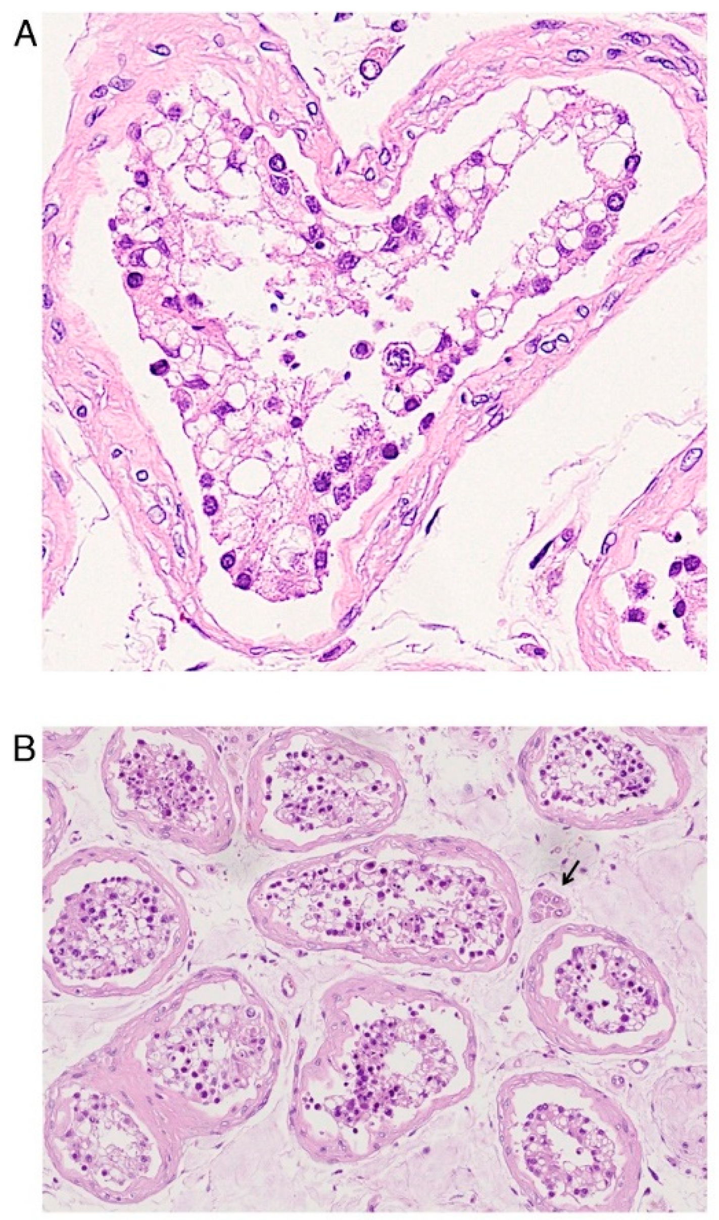
2.4. Histopathological Evidence in the Urinary System: The Kidney
2.4.1. Histopathological Findings: Light Microscopy
2.4.2. Histopathological Findings: Transmission Electron Microscopy
2.5. Histopathological Evidence in Endocrine System: Adrenal Glands
Histopathological Findings: Light Microscopy
2.6. Histopathological Evidence in Male Reproductive Tissue
2.6.1. Histopathological Findings: Light Microscopy
2.6.2. Histopathological Findings: Transmission Electron Microscopy
2.7. Histopathological Evidence in Female Reproductive Tissue: Placenta
2.7.1. Histopathological Findings: Light Microscopy
2.7.2. Histopathological Findings: Transmission Electron Microscopy
2.8. Histopathological Evidence in the Integumentary System
Histopathological Findings: Light Microscopy
3. Discussion
3.1. Microscopical Evidence in the Histopathology of the Myocardium
3.2. Microscopical Evidence in the Histopathology of Liver and Hepatocytes
3.3. Microscopical Evidence in the Histopathology of White and Red Pulp in the Spleen
3.4. Microscopical Evidence in the Histopathology of Tubular Epithelial and Glomerular Endothelial Cells
3.5. Microscopical Evidence in the Histopathology of Endocrine System
3.6. Microscopical Evidence in the Histopathology of Male Reproductive Tissue
3.7. Microscopical Evidence in the Histopathology of Female Reproductive Tissue
3.8. Microscopical Evidence in the Histopathology of the Skin
3.9. Strengths and Limitation
4. Materials and Methods
4.1. Search Strategy
4.2. Search Terms
4.3. Inclusion and Exclusion Criteria
4.4. Study Selection
5. Conclusions
Author Contributions
Funding
Institutional Review Board Statement
Informed Consent Statement
Data Availability Statement
Conflicts of Interest
Abbreviations
| SARS-CoV-2 | Severe acute respiratory syndrome coronavirus 2 |
| PRISMA | Preferred Reporting Items for Systematic Reviews and Meta-Analyses |
| COVID-19 | Coronavirus disease 2019 |
| WHO | World Health Organization |
| S | Spike protein |
| M | Membrane protein |
| N | Nucleocapsid protein |
| E | Envelope protein |
| HE | Hemagglutinin-esterase protein |
| RBD | Receptor-binding domain |
| ACE-2 | Angiotensin converting enzyme 2 |
| TMPRSS2 | Transmembrane protease serine 2 |
| LM | Light microscopy |
| TEM | Transmission electron microscopy |
| SEM | Scanning electron microscopy |
| CM | Confocal microscopy |
References
- Deshmukh, V.; Motwani, R.; Kumar, A.; Kumari, C.; Raza, K. Histopathological observations in COVID-19: A systematic review. J. Clin. Pathol. 2021, 74, 76–83. [Google Scholar] [CrossRef] [PubMed]
- Varvara, G.; Bernardi, S.; Bianchi, S.; Sinjari, B.; Piattelli, M. Dental education challenges during the COVID-19 pandemic period in Italy: Undergraduate student feedback, future perspectives, and the needs of teaching strategies for professional development. Healthcare 2021, 9, 454. [Google Scholar] [CrossRef]
- Umakanthan, S.; Sahu, P.; Ranade, A.V.; Bukelo, M.M.; Rao, J.S.; Abrahao-Machado, L.F.; Dahal, S.; Kumar, H.; Kv, D. Origin, transmission, diagnosis and management of coronavirus disease 2019 (COVID-19). Postgrad. Med. J. 2020, 96, 753–758. [Google Scholar] [CrossRef] [PubMed]
- Bianchi, S.; Gatto, R.; Fabiani, L. Effects of the SARS-CoV-2 pandemic on medical education in Italy: Considerations and tips. EuroMediterranean Biomed. J. 2020, 15, 100–101. [Google Scholar] [CrossRef]
- Lu, R.; Zhao, X.; Li, J.; Niu, P.; Yang, B.; Wu, H.; Wang, W.; Song, H.; Huang, B.; Zhu, N.; et al. Genomic characterisation and epidemiology of 2019 novel coronavirus: Implications for virus origins and receptor binding. Lancet 2020, 395, 565–574. [Google Scholar] [CrossRef] [Green Version]
- Wu, M.; Ma, L.; Xue, L.; Zhu, Q.; Zhou, S.; Dai, J.; Yan, W.; Zhang, J.; Wang, S. Co-Expression of the SARS-CoV-2 entry molecules ACE2 and TMPRSS2 in human ovaries: Identification of cell types and trends with age. Genomics 2021, 113, 3449–3460. [Google Scholar] [CrossRef]
- Bourgonje, A.R.; Abdulle, A.E.; Timens, W.; Hillebrands, J.; Navis, G.J.; Gordijn, S.J.; Bolling, M.C.; Dijkstra, G.; Voors, A.A.; Osterhaus, A.D.; et al. Angiotensin-converting Enzyme 2 (ACE2), SARS-CoV-2 and the pathophysiology of coronavirus disease 2019 (COVID-19). J. Pathol. 2020, 251, 228–248. [Google Scholar] [CrossRef]
- Huang, C.; Wang, Y.; Li, X.; Ren, L.; Zhao, J.; Hu, Y.; Zhang, L.; Fan, G.; Xu, J.; Gu, X.; et al. Clinical features of patients infected with 2019 novel coronavirus in Wuhan, China. Lancet 2020, 395, 497–506. [Google Scholar] [CrossRef] [Green Version]
- Bianchi, S.; Bernardi, S.; Continenza, M.A.; Vincenti, E.; Antonouli, S.; Torge, D.; Macchiarelli, G. Scanning electron microscopy approach for evaluation of hair dyed with Lawsonia inermis powder: In vitro study. Int. J. Morphol. 2020, 38, 96–100. [Google Scholar] [CrossRef] [Green Version]
- Giusti, I.; Bianchi, S.; Nottola, S.A.; Macchiarelli, G.; Dolo, V. Clinical electron microscopy in the study of human ovarian tissues. EuroMediterranean Biomed. J. 2019, 14, 145–151. [Google Scholar] [CrossRef]
- Bianchi, S.; Macchiarelli, G.; Micara, G.; Aragona, C.; Maione, M.; Nottola, S.A. Ultrastructural and morphometric evaluation of aged cumulus-oocyte- complexes. Ital. J. Anat. Embryol. 2013, 118, 28. [Google Scholar]
- Witte, R.; Andriasyan, V.; Georgi, F.; Yakimovich, A.; Greber, U. Concepts in light microscopy of viruses. Viruses 2018, 10, 202. [Google Scholar] [CrossRef] [PubMed] [Green Version]
- Martin-Cardona, A.; Lloreta Trull, J.; Albero-González, R.; Paraira Beser, M.; Andújar, X.; Ruiz-Ramirez, P.; Tur-Martínez, J.; Ferrer, C.; De Marcos Izquierdo, J.A.; Pérez-Madrigal, A.; et al. SARS-CoV-2 identified by transmission electron microscopy in lymphoproliferative and ischaemic intestinal lesions of COVID-19 patients with acute abdominal pain: Two case reports. BMC Gastroenterol. 2021, 21, 334. [Google Scholar] [CrossRef]
- Richert-Pöggeler, K.R.; Franzke, K.; Hipp, K.; Kleespies, R.G. Electron microscopy methods for virus diagnosis and high resolution analysis of viruses. Front. Microbiol. 2019, 9, 3255. [Google Scholar] [CrossRef] [PubMed]
- Bradley, B.T.; Maioli, H.; Johnston, R.; Chaudhry, I.; Fink, S.L.; Xu, H.; Najafian, B.; Deutsch, G.; Lacy, J.M.; Williams, T.; et al. Histopathology and ultrastructural findings of fatal COVID-19 infections in Washington State: A case series. Lancet 2020, 396, 320–332. [Google Scholar] [CrossRef]
- Fox, S.E.; Akmatbekov, A.; Harbert, J.L.; Li, G.; Quincy Brown, J.; Vander Heide, R.S. Pulmonary and cardiac pathology in African American patients with COVID-19: An autopsy series from New Orleans. Lancet Respir. Med. 2020, 8, 681–686. [Google Scholar] [CrossRef]
- Kudose, S.; Batal, I.; Santoriello, D.; Xu, K.; Barasch, J.; Peleg, Y.; Canetta, P.; Ratner, L.E.; Marasa, M.; Gharavi, A.G.; et al. Kidney biopsy findings in patients with COVID-19. J. Am. Soc. Nephrol. 2020, 31, 1959–1968. [Google Scholar] [CrossRef]
- Hosier, H.; Farhadian, S.F.; Morotti, R.A.; Deshmukh, U.; Lu-Culligan, A.; Campbell, K.H.; Yasumoto, Y.; Vogels, C.B.F.; Casanovas-Massana, A.; Vijayakumar, P.; et al. SARS-CoV-2 infection of the placenta. J. Clin. Investig. 2020, 130, 4947–4953. [Google Scholar] [CrossRef]
- Bertero, L.; Borella, F.; Botta, G.; Carosso, A.; Cosma, S.; Bovetti, M.; Carosso, M.; Abbona, G.; Collemi, G.; Papotti, M.; et al. Placenta histopathology in SARS-CoV-2 infection: Analysis of a consecutive series and comparison with control cohorts. Virchows Arch. 2021, 479, 715–728. [Google Scholar] [CrossRef]
- Zaigham, M.; Gisselsson, D.; Sand, A.; Wikström, A.; von Wowern, E.; Schwartz, D.A.; Iorizzo, L.; Nelander, M.; Blomberg, M.; Papadogiannakis, N.; et al. Clinical-pathological features in placentas of pregnancies with SARS-CoV-2 infection and adverse outcome: Case series with and without congenital transmission. BJOG An Int. J. Obstet. Gynaecol. 2022, 129, 1361–1374. [Google Scholar] [CrossRef]
- Gianotti, R.; Zerbi, P.; Dodiuk-Gad, R.P. Clinical and histopathological study of skin dermatoses in patients affected by COVID-19 infection in the northern part of Italy. J. Dermatol. Sci. 2020, 98, 141–143. [Google Scholar] [CrossRef]
- Kolivras, A.; Dehavay, F.; Delplace, D.; Feoli, F.; Meiers, I.; Milone, L.; Olemans, C.; Sass, U.; Theunis, A.; Thompson, C.T.; et al. Coronavirus (COVID-19) infection–induced chilblains: A case report with histopathologic findings. JAAD Case Rep. 2020, 6, 489–492. [Google Scholar] [CrossRef]
- Sohier, P.; Matar, S.; Meritet, J.F.; Laurent-Roussel, S.; Dupin, N.; Aractingi, S. Histopathologic features of chilblainlike lesions developing in the setting of the coronavirus disease 2019 (COVID-19) pandemic. Arch. Pathol. Lab. Med. 2021, 145, 137–144. [Google Scholar] [CrossRef] [PubMed]
- Fanni, D.; Cerrone, G.; Saba, L.; Demontis, R.; Congiu, T.; Piras, M.; Gerosa, C.; Suri, J.S.; Coni, P.; Caddori, A.; et al. Thrombotic sinusoiditis and local diffuse intrasinusoidal coagulation in the liver of subjects affected by COVID-19: The evidence from histology and scanning electron microscopy. Eur. Rev. Med. Pharmacol. Sci. 2021, 25, 5904–5912. [Google Scholar] [CrossRef]
- Tavazzi, G.; Pellegrini, C.; Maurelli, M.; Belliato, M.; Sciutti, F.; Bottazzi, A.; Sepe, P.A.; Resasco, T.; Camporotondo, R.; Bruno, R.; et al. Myocardial localization of coronavirus in COVID-19 cardiogenic shock. Eur. J. Heart Fail. 2020, 22, 911–915. [Google Scholar] [CrossRef] [PubMed] [Green Version]
- Kresch, E.; Achua, J.; Saltzman, R.; Khodamoradi, K.; Arora, H.; Ibrahim, E.; Kryvenko, O.N.; Almeida, V.W.; Firdaus, F.; Hare, J.M.; et al. COVID-19 endothelial dysfunction can cause erectile dysfunction: Histopathological, immunohistochemical, and ultrastructural study of the human penis. World J. Mens Health 2021, 39, 466. [Google Scholar] [CrossRef] [PubMed]
- Facchetti, F.; Bugatti, M.; Drera, E.; Tripodo, C.; Sartori, E.; Cancila, V.; Papaccio, M.; Castellani, R.; Casola, S.; Boniotti, M.B.; et al. SARS-CoV2 vertical transmission with adverse effects on the newborn revealed through integrated immunohistochemical, electron microscopy and molecular analyses of placenta. eBioMedicine 2020, 59, 102951. [Google Scholar] [CrossRef]
- Ramos-Rincon, J.-M.; Herrera-García, C.; Silva-Ortega, S.; Portilla-Tamarit, J.; Alenda, C.; Jaime-Sanchez, F.-A.; Arenas-Jiménez, J.; Fornés-Riera, F.-E.; Scholz, A.; Escribano, I.; et al. Pathological findings associated with SARS-CoV-2 on postmortem core biopsies: Correlation with clinical presentation and disease course. Front. Med. 2022, 9, 4307. [Google Scholar] [CrossRef]
- Tian, S.; Xiong, Y.; Liu, H.; Niu, L.; Guo, J.; Liao, M.; Xiao, S.-Y. Pathological study of the 2019 novel coronavirus disease (COVID-19) through postmortem core biopsies. Mod. Pathol. 2020, 33, 1007–1014. [Google Scholar] [CrossRef] [PubMed] [Green Version]
- Wang, Y.; Liu, S.; Liu, H.; Li, W.; Lin, F.; Jiang, L.; Li, X.; Xu, P.; Zhang, L.; Zhao, L.; et al. SARS-CoV-2 Infection of the liver directly contributes to hepatic impairment in patients with COVID-19. J. Hepatol. 2020, 73, 807–816. [Google Scholar] [CrossRef] [PubMed]
- Yang, M.; Chen, S.; Huang, B.; Zhong, J.-M.; Su, H.; Chen, Y.-J.; Cao, Q.; Ma, L.; He, J.; Li, X.-F.; et al. Pathological findings in the testes of COVID-19 patients: Clinical implications. Eur. Urol. Focus 2020, 6, 1124–1129. [Google Scholar] [CrossRef] [PubMed]
- Achua, J.K.; Chu, K.Y.; Ibrahim, E.; Khodamoradi, K.; Delma, K.S.; Iakymenko, O.A.; Kryvenko, O.N.; Arora, H.; Ramasamy, R. Histopathology and ultrastructural findings of fatal COVID-19 infections on testis. World J. Mens Health 2021, 39, 65. [Google Scholar] [CrossRef] [PubMed]
- Ma, X.; Guan, C.; Chen, R.; Wang, Y.; Feng, S.; Wang, R.; Qu, G.; Zhao, S.; Wang, F.; Wang, X.; et al. Pathological and molecular examinations of postmortem testis biopsies reveal SARS-CoV-2 infection in the testis and spermatogenesis damage in COVID-19 patients. Cell. Mol. Immunol. 2021, 18, 487–489. [Google Scholar] [CrossRef] [PubMed]
- Nava-Santana, C.; Rodríguez-Armida, M.; Jiménez, J.V.; Vargas-Parra, N.; León, D.E.A.; Campos-Murguia, A.; Macías-Rodriguez, R.; Arteaga-Garrido, A.; Hernández-Villegas, A.C.; Dominguez-Cherit, G.; et al. Clinicopathologic characteristics of severe COVID-19 patients in Mexico City: A post-mortem analysis using a minimally invasive autopsy approach. PLoS ONE 2022, 17, e0262783. [Google Scholar] [CrossRef] [PubMed]
- Falasca, L.; Nardacci, R.; Colombo, D.; Lalle, E.; Di Caro, A.; Nicastri, E.; Antinori, A.; Petrosillo, N.; Marchioni, L.; Biava, G.; et al. Postmortem findings in Italian patients with COVID-19: A descriptive full autopsy study of cases with and without comorbidities. J. Infect. Dis. 2020, 222, 1807–1815. [Google Scholar] [CrossRef]
- Bois, M.C.; Boire, N.A.; Layman, A.J.; Aubry, M.C.; Alexander, M.P.; Roden, A.C.; Hagen, C.E.; Quinton, R.A.; Larsen, C.; Erben, Y.; et al. COVID-19-associated nonocclusive fibrin microthrombi in the heart. Circulation 2021, 143, 230–243. [Google Scholar] [CrossRef]
- Hartard, C.; Chaqroun, A.; Settembre, N.; Gauchotte, G.; Lefevre, B.; Marchand, E.; Mazeaud, C.; Nguyen, D.T.; Martrille, L.; Koscinski, I.; et al. Multiorgan and vascular tropism of SARS-CoV-2. Viruses 2022, 14, 515. [Google Scholar] [CrossRef]
- Mezache, L.; Nuovo, G.J.; Suster, D.; Tili, E.; Awad, H.; Radwański, P.B.; Veeraraghavan, R. Histologic, viral, and molecular correlates of heart disease in fatal COVID-19. Ann. Diagn. Pathol. 2022, 60, 151983. [Google Scholar] [CrossRef]
- Xiang, Q.; Feng, Z.; Diao, B.; Tu, C.; Qiao, Q.; Yang, H.; Zhang, Y.; Wang, G.; Wang, H.; Wang, C.; et al. SARS-CoV-2 induces lymphocytopenia by promoting inflammation and decimates secondary lymphoid organs. Front. Immunol. 2021, 12, 66105. [Google Scholar] [CrossRef]
- González Pessolani, T.; Muñóz Fernández de Legaria, M.; Elices Apellániz, M.; Salinas Moreno, S.; Lorido Cortés, M.d.M.; García Sánchez, S. Multi-organ pathological findings associated with COVID-19 in postmortem needle core biopsies in four patients and a review of the current literature. Rev. Española Patol. 2021, 54, 275–280. [Google Scholar] [CrossRef] [PubMed]
- Farkash, E.A.; Wilson, A.M.; Jentzen, J.M. Ultrastructural evidence for direct renal infection with SARS-CoV-2. J. Am. Soc. Nephrol. 2020, 31, 1683–1687. [Google Scholar] [CrossRef] [PubMed]
- Freire Santana, M.; Borba, M.G.S.; Baía-da-Silva, D.C.; Val, F.; Alexandre, M.A.A.; Brito-Sousa, J.D.; Melo, G.C.; Queiroga, M.V.O.; Leão Farias, M.E.; Camilo, C.C.; et al. Case report: Adrenal pathology findings in severe COVID-19: An autopsy study. Am. J. Trop. Med. Hyg. 2020, 103, 1604–1607. [Google Scholar] [CrossRef] [PubMed]
- Fitzek, A.; Gerling, M.; Püschel, K.; Saeger, W. Post-mortem histopathology of pituitary and adrenals of COVID-19 patients. Leg. Med. 2022, 57, 102045. [Google Scholar] [CrossRef] [PubMed]
- Paul, T.; Ledderose, S.; Bartsch, H.; Sun, N.; Soliman, S.; Märkl, B.; Ruf, V.; Herms, J.; Stern, M.; Keppler, O.T.; et al. Adrenal tropism of SARS-CoV-2 and adrenal findings in a post-mortem case series of patients with severe fatal COVID-19. Nat. Commun. 2022, 13, 1589. [Google Scholar] [CrossRef]
- Chu, H.; Peng, L.; Hu, L.; Zhu, Y.; Zhao, J.; Su, H.; Yao, L.; Zhu, Q.; Nie, X.; Yang, L.; et al. Liver histopathological analysis of 24 postmortem findings of patients with COVID-19 in China. Front. Med. 2021, 8, 749318. [Google Scholar] [CrossRef]
- Oleszak, F.; Maryniak, A.; Botti, E.; Abrahim, C.; Salifu, M.O.; Youssef, M.; Henglein, V.L.; McFarlane, S.I. Myocarditis associated with COVID-19. Am. J. Med. Case Rep. 2020, 8, 498–502. [Google Scholar] [CrossRef] [PubMed]
- Maiese, A.; Manetti, A.C.; La Russa, R.; Di Paolo, M.; Turillazzi, E.; Frati, P.; Fineschi, V. Autopsy findings in COVID-19-related deaths: A literature review. Forensic Sci. Med. Pathol. 2021, 17, 279–296. [Google Scholar] [CrossRef]
- Maiese, A.; Frati, P.; Del Duca, F.; Santoro, P.; Manetti, A.C.; La Russa, R.; Di Paolo, M.; Turillazzi, E.; Fineschi, V. Myocardial pathology in COVID-19-associated cardiac injury: A systematic review. Diagnostics 2021, 11, 1647. [Google Scholar] [CrossRef]
- Nan, J.; Jin, Y.-B.; Myo, Y.; Zhang, G. Hypoxia in acute cardiac injury of coronavirus disease 2019: Lesson learned from pathological studies. J. Geriatr. Cardiol. 2020, 17, 221–223. [Google Scholar] [CrossRef]
- van Eijk, L.E.; Binkhorst, M.; Bourgonje, A.R.; Offringa, A.K.; Mulder, D.J.; Bos, E.M.; Kolundzic, N.; Abdulle, A.E.; Voort, P.H.; Olde Rikkert, M.G.; et al. COVID-19: Immunopathology, pathophysiological mechanisms, and treatment options. J. Pathol. 2021, 254, 307–331. [Google Scholar] [CrossRef]
- Bonaventura, A.; Vecchié, A.; Dagna, L.; Martinod, K.; Dixon, D.L.; Van Tassell, B.W.; Dentali, F.; Montecucco, F.; Massberg, S.; Levi, M.; et al. Endothelial dysfunction and immunothrombosis as key pathogenic mechanisms in COVID-19. Nat. Rev. Immunol. 2021, 21, 319–329. [Google Scholar] [CrossRef]
- Varga, Z.; Flammer, A.J.; Steiger, P.; Haberecker, M.; Andermatt, R.; Zinkernagel, A.S.; Mehra, M.R.; Schuepbach, R.A.; Ruschitzka, F.; Moch, H. Endothelial cell infection and endotheliitis in COVID-19. Lancet 2020, 395, 1417–1418. [Google Scholar] [CrossRef]
- Holland, D.J.; Blazak, P.L.; Martin, J.; Broom, J.; Poulter, R.S.; Stanton, T. Myocarditis and cardiac complications associated with COVID-19 and mRNA vaccination: A pragmatic narrative review to guide clinical practice. Hear. Lung Circ. 2022, 31, 924–933. [Google Scholar] [CrossRef] [PubMed]
- Jakovac, H.; Ferenčić, A.; Stemberger, C.; Mohar Vitezić, B.; Cuculić, D. Detection of SARS-CoV-2 antigens in the AV-node of a cardiac conduction system—a case report. Trop. Med. Infect. Dis. 2022, 7, 43. [Google Scholar] [CrossRef]
- Barton, L.M.; Duval, E.J.; Stroberg, E.; Ghosh, S.; Mukhopadhyay, S. COVID-19 autopsies, Oklahoma, USA. Am. J. Clin. Pathol. 2020, 153, 725–733. [Google Scholar] [CrossRef] [PubMed] [Green Version]
- Philips, C.A.; Ahamed, R.; Augustine, P. SARS-CoV-2 related liver impairment—Perception may not be the reality. J. Hepatol. 2020, 73, 991–992. [Google Scholar] [CrossRef]
- Baroiu, L.; Dumitru, C.; Iancu, A.; Leșe, A.-C.; Drăgănescu, M.; Baroiu, N.; Anghel, L. COVID-19 impact on the liver. World J. Clin. Cases 2021, 9, 3814–3825. [Google Scholar] [CrossRef] [PubMed]
- McConnell, M.J.; Kondo, R.; Kawaguchi, N.; Iwakiri, Y. COVID-19 and liver injury: Role of inflammatory endotheliopathy, platelet dysfunction, and thrombosis. Hepatol. Commun. 2022, 6, 255–269. [Google Scholar] [CrossRef]
- Lin, L.; Luo, S.; Qin, R.; Yang, M.; Wang, X.; Yang, Q.; Zhang, Y.; Wang, Q.; Zhu, R.; Fan, H.; et al. Long-term infection of SARS-CoV-2 changed the body’s immune status. Clin. Immunol. 2020, 218, 108524. [Google Scholar] [CrossRef]
- Cococcia, S.; Lenti, M.V.; Santacroce, G.; Achilli, G.; de Andreis, F.B.; Di Sabatino, A. Liver-spleen axis dysfunction in COVID-19. World J. Gastroenterol. 2021, 27, 5919–5931. [Google Scholar] [CrossRef] [PubMed]
- Naicker, S.; Yang, C.W.; Hwang, S.J.; Liu, B.C.; Chen, J.H.; Jha, V. The novel coronavirus 2019 epidemic and kidneys. Kidney Int. 2020, 97, 824–828. [Google Scholar] [CrossRef] [PubMed]
- Wang, M.; Xiong, H.; Chen, H.; Li, Q.; Ruan, X.Z. Renal injury by SARS-CoV-2 infection: A systematic review. Kidney Dis. 2021, 7, 100–110. [Google Scholar] [CrossRef] [PubMed]
- Shetty, A.A.; Tawhari, I.; Safar-Boueri, L.; Seif, N.; Alahmadi, A.; Gargiulo, R.; Aggarwal, V.; Usman, I.; Kisselev, S.; Gharavi, A.G.; et al. COVID-19–associated glomerular disease. J. Am. Soc. Nephrol. 2021, 32, 33–40. [Google Scholar] [CrossRef] [PubMed]
- Frankel, M.; Feldman, I.; Levine, M.; Frank, Y.; Bogot, N.R.; Benjaminov, O.; Kurd, R.; Breuer, G.S.; Munter, G. Bilateral adrenal hemorrhage in coronavirus disease 2019 patient: A case report. J. Clin. Endocrinol. Metab. 2020, 105, 3745–3749. [Google Scholar] [CrossRef] [PubMed]
- Mao, Y.; Xu, B.; Guan, W.; Xu, D.; Li, F.; Ren, R.; Zhu, X.; Gao, Y.; Jiang, L. The adrenal cortex, an underestimated site of SARS-CoV-2 infection. Front. Endocrinol. 2020, 11, 593179. [Google Scholar] [CrossRef] [PubMed]
- Li, H.; Xiao, X.; Zhang, J.; Zafar, M.I.; Wu, C.; Long, Y.; Lu, W.; Pan, F.; Meng, T.; Zhao, K.; et al. Impaired spermatogenesis in COVID-19 patients. eClinicalMedicine 2020, 28, 100604. [Google Scholar] [CrossRef] [PubMed]
- Nassau, D.E.; Best, J.C.; Kresch, E.; Gonzalez, D.C.; Khodamoradi, K.; Ramasamy, R. Impact of the SARS-CoV-2 virus on male reproductive health. BJU Int. 2022, 129, 143–150. [Google Scholar] [CrossRef] [PubMed]
- Shen, Q.; Xiao, X.; Aierken, A.; Yue, W.; Wu, X.; Liao, M.; Hua, J. The ACE2 Expression in Sertoli cells and germ cells may cause male reproductive disorder after SARS-CoV-2 infection. J. Cell. Mol. Med. 2020, 24, 9472–9477. [Google Scholar] [CrossRef]
- Wang, Y.; Biology, Z.S.V. Chapter 4 Cell Types of the Placenta; Morgan & Claypool Life Sciences: San Rafael, CA, USA, 2010. [Google Scholar]
- Bos, M.; Harris-Mostert, E.T.M.S.; van der Meeren, L.E.; Baelde, J.J.; Williams, D.J.; Nikkels, P.G.J.; Bloemenkamp, K.W.M.; van der Hoorn, M.L.P. Clinical outcomes in chronic intervillositis of unknown etiology. Placenta 2020, 91, 19–23. [Google Scholar] [CrossRef]
- Drenovska, K.; Schmidt, E.; Vassileva, S. Covid-19 pandemic and the skin. Int. J. Dermatol. 2020, 59, 1312–1319. [Google Scholar] [CrossRef] [PubMed]
- Xue, X.; Mi, Z.; Wang, Z.; Pang, Z.; Liu, H.; Zhang, F. High expression of ACE2 on keratinocytes reveals skin as a potential target for SARS-CoV-2. J. Investig. Dermatol. 2021, 141, 206–209.e1. [Google Scholar] [CrossRef] [PubMed]
- Fineschi, V.; Aprile, A.; Aquila, I.; Arcangeli, M.; Asmundo, A.; Bacci, M.; Cingolani, M.; Cipolloni, L.; D’Errico, S.; De Casamassimi, I.; et al. Management of the corpse with suspect, probable or confirmed COVID-19 respiratory infection—Italian interim recommendations for personnel potentially exposed to material from corpses, including body fluids, in morgue structures and during autopsy practice. Pathologica 2020, 112, 64–77. [Google Scholar] [CrossRef] [PubMed]
- Bianchi, S.; Nottola, S.A.; Torge, D.; Palmerini, M.G.; Necozione, S.; Macchiarelli, G. Association between female reproductive health and Mancozeb: Systematic review of experimental models. Int. J. Environ. Res. Public Health 2020, 17, 2580. [Google Scholar] [CrossRef] [PubMed] [Green Version]


| System | Number of Studies |
|---|---|
| Cardiovascular System | 8 |
| Digestive System | 7 |
| Lymphatic System | 4 |
| Urinary System | 4 |
| Endocrine System | 3 |
| Male Reproductive System | 4 |
| Female Reproductive System | 4 |
| Integumentary System | 3 |
| Methods of Sampling | Light Microscopy | Transmission Electron Microscopy | Scanning Electron Microscopy | |
|---|---|---|---|---|
| Methods of Microscopy | ||||
| Living Biopsy | Kudose et al. 2020 [17] Hosier et al. 2020 [18] Bertero et al. 2021 [19] Zaigham et al. 2022 [20] Gianotti et al. 2020 [21] Kolivras et al. 2020 [22] Sohier et al. 2021 [23] Fanni et al. 2021 [24] | Tavazzi et al. 2020 [25] Kresh et al. 2021 [26] Hosier et al. 2020 [18] Kudose et al. 2020 [17] Facchetti et al. 2020 [27] | Fanni et al. 2021 [24] | |
| Post-mortem Biopsy | Ramos-Rincon et al. 2022 [28] Tian et al. 2020 [29] Wang et al. 2020 [30] Fanni et al. 2021 [24] Yang et al. 2020 [31] Achua et al. 2020 [32] Ma et al. 2021 [33] Nava-Santana et al. 2022 [34] | Wang et al. 2020 [30] Achua et al. 2020 [32] Ma et al. 2021 [33] | Fanni et al. 2021 [24] | |
| Autopsy | Fox et al. 2020 [16] Bradley et al. 2020 [15] Falasca et al. 2020 [35] Bois et al. 2021 [36] Hartard et al. 2022 [37] Mezache et al. 2022 [38] Xiang et al. 2021 [39] González Pessolani et al. 2021 [40] Farkash et al. 2020 [41] Santana et al. 2020 [42] Fitzek et al. 2022 [43] Paul et al. 2022 [44] | Chu et al. 2021 [45] Farkash et al. 2020 [41] Bradley et al. 2020 [15] | ||
| Author Year | Article Type | Tissue | Type of Sample | Microscopy | Histopathological Findings |
|---|---|---|---|---|---|
| Tavazzi et al. 2020 [25] | Case Report | Myocardium | Living Myocardial biopsy | Transmission Electron Microscopy | Cytopathy, with membrane damage and cytoplasmic vacuoles. Single or small groups of viral particles within the interstitial cells of the myocardium of the patient. |
| Fox et al. 2020 [16] | Original Research Article | Myocardium | Autopsy | Light Microscopy | Scattered individual cell myocyte necrosis. Cardiac myocytes showing focal, atypical myocyte degeneration. |
| Bradley et al. 2020 [15] | Original Research Article | Myocardium | Autopsy | Light Microscopy | Lymphocytic myocarditis. Necrotic myocytes. |
| Falasca et al. 2020 [35] | Original Research Article | Myocardium | Autopsy | Light Microscopy | Myocardial ischemic or inflammatory changes. Pale and flabby myocardium. Hypertrophy of myocytes. |
| Bois et al. 2021 [36] | Comparative Study | Myocardium | Autopsy | Light Microscopy | Active myocarditis, with focal active lymphocytic myocarditis. |
| Hartard et al. 2022 [37] | Case Report | Myocardium | Autopsy | Light Microscopy | Mild perivascular lymphocytic infiltrate, without cardiomyocytes alteration. |
| Mezache et al. 2022 [38] | Original Research Article | Myocardium | Autopsy | Light Microscopy | Scattered microvessels, enlarged, atypical endothelial cells. Microthrombi, perivascular extravasation of red blood cells. Perivascular edema and perivascular collections of mononuclear cells. |
| Ramos-Rincon et al. 2022 [28] | Original Research Article | Myocardium | Post-mortem biopsy | Light Microscopy | Myocardial edema. Mononuclear interstitial infiltrate. Necrotic myocardial cells. |
| Author Year | Article Type | Tissue | Type of Sample | Microscopy | Histopathological Findings |
|---|---|---|---|---|---|
| Tian et al. 2020 [29] | Original Research Article | Liver | Post-mortem biopsy | Light Microscopy | Hepatic cell degeneration. Nuclear glycogenation in hepatocytes. Focal necrosis. |
| Bradley et al. 2020 [15] | Original Research Article | Liver | Autopsy | Light Microscopy | Centrilobular necrosis. Mild periportal lymphocytic inflammation. |
| Wang et al. 2020 [30] | Original Research Article | Liver | Post-mortem biopsy | Light Microscopy | Apoptotic hepatocytes. Multinuclear syncytial hepatocytes. Vesicular steatosis. Lobular and portal tracts inflammation with lymphocytic infiltrate. |
| Original Research Article | Hepatocytes | Post-mortem biopsy | Transmission Electron Microscopy | Viral particles in cytoplasm of hepatocytes. Enlarged mitochondria displaying obscure cristae and electron dense materials. Glycogen granule decrease. | |
| Falasca et al. 2020 [35] | Original Research Article | Liver | Autopsy | Light Microscopy | Sinusoidal congestion. Extravasation of red blood cells. Hepatic necrosis. Inflammatory infiltration. |
| Chu et al. 2021 [45] | Original Research Article | Liver | Autopsy | Light Microscopy | Swelling of hepatocytes. |
| Original Research Article | Hepatocytes | Autopsy | Transmission Electron Microscopy | Vacuolar degeneration of hepatocytes. Edema of mitochondria with disruption of cristae. Expansion of endoplasmic reticulum. | |
| Fanni et al. 2021 [24] | Case Report | Liver | Post-mortem and living biopsy | Light Microscopy | Enlarged hepatocytes, with abundant granular cytoplasm, focally showing ground-glass appearance. Vesicular steatosis. Enlarged sinusoids, with abundant fibrillary aggregates. Sinusoidal dilatation, associated with scattered lymphocytes and red blood cells. Intrasinusoidal thrombi. |
| Case Report | Liver | Post-mortem and living biopsy | Scanning Electron Microscopy | Fibrillary aggregates of fibrin inside the sinusoidal lumen. | |
| Nava-Santana et al. 2022 [34] | Original Research Article | Liver | Post-mortem biopsy | Light Microscopy | Neutrophilic sinusoidal inflammation. Ballooning degeneration of hepatocytes. |
| Author Year | Article Type | Tissue | Type of Sample | Microscopy | Histopathological Findings |
|---|---|---|---|---|---|
| Bradley et al. 2020 [15] | Original Research Article | White pulp | Autopsy | Light Microscopy | White pulp depletion. |
| Falasca et al. 2020 [35] | Original Research Article | White pulp Red pulp | Autopsy | Light Microscopy | Lymphoid hypoplasia in white pulp. Red pulp congestion. |
| Xiang et al. 2021 [39] | Original Research Article | White pulp Red pulp | Autopsy | Light Microscopy | White pulp atrophy. Congested and hemorrhagic red pulp. |
| González Pessolani et al. 2021 [40] | Brief Report | White pulp | Post-mortem biopsy | Light Microscopy | White pulp with lymphocytic depletion and diminished lymphoid follicles. |
| Author Year | Article Type | Tissue | Type of Sample | Microscopy | Histopathological Findings |
|---|---|---|---|---|---|
| Bradley et al. 2020 [15] | Original Research Article | Kidney | Autopsy | Light Microscopy | Inflammation of the renal parenchyma. Focal segmental glomerulosclerosis. Tubular epithelial vacuolization. |
| Original Research Article | Kidney | Autopsy | Transmission Electron Microscopy | Viral particles in endothelial cells and proximal tubular epithelial cells. | |
| Farkash et al. 2020 [41] | Case Report | Tubular epithelial cells | Autopsy | Light Microscopy | Vacuolization of proximal tubular epithelial cells. |
| Case Report | Tubular epithelial cells | Autopsy | Transmission Electron Microscopy | Vacuolization and degeneration of tubular epithelial cells, containing viral particles. | |
| Kudose et al. 2020 [17] | Original Research Article | Kidney | Living biopsy | Light Microscopy | Hyperplasia of glomerular epithelial cells (podocytes). Collapse of glomerular capillaries. |
| Original Re-search Article | Renal corpuscle | Living bi-opsy | Transmission Electron Microscopy | Podocytopathy. Glomerular endothelial inclusions. | |
| Falasca et al. 2020 [35] | Original Research Article | Kidney | Autopsy | Light Microscopy | Swollen glomerular endothelial cells. Fibrin deposit under the Bowman capsule. Tubulointerstitial inflammation. Glomerular sclerosis. |
| Author Year | Article Type | Tissue | Type of Sample | Microscopy | Histopathological Findings |
|---|---|---|---|---|---|
| Santana et al. 2020 [42] | Case report | Adrenal glands | Autopsy | Light Microscopy | Extensive areas of hemorrhagic necrosis. Cortical lipid degeneration and focal inflammation. |
| Fitzek et al. 2022 [43] | Original Research Article | Adrenal glands | Autopsy | Light Microscopy | Reduction of cytoplasmic lipid vacuoles. |
| Paul et al. 2022 [44] | Original Research Article | Adrenal glands | Autopsy | Light Microscopy | Inflammation, with perivascular accentuation of small foci of lymphocytes and histiocytes. Inflammatory cell death. |
| Author Year | Article Type | Tissue | Type of Sample | Microscopy | Histopathological Findings |
|---|---|---|---|---|---|
| Yang et al. 2020 [31] | Original Research Article | Testis | Post-mortem biopsy | Light Microscopy | Defoliated and edematous Sertoli cells. Scattered Leydig cells. Extensive germ cell destruction. Vacuolation and cytoplasmic rarefaction of Sertoli cells. |
| Achua et al. 2020 [32] | Original Research Article | Testis | Autopsy | Light Microscopy | Hyalinization and thickening of the basement membrane of the seminiferous tubules, with lymphocyte infiltration. |
| Original Research Article | Testis | Autopsy | Transmission Electron Microscopy | Coronavirus-like particles in the cytoplasm of the interstitial cells of the testes. | |
| Ma et al. 2021 [33] | Correspondence Article | Testis | Post-mortem biopsy | Light Microscopy | Degenerated germ cells sloughing into the lumen of the seminiferous tubules. |
| Correspondence Article | Testis | Post-mortem biopsy | Transmission Electron Microscopy | Coronavirus-like particles in the interstitial compartment of testes. | |
| Kresch et al. 2021 [26] | Original Research Article | Penis | Living Biopsy | Transmission Electron Microscopy | Extracellular viral particles in the peri-vascular erectile tissue. |
| Author Year | Article Type | Tissue | Type of Sample | Microscopy | Histopathological Findings |
|---|---|---|---|---|---|
| Hosier et al. 2020 [18] | Case Report | Placenta | Living Biopsy | Light Microscopy | Diffuse perivillous fibrin and inflammatory infiltrate, with macrophages and T lymphocytes. |
| Case Report | Placenta | Living Biopsy | Transmission Electron Microscopy | Virus particles in the cytosol of syncytiotrophoblast cells, cytotrophoblast cell and fibroblasts. | |
| Facchetti et al. 2020 [27] | Original Research Article | Placenta | Living Biopsy | Transmission Electron Microscopy | Viral particles in the cytoplasm of syncytiotrophoblast cells and fibroblasts. |
| Bertero et al. 2021 [19] | Original Research Article | Placenta | Living Biopsy | Light Microscopy | Placental intervillous hematomas and microvascular thrombosis. Significant foci of decidual and villous inflammation. |
| Zaigham et al. 2022 [20] | Original Research Article | Placenta | Living Biopsy | Light Microscopy | Mixed inflammatory infiltrate in the intervillous space (intervillositis). Fibrinoid deposition in the intervillous space. |
| Author Year | Article Type | Tissue | Type of Sample | Microscopy | Histopathological Findings |
|---|---|---|---|---|---|
| Gianotti et al. 2020 [21] | Letter to Editor | Skin | Living Biopsy | Light Microscopy | Parakeratosis, acanthosis, dyskeratotic keratinocytes, and small acantholytic cleft. Necrotic keratinocytes with lymphocyte satellitosis. |
| Kolivras et al. 2020 [22] | Case Report | Skin | Living Biopsy | Light Microscopy | Scattered, necrotic and apoptotic keratinocytes, with smudging of the basement membrane. Vacuolar alteration along the basal layer of the epidermis. Superficial and deep infiltrate of lymphocytes. |
| Sohier et al. 2021 [23] | Case Series | Skin | Living Biopsy | Light Microscopy | Superficial and deep infiltrate of lymphocytes. Vacuolar alteration of the basal layer of the epidermis. |
Publisher’s Note: MDPI stays neutral with regard to jurisdictional claims in published maps and institutional affiliations. |
© 2022 by the authors. Licensee MDPI, Basel, Switzerland. This article is an open access article distributed under the terms and conditions of the Creative Commons Attribution (CC BY) license (https://creativecommons.org/licenses/by/4.0/).
Share and Cite
Torge, D.; Bernardi, S.; Arcangeli, M.; Bianchi, S. Histopathological Features of SARS-CoV-2 in Extrapulmonary Organ Infection: A Systematic Review of Literature. Pathogens 2022, 11, 867. https://doi.org/10.3390/pathogens11080867
Torge D, Bernardi S, Arcangeli M, Bianchi S. Histopathological Features of SARS-CoV-2 in Extrapulmonary Organ Infection: A Systematic Review of Literature. Pathogens. 2022; 11(8):867. https://doi.org/10.3390/pathogens11080867
Chicago/Turabian StyleTorge, Diana, Sara Bernardi, Mauro Arcangeli, and Serena Bianchi. 2022. "Histopathological Features of SARS-CoV-2 in Extrapulmonary Organ Infection: A Systematic Review of Literature" Pathogens 11, no. 8: 867. https://doi.org/10.3390/pathogens11080867
APA StyleTorge, D., Bernardi, S., Arcangeli, M., & Bianchi, S. (2022). Histopathological Features of SARS-CoV-2 in Extrapulmonary Organ Infection: A Systematic Review of Literature. Pathogens, 11(8), 867. https://doi.org/10.3390/pathogens11080867