Article

Genome Analysis of ESBL-Producing Escherichia coli Isolated from Pigs

by
Luria Leslie Founou
1,2,3,*,
Raspail Carrel Founou
1,4,5,
Mushal Allam
6,7,
Arshad Ismail
6 and
Sabiha Yusuf Essack
1
1
Antimicrobial Research Unit, University of KwaZulu-Natal, Durban 4000, South Africa
2
Reproductive, Maternal, Newborn and Child Health (ReMarch) Research Unit, Research Institute of the Centre of Expertise and Biological Diagnostic of Cameroon (CEDBCAM-RI), Yaoundé P.O. Box 8242, Cameroon
3
Bioinformatics and Applied Machine Learning Research Unit, EDEN Foundation, Yaoundé P.O. Box 8242, Cameroon
4
Department of Microbiology, Haematology and Immunology, Faculty of Medicine and Pharmaceutical Sciences, University of Dschang, Dschang P.O. Box 96, Cameroon
5
Antimicrobial Resistance and Infectious Diseases (ARID) Research Unit, Research Institute of the Centre of Expertise and Biological Diagnostic of Cameroon (CEDBCAM-RI), Yaoundé P.O. Box 8242, Cameroon
6
Sequencing Core Facility, National Health Laboratory Service, Johannesburg 2131, South Africa
7
Department of Genetics and Genomics, College of Medicine and Genomics, Al Ain 15551, United Arab Emirates
*
Author to whom correspondence should be addressed.
Pathogens 2022, 11(7), 776; https://doi.org/10.3390/pathogens11070776
Submission received: 5 June 2022 / Revised: 4 July 2022 / Accepted: 5 July 2022 / Published: 7 July 2022

Abstract

:
The resistome, virulome and mobilome of extended spectrum ß-lactamase (ESBL)-producing Escherichia coli (ESBL-Ec) isolated from pigs in Cameroon and South Africa were assessed using whole genome sequencing (WGS). Eleven clonally related phenotypic ESBL-Ec isolates were subjected to WGS. The prediction of antibiotic resistance genes, virulence factors (VFs) and plasmids was performed using ResFinder, VirulenceFinder and PlasmidFinder, respectively. Diverse sequence types (STs) were detected with ST2144 and ST88 being predominant and blaCTX-M-15 (55%) being the principal ESBL gene. All except two isolates harboured various aminoglycoside resistance genes, including aph(3″)-Ib (6/11, 55%) and aph(6)-1d (6/11, 55%), while the qnrS1 gene was identified in four of the isolates. The ESBL-Ec isolates showed a 93.6% score of being human pathogens. The fim, ehaB, ibeB/C were the leading virulence factors detected. All isolates harboured at least three extraintestinal pathogenic E. coli (ExPEC) VFs, with one isolate harbouring up to 18 ExPEC VFs. Five isolates (45.45%) harboured the plasmid incompatibility group IncF (FII, FIB, FIC, FIA). The study revealed that there is an urgent need to implement effective strategies to contain the dissemination of resistant and virulent ESBL-Ec through the food chain in Cameroon and South Africa.

1. Introduction

Antibiotic resistance (ABR) is a global public health issue that has severe multi-dimensional repercussions not only in humans, but also in the food production industry. The extensive use of antibiotics in food animal production is widely acknowledged as the driving force behind antibiotic resistance in humans and animals. Antibiotics are used for a variety of purposes, including therapeutic and non-therapeutic uses, as metaphylactics, prophylactics, and growth promoters [1]. Hence, the emergence and spread of ABR across the farm-to-plate continuum puts occupationally exposed workers (viz. farmers, agricultural practitioners, abattoir workers, food handlers, etc.), their close contacts and consumers at the end of the food chain at risk of contamination or infection by antibiotic-resistant bacteria (ARB) and/or antibiotic resistance genes (ARGs) [1,2]. ABR prevention and containment measures should focus not only on humans but also on animals and their associated environments [2].
Escherichia coli is a recognized commensal bacterium of the gastrointestinal tract of humans and animals. The genomic plasticity of E. coli strains allows their adaptation to different environments, hence their wide implication in intestinal and extraintestinal infections in both humans and animals worldwide [3]. E. coli displays a clonal population structure delineating four main phylogenetic groups (A, B1, B2, and D) with few phylogroups being involved in infections and others being commensal [4,5].
E. coli has been suggested as the putative reservoir for extended-spectrum β-lactamase (ESBL) resistance. It has been demonstrated that substantial resistance emerges in commensal bacteria, especially those present in the gastrointestinal tract where horizontal gene transfer prevails and does occur within and between species and genera [2]. ESBL production in E. coli is associated with different resistance genes but is most frequently caused by the production of ESBLs encoded by the blaTEM, blaSHV and blaCTX-M families, with the latter being the predominant type [6]. ESBL-producing E. coli (ESBL-Ec) have been detected across the animal, human and environmental interface worldwide, with the emergence of specific clones able to acquire ARGs and virulence factors (VFs) via mobile genetic elements (MGEs), such as plasmids, transposons, gene cassettes and other integrative genetic elements [2].
ESBL-Ec emerging on farms and/or abattoirs can disseminate directly to occupationally exposed workers, and indirectly through the food chain via contact with or consumption of contaminated food products. This direct and indirect transmission of ESBL-Ec facilitate the likelihood of their subsequent entrance and spread into communities and hospitals [7]. This is further exacerbated by international travels and the globalization of trade in animals and food products, meaning that there are no species, nor geographical frontiers to contain such resistant pathogens [7]. Chishimba et al., (2016) and Aworh et al., (2020) reported 20 and 32% of ESBL-Ec in poultry abattoirs in Zambia and Nigeria, respectively [8,9]. Both studies concluded that the presence of ESBL-Ec in food animals and food products poses a significant public health threat for the general population that requires urgent and appropriate containment measures.
Understanding the evolution, transmission dynamics of ESBL-Ec are thus essential and whole genome sequencing (WGS) has been recognized as a highly discriminatory bacterial typing technique that surpasses previous methods [10]. WGS was for instance used to evidence high resolution and the transmission of ESBL-Ec between broilers, farmers and household members in a Dutch farm [11].
Despite the evidence of increasing prevalence and the potential zoonotic transmission of ESBL-Ec in food animals, there is still limited information regarding the genetic structure, diversity and relationship of ESBL-Ec isolates from food animals, especially in the pig industry in sub-Saharan African countries, such as Cameroon and South Africa.
As part of a previous multi-centre study conducted from March to October 2016 in Cameroon and South Africa, a total of 432 nasal and rectal swabs were collected from pigs in both countries [12]. Three individual swabs were pooled according to the gender, area of breeding, farm and age of animals, leading to 288 pool samples (144 nasal and 144 rectal pools). Upon identification and antimicrobial susceptibility testing, all putative ESBL-Ec underwent enterobacterial-repetitive-polymerase chain reaction (ERIC-PCR) where they were grouped into 14 clusters. The ERIC genotypes revealed that isolates spread within and between abattoirs, as well as within and across countries, with some isolates originating from Cameroon being highly related to those from South Africa. In order to assess further the transmission of ESBL-Ec across the food chain, within each generated cluster, ESBL-Ec isolates having high genetic relationships with those from another abattoir or country were considered for WGS.
The objectives of this study were thus to use WGS and bioinformatics tools to investigate the pathogenicity, genetic diversity and resistome, virulome and mobilome of ESBL-Ec isolates from pigs in Cameroon and South Africa in order to ascertain their potential threat in human health.

2. Results

2.1. Baseline Characteristics and Phenotypic Analyses

All the E. coli isolates were ESBL producers and had a high level of resistance to ampicillin, cefuroxime, cefuroxime acetyl, as well as to third (cefotaxime, ceftazidime) and fourth generation (cefepime) cephalosporins were observed (Table S1). All isolates were resistant to trimethoprim–sulfamethoxazole and susceptible to cefoxitin, ertapenem, meropenem, imipenem and tigecycline (Table S1). Two isolates displayed multidrug resistance (MDR; resistance to three or more antibiotic families) with one isolate, PN256E8, being resistant to colistin with a minimum inhibitory concentration (MIC) of 8 mg/L. Relevant population data, specimen source, phenotypic and genotypic characteristics for these isolates are summarized in Table 1.

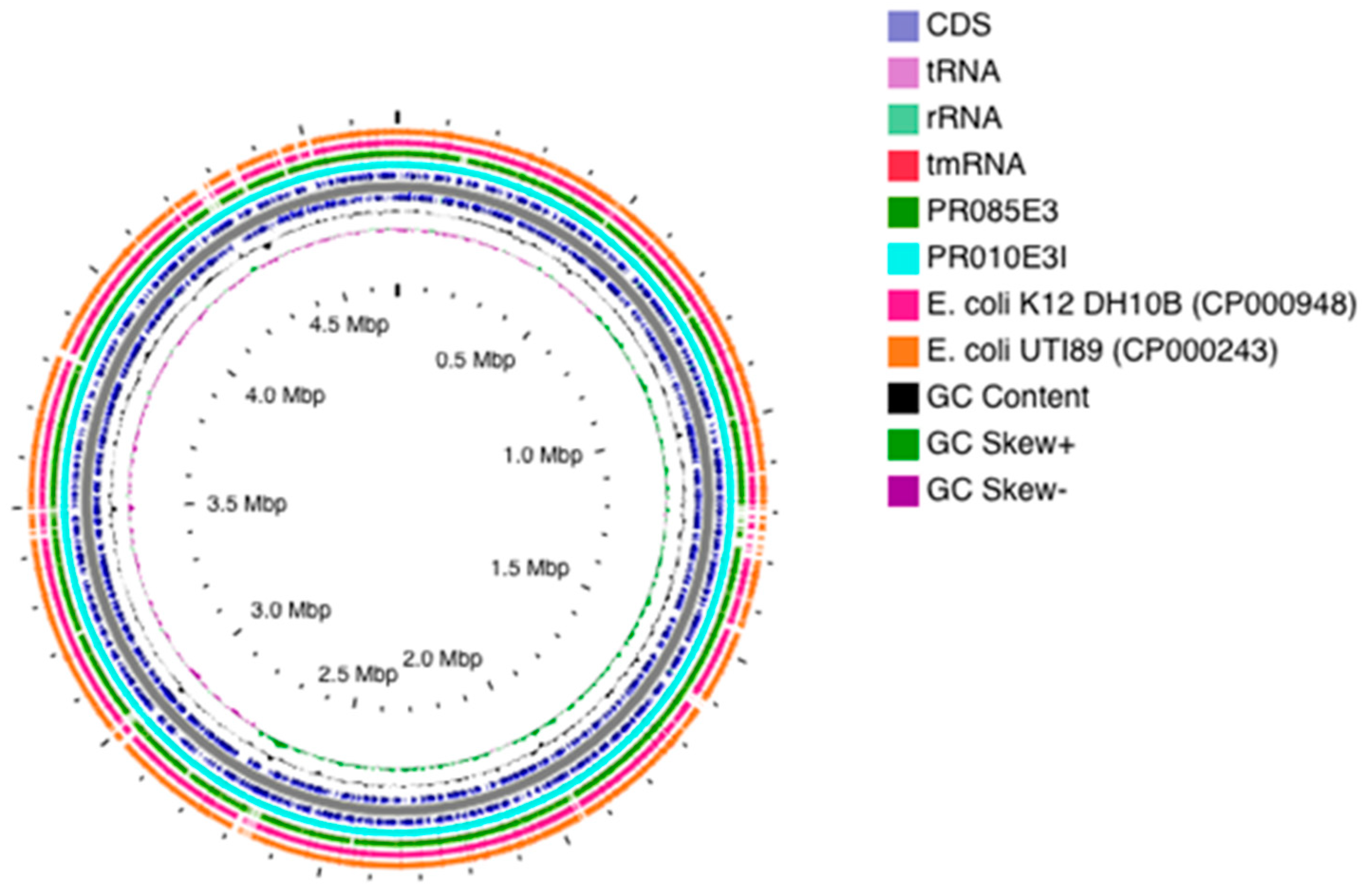
2.2. Genomic Features

Table 1 and Table S2 depict all the genomic characteristics, including length, GC content, N50, coverage, coding sequences, RNAs, rMLST, phylotype and serotype, of the isolates. The genome size of the isolates ranged from 4.5 Mb to 5.3 Mb with a GC content of 50.5 to 50.9 and coverage of 111 to 188 (Table S2).

2.3. Antimicrobial Resistance Phenotypes and Genotypes

Whole genome-based resistome analyses revealed that all isolates evidenced relatively similar combinations of resistance genes encoding target modification, antibiotic inactivation, antibiotic efflux pumps and regulators. Six (54.54%) E. coli isolates harboured blaCTX-M-15 with other blaCTX-M-group being detected in the others. Three isolates simultaneously harboured the blaCTX-M-15 and blaTEM-1B whilst one isolate, PN256E8 harboured concomitantly the blaCTX-M-15, blaTEM-1B, blaTEM-141 and blaTEM-206 (Table 2).
All except two isolates (PR256E1, PN256E2) harboured various aminoglycoside resistance genes, including aph(3″)-Ib (6/11, 55%) and aph(6)-1d (6/11, 55%) genes (Table 2). Eight (73%) isolates harboured different aad genes, including aadA5 (3/11; 27%) and aadA1 (3/11; 27%) (Table 2). Several types of plasmid-mediated quinolone resistance (PMQR) genes were also identified in the isolates (Table 2) with the qnrS1 gene being detected in four (36%) isolates, whilst the aac(6′)Ib-cr and oqxAB genes were identified in three isolates (27%) each. Mutations in the gyrA quinolone resistance-determining region (QRDR) genes were observed in two isolates (PR010E3I, PN091E1II) where three mutations were observed in gyrA with two (S83L, D87N) occurring within PR010E3I and one (S83A) in PN091E1II (Table 2). All isolates, except for PN256E8 and PR010E3I, for which the PMQR gene was identified in both and additionally QRDR in the latter, were susceptible to ciprofloxacin.
All isolates displayed concomitant resistance to trimethoprim and sulfamethoxazole with all harbouring at least one sul gene variant. Specifically, sul2 gene was identified in nine (82%) isolates alone and in combination with sul1 gene in one (PR010E3I). Similarly, the dfr gene was identified in 7/11 (64%) isolates, specifically dfrA17 (n = 3) and dfrA14 (n = 3). Diverse permutations of dfr and sul genes occurred in the isolates with sul2 and dfrA14 being detected in three (27%) isolates while sul2 and dfrA17 were identified in two isolates (Table 2). One isolate (9%) harboured the mcr-1 gene encoding for colistin resistance (Table 2).

2.4. Whole-Genome Virulome Profiling and Pathogenicity

The virulomes of all E. coli displayed high level of pathogenicity (Table 3). The ESBL-Ec isolates showed a 93.6% mean probability (P score) of being human pathogens. The pathogenic species with the highest linkage (100% identity) were the E. coli APEC O1 (Accession numbers: DQ517526, DQ381420), E. coli UMN026 (Accession number: CU928163) and E. coli UTI89 (Accession number: CP000243), which are all extraintestinal pathogenic strains in animals (poultry) and humans belonging to the pathogenic phylogroup B2.
The VFs detected belonged to major functional categories including: adhesins, toxins, protectins and invasins, iron uptake/siderophores, anti-phagocytosis, secretion systems and autotransporters (Table 3). The isolate PR85E3 harboured the highest number (72) of VFs, followed by the isolates PN256E2 and PR010E3I with 39 and 24 VFs, respectively. Analysis of the type I fimbriae fim showed that it was present in 64% of the isolates. Among putative VFs, autotransporter adhesin ehaB, invasin of brain endothelial cells locus B (ibeB) and invasin of brain endothelial cells locus C (ibeC) belonging to autotransporter protein and invasins, were the most prevalent (73%, 8/11) VFs across the isolates (Table 3 and Table S3). Interestingly, the avian hemolysin gene F (hlyF) that enhanced the production of outer membrane vesicles (OMVs) and led to autophagy of eukaryotic cells was detected in one isolate while the hemolysin E (hlyE) a pore-forming toxin was observed in 34% (4/11) of isolates.
Our findings showed that rectal E. coli isolates harboured significantly more VFs than nasal isolates (141 VFs in rectal isolates vs 104 in nasal isolates, p = 0.018). Putative VFs for invasion, such as the outer membrane protein T (OmpT) and the traT genes were more prevalent in rectal than nasal isolates. However, the polysialic acid transport protein group 3 (KpsMIII) gene encoding for group 3 capsule, was detected only in nasal isolates, as were the unique vat and astA. Specifically, all E. coli isolates harboured at least one ExPEC VF from each of the major functional categories and up to 18 ExPEC VFs (Table 3 and Supplementary Table S1).

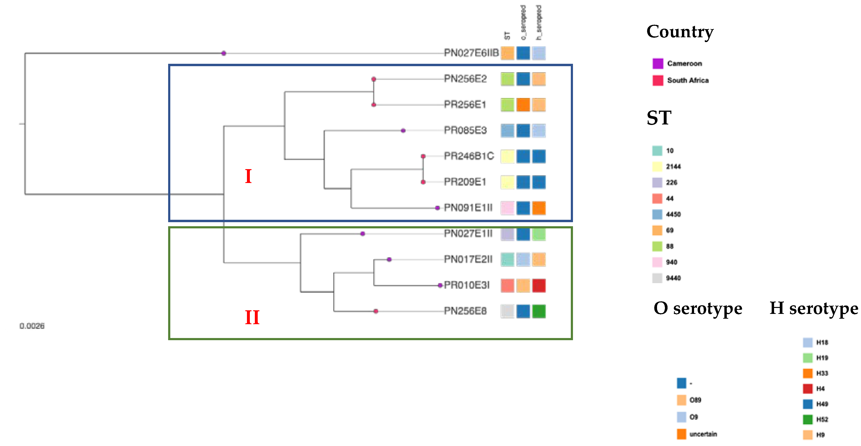
2.5. Phylogenetic Groups and Multilocus Sequence Typing, Serotyping and Phylotyping

Based on in silico MLST results, four E. coli isolates were assigned to the pandemic ST88 (n = 2) and ST2144 (n = 2) clones, while the remaining isolates were assigned to six single-locus variants, namely, ST10, ST69, ST226, ST944, ST4450 and ST44. Interestingly, the E. coli ST2144 were both isolated from two rectal samples processed within the same abattoir (SH004).
The majority of the isolates were assigned to commensal phylogroups A (45%), B1 (28%) and C (18%) but one belonged to the virulence phylogroup D (9%). The serotype O-:H49 (18.18%) and O-:H18 (18.18%) were the principal serotypes detected, while the fimH1250 (18.18%) and fimH87 (18.18%) were the predominant fimH gene observed.

2.6. Mobile Genetic Elements

WGS analysis identified 15 different plasmid replicons in all the isolates, which further all harboured multiple plasmid replicons concomitantly. Ten types of incompatibility (Inc) plasmid replicons were identified with different frequencies, including IncY, IncFIA, IncFIB (AP001918), IncFIC(FII), IncFII, IncN, IncHI2, IncHI2A, IncI1, IncI2 and IncX (Table 2 and Table 4). The majority of isolates (5/11; 45.45%) harboured the IncF (FII, FIB, FIC, FIA) and IncY (4/11; 36%). Four isolates harbouring the IncF incompatibility group also harboured the IncH (n = 2) and IncI (n = 2) groups.
In silico plasmid MLST-analyses assigned the IncF plasmid incompatibility group to STs K-:A-:B1 and K89:A-:B57, while IncH and IncI plasmids were assigned to ST3. Additionally, nine (82%) isolates harboured an array of insertion sequences (IS) with IS26, IS421, Isec1 being the most frequent with a 55% prevalence. An array of three IS (IS26, ISVsa3, ISEc9) were harboured on the plasmid IncI that also encoded the sulphonamide resistance gene (sul2) and virulence factor (cib) in the two E. coli ST88 (PR256E1 and PN256E2). Similarly, three isolates harboured transposons (Tn), including Tn6082 (18%) and Tn7 (9%), with the trimethoprim resistance gene dfrA1 being encoded in transposon Tn7 (Table 4).

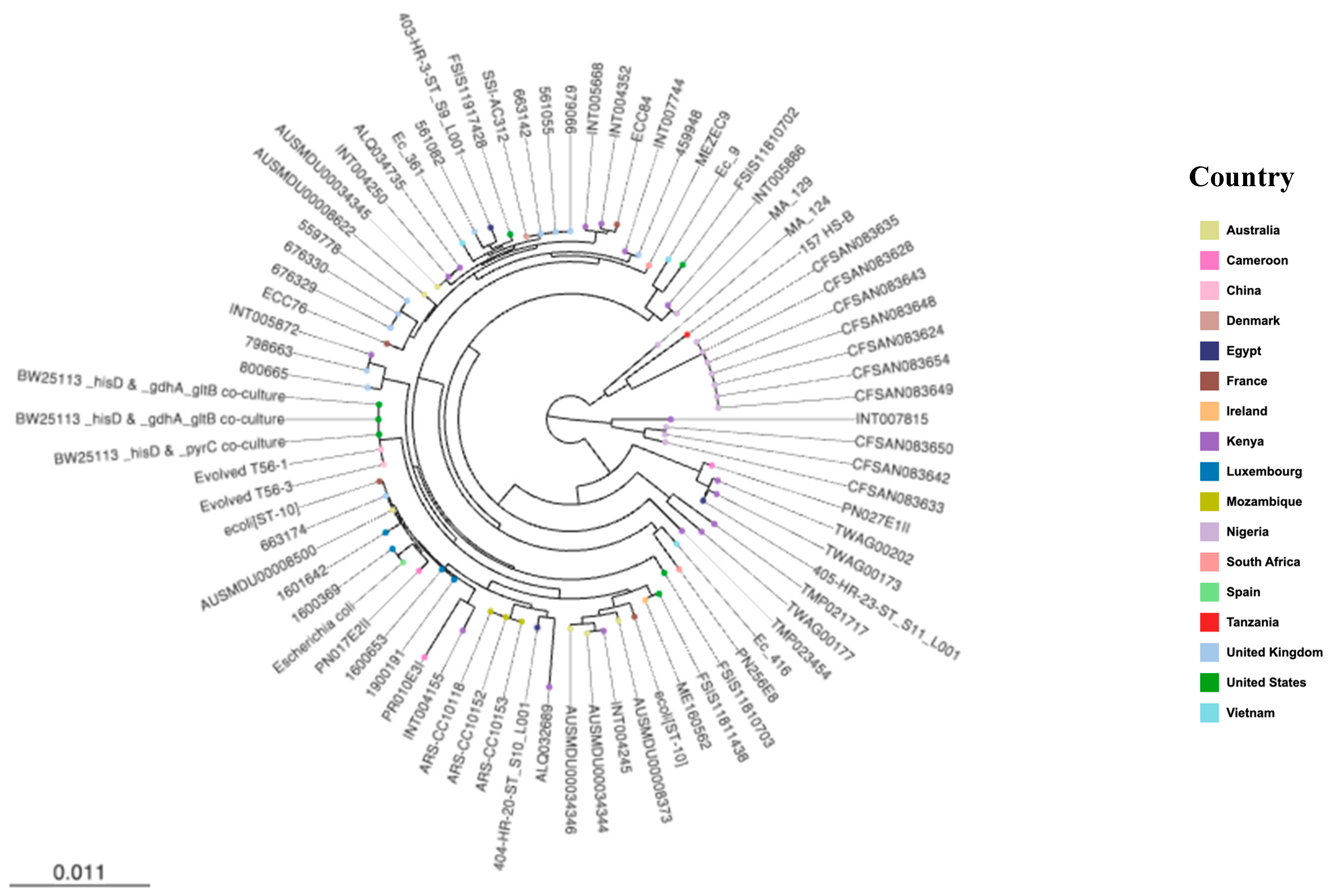
2.7. Phylogenetic Analysis

The contigs of the ESBL-Ec harbouring the most VFs were mapped against the complete genome of E. coli Ecol_AZ155 (NZ_CP019005.1) for visualization of the genomic organisation (Figure 1). The results of the comparative genomic analyses revealed specific similarities and dissimilarities, i.e., isolates had similar and dissimilar arrangements of genomic regions towards representative and reference genomes (Figure 1).
Whole genome phylogenetic analysis grouped the study E. coli isolates (n = 11) into two major clusters (Figure 2). The first one grouped six isolates including two E. coli ST2144 (PR209E1, PR246B1C), two ST88 strains (PR256E1, PN256E2), one ST940 (PN091E1II) and one ST4450 (PR085E3). The two E. coli ST2144 isolates identified from the same abattoir (SH004) in South Africa had 100% identity and shared a close common ancestor with E. coli ST940 (PN091E1II) and E. coli ST4450 (PR085E3), which both originated from one Cameroonian abattoir (SH002). Similarly, the two ST88 isolates (PR256E1 and PN256E2) displayed 100% identity and were found to share common ancestor with the E. coli ST2144, ST940 and ST4450. The second clade included four isolates belonging to various STs (i.e., PN256E8: ST9440; PR010E3I: ST44; PN017E1: ST10; PN027E1II: ST226) of which E. coli ST10 (PN017E1) and ST44 (PR010E3I) isolated in the same abattoir Cameroon were closely related and shared a common ancestor with E. coli ST9440 originating from South Africa.
The phylogenomic analyses of the study’s isolates with international strains revealed that they were more closely related to strains from African countries, such as Kenya, Ghana, Egypt and Morocco, than to any other country (Figure 3). None were phylogenetically related to any strain from the United States or United Kingdom. The genomes from livestock and humans clustered together at some level. Specifically, PR246B1C and PR209E1 (ST2144), described above to have the same virulome, resistome and mobilome, as well as PN091E1II (ST940), were in the same cluster and shared common ancestors with strains isolated from humans and livestock in Kenya, Uganda, South Africa, Mozambique and Ghana (Figure 3). Similarly, hierarchical clustering analyses provided evidence of the genomic relationship between strains originating from livestock and humans with the sole E. coli ST10 strain (PN017E2) sharing a common ancestor with the E. coli ST10 isolated from humans in Spain and livestock in Luxembourg. Intriguingly, the ST9440 isolate (PN256E8) was closest to an ST10 isolate (Ec416) from livestock in Vietnam (Figure 4).

3. Discussion

In this study, genotypic and pathogenic characteristics of ESBL-Ec isolated from pigs collected at Cameroonian and South African abattoirs were investigated using WGS. A diverse population of ESBL-Ec harbouring an extensive repertoire of resistance genes and virulence factors have been detected.
Each genome of ESBL-Ec isolated from both countries harboured numerous ARGs, especially the blaCTX-M-15, supporting other contemporary studies that showed that blaCTX-M-15 is the most prevalent ESBL variant among E. coli [13,14]. In fact, Rafai et al., (2015) detected 63.7% of ESBL producers in surgical site infections of humans in the Central African Republic [14]. The authors showed that blaCTX-M-15 was present in all isolates along with aac(6′)-Ib-cr. Similar findings were also evidenced by Mbelle et al., (2019) who reported a 70% occurrence of blaCTX-M-15-carrying strains among hospitalized patients in South Africa [15].
A major observation was the phenotypic multi-drug resistance (MDR) of the isolates PR010E3I and PN256E8, although all isolates, except PR256E1 and PN256E2, harboured concomitant resistance genes encoding for resistance to aminoglycosides, tetracyclines and fluoroquinolones. This finding is in contrast with data readily available from the literature, which suggest that machine or deep learning can be used to predict adequately phenotypic antimicrobial resistance based on genome sequence data [16]. Although transcription analyses could not be undertaken to assess the expression of these genes, these observations led us to posit that the PMQR and QRDR genes, as well as aminoglycoside resistance genes present in these isolates might not have been expressed or be silent. Similar discrepancies regarding the phenomes and genomes of isolates harbouring resistance genes but not expressing associated phenotypic resistance were reported elsewhere [16] and can be further observed in Table 2 and Table S1. Our finding further reveals that the application of machine or deep learning, as well as the comparison between phenome and genome are still needed at a large-scale from various environments and sources.
All isolates were seen as human pathogens with over 93% of pathogenicity score and the avian pathogenic E. coli APEC-O1-ColBM (DQ381420) being the closest related strain. There is increasing evidence that food-producing animals and food products may contribute to the spread of ExPEC in the community [17]. In our study, all ESBL-Ec harboured at least three VFs associated with ExPEC, such as iss, iutA, traT, ompT, hlyA, iroN, papC and fimH [3]. Of great concern is that the isolate PR085EE3 carried over to 72 VFs with the majority being identified in clinical ExPEC. This suggests that commensal bacteria prevailing in the microbiome of food animals are not only reservoirs of resistance genes, but more so, of virulence factors which might be transmitted via horizontal gene transfer to other bacteria and disseminate to humans via the food chain. It reemphasizes the need to ensure adequate food safety measures throughout the farm-to-plate continuum along with effective infection prevention and control measures in hospitals.
The detection of the heat stable enterotoxin 1 (astA) gene, encoding for the enteroaggregative E. coli heat-stable toxin 1 (EAST1), in the sole ST9440 strain (PN256E8), as well as the avian hemolysin (hlyF) in PR256E1 (ST88) gives credence to the fact that commensal E. coli prevailing in the gut microbiome have a propensity to acquire various virulence genes, which might, therefore, evolve as progenitor lineages from which heteropathogenic E. coli, including uropathogenic E. coli (UPEC), neonatal meningitis-associated E. coli (NMEC) and enteroaggregative E. coli (EAEC) strains, will emerge.
The majority of commensal E. coli strains belong to the phylogenetic groups A and B1, whereas the most common virulent ExPEC are associated with group B2 and D. Our ESBL-Ec isolates belonged to group A (45%) and B1 (28%). Several reports confirmed that phylogroups A and B1 are the leading phylogroups among E. coli isolates especially in the gut microbiome [18,19]. A study from Nigeria showed that 62% of E. coli isolates tended towards the commensal phylogroup B1 and A [20]. The relationship between phylogenetic groups and ABR has been established previously [20], and studies have shown that the group B2 strains are mainly MDR [18,21]. However, our study revealed that isolates from other phylogroups, such as group A and B1, could also display MDR and a high level of virulence certainly as a result of horizontal gene transfer. We posit that isolates from other phylogroups, such as group A and B1, though commensals, could also display MDR and high level of virulence, likely due to horizontal acquisition of resistance genes and virulence factors, which might allow commensal bacteria to become putatively virulent in case of extra-intestinal infections.
The ESBL-Ec isolates were mainly circulating in two clonal lineages since four out of seven isolated strains belonged to the ST2144 (n = 2) and ST88 (n = 2). In addition, the MDR-high-risk clone ST69 and the ST10 were also detected. The ST10 complex, including ST10, commonly associated with spread of CTX-M-1, CTX-M-2 and CTX-M-9 groups, is highly distributed among humans and various livestock species and has been linked with intestinal and extra-intestinal infections in several African countries [14]. E. coli ST10 was the main ST along with ST131 identified in surgical site infection in the Central African Republic [14]. Like other high-risk clones, E. coli ST69 possesses biological factors, such as usp, ompT, secreted autotransporter toxin (sat) and iutA genes corresponding specifically to ST131 [13], that increase bacterial fitness allowing these strains to out-compete other bacterial strains and become the principal part of the bacterial population in the gut [13]. The ß-lactamase genes detected in this study is well in line with that described elsewhere in the world. The detection of the mcr-1 gene in one isolate suggests that colistin-resistant Enterobacterales are also emerging among food-producing animals in Africa and demonstrates the urgent need of antimicrobial usage stewardship in food production systems and implementation of effective monitoring programmes to curb the spread of MDR-E. coli.
Comparative hierarchical clustering suggested that the majority of our strains belong to two clusters (Figure 2 and Figure 3). Interestingly, in cluster I, the two ST2144 isolates (PR246B1C and PR209E1) originating from South Africa were identical and shared common ancestors with two Cameroonian isolates ST940 (PN091EII) and ST4450 (PR085E3). Likewise, in cluster II, the sole ST9440 isolate (PN256E8) originating from South Africa shared common ancestors with three Cameroonian isolates ST44 (PR010E3I), ST10 (PN017E2I) and ST226 (PN27E1II). This gives credence to the hypothesis that ESBL-Ec emerging in one part of the world can spread to another part due to the globalization of trade and international travels [7].
The study further confirmed that the ST10 complex is common in African livestock as all our ST10 complex belong to a unique cgMLST cluster containing closely related isolates from Cameroonian (PR010E3I, PN027E1II) and South African pigs (PN256E8). The comparative phylogenomic analysis further confirms that our ESBL-Ec ST10 demonstrated overlap with ST10 strains isolated from livestock and human populations in Africa (Mozambique, Egypt, Morocco), Asia (Vietnam), Europe (United Kingdom, Denmark, France) and Oceania (Australia) and display the same phylogroup A. Similarly, our ESBL-Ec ST69 share high levels of similarity with an UPEC ST69 phylogroup D that was involved in pyelonephritis in France (unpublished data). This gives credence to the hypothesis that commensal E. coli of the gut microbiome might be the reservoir of virulence and resistance genes that allow the emergence of hetero-pathogenic E. coli strains [18,19].
Mobile genetic elements (MGEs) play an essential role in the mobility of ARGs and VFs between different bacterial species. Our isolates harboured multiple plasmids belonging to major replicon types, especially the IncF (9/11; 81%) plasmid. Similar plasmid replicons associated with blaCTX-M-group were reported in humans and livestock in Africa and across the world [22]. Given the presence of ESBL-Ec in clinically healthy animals and humans, it is likely that the presence of these plasmids could contribute to the long-term persistence of resistance traits in animal and environmental microbiome.
Though our study was limited by the isolates numbers and geographic area, our results sufficiently reinforce the need to closely monitor pathogenic and commensal bacteria prevailing in the food production systems on the continent [22].

4. Conclusions

Our study demonstrates that the population structure of ESBLs-E. coli in pigs is highly diverse with the blaCTX-M-15 gene being the leading CTX-M variant. Although the phylogenetic diversity observed precludes any suggestion for clonal dissemination, the resistance and high human pathogenic potential demonstrate the urgent need to implement effective strategies to contain the dissemination of antibiotic-resistant bacteria in Cameroon and South Africa. Our study underlines the necessity of long-term genomic studies investigating commensal and pathogenic bacteria in (food) animals, food products and associated environments, as well as in occupationally exposed workers, in line with the One Health approach, not only to preserve antibiotics for future generations, but also to gain new insights into the diversity, evolutionary history and emergence of ESBL-ExPEC, as basis for sustainable containment of this resistant pathogen.

5. Materials and Methods

5.1. Study Design and Bacterial Isolates

The study sample consisted of eleven putative ESBL-Ec isolates that were collected between March and October 2016 as part of a larger study where ESBL-producing Enterobacterales were collected from three abattoirs in Cameroon and two in South Africa. These isolates originating from nasal (n = 6) and rectal swabs (n = 5) from healthy pigs processed at abattoirs, were identified as ESBL producers via VITEK 2 system and as closely related isolates via the enterobacterial-repetitive-polymerase chain reaction (ERIC-PCR) analysis, respectively [12]. They were then selected to assess further the clonal relatedness between Cameroonian and South African isolates.

5.2. Identification, ESBL Screening and Antimicrobial Susceptibility Testing

All samples were cultured on MacConkey agar supplemented with 2 mg/L cefotaxime and incubated for 18–24 h at 37 °C in aerobic conditions [7]. All putative ESBL-producers were phenotypically characterized to the genus level using Gram staining and biochemical tests (catalase and oxidase tests). The isolates were thereafter phenotypically confirmed using the VITEK 2 system.
The VITEK 2 system was further used for ESBL screening along with the double disk synergy test as previously described [12]. A series of 18 antibiotics encompassed in the Vitek® 2 Gram Negative Susceptibility card (AST-N255) were tested using Vitek® 2 System (BioMérieux, Marcy l’Etoile, France). Breakpoints of the CLSI guidelines [8] were used except for the colistin, amoxicillin and clavulanic acid, piperacillin/tazobactam, amikacin for which EUCAST breakpoints [23] were considered with E. coli ATCC 25922 and K. pneumoniae ATCC 700603 being used as controls.

5.3. Whole Genome Sequencing and Data Analysis

5.3.1. Purification, Sequencing and Pre-Processing of Genomic Data

GenElute® bacterial genomic DNA kit (Sigma-Aldrich, St. Louis, MO, USA) was used for genomic DNA (gDNA) extraction with the concentration and purity assessed using agarose gel electrophoresis, NanoDrop 8000 spectrophotometer (Thermo Scientific, Waltham, MA, USA), and fluorometric analysis Qubit® (Thermo Scientific, Waltham, MA. USA). Libraries were constructed using the Nextera XT DNA Library Preparation kit (Illumina Inc., San Diego, CA, USA) and subjected to paired-end (2×300 bp) sequencing on an Illumina MiSeq (Illumina Inc., San Diego, CA, USA) machine with 100× coverage. The generated paired-end reads were merged, checked for quality, trimmed, and de novo assembled into contigs with SPAdes version 3.11 [24].

5.3.2. WGS-Based Molecular Typing

WGS data were used to predict in silico multi-locus sequence type (MLST) based on the Achtman scheme, which considers allelic variation amongst seven housekeeping genes (adk, fumC, gyrB, icd, mdh, purA and recA) to assign STs [25]. In addition to generating an E. coli MLST assignment for each isolate, core-genome MLST (cgMLST) was assigned based on a scheme from EnteroBase server (https://enterobase.warwick.ac.uk/species/index/ecoli accessed on 20 April 2021) that uses 2513 loci [26]. EnteroBase was further used for in silico phylotype predictions following the Clermont scheme [27], as well as for fimH allelic designations [26]. Ribosomal MLST, hierarchical cgMLST clustering, wgMLST were further performed using core genome data in EnteroBase.

5.3.3. In Silico Resistome and Virulome Profiling

ARGs of the E. coli genomes were annotated and identified with ResFinder [28] through the bacterial analysis online platform of GoSeqIt tool (www.goseqit.com accessed on 15 November 2020). The Comprehensive Antibiotic Resistance Database (CARD, https://card.mcmaster.ca/ accessed on 20 April 2021) [29] platform was concomitantly used for prediction of ARGs and detection of chromosomal mutations (SNPs) in quinolone-resistant genes of gyrA, gyrB, parC and parE. The ARGs predicted are combinations of both database with a threshold set at 90% identity for a positive match between the reference database and a target genome. VirulenceFinder [30] available from the GoSeqIt tools server along with the comparative pathogenomics platform VFanalyzer from Virulence Factor Database (VFDB) [31] were similarly used to predict and annotate virulence factors (VFs), respectively. Likewise, virulence factors detected in our study had a threshold of 90% identity with reference genes and represent combinations of both databases. ExPEC virulence genes, including ferric aerobactin receptor (iutA), increased serum survival (iss), heat-resistant agglutinin (hra), temperature sensitive haemagglutinin (tsh), P fimbrial adhesin (papC), colicin V (cvaC), capsular polysialic acid virulence factor group 2 (kpsII) and invasive factor of brain endothelial cells locus A (ibeA) of E. coli strains responsible for neonatal meningitis in humans were investigated in silico. Moreover, the pathogenicity prediction web-server PathogenFinder [32] was used to predict bacteria pathogenic potential towards human hosts with a threshold ≥90% being considered as isolates with significant pathogenic human potential.

5.3.4. Detection of Mobile Genetic Elements

The RAST SEED viewer [33] and Artemis Comparison Tool (ACT, [34]) were used to identify the presence of transposases and integrons flanking resistance and virulence genes. MGEfinder was used for the in-silico detection of insertion sequences (IS), conjugative genetic elements and transposons allowing investigation of synteny of mobile genetic elements with VFs and antibiotic resistance genes [35]. PHAge Search Tool Enhanced Release (PHASTER) server was used for the identification, annotation and visualization of prophage sequences [36]. The profile of bacterial plasmid replicons and plasmid incompatibility groups was assessed through PlasmidFinder 2.1 (https://cge.cbs.dtu.dk/services/PlasmidFinder/ accessed on 20 April 2021) and pMLST 2.0 (https://cge.cbs.dtu.dk/services/pMLST/ accessed on 20 April 2021) [37]. Putative CRISP system and Cas cluster were assessed through CRISPRCasFinder (https://crisprcas.i2bc.paris-saclay.fr/CrisprCasFinder/Index accessed on 20 April 2021).

5.3.5. Genome Visualization and Gene Annotation

The de novo assembled raw reads were annotated using the Rapid Prokaryotic Genome (PROKKA) version 1.12 beta available from EnteroBase, the NCBI Prokaryotic Genome Annotation Pipeline (PGAP) and RAST 2.0 server (http://rast.nmpdr.org accessed on 15 November 2020) [38], which identified encoding proteins, rRNA and tRNA, assigned functions to the genes and predicted subsystems represented in the genome. The size, GC content, average coverage, length, N50, L50, RNAs and protein coding sequences were obtained for each isolate. The annotated in silico predicted proteins and regions were visualized via the JSBrowse of EnteroBase server and RAST. The genomes of the isolates were visualized using the CG Viewer Server [39]. In addition, the contigs of selected isolates were mapped against the complete genome of E. coli Ecol_AZ155 (NZ_CP019005.1) for visualization of the genomic organization.

5.3.6. Comparative Phylogenomic Analyses

The whole genome phylogenetic relationship was assessed within the study isolates and with a collection of international E. coli genomes (n = 118) available at the EnteroBase E. coli genomes repository as of 12 November 2020 (Supplementary Dataset S1). The international isolates were closely related E. coli strains of similar STs isolated from various sources (humans, livestock and environment).
The E. coli isolate YA00194039 (ERS4920643) was used as reference genome with all assembled contigs being aligned against it to determine SNP locations. The phylogeny of the E. coli isolates was characterised using the whole genome MLST (wgMLST), core genome MLST (cgMLST) and accessory genome MLST. Phylogenetic relationships among study isolates and between study and international isolates were assessed based on nucleotide alignments of all the genes in the entire genome (wgMLST) and core genome content (core genes that are present in most genomes with ≥95% of nucleotide identity; cgMLST). Moreover, the accessory gene, including ARGs, plasmid replicons and phages content, was analysed using the EnteroBase server, which scans the genome against the core ResFinder and PlasmidFinder databases based on a percentage identity of ≥ 90% and coverage of ≥ 70% in order to generate a customized phylogenetic tree to infer the evolutionary relationship within the study isolates and between the study and international isolates. Minimum spanning trees constructed using GrapeTree software and phylogenetic trees were further built to describe the relatedness among the study isolates and between the study and international isolates [40]. The generated phylogenomic trees were downloaded, and, subsequently, visualized and edited using MicroReact (www.microreact.org accessed on 4 June 2022).

5.4. Nucleotide Accession Number

This whole-genome shotgun bioproject PRJNA548686 of E. coli strains PN017E2II, PR010E3I, PN027E6IIB, PR256E1, PN256E2, PN027E1II, PN091E1II, PN256E8, PR209E1, PR246B1C, and PR085E3 has been deposited at DDBJ/EMBL/GenBank under accession numbers VMKK00000000, VKOQ00000000, VKOV00000000, VKOS00000000, VKOT00000000, VKOW00000000, VKOU00000000, QJRZ00000000, VKOO00000000, WHRW00000000, and VKOP000000000 respectively. The versions described in this paper are the versions, VMKK01000000, VKOQ01000000, VKOV01000000, VKOS01000000, VKOT01000000, VKOW01000000, VKOU01000000, QJRZ00000000.1, VKOO01000000, WHRW0000000.1, and VKOP010000000, respectively.

Supplementary Materials

The following supporting information can be downloaded at: https://www.mdpi.com/article/10.3390/pathogens11070776/s1, Table S1: Antimicrobial resistance phenotype of the isolates ESBL-E. coli; Table S2: Summary of genotypic features of ESBL-E. coli; Table S3: Virulence-associated-traits of nasal and rectal ESBL-E. coli; Supplementary Dataset S1: Metadata of 129 E. coli genomes.

Author Contributions

L.L.F. co-conceptualized the study, undertook sample collection, microbiological laboratory and data analyses, prepared tables and figures, interpreted results, performed bioinformatics analysis and drafted the manuscript. R.C.F. undertook sample collection, microbiological laboratory analyses, contributed to bioinformatics analysis, vetting of the results and writing of the manuscript. M.A. contributed to and vetted bioinformatics analyses. A.I. performed whole genome sequencing analysis. S.Y.E. co-conceptualized the study and undertook critical revision of the manuscript. All authors have read and agreed to the published version of the manuscript.

Funding

L.L.F. Founou and R.C. Founou were funded by the Antimicrobial Research Unit (ARU) and College of Health Sciences (CHS) of the University of KwaZulu-Natal, South Africa. The National Research Foundation funded this study through the NRF Incentive Funding for Rated Researchers (Grant No. 85595), and the DST/NRF South African Research Chair in Antibiotic Resistance and One Health (Grant No. 98342) awarded to S.Y. Essack. The South African Medical Research Council also funded the study through the Self-Initiated Researcher (SIR) Grant awarded to S.Y Essack. The funders had no role in the study design, preparation of the manuscript nor the decision to submit the work for publication.

Institutional Review Board Statement

Ethical approvals were obtained from the National Ethics Committee for Research in Human Health of Cameroon (Ref. 2016/01/684/CE/CNERSH/SP), as well as from the Biomedical Research Ethics Committee (Ref. BE365/15) and Animal Research Ethics Committee (Ref. AREC/091/015D) of the University of KwaZulu-Natal. Approvals were additionally obtained from the Cameroonian Ministry of Livestock, Fisheries and Animal Industries (Ref. 061/L/MINEPIA/SG/DREPIA/CE) and Ministry of Scientific Research and Innovation (Ref. 015/MINRESI/B00/C00/C10/C14). This study was further placed on record with the South African National Department of Agriculture, Forestry and Fisheries [Ref. 12/11/1/5 (878)].

Informed Consent Statement

Not applicable.

Data Availability Statement

Not applicable.

Acknowledgments

The authors would like to thank all included abattoirs, participants and other involved employees from Cameroon and South Africa for participation and all field work assistance. Preliminary results from this study were presented in a poster and ePoster at the 30th and 31st European Congress of Clinical Microbiology and Infectious Diseases (ECCMID) in Paris, France, 2020 (abstract 2297) and Austria, 2021 (abstract 276), respectively. Preliminary results from this study were also presented in oral presentation at the World Microbe Forum in 2021.

Conflicts of Interest

Essack is a member of the Global Respiratory Infection Partnership and Global Hygiene Council both sponsored by unrestricted educational grants from Reckitt and Benckiser Ltd., UK. All other authors declare that there is no competing financial interest.

References

  1. O’Neill, J. Tackling Drug-Resistant Infections Globally: Final Report and Recommendations; The Review on Antimicrobial Resistance: London, UK, 2016. [Google Scholar]
  2. Founou, L.L.; Founou, R.C.; Essack, S.Y. Antibiotic Resistance in the Food Chain: A Developing Country-Perspective. Front. Microbiol. 2016, 7, 1881. [Google Scholar] [CrossRef] [PubMed]
  3. Sarowska, J.; Futoma-Koloch, B.; Jama-Kmiecik, A.; Frej-Madrzak, M.; Ksiazczyk, M.; Bugla-Ploskonska, G.; Choroszy-Krol, I. Virulence factors, prevalence and potential transmission of extraintestinal pathogenic Escherichia coli isolated from different sources: Recent reports. Gut Pathog. 2019, 11, 10. [Google Scholar] [CrossRef] [PubMed] [Green Version]
  4. Selander, R.K.; Caugant, D.A.; Whittam, T.S. Genetic Structure and Variation in Natural Populations of Escherichia coli and Salmonella typhimurium: Cellular and Molecular Biology; Ingraham, J.L., Magasanik, B., Schaechter, M., Low, K.B., Neidhardt, F.C., Umbarger, H.E., Eds.; American Society for Microbiology: Washington, DC, USA, 1987; pp. 1625–1648. [Google Scholar]
  5. Herzer, P.J.; Inouye, S.; Inouye, M.; Whittam, T.S. Phylogenetic distribution of branched RNA-linked multicopy single-stranded DNA among natural isolates of Escherichia coli. J. Bacteriol. 1990, 172, 6175–6181. [Google Scholar] [CrossRef] [PubMed] [Green Version]
  6. Perovic, O.; Singh-Moodley, A.; Dusé, A.; Bamford, C.; Elliott, G.; Han, K.S.S.; Kularatne, R.; Lowman, W.; Whitelaw, A.; Nana, T.; et al. National sentinel site surveillance for antimicrobial resistance in Klebsiella pneumoniae isolates in South Africa, 2010–2012. S. Afr. Med. J. 2014, 104, 563–568. [Google Scholar] [CrossRef] [Green Version]
  7. Founou, L.L.; Founou, R.C.; Essack, S.Y. Antimicrobial resistance in the farm-to-plate continuum: More than a food safety issue. Futur. Sci. OA 2021, 7, FSO692. [Google Scholar] [CrossRef]
  8. Chishimba, K.; Hang’ombe, B.M.; Muzandu, K.; Mshana, S.E.; Matee, M.I.; Nakajima, C.; Suzuki, Y. Detection of Extended-Spectrum Beta-Lactamase-Producing Escherichia coli in Market-Ready Chickens in Zambia. Int. J. Microbiol. 2016, 2016, 5275724. [Google Scholar] [CrossRef] [Green Version]
  9. Aworh, M.K.; Kwaga, J.; Okolocha, E.; Harden, L.; Hull, D.; Hendriksen, R.S.; Thakur, S. Extended-spectrum ß-lactamase-producing Escherichia coli among humans, chickens and poultry environments in Abuja, Nigeria. One Health Outlook 2020, 2, 8. [Google Scholar] [CrossRef]
  10. Runcharoen, C.; Raven, K.E.; Reuter, S.; Kallonen, T.; Paksanont, S.; Thammachote, J.; Anun, S.; Blane, B.; Parkhill, J.; Peacock, S.J.; et al. Whole genome sequencing of ESBL-producing Escherichia coli isolated from patients, farm waste and canals in Thailand. Genome Med. 2017, 9, 81. [Google Scholar] [CrossRef]
  11. Hoek, A.H.A.M.V.; Dierikx, C.; Bosch, T.; Schouls, L.; Van Duijkeren, E.; Visser, M. Transmission of ESBL-producing Escherichia coli between broilers and humans on broiler farms. J. Antimicrob. Chemother. 2020, 75, 543–549. [Google Scholar] [CrossRef]
  12. Founou, L.L.; Founou, R.C.; Ntshobeni, N.; Govinden, U.; Bester, L.A.; Chenia, H.Y.; Djoko, C.F.; Essack, S.Y. Emergence and Spread of Extended Spectrum β-Lactamase Producing Enterobacteriaceae (ESBL-PE) in Pigs and Exposed Workers: A Multicentre Comparative Study between Cameroon and South Africa. Pathogens 2019, 8, 10. [Google Scholar] [CrossRef] [Green Version]
  13. Mathers, A.J.; Peirano, G.; Pitout, J.D.D. The Role of Epidemic Resistance Plasmids and International High-Risk Clones in the Spread of Multidrug-Resistant Enterobacteriaceae. Clin. Microbiol. Rev. 2015, 28, 565–591. [Google Scholar] [CrossRef] [PubMed] [Green Version]
  14. Rafaï, C.; Frank, T.; Manirakiza, A.; Gaudeuille, A.; Mbecko, J.-R.; Nghario, L.; Serdouma, E.; Tekpa, B.; Garin, B.; Breurec, S. Dissemination of IncF-type plasmids in multiresistant CTX-M-15-producing Enterobacteriaceae isolates from surgical-site infections in Bangui, Central African Republic. BMC Microbiol. 2015, 15, 15. [Google Scholar] [CrossRef] [PubMed] [Green Version]
  15. Mbelle, N.M.; Feldman, C.; Sekyere, J.O.; Maningi, N.E.; Modipane, L.; Essack, S.Y. The Resistome, Mobilome, Virulome and Phylogenomics of Multidrug-Resistant Escherichia coli Clinical Isolates from Pretoria, South Africa. Sci. Rep. 2019, 9, 16457. [Google Scholar] [CrossRef]
  16. Hendriksen, R.S.; Bortolaia, V.; Tate, H.; Tyson, G.H.; Aarestrup, F.M.; McDermott, P.F. Using Genomics to Track Global Antimicrobial Resistance. Front. Public Health 2019, 7, 242. [Google Scholar] [CrossRef] [PubMed] [Green Version]
  17. Hammad, A.M.; Hoffmann, M.; Gonzalez-Escalona, N.; Abbas, N.H.; Yao, K.; Koenig, S.; Allué-Guardia, A.; Eppinger, M. Genomic features of colistin resistant Escherichia coli ST69 strain harboring mcr-1 on IncHI2 plasmid from raw milk cheese in Egypt. Infect. Genet. Evol. 2019, 73, 126–131. [Google Scholar] [CrossRef]
  18. Li, B.; Sun, J.-Y.; Han, L.-Z.; Huang, X.-H.; Fu, Q.; Ni, Y.-X. Phylogenetic Groups and Pathogenicity Island Markers in Fecal Escherichia coli Isolates from Asymptomatic Humans in China. Appl. Environ. Microbiol. 2010, 76, 6698–6700. [Google Scholar] [CrossRef] [PubMed] [Green Version]
  19. Stoppe, N.D.C.; Silva, J.S.; Carlos, C.; Sato, M.I.Z.; Saraiva, A.M.; Ottoboni, L.M.M.; Torres, T.T. Worldwide Phylogenetic Group Patterns of Escherichia coli from Commensal Human and Wastewater Treatment Plant Isolates. Front. Microbiol. 2017, 8, 2512. [Google Scholar] [CrossRef] [Green Version]
  20. Olowe, O.A.; Adefioye, O.J.; Ajayeoba, T.A.; Schiebel, J.; Weinreich, J.; Ali, A.; Burdukiewicz, M.; Rödiger, S.; Schierack, P. Phylogenetic grouping and biofilm formation of multidrug resistant Escherichia coli isolates from humans, animals and food products in South-West Nigeria. Sci. Afr. 2019, 6, e00158. [Google Scholar] [CrossRef]
  21. Tadesse, D.A.; Zhao, S.; Tong, E.; Ayers, S.; Singh, A.; Bartholomew, M.J.; McDermott, P.F. Antimicrobial Drug Resistance in Escherichia coli from Humans and Food Animals, United States, 1950–2002. Emerg. Infect. Dis. 2012, 18, 741–749. [Google Scholar] [CrossRef]
  22. Patil, S.; Chen, X.; Lian, M.; Wen, F. Phenotypic and genotypic characterization of multi-drug-resistant Escherichia coli isolates harboring blaCTX-M group extended-spectrum β-lactamases recovered from pediatric patients in Shenzhen, southern China. Infect. Drug Resist. 2019, 12, 1325–1332. [Google Scholar] [CrossRef] [Green Version]
  23. EUCAST. Breakpoint Tables for Interpretation of MICs and Zone Diameters. 2016. Available online: https://www.eucast.org/ (accessed on 28 March 2017).
  24. Bankevich, A.; Nurk, S.; Antipov, D.; Gurevich, A.A.; Dvorkin, M.; Kulikov, A.S.; Lesin, V.M.; Nikolenko, S.I.; Pham, S.; Prjibelski, A.D.; et al. SPAdes: A new genome assembly algorithm and its applications to single-cell sequencing. J. Comput. Biol. 2012, 19, 455–477. [Google Scholar] [CrossRef] [PubMed] [Green Version]
  25. Larsen, M.V.; Cosentino, S.; Rasmussen, S.; Friis, C.; Hasman, H.; Marvig, R.L.; Jelsbak, L.; Sicheritz-Pontén, T.; Ussery, D.W.; Aarestrup, F.M.; et al. Multilocus Sequence Typing of Total-Genome-Sequenced Bacteria. J. Clin. Microbiol. 2012, 50, 1355–1361. [Google Scholar] [CrossRef] [PubMed] [Green Version]
  26. Alikhan, N.-F.; Zhou, Z.; Sergeant, M.J.; Achtman, M. A genomic overview of the population structure of Salmonella. PLoS Genet. 2018, 14, e1007261. [Google Scholar] [CrossRef] [PubMed] [Green Version]
  27. Clermont, O.; Christenson, J.K.; Denamur, E.; Gordon, D.M. The Clermont Escherichia coli phylo-typing method revisited: Improvement of specificity and detection of new phylo-groups. Environ. Microbiol. Rep. 2013, 5, 58–65. [Google Scholar] [CrossRef]
  28. Zankari, E.; Hasman, H.; Cosentino, S.; Vestergaard, M.; Rasmussen, S.; Lund, O.; Aarestrup, F.M.; Larsen, M.V. Identification of acquired antimicrobial resistance genes. J. Antimicrob. Chemother. 2012, 67, 2640–2644. [Google Scholar] [CrossRef]
  29. Alcock, B.P.; Raphenya, A.R.; Lau, T.T.Y.; Tsang, K.K.; Bouchard, M.; Edalatmand, A.; Huynh, W.; Nguyen, A.-L.V.; Cheng, A.A.; Liu, S.; et al. CARD 2020: Antibiotic resistome surveillance with the comprehensive antibiotic resistance database. Nucleic Acids Res. 2020, 48, D517–D525. [Google Scholar] [CrossRef]
  30. Joensen, K.G.; Scheutz, F.; Lund, O.; Hasman, H.; Kaas, R.S.; Nielsen, E.M.; Aarestrup, F.M. Real-Time Whole-Genome Sequencing for Routine Typing, Surveillance, and Outbreak Detection of Verotoxigenic Escherichia coli. J. Clin. Microbiol. 2014, 52, 1501–1510. [Google Scholar] [CrossRef] [Green Version]
  31. Liu, B.; Zheng, D.; Jin, Q.; Chen, L.; Yang, J. VFDB 2019: A comparative pathogenomic platform with an interactive web interface. Nucleic Acids Res. 2018, 47, D687–D692. [Google Scholar] [CrossRef]
  32. Cosentino, S.; Voldby Larsen, M.; Møller Aarestrup, F.; Lund, O. PathogenFinder—Distinguishing Friend from Foe Using Bacterial Whole Genome Sequence Data. PLoS ONE 2013, 8, e77302. [Google Scholar] [CrossRef]
  33. Overbeek, R.; Olson, R.; Pusch, G.D.; Olsen, G.J.; Davis, J.J.; Disz, T.; Edwards, R.A.; Gerdes, S.; Parrello, B.; Shukla, M.; et al. The SEED and the Rapid Annotation of microbial genomes using Subsystems Technology (RAST). Nucleic Acids Res. 2014, 42, D206–D214. [Google Scholar] [CrossRef]
  34. Carver, T.J.; Rutherford, K.M.; Berriman, M.; Rajandream, M.-A.; Barrell, B.G.; Parkhill, J. ACT: The Artemis comparison tool. Bioinformatics 2005, 21, 3422–3423. [Google Scholar] [CrossRef] [PubMed] [Green Version]
  35. Durrant, M.G.; Li, M.M.; Siranosian, B.A.; Montgomery, S.B.; Bhatt, A.S. A Bioinformatic Analysis of Integrative Mobile Genetic Elements Highlights Their Role in Bacterial Adaptation. Cell Host Microbe 2020, 27, 140–153.e149. [Google Scholar] [CrossRef] [PubMed]
  36. Zhou, Y.; Liang, Y.; Lynch, K.H.; Dennis, J.J.; Wishart, D.S. PHAST: A Fast Phage Search Tool. Nucleic Acids Res. 2011, 39, W347–W352. [Google Scholar] [CrossRef] [PubMed]
  37. Carattoli, A.; Zankari, E.; Garcìa-Fernandez, A.; Voldby Larsen, M.; Lund, O.; Villa, L.; Møller Aarestrup, F.; Hasman, H. In Silico Detection and Typing of Plasmids. Antimicrob using PlasmidFinder and plasmid multilocus sequence typing. Agents Chemother. 2014, 58, 3895–3903. [Google Scholar] [CrossRef] [PubMed] [Green Version]
  38. Aziz, R.K.; Bartels, D.; Best, A.A.; DeJongh, M.; Disz, T.; Edwards, R.A.; Formsma, K.; Gerdes, S.; Glass, E.M.; Kubal, M.; et al. The RAST server: Rapid annotations using subsystems technology. BMC Genom. 2008, 9, 75. [Google Scholar] [CrossRef] [Green Version]
  39. Grant, J.R.; Arantes, A.S.; Stothard, P. Comparing thousands of circular genomes using the CGView Comparison Tool. BMC Genom. 2012, 13, 202. [Google Scholar] [CrossRef] [Green Version]
  40. Zhou, Z.; Alikhan, N.-F.; Sergeant, M.J.; Luhmann, N.; Vaz, C.; Francisco, A.P.; Carriço, J.A.; Achtman, M. GrapeTree: Visualization of core genomic relationships among 100,000 bacterial pathogens. Genome Res. 2018, 28, 1395–1404. [Google Scholar] [CrossRef] [Green Version]
Figure 1. Circular genome representation of selected ESBL-producing E. coli aligned with reference genome and closely related strains. Circular map of selected ESBL-producing E. coli (PR010E3I, PN256E8 and PR085E3) and closely related strains (E. coli K12, E. coli UTI89, E. coli UMN026), with comparative alignment against E. coli APEC_O1 (NZ_CP019005.1), generated using CGView Server V1.0. Coloured arrows in the outer ring represent different gene families of the reference genome. A key of the coloured arrows representing different gene families is presented in the inset. The inner coloured circles representing different strains are also listed in the inset. Innermost circles show GC content indicated in black and GC Skew, with green and purple indicating positive and negative values, respectively.
Pathogens 11 00776 g001
Figure 2. Comparative genome analysis based on the core genome MLST of study’s ESBL-producing E. coli isolates. Each node represents an isolate, each of which is coloured according to country of origin, as defined in the legend. Clusters of isolates belonging to the same sequence cluster are encircled and annotated. Serotype and sequence types are also indicated via a heatmap. Core-genome phylogenetic tree based on comparison of conserved clusters of orthologous genes (COGs). Interactive map of geographic locations and genetic attributes can be visualized within Microreact at https://microreact.org/project/tYENaUrCix7jMS7RBrFeBi-population-structure-and-pangenome-comparative-analysis-of-esbl-e-coli accessed on 4 June 2022.
Pathogens 11 00776 g002
Figure 3. Phylogenetic relationship between study’s and international E. coli isolates. Isolates of the same continent share the same leaf node colour as depicted in the legend. A total of 118 genomes were used to contextualise our isolates. Mid-point rooted maximum likelihood phylogenetic tree was built using core genome MLST. The inner ring indicates country and the outer ring indicates host source. Interactive map of geographic locations and genetic attributes can be visualized within Microreact at https://microreact.org/project/5CNJcAYrnVvLRLXZX2bXCt-population-structure-and-pangenome-comparative-analysis-of-esbl-e-coli accessed on 4 June 2022.
Pathogens 11 00776 g003
Figure 4. Phylogenetic relationship between E. coli isolates belonging to CC10. Isolates of the same country share the same colour. Eighty isolates were used to contextualise our isolates. Mid-point rooted maximum likelihood phylogenetic tree was built using core genome MLST.
Pathogens 11 00776 g004
Table 1. Genotypic characteristics of ESBL-producing E. coli isolates (Bioproject PRJNA412434).
IsolateAccession NumberCountrySample TypeAbattoirMLST *Clonal ComplexFimHPhylogroupSerotype
PN017E2IIVMKK00000000CameroonNasal swabSH00110ST10 CplxFimH215AO9:H:9
PR010E3IVKOQ00000000CameroonRectal swabSH00144ST10 CplxFimH54AO89:H4
PN027E6IIBVKOV00000000CameroonNasal swabSH00169ST69 CplxFimH27DO-:H18
PR256E1VKOS00000000South AfricaRectal swabSH00588ST23 CplxFimH1250CO: Uncertain H9
PN256E2VKOT00000000South AfricaNasal swabSH00588ST23 CplxFimH1250CO-:H9
PN027E1IIVKOW00000000CameroonNasal swabSH001226ST226 CplxFimH43AO-:H19
PN091E1IIVKOU00000000CameroonNasal swabSH002940ST448 CplxUnknownB1O-:H33
PN256E8QJRZ00000000South AfricaNasal swabSH0059440ST10 CplxFimH23AO-:H52
PR209E1VKOO00000000South AfricaRectal swabSH0042144-FimH87B1O-:H49
PR246B1CWHRW00000000South AfricaRectal swabSH0042144-FimH87B1O-:H49
PR085E3VKOP000000000CameroonRectal swabSH0024450-FimH566AO-:H18
* MLST. Multi-locus sequence typing.
Table 2. Overview of resistome and mobilome in ESBL-producing E. coli isolates.
IsolateCountrySample TypeAbattoirMLST #β-Lactamase Resistance GenesFluoroquinolone Resistance GenesOther Resistance GenesPlasmidspMLST *
CTX-MTEMOXAQRDRPMQR
PN017E2IICameroonNasal swabSH00110CTX-M-15TEM-1B--qnrS1aph(6)-Id, aph(3″)-Ib, tet(A), mph(A), sul2, dfrA14,IncY, Col(MG828), Col440I, rep21-
PR010E3ICameroonRectal swabSH00144CTX-M-15-OXA-1gyrA (p.S83L) gyrA (p.D87N) aac(6′)-Ib-craac(3)-IIa, aph(3″)-Ib, aadA5, aph(6)-Id, tet(B), tet(A), sul1, sul2, dfrA17, floR, catB3IncFIA, Col440I, IncFII, IncFIB, Col(MG828), rep21IncF [F36:A20:B1]
PN027E6IIBCameroonNasal swabSH00169CTX-M-15TEM-1B--qnrS1strA, strB, sul2, tet(A), dfrA14IncY, Col(MG828)-
PR256E1South AfricaRectal swabSH00588CTX-M-1----tet(A), sul2,IncI1 &, IncI2, Col(MG828), ColPVC, IncFIB,IncF [K-:A-:B1];
IncI1[ST3]
PN256E2South AfricaNasal swabSH00588CTX-M-1----tet(A), sul2,IncI1 **, IncFIB, Col(MG828), Col440I, rep10IncF [K-:A-:B1]; IncI1[ST3]
PN027E1IICameroonNasal swabSH001226CTX-M-15TEM-1B--qnrS1aph(3″)-Ib, aph(6)-Id, tet(A), mdf(A), sul2, dfrA14,IncY, Col440I, colRNAI, Col(MG828)-
PN091E1IICameroonNasal swabSH002940CTX-M-15TEM-1B-gyrA (p.S83A),-aph(3″)-Ib, aph(6)-Id, aadA1, 16S_rrsC (g.926_926del), tet(B), mph(A), sul2, dfrA1,IncX, Col440I-
PN256E8South AfricaNasal swabSH005944CTX-M-55TEM-1B
TEM-141
TEM-206
--oqxA, oqxB
aac(6′)-Ib-cr
aac(6′)-Ib3, aadA5, tet(A), sul2, dfrA17, floR, mcr-1.1, fosA3IncN, IncHI2A, IncHI2IncN [ST1];
IncHI2 [ST3-like]
PR209E1South AfricaRectal swabSH0042144CTX-M-14---oqxB, oqxAaph(3″)-Ib, aph(6)-Id, aadA2b, aadA1, sul3, cmlA1, fosA3IncFIC(FII), IncFIB, IncHI2A, IncHI2 rep21IncF [K89:A-:B57]
IncHI2[ST3]
PR246B1CSouth AfricaRectal swabSH0042144CTX-M-14---oqxA, oqxBaph(3″)-Ib, aadA2b, aph(6)-Id, aadA1, aph(3″)-Ib, sul3, fosA3, cmlA1IncFIC(FII), Col440II, IncHI2A, IncHI2, IncFIBIncF [K89:A-:B57]
IncHI2 [ST3]
PR085E3CameroonRectal swabSH0024450CTX-M-15---qnrS1AadA5, sul2, dfrA17IncY-
# MLST. Multi-locus sequence typing; * pMLST. Plasmid multi-locus sequence typing; QRDR: quinone resistance determining-regions; PMQR: plasmid-mediated quinolone resistance. & IncI1 (harbours 3 MGEs i.e IS26, ISVsa3, ISEc9 and encoded sul2 and cib); ** IncI1 (harbours 3 MGEs i.e IS26, ISVsa3, ISEc9 and encoded sul2 and cib.
Table 3. In silico identification of human pathogenicity and virulence factors in the ESBL-E. coli isolates.
Pathogenicity FeatureNasal IsolatesRectal Isolates
PN017E2IIPN027E6IIBPN027E1IIPN091E1IIPN256E2PN256E8PR010E3IPR209E1PR246B1CPR256E1PR085E3
Pathogenicity Score
(No. of Pathogenic Families)
0.934 (615)0.937 (889)0.94 (526)0.941 (665)0.927 (735)0.932 (625)0.94 (677)0.939 (710)0.937 (682)0.929 (729)0.939 (666)
Human PathogenicityYesYesYesYesYesYesYesYesYesYesYes
Virulence Factors
Adherenceecp, elf, eae, hcp, fimlpfA,elf, hcp, fimcfa, ecp, elf, eae, hcp, lpfAhra, lpfA, tsh, cfa, ecp, elf, eae, foc, hcp, pap, fim, pil-hra, papA_F19, ecp, elf, eae, hcp, papI, fim,lpfA, cfa, ecp, elf, eae, hcp, papI, fimlpfApap, foc, lpfA, tsh,hra, lpfA, pap, tsh, cfa, ecp, elf, eae, foc, hcp, fim, lfhA, prl/gapA, cgs, pilW, sta, stf, stgB
AutotransporterEhaB-aatA, ehaB, upaG/ehaGehaB, upaG/ehaGagn43, ehaB, upaG/ehaG,-cah, ehaBeha, upaG/ehaG,--cah, ehaAB, upaG/ehaG,
Iron Uptake-fyuA, irp, sitA,-fyuA, irp, ybtiuc, iut, sitABC, iro, iroN, fyuA, irp, ybt-iut, sitABCD--irp, iuc, iutA, iroN, fyuAiroN, ccmF, ent, fep, hem
Secretion systemaec--aecaec-aecaec-etsCaec, flg, flh, fli, ipaH, gsp, clpB
Antiphagocytosiswzc, wzi--wzc, wzi, wbaZ--wzi- --rmkB, wbjD/wecB, wecC, galF, ugd, wcal, wzc
ToxinshlyE---hlyF, astA, vatastAhlyEhlyE-hlyF,hlyAE
Protectins and invasinsibeBCKpsE, kpsMIII_K96; iss, ompTibeBCiss ibeBCKpsE, kpsMIII_K96, iss, ompT, traT, ibeBCompT,traT, ibeBC, tiaiss, ompT, ibeBC,traT, ompT, iss,iss, ompT, traT,ibeBC, che, motA,
MiscellaneousespL espX, galE, rmlD,
gad, terC
air, terC, gad, chuA, eilAespL, espX, rmlDespL, espX, terC, gad,mch, mcmA, terC, gad, eilA, airterC, gad,esp, gad, terC, rmlD, galE, ceaesp, terC, gad, adeG, airgad, terCcea, cib, mch, mcmA, terCesp, gal, mrsA/glmM, pgi, acpXL, rml, rpoS, phoQ, glnA1, narH, sugC, acrB, farB, icl, mgtB, motB, bioB, katG, gmhA/lpcA, htrB, kdsA, kdtA, lpxABK, msbA, opsX/rfaC, rfa, wecA, air
Table 4. Mobile genetic elements detected in the ESBL-E. coli isolates.
Isolate (ST)PlasmidsInsertion SequenceTransposonsPhagesCRISPR Array
(Cas System)
TR
PN017E2II (10)IncY, Col(MG828), Col440I, rep21---6 (Cas1)54
PR010E3I (44)IncFIA, Col440I, IncFII, IncFIB, Col(MG828), rep21---8 (Cas1, Cas3)48
PN027E6IIB (69)IncY, Col(MG828)ISKpn19, ISEc1, ISEc31, IS4, ISSfl10, IS911, cn_5813_IS911, MITEEc1, ISEc38, IS629, ISEc46, IS5075-PHAGE_Entero_mEp460_NC_0197165 (Cas1)54
PR256E1 (88)IncI1 &, IncI2, Col(MG828), ColPVC, IncFIB,IS26, ISVsa3, ISSbo1, cn_3792_ISSbo1, ISEc9, ISEc40, ISEc38, ISEc13-PHAGE_Entero_fiAA91_ss_NC_022750
PHAGE_Shigel_SfII_NC_021857(34)
PHAGE_Entero_HK544_NC_019767
6 (Cas2)51
PN256E2 (88)IncI1 *, IncFIB, Col(MG828), Col440I, rep10IS26, ISVsa3, ISEc9--10 (Cas3)101
PN027E1II (226)IncY, Col440I, colRNAI, Col(MG828)ISKpn19, ISEsa1, IS5075, MITEEc1, IS100, ISEc30, IS5, ISEc26, ISKpn8, IS421, IS609, ISEc38, IS30, IS903--11 (Cas3, Cas1)55
PN091E1II (940)IncX, Col440IIS6100, MITEEc1, IS421, ISEc30, ISSfl10, IS30, ISEc38, ISEc1, IS100, ISKpn8Tn7 #PHAGE_Entero_BP_4795_NC_0048135 (Cas2)40
PN256E8 (944)IncN, IncHI2A, IncHI2ISVsa3, IS640, IS100, ISEam1, IS30, MITEEc1, ISEc1, ISKpn26, IS421, ISVsa5, IS609-PHAGE_Shigel_SfII_NC_021857(34)8 (Cas2)87
PR209E1 (2144)IncFIC(FII), IncFIB, IncHI2A, IncHI2 rep21IS102, IS629, MITEEc1, ISKpn8, ISVsa5, IS421, IS3, IS26Tn6082PHAGE_Shigel_Sf6_NC_005344
PHAGE_Shigel_Sf6_NC_005344
PHAGE_Shigel_SfII_NC_021857(34)
6 (Cas2)44
PR246B1C (2144)IncFIC(FII), Col440II, IncHI2A, IncHI2, IncFIBIS102, IS3, IS629, IS26, ISEc1, ISKpn8, ISVsa5, IS421, MITEEc1Tn6082PHAGE_Shigel_Sf6_NC_005344
PHAGE_Shigel_SfII_NC_021857
PHAGE_Entero_fiAA91_ss_NC_022750
841
PR085E3 (4450)IncYISVsa3, ISEc9, IS421, ISKpn26, IS3, ISEc1, ISEc38, MITEEc1, IS26, IS102-PHAGE_Entero_mEp460_NC_019716
PHAGE_Pseudo_phiPSA1_NC_024365
PHAGE_Entero_fiAA91_ss_NC_022750
4 (Cas3)39
TR: Tandem Repeat; Synteny of resistance and virulence genes and MGEs; & IncI1 (harbours 3 MGEs, i.e, IS26, ISVsa3, ISEc9 and encoded sul2 and cib); * IncI1 (harbours 3 MGEs, i.e, IS26, ISVsa3, ISEc9 and encoded sul2 and cib); # Tn7 (harbouring dfrA1).
Publisher’s Note: MDPI stays neutral with regard to jurisdictional claims in published maps and institutional affiliations.

