Article

Identification of Three Novel Conidiogenesis-Related Genes in the Nematode-Trapping Fungus Arthrobotrys oligospora

State Key Laboratory for Conservation and Utilization of Bio-Resources in Yunnan, Yunnan University, Kunming 650091, China
*
Author to whom correspondence should be addressed.
These authors contributed equally to this work.
Pathogens 2022, 11(7), 717; https://doi.org/10.3390/pathogens11070717
Submission received: 26 May 2022 / Revised: 15 June 2022 / Accepted: 20 June 2022 / Published: 23 June 2022
(This article belongs to the Special Issue Microbe-Nematode Interactions)

Abstract

:
For filamentous fungi, conidiogenesis is the most common reproductive strategy for environmental dispersal, invasion, and proliferation. Understanding the molecular mechanisms controlling conidiation and increasing conidium yield may provide promising applications in commercial development in the future for nematode-trapping fungi. However, the molecular mechanism for regulating conidium production of filamentous fungi is not fully understood. In this study, we characterized three novel conidiogenesis-related genes via gene knockout in A. oligospora. The absence of the genes AoCorA and AoRgsD caused significant increases in conidia production, while the absence of AoXlnR resulted in a decrease in conidiogenesis. Moreover, we characterized the ortholog of AbaA, a well-known conidiogenesis-related gene in Aspergillus nidulans. The deletion of AoAbaA not only completely abolished conidium production but also affected the production of nematode-trapping traps.

1. Introduction

For decades, plant-parasitic nematodes have caused terrible damage to economic crops across the world [1]. With the increasing ban of chemical nematicides, there is an urgent need to develop biocontrol agents with high efficiencies to control plant-parasitic nematodes. In soil ecology, there is a special type of fungi called nematode-trapping fungi (NTFs) which can produce nematode-trapping devices (traps) such as adhesive networks, adhesive branches, adhesive knobs, constricting rings, etc. to capture nematodes [2]. Despite showing promising applications in agriculture, the development of NTFs is still restricted as the molecular mechanisms of the NTFs interacting with nematodes are poorly understood.
Among NTFs, Arthrobotrys oligospora, a model nematode-trapping fungus that produces adhesive networks to capture nematodes, is the most studied fungus at present. The genome of A. oligospora was sequenced in 2011 [3]. As trap production is closely correlated with nematode predation ability, studies have focused on how this fungus shifts its lifestyle from saprophytic to pathogenic to capture nematodes [4,5]. This fungus shows a promising agricultural utilization potential as it would be an alternative to chemical nematicides to control plant-parasitic nematodes in the soil. A better understanding of the molecular interaction upon fungus–nematode interaction would help in the development of efficient biocontrol agents. Moreover, with A. oligospora, the yield of conidia is always low and the conidia are sensitive to soil environmental factors, which can reduce its predictability in field applications for controlling nematodes. Therefore, a better understanding of the molecular mechanisms of conidiation with A. oligospora would help to increase the conidium yield through genetic manipulation, benefitting the development of biocontrol agents to control parasitic nematodes.
For filamentous fungi, conidiogenesis is an important strategy in resisting diverse environmental stresses and in dispersing to new habitats. At present, the genetic regulation of conidiogenesis has been extensively studied in the model fungi Neurospora crassa and Aspergillus nidulans, providing an in-depth understanding of the biological processes in the asexual development of fungi [6,7]. Three key regulators, BrlA, AbaA, and WetA, are considered to comprise the central regulatory pathway in asexual development [7]. The C2H2 zinc finger transcription factor BrlA governs early development by regulating genes including AbaA, RodA, and yA [8]. WetA, the expression of which is induced by AbaA in the late stage of conidiation, then activates the expression of proteins involved in the synthesis of conidium wall components which are required for conidial maturation [9,10]. Many regulatory genes including negative regulators, upstream activators, and light-responsive genes have also been identified to be involved in conidiogenesis [6,7]. Inside the central regulatory pathway in A. oligospora, only the homolog of AbaA was found in its genome, while the homologs of brlA and wetA were not found. This suggests that the regulatory mechanisms of conidiation in A. oligospora may be different from those of N. crassa and A. nidulans.
Thus, finding novel genes related to conidiogenesis in A. oligospora would provide valuable information for us to understand the regulatory mechanisms of conidiogenesis in A. oligospora. A previous study revealed that the G-protein signaling (RGS) protein RgsD is involved in the asexual development of Aspergillus fumigatus [11]; the homolog of RgsD in A. oligospora (AoRgsD) could show a similar function. In addition, whether the transportation of metal elements and the metabolism of cellulose and hemicellulose are involved in the conidiogenesis in A. oligospora remains an open question. Thus, AoCorA, the homolog of the membrane protein CorA whose primary function is to transport magnesium, nickel, and cobalt to maintain the homeostasis of this divalent cation in cells [12], and AoXlnR, the homolog of XlnR that mediates cellulose and hemicellulose metabolism in many other filatmentous fungi [13,14,15] were chosen as the objects of study here. The absence of AoCorA and AoRgsD caused significant increases of conidia production, while the absence of AoXlnR resulted in a decrease in conidiogenesis. In addition, the disruption of AoAbaA abolished the capacity for conidiation. This study provides novel insights to understand the mechanisms of conidiation in A. oligospora.

2. Results

2.1. Identification of AoAbaA, AoXlnR, AoCorA, and AoRgsD in A. oligospora

The ORF (open reading frame) of AoAbaA of A. oligospora consists of 1767 bp nucleotides with two introns and is predicted to encode a protein of 588 amino acids. As shown in Figure 1A, AoAbaA contains a TEA/ATTS domain (38 to 103 aa).
The ORF of AoCorA of A. oligospora consists of 1878 bp nucleotides with four introns and is predicted to encode a protein of 625 amino acids. As shown in Figure 1A, AoCorA contains a MIT_CorA-like (153 to 501 aa) domain belonging to the Thermotoga maritima CorA (TmCorA)-family of the MIT superfamily.
The ORF of AoXlnR of A. oligospora consists of 2979 bp nucleotides with one intron and is predicted to encode a protein of 992 amino acids. As shown in Figure 1A, AoXlnR contains a GAL4-like zinc binuclear cluster (79 to 134 aa) and a fungal specific transcription factor domain called fungal_TF_MHR (380 to 652 aa).
The ORF of AoRgsD of A. oligospora consists of 1065 bp nucleotides with no introns and is predicted to encode a protein of 354 amino acids. As shown in Figure 1A, AoRgsD contains an RGS domain (78 to 199 aa).

2.2. Phenotypes of AoAbaA, AoXlnR, AoCorA, and AoRgsD Deletion Mutants

In this study, the encoding genes AoAbaA, AoXlnR, AoCorA, and AoRgsD in A. oligospora were respectively replaced with hph via a homologous recombination method (Figure 1B). Briefly, the 5′- and 3′-flanking fragments of each encoding gene were amplified from the genome of A. oligospora using corresponding primer pairs (Table S1) and the hygromycin-resistance gene cassette (hph) was amplified from the plasmid pCSN44. The plasmid pRS426 was linearized with EcoRI and XhoI. Then, the linearized pRS426 and the three purified fragments were co-transformed into Saccharomyces cerevisiae using electroporation. The recombinant plasmids were individually transformed into A. oligospora protoplasts. The transformants were identified using the primers to confirm whether the encoding genes had been disrupted correctly. Southern blot analysis was used to further investigate the differences between the wild-type strain and the transformants (Figure 1C).

2.3. Effects of ΔAoAbaA, ΔAoXlnR, ΔAoCorA, and ΔAoRgsD Deletion Mutations on Hyphal Growth and Responses to Different Stresses

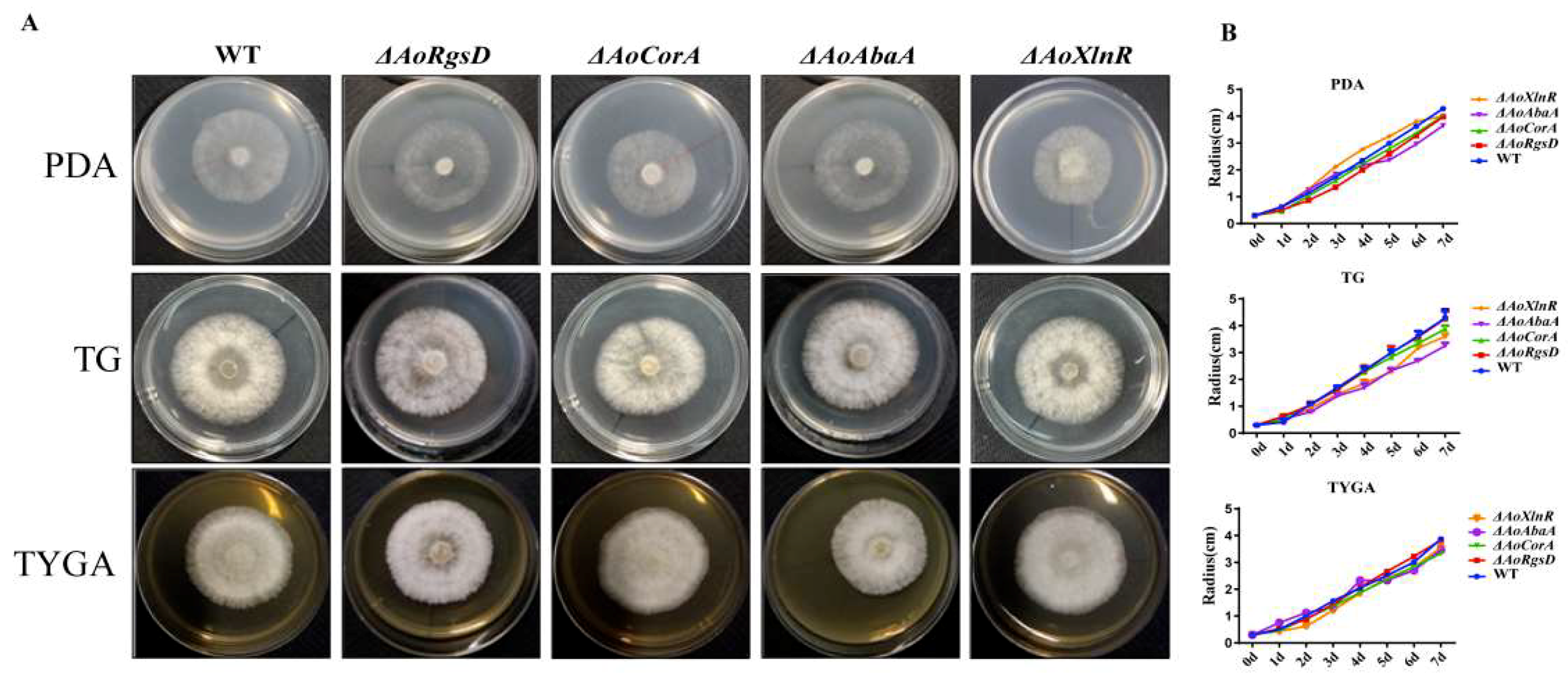
The hyphal growth rates of the WT and ΔAoAbaA, ΔAoXlnR, ΔAoCorA, and ΔAoRgsD mutant strains were compared on potato dextrose agar (PDA), tryptone-glucose (TG), and tryptone yeast-extract glucose agar (TYGA) media, respectively. However, the radial growth and the morphology showed no apparent differences between the wild-type strain and the mutants on different media (Figure 2A,B). In addition, the growth rate of the WT and mutants were compared on TG medium with different stress factors. No obvious differences were observed between the growth of the mutants and the wild-type strain on media with SDS, H2O2, and NaCl at different concentrations (Figure S1). This indicated that the wild-type and mutant strains were similar in their resistance to different stresses.

2.4. AoAbaA Is Also Involved in Trap Formation in A. oligospora

For nematode-trapping fungi, the capacity to produce traps is crucial for them to capture nematodes. Thus, we also compared the trap numbers among the mutant strains by adding nematodes. The WT and mutant strains were incubated on WA medium at 28 °C for 3 days. Then, approximately 200 nematodes were added to induce the formation of adhesive networks. Our results showed that all the mutant strains produced traps with the addition of nematodes. However, the number of traps in the ΔAoAbaA mutant was significantly reduced after adding the nematodes for 48 h, while no obvious difference was observed from the other three mutants over the same period (Figure S2A,B).
Additionally, we counted the number of nematodes that were captured and killed by these strains. Our results showed that after 36 h, 63.9%, 57.0%, 48.7%, 39.3%, and 76.8% of the nematodes had survived in the WT, ΔAoXlnR, ΔAoCorA, ΔAoRgsD, and ΔAoAbaA strains, respectively (Figure S2C).

2.5. AoAbaA, AoXlnR, AoCorA, and AoRgsD Were Required for Conidiation

The WT and each mutant strain were incubated on CMY medium at 28 °C for 14 days to allow conidiation. Our analyses identified that the deletion of AoCorA and AoRgsD resulted in a significantly enhanced number of conidiophores. In the WT strain from the 14-day-old cultures, the averaged conidial yields were quantified at 4.6 × 10⁵ conidia cm−2, whereas the conidial yields of ΔAoCorA and ΔAoRgsD were quantified at 8.1 × 10⁵ and 8.59 × 10⁵ conidia cm−2, respectively (Figure 3A,B). However, conidiation was completely abolished with the disruption of the AoAbaA gene, and significantly reduced in the absence of the AoXlnR gene. The average conidial yield from ΔAoXlnR was 2.15 × 10⁵, indicating a 53% decrease in conidiation capacity in the absence of AoXlnR (Figure 3A,B).
Although the conidiation capacities of the WT and the mutants were different, their conidial morphology showed no obvious differences (Figure 3C). Furthermore, the conidial germination rates of the mutants showed no significant difference with the WT strain on WA media (Figure 3D,E).

2.6. Relationship between the Expressions of AoAbaA, AoXlnR, AoCorA, and AoRgsD and Conidiation

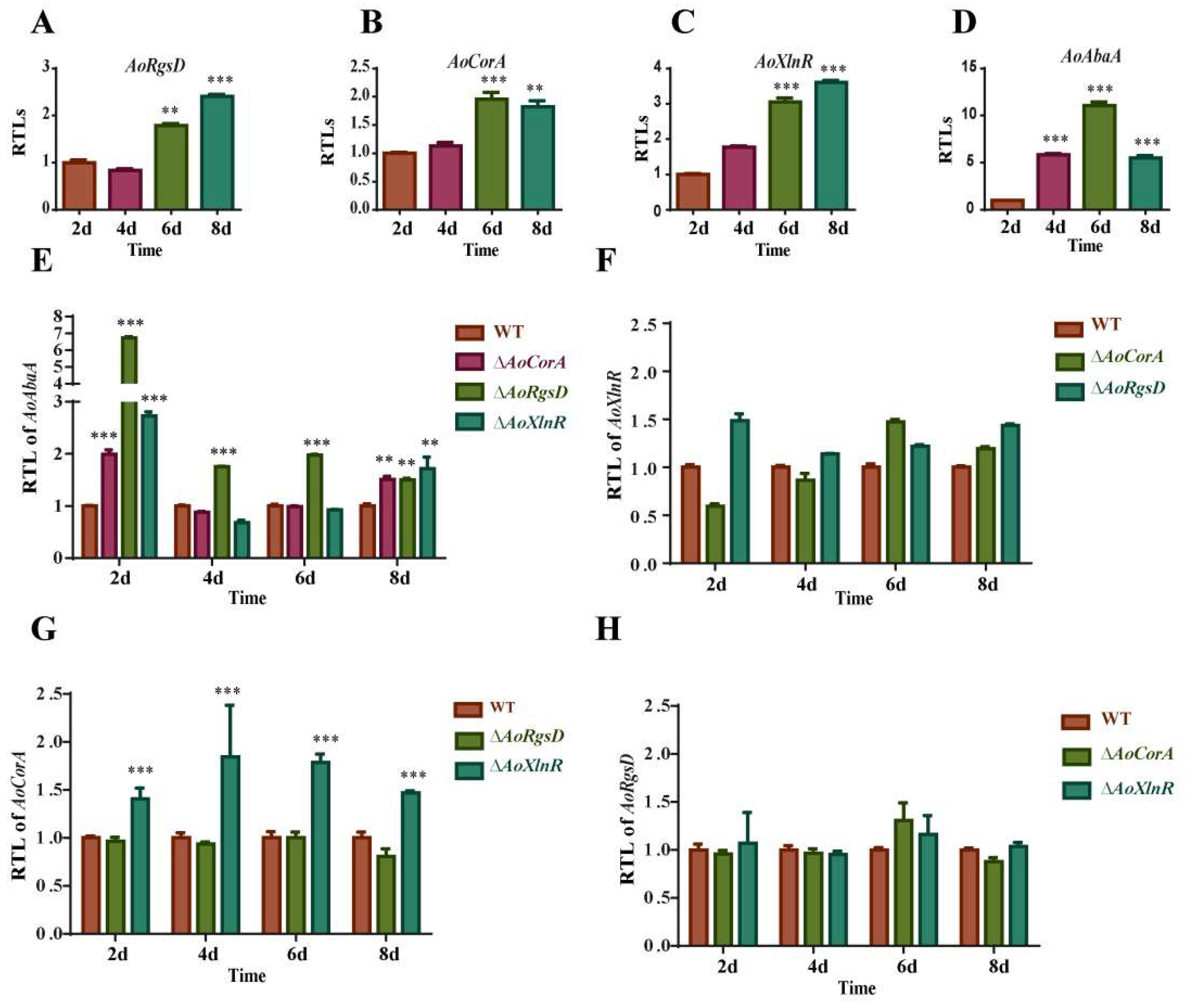
To determine whether AoXlnR, AoCorA, and AoRgsD play a crucial role during the conidiation of A. oligospora, we investigated the expression levels of these four genes at 2, 4, 6, and 8 days. Our results showed that the expression of all four genes showed a significant increase at 6 days (Figure 4A). The expression of AoAbaA was down-regulated at 8 days (Figure 4D), but AoXlnR, AoCorA, and AoRgsD retained a high expression at 8 days (Figure 4A–C).
To understand the regulatory relationships among these four genes, we also compared the transcript levels of these four genes in the other three mutants. Results showed that the transcript level of AoAbaA was enhanced in ΔAoXlnR, ΔAoCorA, and ΔAoRgsD at 2 and 8 days, but showed no obvious changes in ΔAoXlnR and ΔAoCorA at 4 and 6 days though the ΔAoRgsD strain’s expression was still enhanced (Figure 4E). For AoXlnR, no obvious transcript change was observed, suggesting that AoRgsD and AoCorA don’t regulate the expression of AoXlnR (Figure 4F). For AoCorA, the transcript level was enhanced in ΔAoXlnR but showed no obvious change in ΔAoRgsD, suggesting that AoCorA may be directly or indirectly regulated by AoXlnR (Figure 4G). For AoRgsD, its expression level showed no change in ΔAoXlnR and ΔAoCorA strains (Figure 4H). In conclusion, we speculated that AoAbaA was negatively regulated by AoRgsD and it may also be regulated by AoXlnR and AoCorA at the initial and late stages of conidiation. Moreover, AoCorA may be directly or indirectly regulated by AoXlnR.
Furthermore, the expression levels of these genes related to conidiation in A. oligospora were compared between the WT and mutant strains (Figure S3). In ΔAoRgsD, the expression of vosA was up-regulated at all the tested times. However, the transcript levels of lreB, fluG and veA were up-regulated at 2 days, but showed no obvious change at the other three tested times. The expression of pkA was up-regulated at the last stage of conidiation (6 and 8 days). In ΔAoXlnR, flbA, vosA, lreA, lreB, pkA, pkaR, and pkaC1 were up-regulated at all the tested times, while veA, fluG, plp1, and velB were up-regulated during the initial days but showed no changes at the later stage. In ΔAoCorA, the expressions of vosA, pkaR, and pkaC1 were up-regulated, and fluG was up-regulated at 2 days but exhibited no change at the other three tested time points. At 8 days, the expressions of veA, velB, lreA, and nsdD were up-regulated. In addition, flbA, plp1, nsdD, pKA, and p38 MAPK were down-regulated in ΔAoCorA at 2 days.

3. Discussion

A. oligospora is a prominent predatory fungus that can capture nematodes with its specific trap devices. Recently, scientists have mainly focused on studying the molecular mechanisms of trap formation. However, for a filamentous fungus, asexual development is also important for this fungus to survive in various environments. Although previous studies have confirmed that several versatile genes such as the autophagy-related genes (Aoatg1, Aoatg4, Aoatg5, and Aoatg13) [16,17,18], transcription factor-encoding genes (AoStuA and APSES) [19], genes involved in the protein kinase signaling pathway (Slt2, Fus3, and Bck1, and Ime2) [20,21], and GTPases [22,23] may influence the capacity for conidiogenesis, these genes can also regulate multiple biological processes including the growth of trap formation in A. oligospora. In this study, we functionally characterized three genes (AoCorA, AoRgsD, and AoXlnR) that may specifically regulate conidiogenesis in A. oligospora, as the disruption of these three genes neither influences the growth nor the capacity for resistance to different stress and trap formations. However, ΔAoCorA and ΔAoRgsD deletion mutants showed significant increases in conidia production, while the absence of AoXlnR resulted in a decrease in conidiogenesis (Figure 3). Thus, we believe that these three genes may perform specific functions during conidiation in A. oligospora.
The RGS domain is an essential part of the ‘regulator of G-protein signaling (RGS)’ protein family, members of which play critical regulatory roles as GTPase-activating proteins (GAPs) of the heterotrimeric G-protein G-alpha-subunits [24]. In Gram-positive bacteria, the RgsD protein shows a similar structure to the ancillary pilin RrgC from Streptococcus pneumoniae, which is a surface-associated protein that likely participates in covalent adhesion [7]. A previous study implied that RgsD might play a role in the positioning of PG synthesis factors or the regulation of their activity [7]. In filamentous fungi, RGSs regulate diverse signals that control vegetative growth, sporulation, stress responses, secondary metabolism, and virulence [25]. In the human pathogenic fungus Aspergillus fumigatus, RgsD is one of the six regulators of G-protein signaling (RGS) proteins (FlbA, GprK, RgsA, Rax1, RgsC, and RgsD), which have been identified to negatively control development, toxigenesis, stress response, and virulence [11]. A previous study on A. fumigatus revealed that RgsD negatively down-regulates cAMP-PKA signaling pathway to control asexual development, as conidia numbers were drastically increased in the absence of RgsD [11]. In this study, the expression of PKA was up-regulated at the late stage of conidiation (6 and 8 days) in A. oligospora and ΔAoRgsD produced a higher number of conidiophores than WT, which is consistent with the study in A. fumigatus (Figure 3 and Figure 4E). Thus, we believe that RgsD has a conserved function in conidiation in filamentous fungi. Moreover, the mRNA levels of AoAbaA and vosA were significantly increased at all time points tested in ΔAoRgsD (Figure 4E and Figure S2), suggesting that AoAbaA and vosA may also be negatively regulated by AoRgsD during conidiation.
XlnR, a binuclear zinc-finger transcription factor belonging to the GAL4 superfamily, is responsible for the transcription of xylanolytic genes (xlnB, xlnC, and xlnD) [26], endoglucanase (eglA and eglB), and cellobiohydrolase genes, and some galactosidase-encoding genes. This suggests a general role for XlnR in cellulose and hemicellulose degradation and metabolism [13,14,15]. Previous findings have shown that the deletion of the C-terminus of XlnR results in an increase in xylanase activity [27]. In F. oxysporum, XlnR was not an essential virulence determinant [28]. In this study, we found that AoXlnR contains a GAL4-like zinc binuclear cluster and a fungal_TF_MHR domain, which has been suggested to assist the C6 zinc cluster in DNA target discrimination [29]. The conidial yield of A. oligospora was significantly affected by the deletion of AoXlnR (Figure 3). However, RT-PCR analysis showed that the expression of AoXlnR was up-regulated during conidiogenesis (Figure 4C), and at least six genes (flbA, vosA, lreA, lreB, pkA, pkaR) involved in conidiogenesis were up-regulated in ΔAoXlnR (Figure S3), suggesting that AoXlnR may negatively regulate these genes related to conidiation. This is the first confirmation that XlnR conducts the function of conidial regulation.
CorA is a membrane protein whose primary function is to transport magnesium, nickel, and cobalt to maintain the homeostasis of this divalent cation in cells [12]. Previous studies have shown that the MIT_CorA-like domain is an essential membrane protein involved in transporting divalent cations (uptake or efflux) across membranes [12]. In Pectobacterium carotovorum, a phytopathogenic bacterium that causes soft rot disease in plants, CorA affects virulence and extracellular enzyme production [30]. In addition, mutation of the corA gene in Salmonella enterica serovar Typhimurium leads to an attenuation of virulence and other defects [31]. In Magnaporthe oryzae, the CorA transporter is required for mycelial growth and surface hydrophobicity [32]. Intriguingly, the deletion of AoCorA in A. oligospora results in an increase in conidial yield (Figure 3), suggesting that magnesium/nickel/cobalt homeostasis is essential for conidiogenesis. RT-PCR analysis showed that the disruption of AoXlnR can lead to an up-regulation of AoCorA (Figure 4G), suggesting the negative regulation of AoCorA by AoXlnR—though their regulatory mechanism is still unclear. In addition, the transcript levels of several genes related to conidiation were also influenced in ΔAoCorA (Figure S3). Our research confirmed that the AoCorA might also be involved in the regulation of conidiation.
In several model fungi, the three central regulators BrlA, AbaA, and WetA have been identified as playing crucial roles in asexual development [7]. Interestingly, only AoAbaA, the ortholog of AbaA, was identified in A. oligospora, suggesting that the genetic program of conidiation in A. oligospora may be different from other filamentous fungi. In our study, the disruption of AoAbaA resulted in the abolishment of conidiogenesis and a reduction in trap formation (Figure 3). Furthermore, the survival rate of Caenorhabditis elegans in ΔAoAbaA was higher than that of the wild-type strain, suggesting that there may be a common pathway where AoAbaA is involved in both conidiation and trap formation. Although the yield of traps showed no obvious differences among the WT and ΔAoXlnR, ΔAoCorA, ΔAoRgsD, and ΔAoAbaA strains, the survival rates of C. elegans in ΔAoCorA and ΔAoRgsD were significantly lower than in the WT strain (Figure S2), implying that the conidial yields might influence the ability to capture nematodes.
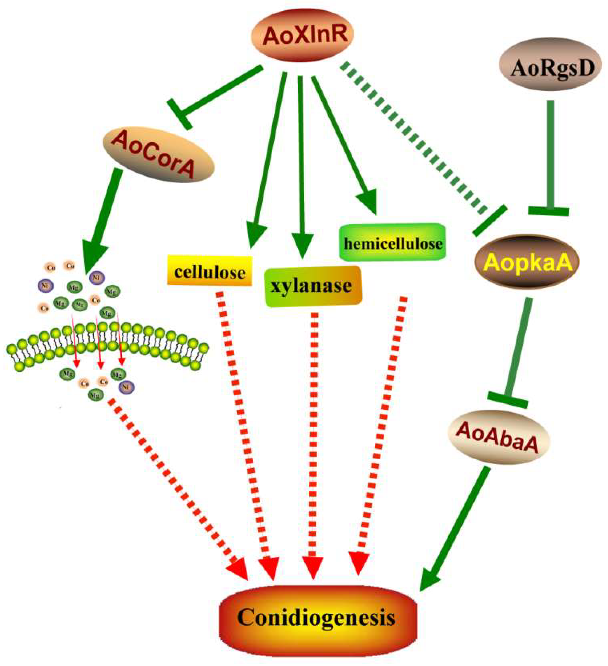
In conclusion, the regulation mechanism for sporulation has both similar and unique features among fungal species. Here, we characterized three novel genes that were involved in conidiation in the model nematode-trapping fungus A. oligospora. AoRgsD regulates conidiation through the PKA/VosA/abaA pathway (Figure 5). AoXlnR may directly or indirectly affect conidiation via AoCorA and/or the metabolisms of cellulose, xylanase, and hemicellulose. AoXlnR may also negatively regulate the PKA signaling pathway, as the expression of AopkaA was enhanced in ΔAoXlnR (Figure 5). In addition, the magnesium, nickel, and cobalt transported by AoCorA may also play a key role during conidiation (Figure 5). Together, our study provides novel insights into the regulation of conidiation in nematode-trapping fungus A. oligospora.

4. Materials and Methods

4.1. Fungal Strains and Culture Conditions

The wild-type strain of A. oligospora (ATCC24927) was cultured on potato dextrose agar (PDA) at 28 °C. S. cerevisiae (FY834) was grown in YPD (yeast extract peptone dextrose) plates. The Escherichia coli strain DH5a (Takara, Shiga, Japan) was used to keep the plasmid pRS426 [33]. The medium PDASS (PDA supplemented with 10 g/L molasses and 0.4 M saccharose) with the addition of 200 µg/mL hygromycin B (Amresco, Solon, OH, USA) was used for selecting transformants. C. elegans nematodes were used in this study, which were grown on oatmeal agar medium at 26 °C and separated using the Baerman funnel technique [34].

4.2. Identification of AoAbaA, AoXlnR, AoCorA, and AoRgsD in A. oligospora

The encoding genes of AoAbaA, AoCorA, AoXlnR, and AoRgsD (GenBank no. XM_011124438, XM_011126761, XM_011119393, and XM_011129373, respectively) were identified from the A. oligospora genome. The conserved functional domain structures of these four proteins were predicted using the InterProScan 5.0 (http://www.ebi.ac.uk/interpro/, accessed on 8 April 2022) with default parameter settings.

4.3. Deletion of the Genes AoAbaA, AoXlnR, AoCorA, and AoRgsD

Genomic DNA of the fungus A. oligospora was extracted using the CTAB (cetyl trimethylammonium bromide) procedure. The encoding genes of AoAbaA, AoCorA, AoXlnR, and AoRgsD were knocked out following the method previously described (Figure 1B) [35]. Briefly, the 5′- and 3′-flanking fragments of each encoding gene were amplified from the genome of A. oligospora using corresponding primer pairs (Table S1). The hygromycin-resistance gene cassette (hph) was amplified from the plasmid pCSN44. The plasmid pRS426 was linearized with EcoRI and XhoI. The linearized pRS426 and the three purified fragments were then co-transformed into S. cerevisiae using electroporation. The recombinant plasmids were individually transformed into A. oligospora protoplasts. The transformants were identified using the primers to confirm whether the encoding genes had been disrupted correctly. Furthermore, Southern analysis was carried out in accordance with our previous reports (Figure 1C) [36]. The primers used to prepare Southern hybridization probes and the restriction enzymes used for different genes are listed in Table S1.

4.4. Comparison in Growth Rate between Mutants and Wild-Type Strain in Different Conditions

The WT and ΔAoAbaA, ΔAoCorA, ΔAoXlnR, and ΔAoRgsD mutants with similar-sized colonies were incubated on PDA, TYGA (10 g/L tryptone, 5 g/L yeast extract, 10 g/L glucose, 5 g/L molasses, 20 g/L agar), CMA (20 g/L maizena, 20 g/L agar), and TGG (10 g/L tryptone,10 g/L glucose, 20 g/L agar) medium at 28 °C for 5 days, respectively. The growth rate and colony morphology were observed and measured daily until mycelia covered the whole surface of the 6 cm diameter disks.
To compare their stress response capacity, mycelial plugs of each strain were inoculated onto TG medium with NaCl (0.1–0.3 M), H2O2 (5–15 mM), and SDS (sodium dodecyl sulfate) (0.02–0.03%) at 28 °C for 5 days, respectively. The diameters of the colonies were measured every day.

4.5. Comparison of Conidial Production, Morphology, and Germination

The fungal plugs (7 mm in diameter) of the WT and mutant strains were cultivated on CMY culture medium at 28 °C for 10 days. The conidia on each plate were washed with 5 mL of sterile water, then mycelial debris was removed using four layers of lens tissues to filtrate. The number of conidia per square millimeter of each colony was determined by hemocytometer [37]. Meanwhile, conidiophores were observed under a light microscope (BX51, Olympus, Tokyo, Japan). To calculate the conidial germination rates, 50-μL aliquots of 105 conidia mL−1 suspensions of the WT strain and mutants were used to inoculate CMY medium (20 g/L maizena, 5 g/L yeast extract, 20 g/L agar). Samples were incubated at 28 °C, and the germinated conidia were counted at 4-h intervals until 24 h post inoculation.

4.6. Trap Formation and Pathogenicity Assays

To characterize the capacity for trap formation in the A. oligospora WT strain and mutants, approximately 104 conidia of the wild-type strain and the ΔAoCorA, ΔAoXlnR and ΔAoRgsD mutants were spread on WA plates and maintained at 28 °C for 5 days. Meanwhile, ΔAoAbaA was incubated on WA medium using colony plugs of 5 mm diameter, as this strain cannot produce conidia. Then, 200–300 nematodes were added to each plate to induce trap formation. The traps were counted under a light microscope (BX51, Olympus, Tokyo, Japan) at specified time points.

4.7. Quantitative Real-Time PCR (RT-PCR) Analysis

To determine the transcript levels of genes involved in sporulation, the WT strain and mutants were incubated on CMY medium at 28 °C for 2, 4, 6, and 8 days, and hyphae were collected for RT-PCR analysis. The total RNA of all samples was extracted using the RNeasy kit (QIAGEN Inc., Hilden, Germany) according to the manufacturer’s instructions. First-strand cDNA was synthesized using the Superscript III First-Strand cDNA Synthesis SuperMix kit (Invitrogen, Carlsbad, CA, USA). The constitutively expressed housekeeping gene β-tubulin (Tub, AOL_s00076g640) was used as the internal standard for the normalization of expression of the other genes studied. Three cDNA samples were analyzed under the action of a SYBR® Premix Ex Taq™ (TaKaRa). Each sample was run in triplicate and two biological replicates were employed. The 2−ΔΔCt method was used to measure the relative transcript levels of each gene [38]. The primers used were designed using Primer 5 and are listed in Table S2.

4.8. Statistical Analysis

Each experiment was performed with three biological replicates and the data from individual treatments are expressed as mean ± SD. GraphPad Prism software, version 6 (GraphPad Software Inc., San Diego, CA, USA) was used for the photographs and statistical analyses (one-way ANOVA). p < 0.05 was considered to indicate significant differences.

Supplementary Materials

The following supporting information can be downloaded at: https://www.mdpi.com/article/10.3390/pathogens11070717/s1, Figure S1: Comparison of growth rate of WT and mutants on TG media with different stress conditions (0.1, 0.2, and 0.3 M NaCl, 5, 10, 15 mM H2O2, and 0.01%, 0.02%, 0.03% SDS). Figure S2: Comparison of trap formation between the WT and mutant strains. (A) Trap formation in the WT and mutant strains induced by nematodes at 48 h (bar: 100 µm); (B) the statistical results of the trap numbers in the WT and mutant strains induced by nematodes at 48 h; (C) comparison of the nematocidal activity. Figure S3: Expression of conidiogenesis-related genes in the wild-type strain and mutants during sporulation. (A) The relative transcriptional levels (RTLs) of genes involved in conidiogenesis in the WT and ΔAoRgsD strains; (B) the relative transcriptional levels (RTLs) of genes involved in conidiogenesis in the WT and ΔAoXlnR strains; (C) the relative transcriptional levels (RTLs) of genes involved in conidiogenesis in the WT and ΔAoCorA strains. *** p < 0.001, ** p < 0.05. Table S1: List of PCR primers used for homologous recombination in this study. Table S2: List of PCR primers used for RT-PCR in this study.

Author Contributions

Conceptualization, X.L. and J.L.; methodology, Q.M.; software, Q.M. and Z.Z.; validation, X.L., Q.M. and Z.Z.; formal analysis, X.L., Q.M., Z.Z. and S.L.; investigation, Q.M.; writing—original draft preparation, J.L.; writing—review and editing, J.L.; visualization, Q.M and Z.Z.; supervision, J.L.; project administration, J.L.; funding acquisition, J.L. All authors have read and agreed to the published version of the manuscript.

Funding

This research was funded by the National Natural Science Foundation of China (approved nos. 32170198 and 31970073).

Institutional Review Board Statement

Not applicable.

Informed Consent Statement

Not applicable.

Data Availability Statement

Not applicable.

Acknowledgments

We are grateful to Jianping Xu of the Department of Biology, McMaster University, for their valuable comments and critical discussions. We also thank the anonymous reviewers for their valuable suggestions.

Conflicts of Interest

The authors declare no conflict of interest.

References

  1. Abad, P.; Gouzy, J.; Aury, J.-M.; Castagnone-Sereno, P.; Danchin, E.G.J.; Deleury, E.; Perfus-Barbeoch, L.; Anthouard, V.; Artiguenave, F.; Blok, V.C.; et al. Genome sequence of the metazoan plant-parasitic nematode Meloidogyne incognita. Nat. Biotechnol. 2008, 26, 909–915. [Google Scholar] [CrossRef] [PubMed] [Green Version]
  2. Zhang, K.-Q.; Hyde, K.D. Nematode-Trapping Fungi; Springer Science & Business: New York, NY, USA, 2014; Volume 23. [Google Scholar]
  3. Yang, J.; Wang, L.; Ji, X.; Feng, Y.; Li, X.; Zou, C.; Xu, J.; Ren, Y.; Mi, Q.; Wu, J.; et al. Genomic and proteomic analyses of the fungus Arthrobotrys oligospora provide insights into nematode-trap formation. PLoS Pathog. 2011, 7, e1002179. [Google Scholar] [CrossRef] [PubMed] [Green Version]
  4. Yang, L.; Li, X.; Bai, N.; Yang, X.; Zhang, K.Q.; Yang, J. Transcriptomic analysis reveals that Rho GTPases regulate trap development and lifestyle transition of the nematode-trapping fungus Arthrobotrys oligospora. Microbiol. Spectr. 2022, 10, e0175921. [Google Scholar] [CrossRef] [PubMed]
  5. Zhu, M.C.; Li, X.M.; Zhao, N.; Yang, L.; Zhang, K.Q.; Yang, J.K. Regulatory mechanism of trap formation in the nematode-trapping fungi. J. Fungi 2022, 8, 406. [Google Scholar] [CrossRef]
  6. Etxebeste, O.; Garzia, A.; Espeso, E.A.; Ugalde, U. Aspergillus nidulans asexual development: Making the most of cellular modules. Trends Microbiol. 2010, 18, 569–576. [Google Scholar] [CrossRef] [Green Version]
  7. Park, H.S.; Yu, J.H. Genetic control of asexual sporulation in filamentous fungi. Curr. Opin. Microbiol. 2012, 15, 669–677. [Google Scholar] [CrossRef]
  8. Boylan, M.T.; Mirabito, P.M.; Willett, C.E.; Zimmerman, C.R.; Timberlake, W.E. Isolation and physical characterization of three essential conidiation genes from Aspergillus nidulans. Mol. Cell. Biol. 1987, 7, 3113–3118. [Google Scholar] [CrossRef]
  9. Sewall, T.C.; Mims, C.W.; Timberlake, W.E. Conidium differentiation in Aspergillus nidulans wild-type and wet-white (wetA) mutant strains. Dev. Biol. 1990, 138, 499–508. [Google Scholar] [CrossRef]
  10. Marshall, M.A.; Timberlake, W.E. Aspergillus nidulans wetA activates spore-specific gene expression. Mol. Cell. Biol. 1991, 11, 55–62. [Google Scholar] [CrossRef]
  11. Kim, Y.; Lee, M.W.; Jun, S.C.; Choi, Y.H.; Yu, J.H.; Shin, K.S. RgsD negatively controls development, toxigenesis, stress response, and virulence in Aspergillus fumigatus. Sci. Rep. 2019, 9, 811. [Google Scholar] [CrossRef]
  12. Kirsten, A.; Herzberg, M.; Voigt, A.; Seravalli, J.; Grass, G.; Scherer, J.; Nies, D.H. Contributions of five secondary metal uptake systems to metal homeostasis of Cupriavidus metallidurans CH34. J. Bacteriol. 2011, 193, 4652–4663. [Google Scholar] [CrossRef] [PubMed] [Green Version]
  13. Noguchi, Y.; Sano, M.; Kanamaru, K.; Ko, T.; Takeuchi, M.; Kato, M.; Kobayashi, T. Genes regulated by AoXlnR, the xylanolytic and cellulolytic transcriptional regulator, in Aspergillus oryzae. Appl. Microbiol. Biotechnol. 2009, 85, 141–154. [Google Scholar] [CrossRef] [PubMed]
  14. Marui, J.; Kitamoto, N.; Kato, M.; Kobayashi, T.; Tsukagoshi, N. Transcriptional activator, AoXlnR, mediates cellulose-inductive expression of the xylanolytic and cellulolytic genes in Aspergillus oryzae. FEBS Lett. 2002, 528, 279–282. [Google Scholar] [CrossRef] [Green Version]
  15. Hasper, A.A.; Visser, J.; de Graaff, L.H. The Aspergillus niger transcriptional activator XlnR, which is involved in the degradation of the polysaccharides xylan and cellulose, also regulates D-xylose reductase gene expression. Mol. Microbiol. 2000, 36, 193–200. [Google Scholar] [CrossRef]
  16. Zhou, D.; Zhu, Y.; Bai, N.; Yang, L.; Xie, M.; Yang, J.; Zhu, M.; Zhang, K.Q.; Yang, J. AoATG5 plays pleiotropic roles in vegetative growth, cell nucleus development, conidiation, and virulence in the nematode-trapping fungus Arthrobotrys oligospora. Sci. China Life Sci. 2022, 65, 412–425. [Google Scholar] [CrossRef]
  17. Zhou, D.; Zhu, Y.; Bai, N.; Xie, M.; Zhang, K.Q.; Yang, J. Aolatg1 and Aolatg13 Regulate Autophagy and Play Different Roles in Conidiation, Trap Formation, and Pathogenicity in the Nematode-Trapping Fungus Arthrobotrys oligospora. Front. Cell. Infect. Microbiol. 2021, 11, 824407. [Google Scholar] [CrossRef]
  18. Zhou, D.; Xie, M.; Bai, N.; Yang, L.; Zhang, K.Q.; Yang, J. The autophagy-related gene Aolatg4 regulates hyphal growth, sporulation, autophagosome formation, and pathogenicity in Arthrobotrys oligospora. Front. Microbiol. 2020, 11, 592524. [Google Scholar] [CrossRef]
  19. Xie, M.; Wang, Y.; Tang, L.; Yang, L.; Zhou, D.; Li, Q.; Niu, X.; Zhang, K.Q.; Yang, J. AoStuA, an APSES transcription factor, regulates the conidiation, trap formation, stress resistance and pathogenicity of the nematode-trapping fungus Arthrobotrys oligospora. Environ. Microbiol. 2019, 21, 4648–4661. [Google Scholar] [CrossRef]
  20. Zhen, Z.; Xing, X.; Xie, M.; Yang, L.; Yang, X.; Zheng, Y.; Chen, Y.; Ma, N.; Li, Q.; Zhang, K.Q.; et al. MAP kinase Slt2 orthologs play similar roles in conidiation, trap formation, and pathogenicity in two nematode-trapping fungi. Fungal Genet. Biol. 2018, 116, 42–50. [Google Scholar] [CrossRef]
  21. Xie, M.; Bai, N.; Yang, J.; Jiang, K.; Zhou, D.; Zhao, Y.; Li, D.; Niu, X.; Zhang, K.Q.; Yang, J. Protein kinase Ime2 is required for mycelial growth, conidiation, osmoregulation, and pathogenicity in nematode-trapping fungus Arthrobotrys oligospora. Front. Microbiol. 2019, 10, 3065. [Google Scholar] [CrossRef] [Green Version]
  22. Yang, X.; Ma, N.; Yang, L.; Zheng, Y.; Zhen, Z.; Li, Q.; Xie, M.; Li, J.; Zhang, K.Q.; Yang, J. Two Rab GTPases play different roles in conidiation, trap formation, stress resistance, and virulence in the nematode-trapping fungus Arthrobotrys oligospora. Appl. Microbiol. Biotechnol. 2018, 102, 4601–4613. [Google Scholar] [CrossRef] [PubMed]
  23. Ma, N.; Zhao, Y.; Wang, Y.; Yang, L.; Li, D.; Yang, J.; Jiang, K.; Zhang, K.Q.; Yang, J. Functional analysis of seven regulators of G protein signaling (RGSs) in the nematode-trapping fungus Arthrobotrys oligospora. Virulence 2021, 12, 1825–1840. [Google Scholar] [CrossRef]
  24. Lafon, A.; Han, K.H.; Seo, J.A.; Yu, J.H.; d’Enfert, C. G-protein and cAMP-mediated signaling in aspergilli: A genomic perspective. Fungal Genet. Biol. 2006, 43, 490–502. [Google Scholar] [CrossRef] [PubMed]
  25. Bayram, O.; Braus, G.H. Coordination of secondary metabolism and development in fungi: The velvet family of regulatory proteins. FEMS Microbiol. Rev. 2012, 36, 1–24. [Google Scholar] [CrossRef] [PubMed] [Green Version]
  26. van Peij, N.N.; Gielkens, M.M.; de Vries, R.P.; Visser, J.; de Graaff, L.H. The transcriptional activator XlnR regulates both xylanolytic and endoglucanase gene expression in Aspergillus niger. Appl. Environ. Microbiol. 1998, 64, 3615–3619. [Google Scholar] [CrossRef] [Green Version]
  27. Marui, J.; Tanaka, A.; Mimura, S.; de Graaff, L.H.; Visser, J.; Kitamoto, N.; Kato, M.; Kobayashi, T.; Tsukagoshi, N. A transcriptional activator, AoXlnR, controls the expression of genes encoding xylanolytic enzymes in Aspergillus oryzae. Fungal Genet. Biol. 2002, 35, 157–169. [Google Scholar] [CrossRef]
  28. Calero-Nieto, F.; Di Pietro, A.; Roncero, M.I.; Hera, C. Role of the transcriptional activator xlnR of Fusarium oxysporum in regulation of xylanase genes and virulence. Mol. Plant Microbe Interact. 2007, 20, 977–985. [Google Scholar] [CrossRef] [Green Version]
  29. Schjerling, P.; Holmberg, S. Comparative amino acid sequence analysis of the C6 zinc cluster family of transcriptional regulators. Nucleic Acids Res. 1996, 24, 4599–4607. [Google Scholar] [CrossRef] [Green Version]
  30. Kersey, C.M.; Agyemang, P.A.; Dumenyo, C.K. CorA, the magnesium/nickel/cobalt transporter, affects virulence and extracellular enzyme production in the soft rot pathogen Pectobacterium carotovorum. Mol. Plant Pathol. 2012, 13, 58–71. [Google Scholar] [CrossRef]
  31. Sermon, J.; Wevers, E.M.; Jansen, L.; De Spiegeleer, P.; Vanoirbeek, K.; Aertsen, A.; Michiels, C.W. CorA affects tolerance of Escherichia coli and Salmonella enterica serovar Typhimurium to the lactoperoxidase enzyme system but not to other forms of oxidative stress. Appl. Environ. Microbiol. 2005, 71, 6515–6523. [Google Scholar] [CrossRef] [Green Version]
  32. Reza, M.H.; Shah, H.; Manjrekar, J.; Chattoo, B.B. Magnesium uptake by CorA transporters is essential for growth, development and infection in the rice blast fungus Magnaporthe oryzae. PLoS ONE 2016, 11, e0159244. [Google Scholar] [CrossRef] [PubMed] [Green Version]
  33. Christianson, T.W.; Sikorski, R.S.; Dante, M.; Shero, J.H.; Hieter, P. Multifunctional yeast high-copy-number shuttle vectors. Gene 1992, 110, 119–122. [Google Scholar] [CrossRef]
  34. Gray, N. Ecology of nematophagous fungi: Comparison of the soil sprinkling method with the Baermann funnel technique in the isolation of endoparasites. Soil Biol. Biochem. 1984, 16, 81–83. [Google Scholar] [CrossRef]
  35. Colot, H.V.; Park, G.; Turner, G.E.; Ringelberg, C.; Crew, C.M.; Litvinkova, L.; Weiss, R.L.; Borkovich, K.A.; Dunlap, J.C. A high-throughput gene knockout procedure for Neurospora reveals functions for multiple transcription factors. Proc. Natl. Acad. Sci. USA 2006, 103, 10352. [Google Scholar] [CrossRef] [Green Version]
  36. Li, J.; Wu, R.; Wang, M.; Borneman, J.; Yang, J.; Zhang, K.Q. The pH sensing receptor AopalH plays important roles in the nematophagous fungus Arthrobotrys oligospora. Fungal Biol. 2019, 123, 547–554. [Google Scholar] [CrossRef]
  37. Xie, X.Q.; Li, F.; Ying, S.H.; Feng, M.G. Additive contributions of two manganese-cored superoxide dismutases (MnSODs) to antioxidation, UV tolerance and virulence of Beauveria bassiana. PLoS ONE 2012, 7, e30298. [Google Scholar] [CrossRef]
  38. Livak, K.J.; Schmittgen, T.D. Analysis of relative gene expression data using real-time quantitative PCR and the 2(-Delta Delta C(T)) Method. Methods 2001, 25, 402–408. [Google Scholar] [CrossRef]
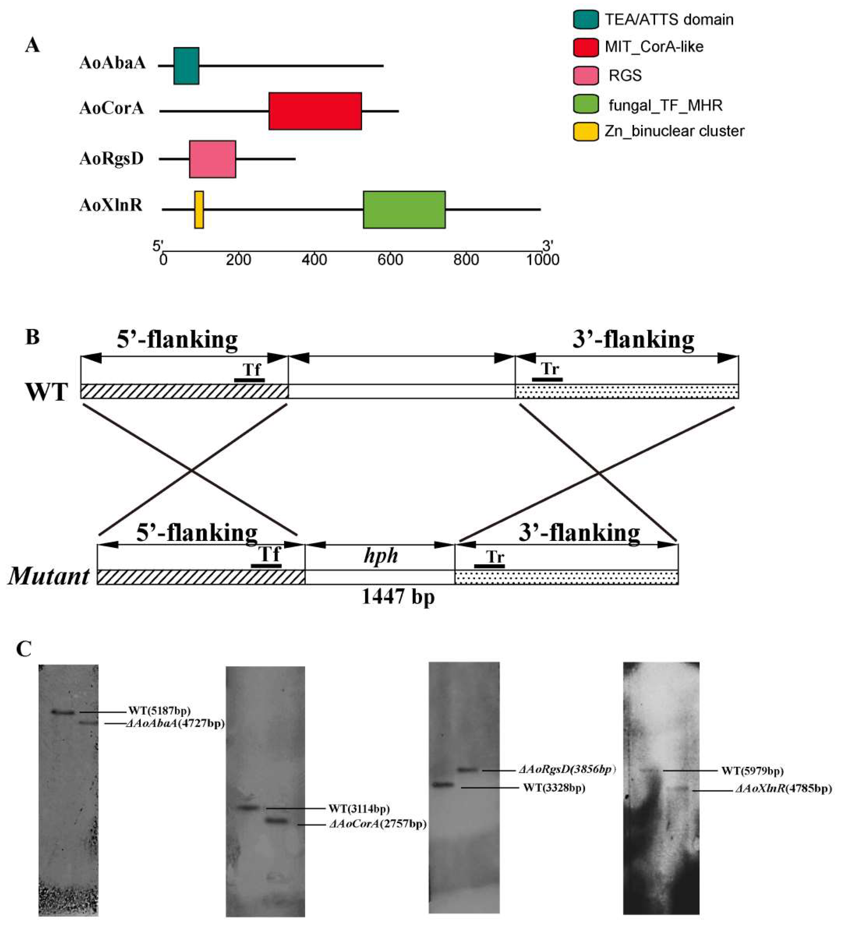
Figure 1. Protein domain structure and deletion of targeting genes in A. oligospora. (A) Protein domain structures of AoAbaA, AoXlnR, AoCorA, and AoRgsD, respectively; (B) the sketch map of replacement of targeting genes using the homologous recombination method; (C) Southern blot analysis of the wild-type (WT) and mutant strains.
Pathogens 11 00717 g001
Figure 2. Comparison of the growth rate and colony morphology of the wild-type strain and mutants. (A) The colony morphology of A. oligospora wild-type strain and mutants on PDA, TG, and TYGA media for 3 days at 28 °C; (B) comparison of the growth rate of the WT and mutants on PDA, TG, and TYGA media.
Pathogens 11 00717 g002
Figure 3. Comparison of the sporulation, spore germination, and conidia morphology of the wild-type strain and mutants. (A) Conidiophore differentiation in the WT and mutant lines. Bar = 100 um; (B) sporulation of the wild-type strain and mutants. ** p < 0.05, *** p < 0.001 versus WT (wild type); (C) comparison of the conidia morphology. Bar = 30 um; (D) comparison of the spore germination rate between the wild-type strain and mutants. Bar = 100 um; (E) statistical results of the germination rate of the wild-type strain and mutant.
Pathogens 11 00717 g003
Figure 4. Expression of genes in the wild-type strain and mutants during sporulation. (A) The relative transcriptional levels (RTLs) of the gene AoRgsD during vegetative growth and conidiation (2, 4, 6, and 8 days) in A. oligospora; (B) the RTLs of the gene AoCorA during vegetative growth and conidiation (2, 4, 6, and 8 days) in A. oligospora; (C) the RTLs of the gene AoXlnR during vegetative growth and conidiation (2, 4, 6, and 8 days) in A. oligospora; (D) the RTLs of the gene AoAbaA during vegetative growth and conidiation (2, 4, 6, and 8 days) in A. oligospora; (E) the RTLs of the gene AoAbaA in the ΔAoXlnR, ΔAoCorA, and ΔAoRgsD strains, respectively; (F) the RTLs of the gene AoXlnR in the ΔAoCorA, and ΔAoRgsD strains, respectively; (G), the RTLs of the gene AoCorA in the ΔAoXlnR and ΔAoRgsD strains, respectively; (H) the RTLs of the gene AoRgsD in the ΔAoXlnR and ΔAoCorA strains, respectively. *** p < 0.001, ** p < 0.05.
Pathogens 11 00717 g004
Figure 5. A proposed model for the regulation of AoAbaA, AoXlnR, AoCorA, and AoRgsD in A. oligospora. Our findings suggest that AoRgsD regulates conidiation through the PKA/VosA/abaA pathway. AoXlnR may directly or indirectly affect conidiation via AoCorA and/or the metabolisms of cellulose, xylanase, and hemicellulose. AoXlnR may negatively regulate the PKA signaling pathway.
Pathogens 11 00717 g005
Publisher’s Note: MDPI stays neutral with regard to jurisdictional claims in published maps and institutional affiliations.