
Seroprevalence of Anti-Theileria equi Antibodies in Horses from Three Geographically Distinct Areas of Romania


 ,
,  ,
,
Abstract
:1. Introduction
2. Results
3. Discussion
4. Materials and Methods
4.1. Blood Samples and Studied Areas
4.2. Competitive Enzyme-Linked Immunosorbent Assay (cELISA)
4.3. Statistical Analysis
5. Conclusions
Author Contributions
Funding
Institutional Review Board Statement
Informed Consent Statement
Conflicts of Interest
References
- Qablan, M.A.; Obroník, M.; Petrželková, K.J.; Sloboda, M.; Shudiefat, M.F.; Hořín, P.; Lukeš, J.; Modrý, D. Infections by Babesia caballi and Theileria equi in Jordanian equids: Epidemiology and genetic diversity. Parasitology 2013, 140, 1096–1103. [Google Scholar] [CrossRef] [PubMed]
- Knowles, D.P.; Kappmeyer, L.S.; Haney, D.; Herndon, D.R.; Fry, L.M.; Munro, J.B.; Sears, K.; Ueti, M.W.; Wise, L.N.; Silva, M.; et al. Discovery of a novel species, Theileria haneyi n. sp. infective to equids, highlights exceptional genomic diversity within the genus Theileria: Implications for apicomplexan parasite surveillance. Int. J. Parasitol. 2018, 48, 679–690. [Google Scholar] [CrossRef] [PubMed]
- Elsawy, B.S.M.; Nassar, A.M.; Alzan, H.F.; Bhoora, R.V.; Ozubek, S.; Mahmoud, M.S.; Kandil, O.M.; Mahdy, O.A. Rapid detection of equine piroplasms using multiplex PCR and first genetic characterization of Theileria haneyi in Egypt. Pathogens 2021, 10, 1414. [Google Scholar] [CrossRef] [PubMed]
- Knowles, D.P., Jr. Control of Babesia equi parasitemia. Parasitol. Today 1996, 12, 195–198. [Google Scholar] [CrossRef]
- Mehlhorn, H.; Schein, E. Redescription of Babesia equi Laveran, 1901 as Theileria equi Mehlhorn, Schein 1998. Parasitol. Res. 1998, 84, 467–475. [Google Scholar] [CrossRef] [PubMed]
- Kumar, S.; Kumar, R.; Sugimoto, C. A perspective on Theileria equi infections in donkeys. Jpn. J. Vet. Res. 2009, 56, 171–180. [Google Scholar] [PubMed]
- Scoles, G.A.; Ueti, M.W. Vector ecology of equine piroplasmosis. Ann. Rev. Entomol. 2015, 60, 561–580. [Google Scholar] [CrossRef]
- Tirosh-Levy, S.; Gottlieb, Y.; Mazuz, M.L.; Savitsky, I.; Steinman, A. Infection dynamics of Theileria equi in carrier horses is associated with management and tick exposure. Ticks Tick-Borne Dis. 2020, 11, 101508. [Google Scholar] [CrossRef]
- Rothschild, C.M. Equine piroplasmosis. J. Equine Vet. Sci. 2013, 23, 115–120. [Google Scholar] [CrossRef]
- Wise, L.N.; Kappmeyer, L.S.; Mealey, R.H.; Knowles, D.P. Review of equine piroplasmosis. J. Vet. Intern. Med. 2013, 27, 1334–1346. [Google Scholar] [CrossRef]
- Tirosh-Levy, S.; Gottlieb, Y.; Arieli, O.; Mazuz, M.L.; King, R.; Horowitz, I.; Steinman, A. Genetic characteristics of Theileria equi in zebras, wild and domestic donkeys in Israel and the Palestinian Authority. Ticks Tick-Borne Dis. 2020, 11, 101286. [Google Scholar] [CrossRef] [PubMed]
- Nadal, C.; Bonnet, S.I.; Marsot, M. Eco-epidemiology of equine piroplasmosis and its associated tick vectors in Europe: A systematic literature review and a meta-analysis of prevalence. Transbound. Emerg. Dis. 2021, 10, tbed.14261. [Google Scholar] [CrossRef] [PubMed]
- Available online: https://www.oie.int/en/what-we-do/animal-health-and-welfare/animal-diseases/old-classification-of-diseases-notifiable-to-the-oie-list-b/ (accessed on 5 September 2021).
- Friedhoff, K.T.; Tenter, A.M.; Müller, I. Haemoparasites of equines: Impact on international trade of horses. Rev. Sci. Tech. 1990, 9, 1187–1194. [Google Scholar] [PubMed]
- Torres, R.; Hurtado, C.; Pérez-Macchi, S.; Bittencourt, P.; Freschi, C.; de Mello, V.V.C.; Machado, R.Z.; André, M.R.; Müller, A. Occurrence and genetic diversity of Babesia caballi and Theileria equi in Chilean thoroughbred racing horses. Pathogens 2021, 10, 714. [Google Scholar] [CrossRef]
- Gallusová, M.; Qablan, M.A.; D’Amico, G.; Oborník, M.; Petrželková, K.J.; Mihalca, A.D.; Modrý, D. Piroplasms in feral and domestic equines in rural areas of the Danube Delta, Romania, with survey of dogs as a possible reservoir. Vet. Parasitol. 2014, 206, 287–292. [Google Scholar] [CrossRef] [PubMed]
- Nardini, R.; Cersini, A.; Del Pino, L.E.B.; Manna, G.; Scarpulla, M.; Di Egidio, A.; Gioradni, R.; Antognetti, V.; Veneziano, V.; Scicluna, M.T. Comparison of direct and indirect methods to maximise the detection of Babesia caballi and Theileria equi infections in Central Southern Italy. Ticks Tick-Borne Dis. 2022, 101939. [Google Scholar] [CrossRef]
- Mihalca, A.D.; Dumitrache, M.O.; Magdaş, C.; Gherman, C.M.; Domşa, C.; Mircean, V.; Ghira, I.V.; Pocora, V.; Ionescu, D.T.; Sikó Barabási, S.; et al. Synopsis of the hard ticks (Acari: Ixodidae) of Romania with update on host associations and geographical distribution. Exp. Appl. Acarol. 2012, 58, 183–206. [Google Scholar] [CrossRef]
- Coipan, E.; Vladimirescu, A.; Ciolpan, O.; Teodorescu, I. Tick Species (Acari: Ixodoidea) Distribution, Seasonality and Host Associations in Romania. Trav. Du Muséum Natl. D’histoire Nat. “Grigore Antipa" 2011, 54, 301–317. [Google Scholar] [CrossRef]
- Grandi, G.; Molinari, G.; Tittarelli, M.; Sassera, D.; Kramer, L.H. Prevalence of Theileria equi and Babesia caballi infection in horses from northern Italy. Vector Borne Zoonotic Dis. 2011, 11, 955–956. [Google Scholar] [CrossRef]
- Ribeiro, A.J.; Cardoso, L.; Maia, J.M.; Coutinho, T.; Cotovio, M. Prevalence of Theileria equi, Babesia caballi, and Anaplasma phagocytophilum in horses from the north of Portugal. Parasitol. Res. 2013, 112, 2611–2617. [Google Scholar] [CrossRef]
- Del Pino, L.E.B.; Nardini, R.; Veneziano, V.; Francesca, I.; Antonella, C.; Luca, A.G.; Francesco, B.; Teresa, M.S. Babesia caballi and Theileria equi infections in horses in Central-Southern Italy: Sero-molecular survey and associated risk factors. Ticks Tick-Borne Dis. 2016, 7, 462–469. [Google Scholar] [CrossRef] [PubMed]
- Camino, E.; Buendia, A.; Dorrego, A.; Pozo, P.; de Juan, L.; Dominguez, L.; Cruz-Lopez, F. Sero-molecular survey and risk factors of equine piroplasmosis in horses in Spain. Equine Vet. J. 2021, 53, 771–779. [Google Scholar] [CrossRef] [PubMed]
- Camino, E.; de la Cruz, M.L.; Dominguez, L.; Carvajal, K.A.; Fores, P.; de Juan, L.; Cruz-Lopez, F. Epidemiological situation of the exposure to agents causing equine piroplasmosis in Spanish purebred horses in Spain: Seroprevalence and associated risk factors. J. Equine Vet. Sci. 2018, 67, 81–86. [Google Scholar] [CrossRef]
- Garcia-Bocanegra, I.; Arenas-Montes, A.; Hernandez, E.; Adaszek, L.; Carbonero, A.; Almeria, S.; Arenas, A. Seroprevalence and risk factors associated with Babesia caballi and Theileria equi infection in equids. Vet. J. 2013, 195, 172–178. [Google Scholar] [CrossRef]
- Guidi, E.; Pradier, S.; Lebert, I.; Leblond, A. Piroplasmosis in an endemic area: Analysis of the risk factors and their implications in the control of Theileriosis and Babesiosis in horses. Parasitol. Res. 2015, 114, 71–83. [Google Scholar] [CrossRef]
- Kouam, M.K.; Kantzoura, V.; Gajadhar, A.A.; Theis, J.H.; Papadopoulos, E.; Theodoropoulos, G. Seroprevalence of equine piroplasms and host-related factors associated with infection in Greece. Vet. Parasitol. 2010, 169, 273–278. [Google Scholar] [CrossRef]
- Montes Cortés, M.G.; Fernández-García, J.L.; Habela Martínez-Estéllez, M.Á. Seroprevalence of Theileria equi and Babesia caballi in horses in Spain. Parasite 2017, 24, 14. [Google Scholar] [CrossRef] [Green Version]
- Papini, R.; Salari, F.; Rocchigiani, G.; Leoni, A.; Ragona, G.; Roncoroni, C.; Mancianti, F. Molecular detection of Theileria equi in donkeys (Equus asinus) in a selected site in central Italy. Large Anim. Rev. 2016, 22, 231–234. [Google Scholar]
- Salib, F.A.; Youssef, R.R.; Rizk, L.G.; Said, S.F. Epidemiology, diagnosis and therapy of Theileria equi infection in Giza, Egypt. Vet. World 2013, 6, 76–82. [Google Scholar] [CrossRef]
- Idoko, I.S.; Edeh, R.E.; Adamu, A.M.; Machunga-Mambula, S.; Okubanjo, O.O.; Balogun, E.O.; Adamu, S.; Johnson, W.; Kappmeyer, L.; Mousel, M.; et al. Molecular and serological detection of piroplasms in horses from Nigeria. Pathogens 2021, 10, 508. [Google Scholar] [CrossRef]
- Farkas, R.; Tánczos, B.; Gyurkovszky, M.; Földvári, G.; Solymosi, N.; Edelhofer, R.; Hornok, S. Serological and molecular detection of Theileria equi infection in horses in Hungary. Vet. Parasitol. 2013, 192, 143–148. [Google Scholar] [CrossRef] [PubMed]
- Piantedosi, D.; D’Alessio, N.; di Loria, A.; di Prisco, F.; Mariani, U.; Neola, B.; Santoro, M.; Montagnaro, S.; Capelli, G.; Veneziano, V. Seroprevalence and risk factors associated with Babesia caballi and Theileria equi infections in donkeys from Southern Italy. Vet. J. 2014, 202, 578–582. [Google Scholar] [CrossRef] [PubMed]
- Kouam, M.K.; Masuoka, P.M.; Kantzoura, V.; Theodoropoulos, G. Geographic distribution modeling and spatial cluster analysis for equine piroplasms in Greece. Infect. Genet. Evol. 2010, 10, 1013–1018. [Google Scholar] [CrossRef] [PubMed]
- Sigg, L.; Gerber, V.; Gottstein, B.; Doherr, M.G.; Frey, C.F. Seroprevalence of Babesia caballi and Theileria equi in the Swiss horse population. Parasitol. Int. 2010, 59, 313–317. [Google Scholar] [CrossRef] [PubMed]
- Villa, L.; Gazzonis, A.L.; Allievi, C.; De Maria, C.; Persichetti, M.F.; Caracappa, G.; Zanzani, S.A.; Manfredi, M.T. Seroprevalence of Tick-Borne Infections in Horses from Northern Italy. Animals 2022, 12, 999. [Google Scholar] [CrossRef] [PubMed]
- Bravo-Barriga, D.; Serrano-Aguilera, F.J.; Barrasa-Rita, R.; Habela, M.A.; Chacón, R.B.; Ezquerra, L.J.; Martín-Cuervo, M. Effects of Competitive ELISA-Positive Results of Piroplasmosis on the Performance of Endurance Horses. Animals 2022, 12, 637. [Google Scholar] [CrossRef]
- Nadal, C.; Marsot, M.; Le Metayer, G.; Boireau, P.; Guillot, J.; Bonnet, S.I. Spatial and Temporal Circulation of Babesia caballi and Theileria equi in France Based on Seven Years of Serological Data. Pathogens 2022, 11, 227. [Google Scholar] [CrossRef]
- Heim, A.; Passos, L.M.; Ribeiro, M.F.; Costa-Júnior, L.M.; Bastos, C.V.; Cabral, D.D.; Hirzmann, J.; Pfister, K. Detection and molecular characterization of Babesia caballi and Theileria equi isolates from endemic areas of Brazil. Parasitol. Res. 2007, 102, 63–68. [Google Scholar] [CrossRef]
- Heuchert, C.M.S.; de Giulli, V., Jr.; de Athaide, D.F.; Böse, R.; Friedhoff, K.T. Seroepidemiologic studies on Babesia equi and Babesia caballi infections in Brazil. Vet. Parasitol. 1999, 85, 1–11. [Google Scholar] [CrossRef]
- Wang, Y.; Zhang, L.P.; Li, J.; Li, D.D.; Zhang, Q.; Li, C. The first report of serological detection of Babesia caballi by cELISA in a horse during serological survey of piroplasmosis in imported horses at Shanghai Port, China. J. Equine Vet. Sci. 2020, 92, 103152. [Google Scholar] [CrossRef]
- Almazán, C.; Scimeca, R.C.; Reichard, M.V.; Mosqueda, J. Babesiosis and Theileriosis in North America. Pathogens 2022, 11, 168. [Google Scholar] [CrossRef] [PubMed]
- Salinas-Estrella, E.; Ueti, M.W.; Lobanov, V.A.; Castillo-Payró, E.; Lizcano-Mata, A.; Badilla, C.; Martínez-Ibáñez, F.; Mosqueda, J. Serological and molecular detection of Babesia caballi and Theileria equi in Mexico: A prospective study. PLoS ONE 2022, 17, e0264998. [Google Scholar] [CrossRef] [PubMed]
- Selim, A.; Khater, H. Seroprevalence and risk factors associated with Equine piroplasmosis in North Egypt. Comp. Immunol. Microbiol. Infect. Dis. 2020, 73, 101549. [Google Scholar] [CrossRef]
- Kamani, J.; Bártová, E.; Kašpárková, N.; Mohammed, J.S.; Budíková, M.; Sedlák, K. Seroprevalence of Theileria equi, Babesia caballi, and Trichinella spp. infections in horses and donkeys from Nigeria, West Africa. Trop. Anim. Health Prod. 2021, 53, 338. [Google Scholar] [CrossRef]
- Onyiche, T.E.; Sivakumar, T.; Tuvshintulga, B.; Nugraha, A.B.; Ahedor, B.; Mofokeng, L.; Luka, J.; Mohammed, A.; Mbaya, A.W.; Biu, A.A.; et al. Serosurvey for equine piroplasms in horses and donkeys from North-Western Nigeria using IFAT and ELISA. J. Immunoass. Immunochem. 2021, 42, 648–668. [Google Scholar] [CrossRef] [PubMed]
- Tirosh-Levy, S.; Gottlieb, Y.; Fry, L.M.; Knowles, D.P.; Steinman, A. Twenty years of equine piroplasmosis research: Global distribution, molecular diagnosis, and phylogeny. Pathogens 2020, 9, 926. [Google Scholar] [CrossRef] [PubMed]
- Coultous, R.M.; Sutton, D.G.M.; Boden, L.A. A risk assessment of equine piroplasmosis entry, exposure and consequences in the UK. Equine Vet. J. 2022, 1–13. [Google Scholar] [CrossRef]
- Tirosh-Levy, S.; Mazuz, M.L.; Savitsky, I.; Pinkas, D.; Gottlieb, Y.; Steinman, A. Serological and molecular prevalence of Babesia caballi in apparently healthy horses in Israel. Pathogens 2021, 10, 445. [Google Scholar] [CrossRef]
- Ionita, M.; Mitrea, I.L.; Pfister, K.; Hamel, D.; Silaghi, C. Molecular evidence for bacterial and protozoan pathogens in hard ticks from Romania. Vet. Parasitol. 2013, 196, 71–76. [Google Scholar] [CrossRef]
- Domsa, C.; Sandor, A.D.; Mihalca, A.D. Climate change and species distribution: Possible scenarios for thermophilic ticks in Romania. Geospat. Health 2016, 11, 421. [Google Scholar] [CrossRef]
- Dirks, E.; de Heus, P.; Joachim, A.; Cavalleri, J.-M.V.; Schwendenwein, I.; Melchert, M.; Fuehrer, H.-P. First case of autochthonous equine theileriosis in Austria. Pathogens 2021, 10, 298. [Google Scholar] [CrossRef] [PubMed]
- Ministry of Agriculture and Rural Development—Romania. Raport Tehnic—Operativ Privind Efectivele de Cabaline, Iepuri şi Animale de Blană la Data de 30 Iunie 2017. Available online: https://www.madr.ro/en/ (accessed on 20 December 2021).
- Stevenson, M.; Nunes, T.; Heuer, C.; Marshall, J.; Sanchez, J.; Thornton, R.; Reiczigel, J.; Robison-Cox, J.; Sebastiani, P.; Solymos, P.; et al. R. epiR: Tools for the Analysis of Epidemiological Data, R Package Version 0.9-93. 2018. Available online: https://shiny.vet.unimelb.edu.au/epi/sample.detect/ (accessed on 24 December 2021).
- Available online: https://vmrd.com/test-kits/detail/theileria-equi-antibody-test-kit-celisa/ (accessed on 7 February 2019).

| Epidemiological Factors | No. of Tested Equines | N * (%) * | T. equi OR * (95%CI *) | p * |
|---|---|---|---|---|
| 522 | 67 (12.84%) | 0.1473 (0.1140–0.1902) | ||
| Gender | ||||
| Female | 308 | 43 (13.96%) | 0.1623 (0.1177–0.2236) | 0.425 |
| Male | 214 | 24 (11.21%) | 0.1263 (0.0829–0.1925) | |
| Age group | ||||
| 0–60 months | 123 | 20 (16.26%) | 0.1942 (0.1208–0.3121) | |
| 60–180 months | 279 | 28 (10.04%) | 0.1116 (0.0757–0.1644) | |
| >180 months | 120 | 19 (15.83%) | 0.1881 (0.1158–0.3056) | |
| 0–60 months vs. 60–180 months | 0.0944 | |||
| 60–180 months vs. >180 months | 0.1265 | |||
| 0–60 months vs. >180 months | 1 | |||
| Breed | ||||
| Mixed-breed | 522 | 67 (12.84%) | 0.1473 (0.1140–0.1902) | |
| Habitat | ||||
| Rural | 522 | 67 (12.84%) | 0.1473 (0.1140–0.1902) | |
| Lifestyle | ||||
| Working animals | 522 | 67 (12.84%) | 0.1473 (0.1140–0.1902) | |
| Counties | ||||
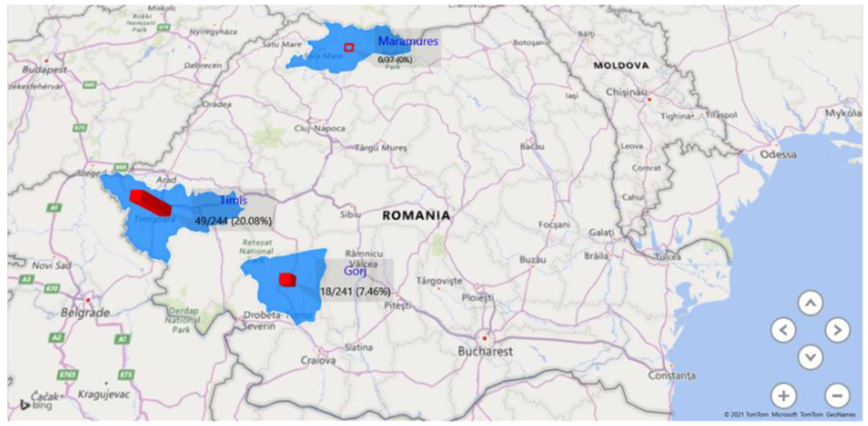
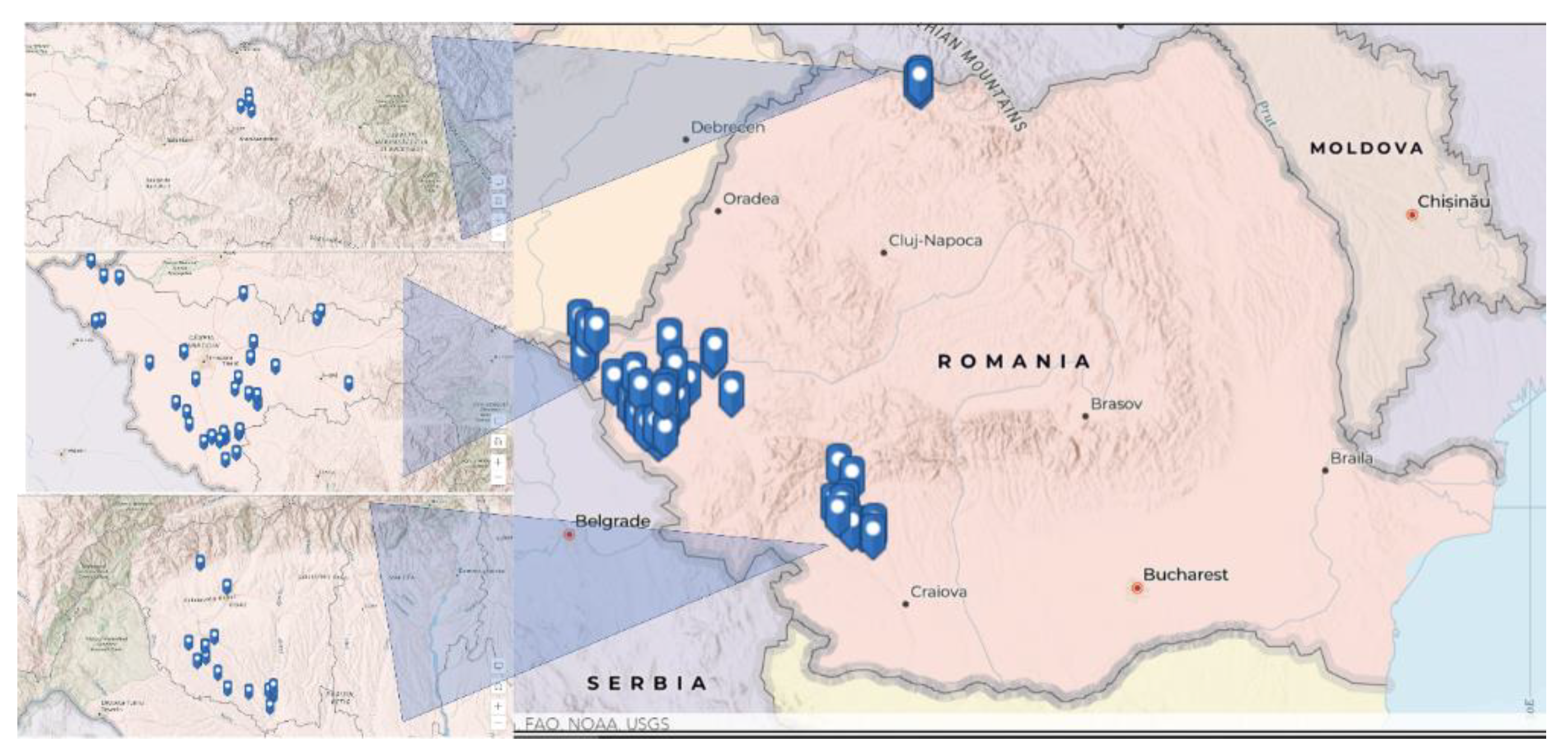
| Gorj | 241 (5/10) ** | 18 (7.46%) (50%) | 0.0807 (0.0502–0.1299) | |
| Timiș | 244 (14/32) ** | 49 (20.08) (43.75) | 0.2513 (0.1839–0.3433) | |
| Maramureș | 37 (0/4) ** | 0 (0%) | 0 (0–0.1038) | |
| Gorj vs. Timiș | <0.0001 | |||
| Gorj vs. Maramureș | 0.1438 | |||
| Timis vs. Maramureș | 0.0008 | |||
Publisher’s Note: MDPI stays neutral with regard to jurisdictional claims in published maps and institutional affiliations. |
© 2022 by the authors. Licensee MDPI, Basel, Switzerland. This article is an open access article distributed under the terms and conditions of the Creative Commons Attribution (CC BY) license (https://creativecommons.org/licenses/by/4.0/).
Share and Cite
Giubega, S.; Ilie, M.S.; Luca, I.; Florea, T.; Dreghiciu, C.; Oprescu, I.; Morariu, S.; Dărăbuș, G. Seroprevalence of Anti-Theileria equi Antibodies in Horses from Three Geographically Distinct Areas of Romania. Pathogens 2022, 11, 669. https://doi.org/10.3390/pathogens11060669
Giubega S, Ilie MS, Luca I, Florea T, Dreghiciu C, Oprescu I, Morariu S, Dărăbuș G. Seroprevalence of Anti-Theileria equi Antibodies in Horses from Three Geographically Distinct Areas of Romania. Pathogens. 2022; 11(6):669. https://doi.org/10.3390/pathogens11060669
Chicago/Turabian StyleGiubega, Simona, Marius Stelian Ilie, Iasmina Luca, Tiana Florea, Cristian Dreghiciu, Ion Oprescu, Sorin Morariu, and Gheorghe Dărăbuș. 2022. "Seroprevalence of Anti-Theileria equi Antibodies in Horses from Three Geographically Distinct Areas of Romania" Pathogens 11, no. 6: 669. https://doi.org/10.3390/pathogens11060669
APA StyleGiubega, S., Ilie, M. S., Luca, I., Florea, T., Dreghiciu, C., Oprescu, I., Morariu, S., & Dărăbuș, G. (2022). Seroprevalence of Anti-Theileria equi Antibodies in Horses from Three Geographically Distinct Areas of Romania. Pathogens, 11(6), 669. https://doi.org/10.3390/pathogens11060669