Article

Proteomic Profiling and In Silico Characterization of the Secretome of Anisakis simplex Sensu Stricto L3 Larvae

Department of Parasitology and Invasive Diseases, National Veterinary Research Institute, Partyzantów Avenue 57, 24-100 Puławy, Poland
*
Author to whom correspondence should be addressed.
Pathogens 2022, 11(2), 246; https://doi.org/10.3390/pathogens11020246
Submission received: 18 January 2022 / Revised: 9 February 2022 / Accepted: 10 February 2022 / Published: 14 February 2022
(This article belongs to the Special Issue Anisakiasis and Anisakidae)

Abstract

:
Anisakis simplex sensu stricto (s.s.) L3 larvae are one of the major etiological factors of human anisakiasis, which is one of the most important foodborne parasitic diseases. Nevertheless, to date, Anisakis secretome proteins, with important functions in nematode pathogenicity and host-parasite interactions, have not been extensively explored. Therefore, the aim of this study was to identify and characterize the excretory-secretory (ES) proteins of A. simplex L3 larvae. ES proteins of A. simplex were subjected to liquid chromatography-tandem mass spectrometry (LC-MS/MS) analysis, and the identified proteins were then analyzed using bioinformatics tools. A total of 158 proteins were detected. Detailed bioinformatic characterization of ES proteins was performed, including Gene Ontology (GO) analysis, identification of enzymes, Kyoto Encyclopedia of Genes and Genomes (KEGG) pathways analysis, protein family classification, secretory pathway prediction, and detection of essential proteins. Furthermore, of all detected ES proteins, 1 was identified as an allergen, which was Ani s 4, and 18 were potential allergens, most of which were homologs of nematode and arthropod allergens. Nine potential pathogenicity-related proteins were predicted, which were predominantly homologs of chaperones. In addition, predicted host-parasite interactions between the Anisakis ES proteins and both human and fish proteins were identified. In conclusion, this study represents the first global analysis of Anisakis ES proteins. The findings provide a better understanding of survival and invasion strategies of A. simplex L3 larvae.

1. Introduction

Anisakis simplex sensu stricto (s.s.), a nematode species belonging to the family Anisakidae, is among the most important foodborne parasites capable of causing a disease in humans called anisakiasis. This roundworm has an indirect lifecycle involving several hosts. Marine mammals are definitive hosts, fish, and cephalopods are intermediate or paratenic hosts, while crustaceans are intermediate hosts [1,2]. Humans, which are accidental hosts of A. simplex, are infected by third-stage (L3) larvae of this nematode, the source of which are infected marine fish or cephalopods [1,3]. A. simplex (s.s.) is mainly distributed in the northern Atlantic and Pacific Oceans [1]. However, other areas of occurrence (e.g., western Mediterranean Sea) were also reported [1,4,5].
The global incidence of anisakiasis is 0.32 cases/100,000 inhabitants [6]. However, according to recent studies, the prevalence of the disease is estimated to be much higher [7]; this discrepancy is linked to the nonspecificity of symptoms and the limitations of diagnostic tools. Furthermore, anisakiasis has become increasingly more important as human health risk, especially in regions where the consumption of raw or only lightly processed fish and seafood is frequent [1,8,9]. Therefore, the majority of anisakiasis cases are noted in Southeast Asia, while cases in Europe are less common and occur mainly in Spain and Italy [1].
Anisakiasis is a parasitic disease that affects the gastrointestinal tract [1,4]. In such cases, patients typically have symptoms such as abdominal pain, nausea, and vomiting [10]. Occasionally, the larva may perforate the alimentary tract and migrate into the peritoneal cavity and internal organs [1]. Another very important form of the disease is gastroallergic anisakiasis, in which abdominal symptoms are accompanied by allergic-like reactions such as urticaria, angioedema, or even anaphylaxis [1,11]. Furthermore, studies have suggested that dead Anisakis larvae can cause allergic reactions in sensitized humans [12,13,14]. From an anatomopathological point of view, Anisakis larva invasion leads to the development of inflammation (predominantly eosinophilic), edema, hemorrhage, formation of granuloma, and ulcers at the larval location, which is usually in the mucosa and submucosa of the gastrointestinal wall [10,12]. Larvae are unable to develop in the human body and die after up to 3 weeks [15].
Because of the risk to human health described above, Anisakis L3 larvae have been the subject of many studies, including investigations of their pathogenicity [16,17,18] and host-parasite interactions [19,20,21]. Excretory-secretory (ES) proteins play a key role in these activities. These molecules react with host proteins and enable development of host-parasite relationships by regulating the host immune system and subsequent pathology associated with the immune response. Furthermore, ES proteins are crucial for the parasite survival in host by inhibiting the inflammatory reactions [22]. Secretome proteins are important components of the structure and metabolism of the parasite, and exploration of A. simplex secretome will identify candidate molecules involved in immune modulation. Furthermore, these proteins are often very valuable targets for diagnostic tests, drugs, or vaccines. Nevertheless, the secretome proteins of A. simplex and other nematodes belonging to the family Anisakidae have not been extensively explored to date, which has resulted in a massive knowledge gap.
One of the most powerful tools used for proteomic profiling is mass spectrometry. This technology, combined with the use of an appropriate protein database, is suitable for the sensitive identification in complex biological samples. The potential for proteomic identification by mass spectrometry was recently greatly enhanced by the improved availability of genome sequencing technology, which has resulted in increased protein sequence database resources. Therefore, mass spectrometry has been successfully used for the proteomic profiling of secretomes of many organisms, including several nematode species, such as Toxocara canis [23], Ascaris suum [24], Ancylostoma caninum [25], and Spirocerca lupi [26].
In light of the above considerations, the aim of this study was to conduct proteomic profiling of the A. simplex (s.s.) L3 secretome. The use of liquid chromatography-tandem mass spectrometry (LC-MS/MS) allowed for the high-throughput identification of ES proteins. Multiple bioinformatics tools were used to comprehensively characterize the detected Anisakis ES proteins. Furthermore, in the present study, particular focus was placed on the discovery of proteins involved in pathogenesis of anisakiasis and host-pathogen interactions.

2. Results

2.1. Mass Spectrometry-Based Identification

LC-MS/MS analysis resulted in the identification of 158 proteins that occurred in all biological replicates. All protein identifications were based on the presence of at least one unique peptide. Of all identified proteins, 85 had at least two unique peptides detected in three biological replicates. The molecular weights (MWs) of proteins ranged from 4 to 811 kDa, and their isoelectric points (pI) ranged from 4 to 8.99. The majority of the proteins had MWs below 50 kDa (116 proteins) and a pI below 6.6 (107 proteins). All of the 158 proteins analyzed in this study are listed in Supplementary File S1.1. Mascot results of proteins identification within individual biological replicates are presented in Supplementary File S1.2.

2.2. Comparison of A. simplex Secretome Proteins with Secretome Proteins of Other Nematodes

The secretome proteins of A. simplex (s.s.) were compared with those of selected nematodes to determine similarities and differences in their profiles. The following secretomes had the largest number of highly similar proteins (≥70% similarity) to those of the A. simplex secretome: adult Ascaris suum (32 proteins), adult Ancylostoma caninum (27 proteins), and L3 larvae of Spirocerca lupi (26 proteins). Fewer highly similar proteins were detected in the secretomes of Toxocara canis larvae (17 proteins) and Ascaris suum L3 larvae (three proteins). Among the highly similar proteins, homologs of the following A. simplex proteins were the most common: peptidyl-prolyl cis-trans isomerase (A0A0M3JT42), which has homologs in adult A. caninum, adult A. suum, L3 larvae of A. suum, and L3 larvae of S. lupi, and an uncharacterized protein (A0A0M3JWS2) and putative actin (A0A0M3J0M4), both of which have homologs in adult A. caninum, adult A. suum, L3 larvae of S. lupi, and T. canis larvae. Details of the comparison of secretome proteomic profiles are presented in Figure 1 and Supplementary Files S1.3–8.

2.3. Protein Family Classification

The proteins in the A. simplex (s.s.) secretome belong to different families, among which the following were most frequently represented: immunoglobulin-like fold (15 proteins), immunoglobulin-like domain superfamily (12 proteins), annexin superfamily (six proteins), and thioredoxin-like superfamily (five proteins). Most protein families (87 protein families) were represented by only one protein. A total of 143 protein families were identified (see Supplementary File S1.9).

2.4. Secretory Pathway Prediction

Based on bioinformatics prediction, 21 proteins with identified signal peptides were classified into the conventional secretory pathway, and 77 proteins were assigned to unconventional protein secretion. Furthermore, among ES proteins the following proteins known to be associated with extracellular vesicles (EVs) released from Anisakis [27] were found: putative actin (A0A0M3J0M4), heat shock 70 kDa protein cognate 1 (A0A0M3K9V2), glutamate dehydrogenase (NAD(P)(+)) (A0A0M3K4H2), uncharacterized protein (A0A0M3KAB8), superoxide dismutase [Cu-Zn] (A0A0M3J718), and pepsin-I3 domain-containing protein (A0A0M3JAH0). Additionally, 24 proteins probably EV-associated were found in the Anisakis secretome. These protein are homologs of EV-associated protein which were identified in the secretomes of the following nematodes: A. suum [28], Brugia malayi [29], and Nippostrongylus brasiliensis [30]. Among the potential EV-associated proteins, the best matches were as follows: proteasome subunit alpha type (A0A0M3K144), proteasome subunit alpha type-3 (A0A0M3JSH7), and triosephosphate isomerase (A0A0M3JVA5). The top 10 best matches from identification of proteins potentially associated with EV are shown in Table 1, and all the results from the secretory pathway and EV-associated protein predictions are presented in Supplementary Files S1.10–12.

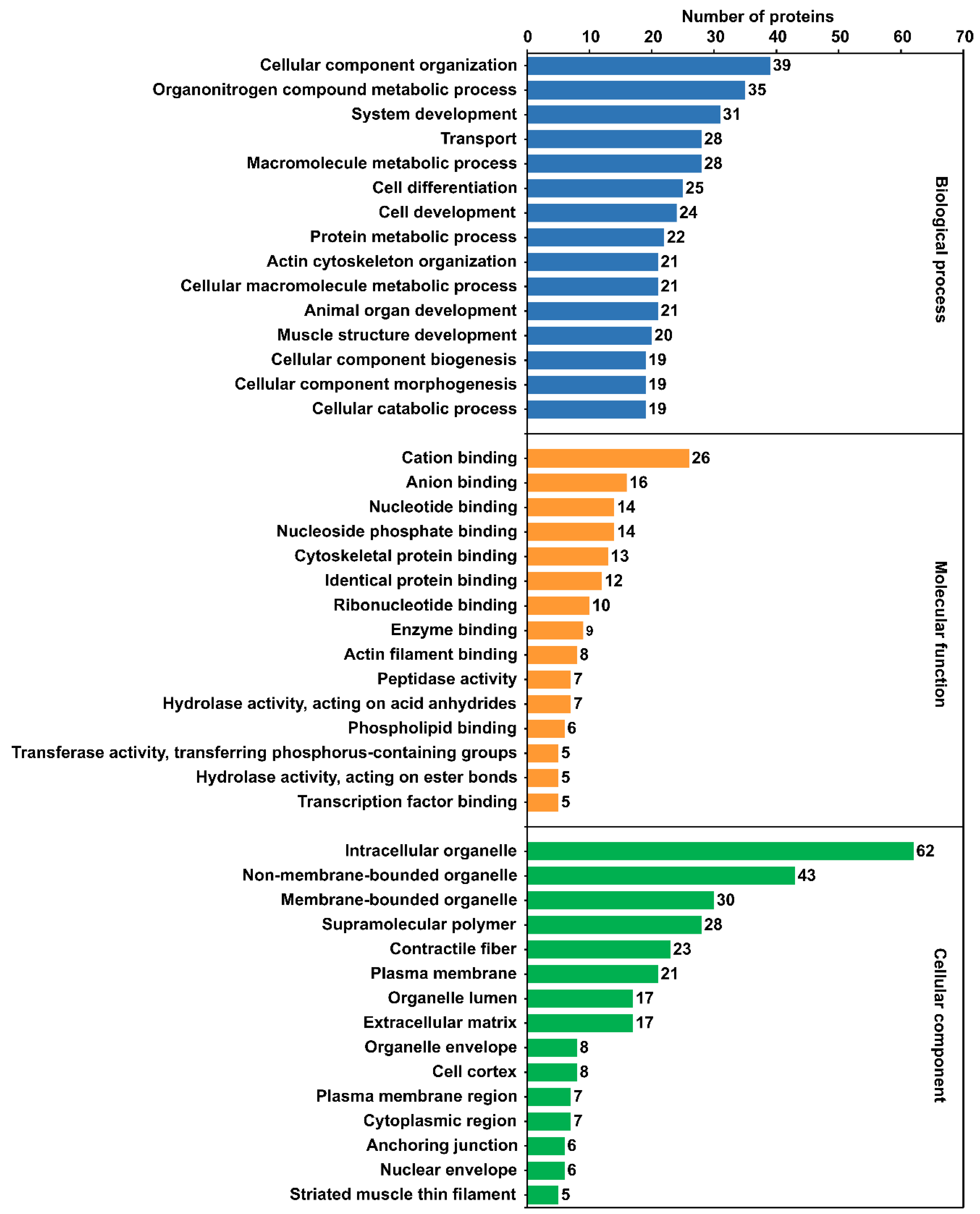
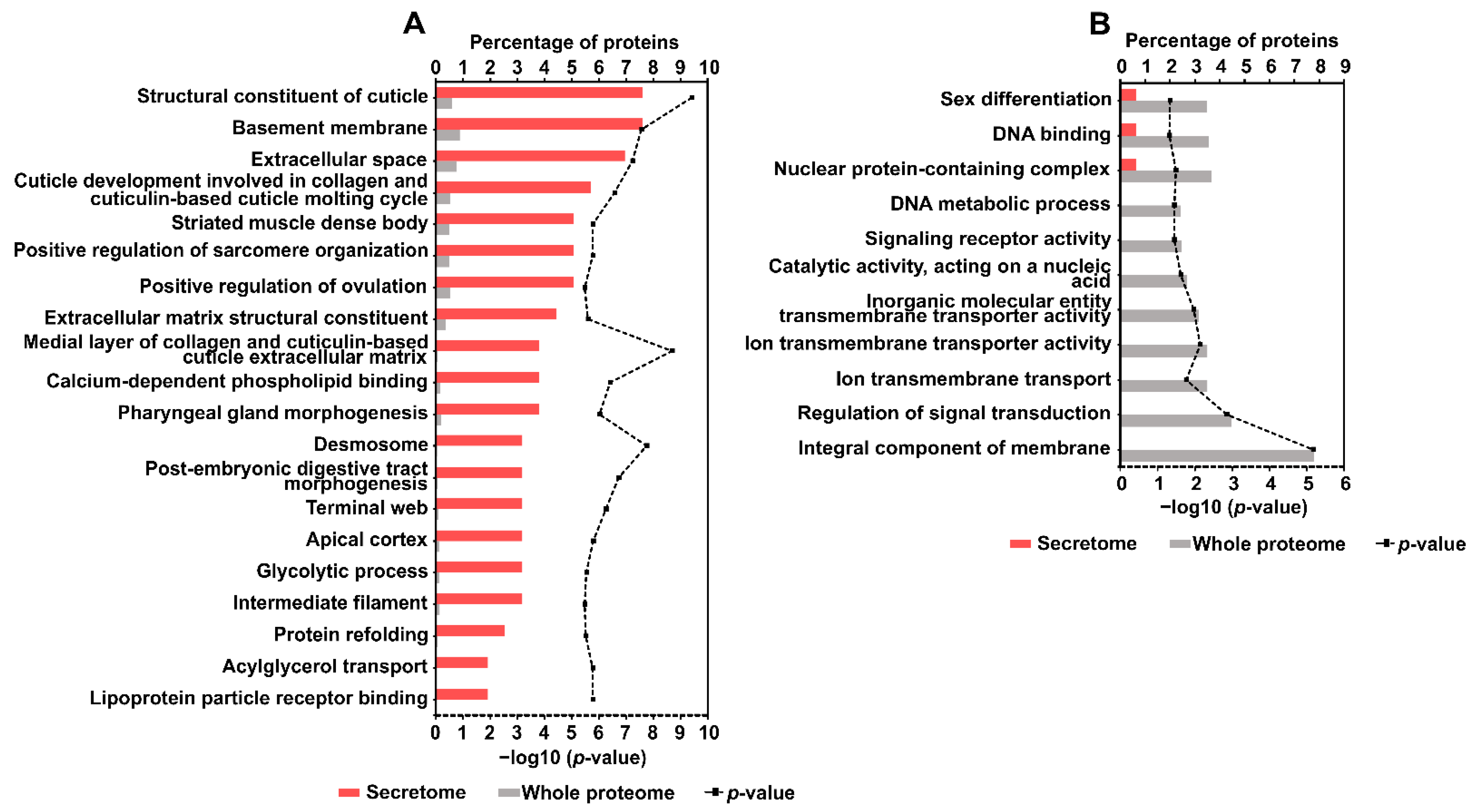
2.5. Gene Ontology (GO) Annotation and Enrichment Analysis

GO annotations of identified A. simplex (s.s.) proteins were grouped into three categories: biological process, molecular function, and cellular component. Around 90% of the proteins were annotated with GO terms. A total of 1242 GO annotations were identified. Only 10 proteins were assigned a single GO annotation, and the remaining proteins were annotated with 2-60 GO terms. In the biological process category, the most frequent GO terms were cellular component organization (39 proteins), organonitrogen compound metabolic process (35 proteins), and system development (31 proteins). In the molecular function category, the following GO terms were the most abundant: cation binding (26 proteins), anion binding (16 proteins), nucleotide binding (14 proteins), and nucleoside phosphate binding (14 proteins). The most abundant terms in the cellular component category were as follows: intracellular organelle (62 proteins), nonmembrane-bounded organelle (43 proteins), and membrane-bounded organelle (30 proteins). The top 15 GO terms in the three ontology categories are shown in Figure 2, and all GO annotations for individual proteins are listed in Supplementary File S1.13.
Enrichment analysis allowed for the mapping of over- and underrepresented GO terms of A. simplex (s.s.) secretome proteins. This analysis was performed by comparing the representation of GO terms for the detected secretome proteins with that for the whole A. simplex proteome. A total of 174 overrepresented and 11 underrepresented GO terms were identified. According to the calculated p-values, the following GO terms were the most overrepresented: structural constituent of cuticle (p-value = 3.88 × 10⁻¹⁰), medial layer of collagen and cuticulin-based cuticle extracellular matrix (p-value = 2.02 × 10⁻⁹), and desmosome (p-value = 1.81 × 10⁻⁸). Conversely, the following GO annotations were the most underrepresented: integral component of membrane (p-value = 6.57 × 10⁻⁶), regulation of signal transduction (p-value = 0.001), and ion transmembrane transporter activity (p-value = 0.007). The top 20 overrepresented and underrepresented GO terms are shown in Figure 3A,B, respectively. The detailed results of the GO enrichment analysis are shown in Supplementary File S1.14.

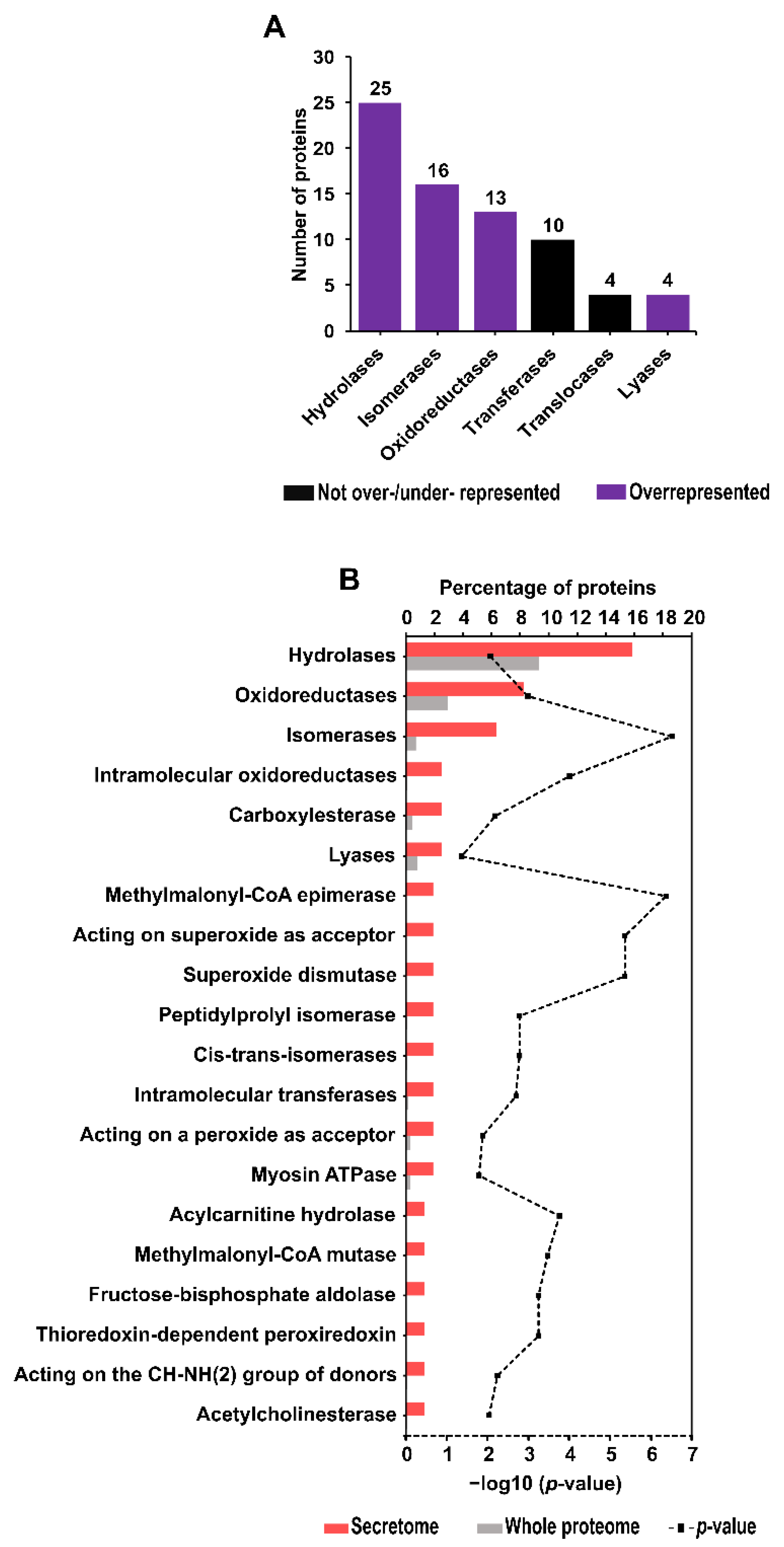
2.6. Enzyme Identification and Enrichment Analysis

Bioinformatics analysis of A. simplex (s.s.) secretome proteins was used to identify enzymes and proteins involved in metabolic pathways. Seventy-two proteins were assigned to six enzyme classes. The most abundant class of enzymes was hydrolases (25 proteins), and the less represented classes were isomerases, transferases, oxidoreductases, translocases, and lyases. No proteins belonging to the ligase class were detected. The distribution of the number of proteins in each enzyme class is shown in Figure 4A.
As determined using the OmicsBox software, 38 enzymes (including enzyme classes/subclasses) were overrepresented in the secretome compared to the whole A. simplex proteome. Based on the calculated P-values, the most overrepresented enzymes were isomerases (p-value = 3.08 × 10⁻⁷), methylmalonyl-CoA epimerase (p-value = 4.31 × 10⁻⁷), acting on superoxide as acceptor (p-value = 4.26 × 10⁻⁶), and superoxide dismutase (p-value = 4.26 × 10⁻⁶). No underrepresented enzymes were found in the Anisakis secretome. Figure 4B shows the 20 overrepresented enzymes in descending order of their abundance in the secretome. Details of the enzyme enrichment analysis are presented in Supplementary File S1.15.
In addition, proteases and protease inhibitors were identified using the MEROPS database. According to the used BLAST search cutoff, 36 of these enzymes were found, of which 27 were proteases and nine were protease inhibitors. Eighteen proteases/protease inhibitors showed 100% similarity to A. simplex proteins reported in the MEROPS database. A large group (eight proteins) also includes proteins that show similarity to proteases/protease inhibitors of other parasitic helminths, such as A. suum, Trichuris suis, Trichinella nativa, Onchocerca volvulus, Hymenolepis nana, and Hymenolepis diminuta. Table 2 shows the top 10 secretome protein matches of proteases/protease inhibitors, and all results are presented in Supplementary File S1.16.

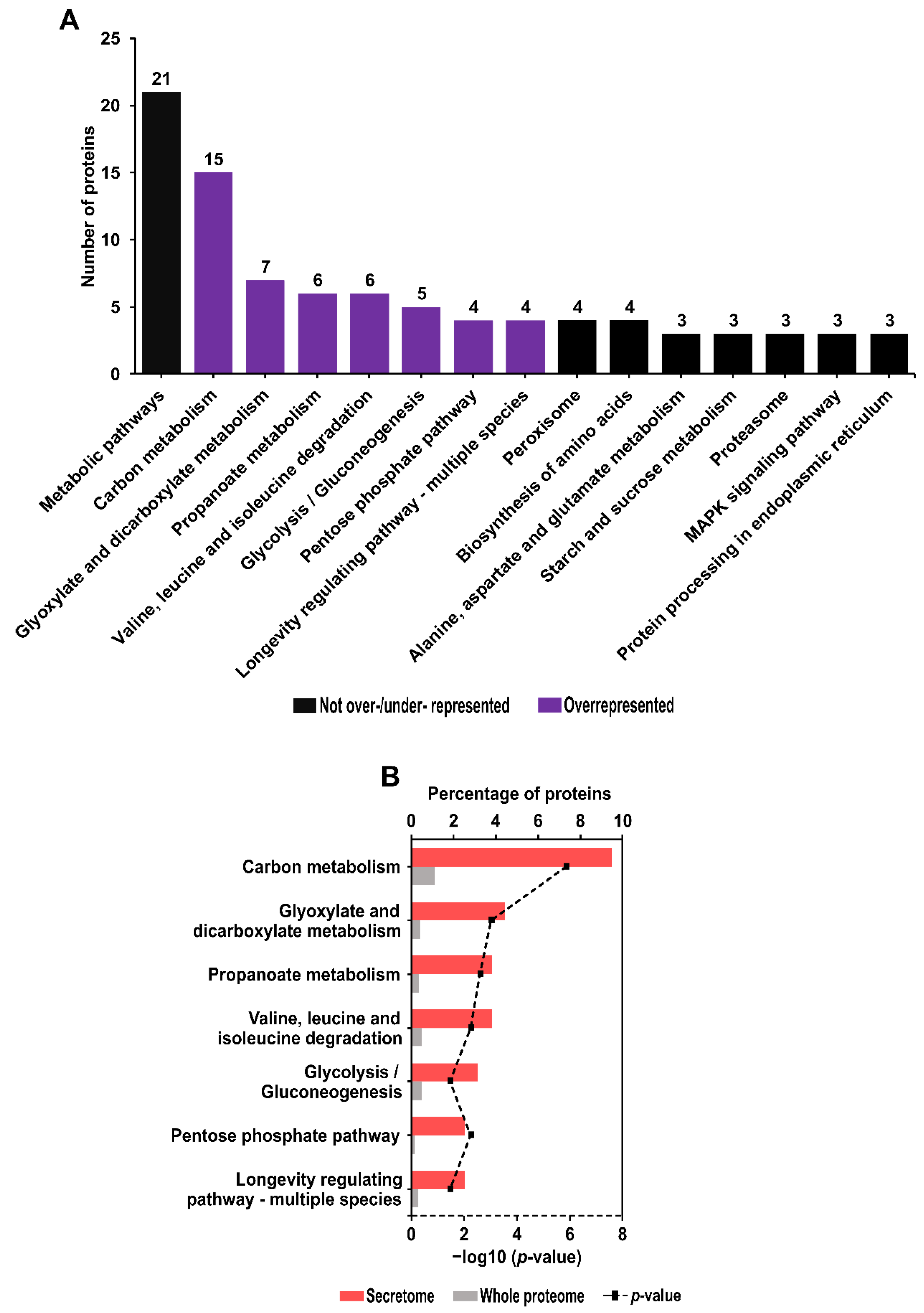
2.7. Kyoto Encyclopedia of Genes and Genomes (KEGG) Pathway Identification and Enrichment Analysis

The KEGG pathway profiling of Anisakis secretome proteins revealed proteins involved in 44 pathways. Among them, the most frequent were metabolic pathways (21 proteins), followed by carbon metabolism (15 proteins), and glyoxylate and dicarboxylate metabolism (seven proteins). The 15 most abundant KEGG pathways are shown in Figure 5A. The other 29 KEGG pathways were represented by 1–2 proteins. All identified KEGG pathways are listed in Supplementary File S1.17.
KEGG pathway enrichment analysis revealed seven overrepresented pathways in the secretome compared to the whole A. simplex proteome (Figure 5B). In ascending order of the calculated p-value, the most overrepresented KEGG pathways were as follows: carbon metabolism (p-value = 1.39 × 10⁻⁶), glyoxylate and dicarboxylate metabolism (p-value = 0.00097), and propanoate metabolism (p-value = 0.00253). No underrepresented KEGG pathways were found. Detailed results of the KEGG pathway enrichment analysis are presented in Supplementary File S1.17.

2.8. Identification of Essential Proteins

Among the identified secretome proteins of A. simplex (s.s.), 33 essential proteins were predicted (see Supplementary File S1.18) using the DEG database. Essential proteins are those indispensable for the survival of an organism. These proteins belong to various protein families. The best three matches against the sequences from the database were as follows: putative actin (A0A0M3J0M4), calmodulin (A0A0M3K916), and an uncharacterized protein (A0A0M3K916) that shows homology to alpha-actinin-4. Table 3 presents the 10 Anisakis proteins with the highest similarity to known essential proteins.

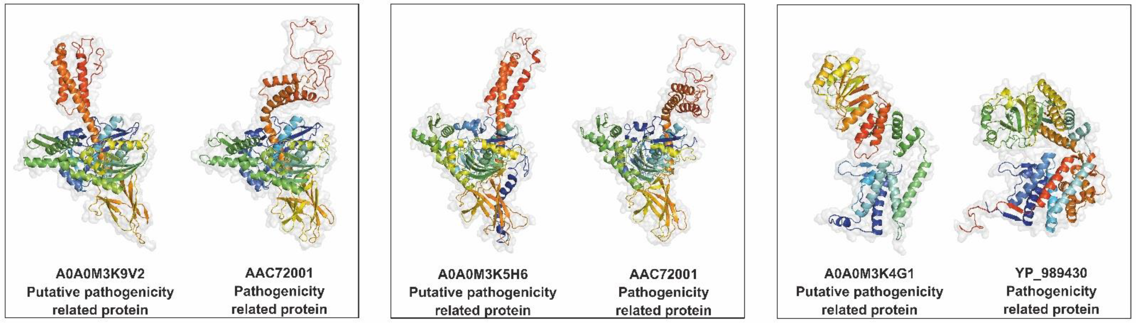
2.9. Identification of Potential Pathogenicity-Related Proteins

Using three databases, nine putative pathogenicity-related proteins were identified in the A. simplex (s.s.) secretome. Four proteins were found in multiple databases, and five proteins were detected in a single database. The highest number of potential pathogenicity-related proteins (eight proteins) was identified using the VICTORS database. Hits with the highest similarity to confirmed pathogenicity-related proteins were as follows: heat shock 70 kDa protein cognate 1 (A0A0M3K9V2), 78 kDa glucose-regulated protein (A0A0M3K5H6), and an uncharacterized protein (A0A0M3K4G1). The 3D structures reveal similarities between these proteins and their homologs with confirmed pathogenic properties (see Figure 6). A relatively high number of detected potential pathogenicity-related proteins (A0A0M3K9V2, A0A0M3K5H6, A0A0M3K4G1, and A0A0M3K0Q9) have homologs in the heat shock protein (HSP) family. Furthermore, the majority of the putative pathogenicity-related proteins in the Anisakis secretome (five proteins) are homologs of bacterial virulence proteins. Three A. simplex proteins show similarity to Cryptococcus neoformans virulence proteins, and two are homologs of Toxoplasma gondii HSP. All of the pathogenicity-related proteins identified in the study are shown in Table 4.

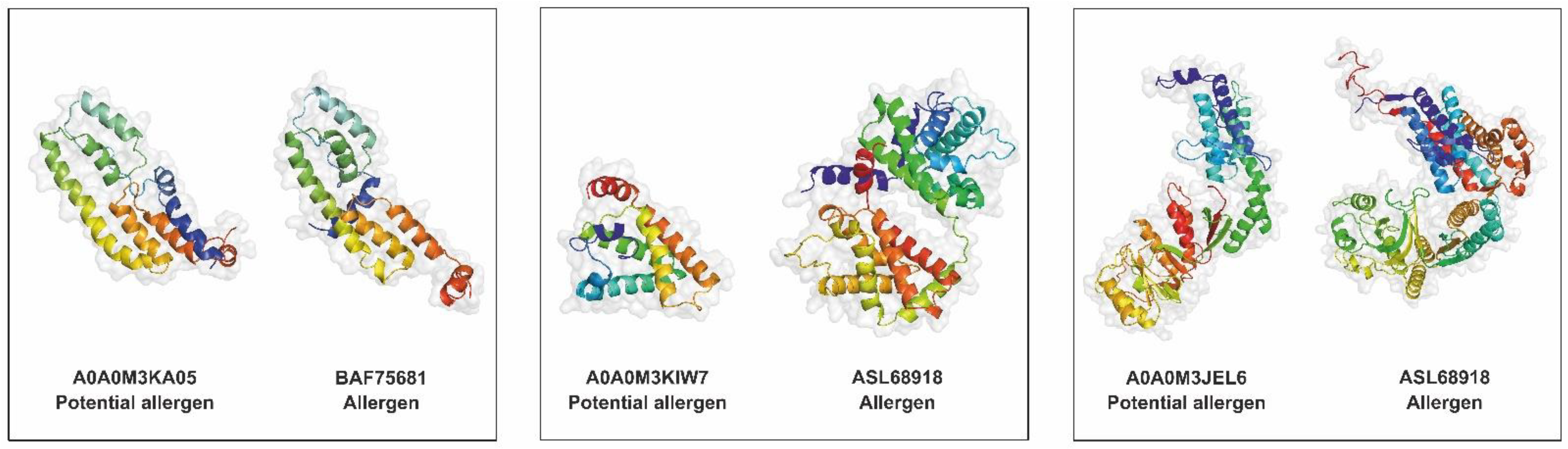
2.10. Allergen and Potential Allergen Identification

Of all identified proteins, only one (Ani s 4) is listed by the World Health Organization and the International Union of Immunological Societies (WHO/IUIS) Allergen Nomenclature Sub-Committee. By contrast, using the FARRP database, 18 potential allergens were identified. The three proteins with the best identification against the FARRP database were as follows: SXP/RAL-2 family protein 2 isoform 1 (A0A0M3KA05) and two globin-like proteins (A0A0M3KIW7, A0A0M3JEL6). The 3D structures of these potential allergens in comparison with their homologous allergens are shown in Figure 7. The AllerCatPro server confirmed that all proteins identified using the FARRP database have possible allergenic potential. The AllerCatPro tool determined the allergenic properties of 11 proteins with high confidence and 7 proteins with low confidence. The five detected potential allergens showed high similarity (>92%) to A. simplex allergens, and four others are highly similar to mite allergens. The other detected putative allergens showed similarity to allergens of A. suum, mosquito, fish, freshwater crayfish, and fungus. All potential allergens found in the A. simplex (s.s.) secretome are presented in Table 5.

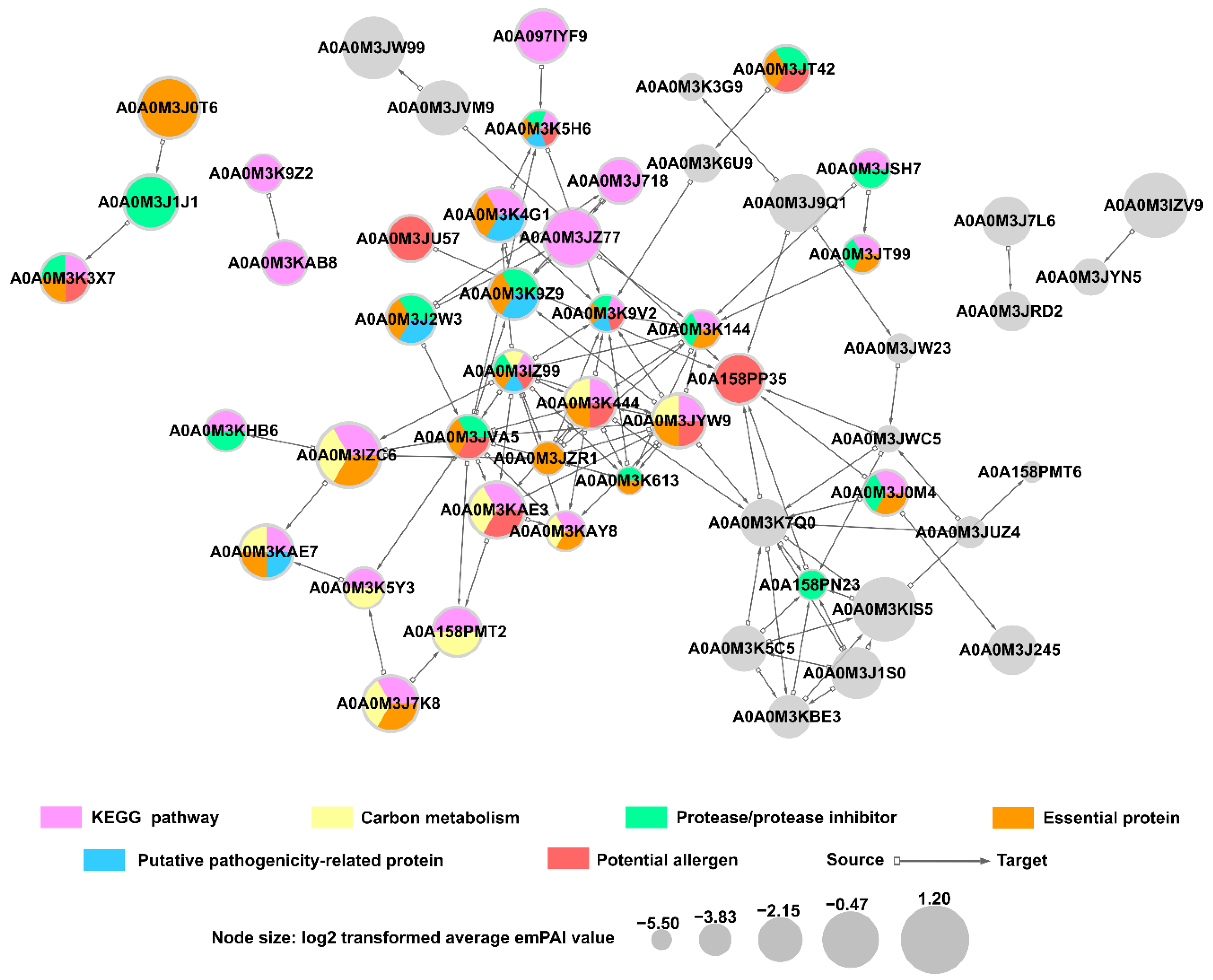
2.11. Predicted Protein-Protein Interactions in A. simplex (s.s.) Secretome

The protein interaction network was established using STRING and showed predicted interactions between ES proteins of A. simplex (s.s.). Fifty-three proteins involved in the interaction network were revealed with high prediction confidence. Twenty-five of these proteins were associated with KEGG metabolic pathways, particularly carbon metabolism, in which ten proteins were involved. Furthermore, proteins involved in the interaction network were associated with the following groups: essential proteins (21 proteins), proteases/protease inhibitors (16 proteins), potential allergens (11 proteins), and potential pathogenicity-related proteins (seven proteins). Twenty-nine proteins were assigned to only one of the groups listed above, and eleven were categorized into several groups simultaneously. Nineteen proteins from the interaction network were not assigned to any of the explored groups. The detailed analysis of the protein interaction network is shown in Figure 8.

2.12. Predicted Host-Parasite Protein Interactions

The HPIDB 3.0 server was used for the prediction of host-parasite interactions between the A. simplex (s.s.) ES proteins and both human and fish (Atlantic herring) proteins.
Eighteen proteins of Anisakis and 87 human proteins were identified in the host-parasite interaction network (see Figure 9A). The following groups of proteins were detected among Anisakis secretome proteins involved in interactions with human proteins: essential proteins (17 proteins), proteases/protease inhibitors (12 proteins), KEGG pathway proteins (12 proteins), potential allergens (seven proteins), and potential pathogenicity-related proteins (seven proteins). Most secretome proteins (16 proteins) were classified into two or more of these groups. Furthermore, it should be noted that the following Anisakis ES proteins showed the highest number of potential interactions with human proteins: transaldolase (A0A0M3KAE3; 22 interactions), proteasome subunit alpha type (A0A0M3JT99; 13 interactions), putative actin (A0A0M3J0M4; 12 interactions), heat shock 70 kDa protein cognate 1 (A0A0M3K9V2; 11 interactions), elongation factor 2 (A0A0M3K613; 11 interactions), and Rab GDP dissociation inhibitor (A0A0M3JZR1; 10 interactions). Human proteins that were predicted to be involved in the host-parasite interaction network belong to many different families, and the most highly represented of them were the following: laminins (17 proteins), methyltransferase proteins (13 proteins), Ras-related proteins (nine proteins), and Hsp70-binding proteins (seven proteins). Furthermore, among human proteins, polyubiquitin-C (P0CG48) showed potential interactions with the highest number of Anisakis proteins (13 interactions).
Five proteins of Anisakis secretome and 19 proteins of Atlantic herring were predicted in the fish-parasite interaction network (see Figure 9B). All Anisakis ES proteins identified in this interactome were identified also in the human-parasite interaction network. The following groups of proteins were detected among Anisakis ES proteins involved in interactions with fish proteins: essential proteins (five proteins), proteases/protease inhibitors (3 proteins), KEGG pathway proteins (three proteins), and potential allergen (one protein). Rab GDP dissociation inhibitor (A0A0M3JZR1), and calmodulin (A0A0M3KFJ2) showed potential interactions with the highest number of fish proteins (10 and four interactions, respectively). The most highly represented fish proteins involved in the interactome were Ras superfamily proteins (10 proteins), mainly members of the Rab family. Phosphodiesterases (four proteins) were the second largest group of the fish-parasite interactome. The detailed results of identification of proteins involved in potential host-parasite interactions are presented in Supplementary Files S1.19–20.

3. Discussion

This study is the first global proteomic analysis of the A. simplex (s.s.) L3 larval secretome. Previous proteomic investigations of Anisakis did not include profiling of the secretome proteins [13,31,32,33,34,35,36,37,38]. Therefore, knowledge on A. simplex ES proteins is very fragmented, although important aspects related to metabolism, pathogenicity, and host-parasite interactions are known to be associated with ES proteins. In this study, LC-MS/MS and bioinformatics analyses were applied to provide insights into these issues.
Prior to identifying Anisakis ES proteins by mass spectrometry, their SDS-PAGE profile was analyzed. Electrophoretic analysis confirmed the distribution of protein bands over a wide range of molecular weights, characteristic of the ES proteins of A. simplex L3 (see Supplementary Figure S1). Subsequently, LC-MS/MS analysis allows identification of 158 proteins in the Anisakis secretome, which is currently the largest proteomic dataset of A. simplex ES proteins. The number of ES proteins identified in this study corresponds to approximately 0.8% of the genes encoding A. simplex proteins. Comparing the number of identified ES proteins of A. simplex to the secretomes of other closely related pathogenic nematodes with similar genome size, such as A. suum and T. canis (see Figure 1), reveals that there is a relatively high number of ES proteins in Anisakis. By contrast, the lower number of identified Anisakis ES proteins compared to the A. caninum secretome is presumably due to the much larger genome size of A. caninum. Furthermore, only about one-quarter of the identified Anisakis ES proteins showed high similarity to the proteins of the Toxocara or Ascaris secretomes. This relatively low similarity is probably due to differences in hosts and life cycles of Anisakis nematodes and those of Toxocara and Ascaris.
The majority of detected Anisakis ES proteins were assigned to an unconventional secretory pathway (approximately 49% of proteins). This prediction is consistent with the secretory pathway analysis of ES proteins of other nematodes, such as Dirofilaria immitis [39] or Strongyloides venezuelensis [40]. Most secretome proteins of these nematodes were also classified into the unconventional secretory pathway. Furthermore, 15% of Anisakis proteins were classified as potentially EV-associated proteins. Prediction of these proteins was based on similarity to EV-associated proteins secreted by other nematodes. This is a particularly important identification because, among other considerations, the EV released by parasites play an important role in delivering molecules that can modulate the host immune response or the transfer of pathogenicity-related factors [28]. In this study, six EV-associated proteins were found which were previously identified by Boysen et al. [27] (see Section 2.4). There are currently no other published studies on the identification of Anisakis EV-associated proteins, and because of their important functions, this topic requires further exploration.
The secretome proteins detected in Anisakis were characterized by high diversity, as evidenced by their classification into 143 protein families. Among the most frequently identified protein families in the Anisakis secretome, attention should be paid to the annexin superfamily. Annexins have multiple functions, such as in cellular anti-inflammation, signal transmission, anticoagulation, ion channel regulation, membrane repair, and membrane transport, and likely participate in cell proliferation, differentiation, and apoptosis [41,42]. Furthermore, parasite annexins are considered potential drug and vaccine targets [43]. The thioredoxin-like superfamily is another of the protein families most frequently detected in the Anisakis secretome that is also important. Thioredoxins, inter alia, regulate thiol-based redox control and prevent the aggregation of cytosolic proteins in the cell [44,45]. The extracellular activities of thioredoxins include anti-inflammatory and antiapoptotic activities and, thus, cytoprotective effects [44,46].
In general, the identified ES proteins of A. simplex (s.s.) have multiple functions, as demonstrated by the GO analysis. On average, nine GO terms were detected for all 142 proteins that were assigned GO annotations. A large variety of GO terms among nematode secretome proteins is quite typical [47]. Many of the detected secretome proteins, such as thioredoxins [44], annexins [48], and HSPs [49], are moonlighting proteins that form a subset of multifunctional proteins in which one polypeptide chain exhibits more than one physiologically relevant biochemical or biophysical function [50].
GO annotation enrichment analysis provided interesting data. Of the many enriched annotations, the most abundant and most enriched GO terms were, in general, related to the glycolytic process, larval development, antioxidants, and cuticle. These annotations cover functions that are important to parasite metabolism, lifestyle, and survival, and they are also found to be enriched in the annotated secretomes of other nematodes [40,47]. Among the enriched GO terms mentioned above, those related to the cuticle may seem to be unassociated with the secretome. However, it should be noted that ES proteins are also released from the surface of the cuticle, in addition to specialized excretory-secretory organs and parasite intestine [24]. Proteins produced and presented at the parasite-host interface during invasion play a critical role in the induction and development of immune responses [24]. Furthermore, secretome proteins could also play essential roles in ensuring cuticle integrity [51].
Possible enzymes were detected among secretome proteins, which is in line with previous studies that confirmed the enzymatic properties of the A. simplex secretome [52]. Furthermore, Kim et al. [53] found that protease related genes are highly expressed in the transcriptome of A. simplex L3 larvae. In the present study, proteases were highly represented in the secretome (17% of ES Anisakis proteins). These enzymes are known to be especially important in the pathogenesis of anisakiasis and other parasitoses [54,55]. Proteases play an important part in host-parasite interactions, such as invasion of the host, migration through host tissues, protection of the parasite against the host immune system, and activation of the inflammatory response [56,57]. Proteases also participate in important biological processes in parasitic nematodes, as they are directly involved in their growth and survival, embryonic development, digestion of protein for nutrients, molting, and numerous metabolic processes [58,59]. Another important enzyme group detected in the secretome is antioxidant enzymes, such as thioredoxin-dependent peroxiredoxin and superoxide dismutase. These enzymes are found to be enriched in the secretome, and they function to protect against the toxic contents released by immune effector cells as a first-line host defense mechanism [22]. Acetylcholinesterase (AChE) was also detected as an enriched enzyme in the secretome. The enrichment of AChE in the Anisakis secretome is in agreement with previous investigations [60]. AChE secretion by Anisakis larvae is presumed to be an adaptive mechanism, and its secretion increases in response to a direct and/or indirect effect of neurotoxic compounds released by the host [60]. Furthermore, AChE has recently received attention as a potential anthelmintic drug and vaccine target in nematodes [61].
Among the KEGG pathways, an important group that was enriched comprises the following carbohydrate metabolism pathways: glyoxylate and dicarboxylate metabolism, propanoate metabolism, glycolysis/gluconeogenesis, and pentose phosphate pathway. Indeed, carbohydrate metabolism is an essential energy source for Anisakis larvae [62]. Carbohydrates play important roles in many basic processes, including development, morphogenesis, immunity, and host-pathogen interactions [63]. Two sugars, trehalose and glycogen, were detected in A. simplex L3 larvae [64]. Łopieńska-Biernat et al. found [65] that trehalose plays a key role in providing energy during thermotolerance and starvation processes. It is also worth mentioning that the secretome was enriched in numerous members of the longevity-regulating pathway, which is associated with the regulation of numerous processes, such as oxidative stress, autophagy, glycogen accumulation, and fat accumulation. In addition, the secretome was found to be enriched in proteins belonging to the valine, leucine, and isoleucine degradation KEGG pathway. This KEGG pathway is important since valine, leucine, and isoleucine are likely to be essential amino acids in Anisakis, as is the case in C. elegans [66].
Approximately 21% of ES proteins were predicted to be essential for life. These are proteins that are critical to the survival of the cell or organism under certain conditions [67]. Among the top matches of secretome essential proteins such proteins were found as putative actin (A0A0M3J0M4), calmodulin (A0A0M3KFJ2), elongation factor 2 (A0A0M3K613), and HSPs (A0A0M3K5H6, A0A0M3K9V2, A0A0M3K4G1). Actin is a family of globular multi-functional proteins that form microfilaments [68]. These proteins participates in many important cellular processes, including muscle contraction, cell motility, cell division and cytokinesis, vesicle and organelle movement, cell signaling, and the establishment and maintenance of cell junctions and cell shape [68]. Calmodulin is a highly conserved protein ubiquitously and abundantly expressed in eukaryotic cells [69]. The functions of calmodulin include Ca2+ binding and alteration of calcium signal transduction pathway to control a variety of biological processes, such as cytoskeletal assembly/reorganization, activation of phosphorylase kinase, abiotic stress responses, neurotransmission, smooth muscle contraction, metabolism, and cell motility [70]. Elongation factor 2 catalyzes the guanosine triphosphate-dependent ribosomal translocation step during translation elongation. HSPs are important molecular chaperones for maintaining cellular functions to prevent proteins from misfolding and aggregating in crowded surroundings. HSP expression levels increase when the organism is exposed to stress conditions, such as heat shock, alkaline treatment, and some chemical reagents, in order to help pathogens survive unfavorable conditions in the host [71,72,73]. Furthermore, essential for life proteins may be promising therapeutic targets for drugs and vaccines [74]. Such proteins include phosphoglycerate mutase (A0A0M3KAY8), which in this study was predicted to be an essential protein and shows homology with cofactor-independent phosphoglycerate mutase (iPGM) of C. elegans. This enzyme is involved in glycolytic and gluconeogenic pathways, and inhibition of iPGM activity has been shown to have a lethal effect on C. elegans [75]. Therefore, iPGM is considered a potential drug target or vaccine candidate in several nematodes, such as Wuchereria bancrofti [76], B. malayi [77], and Leishmania donovani [78].
Nine potential pathogenicity-related proteins in the A. simplex (s.s.) secretome were identified using database searching. The majority were HSPs which are the first line of attack and help in fortifying pathogen virulence [79,80]. The HSPs of Anisakis are poorly characterized; to date, only the expression patterns of HSP90 and HSP70 in Anisakis have been analyzed [81,82], whereas the contribution of Anisakis HSPs to pathogenicity has not been investigated. By contrast, HSP70 has a better-known role in the pathogenicity of Toxoplasma gondii, which is based on the modulation of nitric oxide production by macrophages [83]. HSP70 of T. gondii was found to be homologous with two top-matched potential pathogenicity-related proteins in the A. simplex secretome. Another two predicted pathogenicity-related proteins of A. simplex (s.s.) were homologous with GroEL of Bartonella, which is a chaperonin and exhibits pathogenicity via apoptosis inhibition and mitogenic stimulation of host cells [84]. The next two potential pathogenicity-related proteins in the Anisakis secretome were found to be homologous with thiol-specific antioxidant protein 1 of Cryptococcus, which is essential for resistance to oxidative, nitrosative, and temperature stress [85]. A homolog of hypothetical protein CNAG_05449 of Cryptococcus was also detected in the Anisakis secretome. This protein is in the metallothionein family, members of which play a crucial role in the pathogenicity and resistance of Cryptococcus against the host immune response, since they are directly involved in the detoxification of high concentrations of copper produced by macrophages fighting the infection [86]. Another protein detected in the Anisakis secretome is a homolog of the glycine cleavage system H protein of Francisella tularensis, which contributes to the intracellular replication of the pathogen in serine-limiting environments [87]. Another predicted pathogenicity-related protein of the A. simplex (s.s.) secretome is homologous with glucose-6-phosphate isomerase, which is required for the extracellular polysaccharide biosynthesis of Haemophilus influenzae [88].
Of the seven A. simplex ES allergens listed by WHO/IUIS [89], only Ani s 4 was detected in the present study. Ani s 4 is significant because of its heat- and pepsin-resistant properties and its ability to cause anaphylaxis [13,90,91]. Other Anisakis allergens were presumably not expressed in the in vitro culture conditions, or their concentration was below the limit of detection of LC-MS/MS. In addition to known allergens, potential allergens were also identified in this study. In order to increase the specificity of this analysis, hits detected using the FARRP database were confirmed by the AllerCatPro server [92], combining various bioinformatics approaches. The majority of the potential allergens detected in the Anisakis secretome are homologs of nematode and arthropod allergens, which is in line with the cross-reactions between Anisakis antigens and these organisms described by other authors [93,94,95]. Of the 18 potential allergens identified in this study, the following 6 were detected in our previous investigations of extracts from A. simplex L3 larvae: A0A158PP35, A0A0M3K5H6, A0A0M3KA05, A0A0M3JU57, A0A0M3K9V2, and A0A0M3K8L6 [13,33]. The first five are also proteins with potential thermostability [13]. Furthermore, many of the possible allergens detected in the Anisakis secretome show similarity to the potential allergens identified by other authors in the whole proteome and transcriptome of Anisakis larvae [32,96,97]. Fæste et al. [37] found in the A. simplex larvae the following proteins, including potential allergens which were similar to putative allergens identified in the present study: haemoglobin (P26914), troponin-like protein (Q9U3U5), HSP 70 (A8Q5Z6), triosephosphate isomerase (P91919), fructose-bisphosphate aldolase 1 (A8P3E5), and calmodulin (O16305). In particular, many sequences similar to the potential allergens of A. simplex detected in this study can be found at the transcriptome level in the ANISAKIS DB database (http://anisakis.mncn.csic.es/public/, accessed on 17 January 2021) [97]. Furthermore, a comparison of the secretome proteins identified in this study with the immunoreactive proteins of A. simplex esophageal gland cells provides interesting data [38]. Of the 13 immunoreactive proteins detected in esophageal gland cells, we also detected the following 4 in the secretome: uncharacterized protein (A0A0M3K6E2), uncharacterized protein (A0A0M3JQQ1), metalloendopeptidase (A0A0M3K299), and SCP domain-containing protein (A0A0M3K1U4). Thus, these ES proteins may also have allergic and/or diagnostic potential.
Interactome analysis was performed to identify and characterize proteins involved in interactions between A. simplex (s.s.) ES proteins as well as proteins involved in host-pathogen interactions. As was expected, such proteins primarily fall into the following groups: proteins of KEGG pathways, essential proteins, and proteases/protease inhibitors, followed by potential allergens and potential pathogenicity-related proteins. These groups of Anisakis proteins are characterized in detail above, and such profile composition is in accordance with the main functions of helminth ES proteins, i.e., penetration, colonization, survival in host tissues, incorporation of host metabolites, and modulation of the host immune response [98,99].
Among the human proteins involved in the interaction with Anisakis ES proteins, laminins were found to be the most highly represented. Laminins, which are the major component of the basal lamina, are enzymatically degraded by parasites such us Anisakis pegreffii during invasion [100], which facilitates internal migration of parasites [101]. Methyltransferase proteins were also highly abundant human proteins which were predicted in the host-parasite interactome. These proteins are known to contribute in deregulation of host expression profile which lead to host cell transformation, or escape of Apicomplexa parasites from the host immune system [102]. It is worth noting that among all human-parasite protein interactions, human polyubiquitin-C is the main target identified as interacting with Anisakis ES proteins. Indeed, ubiquitin is known to modulate host-pathogen interactions, with a particular focus on host innate immune defenses and pathogen immune evasion [103]. Among Anisakis secretome proteins, transaldolase (A0A0M3KAE3) was found to interact with the largest number of human proteins. This protein is an enzyme of pentose phosphate pathway, and a potential allergen. Another A. simplex (s.s.) protein which was predicted to interact with a large number of human proteins is proteasome subunit alpha type (A0A0M3JT99) which is characterized by its proteolytic activity. Another important Anisakis protein of predicted human-parasite interactome is heat shock 70 kDa protein cognate 1 (A0A0M3K9V2). This protein was predicted to be a potential protease, a potential allergen, a protein essential for life, and a possible pathogenicity-related protein. It is known that heat shock 70 kDa cognate plays an important role in the interactions of many parasites with the host organism, since it is highly immunogenic and a target of B and T cells [104,105].
The number of proteins involved in the human-parasite interaction network is about four times the number of proteins in the fish-parasite interactome. This result could be mainly due to the fact that the available database of the non-human host-pathogen interaction is much more limited than for human-pathogen interactions. Among the fish proteins involved in the interaction with Anisakis secretome proteins, Rab-family proteins were found to be the most abundant. These proteins regulate virtually all membrane trafficking events in eukaryotic cells [106,107]. Other abundant proteins in fish-parasite interactome were phosphodiesterases (PDEs). PDEs are metallohydrolases that control the concentration of second messengers cyclic adenosine monophosphate and cyclic guanosine monophosphate. Among Anisakis secretome proteins, Rab GDP dissociation inhibitor (GDI) (A0A0M3JZR1) was found to interact with the largest number of fish proteins. This protein is involved in regulation of GDP-GTP exchange between Rab-family proteins. Rab GDI is an immunoreactive protein of Trichinella britovi [108], but its role in host-parasite interactions is poorly known. Similarly, calmodulin (A0A0M3KFJ2) of Anisakis secretome was predicted to interact with fish proteins. Furthermore, calmodulin it is known immunogenic protein of Fasciola hepatica secretome [109].

4. Materials and Methods

4.1. A. simplex (s.s.) L3 Larvae Collection and Identification

Anisakis spp. L3 larvae were collected from naturally infected Atlantic mackerel (Scomber scombrus) originated from subarea VII of FAO fishing area 27. As described in our previous publication [110], the larvae were purified by washing with sterile phosphate-buffered saline (PBS, 0.01 M, pH 7.4; Sigma, St. Louis, MO, USA) and microscopically evaluated for integrity and viability. Ten randomly selected larvae were used for identification of Anisakis species by PCR-restriction fragment length polymorphism (PCR-RFLP) [111,112,113,114] (see Supplementary Figure S2).

4.2. ES Proteins Preparation

ES proteins were prepared by incubating 100 viable L3 A. simplex (s.s.) larvae in 5 mL of sterile PBS. After 24 h of incubation at 36 °C, medium containing ES proteins was collected and clarified by centrifugation at 20,000× g for 15 min at 4 °C. Supernatants were concentrated by ultrafiltration at 4 °C (3 kDa cutoff membrane; Thermo Fisher Scientific, Rockford, IL, USA). Subsequently, protein concentration was measured using a spectrophotometer (NanoPhotometer P330, Implen, München, Germany) and secretome samples were stored at −80 °C for further experiments. According to this procedure, three independent biological replicates of ES proteins were prepared.

4.3. SDS-PAGE

ES proteins of A. simplex (s.s.) were subjected to 4–20% SDS-PAGE (BioRad, Hercules, CA, USA) under reducing conditions [115]. Gel was fluorescently stained with SYPRO Ruby protein gel stain (Invitrogen, Eugene, OR, USA) and the molecular weights of the electrophoresis bands were calculated using Bio-1D software (ver. 15.07; Vilber Lourmat, Marne-la-Vallée, France) (see Supplementary Figure S1).

4.4. Sample Processing and LC-MS/MS Analysis

Three batches of A. simplex (s.s.) ES proteins were subjected to in-solution digestion and LC-MS/MS analysis, as described in our previous publication [13]. Briefly, secretome samples were analyzed using a Q Exactive mass spectrometer (Thermo Electron Corp., San Jose, CA, USA) coupled with nano-high-performance liquid chromatography (nano-HPLC) RP-18 column (internal diameter 75 µm; Waters, Milford, MA, USA). Proteins were identified with Mascot search engine server (ver. 2.5; Matrix Science, London, UK; http://www.matrixscience.com/server.html, accessed on 25 October 2019) using the A. simplex proteome (proteome ID: UP000036680; 20,789 sequences) obtained from the Universal Protein Resource (UniProt, http://www.uniprot.org/, accessed on 25 October 2019) [116]. The following Mascot search parameters were applied: trypsin digestion allowing one missed cleavage, parent ions was set to 5 parts per million (ppm), and fragment ions was set to 0.01 dalton (Da). The ion type was set as monoisotopic, and protein mass was set as unrestricted. Beta-methylthiolation of cysteine was used as a fixed modification, whereas oxidation of methionine was set as a variable modification. Peptides were accepted at False Discovery Rate (FDR) ≤ 0.98%, ion score ≥ 38, and significant threshold of p ≤ 0.00026. Only proteins detected with at least one unique peptide and proteins identified in all three biological replicates were accepted for further analysis. A detailed procedure of samples processing and LC-MS/MS analysis is presented in Supplementary File S2.

4.5. Bioinformatics Analysis

Theoretical pI and MW values of ES proteins were derived from the Mascot server. Gene ontology (GO) analysis, InterPro protein family classification, and enzyme identification were performed using OmicsBox with the Blast2GO algorithm (ver. 1.4.12; BioBam Bioinformatics SL, Valencia, Spain, https://www.biobam.com/omicsbox/) [117]. Analyses were run with the default settings and filtered using nematode taxonomy, as previously described [13]. Proteases and protease inhibitors were identified using a BLASTP search against the MEROPS database (release 11 September 2020; https://www.ebi.ac.uk/merops/, accessed on 8 April 2021) [118]. Proteins were assigned to the conventional or unconventional secretory pathway using OutCyte 1.0 server (http://www.outcyte.com/, accessed on 1 April 2021) [119]. Putative EV-associated proteins were identified using a BLASTP search against the known EV-associated proteins detected in the secretomes of A. suum [28], B. malayi [29], and N. brasiliensis [30]. Similarities between A. simplex ES proteins and previously identified secretome proteins of the parasites S. lupi L3 larvae [26], adult A. caninum [25], A. suum L3 larvae [24], and T. canis larvae [23] were evaluated using BLASTP. The same bioinformatics tool was used to identify proteins essential for life using reference eukaryote proteins from the DEG database (release 1 September 2020; http://origin.tubic.org/deg/public/index.php/index, accessed on 22 April 2021) [67].
Enriched GO annotations and enzymes of identified secretome proteins were compared with those of the whole proteome of A. simplex (proteome ID: UP000036680) by two-tailed Fisher’s exact test using OmicsBox software. These tests were performed using a p-value cutoff of 0.05 to indicate significance. Proteins involved in KEGG pathways were detected using the KOBAS 3.0 server (http://kobas.cbi.pku.edu.cn/, accessed on 23 April 2021) [120] based on the C. elegans proteome as a reference. Enriched KEGG pathways were identified by Fisher’s exact test using KOBAS 3.0 with the C. elegans proteome as a reference and the whole A. simplex proteome as background. In this case, a p-value less than 0.05 with Benjamini and Hochberg correction was considered to indicate significance.
Potential pathogenicity-related proteins were predicted using BLASTP against the following databases: ProtVirDB (http://bioinfo.icgeb.res.in/protvirdb/home.html, accessed on 22 April 2021) [121], Victors (http://www.phidias.us/victors/, accessed on 23 April 2021) [122], and VFDB (http://www.mgc.ac.cn/VFs/main.htm, accessed on 23 April 2021) [123]. Proteins were evaluated for potential allergenicity by BLASTP searching against The Food Allergy Research and Resource Program (FARRP) AllergenOnline.org database (ver. 21; http://www.allergenonline.com/, accessed on 30 March 2021). Possible allergens detected using the FARRP database were confirmed by the AllerCatPro server (ver. 1.8; https://allercatpro.bii.a-star.edu.sg/, accessed on 10 April 2021) [92]. Unknown 3D structures of pathogenicity-related/potential pathogenicity-related proteins and allergens/potential allergens were predicted by homology modeling using the Phyre2 server (http://www.sbg.bio.ic.ac.uk/phyre2/, accessed on 9 June 2021) in intensive mode (multi-template + ab initio) [124]. The model structures were further improved using 3Drefine (http://sysbio.rnet.missouri.edu/3Drefine/, accessed on 13 June 2021) [125]. Visualization of the 3D structures of proteins was performed using the PyMOL Molecular Graphics System (ver. 2.0; Schrödinger, LLC, New York, NY, USA).
Interactions between A. simplex proteins were predicted with high confidence (score of at least 0.7) using the STRING server (ver. 11.0; https://string-db.org/, accessed on 20 June 2021) [126] based on known interactions of C. elegans orthologs. Host-parasite protein interactions were predicted using the HPIDB 3.0 server (https://hpidb.igbb.msstate.edu/hpi30_index.html, accessed on 12 December 2021) [127,128], which was run with the default setting using A. simplex (s.s.) ES proteins searched against human (Homo sapiens, UniProt proteome ID: UP000005640) and Atlantic herring (Clupea harengus, UniProt proteome ID: UP000515152) proteomes. Interactome networks were visualized and analyzed using Cytoscape software ver. 3.9.0 [129].
All BLASTP analyses in this study were performed using the OmicsBox software. Only hits with a BLAST e-value ≤ 1.0 × 10⁻⁵ and similarity ≥70% were considered. The databases, software tools, and servers used for bioinformatics analyses in this study are listed in Supplementary Table S1.

5. Conclusions

Proteomic analysis was performed for the first broad-scale identification and characterization of ES proteins of A. simplex (s.s.) L3 larvae. A total of 158 proteins, belonging to 143 different proteins families, were identified in Anisakis secretome using mass spectrometry technique. Comparison of Anisakis secretome proteins with ES proteins of closely related nematodes revealed that the A. simplex secretome contains a relatively high number of proteins with a low level of overall similarity to ES proteins of related parasites. Prediction of secretory pathways allowed the classification of the majority of proteins (approximately 49% of ES proteins) to the unconventional route. In addition, six Anisakis proteins previously known to be associated with EVs were detected and 24 new possibly EV-associated proteins were predicted. GO annotations, KEGG pathways, and enzymes were assigned to ES proteins and enrichment analysis of these terms was performed by comparison with whole A. simplex proteome. The most enriched GO annotations were terms related to the glycolytic process, larval development, antioxidants, and cuticle, while among the KEGG pathways the main enriched group was associated with carbohydrate metabolism. Furthermore, proteases were found to be highly represented enzymes in the secretome (17% of ES proteins). Another finding was identification of essential proteins (approximately 21% of ES proteins) that are indispensable for the survival of an organism. Important findings were identification of pathogenicity-related proteins, allergens, and potential allergens. Nine potential pathogenicity-related proteins were predicted, which were mostly homologs of chaperones. Of all secretome proteins, one was identified as an allergen, which was Ani s 4, and 18 were putative allergens, most of which were homologs of nematode and arthropod allergens. Another finding was prediction of proteins possible involved in interactions between A. simplex ES proteins as well as proteins involved in interactions between hosts and parasite.
As summarized above detected ES proteins play an important role in many biological processes and provide a better understanding of A. simplex survival, development, and invasion strategy. In addition, the identified secretome proteins could be used as targets for new drugs, vaccines, and diagnostic assays. Nevertheless, it should be noted that functional analysis of ES protein was performed using a bioinformatics approach. Therefore, future in vitro and in vivo studies are needed to confirm our findings regarding the role of detected proteins.

Supplementary Materials

The following supporting information can be downloaded at: https://www.mdpi.com/article/10.3390/pathogens11020246/s1, File S1: 1. List of proteins identified in all three biological replicates of A. simplex secretome; 2. Mascot results of proteins identification within individual biological replicates of Anisakis secretome; 3. Results of the comparison of the similarity of secretome proteins of A. simplex (s.s.) L3 larvae with S. lupi L3 larvae ES proteins; 4. Results of the comparison of the similarity of secretome proteins of A. simplex (s.s.) L3 larvae with adult A. caninum ES proteins; 5. Results of the comparison of the similarity of secretome proteins of A. simplex (s.s.) L3 larvae with T. canis larvae ES proteins; 6. Results of the comparison of the similarity of secretome proteins of A. simplex (s.s.) L3 larvae with adult A. suum ES proteins; 7. Results of the comparison of the similarity of secretome proteins of A. simplex (s.s.) L3 larvae with A. suum L3 larvae ES proteins; 8. Results of the identification of secretome proteins of A. simplex (s.s.) L3 larvae shared with ES proteins of the following nematodes: adult A. caninum, adult A. suum, A. suum L3 larvae, T. canis larvae, and S. lupi L3 larvae; 9. Results of protein family identification; 10. Results of prediction of secretome proteins associated with extracellular vesicles; 11. Results of identification of proteins secreted through an unconventional pathway; 12. Results of identification of conventionally secreted proteins; 13. Results of protein functional annotation; 14. Results of GO enrichment analysis; 15. Results of enzyme enrichment analysis; 16. Results of protease and protease inhibitor identification using MEROPS database; 17. Results of KEGG pathway enrichment analysis; 18. Results of identification of proteins essential for life; 19. Results of identification of proteins involved in potential human-parasite interactions; 20. Results of identification of proteins involved in potential fish-parasite interactions. File S2: Procedure of sample processing and LC-MS/MS analysis. Table S1: The databases, software tools, and servers used for bioinformatics analyses in the present study. Figure S1: Secretome protein analysis of A. simplex (s.s.) L3 larvae using SDS-PAGE stained with SYPRO Ruby. Molecular weight (MW) estimations are presented in kilodaltons (kDa). Figure S2: Restriction fragment length polymorphism patterns of the ITS region of the rDNA of Anisakis L3 larvae using the HinfI (A) and HhaI (B) restriction enzymes.

Author Contributions

Conceptualization, M.K., M.R., J.D. and T.C.; methodology, investigation, M.K., M.R., J.D., J.K., J.S., A.B. and W.K.-D.; software, M.K., J.D., W.K.-D. and J.S.; visualization, M.K., J.K. and A.B.; supervision, T.C.; writing—original draft, M.K.; writing—review and editing, J.K., J.S., M.R., J.D., A.B., W.K.-D., T.C. and M.K. All authors have read and agreed to the published version of the manuscript.

Funding

This research was funded by National Veterinary Research Institute in Puławy, Poland (statutory funds no. S/472).

Institutional Review Board Statement

Not applicable.

Informed Consent Statement

Not applicable.

Data Availability Statement

The mass spectrometry proteomics data have been deposited in the Zenodo with the digital object identifier (DOI) 10.5281/zenodo.5866516 (https://doi.org/10.5281/zenodo.5866516) accessed on 17 January 2022.

Acknowledgments

LC-MS/MS analysis was performed at the Laboratory of Mass Spectrometry, IBB PAS. (Warsaw, Poland). The equipment used was sponsored in part by the Centre for Preclinical Research and Technology (CePT), a project co-sponsored by European Regional Development Fund and Innovative Economy, The National Cohesion Strategy of Poland.

Conflicts of Interest

The authors declare no conflict of interest.

References

  1. Mattiucci, S.; Cipriani, P.; Levsen, A.; Paoletti, M.; Nascetti, G. Molecular epidemiology of Anisakis and anisakiasis: An ecological and evolutionary road map. Adv. Parasitol. 2018, 99, 93–263. [Google Scholar] [PubMed]
  2. Palomba, M.; Mattiucci, S.; Crocetta, F.; Osca, D.; Santoro, M. Insights into the role of deep-sea squids of the genus Histioteuthis (Histioteuthidae) in the life cycle of ascaridoid parasites in the Central Mediterranean Sea waters. Sci. Rep. 2021, 11, 7135. [Google Scholar] [CrossRef]
  3. Kent, A.J.; Pert, C.C.; Briers, R.A.; Diele, K.; Rueckert, S. Increasing intensities of Anisakis simplex third-stage larvae (L3) in Atlantic salmon of coastal waters of Scotland. Parasites Vectors 2020, 13, 62. [Google Scholar] [CrossRef] [PubMed] [Green Version]
  4. Levsen, A.; Cipriani, P.; Mattiucci, S.; Gay, M.; Hastie, L.C.; MacKenzie, K.; Pierce, G.J.; Svanevik, C.S.; Højgaard, D.P.; Nascetti, G. Anisakis species composition and infection characteristics in Atlantic mackerel, Scomber scombrus, from major European fishing grounds—reflecting changing fish host distribution and migration pattern. Fish. Res. 2018, 202, 112–121. [Google Scholar] [CrossRef]
  5. Santoro, M.; Palomba, M.; Mattiucci, S.; Osca, D.; Crocetta, F. New parasite records for the sunfish Mola mola in the Mediterranean Sea and their potential use as biological tags for long-distance host migration. Front. Vet. Sci. 2020, 7, 579728. [Google Scholar] [CrossRef] [PubMed]
  6. Orphanet. Prevalence and Incidence of Rare Diseases. Prevalence and Incidence of Rare Diseases: Bibliographic Data. 2021. Available online: http://www.orpha.net/orphacom/cahiers/docs/GB/Prevalence_of_rare_diseases_by_alphabetical_list.pdf (accessed on 2 November 2021).
  7. Bao, M.; Pierce, G.J.; Strachan, N.J.; Pascual, S.; González-Muñoz, M.; Levsen, A. Human health, legislative and socioeconomic issues caused by the fish-borne zoonotic parasite Anisakis: Challenges in risk assessment. Trends Food Sci. Technol. 2019, 86, 298–310. [Google Scholar] [CrossRef]
  8. Rahmati, A.R.; Kiani, B.; Afshari, A.; Moghaddas, E.; Williams, M.; Shamsi, S. World-wide prevalence of Anisakis larvae in fish and its relationship to human allergic anisakiasis: A systematic review. Parasitol. Res. 2020, 119, 3585–3594. [Google Scholar] [CrossRef]
  9. Mladineo, I.; Trumbić, Ž.; Radonić, I.; Vrbatović, A.; Hrabar, J.; Bušelić, I. Anisakis simplex complex: Ecological significance of recombinant genotypes in an allopatric area of the Adriatic Sea inferred by genome-derived simple sequence repeats. Int. J. Parasitol. 2017, 47, 215–223. [Google Scholar] [CrossRef]
  10. Audicana, M.T.; Kennedy, M.W. Anisakis simplex: From obscure infectious worm to inducer of immune hypersensitivity. Clin. Microbiol. Rev. 2008, 21, 360–379. [Google Scholar] [CrossRef] [Green Version]
  11. Daschner, A.; Alonso-Gómez, A.; Cabañas, R.; Suarez-de-Parga, J.-M.; López-Serrano, M.-C. Gastroallergic anisakiasis: Borderline between food allergy and parasitic disease—Clinical and allergologic evaluation of 20 patients with confirmed acute parasitism by Anisakis simplex. J. Allergy Clin. Immunol. 2000, 105, 176–181. [Google Scholar] [CrossRef]
  12. Nieuwenhuizen, N.E. Anisakis—Immunology of a foodborne parasitosis. Parasite Immunol. 2016, 38, 548–557. [Google Scholar] [CrossRef] [PubMed] [Green Version]
  13. Kochanowski, M.; Różycki, M.; Dąbrowska, J.; Bełcik, A.; Karamon, J.; Sroka, J.; Cencek, T. Proteomic and bioinformatic investigations of heat-treated Anisakis simplex third-stage larvae. Biomolecules 2020, 10, 1066. [Google Scholar] [CrossRef] [PubMed]
  14. Audicana, M.A.T.; Ansotegui, I.J.; de Corres, L.F.; Kennedy, M.W. Anisakis simplex: Dangerous—Dead and alive? Trends Parasitol. 2002, 18, 20–25. [Google Scholar] [CrossRef]
  15. Pravettoni, V.; Primavesi, L.; Piantanida, M. Anisakis simplex: Current knowledge. Eur Ann. Allergy Clin. Immunol. 2012, 44, 150. [Google Scholar]
  16. Sánchez-Alonso, I.; Carballeda-Sangiao, N.; Gonzalez-Munoz, M.; Navas, A.; Arcos, S.C.; Mendizábal, A.; Tejada, M.; Careche, M. Pathogenic potential of Anisakis L3 after freezing in domestic freezers. Food Control 2018, 84, 61–69. [Google Scholar] [CrossRef]
  17. Romero, M.C.; Valero, A.; Navarro, M.C.; Hierro, I.; Barón, S.D.; Martín-Sánchez, J. Experimental demonstration of pathogenic potential of Anisakis physeteris and Anisakis paggiae in Wistar rats. Parasitol. Res. 2014, 113, 4377–4386. [Google Scholar] [CrossRef]
  18. Jeon, C.-H.; Kim, J.-H. Pathogenic potential of two sibling species, Anisakis simplex (s.s.) and Anisakis pegreffii (Nematoda: Anisakidae): In vitro and in vivo studies. BioMed Res. Int. 2015, 2015, 983656. [Google Scholar] [CrossRef] [Green Version]
  19. Napoletano, C.; Mattiucci, S.; Colantoni, A.; Battisti, F.; Zizzari, I.G.; Rahimi, H.; Nuti, M.; Rughetti, A. Anisakis pegreffii impacts differentiation and function of human dendritic cells. Parasite Immunol. 2018, 40, e12527. [Google Scholar] [CrossRef] [Green Version]
  20. Palomba, M.; Cipriani, P.; Giulietti, L.; Levsen, A.; Nascetti, G.; Mattiucci, S. Differences in gene expression profiles of seven target proteins in third-stage larvae of Anisakis simplex (sensu stricto) by sites of infection in blue whiting (Micromesistius poutassou). Genes 2020, 11, 559. [Google Scholar] [CrossRef]
  21. Zamora, V.; Andreu-Ballester, J.C.; Rodero, M.; Cuéllar, C. Anisakis simplex: Immunomodulatory effects of larval antigens on the activation of Toll like Receptors. Int. Immunopharmacol. 2021, 100, 108120. [Google Scholar] [CrossRef]
  22. Hotterbeekx, A.; Perneel, J.; Vieri, M.K.; Colebunders, R.; Kumar-Singh, S. The Secretome of Filarial Nematodes and Its Role in Host-Parasite Interactions and Pathogenicity in Onchocerciasis-Associated Epilepsy. Front. Cell. Infect. Microbiol. 2021, 11, 360. [Google Scholar] [CrossRef] [PubMed]
  23. Da Silva, M.B.; Oviedo, Y.; Cooper, P.J.; Pacheco, L.G.; Pinheiro, C.S.; Ferreira, F.; Briza, P.; Alcantara-Neves, N.M. The somatic proteins of Toxocara canis larvae and excretory-secretory products revealed by proteomics. Vet. Parasitol. 2018, 259, 25–34. [Google Scholar] [CrossRef] [Green Version]
  24. Wang, T.; Van Steendam, K.; Dhaenens, M.; Vlaminck, J.; Deforce, D.; Jex, A.R.; Gasser, R.B.; Geldhof, P. Proteomic analysis of the excretory-secretory products from larval stages of Ascaris suum reveals high abundance of glycosyl hydrolases. PLoS Negl. Trop. Dis. 2013, 7, e2467. [Google Scholar] [CrossRef] [PubMed] [Green Version]
  25. Morante, T.; Shepherd, C.; Constantinoiu, C.; Loukas, A.; Sotillo, J. Revisiting the Ancylostoma caninum secretome provides new information on hookworm–host interactions. Proteomics 2017, 17, 1700186. [Google Scholar] [CrossRef] [PubMed]
  26. Rojas, A.; Baneth, G. Secretome of the carcinogenic helminth Spirocerca lupi reveals specific parasite proteins associated with its different life stages. Vet. Parasitol. 2019, 275, 108935. [Google Scholar] [CrossRef]
  27. Boysen, A.T.; Whitehead, B.; Stensballe, A.; Carnerup, A.; Nylander, T.; Nejsum, P. Fluorescent labeling of helminth extracellular vesicles using an in vivo whole organism approach. Biomedicines 2020, 8, 213. [Google Scholar] [CrossRef]
  28. Hansen, E.P.; Fromm, B.; Andersen, S.D.; Marcilla, A.; Andersen, K.L.; Borup, A.; Williams, A.R.; Jex, A.R.; Gasser, R.B.; Young, N.D. Exploration of extracellular vesicles from Ascaris suum provides evidence of parasite–host cross talk. J. Extracell. Vesicles 2019, 8, 1578116. [Google Scholar] [CrossRef] [Green Version]
  29. Harischandra, H.; Yuan, W.; Loghry, H.J.; Zamanian, M.; Kimber, M.J. Profiling extracellular vesicle release by the filarial nematode Brugia malayi reveals sex-specific differences in cargo and a sensitivity to ivermectin. PLoS Negl. Trop. Dis. 2018, 12, e0006438. [Google Scholar] [CrossRef] [Green Version]
  30. Eichenberger, R.M.; Ryan, S.; Jones, L.; Buitrago, G.; Polster, R.; Montes de Oca, M.; Zuvelek, J.; Giacomin, P.R.; Dent, L.A.; Engwerda, C.R. Hookworm secreted extracellular vesicles interact with host cells and prevent inducible colitis in mice. Front. Immunol. 2018, 9, 850. [Google Scholar] [CrossRef]
  31. Stryiński, R.; Mateos, J.; Pascual, S.; González, Á.F.; Gallardo, J.M.; Łopieńska-Biernat, E.; Medina, I.; Carrera, M. Proteome profiling of L3 and L4 Anisakis simplex development stages by TMT-based quantitative proteomics. J. Proteom. 2019, 201, 1–11. [Google Scholar] [CrossRef]
  32. Arcos, S.C.; Ciordia, S.; Roberston, L.; Zapico, I.; Jiménez-Ruiz, Y.; Gonzalez-Muñoz, M.; Moneo, I.; Carballeda-Sangiao, N.; Rodriguez-Mahillo, A.; Albar, J.P. Proteomic profiling and characterization of differential allergens in the nematodes Anisakis simplex sensu stricto and A. pegreffii. Proteomics 2014, 14, 1547–1568. [Google Scholar] [CrossRef] [PubMed]
  33. Kochanowski, M.; Dąbrowska, J.; Różycki, M.; Karamon, J.; Sroka, J.; Cencek, T. Proteomic profiling reveals new insights into the allergomes of Anisakis simplex, Pseudoterranova decipiens, and Contracaecum osculatum. J. Parasitol. 2020, 106, 572–588. [Google Scholar] [CrossRef] [PubMed]
  34. Marzano, V.; Pane, S.; Foglietta, G.; Levi Mortera, S.; Vernocchi, P.; Onetti Muda, A.; Putignani, L. Mass Spectrometry Based-Proteomic Analysis of Anisakis spp.: A Preliminary Study towards a New Diagnostic Tool. Genes 2020, 11, 693. [Google Scholar] [CrossRef] [PubMed]
  35. Arcos, S.C.; Robertson, L.; Ciordia, S.; Sánchez-Alonso, I.; Careche, M.; Carballeda-Sanguiao, N.; Gonzalez-Muñoz, M.; Navas, A. Quantitative Proteomics Comparison of Total Expressed Proteomes of Anisakis simplex Sensu Stricto, A. pegreffii, and Their Hybrid Genotype. Genes 2020, 11, 913. [Google Scholar] [CrossRef]
  36. Polak, I.; Łopieńska-Biernat, E.; Stryiński, R.; Mateos, J.; Carrera, M. Comparative proteomics analysis of Anisakis simplex s.s—Evaluation of the response of invasive larvae to ivermectin. Genes 2020, 11, 710. [Google Scholar] [CrossRef]
  37. Fæste, C.K.; Jonscher, K.R.; Dooper, M.M.; Egge-Jacobsen, W.; Moen, A.; Daschner, A.; Egaas, E.; Christians, U. Characterisation of potential novel allergens in the fish parasite Anisakis simplex. EuPA Open Proteom. 2014, 4, 140–155. [Google Scholar] [CrossRef] [Green Version]
  38. Robertson, L.; Arcos, S.C.; Ciordia, S.; Carballeda-Sanguiao, N.; Mena, M.d.C.; Sánchez-Alonso, I.; Gonzalez-Muñoz, M.; Careche, M.; Navas, A. Immunoreactive Proteins in the Esophageal Gland Cells of Anisakis Simplex Sensu Stricto Detected by MALDI-TOF/TOF Analysis. Genes 2020, 11, 683. [Google Scholar] [CrossRef]
  39. Geary, J.; Satti, M.; Moreno, Y.; Madrill, N.; Whitten, D.; Headley, S.A.; Agnew, D.; Geary, T.; Mackenzie, C. First analysis of the secretome of the canine heartworm, Dirofilaria immitis. Parasites Vectors 2012, 5, 140. [Google Scholar] [CrossRef] [Green Version]
  40. Maeda, Y.; Palomares-Rius, J.E.; Hino, A.; Afrin, T.; Mondal, S.I.; Nakatake, A.; Maruyama, H.; Kikuchi, T. Secretome analysis of Strongyloides venezuelensis parasitic stages reveals that soluble and insoluble proteins are involved in its parasitism. Parasites Vectors 2019, 12, 21. [Google Scholar] [CrossRef]
  41. Song, H.; He, X.; Du, X.; Hua, R.; Xu, J.; He, R.; Xie, Y.; Gu, X.; Peng, X.; Yang, G. Molecular characterization and expression analysis of annexin B3 and B38 as secretory proteins in Echinococcus granulosus. Parasites Vectors 2021, 14, 21. [Google Scholar] [CrossRef]
  42. Song, X.; Hu, D.; Zhong, X.; Wang, N.; Gu, X.; Wang, T.; Peng, X.; Yang, G. Characterization of a secretory annexin in Echinococcus granulosus. Am. J. Trop. Med. Hyg. 2016, 94, 626. [Google Scholar] [CrossRef] [Green Version]
  43. Hofmann, A.; Osman, A.; Leow, C.Y.; Driguez, P.; McManus, D.P.; Jones, M.K. Parasite annexins–new molecules with potential for drug and vaccine development. Bioessays 2010, 32, 967–976. [Google Scholar] [CrossRef] [PubMed] [Green Version]
  44. Ditgen, D.; Anandarajah, E.M.; Hansmann, J.; Winter, D.; Schramm, G.; Erttmann, K.D.; Liebau, E.; Brattig, N.W. Multifunctional thioredoxin-like protein from the gastrointestinal parasitic nematodes Strongyloides ratti and Trichuris suis affects mucosal homeostasis. J. Parasitol. Res. 2016, 2016, 8421597. [Google Scholar] [CrossRef] [PubMed] [Green Version]
  45. Fierro-González, J.C.; González-Barrios, M.; Miranda-Vizuete, A.; Swoboda, P. The thioredoxin TRX-1 regulates adult lifespan extension induced by dietary restriction in Caenorhabditis elegans. Biochem. Biophys. Res. Commun. 2011, 406, 478–482. [Google Scholar] [CrossRef] [PubMed] [Green Version]
  46. Bertini, R.; Zack Howard, O.; Dong, H.-F.; Oppenheim, J.J.; Bizzarri, C.; Sergi, R.; Caselli, G.; Pagliei, S.; Romines, B.; Wilshire, J.A. Thioredoxin, a redox enzyme released in infection and inflammation, is a unique chemoattractant for neutrophils, monocytes, and T cells. J. Exp. Med. 1999, 189, 1783–1789. [Google Scholar] [CrossRef]
  47. Cuesta-Astroz, Y.; de Oliveira, F.S.; Nahum, L.A.; Oliveira, G. Helminth secretomes reflect different lifestyles and parasitized hosts. Int. J. Parasitol. 2017, 47, 529–544. [Google Scholar] [CrossRef] [PubMed]
  48. Gomez-Fuentes, S.; Morales-Ruiz, V.; López-Recinos, D.; Guevara-Salinas, A.; Arce-Sillas, A.; Rodríguez, J.; Parada-Colin, C.; Adalid-Peralta, L. Biological role of excretory–secretory proteins in endemic parasites of Latin America and the Caribbean. J. Helminthol. 2020, 94, e53. [Google Scholar] [CrossRef] [PubMed]
  49. Młocicki, D.; Sulima, A.; Bień, J.; Näreaho, A.; Zawistowska-Deniziak, A.; Basałaj, K.; Sałamatin, R.; Conn, D.B.; Savijoki, K. Immunoproteomics and surfaceomics of the adult tapeworm Hymenolepis diminuta. Front. Immunol. 2018, 9, 2487. [Google Scholar] [CrossRef] [Green Version]
  50. Jeffery, C.J. Protein moonlighting: What is it, and why is it important? Philos. Trans. R. Soc. Lond. B Biol. Sci. 2018, 373, 20160523. [Google Scholar] [CrossRef]
  51. Vanhamme, L.; Souopgui, J.; Ghogomu, S.; Ngale Njume, F. The Functional Parasitic Worm Secretome: Mapping the Place of Onchocerca volvulus Excretory Secretory Products. Pathogens 2020, 9, 975. [Google Scholar] [CrossRef]
  52. Bahlool, Q.Z.; Skovgaard, A.; Kania, P.W.; Buchmann, K. Effects of excretory/secretory products from Anisakis simplex (Nematoda) on immune gene expression in rainbow trout (Oncorhynchus mykiss). Fish Shellfish Immunol. 2013, 35, 734–739. [Google Scholar] [CrossRef] [PubMed] [Green Version]
  53. Kim, J.-H.; Kim, J.-O.; Jeon, C.-H.; Nam, U.-H.; Subramaniyam, S.; Yoo, S.-I.; Park, J.-H. Comparative transcriptome analyses of the third and fourth stage larvae of Anisakis simplex (Nematoda: Anisakidae). Mol. Biochem. Parasitol. 2018, 226, 24–33. [Google Scholar] [CrossRef] [PubMed]
  54. Molina-Fernández, D.; Benítez, R.; Adroher, F.J.; Malagón, D. Differential proteolytic activity in Anisakis simplex s.s. and Anisakis pegreffii, two sibling species from the complex Anisakis simplex s.l., major etiological agents of anisakiasis. Acta Trop. 2019, 195, 44–50. [Google Scholar] [CrossRef]
  55. Yang, Y.; jun Wen, Y.; Cai, Y.N.; Vallée, I.; Boireau, P.; Liu, M.Y.; Cheng, S.P. Serine proteases of parasitic helminths. Korean J. Parasitol. 2015, 53, 1. [Google Scholar] [CrossRef] [PubMed]
  56. Haffner, A.; Guilavogui, A.Z.; Tischendorf, F.W.; Brattig, N.W. Onchocerca volvulus: Microfilariae secrete elastinolytic and males nonelastinolytic matrix-degrading serine and metalloproteases. Exp. Parasitol. 1998, 90, 26–33. [Google Scholar] [CrossRef]
  57. McKerrow, J.H.; Caffrey, C.; Kelly, B.; Loke, P.n.; Sajid, M. Proteases in parasitic diseases. Annu. Rev. Pathol. Mech. Dis. 2006, 1, 497–536. [Google Scholar] [CrossRef]
  58. Britton, C.; Murray, L. A cathepsin L protease essential for Caenorhabditis elegans embryogenesis is functionally conserved in parasitic nematodes. Mol. Biochem. Parasitol. 2002, 122, 21–33. [Google Scholar] [CrossRef]
  59. Jolodar, A.; Fischer, P.; Büttner, D.W.; Miller, D.J.; Schmetz, C.; Brattig, N.W. Onchocerca volvulus: Expression and immunolocalization of a nematode cathepsin D-like lysosomal aspartic protease. Exp. Parasitol. 2004, 107, 145–156. [Google Scholar] [CrossRef]
  60. Podolska, M.; Nadolna, K. Acetylcholinesterase secreted by Anisakis simplex larvae (Nematoda: Anisakidae) parasitizing herring, Clupea harengus: An inverse relationship of enzyme activity in the host-parasite system. Parasitol. Res. 2014, 113, 2231–2238. [Google Scholar] [CrossRef] [Green Version]
  61. You, H.; Liu, C.; Du, X.; McManus, D.P. Acetylcholinesterase and nicotinic acetylcholine receptors in schistosomes and other parasitic helminths. Molecules 2017, 22, 1550. [Google Scholar] [CrossRef] [Green Version]
  62. Trumbić, Ž.; Hrabar, J.; Palevich, N.; Carbone, V.; Mladineo, I. Molecular and evolutionary basis for survival, its failure, and virulence factors of the zoonotic nematode Anisakis pegreffii. Genomics 2021, 113, 2891–2905. [Google Scholar] [CrossRef] [PubMed]
  63. Łopieńska-Biernat, E.; Paukszto, Ł.; Jastrzębski, J.P.; Myszczyński, K.; Polak, I.; Stryiński, R. Genome-wide analysis of Anisakis simplex sensu lato: The role of carbohydrate metabolism genes in the parasite’s development. Int. J. Parasitol. 2019, 49, 933–943. [Google Scholar] [CrossRef] [PubMed]
  64. Łopieńska-Biernat, E.; Żółtowska, K.; Rokicki, J. The content of carbohydrates in larval stages of Anisakis simplex (Nematoda, Anisakidae). Helminthologia 2006, 43, 125–129. [Google Scholar] [CrossRef] [Green Version]
  65. Łopieńska-Biernat, E.; Stryiński, R.; Dmitryjuk, M.; Wasilewska, B. Infective larvae of Anisakis simplex (Nematoda) accumulate trehalose and glycogen in response to starvation and temperature stress. Biol. Open 2019, 8, bio040014. [Google Scholar] [CrossRef] [Green Version]
  66. Zečić, A.; Dhondt, I.; Braeckman, B.P. The nutritional requirements of Caenorhabditis elegans. Genes Nutr. 2019, 14, 15. [Google Scholar] [CrossRef]
  67. Luo, H.; Lin, Y.; Liu, T.; Lai, F.-L.; Zhang, C.-T.; Gao, F.; Zhang, R. DEG 15, an update of the Database of Essential Genes that includes built-in analysis tools. Nucleic Acids Res. 2021, 49, D677–D686. [Google Scholar] [CrossRef]
  68. Pfam. Available online: http://pfam.xfam.org/family/actin (accessed on 16 January 2022).
  69. Xue, R.; Meng, H.; Yin, J.; Xia, J.; Hu, Z.; Liu, H. The Role of Calmodulin vs. Synaptotagmin in Exocytosis. Front. Mol. Neurosci. 2021, 162, 691363. [Google Scholar] [CrossRef]
  70. Mousavi, S.M.; Afgar, A.; Mohammadi, M.A.; Mortezaei, S.; Sadeghi, B.; Harandi, M.F. Calmodulin-specific small interfering RNA induces consistent expression suppression and morphological changes in Echinococcus granulosus. Sci. Rep. 2019, 9, 3894. [Google Scholar] [CrossRef]
  71. Lanneau, D.; de Thonel, A.; Maurel, S.; Didelot, C.; Garrido, C. Apoptosis versus cell differentiation: Role of heat shock proteins HSP90, HSP70 and HSP27. Prion 2007, 1, 53–60. [Google Scholar] [CrossRef] [Green Version]
  72. Sun, H.; Zhuo, X.; Zhao, X.; Yang, Y.; Chen, X.; Yao, C.; Du, A. The heat shock protein 90 of Toxoplasma gondii is essential for invasion of host cells and tachyzoite growth. Parasite 2017, 24, 22. [Google Scholar] [CrossRef] [Green Version]
  73. Neckers, L.; Tatu, U. Molecular chaperones in pathogen virulence: Emerging new targets for therapy. Cell Host Microbe 2008, 4, 519–527. [Google Scholar] [CrossRef] [Green Version]
  74. Sharma, O.P.; Kumar, M.S. Essential proteins and possible therapeutic targets of Wolbachia endosymbiont and development of FiloBase-a comprehensive drug target database for lymphatic filariasis. Sci. Rep. 2016, 6, 19842. [Google Scholar] [CrossRef] [PubMed] [Green Version]
  75. Zhang, Y.; Foster, J.M.; Kumar, S.; Fougere, M.; Carlow, C.K. Cofactor-independent phosphoglycerate mutase has an essential role in Caenorhabditis elegans and is conserved in parasitic nematodes. J. Biol. Chem. 2004, 279, 37185–37190. [Google Scholar] [CrossRef] [PubMed] [Green Version]
  76. Dhamodharan, R.; Hoti, S.; Sankari, T. Characterization of cofactor-independent phosphoglycerate mutase isoform-1 (Wb-iPGM) gene: A drug and diagnostic target from human lymphatic filarial parasite, Wuchereria bancrofti. Infect. Genet. Evol. 2012, 12, 957–965. [Google Scholar] [CrossRef] [PubMed]
  77. Singh, P.K.; Kushwaha, S.; Mohd, S.; Pathak, M.; Misra-Bhattacharya, S. In vitro gene silencing of independent phosphoglycerate mutase (iPGM) in the filarial parasite Brugia malayi. Infect. Dis. Poverty 2013, 2, 5. [Google Scholar] [CrossRef] [PubMed] [Green Version]
  78. Tandon, R.; Chandra, S.; Baharia, R.K.; Misra, P.; Das, S.; Rawat, K.; Siddiqi, M.I.; Sundar, S.; Dube, A. Molecular, biochemical characterization and assessment of immunogenic potential of cofactor-independent phosphoglycerate mutase against Leishmania donovani: A step towards exploring novel vaccine candidate. Parasitology 2018, 145, 508–526. [Google Scholar] [CrossRef]
  79. Goulhen, F.; Grenier, D.; Mayrand, D. Oral microbial heat-shock proteins and their potential contributions to infections. Crit. Rev. Oral Biol. Med. 2003, 14, 399–412. [Google Scholar] [CrossRef] [PubMed] [Green Version]
  80. Kamiya, S.; Yamaguchi, H.; Osaki, T.; Taguchi, H. A virulence factor of Helicobacter pylori: Role of heat shock protein in mucosal inflammation after H. pylori infection. J. Clin. Gastroenterol. 1998, 27, S35–S39. [Google Scholar] [CrossRef]
  81. Chen, H.-Y.; Cheng, Y.-S.; Grabner, D.S.; Chang, S.-H.; Shih, H.-H. Effect of different temperatures on the expression of the newly characterized heat shock protein 90 (Hsp90) in L3 of Anisakis spp. isolated from Scomber australasicus. Vet. Parasitol. 2014, 205, 540–550. [Google Scholar] [CrossRef]
  82. Chen, H.-Y.; Cheng, Y.-S.; Shih, H.-H. Expression patterns and structural modelling of Hsp70 and Hsp90 in a fish-borne zoonotic nematode Anisakis pegreffii. Vet. Parasitol. 2015, 212, 281–291. [Google Scholar] [CrossRef]
  83. Dobbin, C.A.; Smith, N.C.; Johnson, A.M. Heat shock protein 70 is a potential virulence factor in murine Toxoplasma infection via immunomodulation of host NF-κB and nitric oxide. J. Immunol. 2002, 169, 958–965. [Google Scholar] [CrossRef] [PubMed] [Green Version]
  84. Harms, A.; Dehio, C. Intruders below the radar: Molecular pathogenesis of Bartonella spp. Clin. Microbiol. Rev. 2012, 25, 42–78. [Google Scholar] [CrossRef] [PubMed] [Green Version]
  85. Missall, T.A.; Pusateri, M.E.; Lodge, J.K. Thiol peroxidase is critical for virulence and resistance to nitric oxide and peroxide in the fungal pathogen, Cryptococcus neoformans. Mol. Microbiol. 2004, 51, 1447–1458. [Google Scholar] [CrossRef]
  86. Palacios, Ò.; Espart, A.; Espín, J.; Ding, C.; Thiele, D.J.; Atrian, S.; Capdevila, M. Full characterization of the Cu-, Zn-, and Cd-binding properties of CnMT1 and CnMT2, two metallothioneins of the pathogenic fungus Cryptococcus neoformans acting as virulence factors. Metallomics 2014, 6, 279–291. [Google Scholar] [CrossRef] [PubMed] [Green Version]
  87. Brown, M.J.; Russo, B.C.; O’Dee, D.M.; Schmitt, D.M.; Nau, G.J. The contribution of the glycine cleavage system to the pathogenesis of Francisella tularensis. Microbes Infect. 2014, 16, 300–309. [Google Scholar] [CrossRef] [Green Version]
  88. Shahbaaz, M.; Ahmad, F.; Hassan, M.I. Structure-based function analysis of putative conserved proteins with isomerase activity from Haemophilus influenzae. 3 Biotech 2015, 5, 741–763. [Google Scholar] [CrossRef] [Green Version]
  89. D’Amelio, S.; Lombardo, F.; Pizzarelli, A.; Bellini, I.; Cavallero, S. Advances in omic studies drive discoveries in the biology of Anisakid nematodes. Genes 2020, 11, 801. [Google Scholar] [CrossRef]
  90. Rodriguez-Mahillo, A.I.; Gonzalez-Muñoz, M.; Moneo, I. Identification and allergenic characterisation of a new isoform of the A. simplex allergen Ani s 4. Mol. Biochem. Parasitol. 2008, 160, 152–156. [Google Scholar] [CrossRef]
  91. Carballeda-Sangiao, N.; Olivares, F.; Rodriguez-Mahillo, A.I.; Careche, M.; Tejada, M.; Moneo, I.; González-Muñoz, M. Identification of autoclave-resistant Anisakis simplex allergens. J. Food Prot. 2014, 77, 605–609. [Google Scholar] [CrossRef]
  92. Maurer-Stroh, S.; Krutz, N.L.; Kern, P.S.; Gunalan, V.; Nguyen, M.N.; Limviphuvadh, V.; Eisenhaber, F.; Gerberick, G.F. AllerCatPro—prediction of protein allergenicity potential from the protein sequence. Bioinformatics 2019, 35, 3020–3027. [Google Scholar] [CrossRef] [Green Version]
  93. Ballestero, S.C.; De Barrio, M.; Baeza, M.; Sotés, M.R. Allergy to chironomid larvae (red midge larvae) in non professional handlers of fish food. J. Investig. Allergol. Clin. Immunol. 2006, 16, 63. [Google Scholar]
  94. Maldonado, J.L.; Hita, L.M.; Sáez, V.D.; Almendros, I.M.; López, A.V.; Bueno, M.C. Cross-reactivity between antigens of Anisakis simplex s.l. and other ascarid nematodes. Parasite 2004, 11, 219–223. [Google Scholar] [CrossRef] [PubMed] [Green Version]
  95. Bernardini, R.; Mistrello, G.; Novembre, E.; Roncarolo, D.; Zanotta, S.; Lombardi, E.; Cianferoni, A.; Pucci, N.; De Martino, M.; Vierucci, A. Cross-reactivity between IgE-binding proteins from Anisakis simplex and Dermatophagoides pteronyssinus. Int J. Immunopathol. Pharm. 2005, 18, 671–675. [Google Scholar] [CrossRef] [Green Version]
  96. Baird, F.J.; Su, X.; Aibinu, I.; Nolan, M.J.; Sugiyama, H.; Otranto, D.; Lopata, A.L.; Cantacessi, C. The Anisakis transcriptome provides a resource for fundamental and applied studies on allergy-causing parasites. PLoS Negl. Trop. Dis. 2016, 10, e0004845. [Google Scholar] [CrossRef] [PubMed] [Green Version]
  97. Llorens, C.; Arcos, S.C.; Robertson, L.; Ramos, R.; Futami, R.; Soriano, B.; Ciordia, S.; Careche, M.; González-Muñoz, M.; Jiménez-Ruiz, Y. Functional insights into the infective larval stage of Anisakis simplex s.s., Anisakis pegreffii and their hybrids based on gene expression patterns. BMC Genom. 2018, 19, 592. [Google Scholar] [CrossRef] [Green Version]
  98. Virginio, V.G.; Monteiro, K.M.; Drumond, F.; de Carvalho, M.O.; Vargas, D.M.; Zaha, A.; Ferreira, H.B. Excretory/secretory products from in vitro-cultured Echinococcus granulosus protoscoleces. Mol. Biochem. Parasitol. 2012, 183, 15–22. [Google Scholar] [CrossRef]
  99. Moreno, Y.; Gros, P.-P.; Tam, M.; Segura, M.; Valanparambil, R.; Geary, T.G.; Stevenson, M.M. Proteomic analysis of excretory-secretory products of Heligmosomoides polygyrus assessed with next-generation sequencing transcriptomic information. PLoS Negl. Trop. Dis. 2011, 5, e1370. [Google Scholar] [CrossRef] [Green Version]
  100. Bušelić, I.; Trumbić, Ž.; Hrabar, J.; Vrbatović, A.; Bočina, I.; Mladineo, I. Molecular and cellular response to experimental Anisakis pegreffii (Nematoda, Anisakidae) third-stage larval infection in rats. Front. Immunol. 2018, 9, 2055. [Google Scholar] [CrossRef] [Green Version]
  101. Fang, W.; Xu, S.; Wang, Y.; Ni, F.; Zhang, S.; Liu, J.; Chen, X.; Luo, D. ES proteins analysis of Angiostrongylus cantonensis: Products of the potential parasitism genes? Parasitol. Res. 2010, 106, 1027–1032. [Google Scholar] [CrossRef] [Green Version]
  102. Villares, M.; Berthelet, J.; Weitzman, J.B. The clever strategies used by intracellular parasites to hijack host gene expression. Semin. Immunopathol. 2020, 42, 215–226. [Google Scholar] [CrossRef]
  103. Li, J.; Chai, Q.-Y.; Liu, C.H. The ubiquitin system: A critical regulator of innate immunity and pathogen–host interactions. Cell. Mol. Immunol. 2016, 13, 560–576. [Google Scholar] [CrossRef] [PubMed] [Green Version]
  104. Selkirk, M.E.; Denham, D.; Partono, F.; Maizels, R.M. Heat shock cognate 70 is a prominent immunogen in Brugian filariasis. J. Immunol. 1989, 143, 299–308. [Google Scholar]
  105. Maresca, B.; Kobayashi, G. Hsp70 in parasites: As an inducible protective protein and as an antigen. Experientia 1994, 50, 1067–1074. [Google Scholar] [CrossRef] [PubMed]
  106. Hutagalung, A.H.; Novick, P.J. Role of Rab GTPases in membrane traffic and cell physiology. Physiol. Rev. 2011, 91, 119–149. [Google Scholar] [CrossRef] [PubMed] [Green Version]
  107. Hall, T.E.; Martel, N.; Lo, H.P.; Xiong, Z.; Parton, R.G. A plasmid library of full-length zebrafish rab proteins for in vivo cell biology. Cell. Logist. 2017, 7, e1301151. [Google Scholar] [CrossRef] [Green Version]
  108. Grzelak, S.; Moskwa, B.; Bień, J. Trichinella britovi muscle larvae and adult worms: Stage-specific and common antigens detected by two-dimensional gel electrophoresis-based immunoblotting. Parasites Vectors 2018, 11, 584. [Google Scholar] [CrossRef]
  109. Becerro-Recio, D.; González-Miguel, J.; Ucero, A.; Sotillo, J.; Martínez-Moreno, Á.; Pérez-Arévalo, J.; Cwiklinski, K.; Dalton, J.P.; Siles-Lucas, M. Recognition Pattern of the Fasciola hepatica Excretome/Secretome during the Course of an Experimental Infection in Sheep by 2D Immunoproteomics. Pathogens 2021, 10, 725. [Google Scholar] [CrossRef]
  110. Kochanowski, M.; González-Muñoz, M.; Gómez-Morales, M.Á.; Gottstein, B.; Dąbrowska, J.; Różycki, M.; Cencek, T.; Müller, N.; Boubaker, G. Comparative analysis of excretory-secretory antigens of Anisakis simplex, Pseudoterranova decipiens and Contracaecum osculatum regarding their applicability for specific serodiagnosis of human anisakidosis based on IgG-ELISA. Exp. Parasitol. 2019, 197, 9–15. [Google Scholar] [CrossRef]
  111. Zhu, X.; Gasser, R.B.; Podolska, M.; Chilton, N.B. Characterisation of anisakid nematodes with zoonotic potential by nuclear ribosomal DNA sequences. Int. J. Parasitol. 1998, 28, 1911–1921. [Google Scholar] [CrossRef]
  112. D’Amelio, S.; Mathiopoulos, K.; Santos, C.P.; Pugachev, O.; Webb, S.; Picanço, M.; Paggi, L. Genetic markers in ribosomal DNA for the identification of members of the genus Anisakis (Nematoda: Ascaridoidea) defined by polymerase-chain-reaction-based restriction fragment length polymorphism. Int. J. Parasitol. 2000, 30, 223–226. [Google Scholar] [CrossRef]
  113. Abollo, E.; Paggi, L.; Pascual, S.; D’Amelio, S. Occurrence of recombinant genotypes of Anisakis simplex ss and Anisakis pegreffii (Nematoda: Anisakidae) in an area of sympatry. Infect. Genet. Evol. 2003, 3, 175–181. [Google Scholar] [CrossRef]
  114. Mattiucci, S.; Acerra, V.; Paoletti, M.; Cipriani, P.; Levsen, A.; Webb, S.; Canestrelli, D.; Nascetti, G. No more time to stay ‘single’ in the detection of Anisakis pegreffii, A. simplex (ss) and hybridization events between them: A multi-marker nuclear genotyping approach. Parasitology 2016, 143, 998–1011. [Google Scholar] [CrossRef] [Green Version]
  115. Laemmli, U.K. Cleavage of structural proteins during the assembly of the head of bacteriophage T4. Nature 1970, 227, 680–685. [Google Scholar] [CrossRef]
  116. Consortium, U. UniProt: The universal protein knowledgebase in 2021. Nucleic Acids Res. 2021, 49, D480–D489. [Google Scholar] [CrossRef]
  117. Götz, S.; García-Gómez, J.M.; Terol, J.; Williams, T.D.; Nagaraj, S.H.; Nueda, M.J.; Robles, M.; Talón, M.; Dopazo, J.; Conesa, A. High-throughput functional annotation and data mining with the Blast2GO suite. Nucleic Acids Res. 2008, 36, 3420–3435. [Google Scholar] [CrossRef]
  118. Rawlings, N.D.; Waller, M.; Barrett, A.J.; Bateman, A. MEROPS: The database of proteolytic enzymes, their substrates and inhibitors. Nucleic Acids Res. 2014, 42, D503–D509. [Google Scholar] [CrossRef] [PubMed] [Green Version]
  119. Zhao, L.; Poschmann, G.; Waldera-Lupa, D.; Rafiee, N.; Kollmann, M.; Stühler, K. OutCyte: A novel tool for predicting unconventional protein secretion. Sci. Rep. 2019, 9, 19448. [Google Scholar] [CrossRef] [PubMed]
  120. Bu, D.; Luo, H.; Huo, P.; Wang, Z.; Zhang, S.; He, Z.; Wu, Y.; Zhao, L.; Liu, J.; Guo, J. KOBAS-i: Intelligent prioritization and exploratory visualization of biological functions for gene enrichment analysis. Nucleic Acids Res. 2021, 29, W317–W325. [Google Scholar] [CrossRef] [PubMed]
  121. Ramana, J.; Gupta, D. ProtVirDB: A database of protozoan virulent proteins. Bioinformatics 2009, 25, 1568–1569. [Google Scholar] [CrossRef] [PubMed] [Green Version]
  122. Sayers, S.; Li, L.; Ong, E.; Deng, S.; Fu, G.; Lin, Y.; Yang, B.; Zhang, S.; Fa, Z.; Zhao, B. Victors: A web-based knowledge base of virulence factors in human and animal pathogens. Nucleic Acids Res. 2019, 47, D693–D700. [Google Scholar] [CrossRef] [PubMed]
  123. Liu, B.; Zheng, D.; Jin, Q.; Chen, L.; Yang, J. VFDB 2019: A comparative pathogenomic platform with an interactive web interface. Nucleic Acids Res. 2019, 47, D687–D692. [Google Scholar] [CrossRef] [PubMed]
  124. Kelley, L.A.; Mezulis, S.; Yates, C.M.; Wass, M.N.; Sternberg, M.J. The Phyre2 web portal for protein modeling, prediction and analysis. Nat. Protoc. 2015, 10, 845–858. [Google Scholar] [CrossRef] [PubMed] [Green Version]
  125. Bhattacharya, D.; Nowotny, J.; Cao, R.; Cheng, J. 3Drefine: An interactive web server for efficient protein structure refinement. Nucleic Acids Res. 2016, 44, W406–W409. [Google Scholar] [CrossRef] [PubMed]
  126. Szklarczyk, D.; Gable, A.L.; Lyon, D.; Junge, A.; Wyder, S.; Huerta-Cepas, J.; Simonovic, M.; Doncheva, N.T.; Morris, J.H.; Bork, P. STRING v11: Protein–protein association networks with increased coverage, supporting functional discovery in genome-wide experimental datasets. Nucleic Acids Res. 2019, 47, D607–D613. [Google Scholar] [CrossRef] [Green Version]
  127. Ammari, M.G.; Gresham, C.R.; McCarthy, F.M.; Nanduri, B. HPIDB 2.0: A curated database for host–pathogen interactions. Database 2016, 2016, baw103. [Google Scholar] [CrossRef] [PubMed]
  128. Kumar, R.; Nanduri, B. HPIDB—A unified resource for host-pathogen interactions. BMC Bioinform. 2010, 11, S16. [Google Scholar] [CrossRef] [Green Version]
  129. Shannon, P.; Markiel, A.; Ozier, O.; Baliga, N.S.; Wang, J.T.; Ramage, D.; Amin, N.; Schwikowski, B.; Ideker, T. Cytoscape: A software environment for integrated models of biomolecular interaction networks. Genome Res. 2003, 13, 2498–2504. [Google Scholar] [CrossRef] [PubMed]
Figure 1. BLASTP-based comparisons of the similarity of the secretome proteins of selected nematodes to secretome proteins of A. simplex (s.s.) L3 larvae.
Pathogens 11 00246 g001
Figure 2. Distribution of the top 15 most abundant Gene Ontology (GO) terms in the categories of biological process, molecular function, and cellular component.
Pathogens 11 00246 g002
Figure 3. The top 20 overrepresented (A) and all underrepresented (B) GO terms in descending order of frequency in the secretome; bars show the percentages of proteins in the secretome and the whole proteome of A. simplex that are associated with the GO term; calculated GO enrichment p-values (−log10(p-value)) are plotted in the chart.
Pathogens 11 00246 g003
Figure 4. Enzyme class identification results (A). The top 20 overrepresented enzymes in descending order of frequency in the secretome; bars show the percentages of proteins in the secretome and the whole proteome of A. simplex that are associated with the enzymes; calculated enzyme enrichment p-values (−log10(p-value)) are plotted in the chart (B).
Pathogens 11 00246 g004
Figure 5. The top 15 most abundant KEGG pathways (A). Overrepresented KEGG pathways in descending order of frequency in the secretome; bars show the percentages of proteins in the secretome and the whole proteome of A. simplex that are associated with the enzymes are shown; calculated enzyme enrichment p-values (−log10(p-value)) are plotted in the chart (B).
Pathogens 11 00246 g005
Figure 6. Comparison of the predicted tertiary structures of the 3 top-ranked putative pathogenicity-related proteins against their homologs with confirmed pathogenic properties. UniProt accession numbers of putative pathogenicity-related proteins and GenBank accession numbers of pathogenicity-related proteins are displayed in the figure.
Pathogens 11 00246 g006
Figure 7. Comparison of the predicted tertiary structures of the 3 top-ranked potential allergens against their homologs with confirmed allergen properties. UniProt accession numbers of potential allergens and GenBank accession numbers of allergens are displayed in the figure.
Pathogens 11 00246 g007
Figure 8. Analysis of the protein-protein interaction network in A. simplex (s.s.) secretome. UniProt accession numbers of proteins are displayed in the interaction network.
Pathogens 11 00246 g008
Figure 9. Analysis of the predicted protein-protein interactions between the A. simplex (s.s.) secretome and hosts. Interactions with human proteins are presented in part (A), and interactions with fish (Atlantic herring) proteins are presented in part (B). UniProt accession numbers of proteins are displayed in the interaction network.
Pathogens 11 00246 g009
Table 1. The top 10 matches of putative proteins associated with extracellular vesicles identified in the secretome of A. simplex (s.s.) L3 larvae.
Secretome ProteinsExtracellular Vesicle-Associated ProteinsBlast Similarity (%)
UniProt Accession No.Protein NameUniProt Accession No.Protein NameOrganism
A0A0M3K144Proteasome subunit alpha typeF1L7Z7Proteasome subunit alpha typeAscaris suum98.79
A0A0M3JSH7Proteasome subunit alpha type-3F1LBE7Proteasome subunit alpha type-3Ascaris suum94.59
A0A0M3JVA5Triosephosphate isomeraseA0A0J9YA50Triosephosphate isomeraseBrugia malayi93.50
A0A0M3K0Q9Chaperonin homolog Hsp-60, mitochondrialA0A0N4YLK1Chaperonin homolog Hsp-60, mitochondrialNippostrongylus brasiliensis92.76
A0A0M3KAY8Phosphoglycerate mutaseQ4VWF82,3-bisphosphoglycerate-independent phosphoglycerate mutaseBrugia malayi92.33
A0A0M3JT99Proteasome subunit alpha typeA0A0N4XV21Proteasome subunit alpha typeNippostrongylus brasiliensis92.02
A0A0M3J0T614-3-3-like protein 2A0A0N4XVA614-3-3-like protein 2Nippostrongylus brasiliensis90.94
A0A0M3K444Fructose-bisphosphate aldolaseA0A0H5S7G0Fructose-bisphosphate aldolaseBrugia malayi90.93
A0A0M3K9Z9Peroxiredoxin 1A0A0N4XCN5Peroxiredoxin 1Nippostrongylus brasiliensis90.72
A0A0M3K4G1Uncharacterized proteinA0A0N4YLK1Chaperonin homolog Hsp-60, mitochondrialNippostrongylus brasiliensis90.71
Table 2. The top 10 matches of proteases/protease inhibitors detected in the secretome of A. simplex (s.s.) L3 larvae.
Secretome ProteinProtease/Protease InhibitorBLAST Similarity (%)
UniProt Accession No.Protein NameMEROPS Accession No.MEROPS ClassificationActivityOrganism
A0A0M3K144Proteasome subunit alpha typeMER1107563Subfamily T1A non-peptidase homologues (T01.UNA)Threonine proteaseAnisakis simplex100.00
A0A0M3JT99Proteasome subunit alpha typeMER1107399Subfamily T1A unassigned peptidases (T01.UPA)Threonine proteaseAnisakis simplex100.00
A0A0M3JSH7Proteasome subunit alpha type-3MER1107379Subfamily T1A non-peptidase homologues (T01.UNA)Threonine proteaseAnisakis simplex100.00
A0A0M3K9E5M20_dimer domain-containing proteinMER1107182Subfamily M20F non-peptidase homologues (M20.UNF)MetalloproteaseAnisakis simplex100.00
A0A0M3JV41M20_dimer domain-containing proteinMER1107451pes-9 g.p. (Caenorhabditis elegans) (M20.A14)MetalloproteaseAnisakis simplex100.00
A0A0M3K810Dipeptidase CMER1107162Subfamily M24B non-peptidase homologues (M24.UNB)MetalloproteaseAnisakis simplex100.00
A0A0M3JXL8Uncharacterized proteinMER1107491F54F11.2 g.p. domain 2 (Caenorhabditis elegans) (M13.A20)MetalloproteaseAnisakis simplex100.00
A0A0M3KCB1Uncharacterized proteinMER1107243Family M1 non-peptidase homologues (M01.UNW)MetalloproteaseAnisakis simplex100.00
A0A0M3K9X8Carboxylic ester hydrolase MER1107193Family S9 non-peptidase homologues (S09.UNW)Serine proteaseAnisakis simplex100.00
A0A0M3JYK8COesterase domain-containing proteinMER1107514Family S9 non-peptidase homologues (S09.UNW)Serine proteaseAnisakis simplex100.00
Table 3. The top 10 matches of essential proteins detected in the secretome of A. simplex (s.s.) L3 larvae.
Secretome ProteinEssential ProteinBlast Similarity (%)
UniProt Accession No.Protein NameDEG Accession No.Protein NameOrganism
A0A0M3J0M4Putative actin DEG20290735Actin gamma 1Homo sapiens99.10
A0A0M3KFJ2CalmodulinDEG20070098Calmodulin CG8472-PA, isoform ADrosophila melanogaster98.08
A0A0M3K916Uncharacterized proteinDEG20051541Alpha-actinin-4 (Non-muscle alpha-actinin 4) (F-actin cross-linking protein)Mus musculus91.16
A0A0M3K5H678 kDa glucose-regulated proteinDEG20040148Immunoglobulin binding protein mRNADanio rerio89.98
A0A0M3K613Elongation factor 2DEG20280147Translation elongation factor 2Bombyx mori88.93
A0A0M3K9V2Heat shock 70 kDa protein cognate 1DEG20330753Heat shock protein family A (Hsp70) member 8Homo sapiens86.16
A0A0M3K9Z9Peroxiredoxin 1 DEG20201416Peroxiredoxin 2Homo sapiens85.49
A0A0M3KAY8Phosphoglycerate mutase (2,3-diphosphoglycerate-independent)DEG20020003F57B10.3aCaenorhabditis elegans84.52
A0A0M3K4G1Uncharacterized proteinDEG20290492Heat shock protein family D (Hsp60) member 1Homo sapiens84.44
A0A0M3K8S1Methylmalonyl-CoA mutaseDEG20051827Methylmalonyl-CoA mutase, mitochondrial precursor (MCM) (Methylmalonyl-CoA isomerase)Mus musculus83.97
Table 4. Results of identification of putative pathogenicity-related proteins in the secretome of A. simplex (s.s.) L3 larvae.
Secretome ProteinPathogenicity-Related ProteinBlast Similarity (%)
UniProt Accession No.Protein NameDatabaseNCBI Accession No.Protein NameOrganism
A0A0M3K9V2Heat shock 70 kDa protein cognate 1ProtVirDBAAC72001Hsp70Toxoplasma gondii88.24
VICTORSBAB20284Hsp70Toxoplasma gondii79.64
A0A0M3K5H678 kDa glucose-regulated proteinProtVirDBAAC72001Hsp70Toxoplasma gondii86.06
VICTORSBAB20284Hsp70Toxoplasma gondii80.36
VFDBNP_219906.1Molecular chaperone DnaKChlamydia trachomatis D/UW-3/CX70.23
A0A0M3K4G1Uncharacterized proteinVICTORSYP_989430Chaperonin GroELBartonella bacilliformis KC58379.22
VFDBYP_001039283Chaperonin GroELRuminiclostridium thermocellum ATCC 2740576.17
A0A0M3IZ99Glucose-6-phosphate isomeraseVFDBNP_439722.1Glucose-6-phosphate isomeraseHaemophilus influenzae Rd KW2076.46
A0A0M3K0Q9Chaperonin homolog Hsp-60, mitochondrialVICTORSYP_989430Chaperonin GroELBartonella bacilliformis KC58374.07
VFDBYP_003454101Molecular chaperone GroELLegionella longbeachae NSW15073.48
A0A0M3K9Z9Peroxiredoxin 1VICTORSAAP68994Thiol-specific antioxidant protein 1Cryptococcus neoformans var. grubii72.77
A0A0M3K6L1Superoxide dismutase [Cu-Zn]VICTORSXP_012053609Hypothetical protein CNAG_05449Cryptococcus neoformans var. grubii H9971.96
A0A0M3KAE7Glycine cleavage system H proteinVICTORSYP_169453Glycine cleavage system H proteinFrancisella tularensis subsp. tularensis SCHU S471.30
A0A0M3J2W3Probable peroxiredoxin prdx-3VICTORSAAP68994Thiol-specific antioxidant protein 1Cryptococcus neoformans var. grubii70.27
Table 5. Results of identification of potential allergens in the secretome of A. simplex (s.s.) L3 larvae.
Secretome ProteinFARRP Database MatchAllerCatPro Prediction
UniProt Accession No.Protein NameNCBI Accession No.Protein NameOrganismBlast Similarity (%)
A0A0M3KA05SXP/RAL-2 family protein 2 isoform 1BAF75681SXP/RAL-2 family protein 2 isoform 1Anisakis simplex100.00Strong evidence
A0A0M3KIW7Globin-like proteinASL68918HemoglobinAnisakis simplex100.00Strong evidence
A0A0M3JEL6Globin-like proteinASL68918HemoglobinAnisakis simplex100.00Strong evidence
A0A0M3JU57Troponin-like proteinCAB58171Troponin-like proteinAnisakis simplex99.38Strong evidence
A0A158PP35ParamyosinQ9NJA9Paramyosin (Ani s 2)Anisakis simplex92.72Strong evidence
A0A0M3K5H678 kDa glucose-regulated proteinABF18258Heat shock cognate 70Aedes aegypti89.69Strong evidence
A0A0M3K8L6Uncharacterized proteinQ06811Polyprotein ABA-1Ascaris suum85.67Weak evidence
A0A0M3J5J0Uncharacterized proteinP46436Glutathione S-transferase 1Ascaris suum82.63Strong evidence
A0A0M3IZ99Glucose-6-phosphate isomeraseXP_026782721LOW QUALITY PROTEIN: glucose-6-phosphate isomerase bPangasianodon hypophthalmus82.27Strong evidence
A0A0M3K9V2Heat shock 70 kDa protein cognate 1AOD75395Heat shock-like proteinTyrophagus putrescentiae81.52Strong evidence
A0A0M3JVA5Triosephosphate isomeraseAEB54655Triosephosphate isomeraseProcambarus clarkii80.74Weak evidence
A0A0M3JT42Peptidyl-prolyl cis-trans isomerase (PPIase)AAP35065Der f Mal f 6 allergenDermatophagoides farinae78.26Weak evidence
A0A0M3K444Fructose-bisphosphate aldolaseXP_026771637Aldolase a, fructose-bisphosphate, bPangasianodon hypophthalmus76.32Weak evidence
A0A0M3KFJ2CalmodulinACL36923Troponin CTyrophagus putrescentiae76.00Weak evidence
A0A0M3JYW9Fructose-bisphosphate aldolaseACH70901Aldolase a, fructose-bisphosphate 1Salmo salar74.93Strong evidence
A0A0M3K3X7Inorganic diphosphataseQAT18643Allergen Der p 32Dermatophagoides pteronyssinus74.51Strong evidence
A0A0M3KAE3TransaldolaseAHY02994TransaldolaseFusarium proliferatum71.16Weak evidence
A0A0M3JTF7Peptidase A1 domain-containing proteinXP_001657556Lysosomal aspartic proteaseAedes aegypti70.82Weak evidence
Publisher’s Note: MDPI stays neutral with regard to jurisdictional claims in published maps and institutional affiliations.

