Next Article in Journal
Reduced Risk of Oat Grain Contamination with Fusarium langsethiae and HT-2 and T-2 Toxins with Increasing Tillage Intensity
Next Article in Special Issue
Toxoplasma gondii and Alternaria sp.: An Original Association in an Immunosuppressed Dog with Persistent Skin Lesions
Previous Article in Journal
Antiviral Activity of Oligonucleotides Targeting the SARS-CoV-2 Genomic RNA Stem-Loop Sequences within the 3′-End of the ORF1b
Previous Article in Special Issue
European Hares, Lepus europaeus, Represent a Reservoir Host for Thelazia callipaeda in Romania
 
 
Correction published on 21 June 2023, see Pathogens 2023, 12(7), 856.
Article

The Presence of Dirofilaria immitis in Domestic Dogs on San Cristobal Island, Galapagos

1
Department of Parasitology and Parasitic Diseases, University of Agricultural Sciences and Veterinary Medicine of Cluj-Napoca, 400372 Cluj-Napoca, Romania
2
Escuela de Medicina Veterinaria, Universidad San Francisco de Quito, Cumbayá, Quito 150157, Ecuador
3
Agencia de Regulación y Control de la Bioseguridad y Cuarentena para Galápagos, Isla San Cristóbal 200152, Galápagos, Ecuador
4
Laboratorio de Entomología Médica & Medicina Tropical LEMMT, Universidad San Francisco de Quito, Cumbayá, Quito 150157, Ecuador
5
Galapagos Science Center, Universidad San Francisco de Quito, Isla San Cristóbal 200150, Islas Galápagos, Ecuador
6
Dirección del Parque Nacional Galápagos, Unidad Técnica Operativa San Cristóbal, Isla San Cristóbal 200150, Islas Galápagos, Ecuador
*
Author to whom correspondence should be addressed.
Pathogens 2022, 11(11), 1287; https://doi.org/10.3390/pathogens11111287
Submission received: 29 September 2022 / Revised: 31 October 2022 / Accepted: 31 October 2022 / Published: 2 November 2022 / Corrected: 21 June 2023
(This article belongs to the Special Issue Pets, Wildlife and Parasites)

Abstract

:
This study's concept was outlined around the principle of conservation medicine in a biodiversity hotspot from the Neotropical realm: the Galapagos Islands. The wildlife balance has been modified by multi-host parasites introduced with some domestic animals (dogs and cats). The endemic and endangered species, the Galapagos sea lion (GSL, Zalophus wollebaeki), has been exposed to pathogens of canine and feline origin that could become a significant conservation problem for this species. One of these potential cases is the filarial heartworm infection, Dirofilaria immitis, which has been reported on other pinnipeds, with fatalities and clinical symptoms. Therefore, this study evaluated the presence of the microfilaria of D. immitis in dogs from Puerto Baquerizo Moreno, San Cristobal Island, where the largest rookery of GSLs lives and where the proximity to domestic dogs is the most intimate compared to other rookeries of the archipelago. Between July and September 2021, 587 blood samples were collected from owned dogs of Puerto Baquerizo Moreno. Overall, 10 dogs (1.7%) were positive for the presence of the microfilaria of D. immitis with a confidence interval of 0.7–2.8%. No other filarial species were identified. Significant differences in prevalence between different dog categories were observed only for the age (p = 0.001). This study represents the first report of D. immitis, the agent of canine heartworm disease, in dogs from San Cristobal Island. Hence, the presence of the microfilaria of D. immitis in the blood of dogs could increase the risk of infection to which the GSL is exposed in the region.

1. Introduction

The Galapagos Islands are located in the Eastern Tropical Pacific, about 906 km from the coast of mainland Ecuador, and have been named a “natural laboratory of evolution” [1,2,3,4]. Approximately, 80% of the land birds, 97% of the reptiles and land mammals, and more than 30% of the plants are endemic and unique to the Galapagos [5]. Despite all the conservation efforts to maintain this biodiversity, 23 animal species are facing extinction or have disappeared already [6]. The main factors endangering the endemic species of Galapagos are habitat loss and/or fragmentation, introduced predators, disease agents, and competitors, human activity, including increased tourism, waste disposal, climate change and El Niño events [6,7,8,9,10]. Among the iconic species of the islands is the endemic Galapagos sea lion (GSL), Zalophus wollebaeki. Due to increased climate variability (i.e., ENSO event) and anthropogenic impacts (e.g., fisheries and introduced species), a drastic decrease in the population of the GSL has been observed and the species is currently listed as endangered by IUCN [10,11]. Another factor negatively impacting GSL populations is the interaction with domestic animals [9,12,13]. This interaction is particularly significant in rookeries living close to human settlements. Puerto Baquerizo Moreno, located on San Cristobal Island, is home to “El Malecon” rookery, which contains the largest population of GSL in the entire archipelago [10,14]. Here, GSLs are found close to human settlements and in close contact with domestic dogs, which increases the risk of pathogen spillover (Figure 1). However, mosquitoes are able to travel relatively long distances between their infection with L1 of D. immitis and the time when the larvae become infective, L3 [15,16].
These conditions have been linked to the identification of various pathogens of canine origin in GSLs, such as canine distemper virus, Leptospira, Mycoplasma spp. and various intestinal parasites [13,17,18]. Moreover, parvoviruses, herpesviruses, caliciviruses, Toxoplasma or Brucella could also pose a threat to this species [19,20,21]. Most of these potential pathogens in GSLs have been identified in local canine populations in the Galapagos Islands [22]. Additionally, cats from the Galapagos Islands have been reported to be infected with several of these pathogens [22]. To highlight the importance of canine and feline pathogens from a conservation medicine perspective, we summarize the findings of these studies in Table 1. The parasites of dogs have been investigated on a few occasions in the Galapagos Islands: Isabela [22,23], Santa Cruz [23,24,25,26], San Cristobal [23] and Floreana [27]. These studies revealed a high diversity of parasitic and vector-borne pathogens, including D. immitis and Leishmania donovani, and also various flea-borne and tick-borne bacteria (Table 1).
Of particular interest is the filarial heartworm, D. immitis, reported prior to the current study with a concerning prevalence of antigens on Isabela Island (34%) in 95 dogs tested [22] and Santa Cruz Island (6.9%) in 58 dogs tested [26]. Additionally, an older study reported that 77% of dogs and 18% of cats on Floreana Island had antibodies against D. immitis [27]. However, none of these studies evaluated the presence of microfilaria, hence the reservoir role of domestic carnivores remains unknown. Since, the presence of D. immitis has not been investigated in dogs that inhabit San Cristobal Island, where the largest GSL rookery resides [10,14], it is particularly important to define the potential impact of this disease in dogs and its possible transmission to the endemic fauna. The canine heartworm is transmitted between mammal hosts by mosquitoes of various genera. In Galapagos, three species of mosquitoes have been reported (all introduced): Aedes aegypti, Aedes taeniorhynchus and Culex quinquefasciatus [26,27,28,29,30], all known to be competent vectors for D. immitis [31,32].
Dirofilaria immitis is often responsible for severe cardiac and respiratory clinical signs and death in dogs and cats. Concerningly, D. immitis has been reported in various pinnipeds such as the hooded seal (Cystophora cristata) [33], captive common seals (Phoca vitulina) in Canada, Portugal [34] and Korea [35], African fur seals (Arctocephalus pusillus) in an oceanographic park in Portugal [34] and California sea lions (Zalophus californianus) in zoological parks in Florida [36], Louisiana [37] and Japan [38] with fatalities and clinical signs such as cardiopulmonary impairment, coughing and labored breathing [34,35,36,37,38,39]. Even though the presence of D. immitis has been documented in the Galapagos Archipelago [22,23,24,25,26,27], several crucial gaps concerning the canine heartworm remain; most importantly, the risk posed to the endemic and endangered GSL, which live on all of the islands and are in close contact with dogs and mosquitoes. Therefore, the aim of this study was to evaluate the presence of the microfilaria of D. immitis in dogs from San Cristobal Island where the contact of GSLs with domestic dogs is the most intimate.

2. Materials and Methods

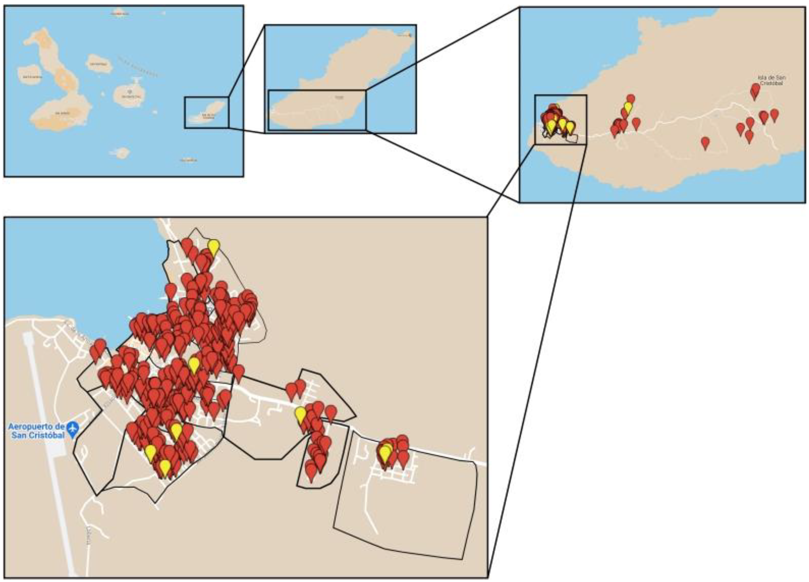
2.1. Sample Collection

Between July and September 2021, 587 blood samples (532 samples screened in the urban area and 55 samples screened in the rural area) were collected from owned dogs (311 males and 276 females) from all 15 neighborhoods of Puerto Baquerizo Moreno (defined as urban) and from rural areas (Figure 2). The sampling strategy consisted of the random selection of blocks of urban and rural neighborhoods. The number of dogs to be selected for blood testing was based on the human population data obtained from the municipality of San Cristobal Island. The observed human–dog ratio on San Cristobal Island was 6:1, which extrapolated to 600 dogs [40].
The minimum sample size was calculated for San Cristobal Island based on an estimated population size [41], the expected prevalence and 95% confidence level (http://www.winepi.net/f101.php, accessed on 21 October 2022 [42]) (Table 2).
The blood collection was performed through a door-to-door approach. Each owner was informed about the importance for the ecosystem of this study through a verbal explanation. For the collection of blood, all owners provided written consent. The animals were restrained manually with the help of a trained assistant and the owner of the dog. Blood was collected from the cephalic vein of the foreleg or the saphenous vein using S-Monovette EDTA K2E tubes. A total volume of 3–5 mL of blood was collected from each animal. At the end of the procedure, all participants received a flyer with a short description of the heartworm disease. Additionally, for each dog, a questionnaire was completed with data concerning the name, age, sex (including the neutered/non-neutered status), breed, type of housing, travel history, previous antiparasitic treatments and vaccinations, medical history, clinical signs at the time of veterinary evaluation. For each dog, the microchip number was recorded, together with the date of the blood collection. Dogs without a microchip were provided with one with the agreement of Galapagos Biosecurity Agency (ABG). Only dogs that were at least 6 months old were included in the study, as the prepatency for D. immitis is usually 6–7 months [43,44].

2.2. Blood Examination

The examination of the blood was conducted at the Galapagos Science Center (GSC) facility of Universidad San Francisco de Quito (USFQ) in San Cristobal Island. All tubes with blood samples collected were stored at 4 °C until examination, for no longer than 12 h. For detection of the microfilariae, the Knott’s test was used [45,46,47,48]. The modified Knott’s test is a concentration method that aims to detect microfilariae species in blood samples [44]. It is a method that is more sensitive than the standard blood smear as it concentrates the microfilariae from a relatively large volume of blood (1 mL). In addition to serologic tests (which detects antigens of adult females), it shows that the dog is still infective to mosquitoes and the infection is patent. The microfilaria of D. immitis was identified based on a morphological differentiation of the character of the species (length of the cephalic space, anterior end to the last body nucleus and the length of the portion of the tail without nuclei) [49,50].

2.3. Statistical Analysis

All statistical analysis was performed using the EpiTools software. Owned dogs were grouped by age, as follows: <1 year, 1–4 years, 4–10 years, >10 years, unknown. Age groups were formed based on the possibility of infection given the time of exposure to possible vectors for each dog. Odds ratios (ORs), 95% confidence intervals (CI) and p-values were calculated for statistically different prevalence by univariate logistic regression. The criterion for statistical significance was p ≤ 0.05. Binomial proportion 95% CI were calculated for positive dogs.

3. Results

Overall, out of 587 tested dogs, 10 were positive for the presence of the microfilaria of D. immitis (1.7%, 95% CI: 0.7–2.8). No other filarial species were identified, and none of the positive dogs presented clinical signs characteristic of D. immitis infection. The population of dogs ranged in age from 6 months to 15 years with a median age of 47 months, and included both non-neutered (190 males, 109 females) and neutered (113 males, 156 females). The prevalence across the different categories is shown in Table 3.
Significant differences for the prevalence between different dog categories were observed only for age (p = 0.001), with a higher risk in dogs aged between 4 and 10 years. No statistical differences were recorded for the sex (p = NS), breed (p = NS), type of housing (p = NS) and environment (p = NS), while the prevalence by neighborhood in the town of Puerto Baquerizo Moreno is shown in Table 4. No significant association was found between neighborhoods and the prevalence of D. immitis (p = NS).

4. Discussion

This study represents the first report to demonstrate the direct presence of circulating microfilaria of D. immitis in the blood of dogs in San Cristobal Island, Galapagos. Similar studies were performed in Floreana [27] and Santa Cruz [26], which assessed the detection of antibodies [27], antigens [22,23,24,25,26], and DNA [26].
Our study completed the previous studies [26,27] regarding the presence of circulating microfilaria of D. immitis in the blood of dogs in the Galapagos Archipelago. Most other studies targeted the detection of antibodies [27], antigens [22,23,24,25,26], or DNA [26]. The presence of an endemic cycle for D. immitis depends on the presence of suitable definitive hosts (dogs), vectors (mosquitoes) and the nematodes. The presence and abundance of mosquitoes and the development of D. immitis larvae in mosquitoes are dependent on climatic factors, the most important being the temperature and availability of mosquito breeding sites [51,52]. Hence, climate and weather have a significant impact on the prevalence of canine heartworm. Dirofilaria immitis L1 larvae need an average temperature higher than 15 °C to develop to L3 in the mosquitoes [53]. Additionally, a recent study demonstrated that cumulative exposure to adequate temperatures can result in the progression of larvae from microfilaria to the L3 infective stage [54]. From this point of view, the Galapagos Archipelago represents a suitable biotope for the development of the mosquito vector and of the D. immitis larvae [55]. Furthermore, sea lions spend more time on land [56], especially in the evening when mosquitoes are active [57].
In the current study, the prevalence of heartworm microfilaremia in dogs (1.7%) was lower than the antigen seroprevalence found in the other islands of the Galapagos Archipelago such as Santa Cruz (6.9%) [26] and Isabela (34%) [22]. Similar studies on other oceanic islands showed a generally higher prevalence: Hawaii (0.7–1.5%) [58], Gran Canaria (12%) [59] and Tenerife (20–34%) [59,60]. Although the climatic conditions on oceanic islands can significantly differ, even over short distances (i.e., coastal vs. highlands), it seems that these are not enough to influence the infection prevalence, as previously demonstrated [60]. Previous studies showed that the prevalence of D. immitis seems to be higher in older dogs [59,60,61], which is consistent with our results. This is associated with the longevity of patent D. immitis infections in dogs, which may be up to 5–7 years [62,63]. Although the prevalence of D. immitis microfilaria in our study was higher in dogs housed outdoors, no statistical significance was found in our study. However, previous studies consider this a risk factor [60,61].
Although the Knott’s test has been shown to be less sensitive than the antigen test [51,52], the method has several advantages: (i) it allows the detection of other filarial species; (ii) it is more specific; (iii) it is cheaper; and (iv) it directly demonstrates the reservoir role of dogs, which was in fact our main rationale for this study. However, antigen detection-based methods can also detect occult heartworm infection even if microfilariae are not present in the blood [53,61,64,65].
Given that: (i) positive dogs were found in the urban environment of the town of Puerto Baquerizo Moreno; (ii) the largest colony of the endangered GSL lives in close vicinity with dogs (closest positive dogs were 350 m away from the GSL colony, which represent a higher risk of infection to mosquitoes that could subsequently feed on sea lions); (iii) dogs are often roaming free in the very close vicinity of GSL; (iv) three mosquito species that have a demonstrated vectorial competence for D. immitis are reported in the town; (v) pinnipeds were previously demonstrated as suitable hosts for D. immitis and can develop clinical signs, we consider that GSLs are exposed to the risk of infection.
Hence, measures to limit the risk of transmission to other dogs, humans or GSLs should be taken, such as: the strict control of dog movement, routine surveillance of dogs for the detection of D. immitis, treatment of positive dogs, implementation of a strategic control program with periodic administration of macrocyclic lactones to all dogs on the island and use of repellent collars on dogs for the prevention of mosquito bites and environmental control of mosquitoes. However, the antiparasitic treatments received by dogs in the Galapagos Islands are very scarce. This is a worrying situation given that antiparasitic treatment in these animals varies over time from a few months to years (Mihalca and Culda, unpublished data). Therefore, treatment campaigns should be carried out for a certain period of time targeting the elimination of the infection from domestic dogs from the island followed by more strict measures to prevent re-introduction
In addition, a neglected reservoir can be represented by cats. These were introduced to the archipelago and they have a negative impact on wildlife [23,55]. Moreover, the majority of cats roam free in the environment and they have a demonstrated role as reservoirs for D. immitis [66,67,68,69].
Although cats are less competent reservoirs than dogs, their role should not be neglected and we recommend their inclusion in future surveillance and control programs for D. immitis.

5. Conclusions

The main scientific impact of the study is that it demonstrates the presence of infective D. immitis larvae in domestic dogs, which, due to their physical proximity to sea lions, could represent a potential infection source. The future perspectives for the research are: (i) to sample the other islands (Isabela, Floreana and Santa Cruz) to complete the data on the prevalence and distribution of D. immitis across the Galapagos Archipelago; (ii) to understand the transmission cycle of D. immitis and identify the most important mosquito vector species; (iii) to evaluate the mosquito blood source around urban and wild rookeries of GSL; (iv) to investigate the possible role of GSLs as reservoirs for D. immitis. Hence, understanding the risk of such an exposure is crucial for future GSL conservation plans and actions.

6. Ethics

This study received approval from the commission for Bioethics within the University of Agricultural Sciences and Veterinary Medicine of Cluj-Napoca (USAMV Cluj-Napoca, Romania) code: 233, and a permit from the Institutional Animal Care and Use Committee (IACUC) (Comité de Ética en el Uso de Animales en Investigación y Docencia de la USFQ); code: 006. Fieldwork at the Galapagos was performed in coordination with the Galapagos Biosecurity Agency ABG under an agreement for Scientific Collaboration between USFQ and the ABG agency.

Supplementary Materials

Interactive Map_San Cristobal Island (Red pins represent the negative dogs and yellow pins represent the positive dogs, and green ones represent sea lions’ location). https://www.google.com/maps/d/edit?mid=1IfYrPtQGO455j76RSZust5gkdF80hCML&usp=sharin.

Author Contributions

Manuscript preparation, C.A.C., D.P.-R., L.R. and A.D.M.; study design, C.A.C., J.G., P.E., R.L.V., M.C., L.R. and A.D.M.; field work, C.A.C., R.D., A.C.B., A.S.C., T.D., J.G., L.R. and A.D.M.; lab work, C.A.C., R.D., A.C.B., A.S.C., T.D. and A.D.M.; permits, J.G., P.E., R.L.V., M.C. and L.R.; manuscript preparation, D.P.-R., L.R. and A.D.M.; conceptualization, A.D.M.; funding, A.D.M. All authors have read and agreed to the published version of the manuscript.

Funding

Part of the study was funded through an ERASMUS+ KA107 mobility grant by the University of Agricultural Sciences and Veterinary Medicine of Cluj-Napoca (USAMV Cluj-Napoca, Romania) and Universidad San Francisco de Quito (USFQ), private funds of the volunteers and co-funding from the Department of Parasitology and Parasitic Diseases at USAMV Cluj-Napoca, Romania.

Institutional Review Board Statement

The animal study protocol was approved by the Ethics Committee of Agricultural Sciences and Veterinary Medicine of Cluj-Napoca (USAMV Cluj-Napoca, Romania) (protocol code 233 and 05.01.2021) and Institutional Animal Care and Use Committee (IACUC) (Comité de Ética en el Uso de Animales en Investigación y Docencia de la USFQ) (protocol code 006 and 19.08.2021).

Informed Consent Statement

Not applicable.

Data Availability Statement

The dataset supporting the conclusions of this study are included in the present article or in the Supplementary Materials.

Acknowledgments

The authors are grateful to the staff of the Galapagos Science Center on San Cristobal Island, Galapagos, for providing laboratory space, equipment and laboratory reagents for the microscopical examination of the samples. We dedicate this paper to the memory of Jaime Grijalva who tragically passed away during the preparation of this manuscript.

Conflicts of Interest

The authors declare they have no actual or potential conflict of interest.

References

  1. Darwin, C. On the Origin of Species; Routledge: London, UK, 1859. [Google Scholar]
  2. Cittadino, E. Nature as the Laboratory: Darwinian Plant Ecology in the German Empire; Cambridge University Press: Cambridge, UK, 1990; pp. 1880–1900. [Google Scholar]
  3. Edgar, G.J.; Banks, S.; Bensted-Smith, R.; Calvopiña, M.; Chiriboga, A.; Garske, L.E.; Henderson, S.; Miller, K.A.; Salazar, S. Conservation of threatened species in the Galapagos Marine Reserve through identification and protection of marine key biodiversity areas. Aquat. Conserv. Mar. Freshw. Ecosyst. 2008, 18, 955–968. [Google Scholar] [CrossRef]
  4. Barrow, M. Nature’s Ghosts: Confronting Extinction from the Age of Jefferson to the Age of Ecology; University of Chicago Press: Chicago, IL, USA, 2009. [Google Scholar]
  5. Swash, A.; Still, R. Birds, Mammals, and Reptiles of the Galapagos Islands; Bloomsbury Publishing PLC: London, UK, 2005. [Google Scholar]
  6. Jiménez-Uzcátegui, G.; Milstead, B.; Márquez, C.; Zabala, J.; Buitrón, P.; Llerena, A.; Salazar, S.; Fessl, B. Galapagos vertebrates: Endangered status and conservation actions. Galapagos Rep. 2007, 2006, 104–110. [Google Scholar]
  7. Harvell, C.D.; Kim, K.; Burkholder, J.M.; Colwell, R.R.; Epstein, P.R.; Grimes, D.J.; Hofmann, E.E.; Lipp, E.K.; Osterhaus, A.D.M.E.; Overstreet, R.M.; et al. Emerging Marine Diseases--Climate Links and Anthropogenic Factors. Science 1999, 285, 1505–1510. [Google Scholar] [CrossRef]
  8. Alava, J.J.; Salazar, S. Status and Conservation of Otariids in Ecuador and the Galápagos Islands. In Sea Lions of the World; AK-SG-06-01; Alaska Sea Grant College Program: Fairbanks, AK, USA, 2006; pp. 495–519. [Google Scholar]
  9. Páez-Rosas, D.; Guevara, N. Management strategies and conservation status in populations of Galapagos sea lion (Zalophus wollebaeki). In Tropical Pinnipeds, Bio-Ecology, Threats and Conservation; CRC Press/Taylor & Francis Group: Boca Raton, FL, USA, 2017. [Google Scholar]
  10. Páez-Rosas, D.; Torres, J.; Espinoza, E.; Marchetti, A.; Seim, H.; Riofrío-Lazo, M. Declines and recovery in endangered Galapagos pinnipeds during the El Niño event. Sci. Rep. 2021, 11, 8785. [Google Scholar] [CrossRef] [PubMed]
  11. Trillmich, F. Zalophus wollebaeki. The IUCN Red List of Threatened Species 2015. e.T41668A45230540. Available online: https://doi.org/10.2305/IUCN.UK.2015-2.RLTS.T41668A45230540.en (accessed on 25 August 2022).
  12. Denkinger, J.; Gordillo, L.; Montero-Serra, I.; Murillo, J.C.; Guevara, N.; Hirschfeld, M.; Fietz, K.; Rubianes, F.; Dan, M. Urban life of Galapagos sea lions (Zalophus wollebaeki) on San Cristobal Island, Ecuador: Colony trends and threats. J. Sea Res. 2015, 105, 10–14. [Google Scholar] [CrossRef]
  13. Denkinger, J.; Guevara, N.; Ayala, S.; Murillo, J.C.; Hirschfeld, M.; Montero-Serra, I.; Fietz, K.; Goldstein, T.; Ackermann, M.; Barragán, V.; et al. Pup Mortality and Evidence for Pathogen Exposure in Galapagos Sea Lions (Zalophus wollebaeki) on San Cristobal Island, Galapagos, Ecuador. J. Wildl. Dis. 2017, 53, 491–498. [Google Scholar] [CrossRef]
  14. Riofrío-Lazo, M.; Arreguín-Sánchez, F.; Páez-Rosas, D. Population Abundance of the Endangered Galapagos Sea Lion Zalophus wollebaeki in the Southeastern Galapagos Archipelago. PLoS ONE 2017, 12, e0168829. [Google Scholar] [CrossRef]
  15. Benelli, G.; Mehlhorn, H. Mosquito-Borne Diseases; Parasitology Research Monographs; Springer: Cham, Switzerland, 2018; Volume 10. [Google Scholar]
  16. Becker, N.; Petrić, D.; Zgomba, M.; Boase, C.; Madon, M.B.; Dahl, C.; Kaiser, A. Mosquitoes: Identification, Ecology and Control; Springer Nature: Singapore, 2020. [Google Scholar]
  17. Sarzosa, M.S.; Duignan, P.; DeRango, E.J.; Field, C.; Ríos, C.; Sanchez, S.; Espinoza, E.; Loyola, A.; Rueda, D.; Páez-Rosas, D. Occurrence of Mycoplasmas in Galapagos Sea Lions (Zalophus wollebaeki) and their Association with Other Respiratory Pathogens. J. Wildl. Dis. 2021, 57, 623–627. [Google Scholar] [CrossRef]
  18. Walden, H.D.; Grijalva, C.J.; Páez-Rosas, D.; Hernandez, J.A. Intestinal Parasites in Galapagos Sea Lions (Zalophus wollebaeki) Sivertsen, 1953 on San Cristóbal Island, Galapagos, Ecuador. J. Parasitol. 2018, 104, 718–721. [Google Scholar] [CrossRef]
  19. Burek, K.A.; Gulland, F.M.D.; Sheffield, G.; Beckmen, K.B.; Keyes, E.; Spraker, T.R.; Smith, A.W.; Skilling, D.E.; Evermann, J.F.; Stott, J.L.; et al. Infectious disease and the decline of Steller sea lions (Eumetopias jubatus) in Alaska, USA: Insights from serologic data. J. Wildl. Dis. 2005, 41, 512–524. [Google Scholar] [CrossRef]
  20. Dubey, J.; Zarnke, R.; Thomas, N.; Wong, S.; Bonn, W.; Briggs, M.; Davis, J.; Ewing, R.; Mense, M.; Kwok, O.; et al. Toxoplasma gondii, Neospora caninum, Sarcocystis neurona, and Sarcocystis canis-like infections in marine mammals. Vet. Parasitol. 2003, 116, 275–296. [Google Scholar] [CrossRef]
  21. Conrad, P.; Miller, M.; Kreuder, C.; James, E.; Mazet, J.; Dabritz, H.; Jessup, D.; Gulland, F.; Grigg, M. Transmission of Toxoplasma: Clues from the study of sea otters as sentinels of Toxoplasma gondii flow into the marine environment. Int. J. Parasitol. 2005, 35, 1155–1168. [Google Scholar] [CrossRef]
  22. Levy, J.; Crawford, P.; Lappin, M.; Dubovi, E.; Levy, M.; Alleman, R.; Tucker, S.; Clifford, E. Infectious Diseases of Dogs and Cats on Isabela Island, Galapagos. J. Vet. Intern. Med. 2008, 22, 60–65. [Google Scholar] [CrossRef] [PubMed]
  23. Gingrich, E.; Scorza, A.; Clifford, E.; Olea-Popelka, F.; Lappin, M. Intestinal parasites of dogs on the Galapagos Islands. Vet. Parasitol. 2010, 169, 404–407. [Google Scholar] [CrossRef] [PubMed]
  24. Adams, D.J.; Rosenberg, D.E.; Yirui, H. Prevalence of vector-borne diseases in a sample of client-owned dogs on Santa Cruz in the Galápagos Islands: A pilot study. Vet. Parasitol. Reg. Stud. Rep. 2016, 6, 28–30. [Google Scholar] [CrossRef]
  25. Diaz, N.M.; Mendez, G.S.; Grijalva, C.J.; Walden, H.S.; Cruz, M.; Aragon, E.; Hernandez, J.A. Dog overpopulation and burden of exposure to canine distemper virus and other pathogens on Santa Cruz Island, Galapagos. Prev. Vet. Med. 2016, 123, 128–137. [Google Scholar] [CrossRef]
  26. Jimenez, I.A.; Mariño, P.A.V.; Stapleton, G.S.; Prieto, J.B.; Bowman, D.D. Canine vector-borne disease in domestic dogs on Isla Santa Cruz, Galápagos. Vet. Parasitol. Reg. Stud. Rep. 2020, 19, 100373. [Google Scholar] [CrossRef]
  27. Barnett, B.D. Dogs of the Galapagos Islands: Evolution, Ecology, Impact and Control (Feral, Galapagos, Dogs). Ph.D. Thesis, University of California, Davis, CA, USA, 1985. [Google Scholar]
  28. Hendrix, C.M.; Brunner, C.J.; Bellamy, L.K. Natural transmission of Dirofilaria immitis by Aedes aegypti. J. Am. Mosq. Control Assoc. 1986, 2, 48–51. [Google Scholar] [PubMed]
  29. Bataille, A.; Cunningham, A.A.; Cedeño, V.; Cruz, M.; Eastwood, G.; Fonseca, D.M.; Causton, C.E.; Azuero, R.; Loayza, J.; Martinez, J.D.C.; et al. Evidence for regular ongoing introductions of mosquito disease vectors into the Galápagos Islands. Proc. R. Soc. B Boil. Sci. 2009, 276, 3769–3775. [Google Scholar] [CrossRef]
  30. Bataille, A.; Cunningham, A.A.; Cedeño, V.; Patiño, L.; Constantinou, A.; Kramer, L.D.; Goodman, S.J. Natural colonization and adaptation of a mosquito species in Galápagos and its implications for disease threats to endemic wildlife. Proc. Natl. Acad. Sci. USA 2009, 106, 10230–10235. [Google Scholar] [CrossRef]
  31. Tiawsirisup, S.; Nithiuthai, S. Vector competence of Aedes aegypti (L.) and Culex quinquefasciatus (Say) for Dirofilaria immitis (Leidy). Southeast Asian J. Trop. Med. Public Health 2006, 37, 110. [Google Scholar] [PubMed]
  32. Vezzani, D.; Eiras, D.F.; Wisnivesky, C. Dirofilariasis in Argentina: Historical review and first report of Dirofilaria immitis in a natural mosquito population. Vet. Parasitol. 2006, 136, 259–273. [Google Scholar] [CrossRef] [PubMed]
  33. King, J.E. Seals of the World; British Museum of Natural History: London, UK, 1964. [Google Scholar]
  34. Alho, A.M.; Marcelino, I.; Colella, V.; Flanagan, C.; Silva, N.; Correia, J.J.; Latrofa, M.S.; Otranto, D.; de Carvalho, L.M. Dirofilaria immitis in pinnipeds and a new host record. Parasites Vectors 2017, 10, 142. [Google Scholar] [CrossRef]
  35. Kang, S.S.; Kim, J.H.; Kwon, Y.B.; Park, S.H. A harbor seal infection with Dirofilaria. J. Vet. Clin. 2002, 19, 92–94. [Google Scholar]
  36. Forrester, D.J.; Jackson, R.F.; Miller, J.F.; Townsend, B.C. Heartworms in captive California sea lions. J. Am. Vet. 1973, 163, 568–570. [Google Scholar]
  37. White, G.L. Dirofilaria immitis and Heartworm Disease in the California Sea Lion. J. Zoo Anim. Med. 1975, 6, 23–24. [Google Scholar] [CrossRef]
  38. Sato, T.; Higuchi, T.; Shibuya, H.; Ohba, S.; Nogami, S.; Shirai, W.; Watanabe, H.; Honda, S. Lingual squamous cell carcinoma in a California sea lion (Zalophus californianus). J. Zoo Wildl. Med. 2002, 33, 367–370. [Google Scholar] [CrossRef]
  39. Farriols, M.; Arellano-Carbajal, F.; Elorriaga-Verplancken, F.R.; Adame-Fernández, K.; Garrido, E.; Álvarez-Martínez, R.C.; Bárcenas, R.T.; Flores-Morán, A.E.; Acevedo-Whitehouse, K. Filarial infections in California sea lions vary spatially within the Gulf of California, Mexico. Parasitol. Res. 2020, 119, 1281–1290. [Google Scholar] [CrossRef] [PubMed]
  40. Grijalva, C.J.; Walden, H.S.; Crawford, P.C.; Levy, J.K.; Pine, W.E.; Hernandez, J.A. Estimating the dog population, responsible pet ownership, and intestinal parasitism in dogs in Quito, Ecuador. Res. Sq. 2021, 1–15. [Google Scholar] [CrossRef]
  41. Vergara, S.P.P. Galápagos, Hábitats Humanos. Ph.D. Thesis, Pontificia Universidad Católica de Chile, Santiago, Chile, 2020. [Google Scholar]
  42. Working in Epidemiology. 2006. Available online: http://www.winepi.net/f101.php (accessed on 21 October 2022).
  43. Patel, J.R.; Devi, S.; Varshney, J.P.; Jadhav, K.M. Epizootiological observations on canine microfilaremia in Gujarat state, India. Vet. World 2018, 11, 1564–1568. [Google Scholar] [CrossRef]
  44. Saari, S.; Näreaho, A.; Nikander, S. Chapter 5-Nematoda (roundworms). In Canine Parasites and Parasitic Diseases; Academic Press: Cambridge, MA, USA, 2019; pp. 83–149. [Google Scholar] [CrossRef]
  45. Knott, J.; Earle, K. A method for making microfilarial surveys on day blood. Trans. R. Soc. Trop. Med. Hyg. 1939, 33, 191–196. [Google Scholar] [CrossRef]
  46. Newton, W.L.; Wright, W.H. The Occurrence of a Dog Filariid Other than Dirofilaria immitis in the United States. J. Parasitol. Res. 1956, 42, 246. [Google Scholar] [CrossRef]
  47. Georgi, J.R.; Georgi, M.E.; Theodorides, V.J. Parasitology for Veterinarians, 5th ed.; W.B. Saunders: Philadelphia, PA, USA, 1990; 412p. [Google Scholar]
  48. Magnis, J.; Lorentz, S.; Guardone, L.; Grimm, F.; Magi, M.; Naucke, T.J.; Deplazes, P. Morphometric analyses of canine blood microfilariae isolated by the Knott’s test enables Dirofilaria immitis and D. repens species-specific and Acanthocheilonema (syn. Dipetalonema) genus-specific diagnosis. Parasites Vectors 2013, 6, 48. [Google Scholar] [CrossRef] [PubMed]
  49. Traversa, D.; Di Cesare, A.; Conboy, G. Canine and feline cardiopulmonary parasitic nematodes in Europe: Emerging and underestimated. Parasites Vectors 2010, 3, 62. [Google Scholar] [CrossRef]
  50. Liotta, J.L.; Sandhu, G.K.; Rishniw, M.; Bowman, D.D. Differentiation of the microfilariae of Dirofilaria immitis and Dirofilaria repens in stained blood films. J. Parasitol. 2013, 99, 421–425. [Google Scholar] [CrossRef]
  51. Montarsi, F.; Ciocchetta, S.; Devine, G.; Ravagnan, S.; Mutinelli, F.; Di Regalbono, A.F.; Otranto, D.; Capelli, G. Development of Dirofilaria immitis within the mosquito Aedes (Finlaya) koreicus, a new invasive species for Europe. Parasites Vectors 2015, 8, 177. [Google Scholar] [CrossRef]
  52. Younes, L.; Barré-Cardi, H.; Bedjaoui, S.; Ayhan, N.; Varloud, M.; Mediannikov, O.; Otranto, D.; Davoust, B. Dirofilaria immitis and Dirofilaria repens in mosquitoes from Corsica Island, France. Parasites Vectors 2021, 14, 427. [Google Scholar] [CrossRef]
  53. Meriem-Hind, B.M.; Mohamed, M. Prevalence of canine Dirofilaria immitis infection in the city of Algiers, Algeria. Afr. J. Agric. Res. 2009, 4, 1097–1100. [Google Scholar] [CrossRef]
  54. Ledesma, N.; Harrington, L. Fine-scale temperature fluctuation and modulation of Dirofilaria immitis larval development in Aedes aegypti. Vet. Parasitol. 2015, 209, 93–100. [Google Scholar] [CrossRef]
  55. Torres, M.L.; Mena, C.F. Understanding Invasive Species in the Galapagos Islands; Springer: Cham, Switzerland, 2018. [Google Scholar]
  56. Montero-Serra, I.; Páez-Rosas, D.; Murillo, J.; Vegas-Vilarrúbia, T.; Fietz, K.; Denkinger, J. Environment-driven changes in terrestrial habitat use and distribution of the Galapagos sea lion. Endanger. Species Res. 2014, 24, 9–19. [Google Scholar] [CrossRef]
  57. Šebesta, O.; Gelbič, I.; Peško, J. Daily and seasonal variation in the activity of potential vector mosquitoes. Open Life Sci. 2011, 6, 422–430. [Google Scholar] [CrossRef]
  58. Little, S.; Braff, J.; Place, J.; Buch, J.; Dewage, B.G.; Knupp, A.; Beall, M. Canine infection with Dirofilaria immitis, Borrelia burgdorferi, Anaplasma spp., and Ehrlichia spp. in the United States, 2013–2019. Parasites Vectors 2021, 14, 10. [Google Scholar] [CrossRef] [PubMed]
  59. Montoya-Alonso, J.; Carretón, E.; Corbera, J.A.; Juste, M.; Mellado, I.; Morchón, R.; Simón, F. Current prevalence of Dirofilaria immitis in dogs, cats and humans from the island of Gran Canaria, Spain. Vet. Parasitol. 2011, 176, 291–294. [Google Scholar] [CrossRef] [PubMed]
  60. Montoya-Alonso, J.A.; Morales, M.; Juste, M.C.; Banares, A.; Simon, F.; Genchi, C. Seroprevalence of canine heartworm disease (Dirofilaria immitis) in Tenerife island: An epidemiological update. Parasitol. Res. 2006, 100, 103–105. [Google Scholar] [CrossRef]
  61. Yaman, M.; Guzel, M.; Koltas, I.; Demirkazik, M.; Aktas, H. Prevalence of Dirofilaria immitis in dogs from Hatay province, Turkey. J. Helminthol. 2009, 83, 255–260. [Google Scholar] [CrossRef]
  62. Bowman, D.D.; Atkins, C.E. Heartworm Biology, Treatment, and Control. Vet. Clin. N. Am. Small Anim. Pract. 2009, 39, 1127–1158. [Google Scholar] [CrossRef]
  63. Nguyen, C.; Koh, W.L.; Casteriano, A.; Beijerink, N.; Godfrey, C.; Brown, G.; Emery, D.; Šlapeta, J. Mosquito-borne heartworm Dirofilaria immitis in dogs from Australia. Parasites Vectors 2016, 9, 535. [Google Scholar] [CrossRef]
  64. Wang, L.-C. Canine filarial infections in north Taiwan. Acta Trop. 1997, 68, 115–120. [Google Scholar] [CrossRef]
  65. Panarese, R.; Iatta, R.; Mendoza-Roldan, J.A.; Szlosek, D.; Braff, J.; Liu, J.; Beugnet, F.; Dantas-Torres, F.; Beall, M.J.; Otranto, D. Comparison of Diagnostic Tools for the Detection of Dirofilaria immitis Infection in Dogs. Pathogens 2020, 9, 499. [Google Scholar] [CrossRef]
  66. Simón, F.; Siles-Lucas, M.; Morchón, R.; González-Miguel, J.; Mellado, I.; Carretón, E.; Montoya-Alonso, J.A. Human and Animal Dirofilariasis: The Emergence of a Zoonotic Mosaic. Clin. Microbiol. Rev. 2012, 25, 507–544. [Google Scholar] [CrossRef]
  67. Ash, L.R. Helminth Parasites of Dogs and Cats in Hawaii. J. Parasitol. Res. 1962, 48, 63–65. [Google Scholar] [CrossRef]
  68. Traversa, D.; Aste, G.; Milillo, P.; Capelli, G.; Pampurini, F.; Tunesi, C.; Santori, D.; Paoletti, B.; Boari, A. Autochthonous foci of canine and feline infections by Dirofilaria immitis and Dirofilaria repens in central Italy. Vet. Parasitol. 2010, 169, 128–132. [Google Scholar] [CrossRef] [PubMed]
  69. Pennisi, M.G.; Tasker, S.; Hartmann, K.; Belák, S.; Addie, D.; Boucraut-Baralon, C.; Egberink, H.; Frymus, T.; Hofmann-Lehmann, R.; Hosie, M.; et al. Dirofilarioses in cats: European guidelines from the ABCD on prevention and management. J. Feline Med. Surg. 2020, 22, 442–451. [Google Scholar] [CrossRef] [PubMed]
Figure 1. Close contact between dogs and Galapagos sea lions.
Pathogens 11 01287 g001
Figure 2. Distribution of sampled and positive dogs. A detailed interactive map is available as Supplementary Materials here (https://www.google.com/maps/d/edit?mid=1IfYrPtQGO455j76RSZust5gkdF80hCML&usp=sharing). Red pins represent the negative dogs and yellow pins represent the positive dogs.
Pathogens 11 01287 g002
Table 1. Distribution of pathogens on the Galapagos Islands.
PathogenIsabelaSanta CruzSan CristobalFloreana
Canine distemper virus++
Canine parvovirus++
Canine adenovirus++
Canine parainfluenza virus++
Ehrlichia spp.++
Anaplasma spp.++
Bartonella spp.+
Mycoplasma haemocanis+
Giardia spp.++
Leishmania donovani+
Sarcocystis canis++
Cystoisospora spp.++
Cryptosporidium spp.+
Toxocara spp.+++
Ancylostoma spp.+++
Dirofilaria immitis+++
+ = presence of pathogens; − = absence/non-testing of pathogens.
Table 2. Sample size selection and randomization.
AreaEstimated Human Population 1Estimated Number of Dogs 2Min. Sample SizeNo. of Collected Samples
San Cristobal73301222269587
1— [41]; 2—based on the 6:1 ratio according to [40].
Table 3. Prevalence of microfilariae of D. immitis in dogs from San Cristobal by sex, age, environment and housing, assessed by Knott’s test.
No. of SamplesPositive (%)OR95% CIp-Value
Total58710 (1.7%) 0.7–2.8%
Sex
Males3115 (1.6%)0.88560.2–3.0%1
Females2765 (1.8%)0.2–3.4%
Age
<1 year740NA0%0.001 *
1–4 years23600%
4–10 years20810 (4.8%)1.9–7.8%
>10 years4300%
unknown2600%
Environment
Urban5328 (1.5%)0.40460.5–2.6%0.5377
Rural552 (3.6%)0–8.6%
Housing
Outdoor2588 (3.1%)NA1–5.2%0.0571
Indoor10900%
Outdoor and indoor2202 (0.9%)0–2.2%
OR—odds ratio; 95% CI—95% confidence interval; NA—not applicable; *—statistically significant.
Table 4. Prevalence of microfilariae of D. immitis in dogs from Puerto Baquerizo Moreno by neighborhood, assessed by Knott’s test.
Neighborhood
(Barrio)
No. of DogsPositivePrevalence (%)95% CIp-Value
Albatros300000.2136
Algarrobos42000
Cactus4912%0–6.0%
Central18000
Divino Niño2414.1%0–12.1%
Manatial and Isla Sur2114.7%0–13.8%
Estacion Terrena8933.3%0–7.2%
Fragatas48000
Frío8000
El Manzanillo23000
Las Palmeras2328.7%0–20.2%
Peñas Altas76000
Peñas Bajas35000
Playa Mann23000
San Francisco23000
95% CI—95% confidence interval.
Publisher’s Note: MDPI stays neutral with regard to jurisdictional claims in published maps and institutional affiliations.

