Case Report

Bacillus simplex as the Most Probable Culprit of Penetrating Trauma Infection: A Case Report

by
Panagiota Xaplanteri
1,*,
Dimitrios S. Serpanos
2,
Ellie Dorva
2,
Tatiana Beqo-Rokaj
3,
Eleni Papadogeorgaki
4 and
Alexandra Lekkou
5,6
1
Department of Microbiology, General Hospital of Eastern Achaia, 25001 Kalavrita, Greece
2
Department of Internal Medicine, General Hospital of Eastern Achaia, 25001 Kalavrita, Greece
3
Department of Primary Healthcare, General Hospital of Eastern Achaia, 25001 Kalavrita, Greece
4
Central Diagnostic Laboratories, Hygeia Hospital Athens, 15123 Athens, Greece
5
Department of Internal Medicine, University General Hospital of Patras, 26504 Patras, Greece
6
Department of Infectious Diseases, University General Hospital of Patras, 26504 Patras, Greece
*
Author to whom correspondence should be addressed.
Pathogens 2022, 11(10), 1203; https://doi.org/10.3390/pathogens11101203
Submission received: 6 October 2022 / Accepted: 17 October 2022 / Published: 18 October 2022
(This article belongs to the Special Issue 10th Anniversary of Pathogens—Feature Papers)

Abstract

:
Bacillus simplex is an environmental microorganism found in soil. Herein, we present the case of a 69-year-old Greek male patient who attended the Emergency Department at our hospital. The patient complained of lower right extremity swelling and pain, after suffering penetrating trauma whilst doing farm work in a rural area. Swab aerobic cultures revealed Bacillus simplex as identified by MALDI-TOF Mass Spectrometry. The strain was susceptible to vancomycin, imipenem, clindamycin, and ciprofloxacin. Our patient refused hospitalization; therefore, both ciprofloxacin and clindamycin were registered for a total of 19 days. No complications were experienced, and he recovered fully. In our case, the thorough cleaning of the ulcer bed prior to sample collection, the fact that it was the only microorganism isolated, and the wound’s aggravating mechanism led the authors of the present study to the conclusion that B. simplex was the most probable culprit of the infection. To our knowledge, this is the second probable case of B. simplex infection described worldwide, and the first in Greece.

1. Introduction

Bacillus simplex is an environmental microorganism found in soil [1]. Herewith presented is the case of a 69-year-old Greek male patient who attended the Emergency Department. The patient complained of lower right extremity swelling and pain, after suffering penetrating trauma whilst doing farm work in a rural area. Swab aerobic cultures revealed a microorganism with the shape and characteristics of Bacillus, identified by MALDI-TOF Mass Spectrometry as Bacillus simplex. To our knowledge this is the second probable case of B. simplex infection described worldwide, and the first in Greece.

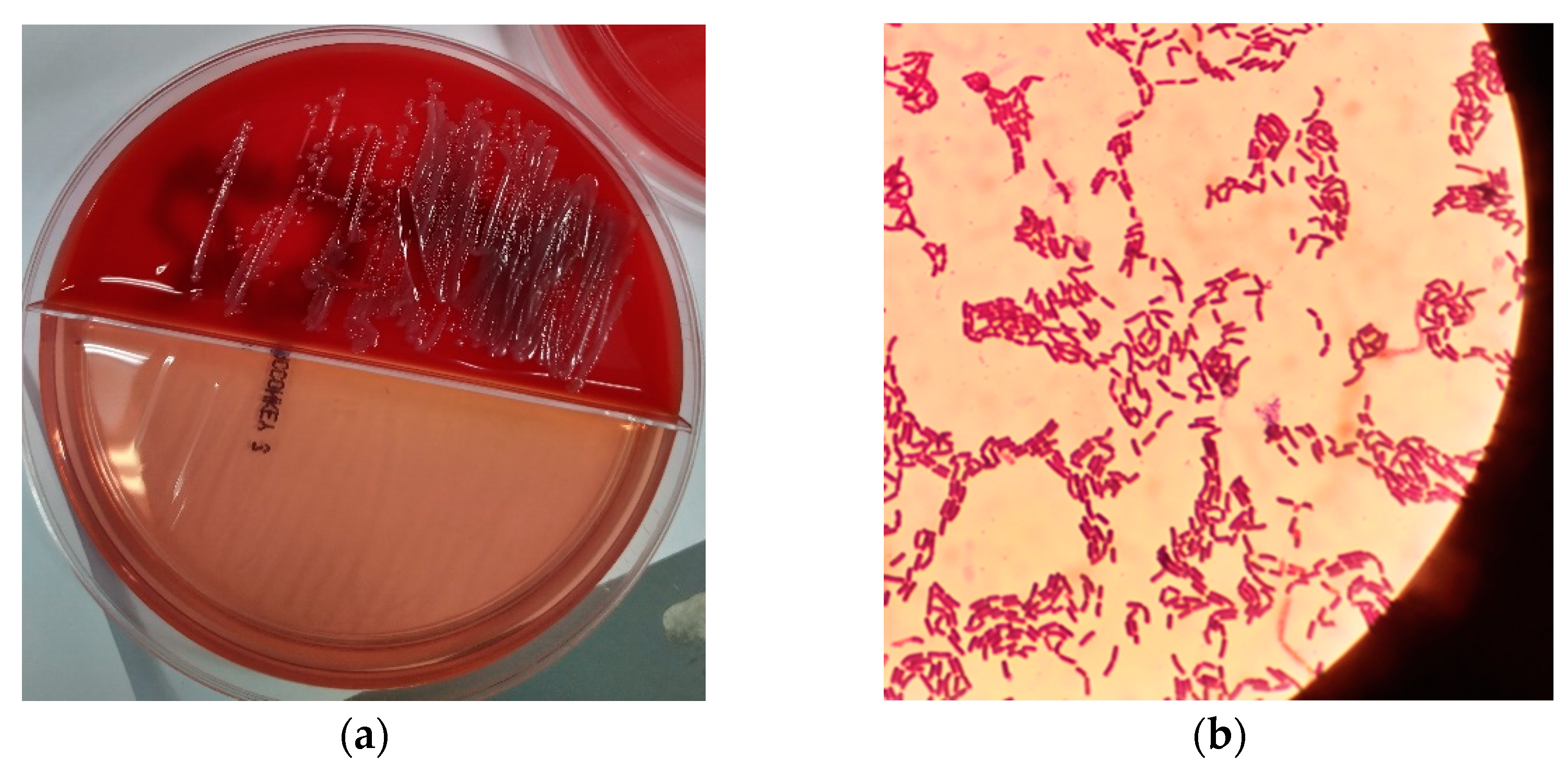
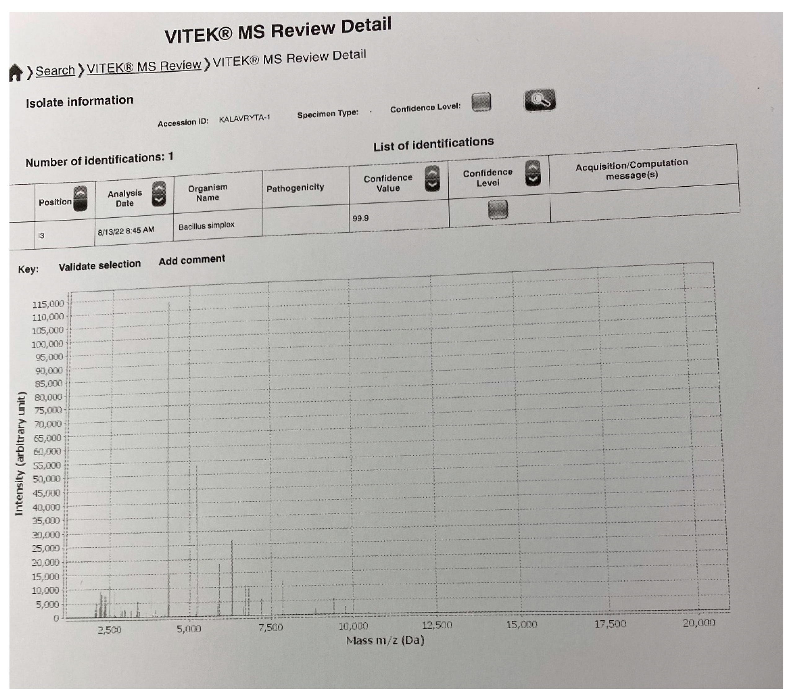
2. Presentation of Case

A 69-year-old male Greek patient with a significant history of chronic obstructive pulmonary disease, hypertension and being an active smoker attended the Emergency Department of our hospital. He complained of lower right extremity swelling and pain, after suffering penetrating trauma whilst doing farm work in a rural area. Physical examination revealed cellulitis, a 10 cm diameter ulcer just above the ankle, tenderness, swelling, erythema, and intense edema from the middle of the leg to the tip of the foot. The patient was afebrile, blood pressure was 160/94 mmHg, pulse rate 99/min, Spo2 98%. The area of the ulcer was cleaned thoroughly, and swab cultures were obtained from the ulcer’s crater according to the Levine technique [2].
Due to the extended edema, Vascular Ultrasound was performed and proved negative for deep vein thrombosis. Blood cultures were not obtained. The patient did not accept hospital admission and was released with ciprofloxacin 500 mg twice a day and clindamycin 600 mg thrice a day whilst awaiting the swab cultures results.
Laboratory findings revealed an elevated white blood cell count of 14300/mm3 (normal values 4000–10000/mm3), elevated C-reactive protein levels of 4.6 mg/dL (normal values 0.0–0.9 mg/dL), whereas serum glucose, renal and liver parameters were within normal range.
The swab samples were inoculated on partitioned agar plates containing 5% sheep blood agar and McConkey agar number 3, by Thermo Scientific™ (Oxoid Ltd., Wade Road, Hampshire, UK). No direct Gram or Ziehl-Neelsen staining were performed from the ulcer. Swab cultures for anaerobes and fungus proved negative. Aerobic culture was positive onto blood agar plate after incubation at 37 °C overnight, with no swarming or hemolysis present (Figure 1a). Gram stain revealed a microorganism with the shape of Bacillus, catalase-positive, with no obvious endospores—if present, they did not alter the shape of the bacterium (Figure 1b). Gram stain was variable. MALDI-TOF Mass Spectrometry (Vitek MS BioMerieux, Athens, Greece, BioMerieux Hellas) identified the strain as Bacillus simplex with confidence value 99.9. The respective image of the bacterium identification is shown in Figure 2.
The breakpoint criteria for our strain were compared to those of Staphylococcus spp. according to the European Committee on Antimicrobial Susceptibility Testing (EUCAST) guidelines [3]. For disk diffusion susceptibility testing, an imipenem disk containing 10 mcg, clindamycin disk containing 2 mcg and ciprofloxacin disk containing 5 mcg were provided from Vaktro Scientific, Panepistimiou 195 Street, 26443, Patras, Achaia, Greece. Using the disk diffusion susceptibility testing our strain proved susceptible to imipenem, clindamycin and ciprofloxacin. As our strain proved susceptible to ciprofloxacin and clindamycin, the initial antibiotic scheme did not change. On follow up after 14 days of the antimicrobial agents’ registration, clinically there was significant improvement but there were still signs of infection. Therefore, the patient continued the therapy for a total of 19 days. No complications were experienced, and the patient recovered fully.

3. Discussion

Bacillus species are spore-forming rods that stain gram positive or gram variable and grow in aerobic conditions. Their natural habitat is the soil [4,5]. Non-Bacillus anthracis infections, most commonly due to Bacillus cereus, are related to food poisoning, ocular infections, deep-seated soft tissue infections, and systemic infections [5]. The taxonomic relationship between many Bacillus species is difficult to decipher as there is similarity in chromosomal structure and phenotypic characteristics among them [4,6]. The formation of endospores is the reaction of the Bacillus to inhospitable environmental conditions, such as extreme temperatures, radiation, and disinfectants [4].
Meyer and Gottheil first described Bacillus simplex in 1901 as an environmental microorganism found in soil [1]. B. simplex bacterial cells produce oval endospores [7]. In our sample no obvious endospores were detected with gram stain (Figure 1b). B. simplex presents a variable gram reaction. Most strains are strictly aerobic [7]. This applies also to our strain, which appeared both gram positive and gram negative and developed under aerobic conditions.
Various virulence factors of B. simplex have been described. The microorganism produces a heat-stable toxin, like cereulide of B. cereus, that causes emesis in humans. This toxin inhibits mitochondrial activity and causes liver failure and cellular damage. It also inhibits human natural killer cells of the immune system [8]. B. simplex strain N65.1 also produces hemolysin BL (HBL), non-hemolytic enterotoxin NHE and CytK (or hemolysin IV) [9,10,11]. Hemolysins lyse erythrocytes. As a result, nutrients and iron-binding proteins are released for the pathogenic bacteria to use for their survival, and they provoke damage to epithelial cells, enhancing the microorganisms’ advance and access to deeper layers [11].
NHE has hemolytic activity towards erythrocytes from several mammalian species [11]. Both HBL and NHE are related to the hemolytic and cytotoxic toxin cytolysin A (ClyA, or HlyE or SheA), expressed during anaerobic growth of Escherichia coli, Shigella flexneri and Salmonella enterica serovars Typhi and Paratyphi A [8]. HBL component B and cytolysin A share a similar tertiary structure [8]. HBL has dermonecrotic activity and is cytotoxic towards Vero cells and retinal tissue [8]. NHE forms transmembrane pores which aggravate colloid osmotic lysis and cell death [8]. The pore-forming ability in phospholipid membranes of NHE is innate and consequently independent of a binding receptor [8]. CytK has cytotoxic, necrotic and hemolytic activities. It was first described in 2000 [11,12]. In B. cereus and Bacillus cytotoxicus strains, CytK relates to bloody diarrhea and increased mortality and has an amino-acid sequence 30% identical to α-hemolysin (α-toxin, Hla) of Staphylococcus aureus [11]. A-hemolysin of S. aureus is a well-described cause of dermonecrosis and lethal infection [13]. S. aureus α-toxin binds on the host cell membrane via its receptor ADAM10, and forms pores through the eukaryotic lipid bilayer. As a result, molecules with low molecular weight, like calcium ions, potassium ions and ATP leak through the pore, leading to cell lysis. The increased intracellular calcium triggers hydrolysis of membrane phospholipids and the conversion of arachidonic acid to leukotrienes, prostanoids, and thromboxane A2. In addition, there is alteration in the cell signaling pathways via the cytoplasmic tail of ADAM10, which induces NF-κB nuclear translocation and the production of proinflammatory cytokines IL-1β, and IL-6. The cascade of alterations that follow includes recruitment of immune cells, increased vascular permeability, tissue edema and inflammation. Pneumocytes and keratinocytes are key targets as well as epithelial and endothelial cells, T cells, monocytes, macrophages and neutrophils [13,14,15]. In addition, α-toxin hastens cell death of Th1 cells via Ca2+-mediated activation. This cell death is not induced via caspases’ activation [16]. B. simplex strain N58.2 has high cytotoxic potential, similar to B. cereus group III strains that have high cytotoxic activity [10]. Although there is little evidence in literature regarding the virulence of B. simplex strains, and most of the aforementioned toxins are best described as emesis-provoking toxins and food poisoning, their cytotoxic, dermonecrotic and inflammatory properties cannot be overlooked regarding soft tissue infection and inflammation and should be taken under consideration in our case.
To our knowledge, there is no reference in the literature regarding B. simplex susceptibility. However, there are studies regarding B. cereus strains. In the referred-to studies, antibiotic susceptibility was tested for ampicillin, cefazolin, cefotaxime, ceftazidime, imipenem, vancomycin, amikacin, gentamicin, erythromycin, levofloxacin, clindamycin, chloramphenicol and rifampin using the Clinical and Laboratory Standards Institute (CLSI) breakpoints as a guide [17,18]. In such studies, susceptibility was detected to imipenem, vancomycin, chloramphenicol, gentamicin, amikacin, linezolid, rifampin, levofloxacin and ciprofloxacin [5,17,18]. As expected, most strains were resistant to β-lactam antibiotics as there is genetical resistance of B. cereus to all beta lactams except carbapenems [18]. Regarding carbapenems, there are in vitro studies demonstrating that B. cereus strains possess metallo-beta lactamase genetically, therefore further elucidation is needed for the empirical use of these antibiotics [18]. Susceptibility to fluoroquinolones and clindamycin is strain-dependent and therefore should always be tested prior to administration [18]. Regarding daptomycin, there are in vitro studies showing that daptomycin resistance occurs under swarming condition [18]. Non-B. cereus strains are usually susceptible to imipenem, vancomycin, clindamycin and ciprofloxacin [17,19]. Erythromycin-resistant genes ermD and ermK localized on an 11.4-kbp plasmid have been reported in all Bacillus licheniformis strains resistant to erythromycin [19]. Resistance to erythromycin has also been reported for Bacillus subtilis, Bacillus clausii and Bacillus anthracis via methylation of the 23S rRNA macrolide binding sites [19]. Bacillus subtilis and Bacillus sonorensis strains have been reported susceptible to erythromycin [19]. Bacillus subtilis strains have been reported resistant to chloramphenicol, tetracycline, rifampin and streptomycin [19]. Disk diffusion susceptibility testing provides highly accurate results in Bacillus strains in comparison to microdilution [17]. There are studies which have revealed that the breakpoint criteria for susceptibility of Bacillus species were based on those respective for Staphylococcus spp. [17,18]. Therefore, the breakpoint criteria for our strain were compared to those of Staphylococcus spp. according to the European Committee on Antimicrobial Susceptibility Testing (EUCAST) guidelines [3]. Our strain, using the disk diffusion susceptibility testing, proved susceptible to imipenem, clindamycin and ciprofloxacin. As our strain proved susceptible to ciprofloxacin and clindamycin, the initial antibiotic scheme did not change. The patient had immediate clinical improvement and an uncomplicated course till full recovery.
Isolation of Bacillus organism requires careful clinical evaluation to rule out possible contaminants [5]. Bacillus species are described as important pathogens when open wounds with contamination from the ground takes place [20].
To our knowledge this is the second case of B. simplex infection described worldwide and the first in Greece. The initial (Italian) case regards a brain abscess [7]. In another study, B. simplex strain P558 has been isolated from a fecal sample of a 25-year-old Saudi male as part of gut flora [21].

4. Conclusions

In our case of the male Greek patient, the thorough cleaning of the ulcer bed prior to sample collection, the fact that it was the only microorganism isolated, and the wound’s aggravating mechanism led the authors of the present study to the conclusion that B. simplex was the most probable culprit of the infection.

Author Contributions

Conceptualization, P.X.; methodology, P.X., D.S.S., E.D., T.B.-R., E.P. and A.L.; software, P.X., D.S.S., E.D., T.B.-R., E.P. and A.L.; validation, P.X., D.S.S., E.D., T.B.-R., E.P. and A.L.; formal analysis, P.X., D.S.S., E.D., T.B.-R., E.P. and A.L.; in-vestigation, P.X., D.S.S., E.D., T.B.-R., E.P. and A.L.; resources, P.X., D.S.S., E.D., T.B.-R., E.P. and A.L.; data curation, P.X., D.S.S., E.D., T.B.-R., E.P. and A.L.; writing—original draft preparation, P.X.; writing—review and editing, P.X.; visualization, P.X., D.S.S., E.D., T.B.-R., E.P. and A.L.; supervision, P.X. and A.L.; project administration, P.X. All authors have read and agreed to the published version of the manuscript.

Funding

This research received no external funding.

Institutional Review Board Statement

The study was conducted in accordance with the Declaration of Helsinki, and approved by the Institutional Review Board of GENERAL HOSPITAL OF EASTERN ACHAIA (protocol code 2791, date of approval 12 October 2022).

Informed Consent Statement

Written informed consent has been obtained from the patient to publish this paper.

Data Availability Statement

Not applicable.

Acknowledgments

Final English edit: Charmaine McAneny.

Conflicts of Interest

The authors declare no conflict of interest.

References

  1. Society for General Microbiology. Validation list no. 28. Int. J. Syst. Bacteriol. 1989, 39, 93–94. [Google Scholar] [CrossRef]
  2. International Wound Infection Institute (IWII). Wound Infection in Clinical Practice. Wounds International 2016. Available online: https://www.woundsme.com/uploads/resources/9b549b9d8a74b2c69a7773aa13157376.pdf (accessed on 28 August 2022).
  3. European Committee on Antimicrobial Susceptibility Testing. Available online: https://www.eucast.org/clinical_breakpoints/ (accessed on 2 August 2022).
  4. Procop, G.W.; Koneman, E.W. Koneman’s Color Atlas and Textbook of Diagnostic Microbiology, 7th ed.; Wolters Kluwer: Baltimore, MD, USA, 2016. [Google Scholar]
  5. Tuazon, C.U. Bacillus species. Available online: http://www.antimicrobe.org/b82.asp (accessed on 27 August 2022).
  6. Drobniewski, F.A. Bacillus cereus and related species. Clin. Microbiol. Rev. 1993, 6, 324–338. [Google Scholar] [CrossRef]
  7. Pesce, A.; Toccaceli, G.; Andrea, G.D. Uncommon Strain for an Intracranial Infection: Bacillus Simplex as Suspected Cause of Brain Abscess. J. Neuroinfect. Dis. 2016, 7, 209. [Google Scholar] [CrossRef]
  8. Stenfors Arnesen, L.P.; Fagerlund, A.; Granum, P.E. From soil to gut: Bacillus cereus and its food poisoning toxins. FEMS Microbiol. Rev. 2008, 32, 579–606. [Google Scholar] [CrossRef] [Green Version]
  9. Taylor, J.M.W.; Sutherland, A.D.; Aidoo, K.E.; Logan, N.A. Heat-stable toxin production by strains of Bacillus cereus, Bacillus firmus, Bacillus megaterium, Bacillus simplex and Bacillus licheniformis. FEMS Microbiol. Lett. 2005, 242, 313–317. [Google Scholar] [CrossRef] [PubMed] [Green Version]
  10. De Bellis, P.; Minervini, F.; Di Biase, M.; Valerio, F.; Lavermicocca, P.; Sisto, A. Toxigenic potential and heat survival of spore-forming bacteria isolated from bread and ingredients. Int. J. Food Microbiol. 2015, 197, 30–39. [Google Scholar] [CrossRef] [PubMed]
  11. Ramarao, N.; Sanchis, V. The pore-forming haemolysins of Bacillus cereus: A review. Toxins 2013, 5, 1119–1139. [Google Scholar] [CrossRef] [Green Version]
  12. Beecher, D.J.; Wong, A.C. Cooperative, synergistic and antagonistic haemolytic interactions between haemolysin BL, phosphatidylcholine phospholipase C and sphingomyelinase from Bacillus cereus. Microbiology 2000, 146, 3033–3039. [Google Scholar] [CrossRef] [Green Version]
  13. Berube, B.J.; Bubeck Wardenburg, J. Staphylococcus aureus α-toxin: Nearly a century of intrigue. Toxins 2013, 5, 1140–1166. [Google Scholar] [CrossRef] [PubMed] [Green Version]
  14. Oliveira, D.; Borges, A.; Simões, M. Staphylococcus aureus Toxins and Their Molecular Activity in Infectious Diseases. Toxins 2018, 10, 252. [Google Scholar] [CrossRef]
  15. Hardy, S.P.; Lund, T.; Granum, P.E. CytK toxin of Bacillus cereus forms pores in planar lipid bilayers and is cytotoxic to intestinal epithelia. FEMS Microbiol. Lett. 2001, 197, 47–51. [Google Scholar] [CrossRef]
  16. Bonifacius, A.; Goldmann, O.; Floess, S.; Holtfreter, S.; Robert, P.A.; Nordengrün, M.; Kruse, F.; Lochner, M.; Falk, C.S.; Schmitz, I.; et al. Staphylococcus aureus Alpha-Toxin Limits Type 1 While Fostering Type 3 Immune Responses. Front. Immunol. 2020, 11, 1579. [Google Scholar] [CrossRef]
  17. Weber, D.J.; Saviteer, S.M.; Rutala, W.A.; Thomann, C.A. In vitro susceptibility of Bacillus spp. to selected antimicrobial agents. Antimicrob. Agents Chemother. 1988, 32, 642–645. [Google Scholar] [CrossRef] [Green Version]
  18. Ikeda, M.; Yagihara, Y.; Tatsuno, K.; Okazaki, M.; Okugawa, S.; Moriya, K. Clinical characteristics and antimicrobial susceptibility of Bacillus cereus blood stream infections. Ann. Clin. Microbiol. Antimicrob. 2015, 14, 43. [Google Scholar] [CrossRef] [PubMed]
  19. Adimpong, D.B.; Sørensen, K.I.; Thorsen, L.; Stuer-Lauridsen, B.; Abdelgadir, W.S.; Nielsen, D.S.; Derkx, P.M.; Jespersen, L. Antimicrobial susceptibility of Bacillus strains isolated from primary starters for African traditional bread production and characterization of the bacitracin operon and bacitracin biosynthesis. Appl. Environ. Microbiol. 2012, 78, 7903–7914. [Google Scholar] [CrossRef] [PubMed] [Green Version]
  20. Wong, M.T.; Dolan, M.J. Significant infections due to Bacillus species following abrasions associated with motor vehicle-related trauma. Clin. Infect. Dis. 1992, 15, 855–857. [Google Scholar] [CrossRef]
  21. Croce, O.; Hugon, P.; Lagier, J.C.; Bibi, F.; Robert, C.; Azhar, E.I.; Raoult, D.; Fournier, P.E. Genome Sequence of Bacillus simplex Strain P558, Isolated from a Human Fecal Sample. Genome Announc. 2014, 2, e01241-14. [Google Scholar] [CrossRef] [PubMed]
Figure 1. (a) Aerobic swab culture on partitioned agar plates containing 5% sheep blood agar and McConkey agar number 3. The culture was positive onto blood agar plate after incubation at 37 °C overnight, with no swarming or hemolysis present; (b) Gram stain revealed a microorganism with the shape of Bacillus, with no obvious endospores, but if present, they did not alter the shape of the bacterium. Gram stain was variable.
Pathogens 11 01203 g001
Figure 2. Spectral fingerprints obtained from the identification analysis of the culprit pathogen (MALDI-TOF Mass Spectrometry Vitek MS BioMerieux, Athens, Greece, BioMerieux Hellas). The strain was identified as Bacillus simplex with confidence value 99.9.
Pathogens 11 01203 g002
Publisher’s Note: MDPI stays neutral with regard to jurisdictional claims in published maps and institutional affiliations.

