Article

A Regulatory SRNA Rli43 Is Involved in the Modulation of Biofilm Formation and Virulence in Listeria monocytogenes

1
College of Animal Science and Technology, Shihezi University, Shihezi 832003, China
2
State Key Lab of Veterinary Etiological Biology, Lanzhou Veterinary Research Institute, Chinese Academy of Agricultural Sciences, Lanzhou 730046, China
*
Author to whom correspondence should be addressed.
Pathogens 2022, 11(10), 1137; https://doi.org/10.3390/pathogens11101137
Submission received: 23 July 2022 / Revised: 11 September 2022 / Accepted: 28 September 2022 / Published: 30 September 2022
(This article belongs to the Section Bacterial Pathogens)

Abstract

:
Small RNAs (sRNAs) are a kind of regulatory molecule that can modulate gene expression at the post-transcriptional level, thereby involving alteration of the physiological characteristics of bacteria. However, the regulatory roles and mechanisms of most sRNAs remain unknown in Listeria monocytogenes (L. monocytogenes). To explore the regulatory roles of sRNA Rli43 in L. monocytogenes, the rli43 gene deletion strain LM-Δrli43 and complementation strain LM-Δrli43-rli43 were constructed to investigate the effects of Rli43 on responses to environmental stress, biofilm formation, and virulence, respectively. Additionally, Rli43-regulated target genes were identified using bioinformatic analysis tools and a bacterial dual plasmid reporter system based on E. coli. The results showed that the intracellular expression level of the rli43 gene was significantly upregulated compared with those under extracellular conditions. Compared with the parental and complementation strains, the environmental adaptation, motility, biofilm formation, adhesion, invasion, and intracellular survival of LM-Δrli43 were significantly reduced, respectively, whereas the LD50 of LM-Δrli43 was significantly elevated in BALB/c mice. Furthermore, the bacterial loads and pathological damages were alleviated, suggesting that sRNA Rli43 was involved in the modulation of the virulence of L. monocytogenes. It was confirmed that Rli43 may complementarily pair with the 5′-UTR (−47–−55) of HtrA mRNA, thereby regulating the expression level of HtrA protein at the post-transcriptional level. These findings suggest that Rli43-mediated control was involved in the modulation of environmental adaptation, biofilm formation, and virulence in L. monocytogenes.

1. Introduction

Listeria monocytogenes (L. monocytogenes) is a facultative intracellular gram-positive zoonotic pathogen that is ubiquitous bacterium in the natural environment, including soil and sewage [1]. As an important food-borne pathogen, this bacterium has not only raised a serious concern for worldwide public health [2], but also has posed a grave threat to the food industry [3]. Existing studies have shown that the expression of virulence genes can be finely regulated at the transcriptional level by regulatory molecules such as PrfA, Sigma B, and VirR in L. monocytogenes, whereby it may adapt to stressful environments rapidly [4].
Recently, a large number of non-coding RNAs (ncRNAs) have been identified in bacteria using bioinformatics, gene chips, and transcriptomic sequencing. Many researchers have revealed that some ncRNAs played important regulatory roles in bacterial adaptation to environmental stress, biofilm formation, motility, quorum sensing, glucose metabolism, and iron homeostasis [5]. As a type of bacterial ncRNAs, small RNA (sRNA) was verified to be involved in the regulation of bacterial virulence and environmental adaptations at the post-transcriptional level through binding to the mRNAs of their target genes [6]. Mraheil et al. found that ncRNA species RliB, Rli33-1, Rli38, and Rli50 are only present in L. monocytogenes [7]. Several studies revealed that the absence of rli31, rli33-1, and rli50 could significantly decrease L. monocytogenes survival and proliferation in mouse macrophages, whereas the absence of RliB could enhance L. monocytogenes proliferation in the mouse liver, which indicated that ncRNA played an important role in regulating L. monocytogenes virulence [7,8,9]. Furthermore, Peng et al. confirmed that rli60 may regulate the adaptability of L. monocytogenes to environmental stresses, such as low temperature, high temperature, and alkaline and alcoholic conditions [10]. Currently, more than 150 sRNAs have been identified in the L. monocytogenes genome [11]; however, the regulatory functions and mechanisms of most sRNAs remain unknown.
Rli43 was identified as an sRNA in L. monocytogenes by Toledo-Arana et al. (2009) [8], while its molecular characteristics and regulatory roles in L. monocytogenes environmental stress, motility, biofilm formation, and virulence are still unclear. The main purpose of this study was to characterize the intracellular and extracellular expression profiles of sRNA Rli43, to explore its regulatory roles in L. monocytogenes environmental stress, motility, biofilm, and virulence, and to identify the target genes regulated by sRNA Rli43. The disclosure of regulatory roles of sRNA Rli43 will provide new insights into the regulatory mechanisms of sRNAs for the environmental adaptation and intracellular parasitism of L. monocytogenes.

2. Materials and Methods

2.1. Primers, Plasmids, Strains, and Culture Conditions

LM EGD-e strain (kindly donated by W. Goebel, University of Wurzburg, Germany) was used as wild-type strain to generate the Δrli43 mutant and complementation strains. The plasmids pHoss1, pHT304, and pMD19-T were employed for the construction of recombinant vectors [12,13], and pUT18 C and pMR-LacZ were employed for the dual plasmid reporter system [14]. These strains were routinely cultured in BHI liquid medium at 37 °C with vigorous shaking or on agar plates containing 1.5% (wt/vol) agar. For the analysis of bacterial growth, overnight cultures of these strains were diluted 100-fold, inoculated into BHI liquid medium, and cultured under different conditions. The primers were designed using Primer Premier 6.0 software (Premier Canada Assurance Managers Ltd., Laval, QC, Canada) based on the LM EGD-e genome sequence deposited in GenBank (accession number: AL591824) (Table 1). The restriction sites were added to the 5′ end of primers. E. coli DH5α was used for plasmid construction, while E. coli H101 was used for validation of the interaction between sRNA and target mRNA. All E. coli strains were grown in Luria Bertani (LB) solid or broth medium at 37 °C.

2.2. Intracellular and Extracellular Expression Profiles of Rli43 Gene

In brief, LM EGD-e strain was inoculated in BHI liquid medium (Sigma, Burlington, MA, USA) and grown to approximately 106 CFU/mL at 37 °C. Following this, mouse RAW264.7 cells cultured in 6-well microplates were infected with LM EGD-e at a macrophage/bacteria ratio of 1:10 for 2 h, and total RNA of LM EGD-e was extracted from the extracellular culture and infected RAW264.7 cells using Trizol (Invitrogen by Life Technologies, Carlsbad, CA, USA), respectively. This was then reversely transcribed into cDNA using an AMV reverse transcription kit (TaKaRa, Shiga, Japan). The Rli43 gene was amplified using cDNA as template, and 16S rRNA gene was used as an internal reference gene to profile the differential expression of Rli43 under intra- and extracellular conditions by qRT-PCR.

2.3. Generation of Rli43 Gene Deletion and Complementation Strains

The rli43 deletion mutant and complementation strains were constructed using the homologous recombination technique. Briefly, the rli43 gene was amplified from the genome of the LM EGD-e strain, and the Δrli43 mutant gene was generated using the SOE-PCR technique [15]. Following this, the Δrli43 mutant gene was cloned into the pMD19-T simple vector (TaKaRa, Japan) to generate the recombinant plasmid pMD19-T-Δrli43. The Δrli43 mutant gene was then ligated with pHoss1 to generate the recombinant shuttle plasmid pHoss1-Δrli43. pHoss1-Δrli43 was then electro-transferred (2.5 kv, 5.0 ms) into LM EGD-e competent cells, and these clones were subjected to homologous recombination at 42 °C and erythromycin resistance (10 μg/mL) to obtain Δrli43 deletion of the mutant strain. For the construction of the complementation strain, the pHT304-rli43 vector was transferred into LM-Δrli43 competent cells, and the positive transformant was screened to obtain the complementation strain LM-Δrli43-rli43.

2.4. Effects of Rli43 Gene Deletion on Responses to Environmental Stresses

The adaptability to environmental stress of LM EGD-e, LM-Δrli43, and LM-Δrli43-rli43 was examined as previously described [16]. In brief, LM EGD-e, LM-Δrli43, and LM-Δrli43-rli43 were inoculated with BHI liquid medium (Sigma, St. Louis, Missouri, USA) and cultured at 30 °C, 37 °C, and 42 °C, respectively, with 200 r/min shaking. The OD600 nm values were measured by full wavelength microplate (Thermo Multiskan SkyHigh, Singapore) at 1.5 h intervals for 12 h, and the bacterial growth curves were plotted. Subsequently, the bacterial solution was inoculated in BHI (Sigma, St. Louis, Missouri, USA) at pH 4 and 9 and containing 3.8% alcohol, respectively, and its OD600 nm value was measured at different times. The growth curve was plotted, and the experiment was repeated 3 times.

2.5. Impacts of Rli43 Gene Deletion on Motility and Biofilm Formation

Bacterial motility was determined using semi-solid agar puncture inoculation and a plate diffusion assay as previously described [17]. Briefly, LM EGD-e, LM-Δrli43, and LM-Δrli43-rli43 were punctured and inoculated with 0.5% semi-solid BHI medium for 5 days at 25 °C, respectively, and their growths were observed and photographed daily. Their motility was also assayed by measuring the diameter of the growth zone compared to control bacteria on 0.5% soft agar plates inoculated with 10 μL of a broth culture and incubated overnight at 25 °C. Meanwhile, 200 μL of these strains was added to 96-well microplates containing polystyrene. The formed biofilms were subsequently prepared by crystalline violet staining [17], followed by the assay of OD570 nm values using a spectrophotometer (Shimadzu, Japan) [18]. Following this, the structure of the biofilm was observed under an inverted microscope. Meanwhile, bacteria were cultured on 316 stainless steel sampling plates (Sigma, St. Louis, MO, USA) at 37 °C for 24 h, and observed under a scanning electron microscope (Hitachi SU8010, Japan) for the observation of the morphological structure of biofilm.

2.6. Cell Infection Experiments

Briefly, mouse RAW264.7 cells were cultured in 6-well microplates with DMEM supplemented with 10% fetal bovine serum at 37 °C with 5% CO2 for 36 h. The cells were infected with LM EGD-e, LM-Δrli43, and LM-Δrli43-rli43 with a macrophage/bacteria ratio of 1:10. Following this, the adhesion, invasion, and intracellular proliferation of RAW264.7 were examined according to the previously described protocol [18,19]. In brief, LM EGD-e, LM-Δrli43, and LM-Δrli43-rli43 were added to the monolayer and incubated at 37 °C with 5% CO2 for 1 h. RAW264.7 cell monolayers were washed with DMEM 3 times, followed by 1 h incubation with gentamycin (50 µg/mL) in each well. The monolayers were washed with DMEM thrice. To enumerate the adhered and invading bacterial cells, cell monolayers were subsequently treated with 0.1% Triton X-100, followed by incubation at 37 °C for 10–15 min, and then appropriate dilutions were plated on BHI agar. Plates were incubated at 37 °C for 18–24 h and bacterial cell counts were expressed as percentage adhesion and invasion. Three independent repeats were set up for each group and each experiment was repeated 3 times.

2.7. Effects of Rli43 Gene Deletion on Virulence of L. monocytogenes

The experiments with mice were performed following the ethical principles in animal research adopted by the National Council for the Control of Animal Experimentation (CONCEA, Shanghai, China), and the protocol was approved by the Research and Ethical Committee of Shihezi University (No. A2019186). The study was carried out in compliance with the ARRIVE guidelines. All experiments were performed in accordance with relevant guidelines and regulations. At the end of the study, all mice were euthanized by intraperitoneal injection of an overdose of 2, 2, 2-tribromoethanol (500 mg/kg, CAS: 75–80-9 Sigma-Aldrich Chemie GmbH, Steinheim, Germany) and cervical dislocation was performed.
Two hundred 8-week-old BALB/c mice were randomly divided into 1 control group and 3 infection groups. Each group was divided into 5 subgroups with 10 mice each. The mice in each subgroup were injected intraperitoneally with LM EGD-e, LM-Δrli43, and LM-Δrli43-rli43 at 0.5 mL/each, respectively, while the control group was injected with a PBS buffer at 0.5 mL/each. After inoculation, the mental status and mortality of mice in each group were recorded daily for 7 days. The LD50 in mice was determined using the Karber method [20], and a Kaplan–Meier survival curve was plotted. For studies measuring bacterial colonization of the liver and spleen, mice were subjected to an intraperitoneal injection of 0.5 mL LM EGD-e, LM-Δrli43, and LM-Δrli43-rli43 at a sublethal dose (5.25 × 10⁴ CFU). At day 1 to day 6 time points post-inoculation, 6 mice in each group were sacrificed by intraperitoneal injection of 2, 2, 2-tribromoethanol (125 mg/kg), which was repeated 3 times. The liver, spleen, and kidney of infected mice were collected and fixed in 4% formaldehyde solution, followed by the preparation of tissue sections. Following this, histopathological changes were observed and recorded.

2.8. Determination of the Transcription Levels of Motility, Biofilm, and Virulence-Related Genes

The transcriptional levels of motility-related (flaA and motB), biofilm-associated (flgE and degU), and virulence-related genes (PrfA, inlA, inlB, lap and actA) were determined by qRT-PCR, respectively [21]. Briefly, LM EGD-e, LM-Δrli43, and LM-Δrli43-rli43 were cultured in BHI liquid medium at 25 °C and 37 °C, respectively, which facilitated bacteria to form flagella and biofilm. Total RNA was extracted using Trizol (Invitrogen by Life Technologies, USA) and was reversely transcribed into cDNA using the AMV reverse transcription kit (TaKaRa, Japan). Following this, the relative transcriptional levels of genes mentioned above were analyzed on a LightCycler 480 instrument (Roche, Switzerland), and the 16S rRNA gene was used as an internal reference gene [10,14]. The experiment was repeated 3 times. The relative transcript levels were calculated according to the 2-ΔΔCT method [22].

2.9. Analysis of Molecular Characteristics and Target Gene of SRNA Rli43

Genetic localization and molecular characteristics of the rli43 gene were analyzed using the online software Softberry, fruitfly, and RNAfold, respectively. The phylogenetic tree-based rli43 gene was then constructed using MEGA10.0 software (NJ method, Bootstrap of 1 000) to reveal the relationships between different strains of L. monocytogenes. The genes with low P-values and long consecutive pairings with rli43 were screened and recognized as target genes by combining TargetRNA2 (http://cs.wellesley.edu/~btjaden/TargetRNA2/) with IntaRNA (http://rna.informatik.uni-freiburg.de/IntaRNA/Input.jsp) (accessed on 21 October 2008) predicting results.

2.10. Verification of Interaction between Rli43 and Target Gene Htra Using Dual Plasmid Reporter System

To understand the interaction between the sRNA Rli43 and htrA mRNA, the dual plasmid reporter system based on E. coli was employed [14]. Briefly, the pUT18C-rli43 and pMR-LacZ-htrA recombinant vectors were constructed by inserting the rli43 gene and the promoter sequences of target gene htrA into pUT18C (Ampicillin resistance) and pMR-LacZ plasmid (Kanamycin and Ampicillin resistance), respectively. The recombinant vectors were co-electrotransferred into E. coli BTH101 receptor cells, followed by cultivation in LB liquid medium (containing 100 mg/mL Kanamycin and 100 mg/mL Ampicillin) for the screening of positive clones. The obtained bacteria were further incubated in LB solid medium containing X-gal and IPTG at 37 °C overnight. The color change of the lawn was observed, and the OD470 nm of bacterial solution rinsed from the plates was determined [23]. Furthermore, the mRNA level of the htrA gene was determined by qRT-PCR according to the method described above.

2.11. Determination of the Expression Level of Target Gene Regulated by Rli43

The protein level of the target gene regulated by Rli43 was detected using SDS-PAGE gels and semi-dry mobile blotting according to the protocols from a previous report [24] with a few changes. Briefly, LM EGD-e, LM-Δrli43, and LM-Δrli43-rli43 protein samples were separated by SDS-PAGE gels and transferred into nitrocellulose membranes (Sigma, St. Louis, MO, USA) using semi-dry mobile blotting (GE Healthcare, Germany). A Western blot was then performed using mouse anti-HtrA recombinant protein antibody as the primary antibody (1: 2000) and goat anti-mouse IgG-HPR (TaKaRa, Japan) (1:4500) as the secondary antibody. Glyceraldehyde-3-phosphate dehydrogenase (GapA) was used as an internal reference protein to analyze the effects of rli43 on the expression of the target protein HtrA. Finally, ImageJ 1.8.0 software (NIH, USA) was applied to quantify the protein bands of the Western blot.

2.12. Statistical Analysis of Data

Each experiment was conducted in triplicate, and all data were presented as mean ± standard deviation (SD). The statistical analysis was performed using GraphPad Prism 5.0 software (GraphPad Software, Inc., San Diego, CA, USA). Independent t-tests were used to analyze differences between 2 groups, and a 1-way analysis of variance (ANOVA, London, UK) was used to analyze differences between multiple groups. A value of p < 0.05 was considered significant, while p < 0.01 was considered extremely significant.

3. Results

3.1. Expression Profiles of SRNA Rli43 Gene under Intra- and Extracellular Conditions

Given that small RNA (sRNA) is a kind of regulatory molecule that plays an important role in bacteria, the role of sRNA rli43 in the response to environmental changes in L. monocytogenes was explored. The sRNA Rli43 was successfully amplified from LM EGD-e by RT-PCR amplification and was verified by sequencing (Figure S1). Following this, we profiled the differential expression of Rli43 under intra- and extracellular conditions by qRT-PCR. The qRT-PCR assay revealed that the transcript level of Rli43 was up-regulated 15.49-fold during cell infection compared with the extracellular culture (Figure 1).

3.2. Deletion of Rli43 Gene Reduced Adaptation to Environmental Stress of L. monocytogenes

To investigate the regulatory roles of Rli43 in L. monocytogenes, the rli43 deletion mutant and its complemented strains were constructed by homologous recombination and the overlap-extension PCR technique. The generation of LM-Δrli43 deletion strain was confirmed by PCR amplification and sequencing verification (Figure S2a). Meanwhile, the genetic stability of the LM-Δrli43 and LM-Δrli43-rli43 strains were analyzed by the PCR method, respectively (Figure S2b,c). To evaluate the effects of rli43 gene deletion on adaptability, responses to different environmental stresses of L. monocytogenes were determined. As shown in Figure 2, the growth of LM-Δrli43 significantly declined in the logarithmic phase at 30 °C and 42 °C when compared with LM EGD-e and LM-Δrli43-rli43 (p < 0.05) (Figure 2a,b), while the difference in growth among the three strains was not significant (p > 0.05) at 37 °C (Figure 2c). Similarly, LM-Δrli43 grew significantly lower than LM EGD-e and LM-Δrli43-rli43 under pH 9 and pH 4 conditions (p < 0.05) (Figure 2d,e). However, the difference in growth among the three strains was not significant (p > 0.05) under the 3.8% alcohol condition (Figure 2f). Together, these results suggested that deletion of the rli43 gene could reduce adaptation to environmental stresses of L. monocytogenes.

3.3. Deletion of Rli43 Gene Weakened L. monocytogenes Motility and Biofilm Formation

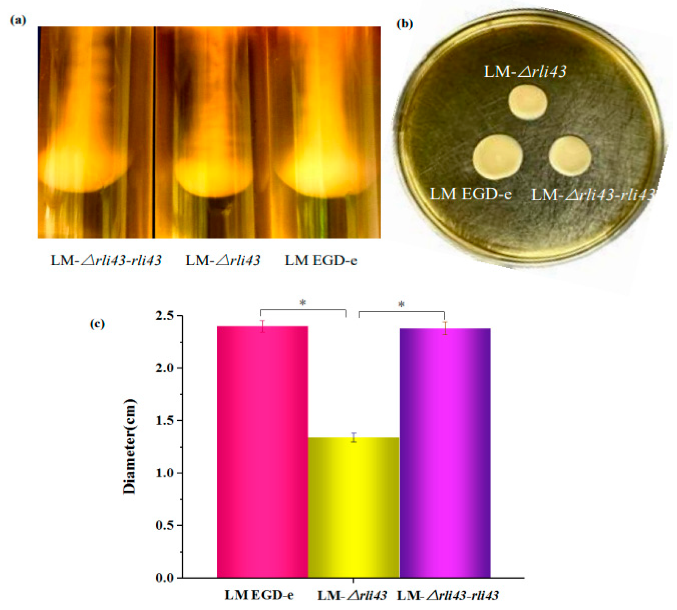
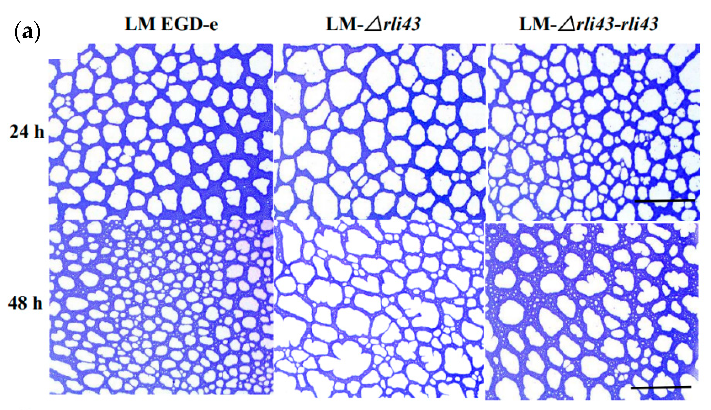
To evaluate the motility of LM EGD-e, LM-Δrli43, and LM-Δrli43-rli43, a semi-solid agar puncture inoculation and a plate diffusion assay were performed. As shown in Figure 3, when grown in semi-solid agar by punctured inoculation, LM-Δrli43 formed a significantly smaller inverted umbrella structure than that of LM EGD-e and LM-Δrli43-rli43 (Figure 3a). Furthermore, when grown in swarming agar plates, the motility of the LM-Δrli43 strain was markedly reduced compared with LM EGD-e and LM-Δrli43-rli43 (Figure 3b). The movement diameter of LM-Δrli43 in swarming agar plates was also significantly decreased compared with LM EGD-e and LM-Δrli43-rli43 (p < 0.05) (Figure 3c). To assess biofilm formation, these strains were subsequently grown statically in 96-well microplates containing polystyrene and 316 stainless steel sampling plates in BHI liquid medium at 37 °C for 24 h and 48 h, respectively. The staining of bacterial cells with crystal violet (CV) showed that LM-Δrli43 formed a looser biofilm structure when compared to LM EGD-e and LM-Δrli43-rli43 (Figure 4a). We also quantitatively measured biofilm formation ability and the results indicated that biofilm formation in LM-Δrli43 (OD570 nm) was significantly lower than that of LM EGD-e and LM-Δrli43-rli43 (p < 0.05) (Figure 4c). In addition, TEM results indicated that the biofilm formation for LM-Δrli43 on the stainless-steel surface was also significantly lower than that of LM EGD-e and LM-Δrli43-rli43 (Figure 4b). The data of the above motility and biofilm formation assays indicates that deletion of the rli43 gene weakens L. monocytogenes motility and biofilm formation.

3.4. Deficiency of Rli43 Gene Impaired Adhesion, Invasion, and Intracellular Survival in Macrophage

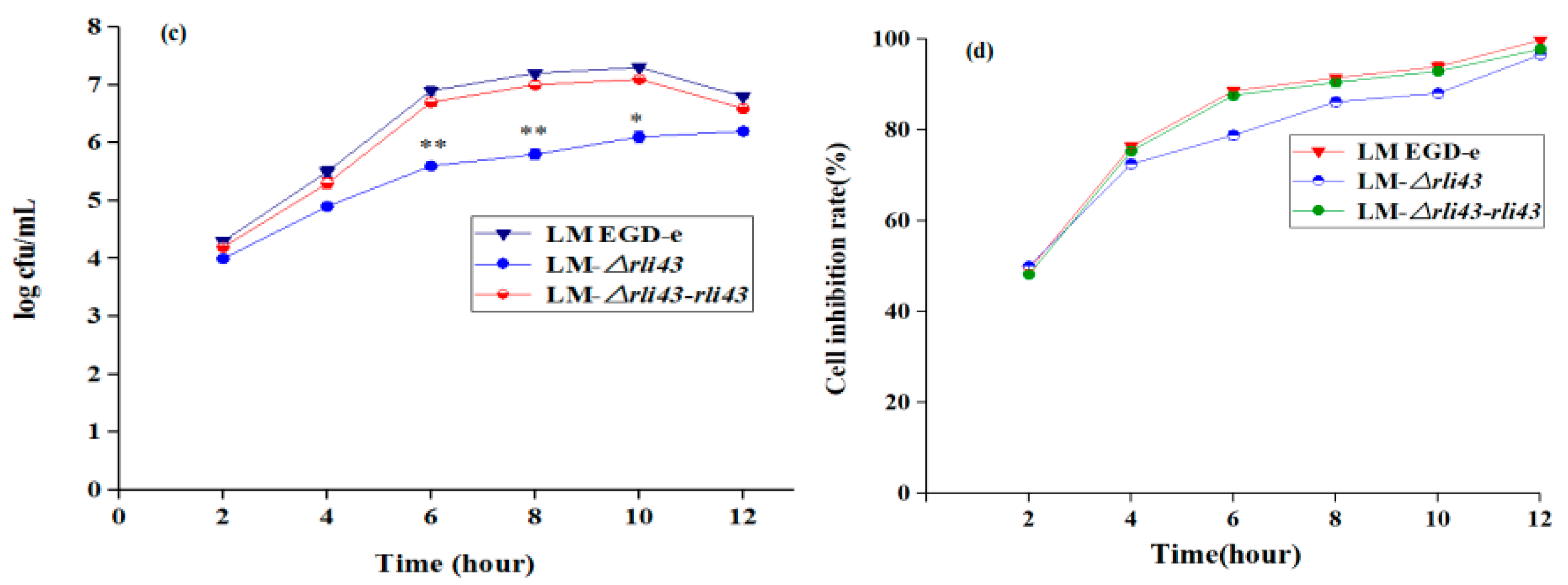
To probe the regulatory role of Rli43 during the infection of a macrophage, the adhesion, invasion, and intracellular proliferation of RAW264.7 were examined by bacterial cell counts. As shown in Figure 5, the adhesion, invasion, and intracellular survival of LM-Δrli43 in RAW264.7 cells were significantly lower when compared with LM EGD-e and LM-Δrli43-rli43 (p < 0.05) (Figure 5a–d), respectively, suggesting that rli43 gene deletion impaired the adhesion, invasion, and intracellular survival in a macrophage.

3.5. Deficiency of Rli43 Gene Weakened the Virulence of L. monocytogenes

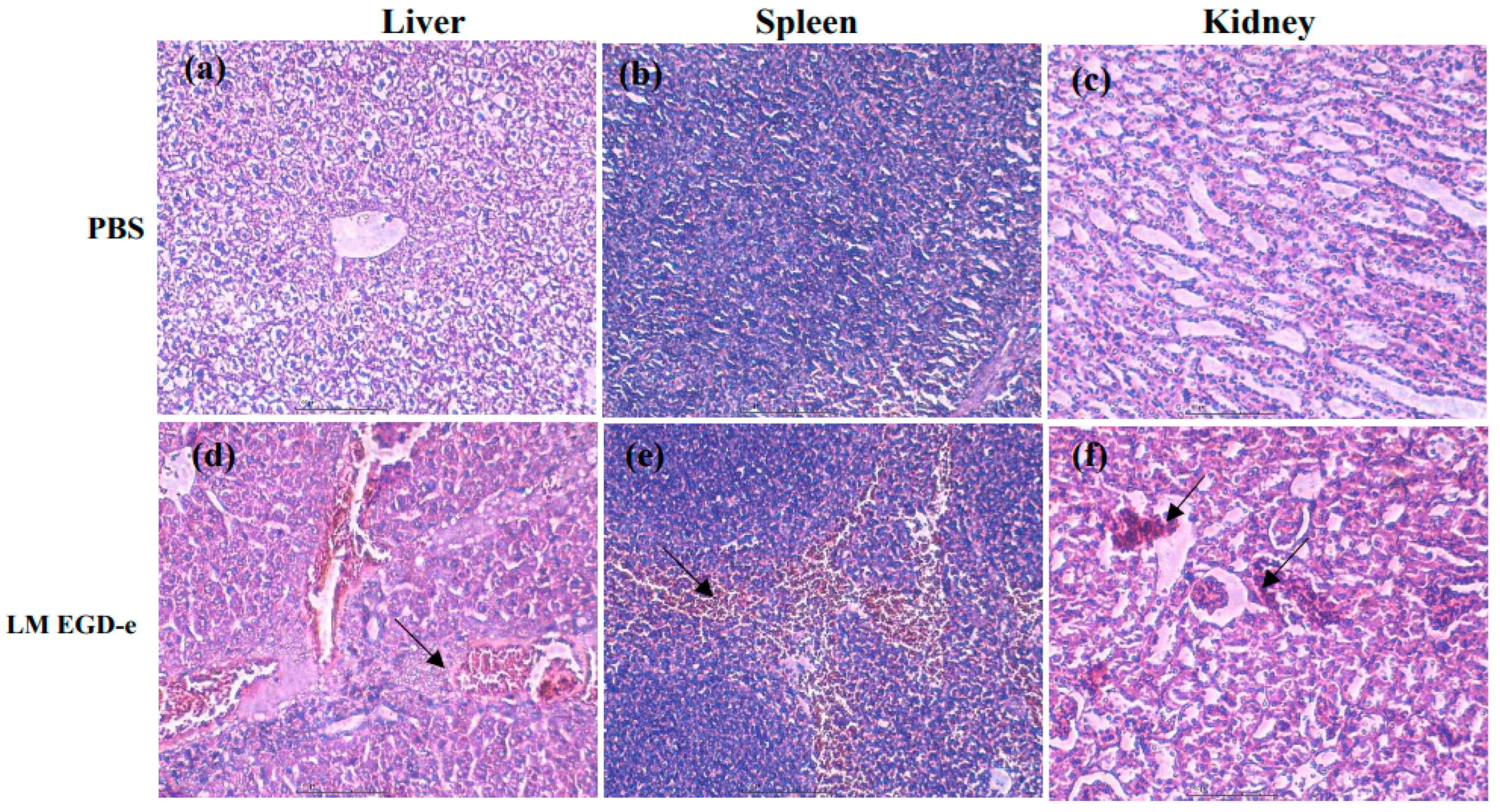
To investigate the regulatory roles of rli43 in the virulence of L. monocytogenes, we compared the lethality of L. monocytogenes in LM EGD-e, LM-Δrli43, and LM-Δrli43-rli43 mice. The mice in each subgroup were injected intraperitoneally with LM EGD-e, LM-Δrli43, and LM-Δrli43-rli43 at 0.5 mL/each, respectively. After inoculation, the mental status and mortality of mice in each group were recorded daily for seven days. As shown in Supplementary Table S1, the LD50 of LM EGD-e, LM-Δrli43, and LM-Δrli43-rli43 on BALB/c mice were 105.56 CFU, 107.32 CFU, and 105.76 CFU, respectively (Table S1). Compared with LM EGD-e and LM-Δrli43-rli43, the LD50 of LM-Δrli43 was elevated by 1.76 and 1.56 logarithmic orders of magnitude, and mice in the LM-Δrli43-infected group survived significantly longer (Figure 6a). Moreover, bacterial loads in both liver and spleen were significantly declined (p < 0.01) (Figure 6b,c). The pathological changes in the livers, spleens, and kidneys of L. monocytogenes-infected mice were further investigated. Compared with the normal control mice (Figure 7a–c), the pathological changes in the livers of LM EGD-e-infected (Black arrows in Figure 7d) and LM-Δrli43-rli43-infected (black arrows in Figure 7g) mice were mainly manifested as partial hepatocyte necrosis and hepatic lobular inflammatory cell infiltration; the tissue structure was unclear and contained a large amount of tissue debris. Transparent degeneration and lymphoid tissue necrosis occurred in spleen reticular fibers, and spleen nodules increased in size. Lymphocytes at the site of lymph nodes were necrotic and lymphoid tissue was bleeding (black arrows in Figure 7e,h). Renal venular hemorrhage occurred, inflammatory cells appeared, and the interstitial volume was enlarged (black arrows in Figure 7f,i). However, compared with LM EGD-e-infected and LM-Δrli43-rli43-infected mice, histopathological damage to the liver, spleen, and kidney was alleviated to some extent in LM-Δrli43-infected mice (Figure 7j–l), indicating that rli43 gene deletion lessened the virulence of L. monocytogenes.

3.6. Determination of the Transcription Levels of Motility-, Biofilm-, and Virulence-Related Genes

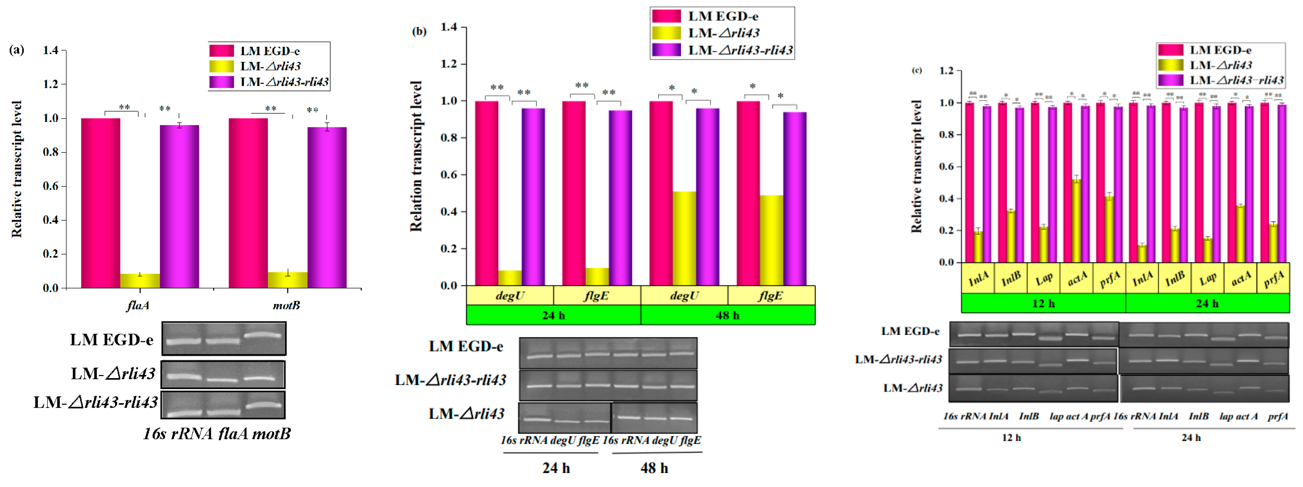
Based on the results which suggested that the deletion of rli43 could weaken L. monocytogenes motility, biofilm formation, and virulence, we quantified the relative transcription levels of motility-related (flaA and motB), biofilm-associated (flgE and degU), and virulence-related genes (PrfA, inlA, inlB, lap and actA) by qRT-PCR, respectively. As shown in Figure 8, the mRNA levels of motility-related genes flaA and motB genes (Figure 8a), and biofilm-associated genes flgE and degU (Figure 8b), were significantly declined in the rli43 gene deletion strain (p < 0.05). Notably, the transcription levels of virulence-related genes (inlA, inlB, lap, actA, PrfA) were also significantly decreased (p < 0.05) in the LM-Δrli43 strain (Figure 8c). These findings were in agreement with the bacterial phenotype assay, indicating that the deletion of the rli43 gene can impair the survival and proliferation of L. monocytogenes in RAW264.7 cells.

3.7. Rli43 Can Modulate the Expression Level of Htra Gene via Targeting Its MRNA

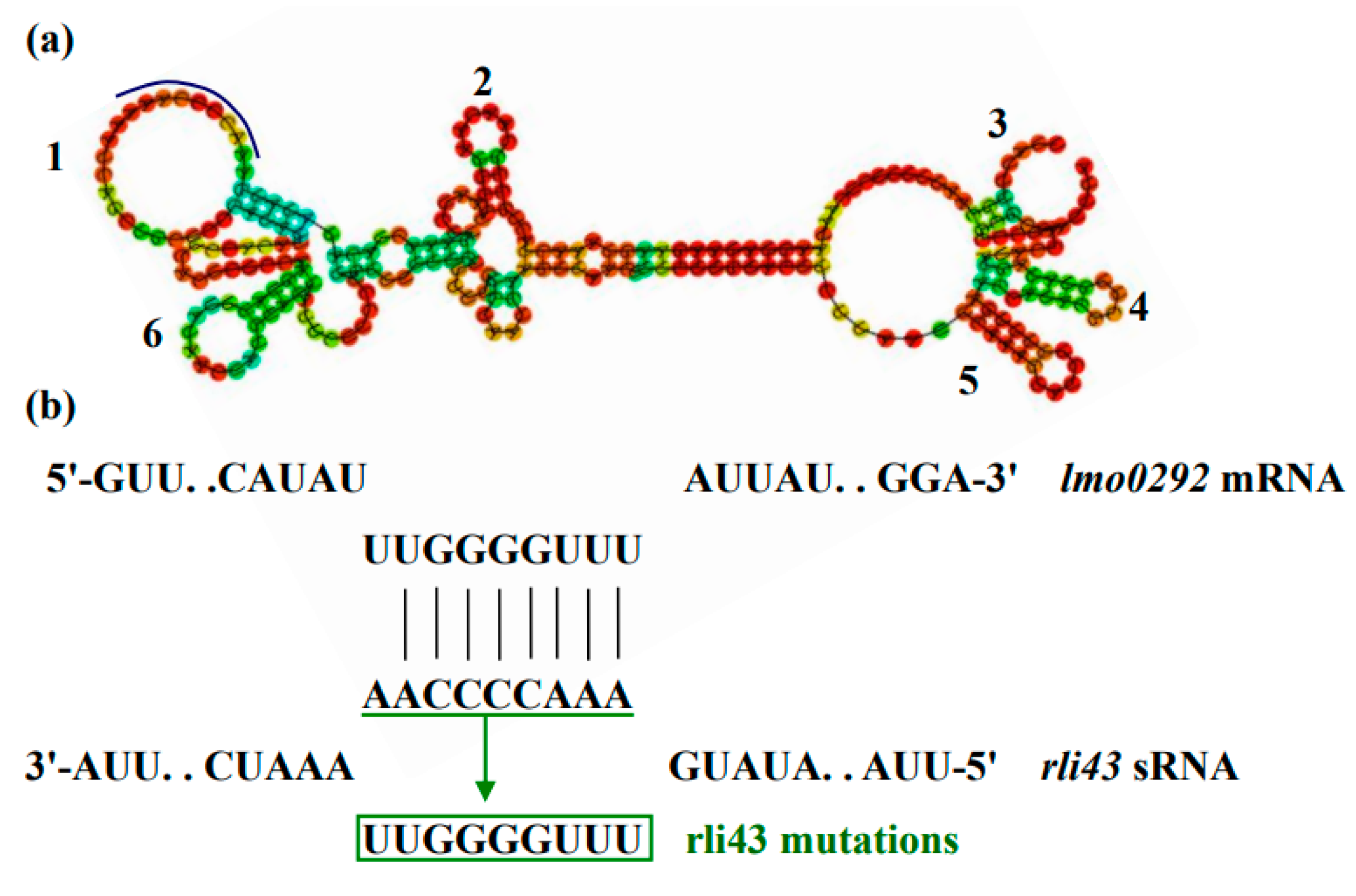
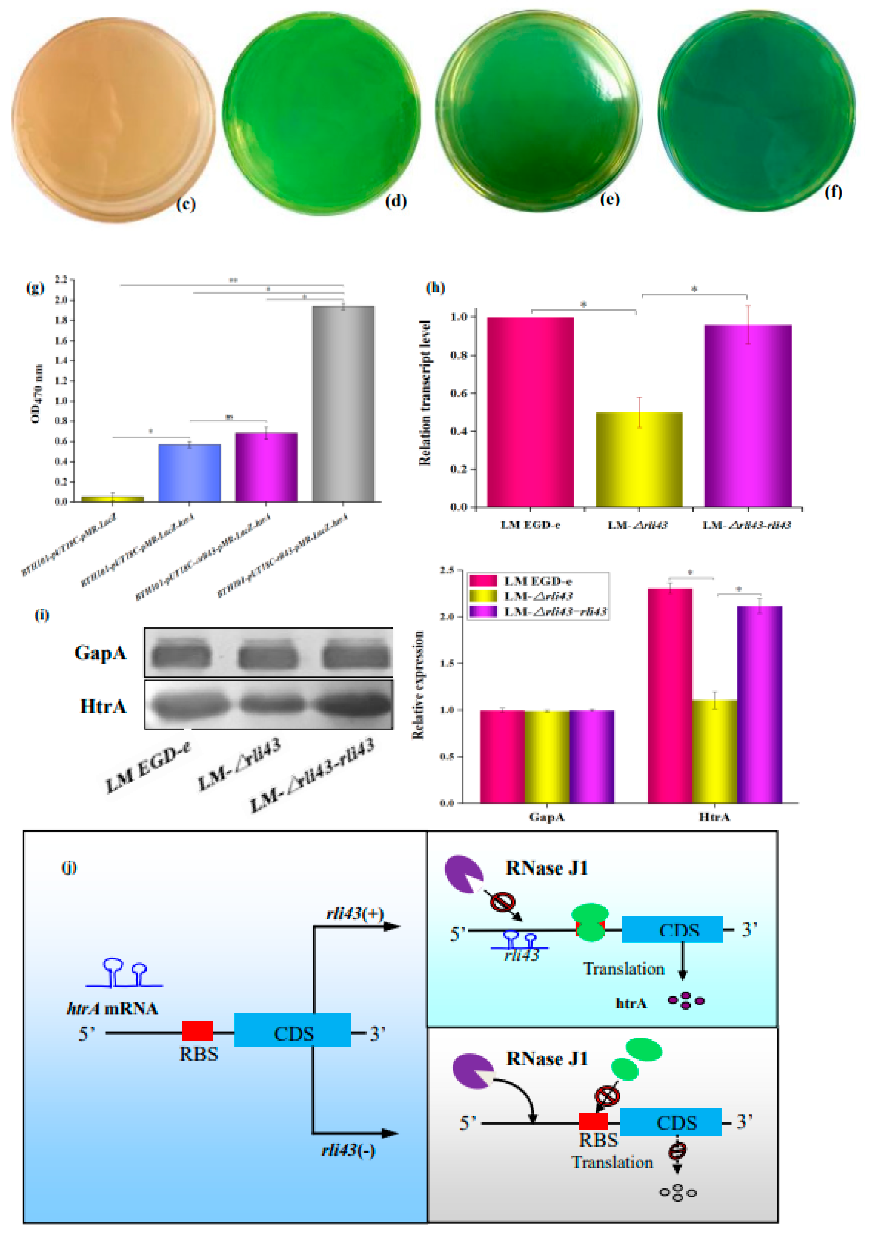
To reveal how Rli43 regulated the phenotypes of motility, biofilm formation, and virulence, we used online software to analyze the genetic localization, molecular characteristics, and target gene of sRNA Rli43. Sequence analysis showed that the rli43 gene was located at the spacer region between the inl C and rplS gene in the genome of LM EGD-e, with a length of 254 bp. Moreover, its promoter region contained an FIS transcription factor binding site (Figure S3a). It was shown that the transcription of sRNA Rli43 owns six stem-loop domains in the secondary structure (Figure 9a). The genetic evolutionary analysis showed that the rli43 gene was highly conserved in L. monocytogenes serotypes ½ a, ½c, 3c, and 4b (Figure S3b). By combining the predicted results of TargetRNA2 and IntaRNA, it was revealed that sRNA Rli43 may complementarily pair with the 5′-UTR (−47–−55) due to the high temperature requirement of htrA mRNA (Figure 9b), suggesting that the htrA gene was one of the potential target genes regulated by Rli43. To verify the interaction between rli43 and htrA mRNA, a dual plasmid reporter system based on E. coli was employed, and the pUT18C-rli43 and pMR-LacZ-htrA plasmids were successfully constructed and verified by PCR and double enzyme digestion, respectively (Figure S4). As shown in Figure 9, the lawn of E. coli strain co-transformed, as pUT18C-rli43 and pMR-LacZ-htrA was displayed as darker blue on the plates containing X-gal compared with those strains of E. coli transformed by pUT18C, pMR-LacZ-htrA, or its co-transformation. The OD470 nm values of bacterial suspensions differed significantly (p < 0.05) (Figure 9c–g), suggesting that Rli43 can interact with the mRNA of target gene htrA. The mRNA and protein levels of the htrA gene were significantly lower (p < 0.05) in LM-Δrli43 when compared with LM EGD-e and LM-Δrli43-rli43 (Figure 9h,i), indicating that Rli43 may modulate the stability of mRNA in the htrA gene, so that sRNA Rli43 can positively regulate the expression of the htrA gene (Figure 9j).

4. Discussion

As a class of ncRNAs, bacterial sRNAs are one of the most important regulators of gene expression and they perform a broad range of physiological functions. Commonly, bacterial sRNAs range from 50–300 nucleotides, and are complementarity with their targeting mRNAs. Currently, a number of sRNAs have been identified in bacteria. However, biological functions have been studied in detail for only a small proportion of sRNAs. Generally speaking, sRNAs are recognized as one of the most important regulators, and they are involved in posttranscriptional or translational control of gene expression through a variety of mechanisms [25]. In L. monocytogenes, some sRNAs, together with PrfA, Sigma B, and VirR regulators, form a complex regulatory network to facilitate its survival and infection. As a newly identified sRNA in L. monocytogenes [8], however, the regulatory roles and mechanism of Rli43 have not yet been revealed. To investigate the regulatory roles of rli43 in L. monocytogenes, the relative transcription levels of rli43 were profiled under extracellular and intracellular conditions. It was shown that the transcriptional level of Rli43 was significantly upregulated in RAW264.7 cells compared with extracellular conditions, which suggested that Rli43 is involved in the processes of infection and intracellular survival.
An in silico analysis revealed that the high temperature requirement (htrA) gene was one of the potential target genes regulated by Rli43. To date, it has been proven that HtrA is an important regulator with a dual function of molecular chaperone and protease activity. It has a critical role in the prevention of severe cellular malfunctions owing to the accumulation of mislocalized or misfolded proteins under physiological and stressful conditions [26]. Currently, HtrA has been proven to be involved in responses to environmental stresses (e.g., high and low temperatures, oxidative stress, high salt, or extreme pH) in Streptococcus pneumonia [27], Lactococcus lactis [28], E. coli [29], and H. pylori [30]. Furthermore, Rebecca and Laura et al. confirmed that the survival capacity of the htrA gene mutant strains of L. monocytogenes is significantly reduced under high temperatures and oxidation conditions [31,32]. Herein, we revealed that Rli43 may facilitate the expression of the htrA gene to modulate the adaptation of L. monocytogenes in response to environmental changes, which is similar to those bacteria mentioned above.
Several studies have shown that HtrA also played important roles in the regulation of motility and biofilm formation in bacteria [33]. Zhang et al. (2019) confirmed that HtrA is a serine protease from B. burgdorferi that regulates the conversion of FlaB [34]. Notably, the motility and the mRNA levels of flaA and motB genes were significantly reduced in LM-Δrli43, suggesting that Rli43 may indirectly regulate motility-related genes through modifying the expression of htrA gene. Furthermore, it has been shown that HtrA also plays important roles in biofilm formation [35] in L. monocytogenes [10], Streptococcus pyogenes [36], E. coli [26], and pneumococcal [37], respectively. In this study, we verified that the biofilm-forming ability was significantly reduced, and the transcriptional levels of flgE and degU genes related to biofilm formation were significantly downregulated in the deletion strain. However, the detailed mechanism of LM Rli43-modulating motility and biofilm formation should be further unraveled in the future.
It was confirmed that HtrA also played important regulatory roles in the expression of bacterial virulence genes [26,27,38,39,40,41,42,43], thereby influencing bacterial pathogenicity [31]. In Campylobacter jejuni, it was confirmed that HtrA could modify the processes of cell adhesion, invasion, and migration ability [44]. In particular, the deficiency of the htrA gene could lessen the survival capacities of Salmonella, Brucella, and Yersinia in macrophages or mice [45]. In L. monocytogenes, htrA gene deletion may result in the accumulation of misfolded proteins on the bacterial membrane, thus impairing intracellular survival [46,47]. In the present study, we verified that the deficiency of the rli43 gene impeded adhesion, invasion, and intracellular survival in a macrophage. In addition, the qRT-PCR results further verified that these findings were in agreement with the results of the bacterial phenotype assay, indicating that the deletion of the rli43 gene can impair the survival of L. monocytogenes in RAW264.7 cells.
Current studies have revealed that the molecular mechanisms underlying sRNA-mediated control are obvious diversity and flexibility in bacteria [3,6]. Commonly, sRNAs may regulate the expression of target genes by base pairing them with the 5′-untranslated region (5′-UTR) of the mRNA, thereby modulating multiple physiological processes, such as virulence and biofilm formation of bacteria [48]. On the one hand, the binding of trans-encoded sRNAs in the vicinity of the ribosomal binding site (RBS) may result in the inhibition of translation initiation or mRNA decay. Alternatively, sRNAs are likely to promote translation or prevent mRNA degradation, especially by base pairing to the far upstream from the RBS [6]. In this study, we verify the interaction between rli43 and target gene htrA mRNA using a two-plasmid system based on E. coli, which suggested that rli43 may bind to the 5′-UTR of htrA mRNA. To date, at least five RNases have been identified in bacteria, namely RNase A, RNase P, RNase E, RNase R, and RNase III. Among them, RNase III is mainly involved in the degradation of double-stranded RNA, while RNase JI is mainly involved in the degradation of single-stranded RNA in gram-positive bacteria. Combined with the results of the qRT-PCR and the Western blot, it is reasonable to infer that Rli43 may protect htrA mRNA from RNase JI degradation, thereby maintaining the stability of its mRNA. This regulatory mechanism mediated by rli43 is similar to the regulatory mode reported by Dutta et al. [48], which further highlights the similarities of sRNA-mediated control in various species of bacteria. However, this detailed regulatory motif in Rli43 should be further experimentally validated by employing site-directed mutagenesis.

5. Conclusions

This study revealed that Rli43 was involved in posttranscriptional control of htrA gene expression by modulating the stability of its mRNA. The regulatory mechanisms of sRNA rli43 provided new insights into the understanding of the diversity and flexibility of sRNA-mediated control in biofilm formation and virulence in L. monocytogenes.

Supplementary Materials

The following supporting information can be downloaded at: https://www.mdpi.com/article/10.3390/pathogens11101137/s1. Table S1: Determination of LD50 of LM EGD-e, LM-Δrli43, and LM-Δrli43-rli43 in mice. Figure S1: Amplification of LM rli43 gene by RT-PCR. Figure S2: Construction of LM-Δrli43 deletion and its complementary strains. Figure S3: Molecular characterization of rli43 gene and its phylogenitic analysis. Figure S4: Screening and identification of pMR-LacZ-htrA and pUT18C-rli43 using PCR and restriction enzyme digestion.

Author Contributions

Conceptualization, methodology, data curation, formal analysis, writing—original draft, writing—review and editing, L.W., Q.M., X.X. and J.Q.; data curation, L.W. and C.J.; funding acquisition, J.Q. and L.W.; supervision, X.C. All authors have read and agreed to the published version of the manuscript.

Funding

This work was supported by the National Natural Science Foundation of China (No. 32160819, 31360596, 30960274), the national key research and development program (No. 2016YFD0500900) and the graduate student research innovation project of Xinjiang Autonomous region (XJ2021G104).

Institutional Review Board Statement

The animal study protocol was approved by the Research and Ethical Committee of Shihezi University (No. A2019226).

Informed Consent Statement

Not applicable.

Data Availability Statement

All data generated or analyzed during this study are included in the manuscript/Supplementary Materials. All study original images are included in the manuscript (supplementary information files: Original Images for Blots-Gels file). The sequencing data are available in the GenBank repository, and the GenBank accession number for LM EGD-e genome sequence is AL591824 (https://www.ncbi.nlm.nih.gov/nuccore/AL591824 (accessed on 27 February 2015)). The sequence of rli43 gene is available in the GenBank repository (https://www.ncbi.nlm.nih.gov/genbank/ (accessed on 6 March 2022)).

Conflicts of Interest

The authors declare no conflict of interest.

References

  1. Matle, I.; Mbatha, K.R.; Madoroba, E. A review of Listeria monocytogenes from meat and meat products: Epidemiology, virulence factors, antimicrobial resistance and diagnosis. Onderstepoort. J. Vet. Res. 2020, 87, e1–e20. [Google Scholar] [CrossRef]
  2. Schonberg, A.; Gerigk, K. Listeria in effluents from the food-processing industry. Rev. Sci. Tech. 1991, 10, 787–797. [Google Scholar] [CrossRef] [PubMed]
  3. Iwu, C.D.; Okoh, A.I. Preharvest Transmission Routes of Fresh Produce Associated Bacterial Pathogens with Outbreak Potentials: A Review. Int. J. Environ. Res. Public Health 2019, 16, 4407. [Google Scholar] [CrossRef] [PubMed]
  4. Oliver, H.F.; Orsi, R.H.; Ponnala, L.; Keich, U.; Wang, W.; Sun, Q.; Cartinhour, S.W.; Filiatrault, M.J.; Wiedmann, M.; Boor, K.J. Deep RNA sequencing of L. monocytogenes reveals overlapping and extensive stationary phase and sigma B-dependent transcriptomes, including multiple highly transcribed noncoding RNAs. BMC Genom. 2009, 10, 641. [Google Scholar] [CrossRef]
  5. Ortega, A.D.; Quereda, J.J.; Pucciarelli, M.G.; Garcia-del, P.F. Non-coding RNA regulation in pathogenic bacteria located inside eukaryotic cells. Front. Cell. Infect. Microbiol. 2014, 4, 162. [Google Scholar] [CrossRef]
  6. Nitzan, M.; Rehani, R.; Margalit, H. Integration of Bacterial Small RNAs in Regulatory Networks. Annu. Rev. Biophys. 2017, 46, 131–148. [Google Scholar] [CrossRef] [PubMed]
  7. Mraheil, M.A.; Billion, A.; Mohamed, W.; Mukherjee, K.; Kuenne, C.; Pischimarov, J.; Krawitz, C.; Retey, J.; Hartsch, T.; Chakraborty, T.; et al. The intracellular sRNA transcriptome of Listeria monocytogenes during growth in macrophages. Nucleic Acids Res. 2011, 39, 4235–4248. [Google Scholar] [CrossRef]
  8. Toledo-Arana, A.; Dussurget, O.; Nikitas, G.; Sesto, N.; Guet-Revillet, H.; Balestrino, D.; Loh, E.; Gripenland, J.; Tiensuu, T.; Vaitkevicius, K.; et al. The Listeria transcriptional landscape from saprophytism to virulence. Nature 2009, 459, 950–956. [Google Scholar] [CrossRef] [PubMed]
  9. Izar, B.; Mraheil, M.A.; Hain, T. Identification and Role of Regulatory Non-Coding RNAs in Listeria monocytogenes. Int. J. Mol. Sci. 2011, 12, 5070–5079. [Google Scholar] [CrossRef] [PubMed]
  10. Peng, Y.L.; Meng, Q.L.; Qiao, J.; Xie, K.; Chen, C.; Liu, T.L.; Hu, Z.X.; Ma, Y.; Cai, X.P.; Chen, C.F. The Regulatory Roles of ncRNA Rli60 in Adaptability of Listeria monocytogenes to Environmental Stress and Biofilm Formation. Curr. Microbiol. 2016, 73, 77–83. [Google Scholar] [CrossRef]
  11. Wehner, S.; Mannala, G.K.; Qing, X.; Madhugiri, R.; Chakraborty, T.; Mraheil, M.A.; Hain, T.; Marz, M. Detection of very long antisense transcripts by whole transcriptome RNA-Seq analysis of Listeria monocytogenes by semiconductor sequencing technology. PLoS ONE 2014, 9, e108639. [Google Scholar] [CrossRef] [PubMed]
  12. Abdelhamed, H.; Karsi, A.; Lawrence, M.L. Efficient Gene Deletion Method for Listeria monocytogenes. Methods Mol. Biol. 2019, 2016, 159–170. [Google Scholar] [CrossRef] [PubMed]
  13. Somwatcharajit, R.; Tiantad, I.; Panbangred, W. Coexpression of the silent cry2Ab27 together with cry1 genes in Bacillus thuringiensis subsp. aizawai SP41 leads to formation of amorphous crystal toxin and enhanced toxicity against Helicoverpa armigera. J. Invertebr. Pathol. 2014, 116, 48–55. [Google Scholar] [CrossRef] [PubMed]
  14. Dong, H.; Peng, X.; Liu, Y.; Wu, T.; Wang, X.; De, Y.; Han, T.; Yuan, L.; Ding, J.; Wang, C.; et al. BASI74, a Virulence-Related sRNA in Brucella abortus. Front. Microbiol. 2018, 9, 2173. [Google Scholar] [CrossRef] [PubMed]
  15. Tan, L.; Chen, H.; Yu, S.; Qiu, X.; Song, C.; Chen, D.; Zhang, S.; Zhang, F.; He, S.; Shen, X.; et al. A SOE-PCR method of introducing multiple mutations into Mycoplasma gallisepticum neuraminidase. J. Microbiol. Methods 2013, 94, 117–120. [Google Scholar] [CrossRef]
  16. Taylor, A.J.; Stasiewicz, M.J. Persistent and sporadic Listeria monocytogenes strains do not differ when growing at 37 degrees C, in planktonic state, under different food associated stresses or energy sources. BMC Microbiol. 2019, 19, 257. [Google Scholar] [CrossRef]
  17. Marini, E.; Magi, G.; Ferretti, G.; Bacchetti, T.; Giuliani, A.; Pugnaloni, A.; Rippo, M.R.; Facinelli, B. Attenuation of Listeria monocytogenes Virulence by Cannabis sativa L. Essential Oil. Front. Cell. Infect. Microbiol. 2018, 8, 293. [Google Scholar] [CrossRef]
  18. Barzelighi, H.M.; Esfahani, B.N.; Bakhshi, B.; Daraei, B.; Moghim, S.; Fazeli, H. Influence of Heterologously Expressed azurin from Pseudomonas aeruginosa on the Adhesion and Invasion of Pathogenic Bacteria to the Caco-2 Cell Line. Probiotics Antimicrob. Proteins 2020, 12, 697–704. [Google Scholar] [CrossRef]
  19. Wang, X.; Ma, W.; Yin, J.; Chen, M.; Jin, H. Retraction Note to: HHIP gene overexpression inhibits the growth, migration and invasion of human liver cancer cells. J. BUON 2021, 26, 1693. [Google Scholar]
  20. Peng, Y.L.; Meng, Q.L.; Qiao, J.; Xie, K.; Chen, C.; Liu, T.L.; Hu, Z.X.; Ma, Y.; Cai, X.P.; Chen, C.F. The roles of noncoding RNA Rli60 in regulating the virulence of Listeria monocytogenes. J. Microbiol. Immunol. Infect. 2016, 49, 502–508. [Google Scholar] [CrossRef]
  21. Kun, X.; Qingling, M.; Qiao, J.; Yelong, P.; Tianli, L.; Cheng, C.; Yu, M.; Zhengxiang, H.; Xuepeng, C.; Chuangfu, C. Impact of rli87 gene deletion on response of Listeria monocytogenes to environmental stress. FEMS Microbiol. Lett. 2014, 359, 50–54. [Google Scholar] [CrossRef] [PubMed] [Green Version]
  22. Livak, K.J.; Schmittgen, T.D. Analysis of relative gene expression data using real-time quantitative PCR and the 2(-Delta Delta C(T)) Method. Methods 2001, 25, 402–408. [Google Scholar] [CrossRef]
  23. Cohen-Berneron, J.; Steinberg, D.; Featherstone, J.D.; Feuerstein, O. Sustained effects of blue light on Streptococcus mutans in regrown biofilm. Lasers Med. Sci. 2016, 31, 445–452. [Google Scholar] [CrossRef] [PubMed]
  24. Wiesner, R.; Scheller, C.; Krebs, F.; Watzig, H.; Oltmann-Norden, I. A comparative study of CE-SDS, SDS-PAGE, and Simple Western: Influences of sample preparation on molecular weight determination of proteins. Electrophoresis 2021, 42, 206–218. [Google Scholar] [CrossRef] [PubMed]
  25. Dutcher, H.A.; Raghavan, R. Origin, Evolution, and Loss of Bacterial Small RNAs. Microbiol. Spectr. 2018, 6, 10. [Google Scholar] [CrossRef]
  26. Lewis, C.; Skovierova, H.; Rowley, G.; Rezuchova, B.; Homerova, D.; Stevenson, A.; Spencer, J.; Farn, J.; Kormanec, J.; Roberts, M. Salmonella enterica Serovar Typhimurium HtrA: Regulation of expression and role of the chaperone and protease activities during infection. Microbiology 2009, 155, 873–881. [Google Scholar] [CrossRef]
  27. Ibrahim, Y.M.; Kerr, A.R.; McCluskey, J.; Mitchell, T.J. Role of HtrA in the virulence and competence of Streptococcus pneumoniae. Infect. Immun. 2004, 72, 3584–3591. [Google Scholar] [CrossRef]
  28. Foucaud-Scheunemann, C.; Poquet, I. HtrA is a key factor in the response to specific stress conditions in Lactococcus lactis. FEMS Microbiol. Lett. 2003, 224, 53–59. [Google Scholar] [CrossRef]
  29. Zhang, Z.; Wu, T.; Li, Y.; Bai, X.; Yan, X.; Gao, Y.; Shi, Q.; Zhu, G. Contribution of the serine protease HtrA in Escherichia coli to infection in foxes. Microb. Pathog. 2019, 135, 103570. [Google Scholar] [CrossRef]
  30. Zarzecka, U.; Modrak-Wojcik, A.; Figaj, D.; Apanowicz, M.; Lesner, A.; Bzowska, A.; Lipinska, B.; Zawilak-Pawlik, A.; Backert, S.; Skorko-Glonek, J. Properties of the HtrA Protease From Bacterium Helicobacter pylori Whose Activity Is Indispensable for Growth Under Stress Conditions. Front. Microbiol. 2019, 10, 961. [Google Scholar] [CrossRef]
  31. Wilson, R.L.; Brown, L.L.; Kirkwood-Watts, D.; Warren, T.K.; Lund, S.A.; King, D.S.; Jones, K.F.; Hruby, D.E. Listeria monocytogenes 10403S HtrA is necessary for resistance to cellular stress and virulence. Infect. Immun. 2006, 74, 765–768. [Google Scholar] [CrossRef] [PubMed] [Green Version]
  32. Wonderling, L.D.; Wilkinson, B.J.; Bayles, D.O. The htrA (degP) gene of Listeria monocytogenes 10403S is essential for optimal growth under stress conditions. Appl. Environ. Microbiol. 2004, 70, 1935–1943. [Google Scholar] [CrossRef] [PubMed]
  33. Xue, R.Y.; Liu, C.; Xiao, Q.T.; Sun, S.; Zou, Q.M.; Li, H.B. HtrA family proteases of bacterial pathogens: Pros and cons for their therapeutic use. Clin. Microbiol. Infect. 2021, 27, 559–564. [Google Scholar] [CrossRef] [PubMed]
  34. Zhang, K.; Qin, Z.; Chang, Y.; Liu, J.; Malkowski, M.G.; Shipa, S.; Li, L.; Qiu, W.; Zhang, J.R.; Li, C. Analysis of a flagellar filament cap mutant reveals that HtrA serine protease degrades unfolded flagellin protein in the periplasm of Borrelia burgdorferi. Mol. Microbiol. 2019, 111, 1652–1670. [Google Scholar] [CrossRef]
  35. Hoy, B.; Brandstetter, H.; Wessler, S. The stability and activity of recombinant Helicobacter pylori HtrA under stress conditions. J. Basic Microbiol. 2013, 53, 402–409. [Google Scholar] [CrossRef]
  36. Ahn, S.J.; Lemos, J.A.; Burne, R.A. Role of HtrA in growth and competence of Streptococcus mutans UA159. J. Bacteriol. 2005, 187, 3028–3038. [Google Scholar] [CrossRef]
  37. Chao, Y.; Bergenfelz, C.; Sun, R.; Han, X.; Achour, A.; Hakansson, A.P. The serine protease HtrA plays a key role in heat-induced dispersal of pneumococcal biofilms. Sci. Rep. 2020, 10, 22455. [Google Scholar] [CrossRef]
  38. Purdy, G.E.; Hong, M.; Payne, S.M. Shigella flexneri DegP facilitates IcsA surface expression and is required for efficient intercellular spread. Infect. Immun. 2002, 70, 6355–6364. [Google Scholar] [CrossRef]
  39. Elzer, P.H.; Phillips, R.W.; Robertson, G.T.; Roop, R.N. The HtrA stress response protease contributes to resistance of Brucella abortus to killing by murine phagocytes. Infect. Immun. 1996, 64, 4838–4841. [Google Scholar] [CrossRef]
  40. Li, S.R.; Dorrell, N.; Everest, P.H.; Dougan, G.; Wren, B.W. Construction and characterization of a Yersinia enterocolitica O:8 high-temperature requirement (htrA) isogenic mutant. Infect. Immun. 1996, 64, 2088–2094. [Google Scholar] [CrossRef]
  41. Boehm, M.; Simson, D.; Escher, U.; Schmidt, A.M.; Bereswill, S.; Tegtmeyer, N.; Backert, S.; Heimesaat, M.M. Function of Serine Protease HtrA in the Lifecycle of the Foodborne Pathogen Campylobacter jejuni. Eur. J. Microbiol. Immunol. 2018, 8, 70–77. [Google Scholar] [CrossRef] [PubMed]
  42. Zhang, L.; Li, Y.; Wen, Y.; Lau, G.W.; Huang, X.; Wu, R.; Yan, Q.; Huang, Y.; Zhao, Q.; Ma, X.; et al. HtrA Is Important for Stress Resistance and Virulence in Haemophilus parasuis. Infect. Immun. 2016, 84, 2209–2219. [Google Scholar] [CrossRef] [PubMed]
  43. Cortes, G.; de Astorza, B.; Benedi, V.J.; Alberti, S. Role of the htrA gene in Klebsiella pneumoniae virulence. Infect. Immun. 2002, 70, 4772–4776. [Google Scholar] [CrossRef] [PubMed]
  44. Simson, D.; Boehm, M.; Backert, S. HtrA-dependent adherence and invasion of Campylobacter jejuni in human vs. avian cells. Lett. Appl. Microbiol. 2020, 70, 326–330. [Google Scholar] [CrossRef] [PubMed]
  45. Pallen, M.J.; Wren, B.W. The HtrA family of serine proteases. Mol. Microbiol. 1997, 26, 209–221. [Google Scholar] [CrossRef] [PubMed]
  46. Ahmed, J.K.; Freitag, N.E. Secretion Chaperones PrsA2 and HtrA Are Required for Listeria monocytogenes Replication following Intracellular Induction of Virulence Factor Secretion. Infect. Immun. 2016, 84, 3034–3046. [Google Scholar] [CrossRef]
  47. Stack, H.M.; Sleator, R.D.; Bowers, M.; Hill, C.; Gahan, C.G. Role for HtrA in stress induction and virulence potential in Listeria monocytogenes. Appl. Environ. Microbiol. 2005, 71, 4241–4247. [Google Scholar] [CrossRef] [PubMed]
  48. Dutta, T.; Srivastava, S. Small RNA-mediated regulation in bacteria: A growing palette of diverse mechanisms. Gene 2018, 656, 60–72. [Google Scholar] [CrossRef] [PubMed]
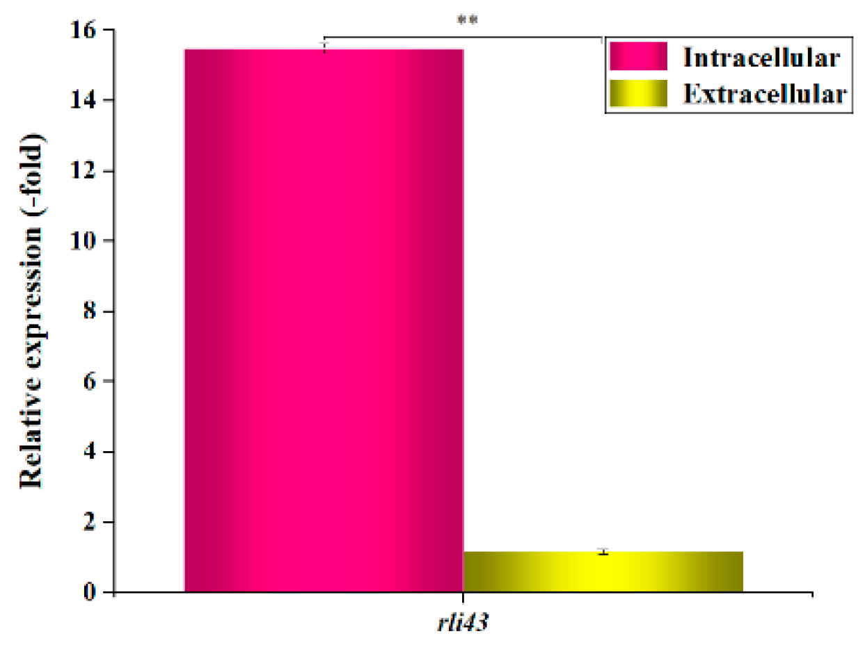
Figure 1. Expression profiles of sRNA rli43 gene under intra- and extracellular conditions. 16S rRNA gene was used as an internal reference gene, error bars represent the standard deviation of three biological replicates (** p-value  <  0.01).
Pathogens 11 01137 g001
Figure 2. Growth curves of LM EGD-e, LM-Δrli43, and LM-Δrli43-rli43 under different stress conditions. (af) growth curves of LM EGD-e, LM-Δrli43, and LM-Δrli43-rli43 at 30 °C, 42 °C, 37 °C, pH 9, pH 4, and 3.8% ethanol, respectively. All data are expressed as the means ± SD (n = 3). The differences between strains were assessed by one-way ANOVA.
Pathogens 11 01137 g002
Figure 3. Comparisons of motility of different strains of L. monocytogenes. (a) puncture inoculation of different strains of on semi-solid medium; (b) swarming motility of different strains on 0.5% soft agar plates; and (c) comparison of the diameters of swarming motility of different strains on 0.5% soft agar plates. Data are the mean ± SD (n = 3). The differences between strains were assessed by one-way ANOVA (* p-value  <  0.05).
Pathogens 11 01137 g003
Figure 4. Determination of capacities of biofilm formation of different strains of L. monocytogenes. (a) biofilm formation on polystyrene surfaces observed by microscopic (20 × 10), scale bars, 0.4 mm; (b) biofilm formation on stainless steel coupons observed by scanning electron micrographs (×10,000), scale bars, 20 μm; and (c) assay of biofilm formation ability using OD570 nm. The differences between strains were tested by one-way ANOVA (* p-value  <  0.05). Data represent the mean ± SD (n = 3).
Pathogens 11 01137 g004aPathogens 11 01137 g004b
Figure 5. Determination of cell adhesion and invasion capacity of different strains of L. monocytogenes. (a) Adhesion rate of LM EGD-e, LM-Δrli43, and LM-Δrli43-rli43 in RAW264.7 cells. (b) Invasion rate of LM EGD-e, LM-Δrli43, and LM-Δrli43-rli43 in RAW264.7. (c) Bacterial numbers in RAW264.7 cells infected by LM EGD-e, LM-Δrli43, and LM-Δrli43-rli43 at various periods. Data represent the mean ± SD (n = 3). Asterisks denote statistically significant difference by one-way ANOVA between strains (α = 0.05) with p < 0.01 and p < 0.05. (d) Inhibition rate of LM EGD-e, LM-Δrli43, and LM-Δrli43-rli43 in RAW264.7 cells after infection at time points. All data are expressed as the means ± SD (n = 3). The differences between strains were assessed by one-way ANOVA (* p-value  <  0.05, ** p-value  <  0.01).
Pathogens 11 01137 g005aPathogens 11 01137 g005b
Figure 6. Survival curves and bacterial loads in mice infected with different strains of L. monocytgenes. (a) survival curves; (b) bacterial loads in livers; and (c) bacterial loads in spleens. Results are represented as means ± SD of three experiments and expressed as log CFU/g. The differences between strains were assessed by one-way ANOVA (* p-value  <  0.05, ** p-value  <  0.01).
Pathogens 11 01137 g006
Figure 7. Histopathological examinations of the liver, spleen, and kidney of mice infected with LM (HE × 400, scale bars, 100 μm). (ac) Liver, spleen, and kidney from mice injected with PBS. (df) Liver, spleen, and kidney from mice infected with LM EGD-e. (gi) Liver, spleen, and kidney from mice infected with LM-Δrli43-rli43. (jl) Liver, spleen, and kidney from mice infected with LM-Δrli43. Arrows indicate major lesion areas.
Pathogens 11 01137 g007aPathogens 11 01137 g007b
Figure 8. Analysis of the relative transcriptional levels of motility-, biofilm-, and virulence-related genes. (a) Relative transcriptional levels and qRT-PCR electrophoretic band analysis of flaA and motB gene of LM EGD-e, LM-Δrli43, and LM-Δrli43-rli43, respectively. Asterisks denote significance in gene transcription compared with different strains according to one-way ANOVA (** p-value  <  0.01). (b) Relative transcriptional levels and qRT-PCR electrophoretic band analysis of degU and flgE gene of LM EGD-e, LM-Δrli43, and LM-Δrli43-rli43, respectively. The differences between strains were tested by one-way ANOVA (* p-value  <  0.05, ** p-value  <  0.01). (c) Relative transcriptional levels and qRT-PCR electrophoretic band analysis of InlA, InlB, Lap, actA, and prfA gene of LM EGD-e, LM-Δrli43, and LM-Δrli43-rli43 at 12 h and 24 h, respectively. All data are expressed as mean ± SD (n = 3). Asterisks denote significance in gene transcription compared with different strains via one-way ANOVA (α = 0.05, * p-value  <  0.05, ** p-value  <  0.01).
Pathogens 11 01137 g008
Figure 9. Verification of the interaction between sRNA Rli43 and target gene htrA using dual plasmid reporter system. (a) Secondary structure of sRNA Rli43 (six stem-loop structures, the blue line represents base-paring region). (b) The predicted potential gene targeted by Rli43, and the nucleotide mutations in the green box for the sRNAs. (c) Bacterial lawn of BTH101 co-transformed by pUT18C and pMR-LacZ. (d) Bacterial lawn of BTH101 co-transformed by pUT18C and pMR-LacZ- htrA. (e) Bacterial lawn of BTH101 co-transformed by pUT18C-Δrli43 and pMR-LacZ- htrA. (f) Bacterial lawn of BTH101 co-transformed by pUT18C-rli43 and pMR-LacZ-htrA. (g) Bacterial suspension determined by OD470 nm. (h) Determination of the mRNA level of htrA genes by qRT-PCR. (i) Determination of the expression of HtrA protein by Western blot. All data represent the mean ± SD (n = 3). The differences between strains were tested by one-way ANOVA (* p-value  <  0.05, ** p-value  <  0.01). (j) The mechanism of sRNA Rli43 to modulate the mRNA level of htrA gene by targeting its 5′-UTR region.
Pathogens 11 01137 g009aPathogens 11 01137 g009b
Table 1. Primers used in the study.
Primer NamesPrimer Sequences (5′→3′)
rli43 FTTATTTGCAATTTTTCTCAATAAGTART-PCR/qRT-PCR
rli43 RACCATCCCGCGCCATTTA
R1CGGGGTACCCACAAAAGAGGATTTTCATATACTTCCSOE-PCR
R2GCAGAAAGTTGGGTATGACTATAGAAAAAAAGCCTTGTCA
R3TGACAAGGCTTTTTTTCTATAGTCATACCCAACTTTCTGC
R4AAAACTGCAGCCTACGTGGTAAAGCGGCTC
P1CCCAAGCTTTTATTTGCAATTTTTCTCAATAAGTComplementation strains
P2GGAATTCTAAATGGCGCGGGATGGT
16S rRNA FGAGCTAATCCCATAAAACTATTCTCAqRT-PCR
16S rRNA RACCTTGTTACGACTTCACCCC
flaA FAACAAGCAACTGAAGCTATTGATGAATT
flaA RTGCGGTGTTTGGTTTGCTTGA
motB FAATCGCCAAAGAAATCGGCG
motB RCGCCGGGGTTTACTTCACTA
flgE FAATGCCAACACGACAGGATA
flgE RTTTGTTCCAGCGTAAAGTCC
degU FGAGGTCGTAGCGGAAGCTGA
degU RCTGTTACATATTCATCTGTATCA
InlB FCTGAAAAAAATGGTGGGCATGAG
InlB RCCGCCATTTCGGGCTTCTCT
PrfA FACGGGAAGCTTGGCTCTATT
PrfA RTGCGATGCCACTTGAATATC
Lap FGCGGCCCAAGAAGTATTAGAA
Lap RCTGTCGCGAAGATGTTTTTAATACA
InlA FTGTGACTGGCGCTTTAATTG
InlA RTCCAATAGTGACAGGTTGGCTA
actA FATGCGGGGAAATGGGTACGT
actA RCCAAGAAGCATTGGTGTCTCTGG
P3GGATCCAACATTCTCCACTCCTTAAAAADual plasmid reporter system
P4GGTACCATCATTTAACGGCTGAAACCCT
P5CCAAGCTTGCTTTTTCATCGTTTTTTCA
P6GGGGTACCATGTCTAGCAGGTTCTGATTCT
P7GGATCCCATTCTCCACTCCTTAAAAATA
P8AACTACATATATGTTTGGGGTTAAATCAGCTGCC
P9GGCAGCTGATTTAACCCCAAACATATATGTAGTT
P10GGGGTACCCAAGGGTTTCAGCCGTTAAATGA
P11ATGGACGAGAAAGAAAAGAATT
P12AATAACACCAATAAGTGCTGTT
Publisher’s Note: MDPI stays neutral with regard to jurisdictional claims in published maps and institutional affiliations.