
Prevalence of Toxoplasma gondii Measured by Western Blot, ELISA and DNA Analysis, by PCR, in Cats of Western Mexico

 , ,
, ,
Abstract
1. Introduction
2. Results
2.1. Questionnaire
2.2. Lifestyle and Risk Factors of the Cats Studied
2.3. Western Blot
2.4. Immunoassay ELISA
2.5. Detection of T. gondii DNA
3. Discussion
4. Conclusions
5. Materials and Methods
5.1. Samples
5.2. Questionnaire
5.3. Toxoplasma gondii Antigen-Derived Preparation
5.4. Western Blot Method
5.5. Immunoassay ELISA Method
5.6. DNA Samples
5.7. Molecular Detection of Toxoplasma gondii
5.8. Statistical Analysis
5.9. Ethical Approval
6. Limitations of the Work
Supplementary Materials
Author Contributions
Funding
Institutional Review Board Statement
Informed Consent Statement
Conflicts of Interest
References
- Torrey, E.F.; Yolken, R.H. Toxoplasma oocysts as a public health problem. Trends Parasitol. 2013, 29, 380–384. [Google Scholar] [CrossRef] [PubMed]
- Galvan-Ramirez, M. Toxoplasmosis Animal, 1st ed.; Universidad de Guadalajara: Guadalajara, México, 2013. [Google Scholar]
- Dabritz, H.A.; Conrad, P.A. Cats and Toxoplasma: Implications for Public Health. Zoonoses Public Health 2010, 57, 34–52. [Google Scholar] [CrossRef] [PubMed]
- Frenkel, J.K.; Ruiz, A.; Chinchilla, M. Soil Survival of Toxoplasma Oocysts in Kansas and Costa Rica. Am. J. Trop. Med. Hyg. 1975, 24, 439–443. [Google Scholar] [CrossRef] [PubMed]
- Yilmaz, S.M.; Hopkins, S.H. Effects of Different Conditions on Duration of Infectivity of Toxoplasma gondii Oocysts. J. Parasitol. 1972, 58, 938. [Google Scholar] [CrossRef] [PubMed]
- Galvan-Ramirez, M.D.L.L.; Troyo, R.; Roman, S.; Calvillo-Sanchez, C.; Bernal-Redondo, R. A systematic review and meta-analysis of Toxoplasma gondii infection among the Mexican population. Parasites Vectors 2012, 5, 271. [Google Scholar] [CrossRef]
- Castillo-Morales, V.J.; Viana, K.Y.A.; Guzmán-Marín, E.D.S.; Jiménez-Coello, M.; Segura-Correa, J.C.; Aguilar-Caballero, A.J.; Ortega-Pacheco, A. Prevalence and Risk Factors of Toxoplasma gondii Infection in Domestic Cats from the Tropics of Mexico Using Serological and Molecular Tests. Interdiscip. Perspect. Infect. Dis. 2012, 2012, 529108. [Google Scholar] [CrossRef] [PubMed]
- Cerro, L.; Rubio, A.; Pinedo, R.; Mendes-De-Almeida, F.; Brener, B.; Labarthe, N. Seroprevalence of Toxoplasma gondii in cats (Felis catus, Linnaeus 1758) living in Lima, Peru. Rev. Bras. De Parasitol. Vet. 2014, 23, 90–93. [Google Scholar] [CrossRef]
- Costa, D.G.C.; Marvulo, M.F.V.; Silva, J.S.A.; Santana, S.C.; Magalhães, F.J.R.; Filho, C.D.F.L.; Ribeiro, V.O.; Alves, L.C.; Mota, R.A.; Dubey, J.P. Seroprevalence of Toxoplasma gondii in Domestic and Wild Animals from the Fernando de Noronha, Brazil. J. Parasitol. 2012, 98, 679–680. [Google Scholar] [CrossRef]
- García-Márquez, L.J.; Gutiérrez-Díaz, M.A.; Correa, D.; Luna-Pastén, H.; Palma, J.M. Prevalence of Toxoplasma gondii Antibodies and the Relation to Risk Factors in Cats of Colima, Mexico. J. Parasitol. 2007, 93, 1527–1528. [Google Scholar] [CrossRef]
- Alvarado-Esquivel, C.; Liesenfeld, O.; Herrera-Flores, R.G.; Ramírez-Sánchez, B.E.; González-Herrera, A.; Martínez-García, S.A.; Dubey, J.P. Seroprevalence of Toxoplasma gondii antibodies in cats from Durango City, Mexico. J. Parasitol. 2007, 93, 1214–1216. [Google Scholar] [CrossRef]
- Besne-Merida, A.; Figueroa-Castillo, J.A.; Martínez-Maya, J.J.; Luna-Pastén, H.; Calderón-Segura, E.; Correa, D. Prevalence of antibodies against Toxoplasma gondii in domestic cats from Mexico City. Vet. Parasitol. 2008, 157, 310–313. [Google Scholar] [CrossRef]
- Galvan Ramirez, M.L.; Sánchez Vargas, G.; Vielma Sandoval, M.; Soto Mancilla, J.L. Presence of anti-Toxoplasma antibodies in humans and their cats in the urban zone of Guadalajara. Rev. Soc. Bras. Med. Trop. 1999, 32, 483–488. [Google Scholar] [CrossRef]
- Área Metropolitana de Guadalajara. Available online: https://www.jalisco.gob.mx/es/jalisco/guadalajara (accessed on 14 January 2022).
- Park, Y.; Noh, J.; Seo, H.-J.; Kim, K.-H.; Min, S.; Yoo, M.-S.; Yun, B.-R.; Kim, J.-H.; Choi, E.-J.; Cheon, D.-S.; et al. Seroprevalence and B1 gene Phylogeny of Toxoplasma gondii of Dogs and Cats in Republic of Korea. Korean J. Parasitol. 2020, 58, 257–265. [Google Scholar] [CrossRef]
- Hong, S.-H.; Jeong, Y.-I.; Kim, J.-Y.; Cho, S.-H.; Lee, W.-J.; Lee, S.-E. Prevalence of Toxoplasma gondii Infection in Household Cats in Korea and Risk Factors. Korean J. Parasitol. 2013, 51, 357–361. [Google Scholar] [CrossRef]
- Lappin, M.R.; Bush, D.J.; Reduker, D.W. Feline serum antibody responses to Toxoplasma gondii and characterization of target an-tigens. J. Parasitol. 1994, 80, 73–80. [Google Scholar] [CrossRef] [PubMed]
- Sohn, W.-M.; Nam, H.-W. Western blot analysis of stray cat sera against Toxoplasma gondii and the diagnostic availability of monoclonal antibodies in sandwich-ELISA. Korean J. Parasitol. 1999, 37, 249–256. [Google Scholar] [CrossRef]
- Mardanly, S.G.; Avdonina, A.S. Immune blotting as a method for diagnosis of toxoplasmosis. Klin. Lab. Diagn. 2020, 65, 693–698. [Google Scholar] [CrossRef] [PubMed]
- Wang, Q.; Jiang, W.; Chen, Y.-J.; Liu, C.-Y.; Shi, J.-L.; Li, X.-T. Prevalence of Toxoplasma gondii antibodies, circulating antigens and DNA in stray cats in Shanghai, China. Parasites Vectors 2012, 5, 190. [Google Scholar] [CrossRef]
- Oi, M.; Yoshikawa, S.; Maruyama, S.; Nogami, S. Comparison of Toxoplasma gondii Seroprevalence in Shelter Cats and Dogs during 1999–2001 and 2009–2011 in Tokyo, Japan. PLoS ONE 2015, 10, e0135956. [Google Scholar] [CrossRef] [PubMed]
- Ahn, K.-S.; Ahn, A.-J.; Park, S.-I.; Sohn, W.-M.; Shim, J.-H.; Shin, S.-S. Excretion of Toxoplasma gondii oocysts from Feral Cats in Korea. Korean J. Parasitol. 2019, 57, 665–670. [Google Scholar] [CrossRef]
- Can, H.; Doskaya, M.; Ajzenberg, D.; Özdemir, H.G.; Caner, A.; Iz, S.G.; Döşkaya, A.D.; Atalay, E.; Çetinkaya, Ç.; Ürgen, S.; et al. Genetic Characterization of Toxoplasma gondii Isolates and Toxoplasmosis Seroprevalence in Stray Cats of İzmir, Turkey. PLoS ONE 2014, 9, e104930. [Google Scholar] [CrossRef]
- Jalal, S.; Nord, C.; Lappalainen, M.; Evengård, B. Rapid and sensitive diagnosis of Toxoplasma gondii infections by PCR. Clin. Microbiol. Infect. 2004, 10, 937–939. [Google Scholar] [CrossRef] [PubMed]
- Lopes, A.P.; Oliveira, A.C.; Granada, S.; Rodrigues, F.T.; Papadopoulos, E.; Schallig, H.; Dubey, J.P.; Cardoso, L. Antibodies to Toxoplasma gondii and Leishmania spp. in domestic cats from Luanda, Angola. Vet. Parasitol. 2017, 239, 15–18. [Google Scholar] [CrossRef] [PubMed]
- Sukhumavasi, W.; Bellosa, M.L.; Lucio-Forster, A.; Liotta, J.L.; Lee, A.; Pornmingmas, P.; Chungpivat, S.; Mohammed, H.O.; Lorentzen, L.; Dubey, J.; et al. Serological survey of Toxoplasma gondii, Dirofilaria immitis, Feline Immunodeficiency Virus (FIV) and Feline Leukemia Virus (FeLV) infections in pet cats in Bangkok and vicinities, Thailand. Vet. Parasitol. 2012, 188, 25–30. [Google Scholar] [CrossRef]
- Fernandez, F.; Ouviña, G.; Clot, E.; Guido, R.F.; Codoni, C. Prevalence of Toxoplasma gondii antibodies in cats in the western part of Great Buenos Aires, Argentina, 1993. Vet. Parasitol. 1995, 59, 75–79. [Google Scholar] [CrossRef]
- Deksne, G.; Petrusēviča, A.; Kirjušina, M. Seroprevalence and Factors Associated with Toxoplasma gondii Infection in Domestic Cats from Urban Areas in Latvia. J. Parasitol. 2013, 99, 48–50. [Google Scholar] [CrossRef]
- Must, K.; Lassen, B.; Jokelainen, P. Seroprevalence of and Risk Factors for Toxoplasma gondii Infection in Cats in Estonia. Vector-Borne Zoonotic Dis. 2015, 15, 597–601. [Google Scholar] [CrossRef]
- Inpankaew, T.; Sattasathuchana, P.; Kengradomkij, C.; Thengchaisri, N. Prevalence of toxoplasmosis in semi-domesticated and pet cats within and around Bangkok, Thailand. BMC Vet. Res. 2021, 17, 252. [Google Scholar] [CrossRef]
- Sævik, B.K.; Krontveit, R.I.; Eggen, K.P.; Malmberg, N.; I Thoresen, S.; Prestrud, K.W. Toxoplasma gondii seroprevalence in pet cats in Norway and risk factors for seropositivity. J. Feline Med. Surg. 2015, 17, 1049–1056. [Google Scholar] [CrossRef] [PubMed]
- Opsteegh, M.; Haveman, R.; Swart, A.; Mensink-Beerepoot, M.; Hofhuis, A.; Langelaar, M.; van der Giessen, J. Seroprevalence and risk factors for Toxoplasma gondii infection in domestic cats in The Netherlands. Prev. Vet. Med. 2012, 104, 317–326. [Google Scholar] [CrossRef]
- Ahmad, N.; Ahmed, H.; Irum, S.; Qayyum, M. Seroprevalence of IgG and IgM antibodies and associated risk factors for toxoplasmosis in cats and dogs from sub-tropical arid parts of Pakistan. Trop. Biomed. 2014, 31, 777–784. [Google Scholar] [PubMed]
- Caballero-Ortega, H.; Uribe-Salas, F.J.; Conde-Glez, C.J.; Cedillo-Pelaez, C.; Vargas-Villavicencio, J.A.; Luna-Pastén, H.; Cañedo-Solares, I.; Ortiz-Alegría, L.B.; Correa, D. Seroprevalence and national distribution of human toxoplasmosis in Mexico: Analysis of the 2000 and 2006 National Health Surveys. Trans. R. Soc. Trop. Med. Hyg. 2012, 106, 653–659. [Google Scholar] [CrossRef] [PubMed]
- Khodaverdi, M.; Razmi, G. Prevalence and genotyping of Toxoplasma gondii in stray cats in Mashhad area, Iran. BMC Vet. Res. 2019, 15, 463. [Google Scholar] [CrossRef]
- Bawm, S.; Phyu, A.Z.; Chel, H.M.; Htun, L.L.; Nakao, R.; Katakura, K. Seroprevalence of Toxoplasma gondii in household cats in Myanmar and molecular identification of parasites using feline faecal oocysts. Food Waterborne Parasitol. 2020, 20, e00094. [Google Scholar] [CrossRef]
- Bastos, B.F.; Brener, B.; Gershony, L.; Willi, L.; Labarthe, N.; Pereira, C.; Mendes-De-Almeida, F. Seroprevalence of Toxoplasma gondii (Nicole & Manceaux, 1909) and retroviral status of client-owned pet cats (Felis cactus, Linnaeus, 1758) in Rio de Janeiro, Brazil. Rev. Inst. Med. Trop. São Paulo 2014, 56, 201–203. [Google Scholar] [CrossRef]
- Etheredge, G.D.; Michael, G.; Muehlenbein, M.P.; Frenkel, J. The roles of cats and dogs in the transmission of Toxoplasma infection in Kuna and Embera children in eastern Panama. Rev. Panam. Salud. Publica 2004, 16, 176–186. [Google Scholar] [CrossRef]
- Galván-Ramírez, M.D.L.L.; Guillén-Vargas, C.; Saavedra-Durán, R.; Islas-Rodríguez, A.; Islos-Rodríguez, A. Analysis of Toxoplasma gondii antigens with sera from toxoplasmosis patients. Rev. Soc. Bras. Med. Trop. 1998, 31, 271–277. [Google Scholar] [CrossRef] [PubMed]
- Towbin, H.; Staehelin, T.; Gordon, J. Electrophoretic transfer of proteins from polyacrylamide gels to nitrocellulose sheets: Procedure and some 270 applications. Biotechnology 1979, 24, 145–149. [Google Scholar] [CrossRef]


| Negative | Positive | Total | p | OR | 95%CI | |||
|---|---|---|---|---|---|---|---|---|
| (n = 253) | (n = 44) | (n = 297) | ||||||
| No. | % | No. | % | |||||
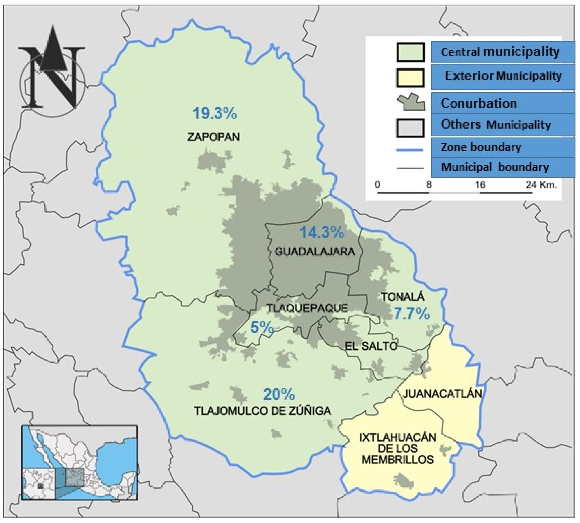
| MUNICIPALITY | 0.515 | |||||||
| Guadalajara | 156 | 85.7 | 26 | 14.3 | 182 | 1.000 | 1.000 | - - - - - |
| Tlajomulco | 20 | 80.0 | 5 | 20.0 | 25 | 0.455 | 1.500 | [0.517–4.348] |
| Tlaquepaque | 19 | 95.0 | 1 | 5.0 | 20 | 0.271 | 0.316 | [0.041–2.461] |
| Tonalá | 12 | 92.3 | 1 | 7.7 | 13 | 0.514 | 0.500 | [0.062–4.010] |
| Zapopan | 46 | 80.7 | 11 | 19.3 | 57 | 0.363 | 1.435 | [0.659–3.123] |
| Total | 253 | 85.2 | 44 | 14.8 | 297 | |||
| AGE GROUP | ||||||||
| Under one year | 119 | 92.2 | 10 | 7.8 | 129 | 1.000 | 1.000 | - - - - - |
| One year and more | 100 | 75.2 | 33 | 24.8 | 133 | p < 0.001 * | 3.927 | [1.844–8.362] |
| Total | 219 | 83.6 | 43 | 16.4 | 262 | |||
| SEX | 0.452 | |||||||
| Female | 128 | 83.7 | 25 | 16.3 | 153 | 1.000 | 1.000 | - - - - - |
| Male | 120 | 86.3 | 19 | 13.7 | 139 | 0.525 | 0.811 | [0.425–1.547] |
| Total | 248 | 84.9 | 44 | 15.1 | 292 | |||
| Variables | Negative | Positive | Total | p | OR | 95% CI | ||
|---|---|---|---|---|---|---|---|---|
| No. | % | No. | % | |||||
| 1 Other animals in the same habitat | 0.326 | |||||||
| Yes | 18 | 84.5 | 42 | 15.5 | 19 | 1.000 | 1.000 | - - - - - |
| No | 229 | 94.7 | 1 | 5.3 | 271 | 0.251 | 3.301 | [0.429–25.398] |
| Total | 247 | 85.2 | 43 | 14.8 | 290 | |||
| Consumption of raw meat | 0.777 | |||||||
| Yes | 226 | 85.6 | 38 | 14.4 | 264 | 1.000 | 1.000 | - - - - - |
| No | 22 | 84.6 | 4 | 15.4 | 26 | 0.891 | 1.081 | [0.353–3.312] |
| Total | 248 | 85.5 | 42 | 14.5 | 290 | |||
| 2 Complete vaccination | 1.000 | |||||||
| No | 236 | 85.2 | 41 | 14.8 | 277 | 1.000 | 1.000 | - - - - - |
| Yes | 11 | 84.6 | 2 | 15.4 | 13 | 0.954 | 1.047 | [0.224– 4.895] |
| Total | 247 | 85.2 | 43 | 14.8 | 290 | |||
| 3 Current deworming | 0.306 | |||||||
| No | 101 | 87.8 | 14 | 12.2 | 115 | 1.000 | 1.000 | - - - - - |
| Yes | 141 | 83.4 | 28 | 16.6 | 169 | 0.308 | 1.433 | [0.718–2.858] |
| Total | 242 | 85.2 | 42 | 14.8 | 284 | |||
| 4 Body condition | 0.330 | |||||||
| Good | 179 | 84.4 | 33 | 15.6 | 212 | 1.000 | 1.000 | - - - - - |
| Bad | 72 | 88.9 | 9 | 11.1 | 81 | 0.333 | 0.678 | [0.309–1.488] |
| Total | 251 | 85.7 | 42 | 14.3 | 293 | |||
| Outdoor access | 0.403 | |||||||
| Yes | 133 | 86.9 | 20 | 13.1 | 153 | 0.404 | 1.000 | [0.396–1.451] |
| No | 116 | 83.5 | 23 | 16.5 | 139 | 1.000 | ||
Publisher’s Note: MDPI stays neutral with regard to jurisdictional claims in published maps and institutional affiliations. |
© 2022 by the authors. Licensee MDPI, Basel, Switzerland. This article is an open access article distributed under the terms and conditions of the Creative Commons Attribution (CC BY) license (https://creativecommons.org/licenses/by/4.0/).
Share and Cite
Galván-Ramírez, M.d.l.L.; Charles-Niño, C.; Pedroza-Roldán, C.; Salazar-Reveles, C.; Ocampo-Figueroa, K.L.; Rodríguez-Pérez, L.R.; Paez-Magallán, V.M. Prevalence of Toxoplasma gondii Measured by Western Blot, ELISA and DNA Analysis, by PCR, in Cats of Western Mexico. Pathogens 2022, 11, 109. https://doi.org/10.3390/pathogens11010109
Galván-Ramírez MdlL, Charles-Niño C, Pedroza-Roldán C, Salazar-Reveles C, Ocampo-Figueroa KL, Rodríguez-Pérez LR, Paez-Magallán VM. Prevalence of Toxoplasma gondii Measured by Western Blot, ELISA and DNA Analysis, by PCR, in Cats of Western Mexico. Pathogens. 2022; 11(1):109. https://doi.org/10.3390/pathogens11010109
Chicago/Turabian StyleGalván-Ramírez, María de la Luz, Claudia Charles-Niño, César Pedroza-Roldán, Carolina Salazar-Reveles, Karen Lissete Ocampo-Figueroa, Laura Roció Rodríguez-Pérez, and Varinia Margarita Paez-Magallán. 2022. "Prevalence of Toxoplasma gondii Measured by Western Blot, ELISA and DNA Analysis, by PCR, in Cats of Western Mexico" Pathogens 11, no. 1: 109. https://doi.org/10.3390/pathogens11010109
APA StyleGalván-Ramírez, M. d. l. L., Charles-Niño, C., Pedroza-Roldán, C., Salazar-Reveles, C., Ocampo-Figueroa, K. L., Rodríguez-Pérez, L. R., & Paez-Magallán, V. M. (2022). Prevalence of Toxoplasma gondii Measured by Western Blot, ELISA and DNA Analysis, by PCR, in Cats of Western Mexico. Pathogens, 11(1), 109. https://doi.org/10.3390/pathogens11010109