
Evaluation of a Novel CLIA Monotest Assay for the Detection of Anti-Hepatitis E Virus-IgG and IgM: A Retrospective Comparison with a Line Blot and an ELISA

 ,
,
Abstract
1. Introduction
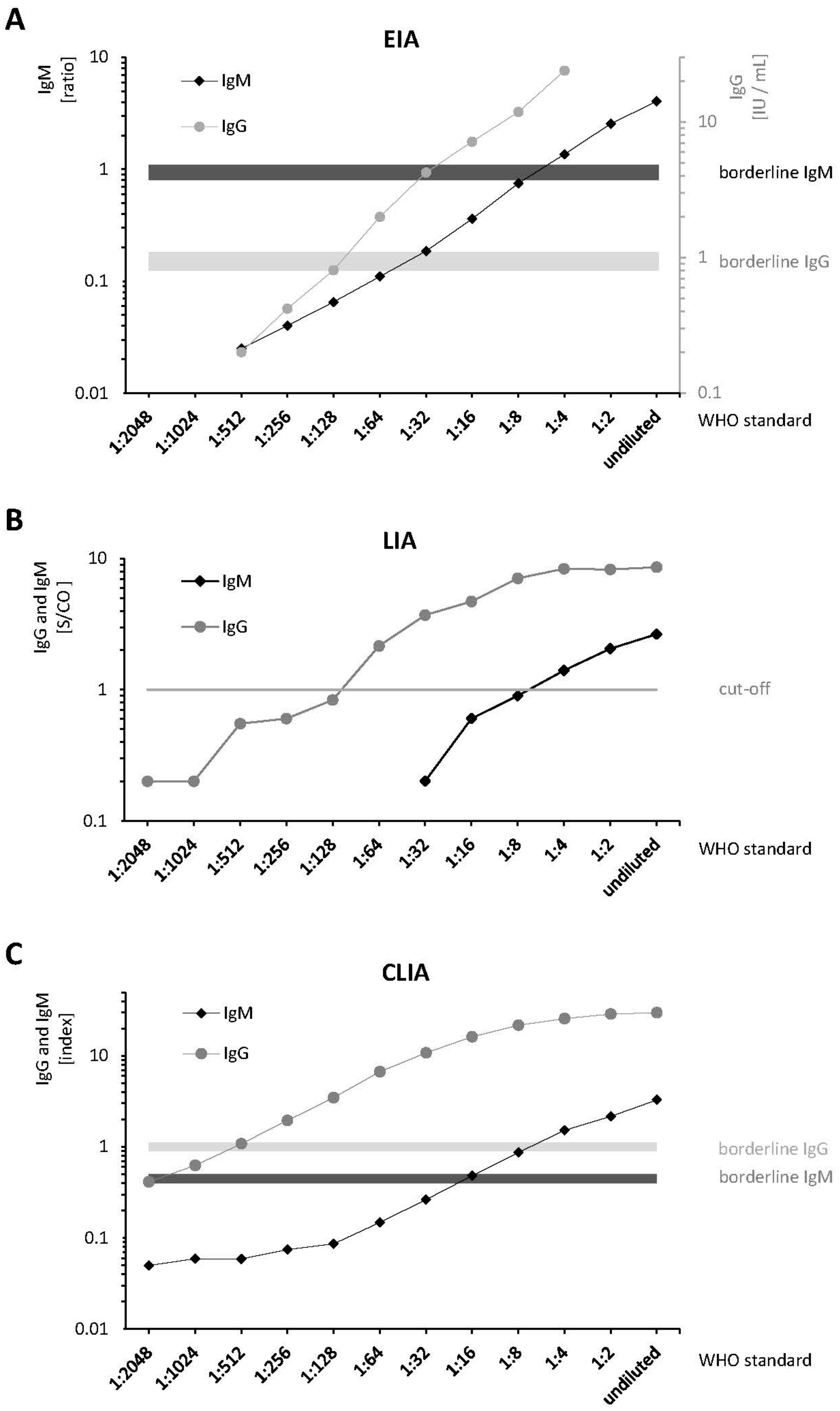
2. Results
3. Discussion
4. Materials and Methods
4.1. Patient Samples
4.2. WHO Reference Reagent
4.3. AST and ALT Testing
4.4. Test Characteristics
4.5. Statistical Analysis
5. Conclusions
Supplementary Materials
Author Contributions
Funding
Institutional Review Board Statement
Informed Consent Statement
Data Availability Statement
Acknowledgments
Conflicts of Interest
Abbreviations
| ALT | Alanine Aminotransferase |
| AST | Aspartate Aminotransferase |
| CLIA | Chemoluminescence Immunoassay |
| C(t) | Cycle threshold |
| EIA | Anti-HEV ELISA (Euroimmun, Lübeck, Germany) |
| ELISA | Enzyme-linked Immunosorbent Assay |
| GT 1 | Genotype 1 |
| GT 3 | Genotype 3 |
| HEV | Hepatitis E Virus |
| IU/mL | International Units/mL |
| κ | Cohen’s correlation coefficient |
| LIA | Line Immunoassay |
| O2CGt3 | C-terminal part of ORF2, Genotype 3 |
| ORF | Open Reading Frame |
| PCR | Polymerase Chain Reaction |
| RLU | Relative Light Units |
| RNA | Ribonucleid acid |
| S/CO | Signal/Cut-Off |
| WHO | World Health Organisation |
References
- Webb, G.W.; Dalton, H.R. Hepatitis E: An expanding epidemic with a range of complications. Clin. Microbiol. Infect. 2020, 26, 828–832. [Google Scholar] [CrossRef]
- Global Burden of Disease Study. Global, regional, and national incidence, prevalence, and years lived with disability for 310 diseases and injuries, 1990–2015: A systematic analysis for the Global Burden of Disease Study 2015. Lancet 2016, 388, 1545–1602. [Google Scholar] [CrossRef]
- Rein, D.B.; Stevens, G.A.; Theaker, J.; Wittenborn, J.S.; Wiersma, S.T. The global burden of hepatitis E virus genotypes 1 and 2 in 2005. Hepatology 2012, 55, 988–997. [Google Scholar] [CrossRef]
- Baki, A.A.; Haque, W.; Giti, S.; Khan, A.A.; Rahman, M.M.; Jubaida, N.; Rahman, M. Hepatitis E virus genotype 1f outbreak in Bangladesh, 2018. J. Med. Virol. 2020. [Google Scholar] [CrossRef] [PubMed]
- Pallerla, S.R.; Harms, D.; Johne, R.; Todt, D.; Steinmann, E.; Schemmerer, M.; Wenzel, J.J.; Hofmann, J.; Shih, J.W.K.; Wedemeyer, H.; et al. Hepatitis E Virus Infection: Circulation, Molecular Epidemiology, and Impact on Global Health. Pathogens 2020, 9, 856. [Google Scholar] [CrossRef] [PubMed]
- Kamar, N.; Bendall, R.; Legrand-Abravanel, F.; Xia, N.S.; Ijaz, S.; Izopet, J.; Dalton, H.R. Hepatitis E. Lancet 2012, 379, 2477–2488. [Google Scholar] [CrossRef]
- Doceul, V.; Bagdassarian, E.; Demange, A.; Pavio, N. Zoonotic Hepatitis E Virus: Classification, Animal Reservoirs and Transmission Routes. Viruses 2016, 8, 270. [Google Scholar] [CrossRef] [PubMed]
- Wenzel, J.J.; Preiss, J.; Schemmerer, M.; Huber, B.; Plentz, A.; Jilg, W. Detection of hepatitis E virus (HEV) from porcine livers in Southeastern Germany and high sequence homology to human HEV isolates. J. Clin. Virol. 2011, 52, 50–54. [Google Scholar] [CrossRef]
- Lewis, H.C.; Wichmann, O.; Duizer, E. Transmission routes and risk factors for autochthonous hepatitis E virus infection in Europe: A systematic review. Epidemiol. Infect. 2010, 138, 145–166. [Google Scholar] [CrossRef]
- Krain, L.J.; Nelson, K.E.; Labrique, A.B. Host immune status and response to hepatitis E virus infection. Clin. Microbiol. Rev. 2014, 27, 139–165. [Google Scholar] [CrossRef]
- Sayed, I.M.; El-Mokhtar, M.A.; Mahmoud, M.A.R.; Elkhawaga, A.A.; Gaber, S.; Seddek, N.H.; Abdel-Wahid, L.; Ashmawy, A.M.; Alkareemy, E.A.R. Clinical Outcomes and Prevalence of Hepatitis E Virus (HEV) Among Non-A-C Hepatitis Patients in Egypt. Infect. Drug Resist. 2021, 14, 59–69. [Google Scholar] [CrossRef]
- Goel, A.; Vijay, H.J.; Katiyar, H.; Aggarwal, R. Prevalence of hepatitis E viraemia among blood donors: A systematic review. Vox Sang. 2020, 115, 120–132. [Google Scholar] [CrossRef]
- Denner, J.; Pischke, S.; Steinmann, E.; Blumel, J.; Glebe, D. Why all blood donations should be tested for hepatitis E virus (HEV). BMC Infect. Dis. 2019, 19, 541. [Google Scholar] [CrossRef]
- Reekie, I.; Irish, D.; Ijaz, S.; Fox, T.; Bharucha, T.; Griffiths, P.; Thorburn, D.; Harber, M.; MacKinnon, S.; Sekhar, M. Hepatitis E infection in stem cell and solid organ transplantpatients: A cross-sectional study: The importance of HEV RNA screening in peri-transplant period. J. Clin. Virol. 2018, 107, 1–5. [Google Scholar] [CrossRef] [PubMed]
- Osterman, A.; Vizoso-Pinto, M.G.; Jung, J.; Jaeger, G.; Eberle, J.; Nitschko, H.; Baiker, A. A novel indirect immunofluorescence test for the detection of IgG and IgA antibodies for diagnosis of Hepatitis E Virus infections. J. Virol. Methods 2013, 191, 48–54. [Google Scholar] [CrossRef]
- European Association for the Study of the Liver. EASL Clinical Practice Guidelines on hepatitis E virus infection. J. Hepatol. 2018, 68, 1256–1271. [Google Scholar] [CrossRef] [PubMed]
- Fogeda, M.; de Ory, F.; Avellon, A.; Echevarria, J.M. Differential diagnosis of hepatitis E virus, cytomegalovirus and Epstein-Barr virus infection in patients with suspected hepatitis E. J. Clin. Virol. 2009, 45, 259–261. [Google Scholar] [CrossRef] [PubMed]
- Hyams, C.; Mabayoje, D.A.; Copping, R.; Maranao, D.; Patel, M.; Labbett, W.; Haque, T.; Webster, D.P. Serological cross reactivity to CMV and EBV causes problems in the diagnosis of acute hepatitis E virus infection. J. Med. Virol. 2014, 86, 478–483. [Google Scholar] [CrossRef] [PubMed]
- Bendall, R.; Ellis, V.; Ijaz, S.; Ali, R.; Dalton, H. A comparison of two commercially available anti-HEV IgG kits and a re-evaluation of anti-HEV IgG seroprevalence data in developed countries. J. Med. Virol. 2010, 82, 799–805. [Google Scholar] [CrossRef] [PubMed]
- Wenzel, J.J.; Preiss, J.; Schemmerer, M.; Huber, B.; Jilg, W. Test performance characteristics of Anti-HEV IgG assays strongly influence hepatitis E seroprevalence estimates. J. Infect. Dis. 2013, 207, 497–500. [Google Scholar] [CrossRef] [PubMed]
- Osterman, A.; Vizoso Pinto, M.G.; Haase, R.; Nitschko, H.; Jager, S.; Sander, M.; Motz, M.; Mohn, U.; Baiker, A. Systematic screening for novel, serologically reactive Hepatitis E Virus epitopes. Virol. J. 2012, 9, 28. [Google Scholar] [CrossRef] [PubMed]
- Al-Sadeq, D.W.; Majdalawieh, A.F.; Mesleh, A.G.; Abdalla, O.M.; Nasrallah, G.K. Laboratory challenges in the diagnosis of hepatitis E virus. J. Med. Microbiol. 2018, 67, 466–480. [Google Scholar] [CrossRef] [PubMed]
- Primadharsini, P.P.; Nagashima, S.; Okamoto, H. Genetic Variability and Evolution of Hepatitis E Virus. Viruses 2019, 11, 456. [Google Scholar] [CrossRef] [PubMed]
- Norder, H.; Karlsson, M.; Mellgren, A.; Konar, J.; Sandberg, E.; Lasson, A.; Castedal, M.; Magnius, L.; Lagging, M. Diagnostic Performance of Five Assays for Anti-Hepatitis E Virus IgG and IgM in a Large Cohort Study. J. Clin. Microbiol. 2016, 54, 549–555. [Google Scholar] [CrossRef] [PubMed]
- Sommerkorn, F.M.; Schauer, B.; Schreiner, T.; Fickenscher, H.; Krumbholz, A. Performance of Hepatitis E Virus (HEV)-antibody tests: A comparative analysis based on samples from individuals with direct contact to domestic pigs or wild boar in Germany. Med. Microbiol. Immunol. 2017, 206, 277–286. [Google Scholar] [CrossRef]
- Dremsek, P.; Wenzel, J.J.; Johne, R.; Ziller, M.; Hofmann, J.; Groschup, M.H.; Werdermann, S.; Mohn, U.; Dorn, S.; Motz, M.; et al. Seroprevalence study in forestry workers from eastern Germany using novel genotype 3- and rat hepatitis E virus-specific immunoglobulin G ELISAs. Med. Microbiol. Immunol. 2012, 201, 189–200. [Google Scholar] [CrossRef] [PubMed]
- Miller, J.M.; Binnicker, M.J.; Campbell, S.; Carroll, K.C.; Chapin, K.C.; Gilligan, P.H.; Gonzalez, M.D.; Jerris, R.C.; Kehl, S.C.; Patel, R.; et al. A Guide to Utilization of the Microbiology Laboratory for Diagnosis of Infectious Diseases: 2018 Update by the Infectious Diseases Society of America and the American Society for Microbiology. Clin. Infect. Dis. 2018, 67, e1–e94. [Google Scholar] [CrossRef]
- Pan, J.S.; Zhang, K.; Zhou, J.; Wu, C.; Zhuang, H.; Zhou, Y.H. Application of truncated immunodominant polypeptide from hepatitis E virus (HEV) ORF2 in an assay to exclude nonspecific binding in detecting anti-HEV immunoglobulin M. J. Clin. Microbiol. 2010, 48, 779–784. [Google Scholar] [CrossRef]
- Park, H.K.; Jeong, S.H.; Kim, J.W.; Woo, B.H.; Lee, D.H.; Kim, H.Y.; Ahn, S. Seroprevalence of anti-hepatitis E virus (HEV) in a Korean population: Comparison of two commercial anti-HEV assays. BMC Infect. Dis. 2012, 12, 142. [Google Scholar] [CrossRef]
- Ferguson, M.; Walker, D.; Mast, E.; Fields, H. Report of a collaborative study to assess the suitability of a reference reagent for antibodies to hepatitis E virus. Biologicals 2002, 30, 43–48. [Google Scholar] [CrossRef] [PubMed]
- Vollmer, T.; Diekmann, J.; Eberhardt, M.; Knabbe, C.; Dreier, J. Monitoring of Anti-Hepatitis E Virus Antibody Seroconversion in Asymptomatically Infected Blood Donors: Systematic Comparison of Nine Commercial Anti-HEV IgM and IgG Assays. Viruses 2016, 8, 232. [Google Scholar] [CrossRef] [PubMed]
- Chapin, K.C.; Dickenson, R.A.; Wu, F.; Andrea, S.B. Comparison of five assays for detection of Clostridium difficile toxin. J. Mol. Diagn 2011, 13, 395–400. [Google Scholar] [CrossRef] [PubMed]
- Landis, J.R.; Koch, G.G. The measurement of observer agreement for categorical data. Biometrics 1977, 33, 159–174. [Google Scholar] [CrossRef] [PubMed]


| IgG | LIA | EIA | CLIA |
| EIA | 0.60 | - | - |
| CLIA | 0.76 | 0.60 | - |
| overall IgG | 0.88 | 0.71 | 0.87 |
| IgM | LIA | EIA | CLIA |
| EIA | 0.46 | - | - |
| CLIA | 0.61 | 0.75 | - |
| overall IgM | 0.64 | 0.79 | 0.96 |
| CMV (n = 11) | EBV (n = 12) | HBV (n = 11) | Total (n = 34) | ||
|---|---|---|---|---|---|
| IgG | |||||
| CLIA | 3 | 2 | 6 | 11 | |
| EIA | 2 | 1 | 1 | 4 | |
| LIA | 3 | 2 | 5 | 10 | |
| IgM | |||||
| CLIA | 0 | 1 | 0 | 1 | |
| EIA | 0 | 2 | 0 | 2 | |
| LIA | 0 | 3 | 0 | 3 | |
| IgG | IgM | |||||
|---|---|---|---|---|---|---|
| Patient ID | CLIA | EIA | LIA | CLIA | EIA | LIA |
| 10 | pos. | pos. | pos. | neg. | pos. | pos. |
| 12 | neg. | neg. | neg. | pos. | neg. | pos. |
| 15 | neg. | neg. | neg. | neg. | neg. | pos. |
| 18 | neg. | neg. | neg. | neg. | pos. | neg. |
| total pos. | 1 | 1 | 1 | 1 | 2 | 3 |
Publisher’s Note: MDPI stays neutral with regard to jurisdictional claims in published maps and institutional affiliations. |
© 2021 by the authors. Licensee MDPI, Basel, Switzerland. This article is an open access article distributed under the terms and conditions of the Creative Commons Attribution (CC BY) license (https://creativecommons.org/licenses/by/4.0/).
Share and Cite
Dichtl, K.; Zimmermann, J.; Koeppel, M.B.; Böhm, S.; Osterman, A. Evaluation of a Novel CLIA Monotest Assay for the Detection of Anti-Hepatitis E Virus-IgG and IgM: A Retrospective Comparison with a Line Blot and an ELISA. Pathogens 2021, 10, 689. https://doi.org/10.3390/pathogens10060689
Dichtl K, Zimmermann J, Koeppel MB, Böhm S, Osterman A. Evaluation of a Novel CLIA Monotest Assay for the Detection of Anti-Hepatitis E Virus-IgG and IgM: A Retrospective Comparison with a Line Blot and an ELISA. Pathogens. 2021; 10(6):689. https://doi.org/10.3390/pathogens10060689
Chicago/Turabian StyleDichtl, Karl, Julia Zimmermann, Martin B. Koeppel, Stephan Böhm, and Andreas Osterman. 2021. "Evaluation of a Novel CLIA Monotest Assay for the Detection of Anti-Hepatitis E Virus-IgG and IgM: A Retrospective Comparison with a Line Blot and an ELISA" Pathogens 10, no. 6: 689. https://doi.org/10.3390/pathogens10060689
APA StyleDichtl, K., Zimmermann, J., Koeppel, M. B., Böhm, S., & Osterman, A. (2021). Evaluation of a Novel CLIA Monotest Assay for the Detection of Anti-Hepatitis E Virus-IgG and IgM: A Retrospective Comparison with a Line Blot and an ELISA. Pathogens, 10(6), 689. https://doi.org/10.3390/pathogens10060689