
Neonatal SARS-CoV-2 Infection: Practical Tips

 , ,
, ,
Abstract
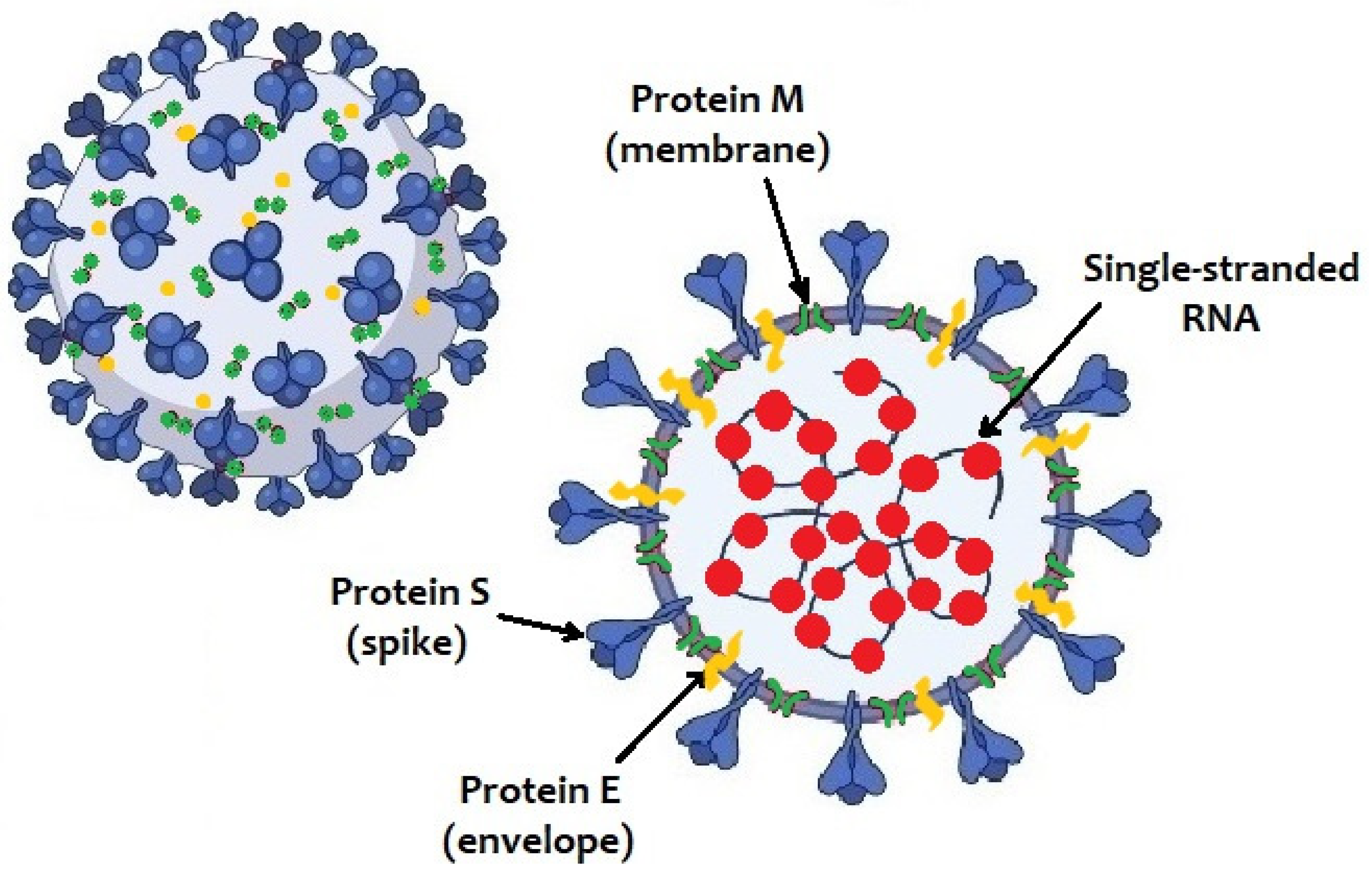
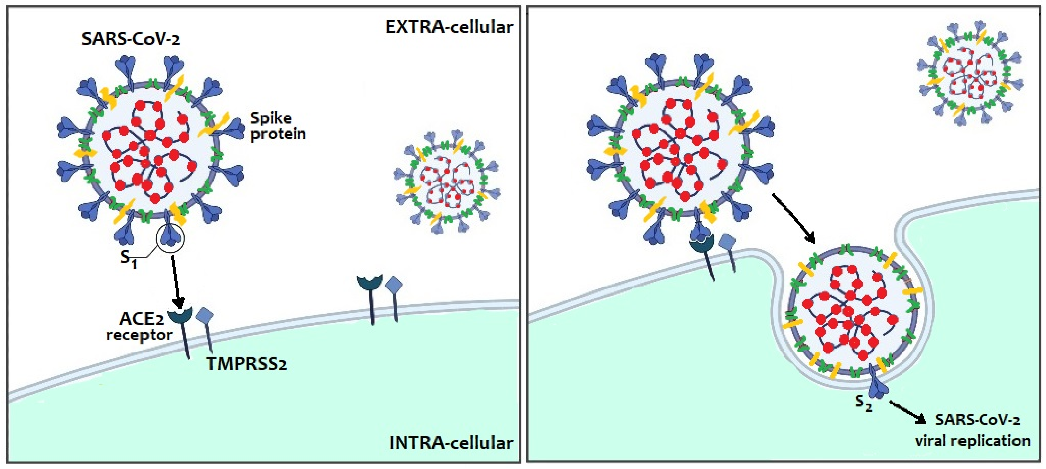
1. Introduction
2. Methods
3. Transplacental Transmission of SARS-CoV-2
4. How to Diagnose a Congenital Infection
5. How to Diagnose a Postnatal Infection
6. Which Tests Are to Be Used in the Neonate?
7. How to Treat SARS-CoV-2 Infection
8. Breastfeeding and SARS-CoV-2 Infection
9. How to Rethink In-Hospital Management of Mother and Neonate
10. Which Indications to Provide upon Discharge?
11. Vaccines and Preventive Strategies
12. Strength, Limitations and Conclusions
Author Contributions
Funding
Institutional Review Board Statement
Informed Consent Statement
Data Availability Statement
Conflicts of Interest
References
- Li, F. Structure, function, and evolution of coronavirus spike proteins. Annu. Rev. Virol. 2016, 3, 237–261. [Google Scholar] [CrossRef] [PubMed]
- Su, S.; Wong, G.; Shi, W.; Liu, J.; Lai, A.C.; Zhou, J.; Liu, W.; Bi, Y.; Gao, G.F. Epidemiology, genetic recombination, and pathogenesis of coronaviruses. Trends Microbiol. 2016, 24, 490–502. [Google Scholar] [CrossRef]
- Cui, J.; Li, F.; Shi, Z.-L. Origin and evolution of pathogenic coronaviruses. Nat. Rev. Microbiol. 2019, 17, 181–192. [Google Scholar] [CrossRef]
- Setti, L.; Passarini, F.; De Gennaro, G.; Barbieri, P.; Perrone, M.G.; Borelli, M.; Palmisani, J.; Di Gilio, A.; Piscitelli, P.; Miani, A. Airborne transmission route of COVID-19: Why 2 meters/6 feet of inter-personal distance could not be enough. Int. J. Environ. Res. Public Health 2020, 17, 2932. [Google Scholar] [CrossRef] [PubMed]
- Amodio, E.; Vitale, F.; Cimino, L.; Casuccio, A.; Tramuto, F. Outbreak of novel coronavirus (SARS-Cov-2): First evidences from international scientific literature and pending questions. Healthcare 2020, 8, 51. [Google Scholar] [CrossRef]
- Zhang, H.; Kang, Z.; Gong, H.; Xu, D.; Wang, J.; Li, Z.; Li, Z.; Cui, X.; Xiao, J.; Zhan, J.; et al. Digestive system is a potential route of COVID-19: An analysis of single-cell coexpression pattern of key proteins in viral entry process. Gut 2020, 69, 1010–1018. [Google Scholar] [CrossRef]
- Arya, R.; Kumari, S.; Pandey, B.; Mistry, H.; Bihani, S.C.; Das, A.; Prashar, V.; Gupta, G.D.; Panicker, L.; Kumar, M. Structural insights into SARS-CoV-2 proteins. J. Mol. Biol. 2021, 433, 166725. [Google Scholar] [CrossRef]
- Malik, Y.A. Properties of coronavirus and SARS-CoV-2. Malays. J. Pathol. 2020, 42, 3–11. [Google Scholar]
- Jing, Y.; Run-Qian, L.; Hao-Ran, W.; Hao-Ran, C.; Ya-Bin, L.; Yang, G.; Fei, C. Potential influence of COVID-19/ACE2 on the female reproductive system. Mol. Hum. Reprod. 2020, 26, 367–373. [Google Scholar] [CrossRef] [PubMed]
- Li, Q.; Guan, X.; Wu, P.; Wang, X.; Zhou, L.; Tong, Y.; Ren, R.; Leung, K.; Lau, E.; Wong, J.Y.; et al. Early transmission dynamics in Wuhan, China, of novel coronavirus-infected pneumonia. N. Engl. J. Med. 2020, 382, 1199–1207. [Google Scholar] [CrossRef]
- Rasmussen, S.A.; Jamieson, D.J.; Bresee, J.S. Pandemic influenza and pregnant women. Emerg. Infect. Dis. 2008, 14, 95–100. [Google Scholar] [CrossRef] [PubMed]
- Valdés, G.; Neves, L.A.; Anton, L.; Corthorn, J.; Chacón, C.; Germain, A.M.; Merrill, D.C.; Ferrario, C.M.; Sarao, R.; Penninger, J.; et al. Distribution of angiotensin-(1-7) and ACE2 in human placentas of normal and pathological pregnancies. Placenta 2006, 27, 200–207. [Google Scholar] [CrossRef]
- Patberg, E.T.; Adams, T.; Rekawek, P.; Vahanian, S.A.; Akerman, M.; Hernandez, A.; Rapkiewicz, A.V.; Ragolia, L.; Sicuranza, G.; Chavez, M.R.; et al. Coronavirus disease 2019 infection and placental histopathology in women delivering at term. Am. J. Obstet. Gynecol. 2021, 224, 382-e1. [Google Scholar] [CrossRef] [PubMed]
- Algarroba, G.N.; Rekawek, P.; Vahanian, S.A.; Khullar, P.; Palaia, T.; Peltier, M.R.; Chavez, M.R.; Vintzileos, A.M. Visualization of severe acute respiratory syndrome coronavirus 2 invading the human placenta using electron microscopy. Am. J. Obstet. Gynecol. 2020, 223, 275–278. [Google Scholar] [CrossRef] [PubMed]
- Schwartz, D.A.; Dhaliwal, A. Infections in pregnancy with COVID-19 and other respiratory RNA virus diseases are rarely, if ever, transmitted to the fetus: Experiences with coronaviruses, parainfluenza, metapneumovirus respiratory syncytial virus, and influenza. Arch. Pathol. Lab. Med. 2020, 144, 920–928. [Google Scholar] [CrossRef]
- Juan, J.; Gil, M.M.; Rong, Z.; Zhang, Y.; Yang, H.; Poon, L.C. Effect of coronavirus disease 2019 (COVID-19) on maternal, perinatal and neonatal outcome: Systematic review. Ultrasound Obstet. Gynecol. 2020, 56, 15–27. [Google Scholar] [CrossRef] [PubMed]
- Crovetto, F.; Crispi, F.; Llurba, E.; Pascal, R.; Larroya, M.; Trilla, C.; Camacho, M.; Medina, C.; Dobaño, C.; Gomez-Roig, M.D.; et al. KidsCorona pregnancy COVID-19 group. Impact of SARS-CoV-2 infection on pregnancy outcomes: A population-based study. Clin. Infect. Dis. 2021, ciab104. [Google Scholar] [CrossRef] [PubMed]
- Li, M.; Chen, L.; Zhang, J.; Xiong, C.; Li, X. The SARS-CoV-2 receptor ACE2 expression of maternal-fetal interface and fetal organs by single-cell transcriptome study. PLoS ONE 2020, 15, e0230295. [Google Scholar] [CrossRef]
- Vivanti, A.J.; Vauloup-Fellous, C.; Prevot, S.; Zupan, V.; Suffee, C.; Do Cao, J.; Benachi, A.; De Luca, D. Transplacental transmission of SARS-CoV-2 infection. Nat. Commun. 2020, 11, 3572. [Google Scholar] [CrossRef] [PubMed]
- Dong, L.; Tian, J.; He, S.; Zhu, C.; Wang, J.; Liu, C.; Yang, J. Possible vertical transmission of SARS-CoV-2 from an infected mother to her newborn. J. Am. Med. Assoc. 2020, 323, 1846–1848. [Google Scholar] [CrossRef]
- Zeng, H.; Xu, C.; Fan, J.; Tang, Y.; Deng, Q.; Zhang, W.; Long, X. Antibodies in Infants Born to Mothers With COVID-19 Pneumonia. J. Am. Med. Assoc. 2020, 323, 1848–1849. [Google Scholar] [CrossRef] [PubMed]
- Raschetti, R.; Vivanti, A.J.; Vauloup-Fellous, C.; Loi, B.; Benachi, A.; De Luca, D. Synthesis and systematic review of reported neonatal SARS-CoV-2 infections. Nat. Commun. 2020, 11, 5164. [Google Scholar] [CrossRef] [PubMed]
- WHO COVID-19 LENS (Living Evidence Synthesis) Working Group. Definition and Categorization of the Timing of Mother-To-Child Transmission of SARS-CoV-2. 2021. Available online: www.who.int/publications/i/item/WHO-2019-nCoV-mother-to-child-transmission-2021.1 (accessed on 29 March 2021).
- Hopwood, A.J.; Jordan-Villegas, A.; Gutierrez, L.D.; Cowart, M.C.; Vega-Montalvo, W.; Cheung, W.L.; McMahan, M.J.; Gomez, M.R.; Laham, F.R. Severe acute respiratory syndrome coronavirus-2 pneumonia in a newborn treated with remdesivir and coronavirus disease 2019 convalescent plasma. J. Pediatr. Infect. Dis. Soc. 2020. [Google Scholar] [CrossRef]
- Chen, Z.M.; Fu, J.F.; Shu, Q.; Chen, Y.H.; Hua, C.Z.; Li, F.B.; Lin, R.; Tang, L.F.; Wang, T.L.; Wang, W.; et al. Diagnosis and treatment recommendations for pediatric respiratory infection caused by the 2019 novel coronavirus. World J. Pediatr. 2020, 16, 240–246. [Google Scholar] [CrossRef] [PubMed]
- Italian National Institute of Health. COVID-19: National Update 24 March 2021 (Italian Version). Available online: www.epicentro.iss.it/coronavirus/bollettino/Bollettino-sorveglianza-integrata-COVID-19_24-marzo-2021.pdf (accessed on 29 March 2021).
- De Rose, D.U.; Piersigilli, F.; Ronchetti, M.P.; Santisi, A.; Bersani, I.; Dotta, A.; Danhaive, O.; Auriti, C.; Study Group of Neonatal Infectious Diseases of The Italian Society of Neonatology (SIN). Novel Coronavirus disease (COVID-19) in newborns and infants: What we know so far. Ital. J. Pediatr. 2020, 46, 56. [Google Scholar] [CrossRef]
- Italian Ministry of Health and Italian National Institute of Health. Laboratory Tests for SARS-CoV-2 and Their Use in Public Health (Italian Version: Test di Laboratorio per SARS-CoV-2 e Loro Uso in Sanità Pubblica). Available online: www.iss.it/documents/20126/0/COVID+19_+test+v4k_last.pdf/9ab1f211-7d88-bcb1-d454-cfed04aa8b05?t=1604483686312 (accessed on 29 March 2021).
- Blumberg, D.A.; Underwood, M.A.; Hedriana, H.L.; Lakshminrusimha, S. Vertical transmission of SARS-CoV-2: What is the optimal definition? Am. J. Perinatol. 2020, 37, 769–772. [Google Scholar] [CrossRef]
- Wang, L.; Shi, Y.; Xiao, T.; Fu, J.; Feng, X.; Mu, D.; Feng, Q.; Hei, M.; Hu, X.; Li, Z.; et al. Chinese expert consensus on the perinatal and neonatal management for the prevention and control of the 2019 novel coronavirus infection. Ann. Transl. Med. 2020, 8, 47. [Google Scholar] [CrossRef]
- Poorisrisak, P.; Bivolarova, M.P.; Bekö, G.; Heiring, C.; Clausen, G.; Greisen, G. Aerosol generation by respiratory support of neonates may be low. J. Pediatric. Infect. Dis. Soc. 2020. [Google Scholar] [CrossRef]
- Taylor, M.M.; Kobeissi, L.; Kim, C.; Amin, A.; Thorson, A.E.; Bellare, N.B.; Brizuela, V.; Bonet, M.; Kara, E.; Thwin, S.S.; et al. Inclusion of pregnant women in COVID-19 treatment trials: A review and global call to action. Lancet Glob. Health 2021, 9, e366–e371. [Google Scholar] [CrossRef]
- Saikia, B.; Tang, J.; Robinson, S.; Nichani, S.; Lawman, K.B.; Katre, M.; Bandi, S. Neonates with SARS-CoV-2 infection and pulmonary disease safely treated with remdesivir. Pediatr. Infect. Dis. J. 2021, 40, e194–e196. [Google Scholar] [CrossRef]
- Frauenfelder, C.; Brierley, J.; Whittaker, E.; Perucca, G.; Bamford, A. Infant with SARS-CoV-2 infection causing severe lung disease treated with remdesivir. Pediatrics 2020, 146, e20201701. [Google Scholar] [CrossRef]
- Wardell, H.; Campbell, J.I.; VanderPluym, C.; Dixit, A. Severe acute respiratory syndrome Coronavirus 2 infection in febrile neonates. J. Pediatric Infect. Dis. Soc. 2020, 9, 630–635. [Google Scholar] [CrossRef] [PubMed]
- Del Borrello, G.; Giraudo, I.; Bondone, C.; Denina, M.; Garazzino, S.; Linari, C.; Mignone, F.; Pruccoli, G.; Scolfaro, C.; Spadea, M.; et al. SARS-COV-2-associated coagulopathy and thromboembolism prophylaxis in children: A single-center observational study. J. Thromb. Haemost. 2021, 19, 522–530. [Google Scholar] [CrossRef] [PubMed]
- Goldenberg, N.A.; Sochet, A.; Albisetti, M.; Biss, T.; Bonduel, M.; Jaffray, J.; MacLaren, G.; Monagle, P.; O’Brien, S.; Raffini, L.; et al. Pediatric/neonatal hemostasis and thrombosis subcommittee of the ISTH SSC. Consensus-based clinical recommendations and research priorities for anticoagulant thromboprophylaxis in children hospitalized for COVID-19-related illness. J. Thromb. Haemost. 2020, 18, 3099–3105. [Google Scholar] [CrossRef]
- Venturini, E.; Montagnani, C.; Garazzino, S.; Donà, D.; Pierantoni, L.; Lo Vecchio, A.; Nicolini, G.; Bianchini, S.; Krzysztofiak, A.; Galli, L.; et al. Italian SITIP-SIP SARS-Cov-2 pediatric infection study group. Treatment of children with COVID-19: Position paper of the Italian Society of Pediatric Infectious Disease. Ital. J. Pediatr. 2020, 46, 139. [Google Scholar] [CrossRef]
- Henderson, L.A.; Canna, S.W.; Friedman, K.G.; Gorelik, M.; Lapidus, S.K.; Bassiri, H.; Behrens, E.M.; Ferris, A.; Kernan, K.F.; Schulert, G.S.; et al. American college of rheumatology clinical guidance for multisystem inflammatory syndrome in children associated with SARS-CoV-2 and hyperinflammation in pediatric COVID-19: Version 2. Arthritis Rheumatol. 2021, 73, e13–e29. [Google Scholar] [CrossRef] [PubMed]
- Götzinger, F.; Santiago-García, B.; Noguera-Julián, A.; Lanaspa, M.; Lancella, L.; Calò Carducci, F.I.; Gabrovska, N.; Velizarova, S.; Prunk, P.; Osterman, V.; et al. COVID-19 in children and adolescents in Europe: A multinational, multicentre cohort study. Lancet Child. Adolesc. Health 2020, 4, 653–661. [Google Scholar] [CrossRef]
- Salvatori, G.; De Rose, D.U.; Concato, C.; Alario, D.; Olivini, N.; Dotta, A.; Campana, A. Managing COVID-19-Positive Maternal-Infant Dyads: An Italian Experience. Breastfeed. Med. 2020, 15, 347–348. [Google Scholar] [CrossRef]
- Lackey, K.A.; Pace, R.M.; Williams, J.E.; Bode, L.; Donovan, S.M.; Järvinen, K.M.; Seppo, A.E.; Raiten, D.J.; Meehan, C.L.; McGuire, M.A.; et al. SARS-CoV-2 and human milk: What is the evidence? Matern Child Nutr. 2020, 2020 16, e13032. [Google Scholar] [CrossRef]
- Groß, R.; Conzelmann, C.; Müller, J.A.; Stenger, S.; Steinhart, K.; Kirchhoff, F.; Münch, J. Detection of SARS-CoV-2 in human breastmilk. Lancet 2020, 395, 1757–1758. [Google Scholar] [CrossRef]
- Vassilopoulou, E.; Feketea, G.; Koumbi, L.; Mesiari, C.; Berghea, E.C.; Konstantinou, G.N. Breastfeeding and COVID-19: From nutrition to immunity. Front. Immunol. 2021, 12, 661806. [Google Scholar] [CrossRef] [PubMed]
- Davanzo, R.; Moro, G.; Sandri, F.; Agosti, M.; Moretti, C.; Mosca, F. Breastfeeding and coronavirus disease-2019: Ad interim indications of the Italian Society of Neonatology endorsed by the Union of European Neonatal & Perinatal Societies. Matern Child. Nutr. 2020, 16, e13010. [Google Scholar] [CrossRef]
- Centers for Disease Control and Prevention. Interim Guidance on Breastfeeding and Breast Milk Feeds in the Context of COVID-19. 2020. Available online: www.cdc.gov/coronavirus/2019-ncov/hcp/care-for-breastfeeding-women.html (accessed on 1 April 2021).
- Pace, R.M.; Williams, J.E.; Järvinen, K.M.; Belfort, M.B.; Pace, C.; Lackey, K.A.; Gogel, A.C.; Nguyen-Contant, P.; Kanagaiah, P.; Fitzgerald, T.; et al. Characterization of SARS-CoV-2 RNA, antibodies, and neutralizing capacity in milk produced by women with COVID-19. Mbio 2021, 12, e03192-20. [Google Scholar] [CrossRef] [PubMed]
- Centers for Disease Control and Prevention. COVID-19—Evaluation and Management Considerations for Neonates at Risk for COVID-19 (Updated on 8 December 2020). Available online: www.cdc.gov/coronavirus/2019-ncov/hcp/caring-for-newborns.html (accessed on 1 April 2021).
- Italian National Institute of Health. Interim Guidance on Pregnancy, Childbirth, Breastfeeding and Care of Infants (0–2 Years) in Response to the COVID-19 Emergency. (Version of 5 February 2021). Available online: https://www.iss.it/documents/5430402/0/Rapporto+ISS+COVID-19+2_2021_EN.pdf/421a0bc6-1933-aa77-6b39-a7860d866a16?t=1615472502820 (accessed on 1 April 2021).
- Chandrasekharan, P.; Vento, M.; Trevisanuto, D.; Partridge, E.; Underwood, M.A.; Wiedeman, J.; Katheria, A.; Lakshminrusimha, S. Neonatal resuscitation and postresuscitation care of infants born to mothers with suspected or confirmed SARS-CoV-2 infection. Am. J. Perinatol. 2020, 37, 813–824. [Google Scholar] [CrossRef] [PubMed]
- Saiman, L.; Acker, K.P.; Dumitru, D.; Messina, M.; Johnson, C.; Zachariah, P.; Abreu, W.; Saslaw, M.; Keown, M.K.; Hanft, E.; et al. Infection prevention and control for labor and delivery, well baby nurseries, and neonatal intensive care units. Semin. Perinatol. 2020, 44, 151320. [Google Scholar] [CrossRef]
- Cavicchiolo, M.E.; Doglioni, N.; Ventola, M.A.; Biban, P.; Baraldi, E.; Trevisanuto, D. Neonatal emergency transport system during COVID-19 pandemic in the Veneto Region: Proposal for standard operating procedures. Pediatr. Res. 2021, 89, 399–401. [Google Scholar] [CrossRef] [PubMed]
- Terheggen, U.; Heiring, C.; Kjellberg, M.; Hegardt, F.; Kneyber, M.; Gente, M.; Roehr, C.C.; Jourdain, G.; Tissieres, P.; Ramnarayan, P.; et al. European consensus recommendations for neonatal and paediatric retrievals of positive or suspected COVID-19 patients. Pediatr. Res. 2020. [Google Scholar] [CrossRef]
- Wölfel, R.; Corman, V.M.; Guggemos, W.; Seilmaier, M.; Zange, S.; Müller, M.A.; Niemeyer, D.; Jones, T.C.; Vollmar, P.; Rothe, C.; et al. Virological assessment of hospitalized patients with COVID-2019. Nature 2020, 581, 465–469. [Google Scholar] [CrossRef] [PubMed]
- Centers for Disease Control and Prevention. COVID-19—Testing Overview: Updated on 17 March 2021. Available online: https://www.cdc.gov/coronavirus/2019-ncov/hcp/testing-overview.html (accessed on 1 April 2021).
- Benitz, W.E.; Watterberg, K.L.; Aucott, S.; Cummings, J.J.; Eichenwald, E.C.; Goldsmith, J.; Poindexteric, B.B.; Puopolo, K.; Stewanrt, D.L. Committee on fetus and newborn, American Academy of Pediatrics.; et al. Hospital stay for healthy term newborn infants. Pediatrics 2015, 135, 948–953. [Google Scholar] [CrossRef]
- Centers for Disease Control and Prevention. COVID-19—Guidance for Fully Vaccinated People: Updated on 8 March 2021. Available online: https://www.cdc.gov/coronavirus/2019-ncov/vaccines/fully-vaccinated-guidance.html (accessed on 1 April 2021).
- The New York Times. The Pfizer-BioNTech Vaccine Is Said to Be Powerfully Protective in Adolescents. Available online: www.nytimes.com/2021/03/31/health/pfizer-biontech-vaccine-adolescents.html (accessed on 1 April 2021).
- Gray, K.J.; Bordt, E.A.; Atyeo, C.; Deriso, E.; Akinwunmi, B.; Young, N.; Medina Baez, A.; Shook, L.L.; Cvrk, D.; James, K.; et al. COVID-19 vaccine response in pregnant and lactating women: A cohort study. Am. J. Obstet. Gynecol. 2021, 187–193. [Google Scholar] [CrossRef]


| IN UTERO Infection | (1) Maternal SARS-CoV-2 Infection | (2) Evidence of In Utero Fetal SARS-CoV-2 Exposure (Birth–24 h) | (3) Viral SARS-CoV-2 Persistence/Immune Response (24–48 h) |
|---|---|---|---|
| Confirmed | Suspect, probable or confirmed maternal infection anytime during pregnancy | Yes | Positive RT-PCR 1 |
| Possible | Yes | Positive RT-PCR 2 OR Positive serology (IgM or IgA) at age 24 h–<7 days | |
| Unlikely | Yes | Negative RT-PCR 1,2 OR Negative serology (IgM or IgA) at age 24 h–<7 days | |
| No | Positive RT-PCR 1,2 OR Positive serology (IgM or IgA) at age 24 h–<7 days | ||
| Indeterminate | Yes | No tests (above) performed | |
| No in utero fetal exposure tests performed | Positive RT-PCR 1,2 OR Positive serology (IgM or IgA) at age 24 h–<7 days |
| INTRA- PARTUM Infection | (1) Maternal SARS-CoV-2 Infection | (2) Lack of In Utero Fetal SARS-CoV-2 Exposure (Birth–24 h) | (3) Intrapartum SARS-CoV-2 Exposure with Viral Persistence/Immune Response (24–48 h) |
|---|---|---|---|
| Confirmed | Suspect, probable or confirmed maternal infection near the time of birth (14 days prior to 2 days after birth) | Negative tests | Positive RT-PCR (from a sterile sample or from repeated non-sterile samples at 2–7 days) OR Positive serology (IgM or IgA) at 7–14 days of age (confirmed by a second positive serology test within 10 days since the first one) |
| Possible | No in utero fetal exposure tests performed | Positive RT-PCR (from a sterile sample or from repeated non-sterile samples at 2–7 days) OR Positive serology (IgM or IgA) at 7–14 days of age (confirmed by a second positive serology test within 10 days since the first one) | |
| Unlikely | No in utero fetal exposure tests performed | Positive RT-PCR (from a sterile sample with a negative second sample at 2-7 days or from a non-sterile sample with a negative second non-sterile sample at 2-7 days) OR Positive serology (IgM or IgA) at 7–14 days of age (with a second negative serology test within 10 days since the first one) |
| POST- NATAL Infection | (1) Maternal SARS-CoV-2 Infection | (2) Lack of In Utero fetal SARS-CoV-2 Exposure (see above) | (3) Intrapartum SARS-CoV-2 Exposure with Viral Persistence/Immune Response (>48 h) |
|---|---|---|---|
| Confirmed | Suspected, probable or confirmed maternal infection near the time of birth (14 days prior to 2 days after birth) | Negative tests before 48 h of age | Positive RT-PCR at age ≥48 h: - from a sterile sample - from non-sterile samples (with a second positive sample within 10 days since the first one) OR Positive serology (IgM or IgA) at age >14 days (confirmed by a second positive serology test within 10 days since the first one) |
| Possible | No in utero/intrapartum exposure tests performed | Positive RT-PCR at age ≥48 h: - from a sterile sample - from non-sterile samples (with a second positive sample within 10 days since the first) OR Positive serology (IgM or IgA) at age >14 days (confirmed by a second positive serology test within 10 days since the first one) | |
| Unlikely | No in utero/intrapartum exposure tests performed | Positive RT-PCR at age ≥48 h from a non-sterile sample (with a second negative sample within 10 days since the first one) OR Positive serology (IgM or IgA) at age >14 days with a negative serology test within 10 days since the first one | |
| Indeterminate | No in utero/intrapartum exposure tests performed | Positive RT-PCR at age ≥48 h from a non-sterile sample (without a second corroboratory test) OR Positive serology (IgM or IgA) at age >14 days without a second corroboratory test |
| Clinical Features | Neonates (%) |
|---|---|
| Respiratory manifestations (signs of respiratory distress, i.e., tachypnea, subcostal retractions and rhinitis) | 51 (52.5%) |
| Fever | 43 (44.3%) |
| Gastrointestinal manifestations (eating disorders, diarrhea, vomiting) | 35 (36%) |
| Neurological manifestations (hypertonia/hypotonia, irritability/lethargy, apnea) | 18 (18.6%) |
| Hemodynamic manifestations (tachycardia, hypotension) | 10 (10.3%) |
| Other signs (conjunctivitis, hypothermia, cutaneous rash) | 9 (9.2%) |
Publisher’s Note: MDPI stays neutral with regard to jurisdictional claims in published maps and institutional affiliations. |
© 2021 by the authors. Licensee MDPI, Basel, Switzerland. This article is an open access article distributed under the terms and conditions of the Creative Commons Attribution (CC BY) license (https://creativecommons.org/licenses/by/4.0/).
Share and Cite
Auriti, C.; De Rose, D.U.; Mondì, V.; Stolfi, I.; Tzialla, C.; on behalf of the Study Group of Neonatal Infectious Diseases. Neonatal SARS-CoV-2 Infection: Practical Tips. Pathogens 2021, 10, 611. https://doi.org/10.3390/pathogens10050611
Auriti C, De Rose DU, Mondì V, Stolfi I, Tzialla C, on behalf of the Study Group of Neonatal Infectious Diseases. Neonatal SARS-CoV-2 Infection: Practical Tips. Pathogens. 2021; 10(5):611. https://doi.org/10.3390/pathogens10050611
Chicago/Turabian StyleAuriti, Cinzia, Domenico Umberto De Rose, Vito Mondì, Ilaria Stolfi, Chryssoula Tzialla, and on behalf of the Study Group of Neonatal Infectious Diseases. 2021. "Neonatal SARS-CoV-2 Infection: Practical Tips" Pathogens 10, no. 5: 611. https://doi.org/10.3390/pathogens10050611
APA StyleAuriti, C., De Rose, D. U., Mondì, V., Stolfi, I., Tzialla, C., & on behalf of the Study Group of Neonatal Infectious Diseases. (2021). Neonatal SARS-CoV-2 Infection: Practical Tips. Pathogens, 10(5), 611. https://doi.org/10.3390/pathogens10050611