Article

Serological and Molecular Prevalence of Babesia caballi in Apparently Healthy Horses in Israel

1
Koret School of Veterinary Medicine, The Hebrew University of Jerusalem, Rehovot 7610001, Israel
2
Division of Parasitology, Kimron Veterinary Institute, Beit Dagan 50250, Israel
*
Author to whom correspondence should be addressed.
Pathogens 2021, 10(4), 445; https://doi.org/10.3390/pathogens10040445
Submission received: 27 February 2021 / Revised: 26 March 2021 / Accepted: 6 April 2021 / Published: 8 April 2021
(This article belongs to the Section Parasitic Pathogens)

Abstract

:
Babesia caballi is a tick-borne hemoparasite of equines and one of the causative agents of equine piroplasmosis, which poses a great concern for the equine industry regarding animal welfare and international horse movement. The parasite is endemic in Israel; however, its seroprevalence in the area was never evaluated due to antigenic heterogenicity in the gene used in the commercially available kit. Blood samples were collected from 257 horses at 19 farms throughout the country and screened for the presence of anti-B. caballi antibodies via an indirect immunofluorescent antibody test (IFAT) and for the presence of parasite DNA by nested PCR. The seroprevalence of B. caballi was 69.6% and its molecular prevalence was 9.7%. The geographical area, horse’s sex, breed, housing, exposure to ticks, and specifically to Hyalomma marginatum, and co-infection with Theileria equi were found to be significantly associated with serologic exposure in univariable analysis, while the geographical area and horses’ sex remained significant in the multivariable analysis. The results of this study demonstrate a high level of exposure to B. caballi and identify important risk factors for infection. The difference between the serological and molecular prevalence, probably related to parasite clearance, is also highlighted.

Graphical Abstract

1. Introduction

Babesia caballi is a tick-borne apicomplexan hemoparasite of equines and one of the causative agents of equine piroplasmosis (EP), along with Theileria equi [1,2,3]. Equine piroplasmosis is endemic in most parts of the world and poses great concern for the equine industry regarding animal welfare and international horse movement [1,2,3,4]. Clinical disease is mostly attributed to intravascular hemolytic anemia, which may vary in its severity, and usually results in long term carriage of parasites [1,2,3]. Both parasites are usually endemic in the same areas and are vectored by similar species of ticks [5]. Babesia caballi is usually less prevalent in most areas, parasitemia in the equine host is usually lower and it tends to cause milder clinical disease than T. equi. Moreover, natural parasite clearance without treatment is possible for B. caballi, in contrast to T. equi [1,2,3,6].
One of the greater challenge EP presents is the identification of carrier horses. This is especially crucial to prevent epizootic spread of the pathogen when horses are transported between endemic and non-endemic regions. Since the parasitemia in carrier horses is low, molecular and serological methods have been developed to increase the sensitivity of the traditional microscopic detection. Currently, the United States Department of Agriculture (USDA)-approved test for screening is a serological competitive enzyme-linked immunosorbent assay (cELISA) based on purified recombinant B. caballi, rhoptry associated protein-1 (RAP-1) as an antigen [7]. Nevertheless, although this protein is fairly conserved between isolates, some heterogenicity has been recorded, and the B. caballi rap-1 cELISA failed to detect infected horses in South Africa and the Middle East, including Israel [8,9,10].
Equine piroplasmosis is endemic in Israel. Several studies have evaluated T. equi seroprevalence as 51–66%, its molecular prevalence as 26–56% and identified three prevalent genotypes (18S rRNA genotypes A, C and D) in the area [11,12,13,14,15,16]. However, the seroprevalence of B. caballi has never been reported, since the commercial cELISA kit did not yield positive results [10]. The molecular prevalence of B. caballi in the area has been estimated as 9.3% by polymerase chain reaction (PCR), more than a decade ago [10]. Restrictions on international movement of horses is based on serology, which is less specific than PCR, but more sensitive. This is attributed to the fact that antibodies remain for prolonged periods of time, while B. caballi parasitemia is usually low and temporary since natural clearance can occur [1,3,16]. The aim of this study was to evaluate the serological prevalence of B. caballi in horses in Israel by indirect immunofluorescent antibody test (IFAT) using local crude antigen, and to compare it to its molecular prevalence evaluated by nested PCR.

2. Results

2.1. Study Population

The study population comprised of 257 horses at 19 farms, between 4 and 36 horses at each farm (Figure 1). Of the horses, 123 were mares (47.9%), 128 were geldings (49.8%) and six were stallions (2.3%). Most of the horses were mixed breeds (186/257, 72.4%) and the rest were Quarter Horses (32/257, 12.5%), Arabians (9/257, 3.5%), Paint Horses (9/257, 3.5%), ponies (8/257, 3.1%) and other various breeds (less than 4 horses each). The horses were between nine months and 30 years old (mean = 10.7, standard deviation (SD) = 5.6). Horses were kept only in stalls (46/257, 17.9%), solely or partly in paddocks (91/257, 35.4%) or solely in pasture (120/257, 46.7%). Ticks were found on 98 of the horses during sampling (38.1%), with Hyalomma marginatum ticks found on 79 of the horses (30.7%), Hyalomma excavatum on 54 horses (21%), Rhipicephalus turanicus on 38 horses (14.8%), Haemaphysalis parva on five horses (1.9%) and Rhipicephalus annulatus on 2 horses (0.8%). Infection with T. equi was recorded in 148 (57.6%) of the horses by either PCR or qPCR, and 168 horses (65.4%) were seropositive for T. equi by IFAT [17].

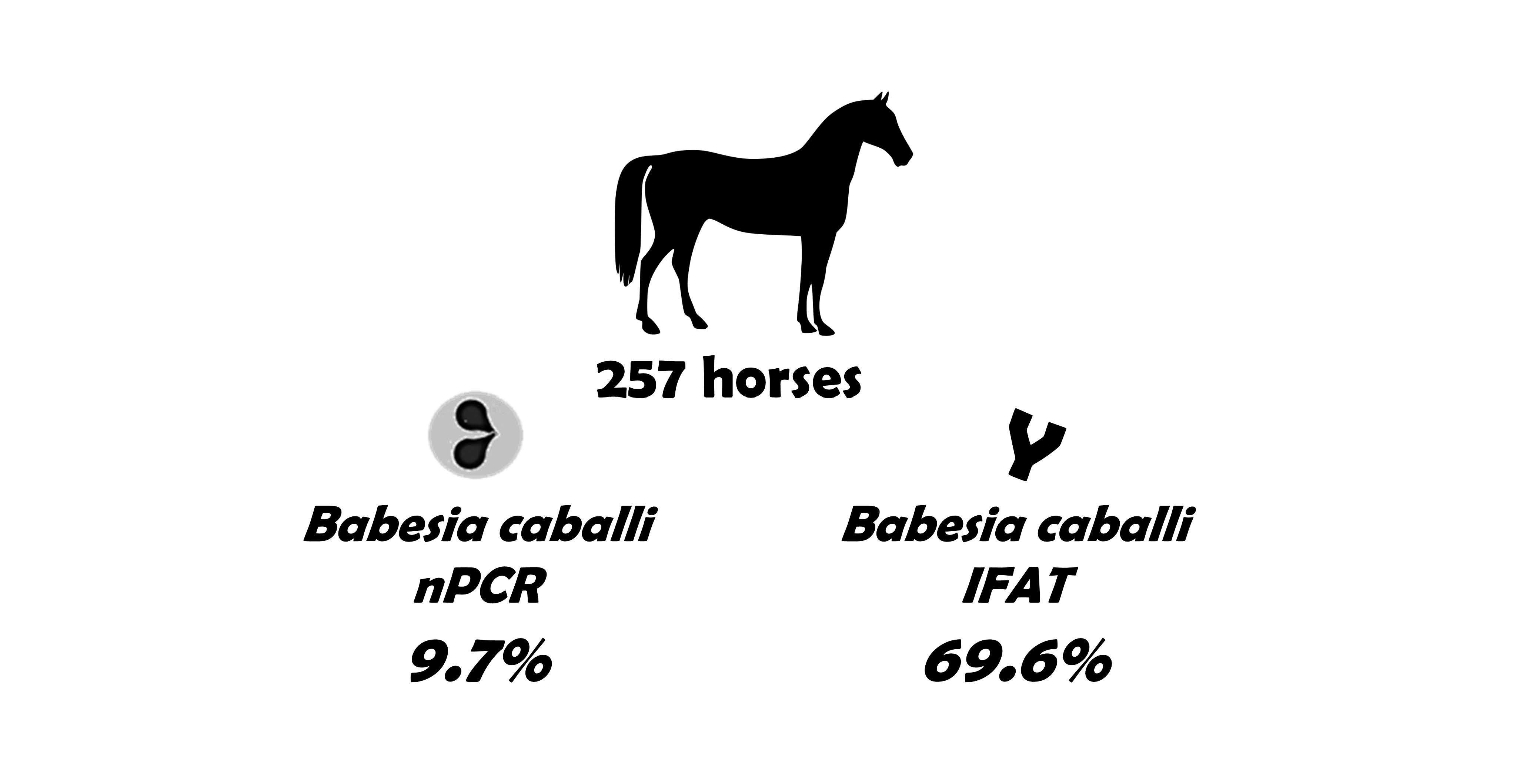
2.2. Detection of B. caballi Infection by IFAT and PCR

Serologic exposure for B. caballi was detected in 179 horses (69.6%, 95% confidence interval (CI): 63.6–75.2), while molecular exposure was detected in 25 horses (9.7%, 95% CI: 6.4–14.0) (Table S1). Babesia caballi seroprevalence varied between 14.3% and 100% between farms (Figure 1). Out of the 179 seropositive horses, 19 were also positive by nPCR (10.6%), as well as six of the 78 (7.7%) seronegative horses (Table 1). The agreement between the molecular and the serological detection was weak (K = 0.019, p = 0.467).

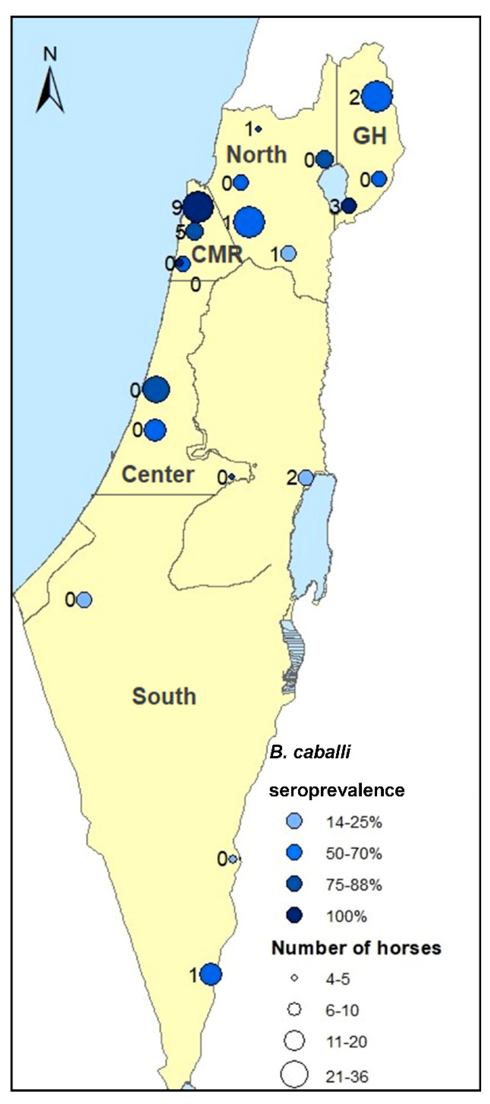
2.3. Risk Factors for Serologic Exposure to B. caballi

Several risk factors were found to be associated with the exposure of horses to B. caballi. Horses residing in the area of the Carmel mountain ridge, being of mixed breeds and geldings, infested with ticks and infested with H. marginatum ticks had higher risk for being seropositive for B. caballi, while horses from the south of Israel and horses kept in paddocks had a lower risk of exposure (univariable analysis; Table 2). Seropositivity was significantly correlated with tick load (ρ = 0.238, p < 0.001), but not with horse age (mean age = 10.9 and 10.3 years in seropositive and seronegative horses respectively, p = 0.37).
The geographical area, housing management, horses’ breed and sex and tick infestation were significantly associated together (all p < 0.001) and therefore, there may have been a confounding effect between them. Nonetheless, only the Carmel mountain ridge and the horses’ sex (gelding) remained significantly associated with B. caballi seroprevalence in the multivariable analysis (Table 2).

2.4. Risk Factors for Molecular Identification of B. caballi Infection

The risk factors that were positively and significantly associated with the molecular detection of B. caballi infection were the geographical area, mixed breed horses, horses infested with ticks and horses infested with H. marginatum. Pure bred horses and horses reared in paddocks had a lower risk of infection (Table 3). A significant correlation was found between the horses’ seropositivity and tick load (ρ = 0.165, p = 0.008). Age was also significantly associated with the molecular detection of infection (mean age = 8.7 and 11.0 years in positive and negative horses respectively, p = 0.038).
The Carmel mountain ridge was the only factor found to be associated with infection in the multivariable analysis (Table 3).

2.5. Potential Clinical Consequences of Exposure or Infection

All horses had normal rectal temperature during sample collection (36.5–38.5 °C). Most horses had normal mucus membrane (MM) color, while for 52 horses (20.2%) MM color was noted as pale.
Pale MM was more often noted in seropositive horses (80% versus 66.8%, p = 0.052). The distribution of body temperature (p = 0.097) and packed cell volume (PCV) (p = 0.525) did not differ between seropositive and seronegative horses, while total solids (TS) values were significantly higher in seropositive horses (Mean = 7.2 and 6.9 respectively, p = 0.004).
Molecular detection of infection was associated with pale MM (17.3% versus 7.8%, p = 0.039). The distribution of body temperature (p = 0.54), PCV (p = 0.347) and TS (p = 0.092) did not differ between positive or negative horses.

3. Discussion

Equine piroplasmosis is endemic in the Middle East, and both parasites have been reported in Israel [10], and in neighboring Egypt [9] and Jordan [18,19]. Reports from all three countries stated that the commercial cELISA failed to detect B. caballi positive horses, which may have led to underestimation of B. caballi prevalence in the area that was generally lower than in Asia or Europe (4.8% versus 25.6% and 12.6% in Asia and Europe respectively) [20]. Further molecular investigation confirmed that B. caballi is endemic in the region, and found polymorphism in rap-1 nucleotide and amino acid sequences, with a considerable difference between African and Middle Eastern isolates, and the American and Caribbean isolates which were used in the development of the cELISA kit [7,8,9,10,19]. This polymorphism may be the reason why the assay based on this protein did not detect a serological response in horses infected by Middle Eastern strains.
In this study we chose to evaluate B. caballi seroprevalence by using IFAT based on a crude antigen of a local isolate, in order to prevent underestimation of the seroprevalence. The results revealed high seroprevalence of B. caballi (69.6%), considerably higher than its molecular detection (9.7%). The molecular prevalence in Israel in 2015 was similar to the prevalence in 2007–2008 (9.3%) [10]. While the seroprevalence of B. caballi was higher than the seroprevalence of T. equi in the same cohort (65.4%), its molecular detection was significantly lower than T. equi (57.8%) [17]. These differences are probably attributed to the difference in the epidemiology and transmission dynamics of T. equi and B. caballi. Since trans–ovarial transmission in vector ticks was documented for B. caballi, and since parasite clearance is possible in the equine host, the main reservoir of this parasite is probably the tick vector. On the other hand, T. equi carriage is usually life-long, and trans-ovarial transmission was not documented in ticks, making the host (horse) the probable main reservoir [5]. This paradigm fits previous findings, which demonstrated that while T. equi prevalence increases with age, the prevalence of B. caballi, does not increase with age and is higher in younger animals [21,22]. Our findings concur with this model since age did not correlate with B. caballi seroprevalence, but its molecular detection was higher in younger animals, and its seroprevalence was higher than that of T. equi, while its molecular detection was lower. These trends were also found in several other serological surveys in endemic areas [23,24,25,26,27,28,29,30,31,32,33,34]. The low molecular detection of B. caballi may be attributed to parasite clearance or to low parasitemia, beneath the detection level of the PCR assay.
Mixed infections of T. equi and B. caballi have widely been reported worldwide (reviewed in [2]), and were also observed in this study, when the results were compared to a previous study evaluating T. equi prevalence in the same cohort [17]. The seroprevalence of T. equi in the study population was 65.4%, its molecular prevalence was 57.6%, with an agreement of 0.804 (p < 0.001) between methods. Of the 179 B. caballi seropositive horses 140 (78.2%) were also seropositive for T. equi, as well as 28 of the 78 (35.9%) seronegative horses. Of the 25 B. caballi nPCR-positive horses, 20 (80%) were also PCR-positive for T. equi, as well as 128 of the 232 negative horses (55.2%). The association between the exposure to both parasites was highly significant (p < 0.001). Although co-infection with both parasites is common, little is known regarding possible interactions between them within the equine host and their immune response.
The considerable difference between the presence of anti-B. caballi antibodies and the presence of parasite DNA (Table 1) probably reflects the fact that parasitemia may be cleared in some horses, while humoral response with the presence of specific antibodies could remain for long periods, even after clearance. Therefore, it should be taken into account that not all seropositive horses are carriers of parasites. While serological methods cannot discriminate between past exposure and current infection, which may have economic implications on international horse trade or competition, molecular methods may fail to detect infection when the parasitemia is low, and increase the chance of false-negative results.
The geographical area, horses’ breed (mixed bred horses), housing management (pasture) and tick infestation were associated with both B. caballi prevalence and seroprevalence, while the horses’ sex (geldings in comparison to mares) was associated only with its seroprevalence and age (younger horses) with its molecular prevalence. A recent meta-analysis of B. caballi global distribution and associated risk factors identified higher prevalence in male horses and in younger horses (<5 years) [20]. However, this study did not differentiate between molecular and serological studies when addressing these factors, which may be significant regarding the circulation of this parasite. The main factor that influences exposure of horses to piroplasms is the availability of vector ticks [2,5]. Tick infestation of horses depends on their habitat (geographic and climatic parameters) and housing management (horses reared indoors are usually less exposed). A previous study investigating the population of ticks found on the same cohort of horses throughout the year demonstrated that horses reared in pasture were more prone to be infested with ticks, and had the highest tick loads [35]. Most pasture areas in Israel are located in two geographical areas: the Golan heights and the Carmel mountain ridge (Figure 1), and comparison of the equine tick populations in both areas revealed that the predominant tick species infesting horses in the Golan heights is H. excavatum, while in the Carmel mountain ridge it was H. marginatum [35]. In the current study, most of the significant risk factors in the univariable analysis associated with each other since mixed-breed and gelded horses are more often kept in pasture (mostly in the Carmel mountain ridge or the Golan heights) and more often infested with ticks. The highest molecular and serological prevalence that were found in horses from the Carmel mountain ridge, where H. marginatum is most prevalent, and therefore the geographical area was the main factor to remain significant in the multivariable models. It has been shown that B. caballi is transmitted transovarially in H. marginatum, but it has not been established for H. excavatum [5]. The significantly higher prevalence in the Carmel mountain ridge may suggest better vector competence of H. marginatum for transmitting B. caballi, which should be further investigated.
Most infected horses in an endemic area are a-symptomatic and may remain carriers for several years [1,2,3]. Investigating possible sub-clinical consequences of infection or exposure revealed that infected horses more often had pale MM than non-infected horses (and this association was close to significant also in seropositive horses, which some may have cleared of infection), although the horses’ PCV was not affected. In addition, seropositive horses had higher TS values, which may be attributed to higher plasma immunoglobulin levels, as demonstrated also for T. equi in the same cohort of horses [17].
Limitations of the study included the use of samples collected in 2015, which were selected to enable comparisons to T. equi infection and tick infestation, which were available for this cohort [17,35]. Nevertheless, the molecular prevalence in 2015 was similar to the prevalence in 2007–2008 [10]. Further studies are required to determine whether the current prevalence is different. The exact molecular prevalence of B. caballi, and any other pathogen for this matter, is limited by the limit of detection of the molecular method used. The limit of detection of the nPCR applied in this study was not determined and, therefore, it is possible that the molecular prevalence is higher.

4. Materials and Methods

4.1. Study Design and Sample Collection

A required sample size of 212 horses was calculated based on previously reported B. caballi prevalence, using PCR (9.3%) [10] and an estimation of the size of the equine population in Israel (n = 35,000).
Nineteen horse farms were sampled to represent the geographical distribution of the equine population in Israel, during May–June 2015. Most of these horses had been previously sampled on three other occasions during 2014–2015, as a part of a seasonal surveillance study [17]. Infection and exposure of these horses to T. equi have been evaluated in the same samples (May–June 2015), by PCR and IFAT [17]. Data including individual horse information and farm management were obtained from farm personnel. Whole blood and serum were collected from the jugular vein of each horse into sterile vacuum tubes containing EDTA and without anticoagulant. Serum was obtained after centrifugation (3000 rpm, 10 min), and all blood and serum samples were kept at −20°C until processing. In addition to blood collection, MM color was noted, rectal temperature was measured, and each horse was scanned for the presence of ticks. All ticks were removed and preserved in ethanol and later identified morphologically [35].

4.2. Serological Screening for B. caballi Exposure

Serological screening for the presence of anti-B. caballi antibodies was performed using IFAT. The assay was performed on all sera using a dilution of 1:80, as a cutoff for screening. The antigen slides were prepared in-house from B. caballi-infected erythrocytes from culture. Blood smears with parasitemia of 5% or higher were prepared on glass slides and stored at −80 °C until use. Test protocol was performed as previously described for T. equi [13,36].

4.3. Identification of B. caballi Infection Using Nested PCR

DNA was extracted from whole blood of each sample and infection with B. caballi was identified by nested PCR directed at the B. caballi rap-1 gene, using the external primers BC-RAPF and BC-RAP1R (amplifying~1800 bp), and the internal primers BC-RAP2F and BC-RAP2R (amplifying~400 bp), as previously described [8,16].

4.4. Statistical Analysis

The agreement between IFAT and nPCR results for identification of B. caballi was assessed using Cohen’s Kappa.
Risk factors for serologic exposure to B. caballi were evaluated using χ2 or Fisher’s exact test, as appropriate, for categorical parameters, and using t-test or Mann–Whitney U-test, as appropriate, for scale parameters. Odds ratios were calculated using univariable logistic regression models for each parameter. All factors that were found significant in the univariable analysis were included in a multivariable generalized estimating equation (GEE), with a logit link function, the horse was defined as the subject and the farm was defined as a random factor.
All statistical analyses were performed using SPSS 25.0® and WinPepi 11.65® statistical software. Statistical significance was set at p < 0.05.

5. Conclusions

The use of local crude antigen proved efficient for the evaluation of the seroprevalence of B. caballi in Israel, and may be beneficial for pre-exportation screening to non-endemic areas. While the molecular prevalence was similar to previous reports, the serological prevalence was considerably higher, suggesting that most horses in the studied area are exposed to this parasite. Risk factor analysis identified one geographical area with the highest rates of infection and exposure, that may be linked to higher prevalence of H. marginatum ticks.

Supplementary Materials

The following are available online at https://www.mdpi.com/article/10.3390/pathogens10040445/s1, Table S1: Data of the horses included in this study including their characteristics, management, tick infestation, clinical parameters and the molecular and serological results of B. caballi and T. equi infection.

Author Contributions

Conceptualization, S.T.-L., M.L.M., Y.G. and A.S.; methodology, M.L.M. and I.S.; formal analysis, S.T.-L.; investigation, S.T.-L., I.S. and D.P.; resources, A.S. and M.L.M.; writing—original draft preparation, S.T.-L.; writing—review and editing, S.T.L., M.L.M., Y.G. and A.S.; visualization, S.T.-L.; supervision, Y.G. and A.S. All authors have read and agreed to the published version of the manuscript.

Funding

This research received no external funding.

Institutional Review Board Statement

The study was conducted according to the guidelines of the Declaration of Helsinki, and approved by the Internal Research Committee of the Koret School of Veterinary Medicine—Veterinary Teaching Hospital (KSVM-VTH/23_2014, 28 December 2014).

Informed Consent Statement

Informed consent was obtained from all horse owners involved in the study.

Data Availability Statement

Data are contained within the article or Supplementary Material.

Acknowledgments

The authors wish to thank the horse owners for their willingness to participate in this study.

Conflicts of Interest

The authors declare no conflict of interest.

References

  1. Rothschild, C.M. Equine piroplasmosis. J. Equine Vet. Sci. 2013, 23, 115–120. [Google Scholar] [CrossRef]
  2. Tirosh-Levy, S.; Gottlieb, Y.; Fry, L.M.; Knowles, D.P.; Steinman, A. Twenty Years of Equine Piroplasmosis Research: Global Distribution, Molecular Diagnosis, and Phylogeny. Pathogens 2020, 9, 926. [Google Scholar] [CrossRef]
  3. Wise, L.N.; Kappmeyer, L.S.; Mealey, R.H.; Knowles, D.P. Review of equine piroplasmosis. J. Vet. Int. Med. 2013, 27, 1334–1346. [Google Scholar] [CrossRef]
  4. Friedhoff, K.T.; Tenter, A.M.; Muller, I. Haemoparasites of equines: Impact on international trade of horses. Rev. Sci. Tech. 1990, 9, 1187–1194. [Google Scholar]
  5. Scoles, G.A.; Ueti, M.W. Vector ecology of equine piroplasmosis. Ann. Rev. Entomol. 2015, 60, 561–580. [Google Scholar] [CrossRef]
  6. De Waal, D. Equine piroplasmosis: A review. Brit. Vet. J. 1992, 148, 6–14. [Google Scholar] [CrossRef]
  7. Kappmeyer, L.S.; Perryman, L.E.; Hines, S.A.; Baszler, T.V.; Katz, J.B.; Hennager, S.G.; Knowles, D.P. Detection of equine antibodies to babesia caballi by recombinant B. caballi rhoptry-associated protein 1 in a competitive-inhibition enzyme-linked immunosorbent assay. J. Clin. Microbiol. 1999, 37, 2285–2290. [Google Scholar] [CrossRef] [Green Version]
  8. Bhoora, R.; Quan, M.; Zweygarth, E.; Guthrie, A.J.; Prinsloo, S.A.; Collins, N.E. Sequence heterogeneity in the gene encoding the rhoptry-associated protein-1 (RAP-1) of Babesia caballi isolates from South Africa. Vet. Parasitol. 2010, 169, 279–288. [Google Scholar] [CrossRef]
  9. Mahmoud, M.S.; El-Ezz, N.T.; Abdel-Shafy, S.; Nassar, S.A.; El Namaky, A.H.; Khalil, W.K.; Knowles, D.; Kappmeyer, L.; Silva, M.G.; Suarez, C.E. Assessment of Theileria equi and Babesia caballi infections in equine populations in Egypt by molecular, serological and hematological approaches. Parasit Vectors 2016, 9, 260. [Google Scholar] [CrossRef] [Green Version]
  10. Rapoport, A.; Aharonson-Raz, K.; Berlin, D.; Tal, S.; Gottlieb, Y.; Klement, E.; Steinman, A. Molecular characterization of the Babesia caballi rap-1 gene and epidemiological survey in horses in Israel. Infect. Genet. Evol. 2014, 23, 115–120. [Google Scholar] [CrossRef]
  11. Aharonson-Raz, K.; Rapoport, A.; Hawari, I.M.; Lensky, I.M.; Berlin, D.; Zivotofsky, D.; Klement, E.; Steinman, A. Novel description of force of infection and risk factors associated with Theileria equi in horses in Israel and in The Palestinian Authority. Ticks Tick-Borne Dis. 2014, 5, 366–372. [Google Scholar] [CrossRef]
  12. Ketter-Ratzon, D.; Tirosh-Levy, S.; Nachum-Biala, Y.; Saar, T.; Qura’n, L.; Zivotofsky, D.; Abdeen, Z.; Baneth, G.; Steinman, A. Characterization of Theileria equi genotypes in horses in Israel, the Palestinian Authority and Jordan. Ticks Tick-Borne Dis. 2017, 8, 499–505. [Google Scholar] [CrossRef] [PubMed]
  13. Shkap, V.; Cohen, I.; Leibovitz, B.; Savitsky; Pipano, E.; Avni, G.; Shofer, S.; Giger, U.; Kappmeyer, L.; Knowles, D. Seroprevalence of Babesia equi among horses in Israel using competitive inhibition ELISA and IFA assays. Vet. Parasitol. 1998, 76, 251–259. [Google Scholar] [CrossRef]
  14. Steinman, A.; Zimmerman, T.; Klement, E.; Lensky, I.M.; Berlin, D.; Gottlieb, Y.; Baneth, G. Demographic and environmental risk factors for infection by Theileria equi in 590 horses in Israel. Vet. Parasitol. 2012, 187, 558–562. [Google Scholar] [CrossRef]
  15. Tirosh-Levy, S.; Gottlieb, Y.; Arieli, O.; Mazuz, M.L.; King, R.; Horowitz, I.; Steinman, A. Genetic characteristics of Theileria equi in zebras, wild and domestic donkeys in Israel and the Palestinian Authority. Ticks Tick Borne Dis. 2020, 11, 101286. [Google Scholar] [CrossRef]
  16. Tirosh-Levy, S.; Steinman, A.; Levy, H.; Katz, Y.; Shtilman, M.; Gottlieb, Y. Parasite load and genotype are associated with clinical outcome of piroplasm-infected equines in Israel. Parasit Vectors 2020, 13, 267. [Google Scholar] [CrossRef]
  17. Tirosh-Levy, S.; Gottlieb, Y.; Mazuz, M.L.; Savitsky, I.; Steinman, A. Infection dynamics of Theileria equi in carrier horses is associated with management and tick exposure. Ticks Tick Borne Dis. 2020, 11, 101508. [Google Scholar] [CrossRef]
  18. Abutarbush, S.M.; Alqawasmeh, D.M.; Mukbel, R.M.; Al-Majali, A.M. Equine babesiosis: Seroprevalence, risk factors and comparison of different diagnostic methods in Jordan. Transbound. Emerg. Dis. 2012, 59, 72–78. [Google Scholar] [CrossRef]
  19. Qablan, M.A.; Obornik, M.; Petrzelkova, K.J.; Sloboda, M.; Shudiefat, M.F.; Horin, P.; Lukes, J.; Modry, D. Infections by Babesia caballi and Theileria equi in Jordanian equids: Epidemiology and genetic diversity. Parasitology 2013, 140, 1096–1103. [Google Scholar] [CrossRef]
  20. Onyiche, T.E.; Taioe, M.O.; Molefe, N.I.; Biu, A.A.; Luka, J.; Omeh, I.J.; Yokoyama, N.; Thekisoe, O. Equine piroplasmosis: An insight into global exposure of equids from 1990 to 2019 by systematic review and meta-analysis. Parasitology 2020, 147, 1411–1424. [Google Scholar] [CrossRef]
  21. Ruegg, S.R.; Heinzmann, D.; Barbour, A.D.; Torgerson, P.R. Estimation of the transmission dynamics of Theileria equi and Babesia caballi in horses. Parasitology 2008, 135, 555–565. [Google Scholar] [CrossRef] [Green Version]
  22. Ruegg, S.R.; Torgerson, P.; Deplazes, P.; Mathis, A. Age-dependent dynamics of Theileria equi and Babesia caballi infections in southwest Mongolia based on IFAT and/or PCR prevalence data from domestic horses and ticks. Parasitology 2007, 134, 939–947. [Google Scholar] [CrossRef] [Green Version]
  23. Kerber, C.E.; Labruna, M.B.; Ferreira, F.; De Waal, D.T.; Knowles, D.P.; Gennari, S.M. Prevalence of equine Piroplasmosis and its association with tick infestation in the State of Sao Paulo, Brazil. Rev. Bras. Parasitol. Vet. 2009, 18, 1–8. [Google Scholar] [CrossRef]
  24. Wang, J.; Liu, J.; Yang, J.; Wang, X.; Li, Z.; Jianlin, X.; Li, X.; Xiang, Q.; Li, Y.; Liu, Z.; et al. The first molecular detection and genetic diversity of Babesia caballi and Theileria equi in horses of Gansu province, China. Ticks Tick Borne Dis. 2019, 10, 528–532. [Google Scholar] [CrossRef]
  25. Xuan, X.; Nagai, A.; Battsetseg, B.; Fukumoto, S.; Makala, L.H.; Inoue, N.; Igarashi, I.; Mikami, T.; Fujisaki, K. Diagnosis of equine piroplasmosis in Brazil by serodiagnostic methods with recombinant antigens. J. Vet. Med. Sci. 2001, 63, 1159–1160. [Google Scholar] [CrossRef] [PubMed] [Green Version]
  26. Nugraha, A.B.; Cahyaningsih, U.; Amrozi, A.; Ridwan, Y.; Agungpriyono, S.; Taher, D.M.; Guswanto, A.; Gantuya, S.; Tayebwa, D.S.; Tuvshintulga, B.; et al. Serological and molecular prevalence of equine piroplasmosis in Western Java, Indonesia. Vet. Parasitol. Reg. Stud. Rep. 2018, 14, 1–6. [Google Scholar] [CrossRef]
  27. Moretti, A.; Mangili, V.; Salvatori, R.; Maresca, C.; Scoccia, E.; Torina, A.; Moretta, I.; Gabrielli, S.; Tampieri, M.P.; Pietrobelli, M. Prevalence and diagnosis of Babesia and Theileria infections in horses in Italy: A preliminary study. Vet. J. 2010, 184, 346–350. [Google Scholar] [CrossRef] [PubMed]
  28. Ikadai, H.; Nagai, A.; Xuan, X.; Igarashi, I.; Tsugihiko, K.; Tsuji, N.; Oyamada, T.; Suzuki, N.; Fujisaki, K. Seroepidemiologic studies on Babesia caballi and Babesia equi infections in Japan. J. Vet. Med. Sci. 2002, 64, 325–328. [Google Scholar] [CrossRef] [Green Version]
  29. Al-Obaidi, Q.; Arshad, M.; Al-Sultan, I.; Azlinda, A.; Mohd-Azam, K. Comparison between microscopic examination and competitive ELISA for diagnosis of equine piroplasmosis in Kelantan, Malaysia. Malays. J. Vet. Res. 2016, 7, 23–29. [Google Scholar]
  30. Munkhjargal, T.; Sivakumar, T.; Battsetseg, B.; Nyamjargal, T.; Aboulaila, M.; Purevtseren, B.; Bayarsaikhan, D.; Byambaa, B.; Terkawi, M.A.; Yokoyama, N.; et al. Prevalence and genetic diversity of equine piroplasms in Tov province, Mongolia. Infect. Genet. Evol. 2013, 16, 178–185. [Google Scholar] [CrossRef] [PubMed]
  31. Asgarali, Z.; Coombs, D.K.; Mohammed, F.; Campbell, M.D.; Caesar, E. A serological study of Babesia caballi and Theileria equi in Thoroughbreds in Trinidad. Vet. Parasitol. 2007, 144, 167–171. [Google Scholar] [CrossRef]
  32. Acici, M.; Umur, S.; Guvenc, T.; Arslan, H.H.; Kurt, M. Seroprevalence of equine babesiosis in the Black Sea region of Turkey. Parasitol. Int. 2008, 57, 198–200. [Google Scholar] [CrossRef] [PubMed]
  33. Mujica, F.F.; Perrone, T.; Forlano, M.; Coronado, A.; Melendez, R.D.; Barrios, N.; Alvarez, R.; Granda, F. Serological prevalence of Babesia caballi and Theileria equi in horses of Lara State, Venezuela. Vet. Parasitol. 2011, 178, 180–183. [Google Scholar] [CrossRef]
  34. Rosales, R.; Rangel-Rivas, A.; Escalona, A.; Jordan, L.S.; Gonzatti, M.I.; Aso, P.M.; Perrone, T.; Silva-Iturriza, A.; Mijares, A. Detection of Theileria equi and Babesia caballi infections in Venezuelan horses using Competitive-Inhibition ELISA and PCR. Vet. Parasitol. 2013, 196, 37–43. [Google Scholar] [CrossRef] [PubMed]
  35. Tirosh-Levy, S.; Gottlieb, Y.; Apanaskevich, D.A.; Mumcuoglu, K.Y.; Steinman, A. Species distribution and seasonal dynamics of equine tick infestation in two Mediterranean climate niches in Israel. Parasit Vectors 2018, 11, 546. [Google Scholar] [CrossRef] [Green Version]
  36. Tirosh-Levy, S.; Gottlieb, Y.; Mimoun, L.; Mazuz, M.L.; Steinman, A. Transplacental Transmission of Theileria equi Is Not a Common Cause of Abortions and Infection of Foals in Israel. Animals 2020, 10, 341. [Google Scholar] [CrossRef] [PubMed] [Green Version]
Figure 1. The geographical distribution of the farms included in the study. The number of horses sampled at each farms in represented by the size of the circle. Babesia caballi seroprevalence (%), as detected by IFAT, is represented by the intensity of the blue color, while the number of horses infected with B. caballi, as tested by nPCR, appears on the left of each point. The division into geographical areas, as used for risk factor analysis appears on the map (GH = Golan heights, CMR = Carmel mountain ridge).
Pathogens 10 00445 g001
Table 1. Detection of B. caballi exposure or infection by IFAT and nPCR in 257 horses from 19 farms in Israel.
nPCR-Negative (%)nPCR-Positive (%)Total
IFAT-Negative72 (92.3)6 (7.7)78
IFAT-Positive160 (89.4)19 (10.6)179
Total23225257
Table 2. Factors associated with serologic exposure to B. caballi in horses in Israel, as detected by an immunofluorescent antibody test (IFAT). Univariable analysis was performed by Chi square test or Fisher’s exact, as appropriate. Multivariable analysis was performed using generalized estimating equation (GEE) with the farm defined as a random factor and included all factors identified as significant in the univariable analysis. Odds ratios (OR) were calculated from the GEE model. Statistically significant associations appear in bold.
NIFAT-Positive (%)p2)p (GEE)OR (95% CI)
North6940 (58)<0.0010.1383.45 (0.67–17.77)
Carmel6459 (92.2) <0.00110.01 (2.98–33.59)
Center4735 (74.5) 0.0625.45 (0.92–32.30)
South3514 (40) 0.2273.11 (0.49–19.57)
Golan heights4231 (73.8)
Gelding128103 (80.5)<0.0010.0372.01 (1.04–3.89)
Stallion64 (66.7) 0.3252.61 (0.39–17.54)
Mare12372 (58.5) ref
Mixed Breed186142 (76.3)<0.0010.2341.58 (0.74–3.33)
Pure breed7036 (51.4)
Stall 4636 (78.3)<0.0010.8410.85 (0.16–4.33)
Paddock9141 (45.1) 0.0640.27 (0.07–1.08)
Pasture120102 (85) ref
Ticks9884 (85.7)<0.0010.1276.98 (0.58–84.34)
No ticks15995 (59.7) ref
Haemaphysalis parva54 (80)1
Not present252175 (69.4)
Hyalomma excavatum5441 (75.9)0.259
Not present203138 (68)
Hyalomma marginatum7967 (84.8)<0.0010.1912.63 (0.62–11.20)
Not present178112 (62.9)
Rhipicephalus annulatus22 (100)1
Not present255177 (69.4)
Rhipicephalus turanicus3828 (73.7)0.558
Not present219151 (68.9)
Table 3. Factors associated with molecular exposure to B. caballi in horses in Israel, as detected by nested polymerase chain reaction (nPCR). Univariable analysis was performed by Chi square test or Fisher’s exact, as appropriate. Multivariable analysis was performed using a generalized estimating equation (GEE) with the farm defined as a random factor and included all factors identified as significant in the univariable analysis. Odds ratios (OR) were calculated from the GEE model. Statistically significant associations appear in bold.
NnPCR-Positive (%)p2)p (GEE)OR (95% CI)
North693 (4.3)0.0010.7980.81 (0.16–4.01)
Carmel6414 (21.9) 0.0253.82 (1.19–12.32)
Center470 nana
South353 (8.6) 0.6861.62 (0.16–16.89)
Golan heights425 (11.9) ref
Gelding12815 (11.7)0.284
Stallion61 (16.7)
Mare1239 (7.3)
Mixed Breed18624 (12.9)0.0060.0839.86 (0.74–130.92)
Pure breed701 (1.4) ref
Stall 4600.001nana
Paddock915 (5.5) 0.9161.11 (0.16–7.79)
Pasture12020 (16.7) ref
Ticks9816 (16.3)0.0050.6671.34 (0.31–6.36)
No ticks1599 (5.7) ref
Haemaphysalis parva501
Not present25225 (9.9)
Hyalomma excavatum547 (13)0.367
Not present20318 (8.9)
Hyalomma marginatum7913 (16.5)0.0150.5280.624 (0.14–2.69)
Not present17812 (6.7) ref
Rhipicephalus annulatus201
Not present25525 (9.8)
Rhipicephalus turanicus385 (13.2)0.388
Not present21920 (9.1)
Age 0.051
Publisher’s Note: MDPI stays neutral with regard to jurisdictional claims in published maps and institutional affiliations.

